Embed Size (px)
Citation preview
D R – A B D U L H A F E D A L H E M Y A R I
Brain tumours
Brain tumours are responsible for approximately
2% of all cancer deaths. Central nervous system
tumours comprise the most common group of
solid tumours in young patients, accounting for
20% of all paediatric neoplasms. The overall incidence
of brain tumours is 8–10 per 100 000 population
per year. A study by the United States
.
Department of Health in 1966 showed the incidence
to be 21 per 100 000 per year at 2 years old
and 1 per 100 000 during the teenage years. The
incidence increases after the 4th decade of life to
reach a maximum of 16 per 100 000 per year in the
7th decade. There has been an intense debate concerning
the increased incidence of brain tumours,
especially in the elderly, but this possible increase
could be explained due to the advent of CT and
MRI leading to better detection of tumours
Classification
The general brain tumour classification is related
to the cell of origin, and is shown in Table 6.1.
Table 6.2 shows the approximate distribution
of the more common brain tumours.
This chapter will discuss the tumours derived
from the neuroectoderm and metastatic tumours.
The following chapters will describe the benign
brain tumours and pituitary tumours.
Aetiology
Epidemiology studies have not indicated any
particular factor (viral, chemical or traumatic)
that causes brain tumours in humans, although a
range of cerebral tumours can be induced in animals
predisposition
but chromosome abnormalities have
been noted in many CNS tumours (Table 6.3).
Neurofibromatosis type 1 (NF1), previously
known as von Recklinghausen’s disease, occurs
with an approximate frequency of 1 in 4000 live
births. It is inherited as an autosomal dominant
pattern and there is a high spontaneous mutation
rate. NF1 is associated with a variety of central
and peripheral nervous system tumours.experimentally. There is no genetic predisposition
optic nerve glioma is the most common CNS tumour
associated with NF1, occurring in about
15% of those affected. Less commonly low-grade
glioma of the hypothalamus, cerebellum, brainstem
or spinal cord may occur. Peripheral neurofibromas
are the hallmark of NF1 (Chapter 17).
Neurofibromas of the spinal roots are a common
feature of NF1 (Chapter 15). The gene causing
NF1 is located on the long arm of chromosome 17(17q 11.2)
Neurofibromatosis type 2 (NF2), previously
known as central neurofibromatosis, is an autosomal
dominant disorder which, beyond a few
superficial similarities, is phenotypically and
genetically distinct from NF1. It has an incidence
of approximately 1 in 100 000 live births. 6.4.)
The hallmark
of NF2 is bilateral acoustic (vestibular)
schwannomas, but patients with NF2 have an increased
risk of other intracranial schwannomas,
multiple meningiomas (both cranial and spinal)
and gliomas. The NF2 gene locus is sited on
the long arm of chromosome 22 (22q 11.2) (Table
There is no specific evidence linking CNS tumours
to environmental carcinogens, although
many chemicals, especially ethyl and methyl
carcinogenic activity in animals and produce
CNS tumours.
Viral induction of brain tumours has been used
in animal models but there is no firm evidence for
viral aetiology in humans.
Ahuman polyoma JC
virus injected into primates produces tumours
similar to human astrocytomas after an 18-month
incubation period. This type of ‘slow virus’ effect
may account for some of the problems of isolating
viruses from human tumours.
Although immunosuppression is known to increase
markedly the risk of primary lymphomanitrosoureaand anthracene derivatives, show
of the brain, particularly in transplant recipients,
there is not the corresponding increased incidence
of gliomas.
At present there is considerable conjecture regarding
the role of other possible aetiological
agents, including trauma, electromagnetic radiation
convincing evidence to implicate these as being
involved with the development of brain tumours
in humans.
The four hallmarks of the development of a
cancer cell are the ability to proliferate, with the
intracellular growth pathways constituitively activated,
the evasion of apoptosis, with the cancer
cells having escaped from cell death pathways,
the attraction and induction of new blood vessels
(angiogenesis) to supply increased metabolic activity
of tumour cells, and tissue invasion.
Each
of these are dependent on ligand–receptor interactions
on the cell surface leading to a cascade of
cytoplasmic events that eventually result in differential
gene expression.and organic solvents but, as yet, there is no
Molecular biology techniques have enabled
the identification of a variety of alterations in the
genome of the tumour cell, including those of
brain tumours. The present concept of oncogenesis
involves both the addition of oncogenes to the
genome and the loss of the normally occurring
tumour suppressor genes. Transformation (spontaneous
or induced) is a multistep process requiring
both initiation and promotion.
. Oncogenes
encode proteins that in the signal
transduction and second messenger systems that
modulate cell metabolism and proliferation.
These proteins include both growth factors and
growth factor receptparticipate ors such as epidermal
growth factor receptor, platelet-derived growth
factor, tryosine-specific protein kinases and
guanine-binding proteins
Tumour suppressor genes are normally presentin the genome and act as a ‘brake’ on celltransformation. Mutations in the p53 tumoursuppressor gene on chromosome 17 are the mostcommon gene alteration found to date in tumoursand have been shown to occur in both astrocytomasand meningiomas. The Li–Fraumenisyndrome is due to a germ line mutation in thep53 gene with the development of numerouscancers including gliomas, ependymomas andmedulloblastomas.
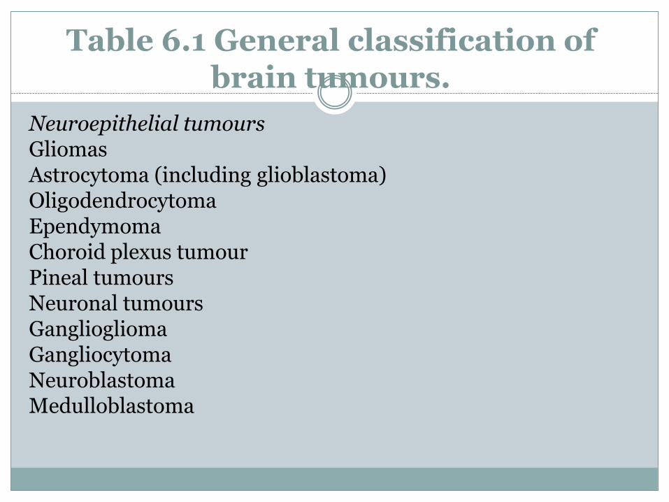
Table 6.1 General classification of brain tumours.
Neuroepithelial tumoursGliomasAstrocytoma (including glioblastoma)OligodendrocytomaEpendymomaChoroid plexus tumourPineal tumoursNeuronal tumoursGangliogliomaGangliocytomaNeuroblastomaMedulloblastoma
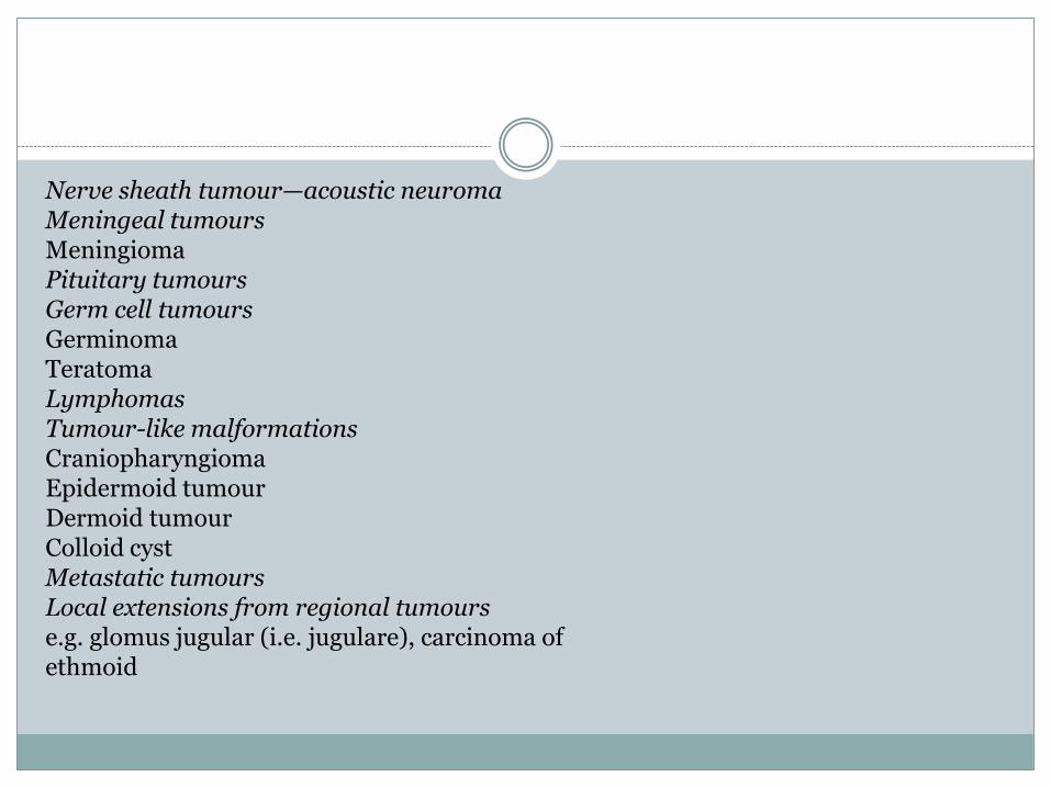
Nerve sheath tumour—acoustic neuromaMeningeal tumoursMeningiomaPituitary tumoursGerm cell tumoursGerminomaTeratomaLymphomasTumour-like malformationsCraniopharyngiomaEpidermoid tumourDermoid tumourColloid cystMetastatic tumoursLocal extensions from regional tumourse.g. glomus jugular (i.e. jugulare), carcinoma ofethmoid
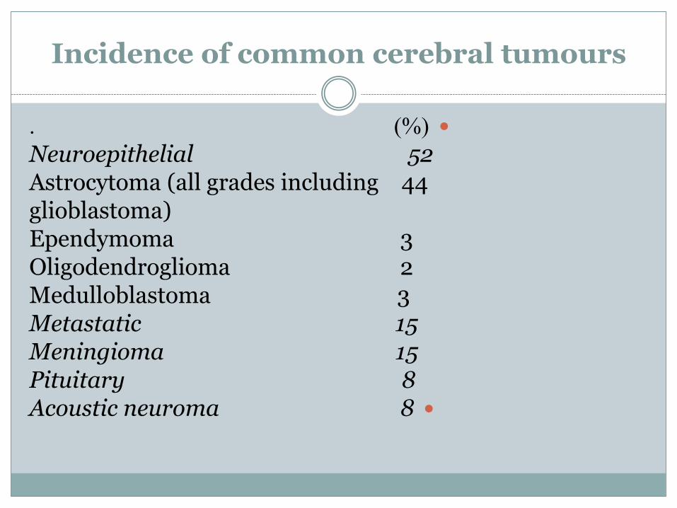
Incidence of common cerebral tumours
. )%(
Neuroepithelial 52Astrocytoma (all grades including 44glioblastoma)Ependymoma 3Oligodendroglioma 2Medulloblastoma 3Metastatic 15Meningioma 15Pituitary 8
Acoustic neuroma 8
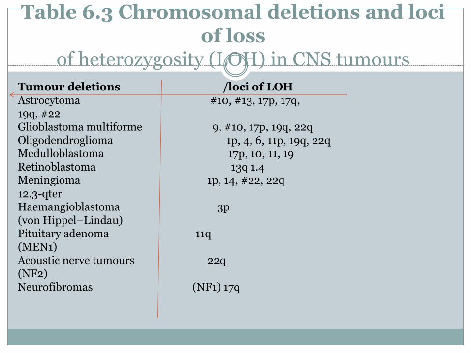
Table 6.3 Chromosomal deletions and loci of loss
of heterozygosity (LOH) in CNS tumours
Tumour deletions /loci of LOHAstrocytoma #10, #13, 17p, 17q,19q, #22 Glioblastoma multiforme 9, #10, 17p, 19q, 22qOligodendroglioma 1p, 4, 6, 11p, 19q, 22qMedulloblastoma 17p, 10, 11, 19Retinoblastoma 13q 1.4Meningioma 1p, 14, #22, 22q12.3-qter Haemangioblastoma 3p(von Hippel–Lindau)Pituitary adenoma 11q(MEN1)Acoustic nerve tumours 22q(NF2) Neurofibromas (NF1) 17q
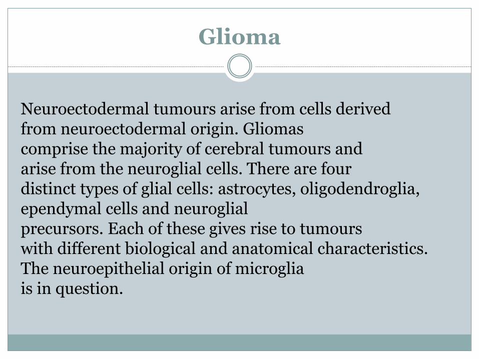
Glioma
Neuroectodermal tumours arise from cells derivedfrom neuroectodermal origin. Gliomascomprise the majority of cerebral tumours andarise from the neuroglial cells. There are fourdistinct types of glial cells: astrocytes, oligodendroglia,ependymal cells and neuroglialprecursors. Each of these gives rise to tumourswith different biological and anatomical characteristics.The neuroepithelial origin of microgliais in question.
Astrocytoma
The most common gliomas arise from the astrocyte
cells which comprise the vast majority of
intraparenchymal cells of the brain. Their main
function appears to be as a supporting tissue for
the neurones. The tumours arising from astro
cytes range from the relatively benign to the
highly malignant. The term ‘malignant’ for brain
tumours differs from its usage for systemic
tumours. Intrinsic brain tumours very rarely
metastasize (except for medulloblastoma and
ependymoma), and ‘malignant’ refers to aggressive
biological characteristics and a poor
prognosis.
Classification
There are many classification systems of brain tumoursin general and gliomas in particular. Theperiod of systematic classification of tumoursbegan in 1846, when Virchow described the neurogliaand related it to brain tumours. AlthoughVirchow created the term ‘glioma’, these tumourshad already been described under othernames. In 1926, Bailey and Cushing described ahistogenetic classification system which comparedthe predominant cell in the tumours withthe embryonal development of the neuroglia..
The comparison with stages of cytogenesis was
probably more of a working hypothesis than an
oncological theory for the origin of the tumours’
cells. The theory that gliomas originate from proliferation
of cells of varying degrees of maturity
lying dormant in the brain is not generally accepted
except in the case of medulloblastoma,
which may arise from a primitive layer in the
cerebellar cortex
A valuable prognostic system of subclassificationof astrocytoma was described by Kernohanin 1949. Astrocytomas were graded from I to IV,with Grade IV being the most malignant andGrade I cytologically, but not necessarily biologically,benign. Ringertz simplified the four-gradeclassification of Kernohan into a three-tiered system;the comparison between the two is shown inFig. 6.1. The glioblastoma multiforme, equivalentto the Kernohan Grade III and IV tumours, is themost common adult cerebral tumour, accountingfor approximately half of all gliomas).
. Thelow-grade gliomas—the astrocytoma, or GradeI or II Kernohan astrocytoma—account for only10–15% of astrocytomas.The World Health Organization (WHO) classificationrecognizes four grades of astrocytoma.Grade I is assigned to the pilocytic astrocytomawhich is biologically distinct from the diffuse astrocytomas,which are classified as astrocytoma(WHO Grade II), anaplastic astrocytoma (WHOGrade III) and glioblastoma multiforme (WHOGrade IV
A grading system proposed by Daumas-
Duport and also known as the St Anne–Mayo
System assessed the tumours according to the
presence or absence of four morphological features—
nuclear atypia, mitosis, endothelial proliferation,
and necrosis—and they are graded
according to the cumulative features score.
II tumours have one feature, Grade III tumours
have two features and Grade IV tumours have
three or four features. Grade I tumours have none of the features, Grade
four morphological features—nuclear atypia, mitosis, endothelial proliferation,and necrosis—and they are gradedaccording to the cumulative features score
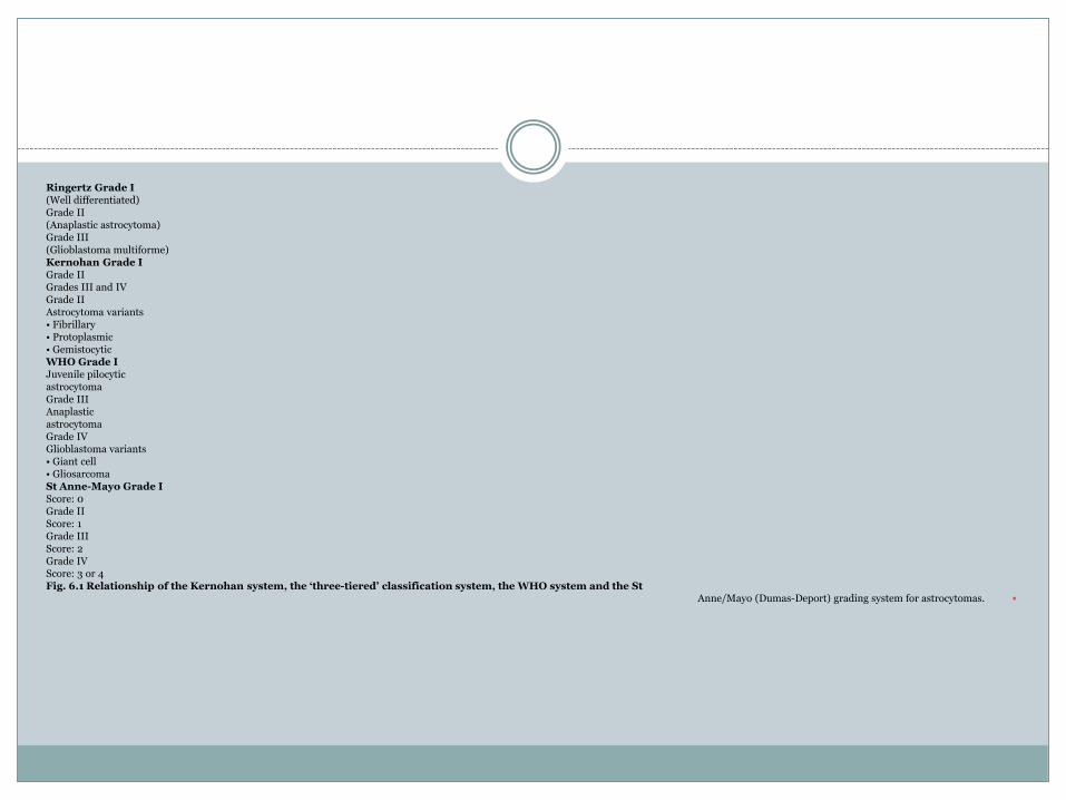
Ringertz Grade I(Well differentiated)Grade II(Anaplastic astrocytoma)Grade III(Glioblastoma multiforme)Kernohan Grade IGrade IIGrades III and IVGrade IIAstrocytoma variants• Fibrillary• Protoplasmic• GemistocyticWHO Grade IJuvenile pilocyticastrocytomaGrade IIIAnaplasticastrocytomaGrade IVGlioblastoma variants• Giant cell• GliosarcomaSt Anne-Mayo Grade IScore: 0Grade IIScore: 1Grade IIIScore: 2Grade IVScore: 3 or 4Fig. 6.1 Relationship of the Kernohan system, the ‘three-tiered’ classification system, the WHO system and the St
Anne/Mayo (Dumas-Deport) grading system for astrocytomas.
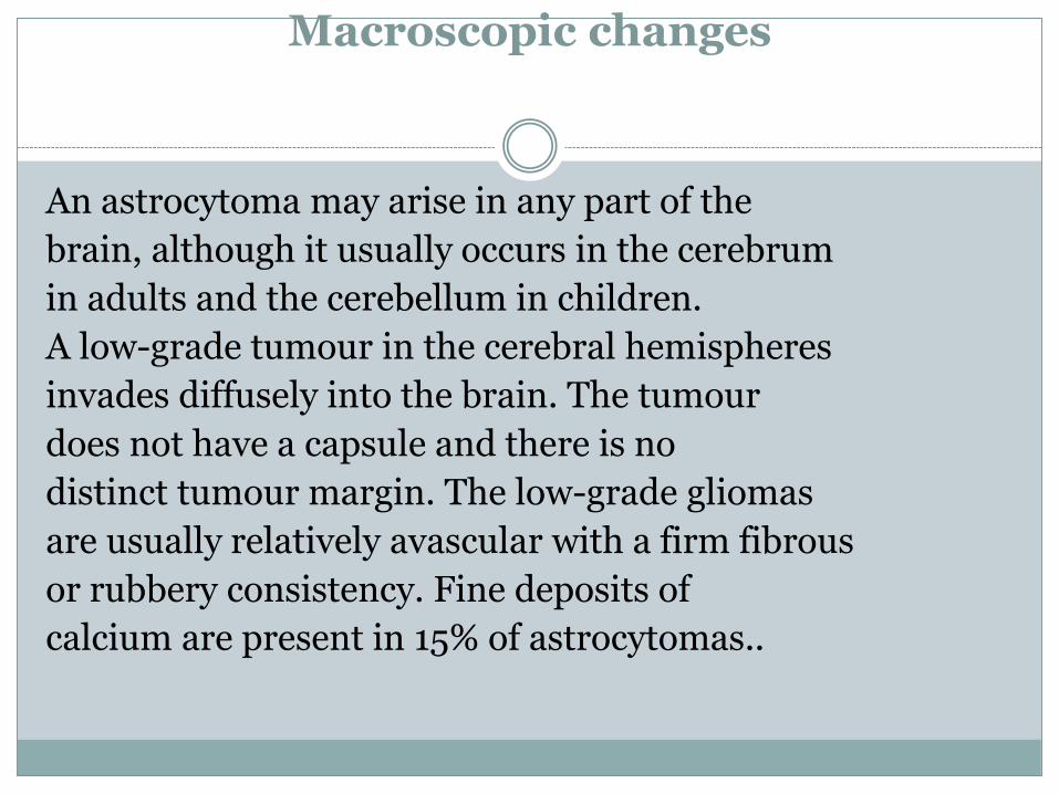
Macroscopic changes
An astrocytoma may arise in any part of the
brain, although it usually occurs in the cerebrum
in adults and the cerebellum in children.
A low-grade tumour in the cerebral hemispheres
invades diffusely into the brain. The tumour
does not have a capsule and there is no
distinct tumour margin. The low-grade gliomas
are usually relatively avascular with a firm fibrous
or rubbery consistency. Fine deposits of
calcium are present in 15% of astrocytomas..
Occasionally,a low-grade astrocytoma may invadediffusely throughout the cerebral hemisphere. Incontrast, the macroscopic appearance of a highgradetumour, the glioblastoma multiforme, ischaracterized by a highly vascular tumour marginwith necrosis in the centre of the tumour. Althoughin certain areas the margin of the tumourmay seem to be macroscopically well definedfrom the surrounding brain, there are microscopicnests of tumour cells extending well outinto the brain
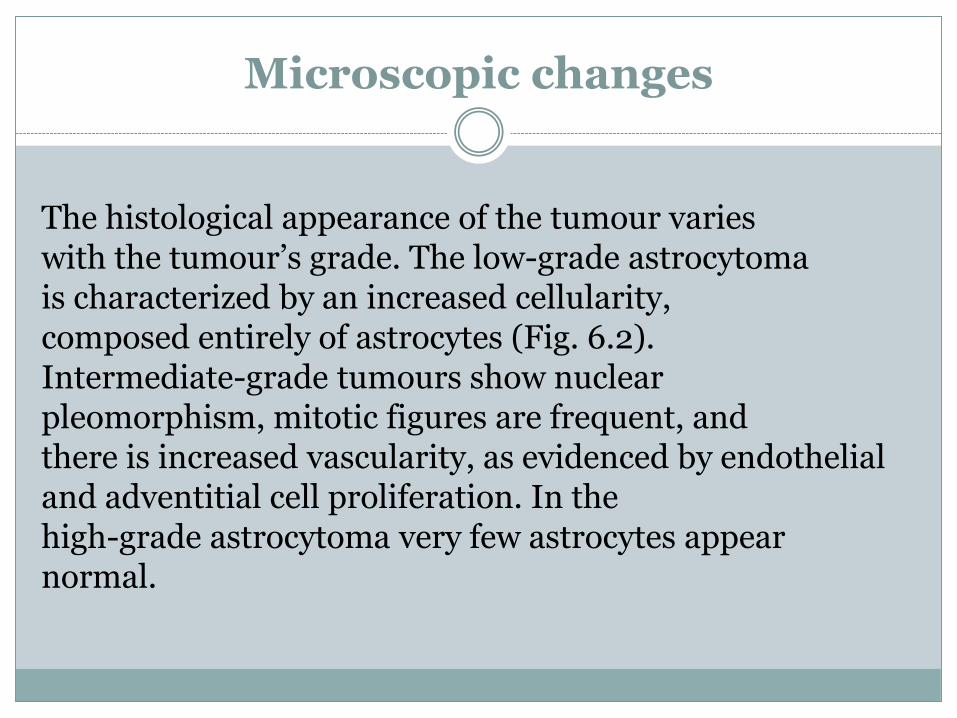
Microscopic changes
The histological appearance of the tumour varieswith the tumour’s grade. The low-grade astrocytomais characterized by an increased cellularity,composed entirely of astrocytes (Fig. 6.2).Intermediate-grade tumours show nuclearpleomorphism, mitotic figures are frequent, andthere is increased vascularity, as evidenced by endothelialand adventitial cell proliferation. In thehigh-grade astrocytoma very few astrocytes appearnormal.
There is marked cellular pleomorphism,
extensive endothelial and adventitial cell
proliferation and numerous mitotic figures with
extensive necrosis (Fig. 6.3).
The major histological features of glioblastoma
multiforme are endothelial proliferation and
necrosis. The anaplastic astrocytoma is characterized
by nuclear pleomorphism and mitoses,
which are absent in the astrocytoma
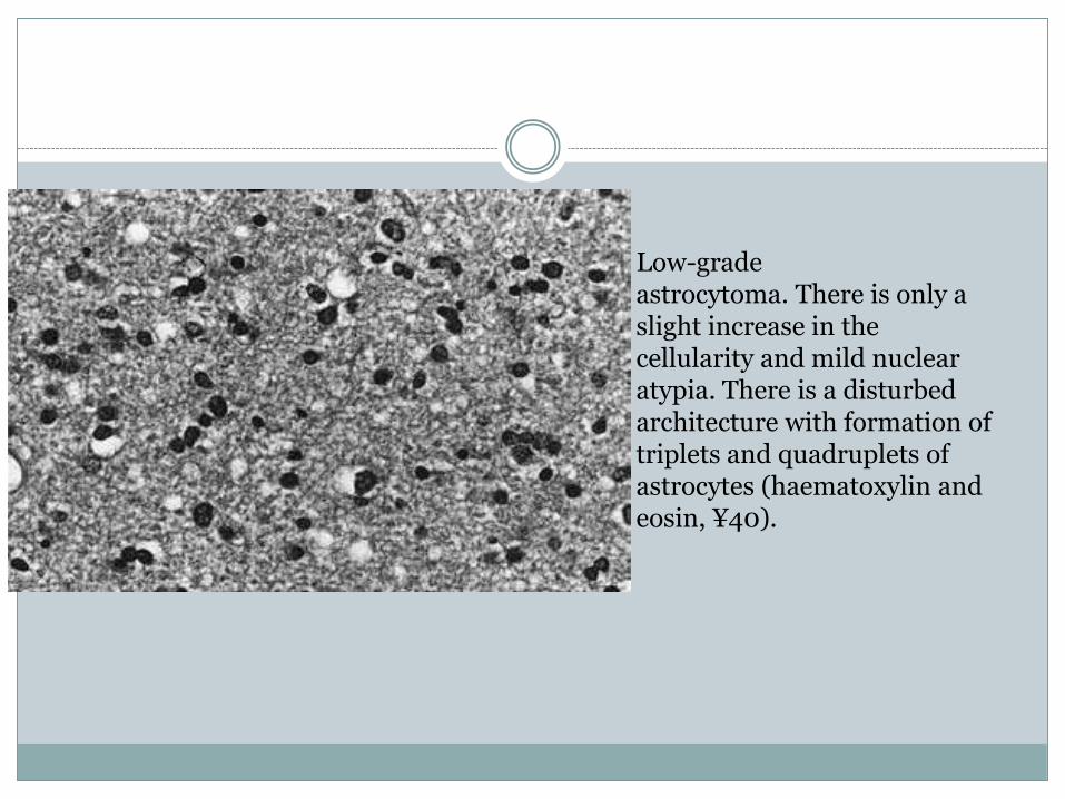
Low-gradeastrocytoma. There is only aslight increase in thecellularity and mild nuclearatypia. There is a disturbedarchitecture with formation oftriplets and quadruplets ofastrocytes (haematoxylin andeosin, ¥40).
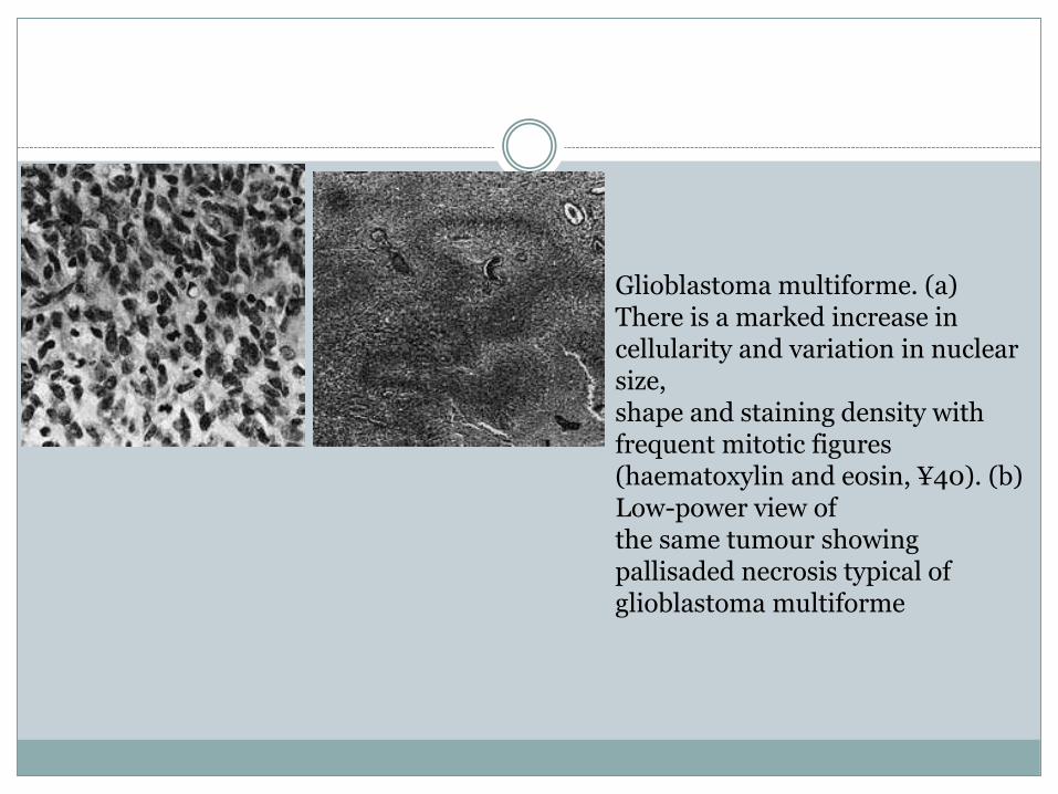
Glioblastoma multiforme. (a) There is a marked increase in cellularity and variation in nuclear size,shape and staining density with frequent mitotic figures (haematoxylin and eosin, ¥40). (b) Low-power view ofthe same tumour showing pallisaded necrosis typical of glioblastoma multiforme
Clinical presentation
The presenting features can be classified under:• raised intracranial pressure• focal neurological signs• epilepsy.The duration of the symptoms and the progressionand evolution of the clinical presentationwill depend on the grade of thetumour—that is, its rate of growth. Apatient presentingwith a low-grade astrocytoma (Grade Ior II) may have a history of seizures extendingover many years, antedating the development of
progressive neurological signs and raised intracranial
pressure. The tumours may evolve histologically
into the more malignant anaplastic
astrocytoma or glioblastoma multiforme. Patients
with the higher-grade tumours present
with a shorter history and glioblastoma multiforme
is characterized by a short illness of weeks
or a few months.
Raised intracranial pressur
eRaised intracranial pressure is due to the tumourmass, surrounding cerebral oedema and hydrocephalusdue to blockage of the CSF pathways.The features of raised intracranial pressure aredescribed in detail in Chapter 3. The major symptomsare headache, nausea and vomiting, anddrowsiness.Headache is the most common symptom inpatients with cerebral astrocytoma and occursin nearly three-quarters of patients; vomitingoccurs in about one-third.
The headaches are
usually gradually progressive and although
frequently worse on the side of the tumours, they
may be bitemporal and diffuse. Characteristically,
the headache is worse on waking and improves
during the day. Nausea and vomiting
occur as the intracranial pressure increases, and
the patient frequently indicates that vomiting
may temporarily relieve the severe headache.
Drowsiness, that is, a deterioration of conscious
state, is the most important symptom and sign of
raised intracranial pressure. The extent of impairment
of conscious state will be related to the
severity of raised intracranial pressure. An alert
patient with severely raised intracranial pressure
may rapidly deteriorate and become deeply unconscious
when there is only a very small further
rise in the pressure within the cranial cavity
Focal neurological deficits
Focal neurological deficits are common in
patients presenting with cerebral gliomas; the
nature of the deficit will depend on the position
of the tumour.
Patients presenting with tumours involving
the frontal lobes frequently have pseudopsychiatric
problems, personality change and mood
disturbance.
. These changes are particularly characteristic
of the ‘butterfly glioma’, so called because
it involves both frontal lobes by spreading
across the corpus callosum, giving it a characteristic
macroscopic (Fig. 6.4) and CT or MRI appearance.
This type of tumour may also occur
posteriorly, with spread across the splenium of
the corpus callosum into both parieto-occipital
lobes
Limb paresis results from interference withthe pyramidal tracts, at either a cortical or a subcorticallevel, and occurs in just under 50% of patients.Field defects associated with tumours ofBRAIN TUMOURS 69Fig. 6.3 Glioblastoma multiforme. (a) There is a marked increase in cellularity and variation in nuclear size,shape and staining density with frequent mitotic figures (haematoxylin and eosin, ¥40). (b) Low-power view ofthe same tumour showing pallisaded necrosis typical of glioblastomamultiforme.(a) (b)70
the temporal, occipital and parietal lobes are
common, but may be evident only on careful testing.
Dysphasia, either expressive or receptive, is
a particularly distressing symptom occurring in
patients with tumours involving the relevant
areas of the dominant hemisphere.
The particular characteristics of posterior fossa
and brainstem gliomas will be discussed in the
following section on paediatric tumours.
Epileptic seizuresSeizures are the most frequent initial symptom inpatients with cerebral astrocytoma and occur in50–75% of all patients. Tumours adjacent to thecortex are more likely to be associated withepilepsy than those deep to the cortex and tumoursinvolving the occipital lobe are less likelyto cause epilepsy than those which are moreanteriorly placed. Astrocytomas may produceeither generalized or focal seizures; the focalcharacteristics will depend on the position withinthe brain and the cortical structures involved.
Investigations
Computerized tomographyCT scan or MRI of the brain are the essential radiologicalinvestigations (Figs 6.5 and 6.6); an accuratediagnosis can be made in nearly all tumours.Low-grade gliomas show decreased density onthe CT scan; this does not enhance with contrastand there is little or no surrounding oedema. Calcificationmay be present. High-grade gliomasare usually large and enhance vividly followingintravenous injection of contrast material (Fig.6.7). The enhancement is often patchy and nonuniformand frequently occurs in a broad, irregularrim around a central area of lower density.e.
Although tumour cysts may occur in the highgradetumours, the central area of low densitysurrounded by the contrast enhancement is usuallydue to tumour necrosis. High-grade tumoursare surrounded by marked cerebral oedema andthere is frequently considerable distortion of thelateral ventricles. Compression of the lateral ventriclein one hemisphere, with pressure extendingacross the midline, may result in an obstructivehydrocephalus involving the opposite lateralventricl
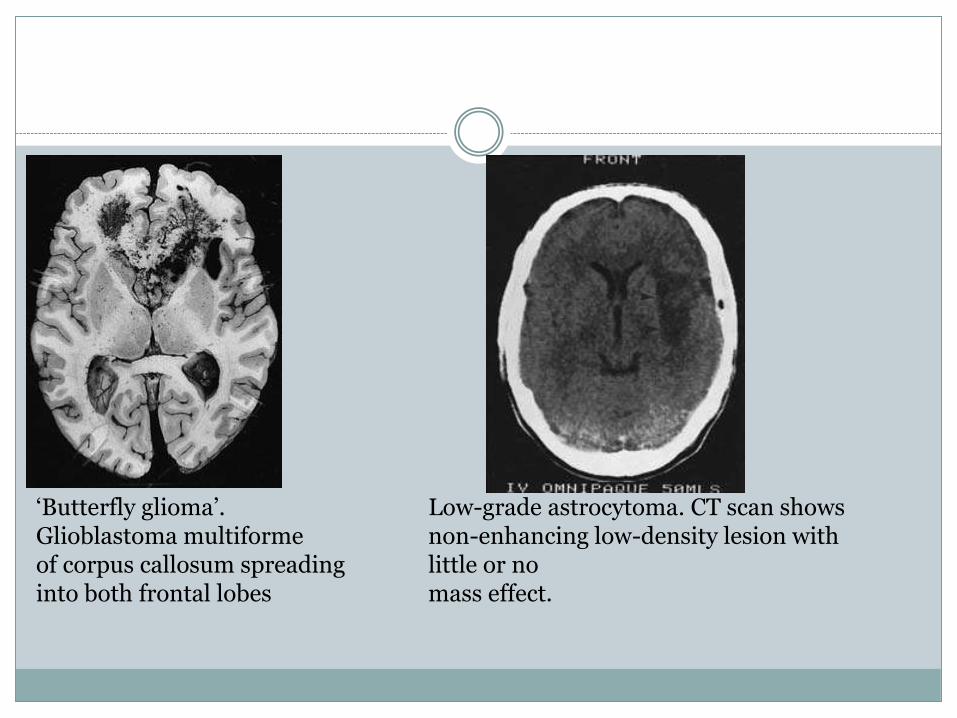
‘Butterfly glioma’. Glioblastoma multiformeof corpus callosum spreading into both frontal lobes
Low-grade astrocytoma. CT scan showsnon-enhancing low-density lesion with little or nomass effect.
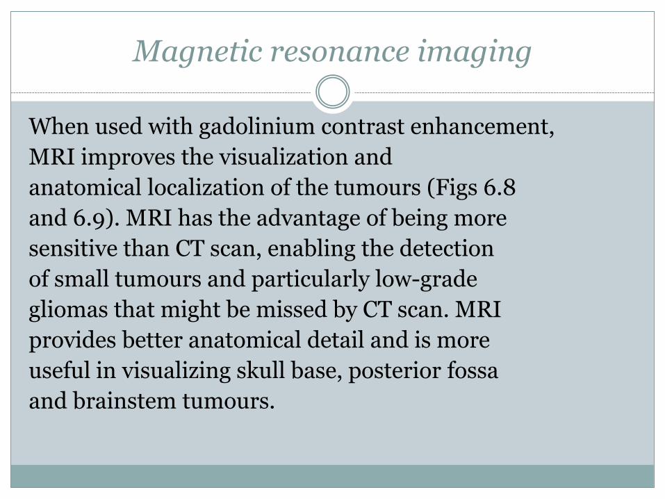
Magnetic resonance imaging
When used with gadolinium contrast enhancement,
MRI improves the visualization and
anatomical localization of the tumours (Figs 6.8
and 6.9). MRI has the advantage of being more
sensitive than CT scan, enabling the detection
of small tumours and particularly low-grade
gliomas that might be missed by CT scan. MRI
provides better anatomical detail and is more
useful in visualizing skull base, posterior fossa
and brainstem tumours.
MRI showing low-grade glioma in posterior frontal region. (a) T1 scan, (b) T2 scan.
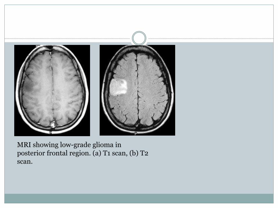
Glioblastoma multiforme. CT shows a largetumour with contrast enhancement particularly atthe margins surrounding a necrotic centre. There ismarked surrounding oedema with compressionof the ventricles.
\Glioblastoma multiforme. MRI shows largeenhancing tumour invading into corpus callosum andventricle.
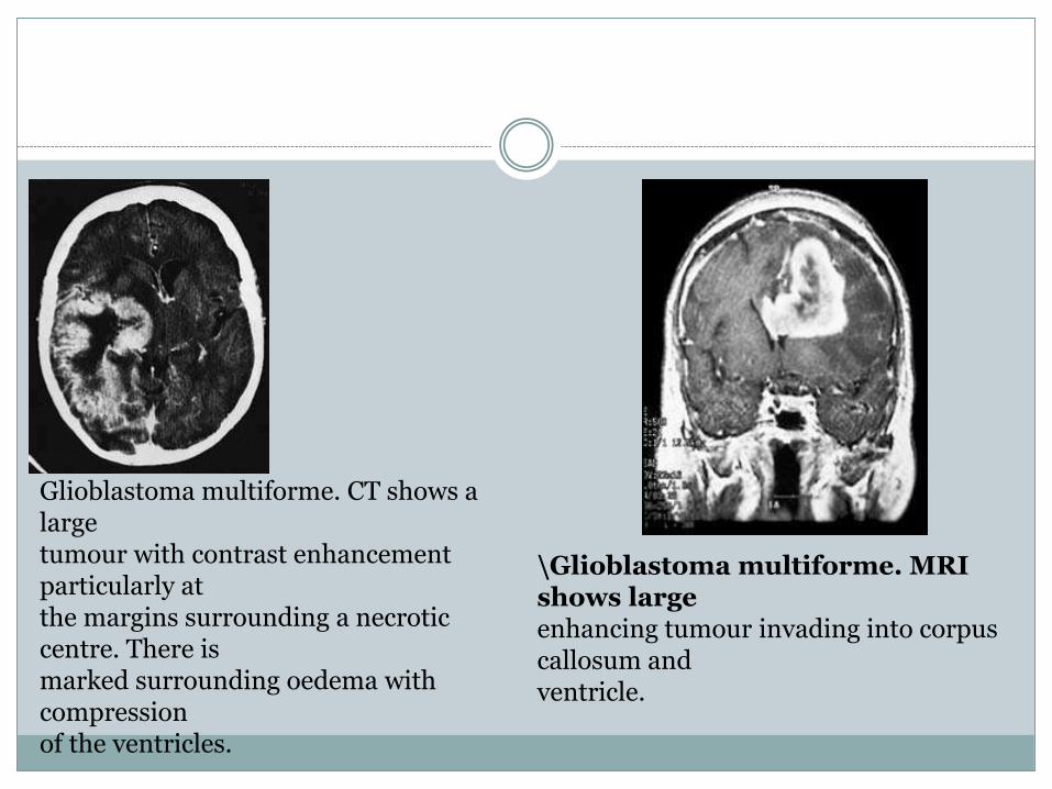
Cystic anaplastic astrocytoma.
Low-grade astrocytomas may show up on MRI
as abnormal areas of increased T2 signal and decreased
T1 signal, even if the CT scan was normal.
High-grade astrocytomas characteristically have
low signal intensity on T1-weighted images and
high signal intensity on T2-weighted images.
Gadolinium enhancement is more likely to occur
in the higher-grade tumour.

Perfusion-weighted MRI is used to determinethe regional cerebral blood volume, which isincreased in high-grade glioma and may be ofvalue in differentiating recurrent tumour fromradiation necrosis. Magnetic resonance spectroscopy(MRS) is a non-invasive technique thatprovides information on the composition andspatial distribution of cellular metabolites. Onproton MRS, tumours have an increased lactateproduction, loss of N-acetyl aspartate (due to lossof neurones in the tumour area) and increasedcholine levels (due to active membranebiosynthesis).
Cerebral angiography
This was the standard study in most patients
with astrocytomas prior to the introduction of
CT. It provides helpful information on the
vascular supply of the tumours but is now only
rarely indicated.
Plain X-rays
Plain X-rays of the skull do not need to be
performed as a routine. The most common
abnormality is erosion of the sella turcica due
to long-standing raised intracranial pressure.
Radiologically visible calcification is present in
about 8% of patients with astrocyte-derived
gliomas.
Management
Following the presumptive diagnosis of a gliomathe management involves:• surgery• radiotherapy• other adjuvant treatments.SurgerySurgery is performed with three principal aims.• To make a definite diagnosis.• Tumour reduction to alleviate the symptomsof raised intracranial pressure.• Reduction of tumour mass as a precursor toadjuvant treatments.
The patient is started on glucocorticoid steroid
therapy (e.g. dexamethasone) when presenting
with clinical features of raised intracranial
pressure with the aim of decreasing the cerebral
oedema prior to surgery.
The type of operation performed will largely
be determined by the position of the tumour and
by the patient’s clinical presentation. In general,
the tumour is excised as radically as possible,
provided the surgery will not result in any disablingneurological deficit. Craniotomy is performedin the position that provides the bestaccess to the tumour and usually with the aid of aframeless stereotactic system to aid accuracy oflocalization. If the tumour has not grown to involvethe cortical surface, a small incision is madein a non-eloquent gyrus or sulcus and the subcorticalbrain is divided down to the tumour mass.The tumour is excised, often with the aid of anultrasonic aspirator..
Occasionally, the tumour
may involve one of the ‘poles’ of the hemisphere
and the excision may entail a partial lobectomy.
Although a craniotomy with radical tumour
excision will alleviate the symptoms of raised
intracranial pressure, there has been controversy
as to whether a radical resection improves survival.
Most high-grade gliomas weigh approximately
100 g at the time of diagnosis and consist
of 1011 cells
A radical tumour excision is able to
excise the macroscopic tumour but cannot remove
the tumour cells that are infiltrating deep
into the adjacent, often vital, areas of normal or
oedematous brain. Consequently, a radical excision
is unlikely to achieve more than a 90–95%
reduction in tumour cell numbers, resulting in
1010 cells remaining.
Whether the 1–2 logs oftumour cell reduction are a significant reductionin tumour burden prior to adjuvant therapyand whether it improves the effectiveness ofsubsequent treatment is still not completelyresolved, although recent clinical studies doseem to show a survival benefit following tumourresection and this is favoured by mostneurosurgeons provided the tumour excisioncan be performed without causing significantneurological mortality
Alternatively, a biopsy, which can be performed
most accurately using stereotactic methods,
may be undertaken to obtain the definite
histological diagnosis, without macroscopic tumour
excision, if:
.
• the tumour is small and deep seated
• the tumour is diffuse, without major features
of raised intracranial pressure, and macroscopic
resection is not feasible
• the tumour involves highly eloquent areas
(e.g. speech centre) without pronounced features
of raised intracranial pressure
Stereotactic biopsy involves localization of the
tumour with a stereotactic frame applied to the
head of the patient using the CT scan or MRI.
The three-dimensional coordinates of the tumour
are ascertained. The surgeon chooses the point of
entry and the desired path through the brain and
a computer program determines the necessary
angles for the biopsy probe and the depth to the
tumour
Postoperative care
The postoperative management of astrocytoma
involves the routine care of a patient following a
craniotomy. Careful neurological observations
are performed, as prompt intervention is essential
if the patient’s neurological state deteriorates
as a result of either increasing cerebral oedema or
postoperative haemorrhage. A postoperative
haematoma may occur in the region of the tumour
excision or it may be extracerebral, either
subdural or extradural.
A CT scan should be
performed urgently if there is neurological
deterioration, to determine the exact pathology.
be so rapid as to require urgent re-exploration Occasionally, postoperative deterioration may
of the craniotomy without prior radiological
assessment.
In the initial postoperative period it is essentialto avoid overhydration of the patient so as not toprecipitate cerebral oedema. The patient isnursed with the head of the bed elevated 20°, soas to promote venous return and reduce intracranialvenous pressure. Steroid medication isusually required in the initial postoperativeperiod and is gradually decreased over thefollowing days. The steroids may need to bere-instituted during the course of radiotherapy.The patient is usually mobilized as soon aspossible, if necessary with the help of aphysiotherapist.
. Opinion varies regardingthe tissue volume that should be treated for malignantglioma but radiation to the tumour areaand a ‘generous volume’ of surrounding brain isnow advocated, rather than radiation to thewhole brain. The selection of the proper radiationdose for gliomas is as controversial. Although increasingthe radiation dosage from 50 to 65 graydoes slightly improve survival, the higher doseof radiation therapy, especially over 65 gray, significantlyincreases the risk of brain necrosis.
Radiation therapy
Postoperative radiation therapy is generally aneffective adjunct to surgery in the treatment ofhigher-grade gliomas. It has been shown todouble the median survival for high-gradegliomas to 37 weeks.Radiation treatment is planned to optimize thehomogeneity of the radiation dose throughoutthe tumour volume selected and to minimizehigh dose regions in normal brain transited bythe radiation beam
. The size of the daily radiation
fraction is related to the incidence of complications
and a maximal daily dose is usually between
1.8 and 2.0 gray. The total radiation dose
varies depending on the tumour type, location
and size of field, but for gliomas it is usually between
45 and 60 gray
Prognosis
At present there is no satisfactory treatment forthe malignant cerebral glioma—the anaplasticastrocytoma and glioblastoma multiforme. Themedian survival following surgery is approximately17 weeks and when radiation therapy isused as an adjuvant the median survival is approximately37 weeks. Chemotherapy for highgradegliomas has been disappointing and thebest results with surgery, radiation therapy andchemotherapy consistently show a median survivaltime of less than 1 year. The median normalfor the low-grade glioma (fibrillary astrocytoma;WHO Grade II, Daumus–Duport Grades I and II)is approximately 8 years, with most tumours progressingto a higher grade
The role of surgery, radiation therapy and
other adjuvant therapies in low-grade gliomas is
even less certain than for the high-grade glioma.
The low-grade tumour may remain relatively
quiescent for some years before it either continues
to grow slowly or changes to a more
anaplastic tumour with resulting debilitating
neurological deterioration. In general, the same
principles for surgical excision apply for lowgrade
gliomas as for high-grade gliomas..
However,
a number of clinical studies have shown
that patients having a radical excision of a tumour
have a longer 5-year survival than those
with a subtotal excision. Radiation therapy has
not been shown to improve the survival in patients
with low-grade tumours. In general, other
adjuvant therapies are not used for the treatment
of low-grade astrocytoma, but may be of benefit
for oligodendrogliomas
Other adjuvant therapies
Many different adjuvant therapies have been investigated
for the treatment of glioma. These include
the use of new chemotherapeutic agents,
new methods of administering cytotoxic chemicals,
immunotherapy, hyperthermia, new techniques
of radiotherapy, photodynamic therapy,
and gene therapy.
The lack of effectiveness of the present treatment
of gliomas is related to the biology of the tumour.
The most common position for tumour
recurrence following conventional treatment is
locally, in the tumour bed, indicating that treatment
has failed in local control. Although light
microscopy shows high-grade gliomas to have a
relatively well-defined border with the adjacent
brain, special staining techniques, includingmonoclonal antibodies, show malignant cells extendingwell out into the surrounding brain. It isthe failure to control the growth of these cells thatis largely responsible for the local tumour recurrence.As indicated previously, a good surgicalresection with 90% of the tumour excised wouldstill result in 1010 cells being present. Effective radiotherapywill result in 1 log (90%) or at the most2 logs (99%) of cell kill and it is unlikely that subsequentchemotherapy would reduce remainingtumour cells by more than 90%.
Consequently,
the effect of cytoreductive surgery, radiotherapy
and chemotherapy would result in approximately
108 cells remaining; the immune system
is unlikely to be able to cope with a tumour
burden of more than 105 cells. It follows that, for
any extra treatment to be effective, whether
surgery or adjuvant therapy, it must provide at
least 1 log of cell kill.
Chemotherapy
Conventional chemotherapy has been disappointing.Many of the chemotherapy agents thatare active in vitro, or in other systemic tumours,have reduced activity in malignant brain tumours,either by exerting an inherently limitedcytotoxic potential on brain tumour cells or bythe inability of the chemotherapeutic agent toreach the cells that are responsible for the tumourrecurrence. A study of brain tumour cell kineticsof high-grade gliomas shows only a small proportionof the cells (5–10%) in an active growthphase.
; this has serious consequences for any cellcycle-specific cytotoxic agent. Until recently themost commonly used single agent cytotoxicregime involves administration of nitrosoureacompounds. The high lipid solubility and lowionization of these agents ensures a relatively effectivepenetration of the cytotoxic compoundinto the tumour. Combination therapy, utilizingmany different cytotoxic compounds, has beenused in various trials but none of the combinationshas been shown to be more beneficial thanthe use of the single nitrosourea
Temozolomide is an alkylating agent that can
be taken orally, penetrates the CNS and is well
tolerated with predictable myelotoxicity. Clinical
studies have shown it to be effective in about 40%
of patients. It is now the initial chemotherapy
agent of choice, but it is usually used at the time
of tumour recurrence, rather than as an adjuvant
to surgery.
It has been postulated that a reason for the lack
of effectiveness of chemotherapy is the inability
of the cytotoxic compound to reach the tumour
cells which are invading the normal adjacent
brain. This has resulted in new techniques
of delivering the cytotoxic agent. High-dose
chemotherapy with bone marrow rescue has
largely been abandoned because of its high
morbidity and lack of effectiveness

Techniques
of disrupting the blood–brain barrier have been
used before chemotherapy infusion to improve
the delivery of chemotherapeutic agents to tumour
cells within the environment of a normal
blood–brain barrier. This has resulted in substantially
increased neurotoxicity to the normal brain
without significantly improving survival..
Intracarotidchemotherapy suffers from serious limitations—the perfusion of the tumour mass is lessthan expected because most tumours are notsupplied entirely by one carotid artery and‘streaming’ of the cytotoxic agent results invery high doses of chemotherapy to small areas,with relative hypoperfusion in other regions.Complications such as serious retinal damageand neurotoxicity have further reduced theattractiveness of this technique
Radiotherapy
Attempts to enhance the effect of radiotherapy
have included the use of radiosensitizers, such as
metronidazole and misonidazole, which increase
the radiosensitivity of hypoxic tumour cells without
a corresponding increase in the sensitivity of
euoxic cells. However, the clinical trials have
shown only a marginal advantage.
The use of interstitial
brachytherapy, involving stereotactically
implanted radioactive sources into the
tumour, has the advantage of applying a high
dose of radiotherapy to the tumour while sparing
the surrounding brain. However, the clinical
trials performed so far have resulted in a high incidence
of radionecrosis to the surrounding brain
and improvements in the technique will need to
be devised before this method would become
acceptable. Similarly, stereotactic radiosurgery,
that involves radiation to a very highly focused
area, is of limited use as it does not target the
infiltrative tumour cells that are responsible for
tumour recurrence.
Hyperthermia
This has inherent basic limitations as, although
cell death occurs at approximately 42°C, damage
to the surrounding brain occurs at 45°C, so there is
a very narrow therapeutic index. In addition, there
is a marked tolerance of tumour cells to hyperthermia
and the treatment has not been effective.
being investigated involve the use of retrovirus,
adenoviruses or adeno-associated viruses to
carry a variety of gene therapies to the cancer cell.
It is hoped that as these treatments are refined
over the next decade they will be useful in the
treatment of cerebral glioma.
Oligodendroglioma
Oligodendrogliomas are responsible for
approximately 5% of all gliomas and occur
throughout the adult age group with a maximal
incidence in the 5th decade. The tumour is rare in
Pathology
Nearly all oligodendrogliomas occur above the
tentorium; most are located in the cerebral
hemispheres and about half of these are in the
frontal lobes. Oligodendrogliomas may project
into either the 3rd or lateral ventricles. children.
Oligodendrogliomas have the same spectrumof histological appearance as astrocytomas, rangingfrom very slow growing, benign tumours to amore rapidly growing, malignant variety withabundant mitotic figures, endothelial proliferationand foci of necrosis. Calcium deposits arefound by histological examination in up to 90% ofoligodendrogliomas. Unlike the astrocyte group,most oligodendrogliomas are well differentiated.Not infrequently tumours have mixed histology,with both oligodendroglial and astroglialfeatures.
Clinical presentation
The presenting features are essentially the sameas for the astrocyte group but, as these tumoursare more likely to be slow growing, epilepsy iscommon, occurring in 80% of patients and seenas an initial symptom in 50%. The features ofraised intracranial pressure and focal neurologicaldeficits are each present in approximatelyone-third of patients.As for astrocyte tumours, MRI with contrastmay be beneficial but other investigations areusually unnecessary.
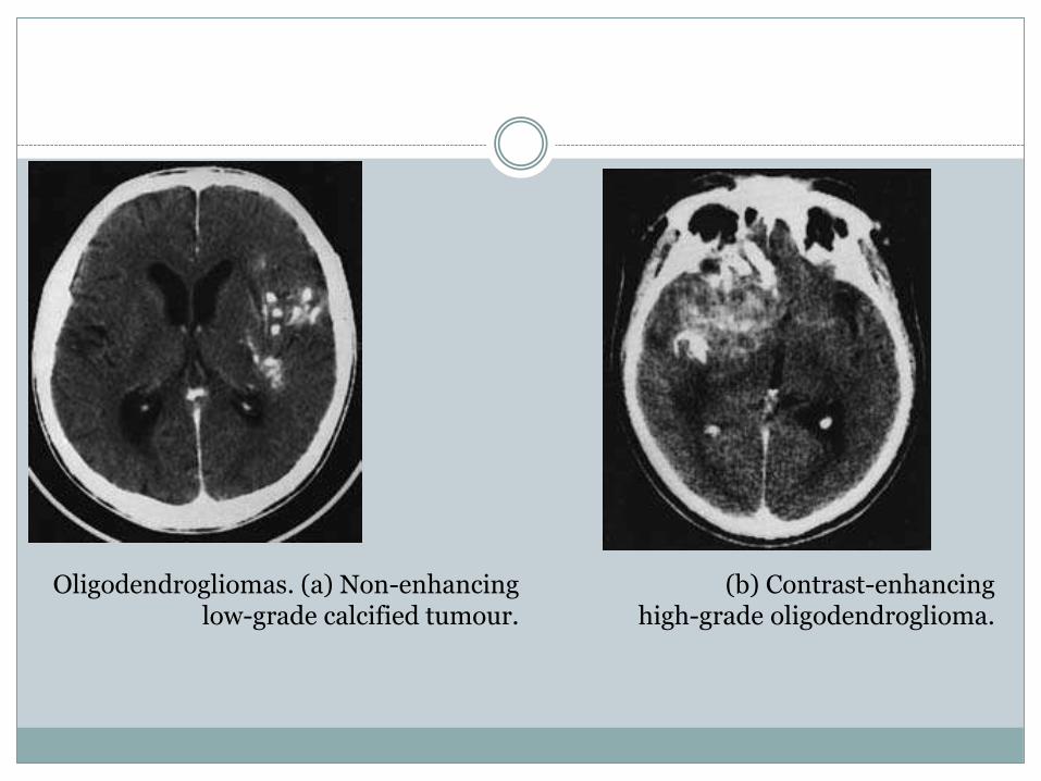
Oligodendrogliomas. (a) Non-enhancing low-grade calcified tumour.
(b) Contrast-enhancinghigh-grade oligodendroglioma.
Radiological investigation
CT scanning and MRI are the fundamental investigations.
They will confirm the diagnosis of an
intracranial tumour and in many cases the diagnosis
of oligodendroglioma will be highly probable.
Calcification will be present in 90% of cases
and over half show contrast enhancement (Fig.
6.11.)
Treatment and results
Treatment involves:• surgical resection• radiotherapy• other adjuvant treatments.The standard treatment for oligodendrogliomahas been an aggressive resection of the tumourfollowed by radiation therapy, although radiotherapywould now not be given to low-grade tumours,and utilized only for the intermediate- orhigh-grade oligodendroglial tumours
. Oligodendrogliomas
have been shown to be more sensitive
to chemotherapy than the astrocytoma
tumours, especially if the oligodendrogliomas
belong to the group with loss of heterozygosity
OF chromosome 1p or 19q.
The survival of patients depends on the degree
of histological malignancy. Five-year survival
rates are between 30 and 50% with a small number
of patients living for many years (up to 5% for
20 years). However, many tumours with histological
features of oligodendroglioma also have a
component of astrocyte-derived cells, usually
anaplastic astrocytoma, and the tumour behaves
biologically and clinically as an anaplastic astrocytoma
rather than an oligodendroglioma
Recurrent cerebral glioma
As discussed earlier, most high-grade cerebral
gliomas will recur within 1 year of the initial
treatment with surgery and radiotherapy. Lowgrade
tumours may either recur as a continuing
progression of the slow growth or, alternatively,
the histological characteristics may alter and
the tumour may become more anaplastic and
rapidly growing.
The clinical presentation of a recurrent tumour
will be evidenced by either a progression of the
focal neurological signs or the signs of an increase
in the intracranial pressure. The diagnosis will be
confirmed by CT scan or MRI in most cases. The
major differential diagnosis is postradiotherapy
radiation necrosis, which may develop as early as
4 months or as late as 9 years after radiotherapy
The radiological features of necrosis are an avascular
mass, and the diagnosis may be suspected
from the dose of radiotherapy that has been
administered. However, there may be considerable
difficulty in differentiating necrosis from
recurrent glioma, and sometimes an operation
is required both for definitive diagnosis and to
remove the mass.
The initial deterioration following a diagnosis
of recurrent glioma can usually be temporarily
halted by the use of steroid medication. The
major decision is whether further surgery and
other adjuvant therapy should be undertaken. In
general, a further operation involving debulking
of the tumour would be considered if
:
• the patient is less than 65 years old
• there has been a symptom-free interval of
1 year or more since the first operation
• debilitating irreversible neurological signs are
absent
• the tumour is in an accessible position and
repeat surgery would not result in additional
morbidity.

Adjuvant therapies
Adjuvant therapies have limited benefit for patients
with recurrent tumour and, considering
the morbidity involved, may not be indicated.
Chemotherapy, utilizing temozolomide administered
orally, has shown to have a temporary benefit
in up to 40% of patients. Other chemotherapy
agents, especially the nitrosourea compounds,
have much greater toxicity.
Ependymoma
Ependymomas are glial neoplasms arising from
the ependyma and constitute approximately
5% of all gliomas. Approximately two-thirds of
ependymomas occur in the infratentorial compartment
and most of these present in children,
adolescents and young adults. The supratentorial
ependymomas occur mostly in adults

Pathology
The tumour arises from the ependyma of the ventricle
and, although predominantly intraventricular,
the tumour often invades into the adjacent
cerebellum, brainstem or cerebral hemisphere.
Fourth ventricular tumours usually arise from
the floor or lateral recess of the 4th ventricle and
they may extend into the subarachnoid space to
encase the medulla or upper cervical spinal cord.
Alternatively, the tumour may grow laterally
through the foramen of Luschka and into the
cerebellopontine angle.
The tumours are well demarcated, nodular,soft and pale. Calcification is common, especiallyin supratentorial ependymomas.There are numerous histological classificationsystems of ependymomas, and the World HealthOrganization classification divides these tumoursinto cellular, papillary and clear cell typesof non-anaplastic ependymoma and anaplasticependymoma.The myxopapillary variety is a slow-growingdistinct variant of ependymoma that occurs
inthe cauda equina and is discussed in the chapteron spinal cord tumours (Chapter 15).In an adult the subependymoma variant maybe encountered as an incidental autopsyfinding—a discrete nodular mass based in thebrain’s ventricular surface, particularly the flooror lateral recess of the 4th ventricle or the septumpellucidum—or it may be large enough to presentclinically. It is usually heavily calcified and iscomposed of cells with astrocytic as well asependymal features.

The papillary and anaplastic varieties of
ependymoma are responsible for the majority of
clinically symptomatic ependymomas. The cellularity
and architecture of the ependymomas vary
but a diagnostic feature is the presence of rosettes,
and most ependymomas contain areas in which
perivascular pseudorosettes are conspicuously
developed
. In these formations the blood vessel is
surrounded by an eosinophilic halo composed of
the radiating tapering processes of the cells. Blepharoplasts
frequently occur in ependymomas
but may be difficult to visualize. These are tiny
intracytoplasmic spherical or rod-shaped structures
which represent the basal bodies of cilia,
and are most frequently encountered in the apical
portion of cells that form ependymal rosettes.
Clinical presentationPosterior fossa ependymomas
Details will be discussed in the section on paediatric
tumours (p. 91). Patients present with features
of raised intracranial pressure due to
hydrocephalus as a result of obstruction of the
4th ventricle, ataxia due to cerebellar involvement,
and occasionally features of brainstem
pressure or infiltration
Supratentorial tumours
Virtually all patients with supratentorialependymomas
present with features of raised intracranial
pressure, often due to hydrocephalus as a
result of obstruction of the CSF pathways. Ataxia
is common and focal neurological deficits may
occur due to involvement of the underlying cerebral
hemisphere.
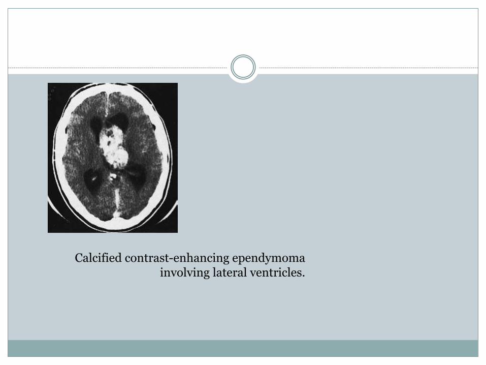
Calcified contrast-enhancing ependymomainvolving lateral ventricles.
Radiological investigation
The CT scan and MRI will show a tumour that
arises in the ventricle and enhances after administration
of intravenous contrast. Calcification is
common in tumours arising from the lateral ventricles
(Fig. 6.12). There is frequently associated
hydrocephalus. In the posterior fossa differentiation
from a medulloblastoma may be difficult.
Treatment
The treatment of ependymomas is initially surgical,
with an attempt to perform a radical macroscopic
resection of the tumour. Supratentorial
tumours are often very large and may extend
throughout the lateral and 3rd ventricles, but the
associated hydrocephalus makes the excision of
the intraventricular portion feasible
. However,
the tumour may arise from the ventricular wall in
the region of the basal ganglia and blend imperceptibly
with the underlying cerebral structures
so that a complete excision is not possible. Fourth
ventricular tumours can be excised from the ventricle
but microscopic infiltration into the underlying
brainstem cannot be removed surgically.
Postoperative radiation therapy is advisable and,
as these tumours may spread through the CSF
pathways, sometimes whole neuraxis radiation
is recommended.
The prognosis is related to the degree of
anaplasia of the tumour and for intratentorial tumours
varies from 20% to 50% 5-year survival.
The prognosis for the supratentorial tumours is
better, particularly in adults.
Pineal tumours
Tumours arising in the region of the pineal glandare mostly not of pineal origin, but are generallycalled ‘pineal’, as they have a similar clinicalpresentation.The tumours are relatively uncommon, accountingfor 0.5% of all intracranial tumours.However, they occur more frequently in Japanand China, where the incidence is up to 5%. Mosttumours make their clinical appearance between10 and 30 years of age.
Classification
Pineal region tumours are classified in decreasing
frequency as
. germinoma
• teratoma
• pineocytoma
• pineoblastoma
• miscellaneous:
• glioma
• cyst
Germinoma is the most common pineal region
tumour and is similar in histological appearance
to germinoma of the gonads and mediastinum; it
occurs predominantly in males. These tumours
may also arise in the suprasellar region, and
synchronous tumours in both the pineal and
suprasellar region occur occasionally. Teratoma
is like germinoma; it also arises from displaced
embryonic tissue. The other tumour types occur
less commonly.
Clinical presentation
Patients with pineal tumours present with:
• raised intracranial pressure
• neurological signs due to focal compression
• endocrine disturbance.
Raised intracranial pressure. The features of
raised intracranial pressure, such as headaches,
drowsiness and papilloedema, are due to
hydrocephalus, which is a result of the tumour
occluding the aqueduct of Sylvius.
Focal compression. Compression of the efferent
cerebellar pathways in the superior cerebellar
peduncle results in limb ataxia and distortion of
the quadrigeminal plate, produces limitation of
upgaze, convergence paresis with impairment
of reaction of pupils to light and accommodation
(Parinaud’s syndrome), and may result in
convergence-retraction nystagmus on upgaze
(Koerber–Salius–Elschnig syndrome).
Endocrine disturbance. Endocrine disturbances are
uncommon but include precocious puberty in
10% of patients, almost invariably male, and
diabetes insipidus in 10%. The endocrine effects
can either be due to direct tumour involvement of
the hypothalamus or result from the secondary
effects of hydrocephalus.
Radiological investigationsCT scan and MRI will show a pineal regiontumour and will often suggest the correct pathologicaldiagnosis (Fig. 6.13). On CT scan, beforecontrast, a germinoma will be a hyperdense lesionin the region of the pineal gland infiltratinginto the surrounding tissue and there will be uniformvivid enhancement following intravenouscontrast. Calcification is uncommon. On MRIgerminomas are relatively isointense to normalwhite matter on T1-weighted images and slightlyhyperintense on T2-weighted scans. Gadoliniumcontrast defines the tumour well as germinomasenhance markedly and homogeneously
ManagementThis consists of surgery and radiotherapy.A ventriculoperitoneal shunt or drainage ofCSF by a 3rd ventriculostomy may be required ifthe hydrocephalus is severe.There is controversy over whether the definitivetreatment should be an attempt at surgicalexcision or radiotherapy. As most of the tumoursare germinomas, and these tumours are veryradiosensitive, a course of radiotherapy can begiven as the initial treatment if the radiologicalappearance is typical for germinoma. If serial CTscans show the tumour has failed to respond toradiotherapy then surgery may be necessary.Alternatively, if the features on the initial CTand MRI scans are atypical, and the lesion doesnot resemble a germinoma, exploration of the tumourand surgical excision may be appropriateas the initial procedure
The preferred surgical approach is usually by a
posterior fossa craniotomy, above the cerebellum
and below the tentorium cerebelli. Alternative
supratentorial surgical exposures include approaching
the tumour through the corpus callosum
or by retracting the occipital lobe
Metastatic tumours
Metastatic tumours are responsible for approximately15% of brain tumours in clinical series butup to 30% of brain tumours reported by pathologists.cancer and 1 in 5 of these have intracranialmetastatic deposits at autopsy. The metastatictumours most commonly originate from:• carcinoma of the lung• carcinoma of the breast• metastatic melanoma• carcinoma of the kidney• gastrointestinal carcinomaApproximately 30% of deaths are due to
In 15% a primary origin is never found.
Most metastatic tumours are multiple and onethird
are solitary. In about half of these systemic
spread is not apparent. The incidence of tumours
in the cerebrum relative to the cerebellum is 8 : 1,
and most occur in the distribution of the middle
cerebral artery. The size of the tumours may
vary considerably if the deposits are multiple.
Metastatic tumours are often surrounded by
intense cerebral oedema.
Clinical presentationThe interval between diagnosis of the primarycancer and cerebral metastases varies considerably.In general, secondary tumours from carcinomaof the lung present relatively soon afterthe initial diagnosis, with a median interval of 5months. Although cerebral metastases may presentwithin a few months of the initial diagnosisof malignant melanoma or carcinoma of thebreast, some patients may live many years (up to15 years) before an intracranial tumour appears.The presenting features are similar to thosedescribed for other intracranial tumours:• raised intracranial pressure• focal neurological signs• epileptic seizures.
Headache and vomiting, indicative of raisedintracranial pressure, occur in most patients andthe presenting history is usually short, often onlya few weeks or months. The increased intracranialpressure will be caused by either the tumourmass and surrounding oedema or, in posteriorfossa tumours, obstructive hydrocephalus.The pattern of focal neurological signs will dependon the position of the tumour deposits andthe patient may present with a progressive hemiparesisor speech disturbance with supratentorialtumours or gait ataxia with cerebellar tumours.
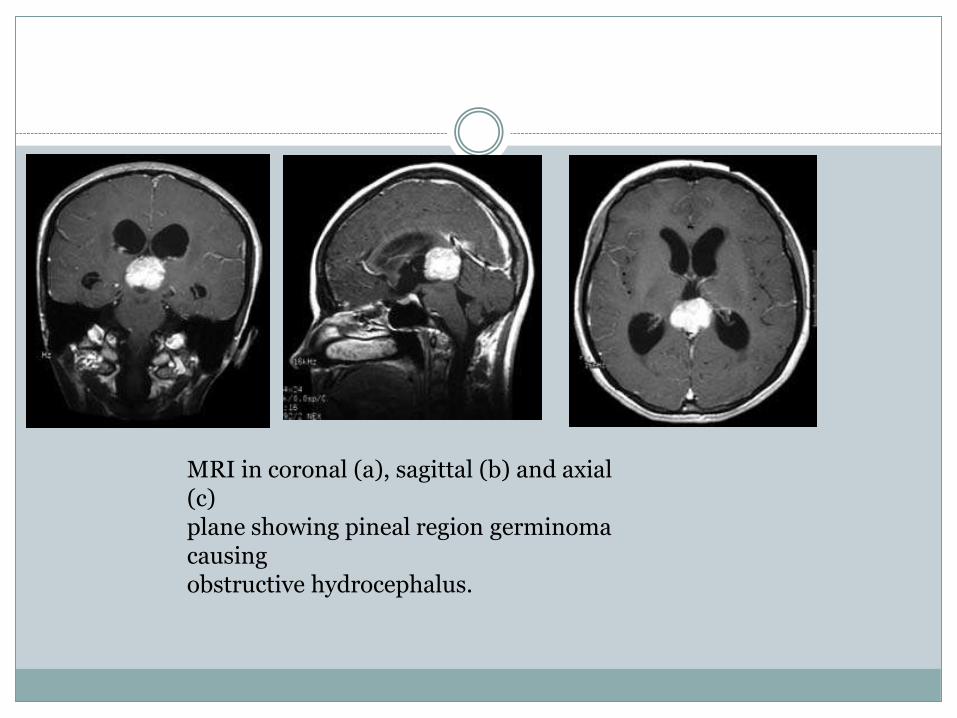
MRI in coronal (a), sagittal (b) and axial (c)plane showing pineal region germinomacausingobstructive hydrocephalus.
Epileptic seizures occur in approximately
25% of patients and may be either focal or
Occasionally, metastases, especially melanoma
or choriocarcinoma, present following an intracerebral
haemorrhage.generalized
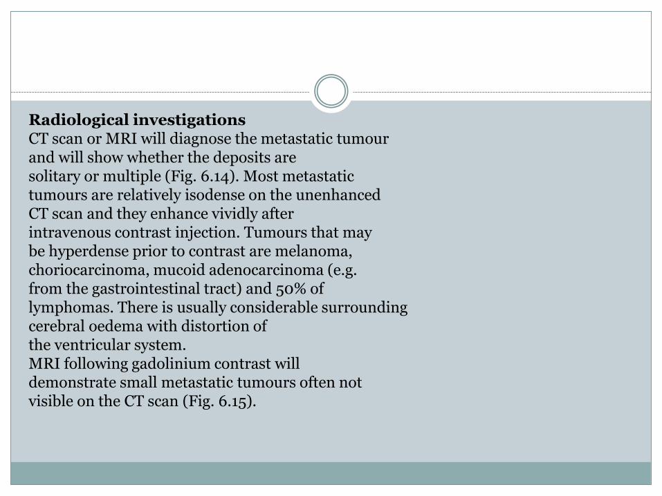
Radiological investigationsCT scan or MRI will diagnose the metastatic tumourand will show whether the deposits aresolitary or multiple (Fig. 6.14). Most metastatictumours are relatively isodense on the unenhancedCT scan and they enhance vividly afterintravenous contrast injection. Tumours that maybe hyperdense prior to contrast are melanoma,choriocarcinoma, mucoid adenocarcinoma (e.g.from the gastrointestinal tract) and 50% oflymphomas. There is usually considerable surroundingcerebral oedema with distortion ofthe ventricular system.MRI following gadolinium contrast willdemonstrate small metastatic tumours often notvisible on the CT scan (Fig. 6.15).
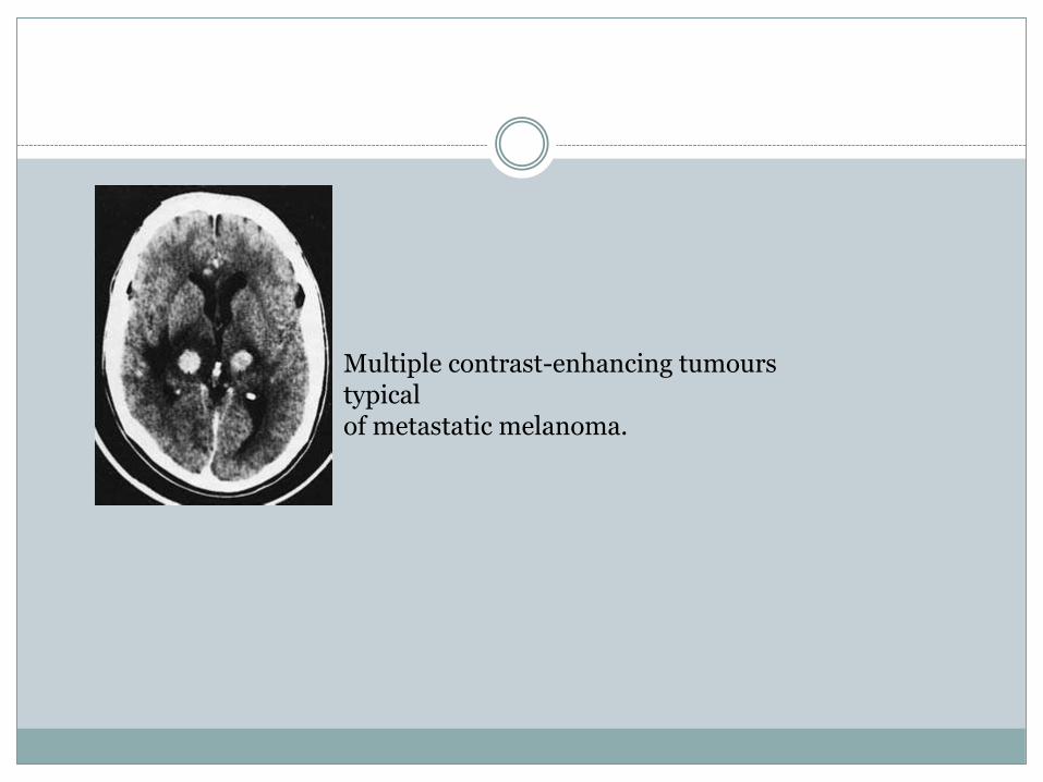
Multiple contrast-enhancing tumourstypicalof metastatic melanoma.
TreatmentSteroid medication (e.g. dexamethasone) willcontrol cerebral oedema and should be commencedimmediately if there is raised intracranialpressure.Surgery to remove the metastasis is indicated if:• there is a solitary metastasis in a surgicallyaccessible position• there is no systemic spread.Removal of a solitary secondary is preferableonly if the primary site of origin has been, or willbe, controlled. However, excision of a singlemetastasis will provide excellent symptomaticrelief and consequently may be indicated even ifthe primary site cannot be treated satisfactorily.Surgery is, of course, mandatory if the diagnosisis uncertain.
Leptomeningeal metastases
Meningeal carcinomatosis is widespread, multifocalseeding of the leptomeninges by systemiccancer. The clinical presentation includes hydrocephalus, causing headaches andvomiting• cranial nerve abnormalities due to directinvasion by the tumours• spinal root involvement due to local infiltration.The CT scan or MRI findings may be subtle butfrequently show excessive enhancement of themeninges. Lumbar puncture can be performedif there is no evidence of raised intracranialpressure. Malignant cells may be seen in theCSF, the protein concentration is increased and
the glucose reduced•.
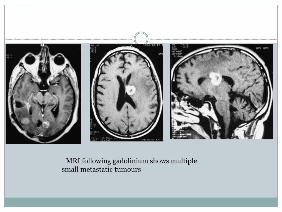
MRI following gadolinium shows multiplesmall metastatic tumours
Cerebral lymphoma
The term cerebral lymphoma encompasses a
number of distinct pathological and clinical
entities. Current nomenclature divides cerebral
lymphoma into: non-Hodgkin’s lymphoma or
Hodgkin’s disease; primary or secondary disease;
and patients who are immunocompetent or
immunosuppressed.
Historically, lymphoma involving the CNS
was considered to be rare, being less than 3% of
all CNS tumours, with about half of these lymphomas
being primary cerebral lymphoma, that
is, the tumour being confined entirely to the CNS.
Over the past decade there has been an unprecedented
increase in the incidence of cerebral lymphoma,
which can be attributed to at least two
known factors: the acquired immune deficiency
syndrome (AIDS) epidemic and the use of immunosuppressive
therapy. However, there has
also been an unexplained increase in the incidence
of primary cerebral lymphoma in nonimmunosuppressed
patients
Cerebral lymphoma may be secondary to systemic
lymphoma and in large studies of patients
with systemic lymphoma, up to 30% of patients
develop clinical or pathological evidence of cerebral
involvement. However, almost all cases are
associated with either relapsed or progressive
systemic disease and an isolated CNS relapse is
Intracerebral Hodgkin’s disease is a very rare
entity.very rare.
Clinical presentationThe most common site for primary cerebrallymphoma is in the frontal lobe, followed by thetemporal lobe, parietal lobe and deep nuclei.Tumours may also occur in the cerebellum andbrainstem. The tumours may be either solitary ormultiple and primary leptomeningeal diseasehas been reported in up to 10% of primary cerebrallymphoma.There are no pathognomonic presenting symptomsor signs in primary cerebral lymphoma andthe presenting features are similar to those describedfor other intracranial tumours: raised intracranialpressure, focal neurological signs andepileptic seizures
The high frequency of frontallobe involvement results in a common mode ofpresentation as memory loss, forgetfulness andaltered affect. Up to 10% of patients with primarycerebral lymphoma present with a seizure. Inview of the incidence of multiple lesions, it isnot surprising that many patients present with aconstellation of symptoms and signs. The presentingfeatures in primary cerebral lymphomaarising in immunodeficient patients (includingAIDS-related primary cerebral lymphoma)do not appear to be different from those in immunocompetentpatients with primary cerebrallymphoma.
Radiological investigations (Figs 6.16 and6.17)
The CT scan characteristically shows a hypodenseor isodense or sometimes hyperdensetumour, which enhances following contrastinjection with associated mild to moderateoedema. Multifocal disease is observed in about40% of CT scans. Most primary cerebral lymphomasarise in the periventricular region. MRIis now the investigation of choice for primarycerebral lymphoma. The lesions are usually hypointenseto isointense on T1-weighted imagesand isointense to hyperintense on T2-weightedimages. The tumours enhance following intravenousinjection of gadolinium.Although CSF examination may show a populationof abnormal lymphocytes, concern regardingraised intracranial pressure usually preventsa lumbar puncture being performed in the majorityof patients
ManagementThe principles concerning the management ofprimary cerebral lymphoma involve:• histological diagnosis• ensuring that the disease is confined to the brain• excluding an underlying, predisposing illness• instituting appropriate therapy.The usual sequence of events is a CT and MRI
In general, it is thought appropriate. scan followed by a biopsy proving intracerebralto exclude systemic disease, although concurrentcerebral and systemic lymphoma is uncommon.Patients usually undergo bone marrow aspirateand trephine, chest X-ray, CT scan of the chestand abdomen and testing for evidence of HIVinfection.lymphoma
Surgical treatmentThere is no clear evidence that craniotomy withexcision of the lymphoma is superior to obtainingan accurate tissue diagnosis using a stereotacticbiopsy. It must be noted that the early use ofcorticosteroids, to treat cerebral oedema, maymake histological assessment difficult due to theexquisite sensitivity of primary cerebral lymphomato steroids. The tumour may disappearon the CT scan or MRI after the commencementof corticosteroids and this has significant implicationsfor obtaining a stereotactic biopsy
Radiotherapy
Primary cerebral lymphoma is usually radiosensitive
with a clinical response rate of up to 80%.
However, cranial radiotherapy alone as a treatment
for primary cerebral lymphoma rarely
produces long-term survivors, despite the high
response rate and an improvement in median
survival time to 15 months.
Chemotherapy
Numerous chemotherapy regimes have been reported,including the use of both intrathecal andintravenous therapies which can be given priorto, synchronous with, or following radiotherapy.The most commonly used drugs are those used inthe therapy of systemic non-Hodgkin’s lymphomaand include methotrexate, corticosteroids,anthracyclines, vinca alkaloids,cytosine, arabinoside and alkylating agents. Mediansurvival times of up to 44 months have beenreported. However, methotrexate given afterradiotherapy increases the risk of leukoencephalopathywith consequent serious neuropsychologicalimpairment. The need forpostchemotherapy radiation has been questioned,in view of the effectiveness of chemotherapy,especially in the elderly.
Paranasal sinus tumours
Tumours of the paranasal sinuses may spread directlyto involve the brain. These uncommon tumoursmost frequently arise from the ethmoid ormaxillary sinuses, less frequently from the sphenoidsinus and rarely from the frontal sinus. Thetumours invade through the floor of the anteriorcranial fossa in the region of the cribriform plateand may extend through the dura into the frontallobe (Fig. 6.18). The tumours are usually squamouscell carcinoma and less frequently adenocarcinomaor adenoid cystic adenocarcinoma.The aesthesioneuroblastoma is a rare nasal
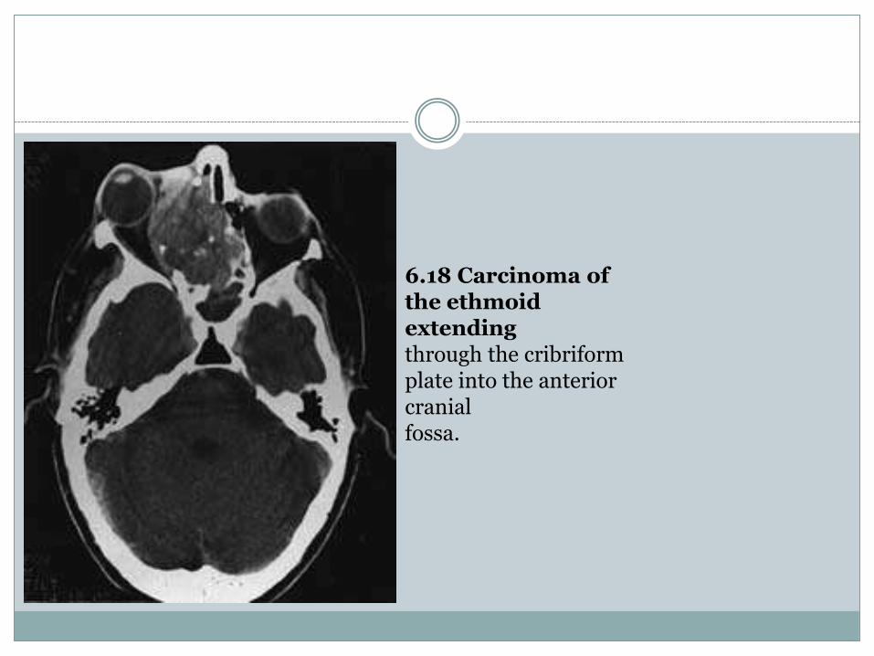
6.18 Carcinoma of the ethmoidextendingthrough the cribriformplate into the anterior cranialfossa.
tumour arising from the olfactory epitheliumthat may invade through the cribriform plate.The patients usually present with a bloodstainedor purulent nasal discharge and pain inthe involved region. CSF rhinorrhoea may occurif the dura has been breached.Surgical excision using a craniofacial resectionmay be the only method of controlling these tumoursand, if the tumour has spread into theorbit, an adequate resection may involve orbitalexenteration.
Chordomas
Chordomas are rare tumours arising from notochord
cell nests. They may arise throughout the
craniospinal axis but occur predominantly at the
ends of the axial skeleton in:
• the basioccipital region
• the sacrococcygeal region.
The intracranial chordoma presents as a skull
base tumour. It infiltrates and erodes the sphenoid
and basiocciput and may spread into the
petrous bones, the paranasal sinuses, the sella
turcica and the cavernous sinuses. The tumour
will compress and distort the adjacent brain and
engulf the cranial nerves and arteries.
These tumours do not have histological features
of malignancy and only rarely metastasize.
However, it is not usually possible to excise the
cranial tumours completely; most patients die
within 10 years of initial presentation.
Spinal chordomas occur predominantly in thesacrococcygeal region, although they may alsoarise in the cervical area. Like the cranial tumours,spinal chordomas invade and destroybone and compress adjacent neural structures.Remote metastases occasionally occur. Patientswith spinal chordomas present with back pain,radicular pain and slowly progressive lumbosacralnerve root involvement resulting insphincter difficulties and sensory and motor disturbancesin the legs.
Histological appearance
The tumours consist of notochord cells and mucoid
stroma. Many of the cells may be coarsely
vacuolated and some will contain a single
large vacuole giving a ‘signet ring’ appearance.
The characteristic histological appearance is
physaliphorous (bubble-bearing) cells containing
multiple vacuoles.
Clinical presentationThe majority of intracranial chordomas arise between20 and 60 years of age. The clinical featuresresult from the widespread tumour extensionand include:• raised intracranial pressure, causingheadaches and vomiting• multiple cranial nerve palsies, often unilateral• nasopharyngeal obstruction.The radiological appearances are of a destructivelesion at the base of the skull or in the vertebralbodies (Fig. 6.19).
Treatment
It is rarely possible to excise all the tumour. Postoperative
radiotherapy is usually administered
but is of doubtful value.
Paediatric tumours
Intracranial tumours are the most common formtumours occurring above the tentorium cerebelli.The most common supratentorial tumours areastrocytomas, followed by anaplastic astrocytomasand glioblastoma multiforme. Craniopharyngiomaoccurs more commonly in childrenthan in adults and is situated in the suprasellarregion; this tumour is described in Chapter 8.Other, less common, supratentorial tumoursinclude primitive neuroectodermal tumours(PNETs), ependymomas, ganglioglioma andpineal region tumours.of solid tumours in childhood, with 40% of the
Posterior fossa tumoursSixty per cent of paediatric brain tumours occurin the posterior fossa. The relative incidence ofthe tumours is:1 cerebellar astrocytoma 30%2 medulloblastoma (infratentorial primitiveneuroectodermal tumour) 30%3 ependymoma 20%4 brainstem glioma 10%5 miscellaneous 10%:(a) choroid plexus papilloma(b) haemangioblastoma(c) epidermoid, dermoid(d) chordoma
Clinical presentation
The presenting clinical features of posterior fossa
neoplasms in children are related to:
• raised intracranial pressure
• focal neurological signs
Raised intracranial pressureThis is the most common presenting feature. It isdue to hydrocephalus caused by obstruction ofthe 4th ventricle and is manifest by headaches,vomiting, diplopia and papilloedema.The headaches begin insidiously, gradually becomingmore severe and frequent; they are worstin the early morning. There is usually no specificheadache localization. Vomiting is frequentlyassociated with the headaches and may temporarilyrelieve the headache. Raised intracranialpressure may result in a strabismus causingdiplopia due to stretching of one or both of the6th (abducens) cranial nerves. This is a so-called
‘false localizing sign’. Papilloedema is usually
present at the time of diagnosis. In infants, an expanding
head size is an additional sign of raised
intracranial pressure
Disturbances of bulbar function, such as difficulty
in swallowing with nasal regurgitation of
fluid, dysarthria and impaired palatal and
pharyngeal reflexes, result from brainstem involvement.
In addition, compression or tumour
invasion of the pyramidal tracts may result in
hemiparesis and, if the ascending sensory pathways
are involved, sensory disturbance will
occur in the trunk and limbs
The tumour may directly envelop the lower
cranial nerves—glossopharyngeal, vagal, spinal
accessory and hypoglossal—as well as the 7th
cranial nerve.
Neck stiffness and head tilt may occur in children
with posterior fossa neoplasms, and may be
due to herniation of a cerebellar tonsil or tumour
tissue resulting in dural irritation.
Investigations
CT scan and MRI have replaced the need for
the previous radiological investigations that
included radio-isotope brain scanning, air ventriculography
and posterior fossa angiography.
The CT scan and/or MRI will show the presence
of a posterior fossa tumour, its position and
whether it arises primarily in the brainstem, 4th
ventricle or the cerebellum (Figs 6.20–6.24
Management
The treatment of posterior fossa tumours
involves:
• surgery
• radiotherapy
• chemotherapy
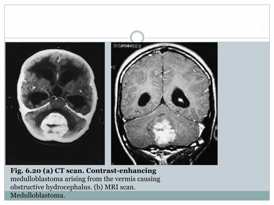
.Fig. 6.20 (a) CT scan. Contrast-enhancingmedulloblastoma arising from the vermis causingobstructive hydrocephalus. (b) MRI scan.Medulloblastoma.
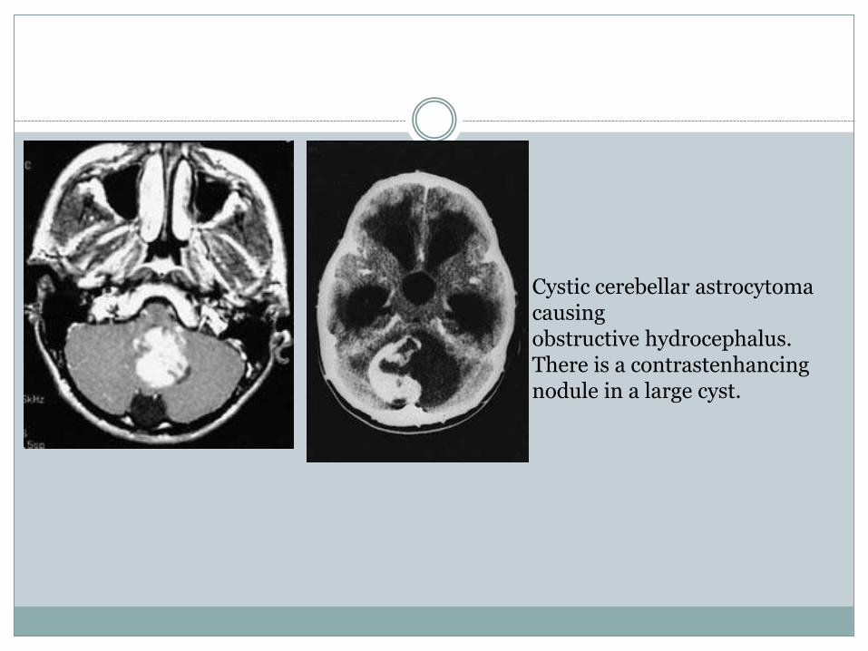
Cystic cerebellar astrocytomacausingobstructive hydrocephalus. There is a contrastenhancingnodule in a large cyst.
Apreliminary CSF shunt may need to be performedin a child with severely raised intracranialpressure due to hydrocephalus. The CSFdiversion can be achieved by either an externaldrain or a ventriculoperitoneal shunt. An externaldrain is a temporary measure only, because ofthe risk of infection. Aventriculoperitoneal shuntprovides immediate and controlled relief ofintracranial hypertension and the subsequentposterior fossa operation can be performedas a planned elective procedure, rather than anurgent operation in suboptimal conditions.
A
criticism of a preoperative ventriculoperitoneal
shunt is that it might promote the metastatic
spread of tumour. A filtering chamber in the
shunt system may lessen this risk but this predisposes
to shunt malfunction.
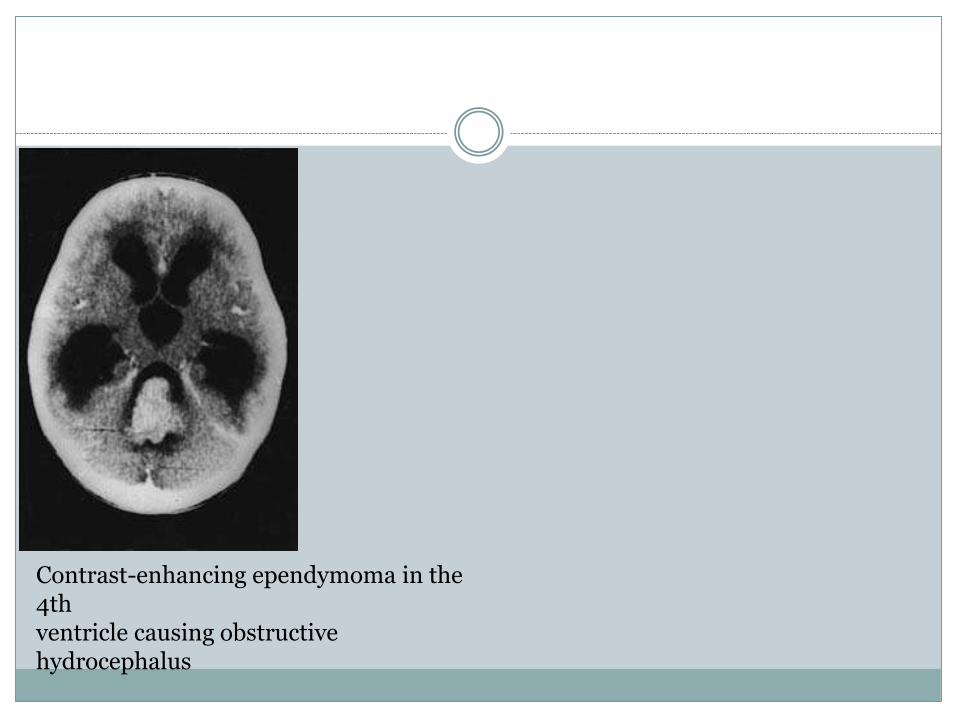
Contrast-enhancing ependymoma in the 4thventricle causing obstructive hydrocephalus
Steroid medication to control local oedema is
commenced preoperatively. The operation is performed
in either the sitting or prone position
through a vertical midline incision. A posterior
fossa craniotomy is performed, usually with excision
of the bone down to and around the foramen
magnum. Tumour excision is aided by the use of
magnifying loupes and illumination with a fibreoptic
headlight, or by the use of an operating
microscope.
Postoperative care involves careful monitoring
of the neurological signs. Postoperative haemorrhage
or oedema may result in rapid deterioration
of the neurological state, and in respiratory
arrest. An urgent CT scan may indicate the cause
and site of the problem but the deterioration may
be so rapid that the wound may need to be reopened
without the benefit of prior scanning

If a ventriculoperitoneal shunt has not been inserted
prior to tumour removal an exacerbation
of the obstructive hydrocephalus may occur if
the tumour excision has failed to relieve the CSF
obstruction. Disturbances of bulbar and lower
cranial nerve function may cause difficulty in
swallowing. Nasogastric feeding may be necessary
until the protective mechanisms return, and
great care should be taken to avoid aspiration.
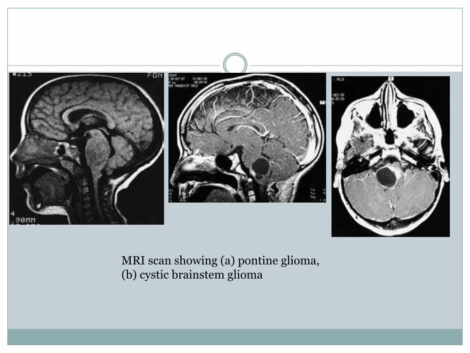
MRI scan showing (a) pontine glioma,(b) cystic brainstem glioma
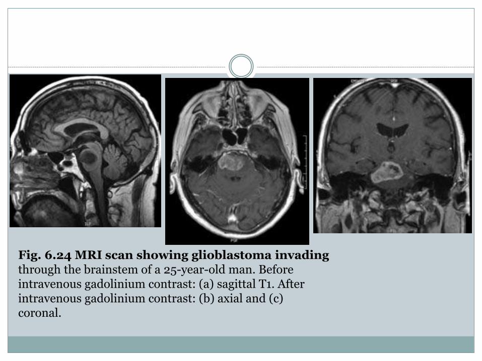
Fig. 6.24 MRI scan showing glioblastoma invadingthrough the brainstem of a 25-year-old man. Beforeintravenous gadolinium contrast: (a) sagittal T1. Afterintravenous gadolinium contrast: (b) axial and (c)coronal.
Medulloblastoma
Medulloblastoma, also referred to as an infratentorial
PNET, is a malignant tumour usually arising
in the midline from the cerebellar vermis,
although it may occur more laterally in a cerebellar
hemisphere in older patients. The tumour expands
to invade the adjacent cerebellum and
large tumours completely fill the 4th ventricle
(see Fig. 6.19).
The tumours arise from the external granular
layer of the fetal cerebellum (Obersteiner’s
layer). Histologically the medulloblastoma is
highly cellular, with numerous mitoses. True
rosettes do not occur but the cells are seen in concentric
patterns around homogeneous material
or blood vessels (pseudorosettes).
Presenting features. The presenting features are
related to hydrocephalus and cerebellar dysfunction.
Truncal ataxia is typically present in children
with medulloblastoma but cranial nerve
deficits, except for a 6th nerve palsy, are uncommon
in the early stages.
Surgery. At surgery the cerebellar vermis is
split in the midline and it is usually possible to obtain
a gross macroscopic excision of the tumour
with complete removal from the 4th ventricle.
Radiation therapy. Medulloblastoma is relatively
radiosensitive and radiation therapy is
recommended to the entire neuraxis because of
the tendency of the tumours to seed along the
CSF pathways.
Chemotherapy. Adjuvant chemotherapy is
usually recommended and numerous protocols
using a variety of chemotherapeutic agents have
been investigated. There is no uniformity of
opinion as to which drugs or routes of administration
are the most effective and whether
chemotherapy should be administered as part of
the initial treatment plan or only used at the time
of recurrence of the tumour.
Young children are exquisitely sensitive to the
neurotoxicity due to radiation therapy, such that
the possibility of utilizing chemotherapand postponing the use of radiotherapy has been
trialled.
Prognosis. Although the combination of radical
surgery and irradiation has improved the prognosis,
the 5-year survival rate is approximately
40.%y alone
Cerebellar astrocytoma
The cerebellar astrocytoma is frequently a benign,slowly growing cystic tumour which is themost favourable of all the intracranial paediatricneoplasms. The tumours may arise in either thehemisphere or vermis and frequently consist of alarge tumour cyst with a relatively small solidcomponent in the wall of the cyst (see Fig. 6.20).Less frequently the tumour may be entirely solidwith little or no cystic component. Histologically,the solid portion of the tumour is usually a Grade1 or 2 astrocytoma.
Presenting features. The clinical presenting features
are similar to those of a medulloblastoma,
but as the tumour may be located more laterally
the presenting features are accompanied by
ipsilateral cerebellar disturbance. The duration of
symptoms is variable but tends to be longer than
with medulloblastoma, averaging 6–12 months.
Surgery. Acomplete surgical excision is usually
possible and it is only necessary to excise the
solid component from the cystic tumour.
Radiation therapy. Postoperative radiation
therapy is not usually indicated if an excision
has been possible. The prognosis is the most
favourable of all intracranial childhood tumours
with a cure rate in excess of 75%.
Ependymoma
The ependymoma of the 4th ventricle arises from
the floor of the 4th ventricle and is attached to,
and may infiltrate, the underlying brainstem (see
Fig. 6.21).
Pathological features. The pathological features
and histology are described earlier in the chapter
(see p. 78).
Presenting features. The presenting features are
similar to those described for a medulloblastoma,
although the initial symptoms and signs are usually
due to hydrocephalus. Involvement of the
dorsal brainstem results in unilateral or bilateral
facial weakness.
Surgery. The surgical excision of the ependymoma
involves splitting the inferior vermis to
obtain access to the 4th ventricle. It is usually possible
to perform a gross macroscopic excision of
the tumour from the ventricle and adjacent cerebellum
but, as the tumour often originates from
the floor of the 4th ventricle, total excision is
rarely possible
Radiation therapy. Postoperative radiation therapy
is usually administered to the posterior fossa
and, as the tumour seeds along the CSF pathways,
entire neural axis irradiation is often
recommended, particularly in the higher-grade
There is no definite advantage from adjuvant
chemotherapy, although it may be used at the
time of tumour recurrence.ependymoma.
Brainstem glioma
The brainstem glioma arises predominantly in
the pons, less frequently in the medulla but may
infiltrate extensively throughout the brainstem.
The tumour infiltrates between the normal
structures with a histological appearance varying
from the relatively benign astrocytoma to
anaplastic astrocytoma and glioblastoma multiforme.
Over 50% of brainstem gliomas examined
at autopsy will have microscopic features of
glioblastoma multiforme.

Clinical presentation. The clinical presentationcharacteristically includes progressive multiplebilateral cranial nerve palsies with involvementof pyramidal tracts and ataxia. Facial weaknessand 6th cranial nerve palsy are common and aninternuclear ophthalmoplegia is indicative of anintrinsic brainstem lesion. The child’s personalityoften changes—they become apathetic. Raisedintracranial pressure is less common than withother paediatric posterior fossa neoplasms, asobstruction of the 4th ventricle or aqueduct ofSylvius occurs late in the illness.
The CT and MRI appearance is of an expandedbrainstem. MRI has considerably improved theaccuracy of the diagnoSurgery. Surgical treatment is not usually indicated,although either an open or a stereotacticbiopsy may be performed to confirm thediagnosis.sis (Fig. Radiation therapy. Palliative radiation therapyis the only treatment. The tumour usually causesdeath within 24 months of diagnosis, althoughsome patients with low-grade tumours will livelonger.Chemotherapy has limited benefit.6.22).


![Detection of Targeted Mutations in Pediatric Brain Tumours … · other types 1of brain tumours [Louis et al., 2007]. Table 1 provides epidemiological data concerning gliomas and](https://img.pdfslide.us/doc/110x75/60caf7cc51125612160ab693/detection-of-targeted-mutations-in-pediatric-brain-tumours-other-types-1of-brain.jpg)