Embed Size (px)
Citation preview

Brain tumours (primary) and brain metastases in over 16s
NICE guideline
Published: 11 July 2018 www.nice.org.uk/guidance/ng99
© NICE 2022. All rights reserved. Subject to Notice of rights (https://www.nice.org.uk/terms-and-conditions#notice-of-rights). Last updated 29 January 2021

Your responsibility Your responsibility The recommendations in this guideline represent the view of NICE, arrived at after careful
consideration of the evidence available. When exercising their judgement, professionals and
practitioners are expected to take this guideline fully into account, alongside the individual needs,
preferences and values of their patients or the people using their service. It is not mandatory to
apply the recommendations, and the guideline does not override the responsibility to make
decisions appropriate to the circumstances of the individual, in consultation with them and their
families and carers or guardian.
Local commissioners and providers of healthcare have a responsibility to enable the guideline to be
applied when individual professionals and people using services wish to use it. They should do so in
the context of local and national priorities for funding and developing services, and in light of their
duties to have due regard to the need to eliminate unlawful discrimination, to advance equality of
opportunity and to reduce health inequalities. Nothing in this guideline should be interpreted in a
way that would be inconsistent with complying with those duties.
Commissioners and providers have a responsibility to promote an environmentally sustainable
health and care system and should assess and reduce the environmental impact of implementing
NICE recommendations wherever possible.
Brain tumours (primary) and brain metastases in over 16s (NG99)
© NICE 2022. All rights reserved. Subject to Notice of rights (https://www.nice.org.uk/terms-and-conditions#notice-of-rights). Last updated 29 January 2021
Page 2of 66

Contents Contents Overview ................................................................................................................................................................................ 5
Who is it for? ...................................................................................................................................................................................... 5
Recommendations .............................................................................................................................................................. 6
1.1 Investigation of suspected glioma ...................................................................................................................................... 6
1.2 Management of glioma ........................................................................................................................................................... 7
1.3 Follow-up for glioma ................................................................................................................................................................ 17
1.4 Investigation and management of meningioma ........................................................................................................... 20
1.5 Follow-up for meningioma ................................................................................................................................................... 24
1.6 Investigation of suspected brain metastases ................................................................................................................ 27
1.7 Management of confirmed brain metastases ................................................................................................................ 27
1.8 Follow-up for brain metastases ........................................................................................................................................... 31
1.9 Care needs of people with brain tumours ....................................................................................................................... 33
1.10 Neurorehabilitation needs of people with brain tumours ..................................................................................... 36
1.11 Surveillance for the late-onset side effects of treatment ...................................................................................... 37
Terms used in this guideline ......................................................................................................................................................... 38
Recommendations for research ....................................................................................................................................39
Key recommendations for research ........................................................................................................................................ 39
Rationale and impact .........................................................................................................................................................44
Investigations for suspected glioma: imaging ....................................................................................................................... 44
Investigations for suspected glioma: molecular markers ................................................................................................. 45
Management of glioma: initial surgery for low-grade glioma ........................................................................................ 45
Management of glioma: further management of low-grade glioma ............................................................................ 47
Management of glioma: grade III glioma following surgery ............................................................................................ 48
Management of glioma: grade IV glioma following surgery ............................................................................................ 48
Management of glioma: recurrent high-grade glioma ....................................................................................................... 50
Management of glioma: techniques for resection of glioma .......................................................................................... 51
Follow-up for glioma ....................................................................................................................................................................... 52
Brain tumours (primary) and brain metastases in over 16s (NG99)
© NICE 2022. All rights reserved. Subject to Notice of rights (https://www.nice.org.uk/terms-and-conditions#notice-of-rights). Last updated 29 January 2021
Page 3of 66

Investigation of suspected meningioma ................................................................................................................................. 53
Management of confirmed meningioma following surgery or if surgery is not possible (or has been declined) .............................................................................................................................................................................................. 54
Follow-up for meningioma ........................................................................................................................................................... 55
Investigation of suspected brain metastases ........................................................................................................................ 56
Management of confirmed brain metastases ....................................................................................................................... 57
Follow-up for brain metastases .................................................................................................................................................. 58
Care needs of people with brain tumours .............................................................................................................................. 59
Neurorehabilitation needs of people with brain tumours ............................................................................................... 60
Surveillance for the late-onset side effects of treatment ................................................................................................ 61
Context ....................................................................................................................................................................................63
Finding more information and committee details .................................................................................................65
Update information ............................................................................................................................................................66
Brain tumours (primary) and brain metastases in over 16s (NG99)
© NICE 2022. All rights reserved. Subject to Notice of rights (https://www.nice.org.uk/terms-and-conditions#notice-of-rights). Last updated 29 January 2021
Page 4of 66

This guideline is the basis of QS203.
Overview Overview This guideline covers diagnosing, monitoring and managing any type of primary brain tumour or
brain metastases in people aged 16 or over. It aims to improve diagnosis and care, including
standardising the care people have, how information and support are provided, and palliative care.
In January 2021January 2021, we replaced our recommendation on surgical cavity radiosurgery and
radiotherapy with a link to the NHS England commissioning policy on stereotactic radiosurgery and
stereotactic radiotherapy to the surgical cavity following resection of cerebral metastases.
Who is it for? Who is it for?
• Healthcare professionals involved in the multidisciplinary care of people with primary brain
tumours or brain metastases
• Commissioners and providers of brain tumour services
• People using services for the diagnosis, management and care of a primary brain tumour or
brain metastases, and their families and carers
Brain tumours (primary) and brain metastases in over 16s (NG99)
© NICE 2022. All rights reserved. Subject to Notice of rights (https://www.nice.org.uk/terms-and-conditions#notice-of-rights). Last updated 29 January 2021
Page 5of 66

Recommendations Recommendations
People have the right to be involved in discussions and make informed decisions about their
care, as described in NICE's information on making decisions about your care.
Making decisions using NICE guidelines explains how we use words to show the strength (or
certainty) of our recommendations, and has information about prescribing medicines
(including off label use), professional guidelines, standards and laws (including on consent and
mental capacity), and safeguarding.
1.1 1.1 Investigation of suspected glioma Investigation of suspected glioma
Imaging for suspected glioma Imaging for suspected glioma
1.1.1 Offer standard structural MRI (defined as T2 weighted, FLAIR, DWI series and
T1 pre- and post-contrast volume) as the initial diagnostic test for suspected
glioma, unless MRI is contraindicated.
1.1.2 Refer people with a suspected glioma to a specialist multidisciplinary team at
first radiological diagnosis for management of their tumour.
1.1.3 Consider advanced MRI techniques, such as MR perfusion and MR
spectroscopy, to assess the potential of a high-grade transformation in a tumour
appearing to be low grade on standard structural MRI.
For a short explanation of why the committee made these recommendations and how they
might affect practice see the rationale and impact section on imaging for suspected glioma.
Full details of the evidence and the committee's discussion are in evidence review A:
investigation, management and follow-up of glioma.
Brain tumours (primary) and brain metastases in over 16s (NG99)
© NICE 2022. All rights reserved. Subject to Notice of rights (https://www.nice.org.uk/terms-and-conditions#notice-of-rights). Last updated 29 January 2021
Page 6of 66

Use of molecular markers to determine prognosis or guide Use of molecular markers to determine prognosis or guide treatment for glioma treatment for glioma
1.1.4 Report all glioma specimens according to the latest version of the World Health
Organization (WHO) classification of tumors of the central nervous system. As
well as histopathological assessment, include molecular markers such as:
• IDH1 and IDH2 mutations
• ATRX mutations to identify IDH mutant astrocytomas and glioblastomas
• 1p/19q codeletion to identify oligodendrogliomas
• histone H3.3 K27M mutations in midline gliomas
• BRAF fusion and gene mutation to identify pilocytic astrocytoma.
1.1.5 Test all high-grade glioma specimens for MGMT promoter methylation to
inform prognosis and guide treatment.
1.1.6 Consider testing IDH-wildtype glioma specimens for TERT promoter mutations
to inform prognosis.
For a short explanation of why the committee made these recommendations and how they
might affect practice see the rationale and impact section on use of molecular markers to
determine prognosis or guide treatment for glioma.
Full details of the evidence and the committee's discussion are in evidence review A:
investigation, management and follow-up of glioma.
1.2 1.2 Management of glioma Management of glioma
Initial surgery for suspected low-grade glioma Initial surgery for suspected low-grade glioma
1.2.1 The surgical expertise in the multidisciplinary team should include:
• access to awake craniotomy with language and other appropriate functional
monitoring and and
Brain tumours (primary) and brain metastases in over 16s (NG99)
© NICE 2022. All rights reserved. Subject to Notice of rights (https://www.nice.org.uk/terms-and-conditions#notice-of-rights). Last updated 29 January 2021
Page 7of 66

• expertise in intraoperative neurophysiological monitoring and and
• access to neuroradiological support and and
• access to intraoperative image guidance.
1.2.2 Consider surgical resection as part of initial management (within 6 months of
radiological diagnosis) to:
• obtain a histological and molecular diagnosis and and
• remove as much of the tumour as safely possible after discussion of the possible extent
of resection at multidisciplinary meeting and with the person with the brain tumour,
and their relatives and carers.
1.2.3 If surgical resection is not appropriate, consider biopsy to obtain a histological
and molecular diagnosis.
1.2.4 Consider active monitoring without a histological diagnosis, for lesions with
radiological features typical of very low-grade tumours, for example, DNET
(dysembryoplastic neuroepithelial tumour) or optic pathway glioma.
1.2.5 If people having active monitoring show radiological or clinical disease
progression, discuss this at a multidisciplinary team meeting and consider:
• surgical resection or or
• biopsy if surgical resection is not possible.
For a short explanation of why the committee made these recommendations and how they
might affect practice see the rationale and impact section on initial surgery for suspected low-
grade glioma.
Full details of the evidence and the committee's discussion are in evidence review A:
investigation, management and follow-up of glioma.
Further management of newly diagnosed low-grade glioma Further management of newly diagnosed low-grade glioma
1.2.6 After surgery, offer radiotherapy followed by up to 6 cycles of PCV
chemotherapy (procarbazine, CCNU [lomustine] and vincristine) for people
Brain tumours (primary) and brain metastases in over 16s (NG99)
© NICE 2022. All rights reserved. Subject to Notice of rights (https://www.nice.org.uk/terms-and-conditions#notice-of-rights). Last updated 29 January 2021
Page 8of 66

who:
• have a 1p/19q codeleted, IDH-mutated low-grade glioma (oligodendroglioma) and and
• are aged around 40 or over, or have residual tumour on postoperative MRI.
1.2.7 After surgery, consider radiotherapy followed by up to 6 cycles of PCV
chemotherapy for people who:
• have a 1p/19q non-codeleted, IDH-mutated low-grade glioma (astrocytoma) and and
• are aged around 40 or over, or have residual tumour on postoperative MRI.
1.2.8 Consider active monitoring for people who are aged around 40 or under with an
IDH-mutated low-grade glioma and have no residual tumour on postoperative
MRI.
1.2.9 Consider radiotherapy followed by up to 6 cycles of PCV chemotherapy for
people with an IDH-mutated low-grade glioma who have not had radiotherapy
before if they have:
• progressive disease on radiological follow-up or or
• intractable seizures.
1.2.10 When delivering radiotherapy for people with IDH-mutated low-grade glioma,
do not use a treatment dose of more than 54 Gy at 1.8 Gy per fraction.
1.2.11 Be aware that the prognosis for people with histologically confirmed IDH-
wildtype grade II glioma may be similar to that of people with glioblastoma if
other molecular features are consistent with glioblastoma. Take this into
account when thinking about management options.
For a short explanation of why the committee made these recommendations and how they
might affect practice see the rationale and impact section on further management of newly
diagnosed low-grade glioma.
Full details of the evidence and the committee's discussion are in evidence review A:
investigation, management and follow-up of glioma.
Brain tumours (primary) and brain metastases in over 16s (NG99)
© NICE 2022. All rights reserved. Subject to Notice of rights (https://www.nice.org.uk/terms-and-conditions#notice-of-rights). Last updated 29 January 2021
Page 9of 66

Management of newly diagnosed gradeManagement of newly diagnosed grade III glioma following III glioma following surgery or if surgery is not possible (or has been declined) surgery or if surgery is not possible (or has been declined)
1.2.12 For guidance on using temozolomide for treating newly diagnosed grade III
glioma, see the NICE technology appraisal guidance on carmustine implants and
temozolomide for the treatment of newly diagnosed high-grade glioma.
1.2.13 After surgery, offer sequential radiotherapy and 4 to 6 cycles of PCV
chemotherapy to people who have:
• a Karnofsky performance status of 70 or more and and
• a newly diagnosed grade III glioma with 1p/19q codeletion (anaplastic
oligodendroglioma).
1.2.14 Agree with the person with the anaplastic oligodendroglioma the order of PCV
chemotherapy and radiotherapy after discussing the potential advantages and
disadvantages of each option with them (see table 1).
TableTable 1 Factors to take into account when deciding whether to have PCV or radiotherapy first for 1 Factors to take into account when deciding whether to have PCV or radiotherapy first for management of anaplastic oligodendroglioma management of anaplastic oligodendroglioma
PCV first PCV first Radiotherapy first Radiotherapy first
Overall Overall
survival survival No clinically important difference. No clinically important difference.
Progression-Progression-
free survival free survival No clinically important difference. No clinically important difference.
Fertility Fertility
preservation preservation
Trying to preserve fertility may cause a
delay in the start of treatment.
Allows additional time for fertility
preservation without delaying
treatment.
Planning Planning
treatment treatment
around around
important life important life
events events
Initially much less contact with the
health system, but potentially more
fatigue.
Harder to give a precise date for when
radiotherapy will start, as people's
tolerance of chemotherapy is less
predictable.
Initially much more contact with the
health system: daily visits to
radiotherapy department lasting
several weeks.
Timing of start of chemotherapy
much more predictable.
Brain tumours (primary) and brain metastases in over 16s (NG99)
© NICE 2022. All rights reserved. Subject to Notice of rights (https://www.nice.org.uk/terms-and-conditions#notice-of-rights). Last updated 29 January 2021
Page 10of 66

1.2.15 After surgery, offer radiotherapy followed by up to 12 cycles of adjuvant
temozolomide to people who have:
• a Karnofsky performance status of 70 or more and and
• a newly diagnosed IDH-wildtype or mutated grade III glioma without 1p/19q
codeletion (anaplastic astrocytoma).
1.2.16 Do not offer nitrosoureas (for example, CCNU [lomustine]) concurrently with
radiotherapy to people with newly diagnosed grade III glioma.
1.2.17 If asked, advise people with an initial diagnosis of grade III glioma (and their
relatives and carers, as appropriate) that the available evidence does not
support the use of:
• cannabis oil
• immunotherapy
• ketogenic diets
• metformin
• statins
• valganciclovir.
For a short explanation of why the committee made these recommendations and how they
might affect practice see the rationale and impact section on management of newly diagnosed
grade III glioma after surgery, or if surgery is not possible or the person declines surgery.
Full details of the evidence and the committee's discussion are in evidence review A:
investigation, management and follow-up of glioma.
Management of newly diagnosed gradeManagement of newly diagnosed grade IV glioma (glioblastoma) IV glioma (glioblastoma) following surgery or if surgery is not possible (or has been following surgery or if surgery is not possible (or has been declined) declined)
The recommendations in this section are also viewable as a visual summary.
Brain tumours (primary) and brain metastases in over 16s (NG99)
© NICE 2022. All rights reserved. Subject to Notice of rights (https://www.nice.org.uk/terms-and-conditions#notice-of-rights). Last updated 29 January 2021
Page 11of 66

1.2.18 For guidance on using temozolomide for treating newly diagnosed grade IV
glioma (glioblastoma), see the NICE technology appraisal guidance on
carmustine implants and temozolomide for the treatment of newly diagnosed
high-grade glioma.
1.2.19 Offer radiotherapy using 60 Gy in 30 fractions with concomitant temozolomide,
followed by up to 6 cycles of adjuvant temozolomide, for people aged around
70 or under who have:
• a Karnofsky performance status of 70 or more and and
• had maximal safe resection, or biopsy when resection is not possible, for a newly
diagnosed grade IV glioma (glioblastoma).
1.2.20 Offer radiotherapy using 40 Gy in 15 fractions with concomitant and up to
12 cycles of adjuvant temozolomide for people aged around 70 or over who
have:
• a Karnofsky performance status of 70 or more and and
• a newly diagnosed grade IV glioma (glioblastoma) with MGMT methylation.
1.2.21 Consider radiotherapy using 40 Gy in 15 fractions with concomitant and up to
12 cycles of adjuvant temozolomide for people aged around 70 or over who
have:
• a Karnofsky performance status of 70 or more and and
• a newly diagnosed grade IV glioma (glioblastoma) without MGMT methylation or for
which methylation status is unavailable.
1.2.22 Consider best supportive care alone for people aged around 70 or over who
have:
• a grade IV glioma (glioblastoma) and and
• a Karnofsky performance status of under 70.
1.2.23 For people with an initial diagnosis of grade IV glioma (glioblastoma) not
covered in recommendations 1.2.19 to 1.2.22, consider the treatment options
of:
Brain tumours (primary) and brain metastases in over 16s (NG99)
© NICE 2022. All rights reserved. Subject to Notice of rights (https://www.nice.org.uk/terms-and-conditions#notice-of-rights). Last updated 29 January 2021
Page 12of 66

• radiotherapy using 60 Gy in 30 fractions with concurrent and up to 6 cycles of adjuvant
temozolomide
• radiotherapy alone using 60 Gy in 30 fractions
• hypofractionated radiotherapy
• up to 6 cycles of temozolomide alone if the tumour has MGMT methylation and the
person is aged around 70 or over
• best supportive care alone.
1.2.24 Assess the person's performance status throughout the postoperative period
and review treatment options for grade IV glioma (glioblastoma) if their
performance status changes.
1.2.25 Do not offer bevacizumab as part of management of a newly diagnosed grade IV
glioma (glioblastoma).
1.2.26 Do not offer tumour-treating fields (TTF) as part of management of a newly
diagnosed grade IV glioma (glioblastoma).
1.2.27 If asked, advise people with an initial diagnosis of grade IV glioma (and their
relatives and carers, as appropriate) that the available evidence does not
support the use of:
• cannabis oil
• immunotherapy
• ketogenic diets
• metformin
• statins
• valganciclovir.
Brain tumours (primary) and brain metastases in over 16s (NG99)
© NICE 2022. All rights reserved. Subject to Notice of rights (https://www.nice.org.uk/terms-and-conditions#notice-of-rights). Last updated 29 January 2021
Page 13of 66

For a short explanation of why the committee made these recommendations and how they
might affect practice see the rationale and impact section on management of newly diagnosed
grade IV glioma (glioblastoma) following surgery, or if surgery is not possible or the person
declines surgery.
Full details of the evidence and the committee's discussion are in evidence review A:
investigation, management and follow-up of glioma.
Management of recurrent high-grade glioma (recurrent gradeManagement of recurrent high-grade glioma (recurrent grade III III and gradeand grade IV glioma) IV glioma)
1.2.28 When deciding on treatment options for people with recurrent high-grade
glioma, take into account:
• Karnofsky performance status
• the person's preferences
• time from last treatment
• tumour molecular markers
• what their last treatment was.
1.2.29 Consider PCV or single agent CCNU (lomustine) as an alternative to
temozolomide for people with recurrent high-grade glioma.
1.2.30 For guidance on using temozolomide as an option for treating recurrent high-
grade glioma, see the NICE technology appraisal guidance on temozolomide for
the treatment of recurrent malignant glioma (brain cancer).
1.2.31 Consider best supportive care alone for high-grade glioma if other treatments
are not likely to be of benefit, or if the person would prefer this. Refer to the
NICE cancer service guidance on improving supportive and palliative care for
adults with cancer.
1.2.32 For people with focally recurrent high-grade glioma, the multidisciplinary team
should also consider the treatment options of:
Brain tumours (primary) and brain metastases in over 16s (NG99)
© NICE 2022. All rights reserved. Subject to Notice of rights (https://www.nice.org.uk/terms-and-conditions#notice-of-rights). Last updated 29 January 2021
Page 14of 66

• further surgery
• further radiotherapy.
1.2.33 Do not offer bevacizumab, erlotinib or cediranib, either alone or in combination
with chemotherapy, as part of management of recurrent high-grade glioma.
1.2.34 Do not offer tumour treating fields (TTF) as part of management of recurrent
high-grade glioma.
1.2.35 If asked, advise people who have recurrent high-grade glioma (and their
relatives and carers, as appropriate) that the available evidence does not
support the use of:
• cannabis oil
• immunotherapy
• ketogenic diets
• metformin
• statins
• valganciclovir.
For a short explanation of why the committee made these recommendations and how they
might affect practice see the rationale and impact section on management of recurrent grade
III and grade IV glioma (recurrent high-grade glioma).
Full details of the evidence and the committee's discussion are in evidence review A:
investigation, management and follow-up of glioma.
Genomic biomarker-based treatment for glioma Genomic biomarker-based treatment for glioma
The point at which to use genomic biomarker-based therapy in solid tumour treatment pathways is
uncertain. See the NICE topic page on genomic biomarker-based cancer treatments.
Brain tumours (primary) and brain metastases in over 16s (NG99)
© NICE 2022. All rights reserved. Subject to Notice of rights (https://www.nice.org.uk/terms-and-conditions#notice-of-rights). Last updated 29 January 2021
Page 15of 66

Techniques for resection of glioma Techniques for resection of glioma
1.2.36 If a person has a radiologically enhancing suspected high-grade glioma and the
multidisciplinary team thinks that surgical resection of all enhancing tumour is
possible, offer 5-aminolevulinic acid (5-ALA)-guided resection as an adjunct to
maximise resection at initial surgery.
1.2.37 Consider intraoperative MRI to help achieve surgical resection of both low-
grade and high-grade glioma while preserving neurological function, unless MRI
is contraindicated.
1.2.38 Consider intraoperative ultrasound to help achieve surgical resection of both
low-grade and high-grade glioma.
1.2.39 Consider diffusion tensor imaging overlays in addition to standard
neuronavigation techniques to minimise damage to functionally important fibre
tracts during resection of both low-grade and high-grade glioma.
1.2.40 Consider awake craniotomy for people with low-grade or high-grade glioma to
help preserve neurological function.
1.2.41 Discuss awake craniotomy and its potential benefits and risks with the person
and their relatives and carers (as appropriate) so that they can make an
informed choice about whether to have it. Only consider the procedure if the
person is likely not to be significantly distressed by it.
1.2.42 Involve other specialists as appropriate, such as neuropsychologists and speech
and language therapists, before, during and after awake craniotomy.
For a short explanation of why the committee made these recommendations and how they
might affect practice see the rationale and impact section on techniques for resection of
glioma.
Full details of the evidence and the committee's discussion are in evidence review A:
investigation, management and follow-up of glioma.
Brain tumours (primary) and brain metastases in over 16s (NG99)
© NICE 2022. All rights reserved. Subject to Notice of rights (https://www.nice.org.uk/terms-and-conditions#notice-of-rights). Last updated 29 January 2021
Page 16of 66

1.3 1.3 Follow-up for glioma Follow-up for glioma 1.3.1 Offer regular clinical review for people with glioma to assess changes in their
physical, psychological and cognitive wellbeing.
1.3.2 Base decisions on the timing of regular clinical reviews and follow-up imaging
for people with glioma on:
• any residual tumour
• life expectancy
• the person's preferences (see table 2 for factors to discuss with them)
• treatments used before
• treatment options available
• tumour subtype.
TableTable 2 Factors to take into account when deciding on frequency of follow-up for people with 2 Factors to take into account when deciding on frequency of follow-up for people with glioma glioma
Possible advantages of more frequent Possible advantages of more frequent
follow-up follow-up
Possible disadvantages of more frequent Possible disadvantages of more frequent
follow-up follow-up
May identify recurrent disease earlier which
may increase treatment options or enable
treatment before people become
symptomatic.
There is no definitive evidence that identifying
recurrent disease early improves outcomes.
May help provide information about the
course of the illness and prognosis.
May increase anxiety if changes of uncertain
significance are detected on imaging.
Some people can find more frequent imaging
and hospital contact reassuring.
Provides an opportunity to identify patient
or carer needs (such as psychosocial support
and late side effects of treatment).
Some people can find more frequent imaging and
hospital contact burdensome and disruptive –
they feel their life revolves around their latest
scan.
There may be a financial cost from taking time off
work and travelling to appointments.
Brain tumours (primary) and brain metastases in over 16s (NG99)
© NICE 2022. All rights reserved. Subject to Notice of rights (https://www.nice.org.uk/terms-and-conditions#notice-of-rights). Last updated 29 January 2021
Page 17of 66
PPossible advantages of more frequent ossible advantages of more frequent
follow-up follow-up
PPossible disadvantages of more frequent ossible disadvantages of more frequent
follow-up follow-up
– More imaging and follow-up is resource intensive
for the NHS.
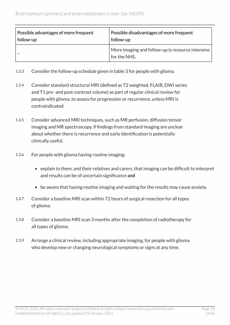
1.3.3 Consider the follow-up schedule given in table 3 for people with glioma.
1.3.4 Consider standard structural MRI (defined as T2 weighted, FLAIR, DWI series
and T1 pre- and post-contrast volume) as part of regular clinical review for
people with glioma, to assess for progression or recurrence, unless MRI is
contraindicated.
1.3.5 Consider advanced MRI techniques, such as MR perfusion, diffusion tensor
imaging and MR spectroscopy, if findings from standard imaging are unclear
about whether there is recurrence and early identification is potentially
clinically useful.
1.3.6 For people with glioma having routine imaging:
• explain to them, and their relatives and carers, that imaging can be difficult to interpret
and results can be of uncertain significance and and
• be aware that having routine imaging and waiting for the results may cause anxiety.
1.3.7 Consider a baseline MRI scan within 72 hours of surgical resection for all types
of glioma.
1.3.8 Consider a baseline MRI scan 3 months after the completion of radiotherapy for
all types of glioma.
1.3.9 Arrange a clinical review, including appropriate imaging, for people with glioma
who develop new or changing neurological symptoms or signs at any time.
Brain tumours (primary) and brain metastases in over 16s (NG99)
© NICE 2022. All rights reserved. Subject to Notice of rights (https://www.nice.org.uk/terms-and-conditions#notice-of-rights). Last updated 29 January 2021
Page 18of 66

TableTable 3 Possible regular clinical review schedule for people with glioma depending on grade of 3 Possible regular clinical review schedule for people with glioma depending on grade of tumour tumour
Grade of tumour Grade of tumour Clinical review schedule Clinical review schedule
GradeGrade I I
Scan at 12 months, then:
• consider discharge if no tumour visible on imaging unless
completely-resected pilocytic astrocytoma
• consider ongoing imaging at increasing intervals for 15 years for
completely-resected pilocytic astrocytoma
• consider if ongoing imaging is needed at a rate of once every 1 to
3 years for the rest of the person's life if the tumour is visible on
imaging.
GradeGrade II 1p/19q non-II 1p/19q non-
codeleted, IDH codeleted, IDH
mutated mutated
Grade II 1p/19q Grade II 1p/19q
codeleted codeleted
Grade III 1p/19q Grade III 1p/19q
codeleted codeleted
• From 0 to 2 years, scan at 3 months, then every 6 months
• From 2 to 4 years, review annually
• From 5 to 10 years, review every 1 to 2 years
• For more than 10 years and for the rest of life consider ongoing
imaging every 1 to 2 years.
GradeGrade II IDH wildtype II IDH wildtype
Grade III 1p/19q non-Grade III 1p/19q non-
codeleted codeleted
Grade IV Grade IV
(glioblastoma) (glioblastoma)
• From 0 to 2 years, review every 3 to 6 months
• From 2 to 4 years, review every 6 to 12 months
• From 5 to 10 years, review annually
• For more than 10 years and for the rest of life - consider ongoing
imaging every 1 to 2 years.
Brain tumours (primary) and brain metastases in over 16s (NG99)
© NICE 2022. All rights reserved. Subject to Notice of rights (https://www.nice.org.uk/terms-and-conditions#notice-of-rights). Last updated 29 January 2021
Page 19of 66

For a short explanation of why the committee made these recommendations and how they
might affect practice see the rationale and impact section on follow up for glioma.
Full details of the evidence and the committee's discussion are in evidence review A:
investigation, management and follow-up of glioma.
1.4 1.4 Investigation and management of meningioma Investigation and management of meningioma
Investigation of suspected meningioma Investigation of suspected meningioma
1.4.1 Offer standard structural MRI (defined as T2 weighted, FLAIR, DWI series and
T1 pre- and post-contrast volume) as the initial diagnostic test for suspected
meningioma, unless MRI is contraindicated.
1.4.2 Consider CT imaging for meningioma (if not already performed) to assess bone
involvement if this is suspected.
For a short explanation of why the committee made these recommendations and how they
might affect practice see the rationale and impact section on investigation of suspected
meningioma.
Full details of the evidence and the committee's discussion are in evidence review B:
investigation, management and follow-up of meningioma.
Management of confirmed meningioma following surgery or if Management of confirmed meningioma following surgery or if surgery is not possible (or has been declined) surgery is not possible (or has been declined)
1.4.3 Base management of meningioma after surgery, or if surgery is not possible or
the person declines surgery, on the extent of any surgery and grade of
meningioma, as described in table 4.
Brain tumours (primary) and brain metastases in over 16s (NG99)
© NICE 2022. All rights reserved. Subject to Notice of rights (https://www.nice.org.uk/terms-and-conditions#notice-of-rights). Last updated 29 January 2021
Page 20of 66

TableTable 4 Treatment choices after surgery by extent, or no excision if surgery was not possible, for 4 Treatment choices after surgery by extent, or no excision if surgery was not possible, for different kinds of meningioma different kinds of meningioma
Grade Grade
Completely Completely
excised excised
(Simpson (Simpson
11 toto 3) 3)
Incompletely excised (Simpson Incompletely excised (Simpson
44 toto 5) 5)
No excision No excision
(radiological (radiological
only diagnosis) only diagnosis)
Recurrent Recurrent
I I Offer active
monitoring.
Consider further surgery (if
possible), radiotherapy or active
monitoring.
Consider active
monitoring or
radiotherapy.
Consider further
surgery or
radiotherapy (if
not previously
used).
II II
Offer a
choice
between
active
monitoring
and
radiotherapy.
Consider further surgery (if
possible). Offer radiotherapy if
surgery is not possible, including if
the person declines surgery, or if the
tumour is incompletely excised
afterwards.
Consider active
monitoring or
radiotherapy
Consider further
surgery and
offer
radiotherapy (if
not previously
used).
III III Offer
radiotherapy.
Consider further surgery (if possible)
and offer radiotherapy.
Consider active
monitoring or
radiotherapy
Consider further
surgery and
offer
radiotherapy (if
not previously
used).
1.4.4 Before a decision is made on radiotherapy for meningioma, take into account:
• comorbidities
• life expectancy
• neurological function
• oedema
• performance status
• rate of tumour progression
Brain tumours (primary) and brain metastases in over 16s (NG99)
© NICE 2022. All rights reserved. Subject to Notice of rights (https://www.nice.org.uk/terms-and-conditions#notice-of-rights). Last updated 29 January 2021
Page 21of 66

• size and location of tumour
• surgical and radiotherapy morbidity
• the person's preferences (see table 5 for factors to discuss with them)
• treatments used before.
TableTable 5 Factors to take into account when deciding on radiotherapy as treatment for a surgically 5 Factors to take into account when deciding on radiotherapy as treatment for a surgically treated meningioma treated meningioma
Radiotherapy Radiotherapy No radiotherapy No radiotherapy
Control of Control of
tumour tumour
There is evidence that radiotherapy is
effective in the local control of a tumour.
Receiving no radiotherapy means
the tumour may continue to grow.
Risk of Risk of
developing developing
subsequent subsequent
symptoms symptoms
Controlling the tumour will reduce the
risk of developing symptoms from the
tumour in the future.
If the tumour grows, it can cause
irreversible symptoms such as loss
of vision.
Risk of re-Risk of re-
treatment treatment
Less risk of needing second surgery
compared with no radiotherapy.
Higher risk of needing second
surgery compared with
radiotherapy.
If the tumour has progressed, then
the surgery might be more
complex.
If the tumour has progressed, then
not all radiotherapy techniques
may be possible.
Brain tumours (primary) and brain metastases in over 16s (NG99)
© NICE 2022. All rights reserved. Subject to Notice of rights (https://www.nice.org.uk/terms-and-conditions#notice-of-rights). Last updated 29 January 2021
Page 22of 66

RadiotherRadiotherapapy y No rNo radiotheradiotherapapy y
Early side Early side
effects of effects of
treatment treatment
Early side effects from radiotherapy can
include:
• fatigue
• hair loss
• headache
• nausea
• seizures
• skin irritation.
No side effects from treatment.
Late side effects Late side effects
of treatment of treatment
Late side effects from radiotherapy can
include:
• effect on cognition
• risk of stroke
• risk of radionecrosis
• risk of second tumours
• cranial nerve effects
• hypopituitarism
• cataracts.
No side effects from treatment.
Management of Management of
side effects side effects
Increased use of steroids to manage side
effects. No side effects from treatment.
1.4.5 When deciding on the radiotherapy technique for people with meningioma, take
into account:
• the preferences of the person (for example, to minimise the number of appointments
or travel distance)
Brain tumours (primary) and brain metastases in over 16s (NG99)
© NICE 2022. All rights reserved. Subject to Notice of rights (https://www.nice.org.uk/terms-and-conditions#notice-of-rights). Last updated 29 January 2021
Page 23of 66

• tumour grade
• tumour location (proximity to optic nerves, optic chiasm and brainstem)
• tumour size.
From the suitable radiotherapy techniques, choose the one which maximises the
chances of local tumour control while minimising the radiation dose to normal brain
tissue.
1.4.6 If the multidisciplinary team thinks that radiotherapy may be appropriate, offer
the person the opportunity to discuss the potential benefits and risks with an
oncologist.
For a short explanation of why the committee made these recommendations and how they
might affect practice see the rationale and impact section on management of confirmed
meningioma following surgery, or if surgery is not possible or the person declines surgery.
Full details of the evidence and the committee's discussion are in evidence review B:
investigation, management and follow-up of meningioma.
Genomic biomarker-based treatment for meningioma Genomic biomarker-based treatment for meningioma
The point at which to use genomic biomarker-based therapy in solid tumour treatment pathways is
uncertain. See the NICE topic page on genomic biomarker-based cancer treatments.
1.5 1.5 Follow-up for meningioma Follow-up for meningioma 1.5.1 Offer regular clinical review for people with meningioma to assess changes in
their physical, psychological and cognitive wellbeing.
1.5.2 Base decisions on the timing of regular clinical reviews and follow-up imaging
for people with meningioma on:
• any residual tumour
• life expectancy
• the person's preferences (see table 6 for factors to discuss with them)
Brain tumours (primary) and brain metastases in over 16s (NG99)
© NICE 2022. All rights reserved. Subject to Notice of rights (https://www.nice.org.uk/terms-and-conditions#notice-of-rights). Last updated 29 January 2021
Page 24of 66

• treatments used before
• treatment options available
• tumour grade.
TableTable 6 Factors to take into account when deciding on frequency of follow-up for people with 6 Factors to take into account when deciding on frequency of follow-up for people with meningioma meningioma
Possible advantages of more frequent Possible advantages of more frequent
follow-up follow-up
Possible disadvantages of more frequent Possible disadvantages of more frequent
follow-up follow-up
May identify recurrent disease earlier which
may increase treatment options or enable
treatment before people become
symptomatic.
There is no definitive evidence that identifying
recurrent disease early improves outcomes.
May help provide information about the
course of the illness and prognosis.
May increase anxiety if changes of uncertain
significance are detected on imaging.
Some people can find more frequent imaging
and hospital contact reassuring.
Provides an opportunity to identify patient
or carer needs (such as psychosocial support
and late side effects of treatment).
Some people can find more frequent imaging and
hospital contact burdensome and disruptive –
they feel their life revolves around their latest
scan.
There may be a financial cost from taking time off
work and travelling to appointments.
– More imaging and follow-up is resource intensive
for the NHS.
1.5.3 Consider the follow-up schedule given in table 7 for people with meningioma.
1.5.4 Consider standard structural MRI (defined as T2 weighted, FLAIR, DWI series
and T1 pre- and post-contrast volume) as part of regular clinical review for
people with meningioma, to assess for progression or recurrence, unless MRI is
contraindicated.
1.5.5 For people with meningioma having routine imaging, be aware that having
routine imaging and waiting for the results may cause anxiety.
1.5.6 Arrange a clinical review, including appropriate imaging, for people with
meningioma (including incidental meningioma) who develop new or changing
Brain tumours (primary) and brain metastases in over 16s (NG99)
© NICE 2022. All rights reserved. Subject to Notice of rights (https://www.nice.org.uk/terms-and-conditions#notice-of-rights). Last updated 29 January 2021
Page 25of 66
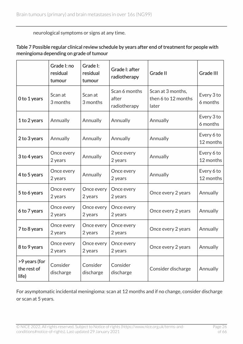
neurological symptoms or signs at any time.
TableTable 7 Possible regular clinical review schedule by years after end of treatment for people with 7 Possible regular clinical review schedule by years after end of treatment for people with meningioma depending on grade of tumour meningioma depending on grade of tumour
Grade I: no Grade I: no
residual residual
tumour tumour
Grade I: Grade I:
residual residual
tumour tumour
Grade I: after Grade I: after
radiotherapy radiotherapy Grade II Grade II Grade III Grade III
0 to 1 years 0 to 1 years Scan at
3 months
Scan at
3 months
Scan 6 months
after
radiotherapy
Scan at 3 months,
then 6 to 12 months
later
Every 3 to
6 months
1 to 2 years 1 to 2 years Annually Annually Annually Annually Every 3 to
6 months
2 to 3 years 2 to 3 years Annually Annually Annually Annually Every 6 to
12 months
3 to 4 years 3 to 4 years Once every
2 years Annually
Once every
2 years Annually
Every 6 to
12 months
4 to 5 years 4 to 5 years Once every
2 years Annually
Once every
2 years Annually
Every 6 to
12 months
5 to 6 years 5 to 6 years Once every
2 years
Once every
2 years
Once every
2 years Once every 2 years Annually
6 to 7 years 6 to 7 years Once every
2 years
Once every
2 years
Once every
2 years Once every 2 years Annually
7 to 8 years 7 to 8 years Once every
2 years
Once every
2 years
Once every
2 years Once every 2 years Annually
8 to 9 years 8 to 9 years Once every
2 years
Once every
2 years
Once every
2 years Once every 2 years Annually
>9 years (for >9 years (for
the rest of the rest of
life) life)
Consider
discharge
Consider
discharge
Consider
discharge Consider discharge Annually
For asymptomatic incidental meningioma: scan at 12 months and if no change, consider discharge
or scan at 5 years.
Brain tumours (primary) and brain metastases in over 16s (NG99)
© NICE 2022. All rights reserved. Subject to Notice of rights (https://www.nice.org.uk/terms-and-conditions#notice-of-rights). Last updated 29 January 2021
Page 26of 66

Note: the presence of any residual tumour can only be established after the first scan at 3 months.
For a short explanation of why the committee made these recommendations and how they
might affect practice see the rationale and impact section on follow up for meningioma.
Full details of the evidence and the committee's discussion are in evidence review B:
investigation, management and follow-up of meningioma.
1.6 1.6 Investigation of suspected brain metastases Investigation of suspected brain metastases 1.6.1 Offer standard structural MRI (defined as T2 weighted, FLAIR, DWI series and
T1 pre- and post-contrast volume) as the initial diagnostic test for suspected
brain metastases, unless MRI is contraindicated.
1.6.2 To help establish current disease status, offer extracranial imaging (appropriate
to the primary tumour type) to people with any radiologically suspected brain
metastases that may be suitable for focal treatment.
1.6.3 Perform all intracranial and extracranial diagnostic imaging and, if appropriate,
biopsy of extracranial disease, before referral to the neuro-oncology
multidisciplinary team.
For a short explanation of why the committee made these recommendations and how they
might affect practice see the rationale and impact section on investigation of suspected brain
metastases.
Full details of the evidence and the committee's discussion are in evidence review C:
investigation, management and follow-up of brain metastases.
1.7 1.7 MaManagement of confirmed brain metastases nagement of confirmed brain metastases 1.7.1 When choosing management options for brain metastases, take into account:
• extracranial disease
• leptomeningeal disease
Brain tumours (primary) and brain metastases in over 16s (NG99)
© NICE 2022. All rights reserved. Subject to Notice of rights (https://www.nice.org.uk/terms-and-conditions#notice-of-rights). Last updated 29 January 2021
Page 27of 66

• location of metastases
• resection cavity size
• the number and volume of metastases
• the person's preference (based on a discussion of the factors listed in tables 8 and 9)
• their age
• their performance status
• the primary tumour site, type, and molecular profile.
1.7.2 Consider systemic anti-cancer therapy for people who have brain metastases
likely to respond effectively, for example, germ cell tumours or small-cell lung
cancer.
1.7.3 Consider maximal local therapy with either surgery, stereotactic radiosurgery
or stereotactic radiotherapy for people with a single brain metastasis.
1.7.4 Base the choice of treatment for people with a single brain metastasis on:
• comorbidities
• extent of oedema
• location of metastasis
• the person's preference (see table 8)
• tumour size.
TableTable 8 Factors to take into account when deciding between surgery and stereotactic 8 Factors to take into account when deciding between surgery and stereotactic radiosurgery/radiotherapy as treatment for a single brain metastasis radiosurgery/radiotherapy as treatment for a single brain metastasis
Surgery Surgery Stereotactic radiosurgery / radiotherapy Stereotactic radiosurgery / radiotherapy
Overall Overall
survival survival No clinically important difference. No clinically important difference.
Brain tumours (primary) and brain metastases in over 16s (NG99)
© NICE 2022. All rights reserved. Subject to Notice of rights (https://www.nice.org.uk/terms-and-conditions#notice-of-rights). Last updated 29 January 2021
Page 28of 66

Surgery Surgery Stereotactic rStereotactic radiosurgery / radiosurgery / radiotheradiotherapapy y
Risk of Risk of
needing needing
additional additional
treatment treatment
Risk that stereotactic
radiosurgery / radiotherapy may
be needed in any case.
Risk that surgery may be needed in any
case. However, has higher local control rate
than surgery (meaning surgery is less likely
after radiotherapy than the other way
around).
Key benefit of Key benefit of
treatment treatment
Has more rapid control of
symptoms.
Additionally, surgery allows for
obtaining an up-to-date
pathological diagnosis which may
guide future treatment, making it
more effective.
Has a higher local control rate than surgery,
meaning more treatment is less likely to be
needed.
Additionally, is an outpatient treatment
and does not need a general anaesthetic.
Key risks of Key risks of
treatment treatment
Surgical procedures carry known
risks that vary depending on the
person and the tumour. These
include infection, stroke, a
prolonged hospital stay and death.
Surgery is more painful than
radiotherapy during recovery.
Radiation carries the risk of delayed effects
such as radionecrosis, which might need
surgical resection.
There is an increased risk of seizures with
this technique, although this appears to
mostly affect people who have pre-existing
epilepsy.
Steroid use Steroid use Early reduction in steroid dose.
Likely to need steroids for longer, and at a
higher dose. Steroids have significant side
effects when used long-term, such as
changes in mood, heart problems and
changes in body fat.
Planning Planning
treatment treatment
around around
important life important life
events events
The wound from the surgery may
affect the ability to carry out
certain activities in the short
term, such as air travel and sport.
The cosmetic appearance of the
wound from surgery may be
important to some people, and
should be discussed.
Some people find the techniques used in
radiotherapy challenging or upsetting,
especially the equipment which
immobilises the head. This is especially
likely to be true for people with
claustrophobia.
Brain tumours (primary) and brain metastases in over 16s (NG99)
© NICE 2022. All rights reserved. Subject to Notice of rights (https://www.nice.org.uk/terms-and-conditions#notice-of-rights). Last updated 29 January 2021
Page 29of 66

Surgery Surgery Stereotactic rStereotactic radiosurgery / radiosurgery / radiotheradiotherapapy y
Other Other
considerations considerations –
Radiotherapy can reach some areas of the
brain that surgery cannot, and might be the
only appropriate technique for certain
tumour types.
1.7.5 Do not offer adjuvant whole-brain radiotherapy to people with a single brain
metastasis treated with stereotactic radiosurgery/radiotherapy or surgery.
1.7.6 See NHS England's clinical commissioning policy on stereotactic radiosurgery
and stereotactic radiotherapy to the surgical cavity following resection of
cerebral metastases. [amended 2021amended 2021]
1.7.7 Consider stereotactic radiosurgery/radiotherapy for people with multiple brain
metastases who have controlled or controllable extracranial disease and
Karnofsky performance status of 70 or more. Take into account the number and
total volume of metastases.
1.7.8 Do not offer whole-brain radiotherapy to people with:
• non-small-cell lung cancer and and
• brain metastases that are not suitable for surgery or stereotactic radiosurgery/
radiotherapy and and
• a Karnofsky performance status of under 70.
1.7.9 For people with multiple brain metastases who have not had stereotactic
radiosurgery/radiotherapy or surgery, decide with them whether to use whole-
brain radiotherapy after a discussion with them and their relatives and carers
(as appropriate) of the potential benefits and risks (see table 9).
TableTable 9 Potential benefits and harms of whole-brain radiotherapy for multiple metastases 9 Potential benefits and harms of whole-brain radiotherapy for multiple metastases
- - Whole-brain radiotherapy Whole-brain radiotherapy No whole-brain radiotherapy No whole-brain radiotherapy
Overall Overall
survival survival No clinically important difference. No clinically important difference.
Brain tumours (primary) and brain metastases in over 16s (NG99)
© NICE 2022. All rights reserved. Subject to Notice of rights (https://www.nice.org.uk/terms-and-conditions#notice-of-rights). Last updated 29 January 2021
Page 30of 66

- - Whole-brWhole-brain rain radiotheradiotherapapy y No whole-brNo whole-brain rain radiotheradiotherapapy y
Quality of life Quality of life Short-term deterioration in quality of
life because of treatment.
No impact on quality of life because of
treatment, but deterioration because
of the disease progression.
Potential Potential
benefits benefits
Can stabilise or reduce the brain
metastases.
Brain metastases may continue to
grow.
Side effects Side effects
Temporary hair loss and fatigue.
Potential for accelerated cognitive loss
because of radiotherapy.
Potential for cognitive loss because of
disease progression.
Time Time
commitment commitment Requires 5 to 10 hospital visits. No time commitment.
Other Other
considerations considerations
People with non-small-cell lung cancer
will not benefit from treatment if their
overall prognosis is poor.
–
1.7.10 Do not offer memantine in addition to whole-brain radiotherapy to people with
multiple brain metastases, unless as part of a clinical trial.
1.7.11 Do not offer concurrent systemic therapy to enhance the efficacy of whole-
brain radiotherapy to people with multiple brain metastases, unless as part of a
clinical trial.
For a short explanation of why the committee made these recommendations and how they
might affect practice see the rationale and impact section on management of confirmed brain
metastases.
Full details of the evidence and the committee's discussion are in evidence review C:
investigation, management and follow-up of brain metastases.
1.8 1.8 Follow-up for brain metastases Follow-up for brain metastases 1.8.1 Offer regular clinical review for people with brain metastases to assess changes
in their physical, psychological and cognitive wellbeing.
Brain tumours (primary) and brain metastases in over 16s (NG99)
© NICE 2022. All rights reserved. Subject to Notice of rights (https://www.nice.org.uk/terms-and-conditions#notice-of-rights). Last updated 29 January 2021
Page 31of 66

1.8.2 Base decisions on the timing of regular clinical reviews and follow-up imaging
for people with brain metastases on:
• extracranial disease status
• life expectancy
• primary cancer
• the person's preferences (see table 10 for factors to discuss with them)
• treatment options available.
TableTable 10 Factors to take into account when deciding on frequency of follow-up for people with 10 Factors to take into account when deciding on frequency of follow-up for people with brain metastases brain metastases
Possible advantages of more frequent Possible advantages of more frequent
follow-up follow-up
Possible disadvantages of more frequent Possible disadvantages of more frequent
follow-up follow-up
May identify recurrent disease earlier which
may increase treatment options or enable
treatment before people become
symptomatic.
There is no definitive evidence that identifying
recurrent disease early improves outcomes.
May help provide information about the
course of the illness and prognosis.
May increase anxiety if changes of uncertain
significance are detected on imaging.
Some people can find more frequent imaging
and hospital contact reassuring.
Provides an opportunity to identify patient
or carer needs (such as psychosocial support
and late side effects of treatment).
Some people can find more frequent imaging and
hospital contact burdensome and disruptive –
they feel their life revolves around their latest
scan.
There may be a financial cost from taking time off
work and travelling to appointments.
– More imaging and follow-up is resource intensive
for the NHS.
1.8.3 Consider the follow-up schedule given in table 11 for people with brain
metastases.
1.8.4 Consider standard structural MRI (defined as T2 weighted, FLAIR, DWI series
and T1 pre- and post-contrast volume) as part of regular clinical review for
Brain tumours (primary) and brain metastases in over 16s (NG99)
© NICE 2022. All rights reserved. Subject to Notice of rights (https://www.nice.org.uk/terms-and-conditions#notice-of-rights). Last updated 29 January 2021
Page 32of 66

people with brain metastases, to assess for progression or recurrence, unless
MRI is contraindicated.
1.8.5 Consider advanced MRI techniques, such as MR perfusion, diffusion tensor
imaging and MR spectroscopy, if findings from standard imaging are unclear
about whether there is recurrence and early identification is potentially
clinically useful.
1.8.6 For people with brain metastases having routine imaging:
• explain to them, and their relatives and carers, that imaging can be difficult to interpret
and results can be of uncertain significance and and
• be aware that having routine imaging and waiting for the results may cause anxiety.
1.8.7 Arrange a clinical review, including appropriate imaging, for people with brain
metastases who develop new or changing neurological symptoms or signs at any
time.
TableTable 11 Possible regular clinical review schedule for 11 Possible regular clinical review schedule for people with brain metastases people with brain metastases
Years after end of treatment Years after end of treatment Clinical review schedule Clinical review schedule
0 to 1 years Every 3 months
1 to 2 years Every 4 to 6 months
2 years and onwards Annually
For a short explanation of why the committee made these recommendations and how they
might affect practice see the rationale and impact section on follow up for brain metastases.
Full details of the evidence and the committee's discussion are in evidence review C:
investigation, management and follow-up of brain metastases.
1.9 1.9 Care needs of people with brain tumours Care needs of people with brain tumours 1.9.1 Be aware that the care needs of people with brain tumours represent a unique
challenge, because (in addition to physical disability) the tumour and treatment
Brain tumours (primary) and brain metastases in over 16s (NG99)
© NICE 2022. All rights reserved. Subject to Notice of rights (https://www.nice.org.uk/terms-and-conditions#notice-of-rights). Last updated 29 January 2021
Page 33of 66

can have effects on:
• behaviour
• cognition
• personality.
1.9.2 Discuss health and social care support needs with the person with a brain
tumour and their relatives and carers (as appropriate). Take into account the
complex health and social care support needs people with any type of brain
tumour and their relatives and carers may have (for example, psychological,
cognitive, physical, spiritual, emotional).
1.9.3 Set aside enough time to discuss the impact of the brain tumour on the person
and their relatives and carers (as appropriate), and to elicit and discuss their
health and social care support needs.
1.9.4 Health and social care professionals involved in the care of people with brain
tumours should address additional complex needs during or at the end of
treatment and throughout follow-up. These include:
• changes to cognitive functioning
• fatigue
• loss of personal identity
• loss of independence
• maintaining a sense of hope
• potential for change in personal and sexual relationships
• the challenges of living with uncertainty
• the impact of brain tumour-associated epilepsy on wellbeing (see the NICE guideline
on epilepsies: diagnosis and management).
1.9.5 Provide a named healthcare professional with responsibility for coordinating
health and social care support for people with brain tumours and their relatives
and carers, for example, a key worker (often a clinical nurse specialist) as defined
Brain tumours (primary) and brain metastases in over 16s (NG99)
© NICE 2022. All rights reserved. Subject to Notice of rights (https://www.nice.org.uk/terms-and-conditions#notice-of-rights). Last updated 29 January 2021
Page 34of 66

in NICE cancer service guidance on improving outcomes for people with brain
and other central nervous system tumours.
1.9.6 Give information to the person with a brain tumour and their relatives and
carers (as appropriate):
• in a realistic and empathetic manner
• in suitable formats (written and spoken, with information available to take away),
following the principles in the NICE guideline on patient experience in adult NHS
services (also see NHS England's guidance on the Accessible Information Standard).
• at appropriate times throughout their care pathway.
1.9.7 Explain to the person that they have a legal obligation to notify the Driver and
Vehicle Licensing Agency (DVLA) if they have a brain tumour, and that this may
have implications for their driving.
1.9.8 Provide and explain clinical results, for example, imaging and pathology reports,
to the person with a brain tumour and their relatives and carers (as appropriate)
as soon as possible.
1.9.9 Offer supportive care to people with brain tumours and their relatives and
carers (as appropriate) throughout their treatment and care pathway
1.9.10 In people aged between 16 and 24 years old, refer to the NICE quality standard
on cancer services for children and young people.
1.9.11 Discuss the potential preservation of fertility with people with brain tumours
where treatment may have an impact on their fertility (see the
recommendations on people with cancer who wish to preserve fertility in
NICE's guideline on fertility problems).
1.9.12 If the person with a brain tumour is likely to be in their last year of life, refer to
the NICE quality standards on end of life care for adults and, when appropriate,
care of dying adults in the last days of life.
Brain tumours (primary) and brain metastases in over 16s (NG99)
© NICE 2022. All rights reserved. Subject to Notice of rights (https://www.nice.org.uk/terms-and-conditions#notice-of-rights). Last updated 29 January 2021
Page 35of 66

For a short explanation of why the committee made these recommendations and how they
might affect practice see the rationale and impact section on care needs of people with brain
tumours.
Full details of the evidence and the committee's discussion are in evidence review D:
supporting people living with a brain tumour.
1.10 1.10 Neurorehabilitation needs of people with brain Neurorehabilitation needs of people with brain tumours tumours 1.10.1 Consider referring the person with a brain tumour for a neurological
rehabilitation assessment of physical, cognitive and emotional function at
diagnosis and every stage of follow-up.
1.10.2 Offer people with brain tumours and their relatives and carers (as appropriate)
information on accessing neurological rehabilitation, and on what needs it can
help address.
1.10.3 Give people with brain tumours and their relatives and carers (as appropriate)
information on:
• neurological rehabilitation options in the community, as an outpatient, or an inpatient
and and
• how to get a neurological rehabilitation assessment.
For a short explanation of why the committee made these recommendations and how they
might affect practice see the rationale and impact section on neurorehabilitation needs of
people with brain tumours.
Full details of the evidence and the committee's discussion are in evidence review D:
supporting people living with a brain tumour.
Brain tumours (primary) and brain metastases in over 16s (NG99)
© NICE 2022. All rights reserved. Subject to Notice of rights (https://www.nice.org.uk/terms-and-conditions#notice-of-rights). Last updated 29 January 2021
Page 36of 66

1.11 1.11 Surveillance for the late-onset side effects of Surveillance for the late-onset side effects of treatment treatment 1.11.1 Be aware that people with brain tumours can develop side effects of treatment
months or years after treatment, which can include:
• cataracts
• cavernoma
• cognitive decline
• epilepsy
• hearing loss
• hypopituitarism
• infertility
• neuropathy (for example, nerve damage causing visual loss, numbness, pain or
weakness)
• radionecrosis
• secondary tumours
• SMART (stroke-like migraine attacks after radiotherapy)
• stroke.
1.11.2 Assess the person's individual risk of developing late effects when they finish
treatment. Record these in their written treatment summary and explain them
to the person (and their relatives and carers, as appropriate).
1.11.3 Encourage people who have had cranial radiotherapy to follow a healthy
lifestyle, including exercise, a healthy diet and stopping smoking (if applicable),
to decrease their risk of stroke. See the NICE guidelines on obesity prevention,
physical activity and tobacco: preventing uptake, promoting quitting and
treating dependence.
1.11.4 For people who are at risk of stroke, consider checking their blood pressure,
Brain tumours (primary) and brain metastases in over 16s (NG99)
© NICE 2022. All rights reserved. Subject to Notice of rights (https://www.nice.org.uk/terms-and-conditions#notice-of-rights). Last updated 29 January 2021
Page 37of 66

HbA1c level and cholesterol profile regularly.
1.11.5 Consider ongoing neuropsychology assessment for people at risk of cognitive
decline.
1.11.6 If a person has had a radiotherapy dose that might affect pituitary function,
consider checking their endocrine function regularly after the end of treatment.
1.11.7 Consider referring people who are at risk of visual impairment for an
ophthalmological assessment.
1.11.8 Consider referring people who are at risk of hearing loss to audiology for a
hearing test.
1.11.9 Consider referring the person to stroke services if an MRI during active
monitoring identifies asymptomatic ischaemic stroke.
For a short explanation of why the committee made these recommendations and how they
might affect practice see the rationale and impact section on surveillance for the late-onset
side effects of treatment.
Full details of the evidence and the committee's discussion are in evidence review D:
supporting people living with a brain tumour.
Terms used in this guideline Terms used in this guideline
Active monitoring Active monitoring
This is regular clinical and radiological review of a person with a brain tumour or brain metastases
who are not currently having treatment for their cancer.
Regular clinical review Regular clinical review
This is outpatient review of the person with a brain tumour or brain metastases at a planned
interval from the previous visit in order to assess symptoms and care needs, to provide support and
treatment and to perform imaging when appropriate.
Brain tumours (primary) and brain metastases in over 16s (NG99)
© NICE 2022. All rights reserved. Subject to Notice of rights (https://www.nice.org.uk/terms-and-conditions#notice-of-rights). Last updated 29 January 2021
Page 38of 66

Recommendations for research Recommendations for research The guideline committee has made the following recommendations for research.
Key recommendations for research Key recommendations for research
1 Managing glioma: management of IDH wildtype grade1 Managing glioma: management of IDH wildtype grade II glioma II glioma
Does the addition of concurrent and adjuvant temozolomide to radiotherapy improve overall
survival in patients with IDH wildtype grade II glioma?
Why this is important Why this is important
The World Health Organization (WHO) 2016 reclassification of brain tumours recognised that the
molecular characteristics of glioma are extremely important in helping differentiate between
disease entities with very different outcomes. Although evidence exists to guide management
recommendations for certain molecular gliomas, such as codeleted and non-codeleted grade III
glioma, currently no studies have investigated the best approach for the management of grade II
glioma with IDH wildtype. The biological behaviour of these tumours is more like a high-grade
glioma with a much shorter prognosis than IDH-mutated grade II glioma.
Because of this, some clinicians have advocated treating such tumours with concurrent
chemoradiation recommended for grade IV glioma (glioblastoma multiforme, GBM). However,
there is currently no research evidence to support this approach and this regimen is more intensive
and people experience increased acute and late side effects compared to radiotherapy alone.
Research is needed to establish whether or not this approach is beneficial in terms of improved
survival, and at what cost in terms of toxicity and, potentially, reduced quality of life.
For a short explanation of why the committee made the recommendation for research, see the
rationale and impact section on managing glioma.
Full details of the evidence and the committee's discussion are in evidence review A:
investigation, management and follow-up of glioma.
Brain tumours (primary) and brain metastases in over 16s (NG99)
© NICE 2022. All rights reserved. Subject to Notice of rights (https://www.nice.org.uk/terms-and-conditions#notice-of-rights). Last updated 29 January 2021
Page 39of 66

2 Managing glioma: supportive care clinics for low-grade glioma 2 Managing glioma: supportive care clinics for low-grade glioma
Does a dedicated supportive care clinic in addition to standard care improve outcomes for people
with low-grade gliomas?
Why this is important Why this is important
People with low-grade gliomas have significant symptoms and complex healthcare needs across
multiple physical, cognitive, emotional and social domains. This is often from the initial diagnosis
onwards. There are indications from research literature and patient reports that these needs are
currently unmet. Helping people with low-grade gliomas maintain their quality of life and function
is important, especially as there is currently no cure, because earlier supportive care interventions
and care plans may help reduce unplanned or emergency contact with secondary and tertiary
providers.
As no research literature exists which establishes the effectiveness of a specific healthcare
intervention, uncertainty exists about the most appropriate intervention to address unmet needs
and improve patient-reported outcome measures (or to establish whether current healthcare
provision can meet these needs). Current uncertainty is likely to have led to variations in service
provision across the UK. It is also possible that no specific intervention is available in some areas.
Research is needed to identify whether, in addition to standard care, a specific supportive care
intervention can significantly improve patient-reported outcome measures, and if so to establish
what this intervention should consist of.
For a short explanation of why the committee made the recommendation for research, see the
rationale and impact section on supportive care clinics for low-grade glioma.
Full details of the evidence and the committee's discussion are in evidence review A:
investigation, management and follow-up of glioma.
3 Managing glioma: early referral to palliative care for 3 Managing glioma: early referral to palliative care for glioblastoma glioblastoma
Does early referral to palliative care improve outcomes for people with glioblastomas in
comparison with standard oncology care?
Brain tumours (primary) and brain metastases in over 16s (NG99)
© NICE 2022. All rights reserved. Subject to Notice of rights (https://www.nice.org.uk/terms-and-conditions#notice-of-rights). Last updated 29 January 2021
Page 40of 66

Why this is important Why this is important
People with grade IV brain tumours (glioblastomas) have a poor prognosis which has not improved
in over a decade. Median overall survival is 14–18 months even with gold-standard chemoradiation
following surgery.
From initial diagnosis people experience multiple complex symptoms resulting from neurological
impairment. These can significantly impact on their quality of life, function and psychological
wellbeing. Their caregivers report high levels of distress and carer burden.
The aim of palliative care is to relieve symptoms and improve people's quality of life and function –
not just towards the end of life but throughout the duration of illness. There is some evidence that
early palliative care referral significantly improves overall survival, quality of life and mood.
Research in this area is important because this group of people have substantial health needs,
which use significant healthcare resources. Supportive care interventions such as early palliative
care may improve quality of life and function throughout the duration of illness. It may also help
people to manage the distress associated with a reduced life expectancy and participate in
advanced care planning.
For a short explanation of why the committee made the recommendation for research, see the
rationale and impact section on early referral to palliative care for glioblastoma.
Full details of the evidence and the committee's discussion are in evidence review A:
investigation, management and follow-up of glioma.
4 Managing glioma: early detection of recurrence after treatment 4 Managing glioma: early detection of recurrence after treatment
Does early detection of recurrence after treatment improve overall survival/outcomes in
molecularly stratified glioma?
Why this is important Why this is important
Prognosis for brain tumours is inherently uncertain, and recent advances in treatment mean many
people with a brain tumour will live for a long time after the initial diagnosis. For these individuals,
follow-up is the longest component of their treatment and it is both expensive for the NHS and
(sometimes) a burden for the person. There is no high-quality evidence that follow-up after
treatment is beneficial, no high-quality evidence on the optimal frequency of imaging, and clinical
Brain tumours (primary) and brain metastases in over 16s (NG99)
© NICE 2022. All rights reserved. Subject to Notice of rights (https://www.nice.org.uk/terms-and-conditions#notice-of-rights). Last updated 29 January 2021
Page 41of 66

uncertainty about whether such follow-up is likely to alter outcomes of importance to people with
tumours (such as overall life expectancy or quality of life).
Research is needed to establish at what point the value of identifying recurrence early is
outweighed by the harms of increasing burden to patients.
For a short explanation of why the committee made the recommendation for research, see the
rationale and impact section on the early detection of recurrence after treatment.
Full details of the evidence and the committee's discussion are in evidence review A:
investigation, management and follow-up of glioma.
5 Managing meningioma: immediate versus deferred 5 Managing meningioma: immediate versus deferred radiotherapy for incompletely excised graderadiotherapy for incompletely excised grade I meningioma I meningioma
Is immediate or deferred radiotherapy better for incompletely excised grade I meningioma?
Why this is important Why this is important
There are no randomised studies on the use of radiotherapy/radiosurgery in the treatment of
grade I meningioma. Though case series have shown that people with inoperable and incompletely
excised grade I meningioma treated with radiotherapy have high rates of control of their tumour,
treatment risks significant side effects. The side effects include: neuropathy, radionecrosis,
significant oedema, neuro-cognitive effects, increased risk of stroke and secondary tumours.
Therefore the timing of treatment is a balance between control of tumour and side effects. It is not
known if early treatment has a greater or lesser chance of long-term tumour control or risk of
tumour complications, or if this just risks complications of treatment earlier.
People with grade I meningioma have traditionally been overlooked as a priority area for research.
This is likely because of the slow nature of the disease resulting in need for long-term follow-up and
the difficulty to obtain funding for radiotherapy-only studies. However, this lack of research is
inequitable, hence the reason for its prioritisation by the committee.
A study on this topic would provide clear information to guide clinicians and people with
meningiomas, hopefully leading to overall improvement in quality of life. Because of the slow-
growing characteristics of grade I meningioma, treatment decisions made early in the management
pathway will have long-term effects on the person with the meningioma's overall quality of life
outcomes, and potentially overall survival.
Brain tumours (primary) and brain metastases in over 16s (NG99)
© NICE 2022. All rights reserved. Subject to Notice of rights (https://www.nice.org.uk/terms-and-conditions#notice-of-rights). Last updated 29 January 2021
Page 42of 66

For a short explanation of why the committee made the recommendation for research, see the
rationale and impact section on managing meningioma.
Full details of the evidence and the committee's discussion are in evidence review B:
investigation, management and follow-up of meningioma.
Brain tumours (primary) and brain metastases in over 16s (NG99)
© NICE 2022. All rights reserved. Subject to Notice of rights (https://www.nice.org.uk/terms-and-conditions#notice-of-rights). Last updated 29 January 2021
Page 43of 66

Rationale and impact Rationale and impact These sections briefly explain why the committee made the recommendations and how they might
affect practice. They link to details of the evidence and a full description of the committee's
discussion.
Investigations for suspected glioma: imaging Investigations for suspected glioma: imaging
The discussion below explains how the committee made recommendations 1.1.1–1.1.3.
Why the committee made the recommendations Why the committee made the recommendations
The evidence indicated that standard structural MRI is useful in distinguishing high-grade from
low-grade glioma. The committee noted that this knowledge will inform management. Based on
their experience, the committee recommended a protocol that they defined as a minimum standard
for imaging acquisition.
No evidence was found on more advanced MRI techniques. However, the committee agreed that in
their experience such techniques can be useful for assessing malignant features of a tumour – in
particular, for ensuring that high-grade tumours are not misdiagnosed as low-grade tumours, which
could have serious consequences for people who receive suboptimal management as a result.
However the committee explained that a specialist multidisciplinary team would be needed to
interpret features of the scan and decide management, even if advanced techniques were used.
How the recommendations might affect practice How the recommendations might affect practice
Currently, various imaging strategies are used in different centres and depending on the person's
circumstances. These recommendations aim to reduce variation in practice, and ensure that images
obtained at different sites and using different equipment can be more accurately compared. Some
centres may need to change their imaging protocols. This might increase or reduce costs depending
on the imaging protocols which are currently in place.
Full details of the evidence and the committee's discussion are in evidence review A: investigation,
management and follow-up of glioma.
Return to recommendations
Brain tumours (primary) and brain metastases in over 16s (NG99)
© NICE 2022. All rights reserved. Subject to Notice of rights (https://www.nice.org.uk/terms-and-conditions#notice-of-rights). Last updated 29 January 2021
Page 44of 66

Investigations for suspected glioma: molecular markers Investigations for suspected glioma: molecular markers
The discussion below explains how the committee made recommendations 1.1.4–1.1.6.
Why the committee made the recommendations Why the committee made the recommendations
Molecular markers are an emerging and important area in the treatment of brain tumours. The
committee looked for evidence on these markers but did not find any. However, they noted that
there are some molecular markers for which the evidence of benefit if tested is overwhelming, as
reported in studies identified in searches for other review questions. This applies in particular for
MGMT promoter methylation and TERT promoter mutations in IDH-wildtype glioma, although the
committee agreed the evidence was of a higher quality in the first case than the second. The
committee agreed that even these markers are not being consistently tested for and that testing
should be standardised. Therefore they made recommendations based on their knowledge and
experience, highlighting the World Health Organization (WHO) classification, to ensure that all
centres follow a consistent process for assessing and interpreting information on molecular
markers. This was important, since failure to consistently report molecular markers can mislead
clinicians or limit treatment options.
How the recommendations might affect practice How the recommendations might affect practice
As testing for molecular markers is relatively new, practice can vary widely and this is to be
expected. In principle there should not be a major change, although the time taken to implement
the new molecular tests will vary significantly between centres.
Full details of the evidence and the committee's discussion are in evidence review A: investigation,
management and follow-up of glioma.
Return to recommendations
Management of glioma: initial surgery for low-grade Management of glioma: initial surgery for low-grade glioma glioma
The discussion below explains how the committee made recommendations 1.2.1–1.2.5.
Why the committee made the recommendations Why the committee made the recommendations
There was evidence that optimal resection of a large percentage of the tumour improved survival
Brain tumours (primary) and brain metastases in over 16s (NG99)
© NICE 2022. All rights reserved. Subject to Notice of rights (https://www.nice.org.uk/terms-and-conditions#notice-of-rights). Last updated 29 January 2021
Page 45of 66

for people with low-grade glioma. The committee noted that it is sometimes not appropriate to
offer maximal safe resection (for example, if the balance of risks and benefits favours not resecting
all areas) and that a specialist surgical team should look at the value of doing an operation given its
safe extent. They agreed that biopsy should be considered in these cases, based on limited evidence
showing improved overall survival after biopsy compared with active monitoring. However, the
committee also concluded that some tumours were of such limited risk that the risks of surgery
outweighed the possible gain of biopsy or resection.
The committee described how there was no evidence for immediate intervention, but that
intervention should not be delayed due to the probability that surgical resection would have
benefit for the person with the tumour. They therefore recommended intervention within
6 months, to allow for time to discuss treatment options with the person with the tumour. This also
allows for the possibility of a second imaging sequence to be done later to look for progression and
to assess for symptom change, as the committee also recognised that a proportion of low-grade
gliomas have unfavourable gene profiles (for example, IDH wildtype) that make them more like
high-grade tumours from a prognostic perspective.
A small number of people might have had initial treatment before it was standard practice to save a
tissue sample for biopsy, and these people would currently be actively monitored. Based on their
experience the committee agreed that these people may not need further surgery as long as their
condition is stable (that is, they are not showing radiological or clinical disease progression).
How the recommendations might affect practice How the recommendations might affect practice
The recommendations are likely to change practice at some centres, and remove unnecessary
variation. There are currently differences between centres in which molecular diagnoses are
performed and in treatment of very low-risk, low-grade tumours. This is partly because low-grade
gliomas may be managed by non-expert surgical teams.
The recommendation about the management of low-grade gliomas that have been managed but
then progress is unlikely to substantially change practice, as management would be largely
unchanged.
Full details of the evidence and the committee's discussion are in evidence review A: investigation,
management and follow-up of glioma.
Return to recommendations
Brain tumours (primary) and brain metastases in over 16s (NG99)
© NICE 2022. All rights reserved. Subject to Notice of rights (https://www.nice.org.uk/terms-and-conditions#notice-of-rights). Last updated 29 January 2021
Page 46of 66

Management of glioma: further management of low-Management of glioma: further management of low-grade glioma grade glioma
The discussion below explains how the committee made recommendations 1.2.6–1.2.11.
Why the committee made the recommendations Why the committee made the recommendations
There was evidence that PCV chemotherapy (procarbazine, CCNU [lomustine] and vincristine)
after radiotherapy improved overall survival and progression-free survival compared with
radiotherapy alone. The committee discussed how the evidence for the exact regime was complex,
and used their judgement to recommend possible sequence and dose. In addition, the committee
noted that there are some circumstances where radiotherapy and PCV might not be appropriate
(particularly for the very lowest-concern and highest-concern low-grade tumours) and made
recommendations based on their experience in these cases.
The committee included approximate age cut-offs based on evidence showing that treatment
improved survival in people aged around 40 or over with or without residual tumour, and their
clinical judgement that treatment would be unlikely to be of benefit for people aged around 40 or
under without residual tumour.
The committee found no evidence on the treatment of IDH wildtype grade II glioma. They
determined that management of this type of glioma was likely to be different from other low-grade
glioma, as IDH wildtype grade II glioma behaves more like a high-grade glioma. The committee
therefore made a research recommendation on whether treating this tumour type more like a
grade II glioma or grade IV glioma was most beneficial.
How the recommendations might affect practice How the recommendations might affect practice
These recommendations aim to standardise practice. They will probably result in the same amounts
of chemotherapy and radiotherapy being given, but these treatments will be more precisely
targeted and it is possible that they will be given earlier. This would result in more people requiring
long-term treatment for the side effects of radiation and chemotherapy. More people are likely to
have active monitoring alone, which is not likely to create a resource impact.
Full details of the evidence and the committee's discussion are in evidence review A: investigation,
management and follow-up of glioma.
Return to recommendations
Brain tumours (primary) and brain metastases in over 16s (NG99)
© NICE 2022. All rights reserved. Subject to Notice of rights (https://www.nice.org.uk/terms-and-conditions#notice-of-rights). Last updated 29 January 2021
Page 47of 66

Management of glioma: gradeManagement of glioma: grade III glioma following III glioma following surgery surgery
The discussion below explains how the committee made recommendations 1.2.12–1.2.17.
Why the committee made the recommendations Why the committee made the recommendations
The committee considered evidence for grade III and grade IV glioma separately.
Treatments to be offered Treatments to be offered
Based on randomised controlled trial evidence, the committee recommended radiotherapy and
either PCV or temozolomide chemotherapy, depending on tumour subtype and performance
status, for people with grade III glioma.
Treatments that should not be offered Treatments that should not be offered
Based on the available evidence, the committee recommended that some treatments should not be
offered because they were harmful. They also agreed, based on their experience, that it would be
useful for healthcare professionals to tell people with glioma that no evidence had been found to
indicate that certain treatments are beneficial.
How the recommendations might affect practice How the recommendations might affect practice
Adjuvant PCV for treating codeleted grade III glioma is standard practice, but adjuvant
temozolomide for non-codeleted grade III gliomas is a change in practice. However, some centres
may already have started to adopt this as standard care, since the results of the study supporting
this treatment were made publicly available in 2016.
Full details of the evidence and the committee's discussion are in evidence review A: investigation,
management and follow-up of glioma.
Return to recommendations
Management of glioma: gradeManagement of glioma: grade IV glioma following IV glioma following surgery surgery
The discussion below explains how the committee made recommendations 1.2.18–1.2.27.
Brain tumours (primary) and brain metastases in over 16s (NG99)
© NICE 2022. All rights reserved. Subject to Notice of rights (https://www.nice.org.uk/terms-and-conditions#notice-of-rights). Last updated 29 January 2021
Page 48of 66

Why the committee made the recommendations Why the committee made the recommendations
The committee considered evidence for grade III and grade IV glioma separately.
Treatments to be used Treatments to be used
The committee saw some evidence demonstrating improved overall survival in some groups of
people with grade IV glioma who had radiotherapy with concurrent and adjuvant temozolomide
(compared with radiotherapy alone). However, based on their clinical experience they were unsure
that these results could be generalised to all people with grade IV glioma, so suggested a range of
possible treatments that can be considered for other groups, depending on the exact clinical
characteristics of the tumour.
Approximate age cut-offs for people with grade IV glioma were specified by the committee based
on evidence that a radiotherapy dose of 40 Gy did not result in lower survival in people aged
around 70 or over compared with a 60 Gy dose. Therefore a lower radiotherapy dose is likely to
cause fewer side effects without compromising clinical effectiveness for this group.
The committee were aware that the prognosis of people with a grade IV glioma and a low
performance status was poor, and recommended palliative care be considered. However the
committee did not find any evidence on whether earlier or later palliative care was most beneficial
for people who might need it. They therefore made a research recommendation on this topic, with
the aim of finding out the point in the treatment pathway when it would be most beneficial for
people with this type of glioma to have palliative care.
Treatments that should not be used Treatments that should not be used
Based on the available evidence, the committee recommended that certain treatments should not
be offered. This included tumour treating fields (TTF) based on published health economic evidence
that they are not an efficient use of NHS resources. They also agreed, based on their clinical
experience, that it would be useful for healthcare professionals to tell people with glioma that no
evidence had been found to suggest that certain treatments are beneficial.
How the recommendations might affect practice How the recommendations might affect practice
For younger people with a grade IV glioma and a good performance status, a course of radiotherapy
with concurrent and adjuvant temozolomide is standard care. However, for people aged around
70 and over, particularly those with a glioma with methylated MGMT, the use of concurrent and
adjuvant temozolomide with 15 fractions of radiotherapy is a change of practice that will probably
Brain tumours (primary) and brain metastases in over 16s (NG99)
© NICE 2022. All rights reserved. Subject to Notice of rights (https://www.nice.org.uk/terms-and-conditions#notice-of-rights). Last updated 29 January 2021
Page 49of 66

result in more people being treated. This is a relatively small group of people, and so the
recommendation is unlikely to have a significant resource impact.
Full details of the evidence and the committee's discussion are in evidence review A: investigation,
management and follow-up of glioma.
Return to recommendations
Management of glioma: recurrent high-grade glioma Management of glioma: recurrent high-grade glioma
The discussion below explains how the committee made recommendations 1.2.28–1.2.35.
Why the committee made the recommendations Why the committee made the recommendations
Treatments to be offered Treatments to be offered
Based on the available evidence, the committee recommended that treatment options for people
with recurrent glioma should include temozolomide, PCV and single-agent CCNU (lomustine). No
evidence was found to indicate which of these 3 options is likely to lead to the best outcomes, and
on the basis of their clinical experience the committee concluded that the choice of treatment
should take several factors into account, including the individual features of the tumour and the
preferences of the person. The committee also highlighted the possibility of considering supportive
care alone.
Treatments that should not be offered Treatments that should not be offered
Based on the available evidence, the committee recommended that certain treatments should not
be offered. This included tumour treating fields (TTF) on the basis of evidence of some clinical
benefit but indirect published health economic evidence, in people with newly diagnosed high-
grade glioma, that they are not cost effective. They also agreed, based on their clinical experience,
that it would be useful for healthcare professionals to tell people with glioma that no evidence had
been found to suggest that certain treatments are beneficial.
How the recommendations might affect practice How the recommendations might affect practice
These recommendations reflect standard treatment for recurrent high-grade glioma, and therefore
should not represent a substantial change in practice.
Full details of the evidence and the committee's discussion are in evidence review A: investigation,
Brain tumours (primary) and brain metastases in over 16s (NG99)
© NICE 2022. All rights reserved. Subject to Notice of rights (https://www.nice.org.uk/terms-and-conditions#notice-of-rights). Last updated 29 January 2021
Page 50of 66

management and follow-up of glioma.
Return to recommendations
Management of glioma: techniques for resection of Management of glioma: techniques for resection of glioma glioma
The discussion below explains how the committee made recommendations 1.2.36–1.2.42.
Why the committee made the recommendations Why the committee made the recommendations
There was evidence that 5-aminolevulinic acid (5-ALA), intraoperative MRI and diffusion tensor
imaging could improve either the extent or safety of resection (particularly the preservation of
neurological function). The committee noted that a combination of techniques might be needed to
optimise both the extent and safety of resection for a particular surgical plan. The committee
concluded that the evidence for MRI could be generalised to intraoperative ultrasound on the basis
of their clinical experience, and therefore that clinicians should be able to choose either technique
depending on availability.
The evidence for awake craniotomy was equivocal (non-significant differences compared with
surgery under general anaesthesia), therefore from the evidence it was not possible to conclude
that awake craniotomy would benefit all people with glioma. This is in line with the committee's
clinical experience that some people benefit from the procedure (in terms of preserving language,
motor and visual function) but others are harmed – particularly from psychological effects which
act as a contraindication to awake craniotomy. The committee described how better preoperative
procedures could reduce the number of people distressed by the procedure.
How the recommendations might affect practice How the recommendations might affect practice
Some techniques recommended by the committee require a very high level of intraoperative skill,
and this might have resource implications for hospitals recruiting people with these specialist skills.
There is significant variation in the current provision of psychological support for people before
and during awake craniotomy, and implementing this could carry a high cost to an individual unit.
If a unit does not have access to intraoperative ultrasound or MRI, the cost of acquiring this
equipment could be substantial (MRI is relatively expensive, ultrasound is relatively cheap).
However the committee concluded that most units should have access to one or the other already.
Therefore the only resource impact would be if a unit currently using intraoperative ultrasound
Brain tumours (primary) and brain metastases in over 16s (NG99)
© NICE 2022. All rights reserved. Subject to Notice of rights (https://www.nice.org.uk/terms-and-conditions#notice-of-rights). Last updated 29 January 2021
Page 51of 66

decided that the additional evidence for preservation of neurological function in intraoperative
MRI justified the cost of switching machines. However, the committee thought this was unlikely to
happen.
Using 5-ALA is associated with a high cost, and 5-ALA-guided surgery needs a non-standard
fluorescence-detecting microscope. Therefore the resource impact of this recommendation is
likely to be high in all settings, and very high in settings without access to a fluorescence-detecting
microscope. The anticipated resource impact of this recommendation is greater than £1 million per
year.
Full details of the evidence and the committee's discussion are in evidence review A: investigation,
management and follow-up of glioma.
Return to recommendations
Follow-up for glioma Follow-up for glioma
The discussion below explains how the committee made recommendations 1.3.1–1.3.9.
Why the committee made the recommendations Why the committee made the recommendations
In the absence of evidence, the committee made recommendations based on their clinical
experience. They recommended regular clinical review as the only plausible way of identifying and
potentially managing recurrence or changing symptoms. They also recommended the review
schedule take into account all of the person's relevant characteristics, including grade of tumour. As
this is quite difficult to work out, the committee suggested a schedule of clinical reviews that is
likely to be beneficial for a 'typical' person, which can be amended as needed to take into account
individual variation. The committee did not uncover evidence on who should do the follow-up and
so did not make a recommendation on this topic as it would vary according to clinical need, but
discussed how it could be – for example – the local oncologist, neuro-oncologist, neurologist,
neurosurgeon, clinical nurse specialist or GP.
As regular clinical review should include imaging, based on their experience the committee
suggested an MRI sequence which they believed would be suitable to monitor for recurrence. They
discussed how advanced MRI techniques might be valuable, but as these techniques are time-
consuming and difficult to interpret the committee concluded they should only be recommended
under certain circumstances where extra information was likely to substantially alter treatment
plans. The committee recommended that any change in neurological signs or symptoms (which
Brain tumours (primary) and brain metastases in over 16s (NG99)
© NICE 2022. All rights reserved. Subject to Notice of rights (https://www.nice.org.uk/terms-and-conditions#notice-of-rights). Last updated 29 January 2021
Page 52of 66

would include changes in behavioural, emotional and psychological signs and symptoms) be treated
as a sign of a potential change to the tumour, and therefore recommended clinical review outside
the usual schedule in order to investigate this.
The committee believed that a dedicated supportive care clinic could improve outcomes for people
with low-grade glioma, but did not find any evidence on this. Therefore they made a research
recommendation on improving the long-term outcomes of people with low-grade glioma.
How the recommendations might affect practice How the recommendations might affect practice
The recommendations are in line with current best practice, and should standardise practice. They
are unlikely to cause a significant increase in resource use, but there may be some additional costs
or changes in service configuration if practice differs in a particular centre.
The imaging sequences are recommended on the basis of evidence for the appropriate sequences
for initial diagnosis, and so might not be the standard sequence for follow-up in all centres. As a
result, adopting the recommended sequences might create some additional workload for some
centres. However the recommendations for exact schedules are examples based on consensus in
the committee, and there is therefore flexibility for centres to adapt these to their own models,
limiting resource impact.
Full details of the evidence and the committee's discussion are in evidence review A: investigation,
management and follow-up of glioma.
Return to recommendations
Investigation of suspected meningioma Investigation of suspected meningioma
The discussion below explains how the committee made recommendations 1.4.1 and 1.4.2.
Why the committee made the recommendations Why the committee made the recommendations
Evidence indicated that standard structural MRI is useful in distinguishing high-grade from low-
grade glioma, and the committee agreed that it is appropriate to extrapolate from this evidence to a
belief that MRI can be used to distinguish meningioma from healthy brain tissue. In the committee's
experience, CT scans can be more accurate than MRI for assessing meningioma with bone
involvement.
Brain tumours (primary) and brain metastases in over 16s (NG99)
© NICE 2022. All rights reserved. Subject to Notice of rights (https://www.nice.org.uk/terms-and-conditions#notice-of-rights). Last updated 29 January 2021
Page 53of 66

How the recommendations might affect practice How the recommendations might affect practice
Currently, various imaging strategies are used depending on the centre and the person's
circumstances. These recommendations aim to reduce variation in practice, and ensure that images
obtained at different sites and using different equipment can be more accurately compared. Some
centres may need to change their imaging protocols as a result, but this should not require the
purchase of additional equipment.
Full details of the evidence and the committee's discussion are in evidence review B: investigation,
management and follow-up of meningioma.
Return to recommendations
Management of confirmed meningioma following Management of confirmed meningioma following surgery or if surgery is not possible (or has been surgery or if surgery is not possible (or has been declined) declined)
The discussion below explains how the committee made recommendations 1.4.3–1.4.6.
Why the committee made the recommendations Why the committee made the recommendations
Based on limited evidence and their clinical experience, the committee concluded that
management of this group of meningiomas will depend on the type of meningioma. They noted that
evidence for 1 grade of meningioma could not normally be used to suggest best management for
another grade. Therefore the committee made recommendations for each grade of meningioma
separately, using evidence if this was available and their judgement if it was not. The committee
identified that management could be more conservative if the tumour grade was lower and initial
resection more complete, and should be more aggressive if the tumour grade was higher or initial
resection more partial.
The committee agreed that the 3 management options – further radiotherapy, surgery and active
monitoring – had different balances of benefits and harms in different situations. However they
also agreed that serious harm could be done to a person with a tumour if they were over- or under-
treated given the risk profile of their tumour, and so made recommendations according to this risk.
For example, for a low-grade almost completely-resected tumour (grade I, Simpson 2 excision),
radiotherapy or further surgery could expose the person to risk of harm for no expected clinical
gain.
Brain tumours (primary) and brain metastases in over 16s (NG99)
© NICE 2022. All rights reserved. Subject to Notice of rights (https://www.nice.org.uk/terms-and-conditions#notice-of-rights). Last updated 29 January 2021
Page 54of 66

Based on limited evidence, the committee made recommendations on how to deliver radiotherapy
or radiosurgery where this was appropriate. They used their experience to highlight features of the
tumour or preferences of the person that might help select the most appropriate radiotherapy or
radiosurgery modality, and explained that the best results would come through minimising the dose
of radiation delivered to healthy brain tissue while maximising the chance of local control.
The committee were unable to find evidence comparing different timings of radiotherapy in
incompletely excised grade I meningioma. As the disease is slow growing it can be difficult to assess
the risks of immediate side effects from treatment compared to the longer-term benefits of tumour
control. Therefore the committee made a research recommendation to investigate this topic.
How the recommendations might affect practice How the recommendations might affect practice
The recommendations reflect standard practice in many centres, and should make treatment more
consistent.
An appointment with an oncologist for all people who may have radiotherapy is not currently
standard practice. However, for most people this is likely to mean a change in the timing of their
first appointment with an oncologist, rather than many more people having oncologist
appointments.
Full details of the evidence and the committee's discussion are in evidence review B: investigation,
management and follow-up of meningioma.
Return to recommendations
Follow-up for meningioma Follow-up for meningioma
The discussion below explains how the committee made recommendations 1.5.1–1.5.6.
Why the committee made the recommendations Why the committee made the recommendations
In the absence of evidence, the committee made recommendations based on their clinical
experience. They recommended regular clinical review as the only plausible way of identifying and
potentially managing recurrence or changing symptoms. They also recommended the review
schedule take into account all of the person's relevant characteristics, including grade of tumour. As
this is quite difficult to work out, the committee suggested a schedule of clinical reviews that is
likely to be beneficial for a 'typical' person, which can be amended as needed to take into account
individual variation. The committee did not uncover evidence on who should do the follow-up and
Brain tumours (primary) and brain metastases in over 16s (NG99)
© NICE 2022. All rights reserved. Subject to Notice of rights (https://www.nice.org.uk/terms-and-conditions#notice-of-rights). Last updated 29 January 2021
Page 55of 66

so did not make a recommendation on this topic as it would vary according to clinical need, but
discussed how it could be – for example – the local oncologist, neuro-oncologist, neurologist,
neurosurgeon, clinical nurse specialist or GP.
As regular clinical review should include imaging, based on their experience the committee
suggested an MRI sequence which they believed would be suitable to monitor for recurrence. They
discussed how advanced MRI techniques might be valuable, but as these techniques are time-
consuming and difficult to interpret the committee concluded they should only be recommended
under certain circumstances where extra information was likely to substantially alter treatment
plans. The committee recommended that any change in neurological signs or symptoms (which
would include changes in behavioural, emotional and psychological signs and symptoms) be treated
as a sign of a potential change to the tumour, and therefore recommended clinical review outside
the usual schedule in order to investigate this.
How the recommendations might affect practice How the recommendations might affect practice
The recommendations are in line with current best practice, and should standardise practice. They
are unlikely to cause a significant increase in resource use, but there may be some additional costs
or changes in service configuration if practice differs in a particular centre.
The imaging sequences are recommended on the basis of evidence for the appropriate sequences
for initial diagnosis, and so might not be the standard sequence for follow-up in all centres. As a
result, adopting the recommended sequences might create some additional workload for some
centres. However the recommendations for exact schedules are examples based on consensus in
the committee, and there is therefore flexibility for centres to adapt these to their own models,
limiting resource impact.
Full details of the evidence and the committee's discussion are in evidence review B: investigation,
management and follow-up of meningioma.
Return to recommendations
Investigation of suspected brain metastases Investigation of suspected brain metastases
The discussion below explains how the committee made recommendations 1.6.1–1.6.3.
Why the committee made the recommendations Why the committee made the recommendations
In the absence of evidence, the committee recommended standard structural MRI based on their
Brain tumours (primary) and brain metastases in over 16s (NG99)
© NICE 2022. All rights reserved. Subject to Notice of rights (https://www.nice.org.uk/terms-and-conditions#notice-of-rights). Last updated 29 January 2021
Page 56of 66

experience, because it is important to establish the exact number of metastases in the brain, which
can guide further treatment. The committee described how failing to establish this could be
dangerous. Extracranial imaging, biopsy of the extracranial disease (where indicated) and
performing all imaging before multidisciplinary team discussions should ensure that all necessary
information is available so that appropriate decisions are made and delays in treatment avoided.
How the recommendations might affect practice How the recommendations might affect practice
The recommendations reinforce current best practice and should reduce delays to local
intracranial treatment.
Full details of the evidence and the committee's discussion are in evidence review C: investigation,
management and follow-up of brain metastases.
Return to recommendations
Management of confirmed brain metastases Management of confirmed brain metastases
The discussion below explains how the committee made recommendations 1.7.1–1.7.11.
Why the committee made the recommendations Why the committee made the recommendations
The committee made recommendations based on the available evidence and their judgement. They
described how features of brain metastases, including the number and volume (which is important
for establishing prognosis), should be evaluated before starting treatment, and decisions about
treatment made on the basis of these features and the person's preferences.
The committee described how systematic anti-cancer therapies were widely used in the
management of other types of metastases, and therefore they might be expected to work for brain
tumours. In the absence of evidence, the committee recommended considering systematic anti-
cancer therapies on the basis of their clinical experience. Whether or not these therapies should be
given depends on the type of metastasis: if it is not likely to respond then the side effects would not
justify giving the therapy, whereas if the metastasis was likely to respond then the therapy was
likely to be beneficial.
Evidence indicated that surgery, stereotactic radiosurgery and stereotactic radiotherapy are
effective for treating a single brain metastasis, but there was no evidence to recommend
1 technique over the other. There was some evidence that irradiation of the cavity site improved
local control, so the committee recommended it on the basis that improving local control should
Brain tumours (primary) and brain metastases in over 16s (NG99)
© NICE 2022. All rights reserved. Subject to Notice of rights (https://www.nice.org.uk/terms-and-conditions#notice-of-rights). Last updated 29 January 2021
Page 57of 66

improve quality of life.
January 2021January 2021: the recommendations on this surgical cavity radiosurgery and radiotherapy have
been updated. For details see the update information.
For people with multiple brain metastases, the committee described how treatment options are
more variable, and that resection, stereotactic radiosurgery, stereotactic radiotherapy and whole-
brain radiotherapy could all be considered in certain circumstances.
The committee recommended that neither memantine nor concurrent systemic therapy should be
offered to enhance the efficacy of whole-brain radiotherapy, on the basis of evidence of no benefit
and a potential risk of harm. However, there were biological reasons to think these treatments
might be beneficial in some settings, so the committee agreed these therapies could be offered in
the context of a clinical trial to investigate this.
How the recommendations might affect practice How the recommendations might affect practice
Current practice varies greatly between centres. Some of the variation reflects clinically relevant
factors such as expertise in a particular technique or the patient population. The recommendations
should help to standardise care and prevent some harmful and wasteful practices from continuing.
Economic modelling identified that the recommendations will likely increase costs, but the
committee believed that this was still an efficient use of NHS resources, as the improvement to
quality of life was significant.
Full details of the evidence and the committee's discussion are in evidence review C: investigation,
management and follow-up of brain metastases.
Return to recommendations
Follow-up for brain metastases Follow-up for brain metastases
The discussion below explains how the committee made recommendations 1.8.1–1.8.7.
Why the committee made the recommendations Why the committee made the recommendations
In the absence of evidence, the committee made recommendations based on their clinical
experience. They recommended regular clinical review as the only plausible way of identifying and
potentially managing recurrence or changing symptoms. They also recommended the review
schedule take into account all of the person's relevant characteristics, including grade of tumour. As
Brain tumours (primary) and brain metastases in over 16s (NG99)
© NICE 2022. All rights reserved. Subject to Notice of rights (https://www.nice.org.uk/terms-and-conditions#notice-of-rights). Last updated 29 January 2021
Page 58of 66

this is quite difficult to work out, the committee suggested a schedule of clinical reviews that is
likely to be beneficial for a 'typical' person, which can be amended as needed to take into account
individual variation. The committee did not uncover evidence on who should do the follow-up and
so did not make a recommendation on this topic as it would vary according to clinical need, but
discussed how it could be – for example – the local oncologist, neuro-oncologist, neurologist,
neurosurgeon, clinical nurse specialist or GP.
As regular clinical review should include imaging, based on their experience the committee
suggested an MRI sequence which they believed would be suitable to monitor for recurrence. They
discussed how advanced MRI techniques might be valuable, but as these techniques are time-
consuming and difficult to interpret the committee concluded they should only be recommended
under certain circumstances where extra information was likely to substantially alter treatment
plans. The committee recommended that any change in neurological signs or symptoms (which
would include changes in behavioural, emotional and psychological signs and symptoms) be treated
as a sign of a potential change to the tumour, and therefore recommended clinical review outside
the usual schedule in order to investigate this.
How the recommendations might affect practice How the recommendations might affect practice
The recommendations are in line with current best practice, and should standardise practice. They
are unlikely to cause a significant increase in resource use, but there may be some additional costs
or changes in service configuration if practice differs in a particular centre.
The imaging sequences are recommended on the basis of evidence for the appropriate sequences
for initial diagnosis, and so might not be the standard sequence for follow-up in all centres. As a
result, adopting the recommended sequences might create some additional workload for some
centres. However the recommendations for exact schedules are examples based on consensus in
the committee, and there is therefore flexibility for centres to adapt these to their own models,
limiting resource impact.
Full details of the evidence and the committee's discussion are in evidence review C: investigation,
management and follow-up of brain metastases.
Return to recommendations
Care needs of people with brain tumours Care needs of people with brain tumours
The discussion below explains how the committee made recommendations 1.9.1–1.9.12.
Brain tumours (primary) and brain metastases in over 16s (NG99)
© NICE 2022. All rights reserved. Subject to Notice of rights (https://www.nice.org.uk/terms-and-conditions#notice-of-rights). Last updated 29 January 2021
Page 59of 66

Why the committee made the recommendations Why the committee made the recommendations
Based on the evidence and their own experience, the committee determined that people with brain
tumours have very specific needs that are often not met. In particular, they highlighted ways in
which the care needs of people with brain tumours differ from those of people with other types of
cancer, such as the impact on the person's sense of identity and legal requirements related to
driving. Losing the ability or legal right to drive can have a profound effect on the patient's
independence, employment status and self-esteem. The committee's aim was to improve the
support and information offered to people with brain tumours.
The committee described how the care needs of people with brain tumours were often more
complex than could be considered in a single guideline. In particular, young people, people wishing
to preserve their fertility, and people nearing the end of their life have especially complex needs. In
order to address these needs, the committee signposted to existing NICE guidance in the specific
area.
How the recommendations might affect practice How the recommendations might affect practice
The recommendations should improve care for both people living with brain tumours and their
relatives and carers. It is likely that there will be a short-term resource impact in some areas, as
supportive care for people with brain tumours is currently variable, with very little support
available in some areas.
Full details of the evidence and the committee's discussion are in evidence review D: supporting
people living with a brain tumour.
Return to recommendations
Neurorehabilitation needs of people with brain tumours Neurorehabilitation needs of people with brain tumours
The discussion below explains how the committee made recommendations 1.10.1–1.10.3.
Why the committee made the recommendations Why the committee made the recommendations
No evidence was found for this topic. Based on their experience, the committee agreed that
neurological rehabilitation is likely to be suitable for many people with brain tumours. Given that
neurological rehabilitation is time-consuming (especially if the person with a tumour lives a long
way from the rehabilitation centre) and sometimes not appropriate, the committee agreed that
assessment should be carried out at every stage of diagnosis and follow-up to identify which, if any,
Brain tumours (primary) and brain metastases in over 16s (NG99)
© NICE 2022. All rights reserved. Subject to Notice of rights (https://www.nice.org.uk/terms-and-conditions#notice-of-rights). Last updated 29 January 2021
Page 60of 66

forms of rehabilitation are suitable for the person. The aim of the recommendations is to ensure
that neurological rehabilitation is considered at every stage of treatment and follow-up.
How the recommendations might affect practice How the recommendations might affect practice
There is currently variation in practice in assessing whether people with a brain tumour need
neurological rehabilitation. Some of this reflects the availability of neurological rehabilitation
services. The recommendations reinforce current best practice, and will mean a change in practice
in some areas, including where assessment is 'ad hoc' rather than systematic.
People with a brain tumour make up a small percentage of people referred for neurological
rehabilitation, so only a small increase in demand on resources is expected. There should not be any
extra training needs because professionals already have the knowledge and skills to provide the
services.
Full details of the evidence and the committee's discussion are in evidence review D: supporting
people living with a brain tumour.
Return to recommendations
Surveillance for the late-onset side effects of treatment Surveillance for the late-onset side effects of treatment
The discussion below explains how the committee made recommendations 1.11.1–1.11.9.
Why the committee made the recommendations Why the committee made the recommendations
No evidence was found for this topic. Some people experience late effects after treatment for a
brain tumour. With the possible exception of stroke risk it is unknown if these late effects can be
prevented, but the committee agreed that any negative impact can be managed through clinical
vigilance and referral into appropriate specialist monitoring pathways. The committee explained
that it was important to consider referral for anyone at risk of late effects – not just those at 'high'
risk – but that there may be no value in such a referral overall in lower risk groups.
How the recommendations might affect practice How the recommendations might affect practice
The recommendations should not significantly alter practice, as they reflect common clinical
practice.
Full details of the evidence and the committee's discussion are in evidence review D: supporting
Brain tumours (primary) and brain metastases in over 16s (NG99)
© NICE 2022. All rights reserved. Subject to Notice of rights (https://www.nice.org.uk/terms-and-conditions#notice-of-rights). Last updated 29 January 2021
Page 61of 66

people living with a brain tumour.
Return to recommendations
Brain tumours (primary) and brain metastases in over 16s (NG99)
© NICE 2022. All rights reserved. Subject to Notice of rights (https://www.nice.org.uk/terms-and-conditions#notice-of-rights). Last updated 29 January 2021
Page 62of 66

Context Context It is estimated there are around 10,000 new cases of primary brain tumours per year. These
tumours come from the brain tissue or its coverings – the meninges. Malignant high-grade gliomas
(anaplastic gliomas and glioblastomas) and pre-malignant low-grade gliomas come from the brain
tissue glial cells, and make up over 60% of primary brain tumours. Meningiomas make up a further
30%. Although often thought benign, meningiomas can have an acute presentation and are
associated with significant long-term neurological morbidity. Because of this, they can behave in a
malignant fashion in terms of recurrence and impact.
Over 60% of people with primary brain tumours present at, and are diagnosed by, accident and
emergency services rather than from conventional GP or specialist referral. This causes a
significant demand on these services. Although primary malignant brain tumours represent only
3% of all cancers, they result in the most life-years lost of any cancer. There is concern that the true
incidence of these tumours is rising.
Cancers that have spread to the brain from somewhere else in the body are called secondary brain
tumours, or brain metastases. Many different cancer types can spread to the brain, with lung and
breast cancers being the most common. More people with systemic cancers are surviving longer
and are referred to neuroscience multidisciplinary teams for management of their brain
metastases. The number of people needing assessment for cranial treatment is now over
10,000 per year in the UK and rising.
The specialist nature of neuro-imaging and the need for complex diagnostic and reductive surgery
emphasises the importance of well-organised service delivery by dedicated units. The singular
effects of brain tumours on mental performance (both psychological state and cognitive decline)
are a particular challenge to carers and professionals alike, especially in delivering support to
people at home. The peak age of presentation of brain cancer is between 65 and 69, and there are
concerns that delivery of all services to these older people is suboptimal. There are also concerns
that the transition from paediatric to adult units could create a care gap. This would most
specifically affects patients who are between 18 and 30 years old.
Survival with malignant brain tumours has remained poor despite some improvements in surgery,
radiotherapy and chemotherapy, and a greater understanding of molecular classification. The
management of a low-grade glioma that is likely to transform to high grade remains controversial,
and presents issues for ongoing care. Follow-up for people with meningiomas after primary
treatment is often long term, and there is variation in both follow-up and treatments for
Brain tumours (primary) and brain metastases in over 16s (NG99)
© NICE 2022. All rights reserved. Subject to Notice of rights (https://www.nice.org.uk/terms-and-conditions#notice-of-rights). Last updated 29 January 2021
Page 63of 66

recurrence.
Conventional whole-brain irradiation as optimal therapy for brain metastases is being challenged
by concerns about its effectiveness and toxicity, as well as the availability and immediacy of surgery
and stereotactic radiotherapy.
Brain tumours (primary) and brain metastases in over 16s (NG99)
© NICE 2022. All rights reserved. Subject to Notice of rights (https://www.nice.org.uk/terms-and-conditions#notice-of-rights). Last updated 29 January 2021
Page 64of 66

Finding more information and committee details Finding more information and committee details To find NICE guidance on related topics, including guidance in development, see the NICE webpage
on brain cancers.
For full details of the evidence and the guideline committee's discussions, see the evidence reviews.
You can also find information about how the guideline was developed, including details of the
committee.
NICE has produced tools and resources to help you put this guideline into practice. For general help
and advice on putting our guidelines into practice, see resources to help you put NICE guidance
into practice.
Brain tumours (primary) and brain metastases in over 16s (NG99)
© NICE 2022. All rights reserved. Subject to Notice of rights (https://www.nice.org.uk/terms-and-conditions#notice-of-rights). Last updated 29 January 2021
Page 65of 66

Update information Update information January 2021January 2021: We replaced recommendation 1.7.6 with a link to the NHS England commissioning
policy on stereotatic radiosurgery and stereotatic radiotherapy to the surgical cavity after
resection of brain metastases.
Minor changes since publication Minor changes since publication
January 2022: January 2022: Minor changes to redirect NICE Pathways links.
ISBN: 978-1-4731-3001-2
Accreditation Accreditation
Brain tumours (primary) and brain metastases in over 16s (NG99)
© NICE 2022. All rights reserved. Subject to Notice of rights (https://www.nice.org.uk/terms-and-conditions#notice-of-rights). Last updated 29 January 2021
Page 66of 66


![Brain metastases - University at Buffalo · related, and cancer remains the second leading cause of death [1]. Brain metastases are among the most feared ... Metastases from breast,](https://img.pdfslide.us/doc/110x75/5b0afd697f8b9aba628d14a0/brain-metastases-university-at-and-cancer-remains-the-second-leading-cause-of.jpg)