Embed Size (px)
Citation preview
IMMUNOHISTOCHEMISTRYIMMUNOHISTOCHEMISTRYOF BRAIN TUMOURSOF BRAIN TUMOURS
IMMUNOHISTOCHEMISTRYIMMUNOHISTOCHEMISTRYOF BRAIN TUMOURSOF BRAIN TUMOURS
IMMUNOHISTOCHEMISTRYIMMUNOHISTOCHEMISTRYOF BRAIN TUMOURSOF BRAIN TUMOURS
IMMUNOHISTOCHEMISTRYIMMUNOHISTOCHEMISTRYOF BRAIN TUMOURSOF BRAIN TUMOURS
Dr. Meera GovindarajanDr. Meera Govindarajan
Role of a pathologist remains a time honoredone, namely, Accurate Diagnosis and
assessment of prognosis.
IHC has made diagnosis more accurate
Role of a pathologist remains a time honoredone, namely, Accurate Diagnosis and
assessment of prognosis.
IHC has made diagnosis more accurate
IHC and Molecular Markers haverevolutionized a Pathologist role in Patient
care by providing the following pivotalinformation.
1.Predicting the response to treatment2.Selection of treatment based on molecular
targets
IHC and Molecular Markers haverevolutionized a Pathologist role in Patient
care by providing the following pivotalinformation.
1.Predicting the response to treatment2.Selection of treatment based on molecular
targets
Applications of IHC
• Diagnostic Immunohistochemistry• Understanding of Molecular biology
of Brain tumours• Assessment of Proliferation of Brain
tumours and their role in therapy• Targeted therapies and biological
modifiers for Brain tumours
• Diagnostic Immunohistochemistry• Understanding of Molecular biology
of Brain tumours• Assessment of Proliferation of Brain
tumours and their role in therapy• Targeted therapies and biological
modifiers for Brain tumours
Overview
• Role of IHC in diagnosis.• Gliosis versus Glioma• Prolferation studies in Glioma• Targeted therapies in Brain tumour
• Role of IHC in diagnosis.• Gliosis versus Glioma• Prolferation studies in Glioma• Targeted therapies in Brain tumour
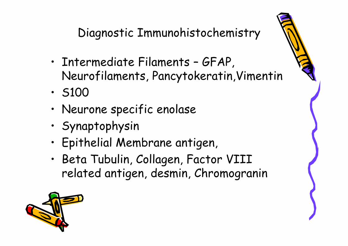
Diagnostic Immunohistochemistry
• Intermediate Filaments – GFAP,Neurofilaments, Pancytokeratin,Vimentin
• S100• Neurone specific enolase• Synaptophysin• Epithelial Membrane antigen,• Beta Tubulin, Collagen, Factor VIII
related antigen, desmin, Chromogranin
• Intermediate Filaments – GFAP,Neurofilaments, Pancytokeratin,Vimentin
• S100• Neurone specific enolase• Synaptophysin• Epithelial Membrane antigen,• Beta Tubulin, Collagen, Factor VIII
related antigen, desmin, Chromogranin
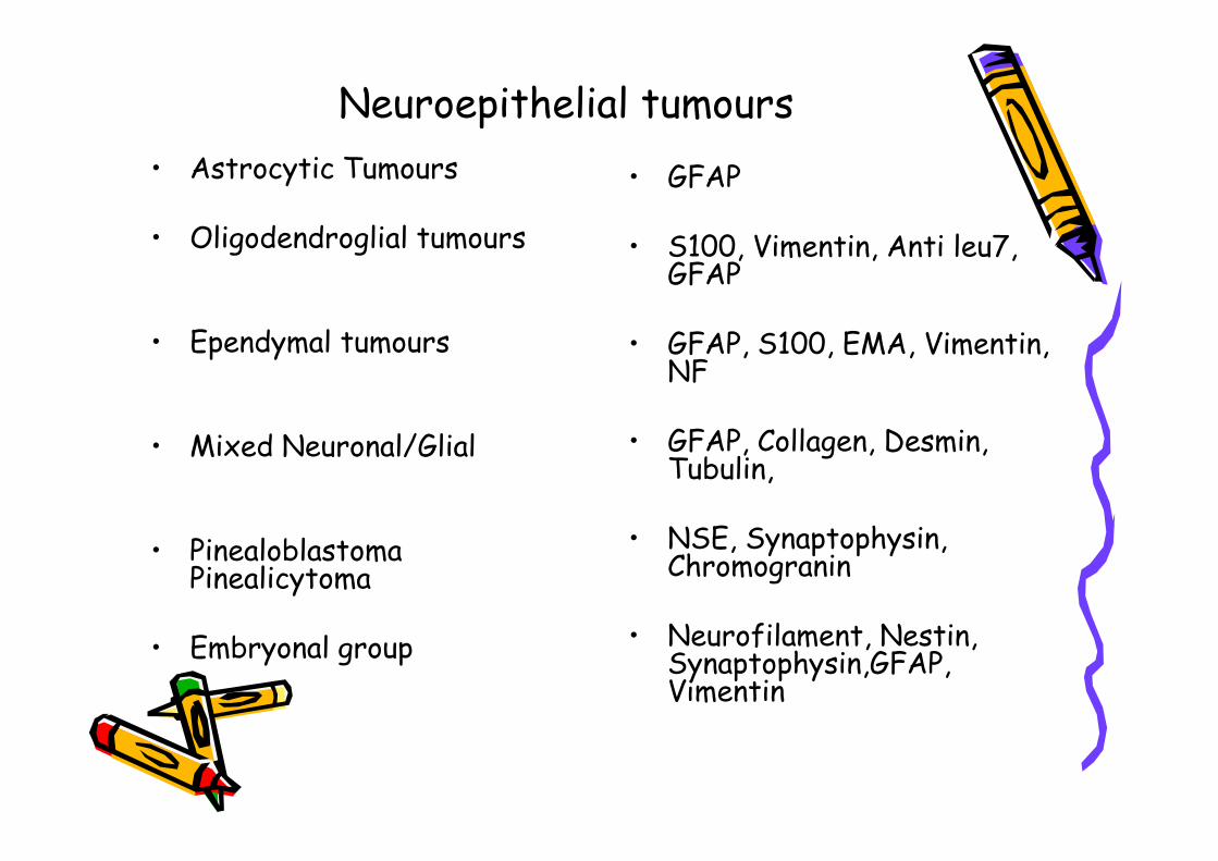
Neuroepithelial tumours• Astrocytic Tumours
• Oligodendroglial tumours
• Ependymal tumours
• Mixed Neuronal/Glial
• PinealoblastomaPinealicytoma
• Embryonal group
• GFAP
• S100, Vimentin, Anti leu7,GFAP
• GFAP, S100, EMA, Vimentin,NF
• GFAP, Collagen, Desmin,Tubulin,
• NSE, Synaptophysin,Chromogranin
• Neurofilament, Nestin,Synaptophysin,GFAP,Vimentin
• Astrocytic Tumours
• Oligodendroglial tumours
• Ependymal tumours
• Mixed Neuronal/Glial
• PinealoblastomaPinealicytoma
• Embryonal group
• GFAP
• S100, Vimentin, Anti leu7,GFAP
• GFAP, S100, EMA, Vimentin,NF
• GFAP, Collagen, Desmin,Tubulin,
• NSE, Synaptophysin,Chromogranin
• Neurofilament, Nestin,Synaptophysin,GFAP,Vimentin
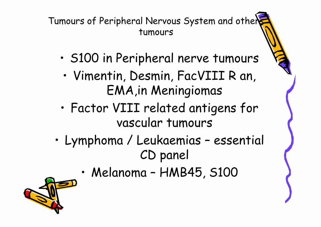
Tumours of Peripheral Nervous System and otherstumours
• S100 in Peripheral nerve tumours• Vimentin, Desmin, FacVIII R an,
EMA,in Meningiomas• Factor VIII related antigens for
vascular tumours• Lymphoma / Leukaemias – essential
CD panel• Melanoma – HMB45, S100
• S100 in Peripheral nerve tumours• Vimentin, Desmin, FacVIII R an,
EMA,in Meningiomas• Factor VIII related antigens for
vascular tumours• Lymphoma / Leukaemias – essential
CD panel• Melanoma – HMB45, S100
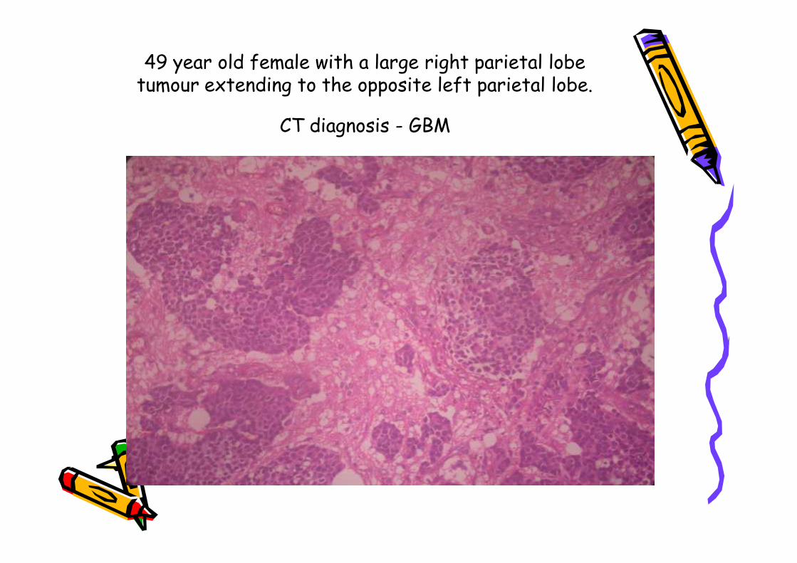
49 year old female with a large right parietal lobetumour extending to the opposite left parietal lobe.
CT diagnosis - GBM
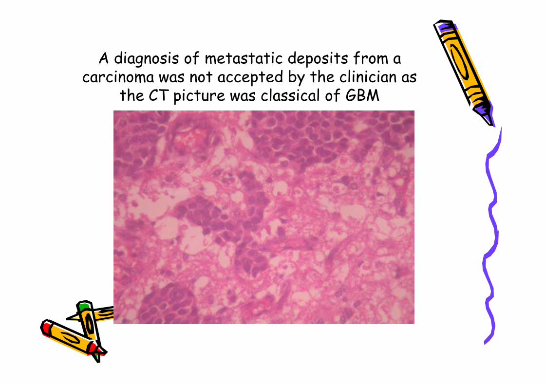
A diagnosis of metastatic deposits from acarcinoma was not accepted by the clinician as
the CT picture was classical of GBM
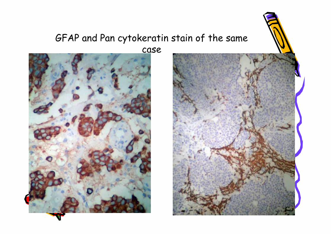
GFAP and Pan cytokeratin stain of the samecase
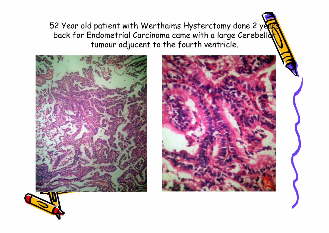
52 Year old patient with Werthaims Hysterctomy done 2 yearsback for Endometrial Carcinoma came with a large Cerebellar
tumour adjucent to the fourth ventricle.
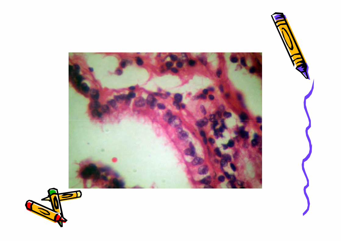
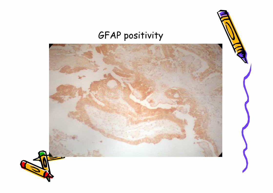
GFAP positivity
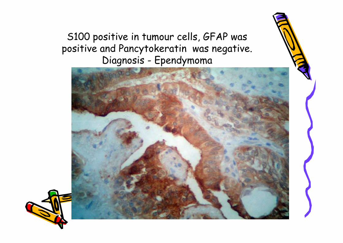
S100 positive in tumour cells, GFAP waspositive and Pancytokeratin was negative.
Diagnosis - Ependymoma
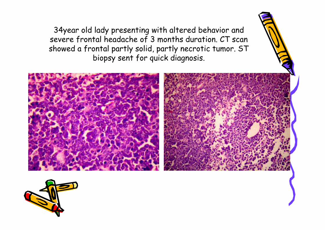
34year old lady presenting with altered behavior andsevere frontal headache of 3 months duration. CT scanshowed a frontal partly solid, partly necrotic tumor. ST
biopsy sent for quick diagnosis.
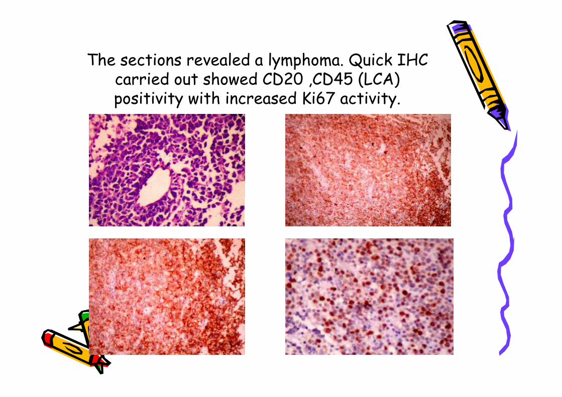
The sections revealed a lymphoma. Quick IHCcarried out showed CD20 ,CD45 (LCA)positivity with increased Ki67 activity.
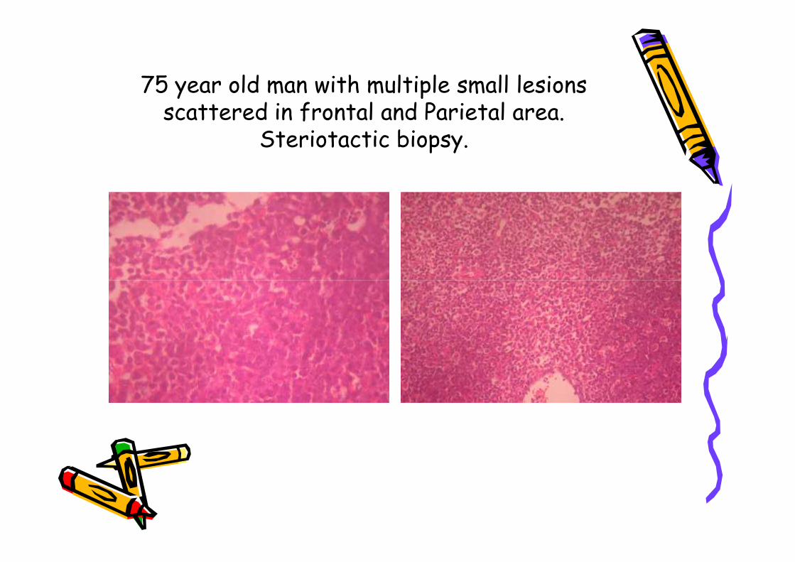
75 year old man with multiple small lesionsscattered in frontal and Parietal area.
Steriotactic biopsy.
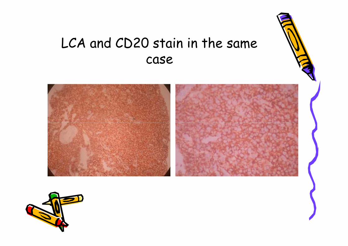
LCA and CD20 stain in the samecase
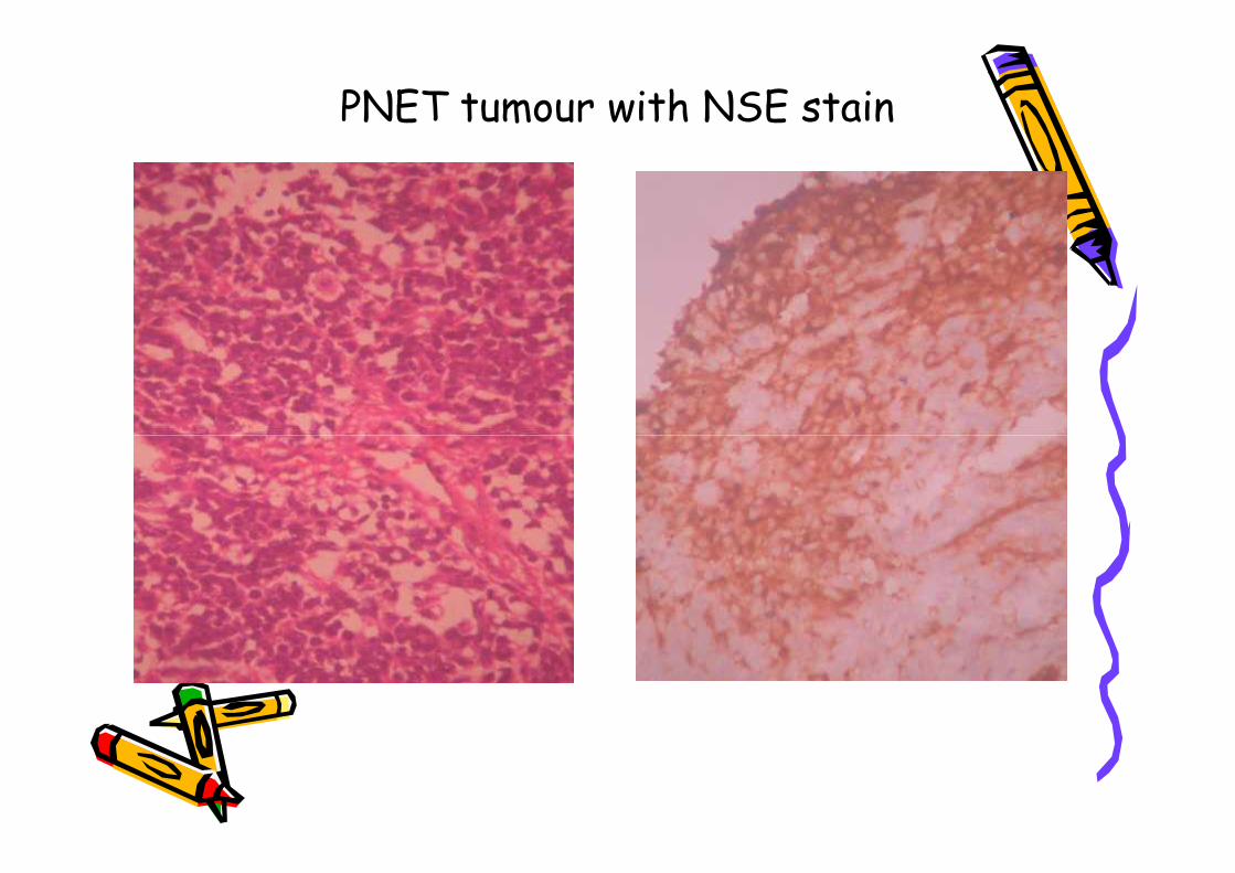
PNET tumour with NSE stain
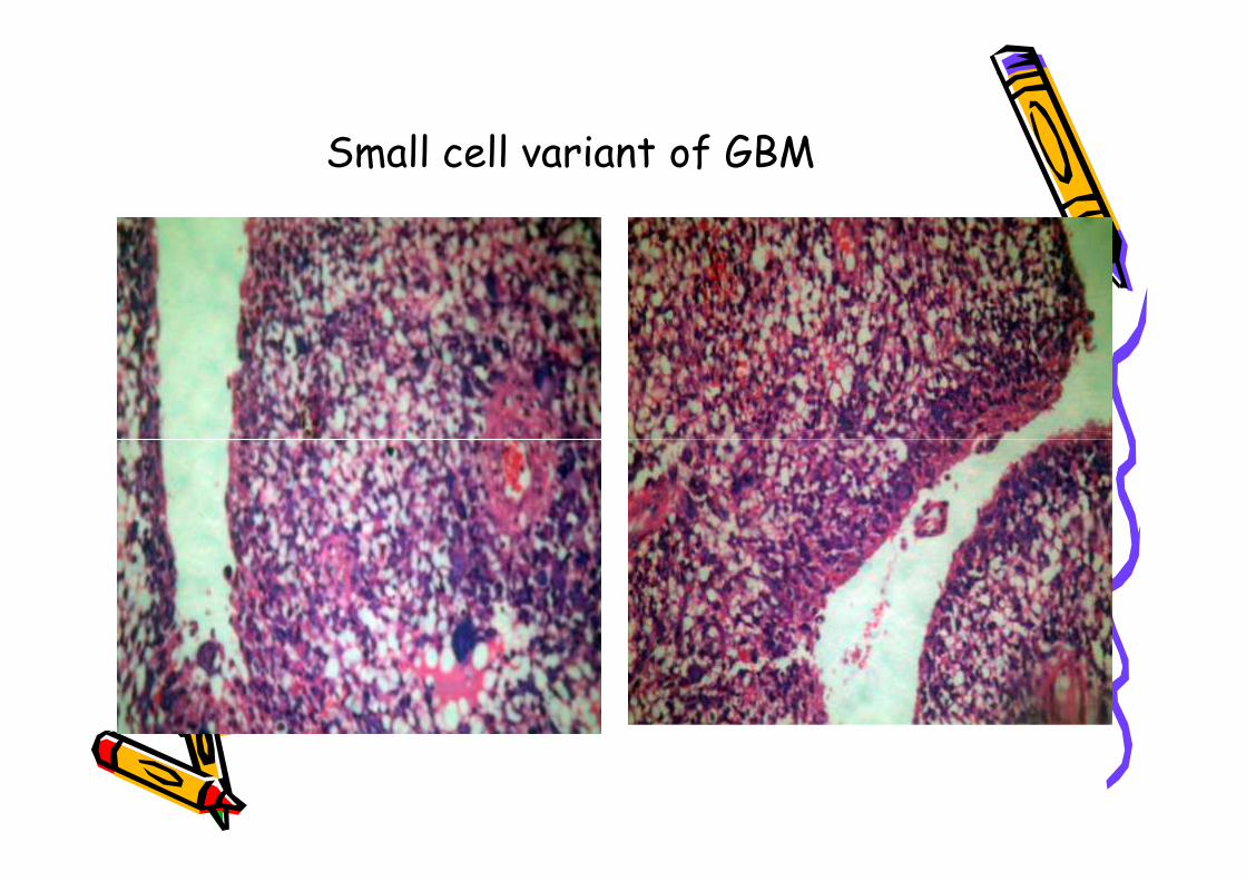
Small cell variant of GBM
Gliosis versus GliomaGliosis versus Glioma
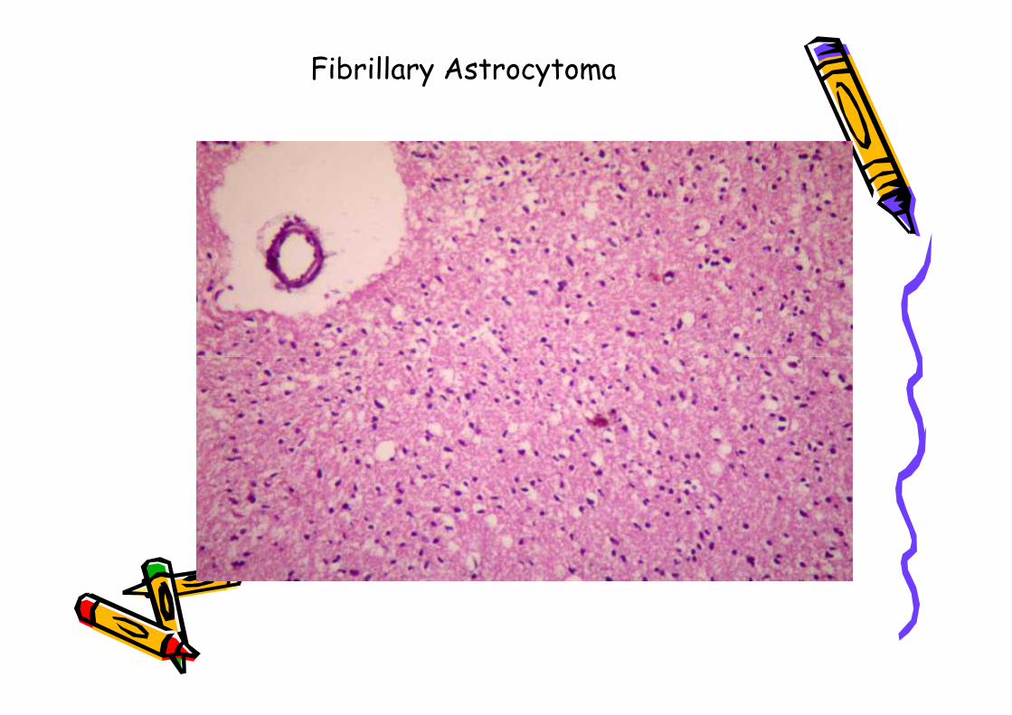
Fibrillary Astrocytoma
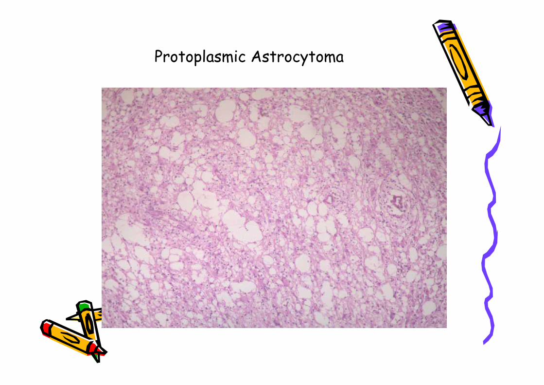
Protoplasmic Astrocytoma
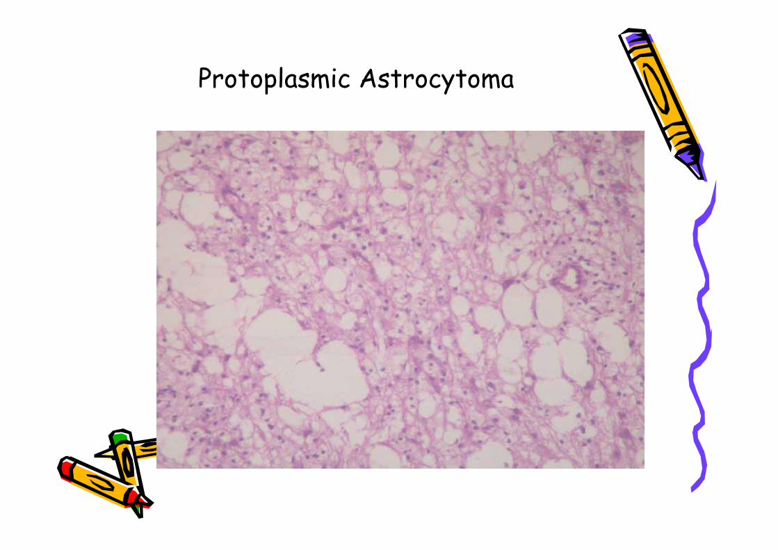
Protoplasmic Astrocytoma
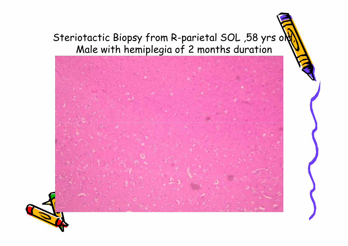
Steriotactic Biopsy from R-parietal SOL ,58 yrs old,Male with hemiplegia of 2 months duration
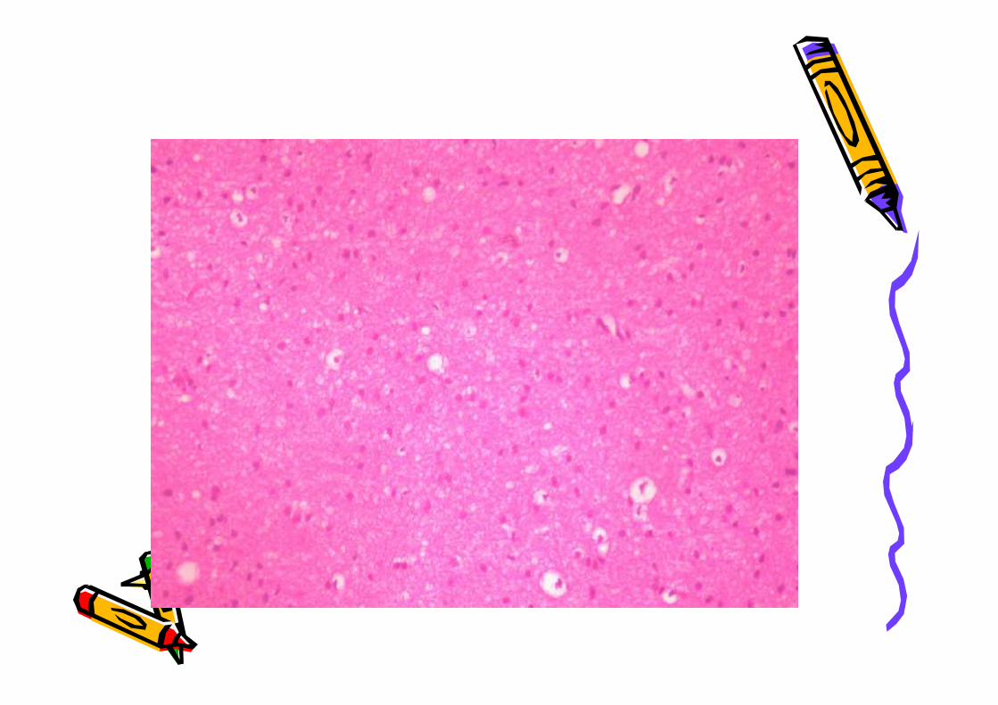
Is It a low grade glioma ?
Is It the infiltrating edge of a glioma?
Or
Is the diagnosis “reactive gliosis”
Is It a low grade glioma ?
Is It the infiltrating edge of a glioma?
Or
Is the diagnosis “reactive gliosis”
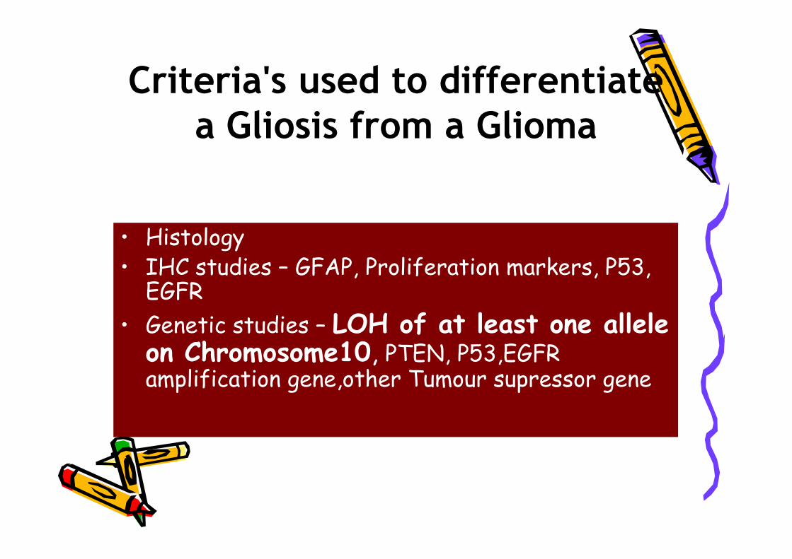
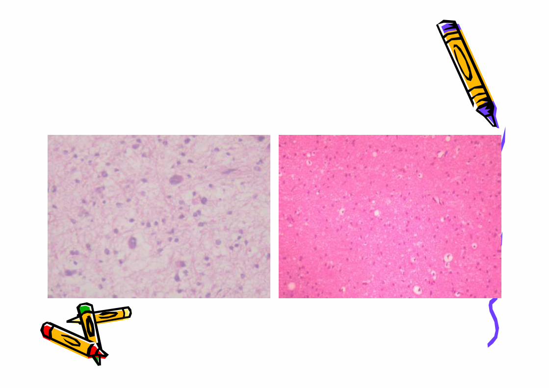
Criteria's used to differentiatea Gliosis from a Glioma
• Histology• IHC studies – GFAP, Proliferation markers, P53,
EGFR• Genetic studies – LOH of at least one allele
on Chromosome10, PTEN, P53,EGFRamplification gene,other Tumour supressor gene
• Histology• IHC studies – GFAP, Proliferation markers, P53,
EGFR• Genetic studies – LOH of at least one allele
on Chromosome10, PTEN, P53,EGFRamplification gene,other Tumour supressor gene
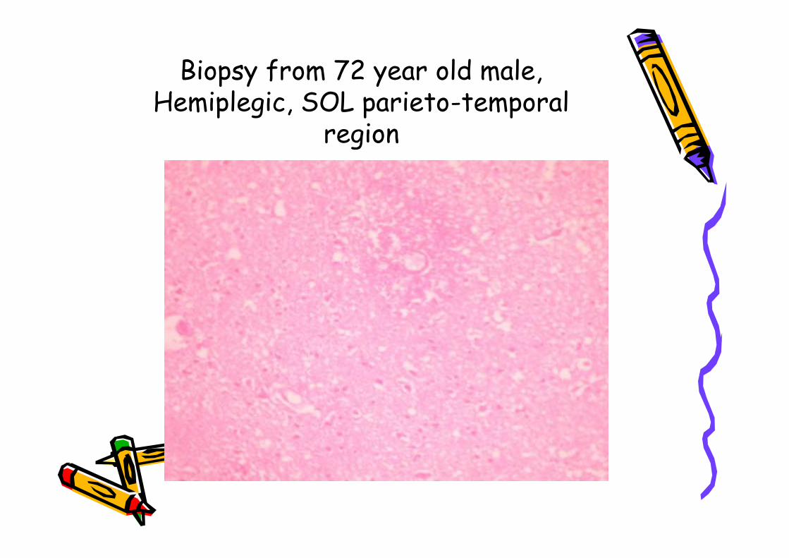
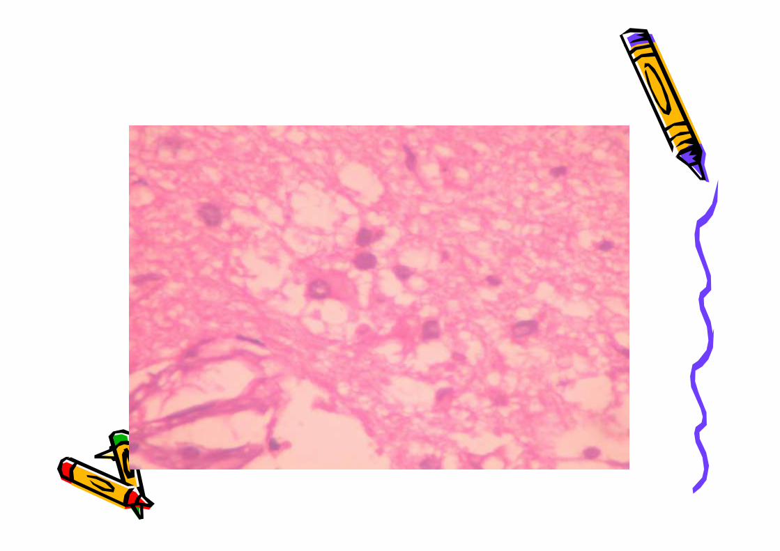
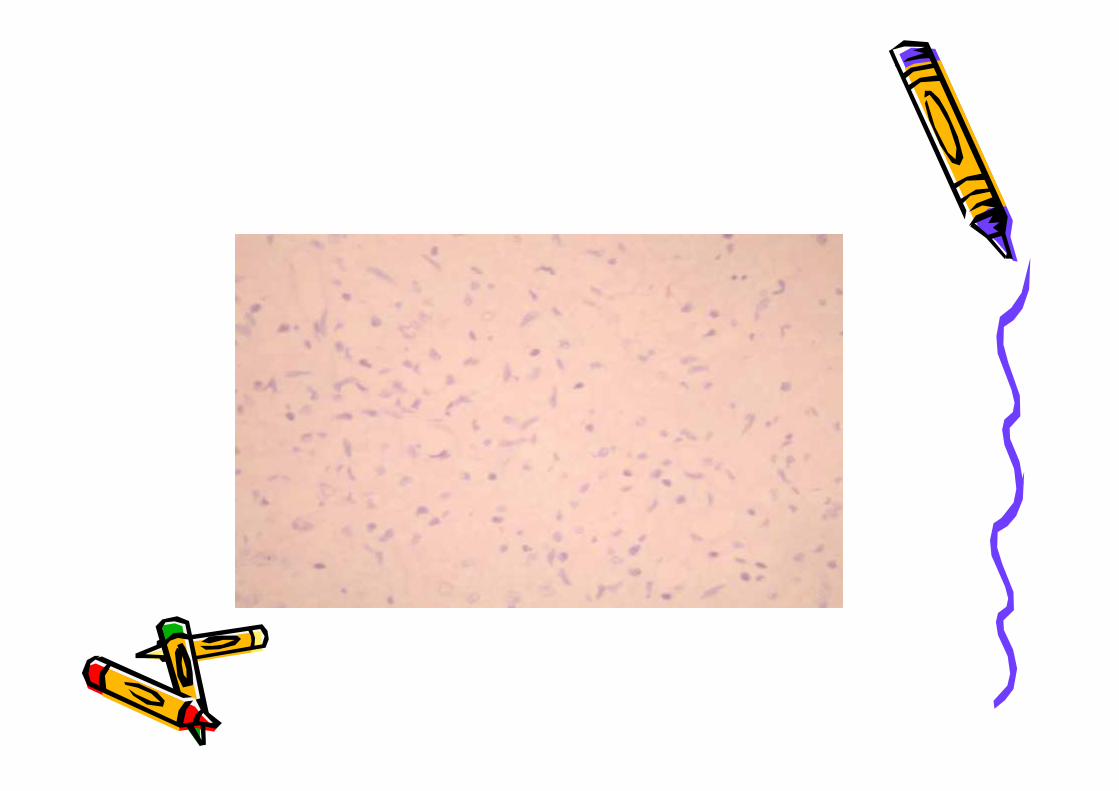
Biopsy from 72 year old male,Hemiplegic, SOL parieto-temporal
region
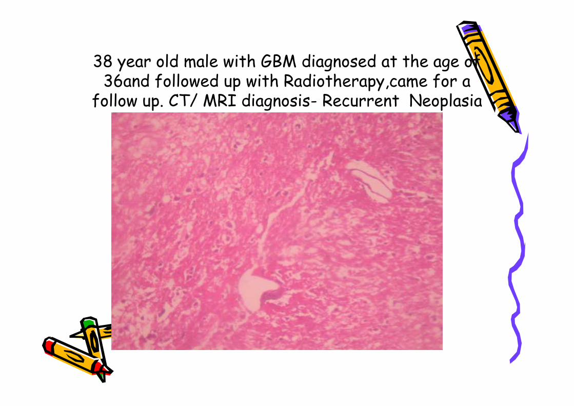
38 year old male with GBM diagnosed at the age of36and followed up with Radiotherapy,came for a
follow up. CT/ MRI diagnosis- Recurrent Neoplasia
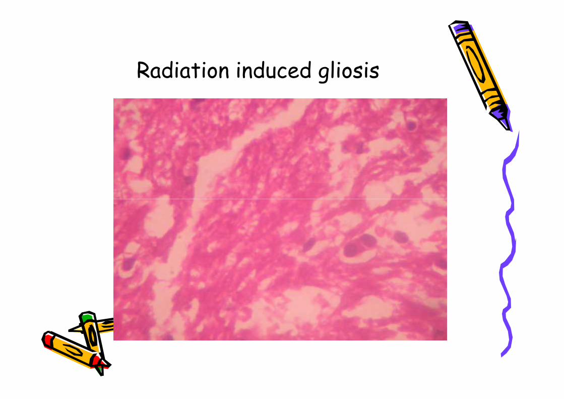
Radiation induced gliosis
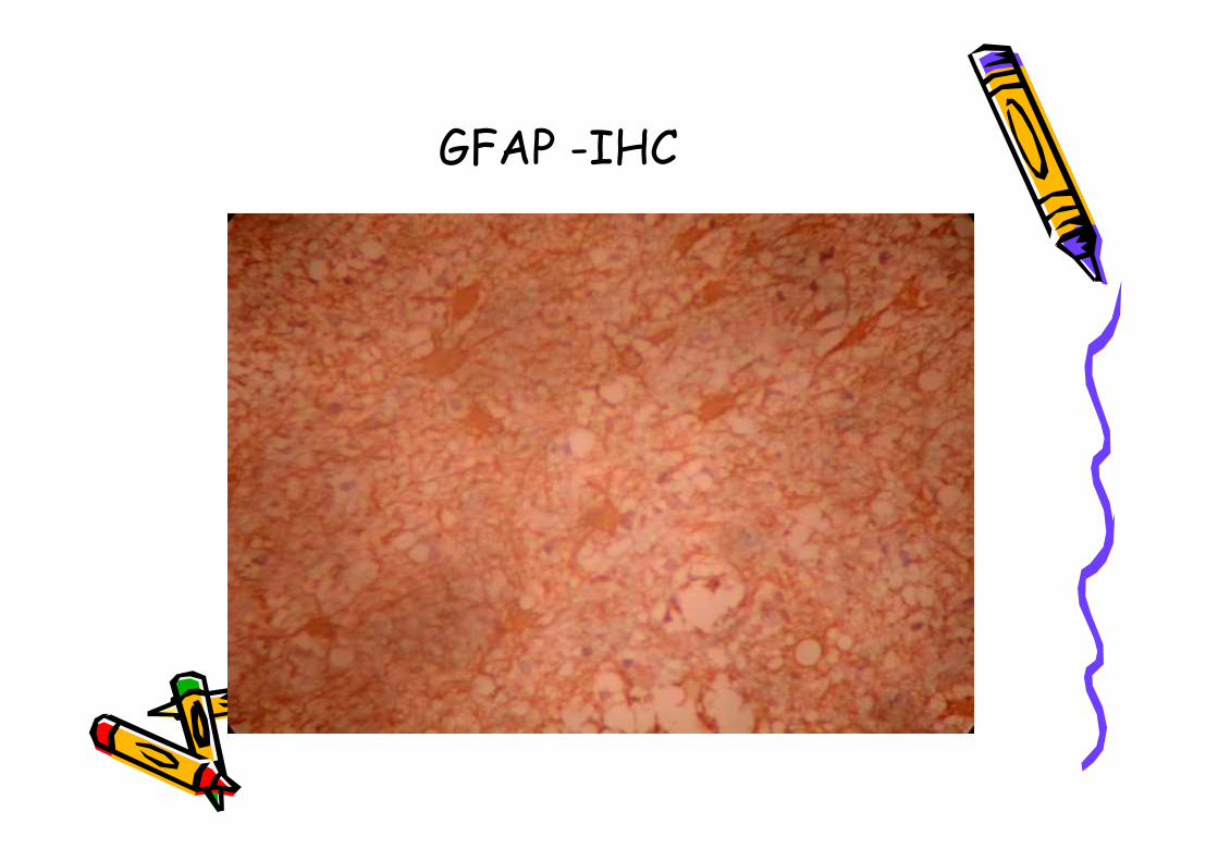
GFAP -IHC
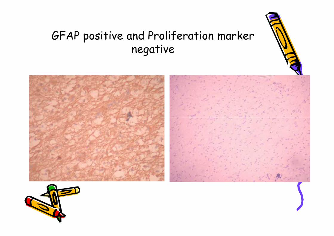
GFAP positive and Proliferation markernegative
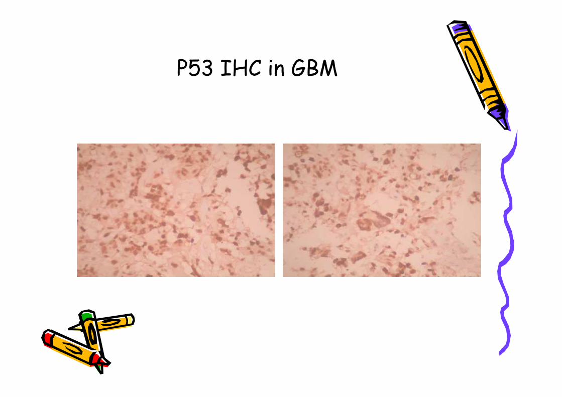
P53 IHC in GBM
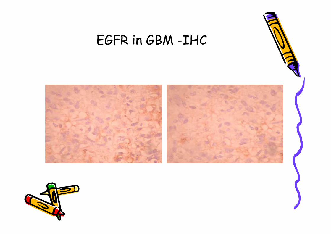
EGFR in GBM -IHC
DEFINITION
Reactive Gliosis is theproliferation of Astrocytes as ahypertrophic and hyperplasticresponse to injury within the
CNS resulting in the formationof scars
Reactive Gliosis is theproliferation of Astrocytes as ahypertrophic and hyperplasticresponse to injury within the
CNS resulting in the formationof scars
Conditions associated with Gliosis• Around Tumours• Hypoxia / Traumatic Injury• Post-Radiation effect• Infections like Abscess, Tuberculoma,
fungal infections• Viral infections involving CNS• Epilepsy• Alzeimer’s disease• Parkinsonism• Stroke• Multiple Sclerosis
• Around Tumours• Hypoxia / Traumatic Injury• Post-Radiation effect• Infections like Abscess, Tuberculoma,
fungal infections• Viral infections involving CNS• Epilepsy• Alzeimer’s disease• Parkinsonism• Stroke• Multiple Sclerosis
Pathogenesis of Gliosis
• How do these agents induce gliosis?NOT KNOWN
• What type of Astrocyte reacts to injury?NOT KNOWN
? Pre existing Astrocyte? Neuro-Glial precursors
• How do these agents induce gliosis?NOT KNOWN
• What type of Astrocyte reacts to injury?NOT KNOWN
? Pre existing Astrocyte? Neuro-Glial precursors
• Histological criteria's are insufficient inmost cases, to distinguish gliosis from lowgrade glioma.
• Use of IHC with proliferation marker hasabout 95 to 98% accuracy. P53 has 65%accuracy and EGFR about 30%.
• Genetic study with Loss of hetrozygosityof at least one allele is considered adefinite indicator of Glioma.Unfortunately it is not available as aroutine diagnostic method.
• Proliferation marker studies inGlioma and their implications in
treatment of Gliomas
• Proliferation marker studies inGlioma and their implications in
treatment of Gliomas
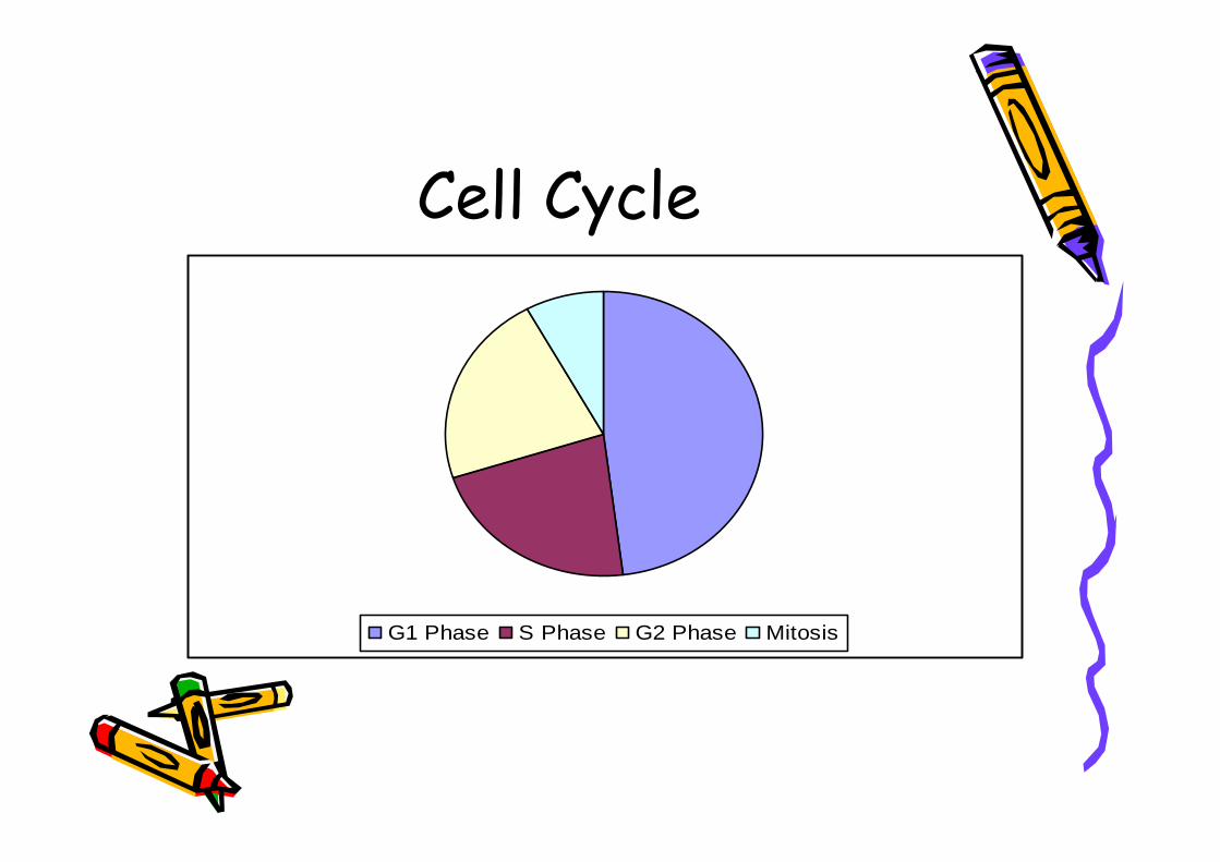
Cell Cycle
G1 Phase S Phase G2 Phase MitosisG1 Phase S Phase G2 Phase Mitosis
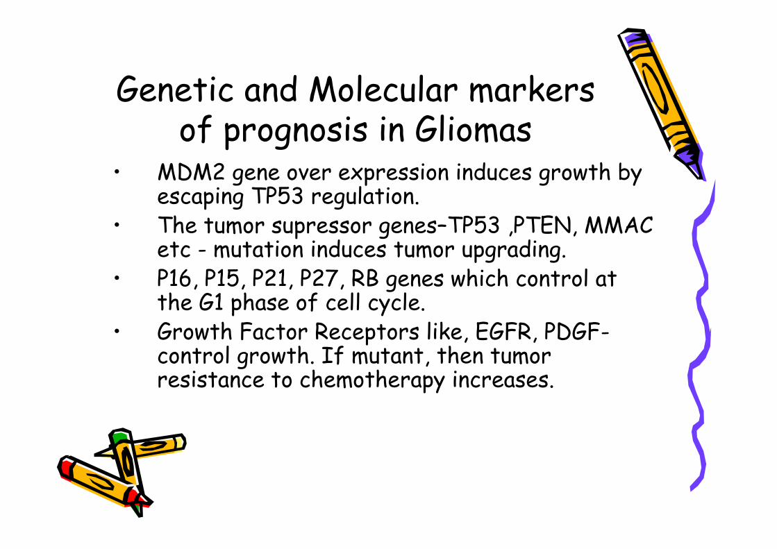
Genetic and Molecular markersof prognosis in Gliomas
• MDM2 gene over expression induces growth byescaping TP53 regulation.
• The tumor supressor genes–TP53 ,PTEN, MMACetc - mutation induces tumor upgrading.
• P16, P15, P21, P27, RB genes which control atthe G1 phase of cell cycle.
• Growth Factor Receptors like, EGFR, PDGF-control growth. If mutant, then tumorresistance to chemotherapy increases.
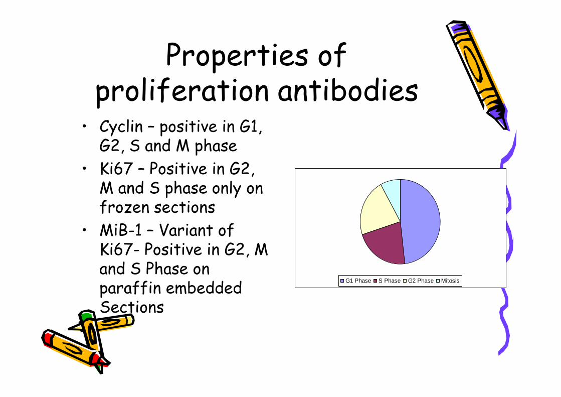
Properties ofproliferation antibodies
• Cyclin – positive in G1,G2, S and M phase
• Ki67 – Positive in G2,M and S phase only onfrozen sections
• MiB-1 – Variant ofKi67- Positive in G2, Mand S Phase onparaffin embeddedSections
G1 Phase S Phase G2 Phase Mitosis
• Cyclin – positive in G1,G2, S and M phase
• Ki67 – Positive in G2,M and S phase only onfrozen sections
• MiB-1 – Variant ofKi67- Positive in G2, Mand S Phase onparaffin embeddedSections
G1 Phase S Phase G2 Phase Mitosis
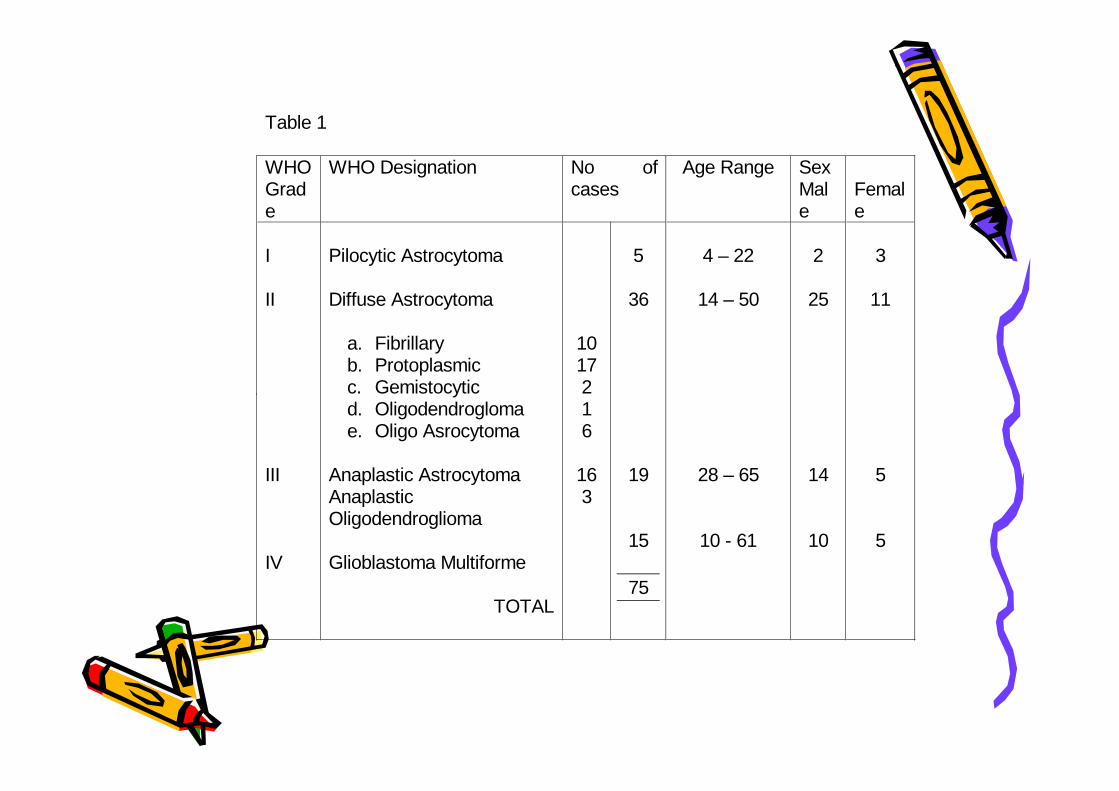
Table 1
WHOGrade
WHO Designation No ofcases
Age Range SexMale
Female
I
II
III
IV
Pilocytic Astrocytoma
Diffuse Astrocytoma
a. Fibrillaryb. Protoplasmicc. Gemistocyticd. Oligodendroglomae. Oligo Asrocytoma
Anaplastic AstrocytomaAnaplasticOligodendroglioma
Glioblastoma Multiforme
TOTAL
1017216
163
5
36
19
15
75
4 – 22
14 – 50
28 – 65
10 - 61
2
25
14
10
3
11
5
5
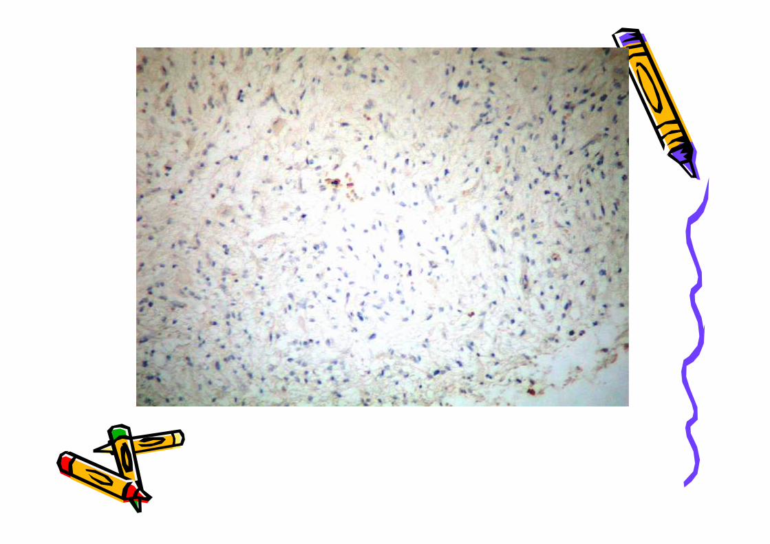
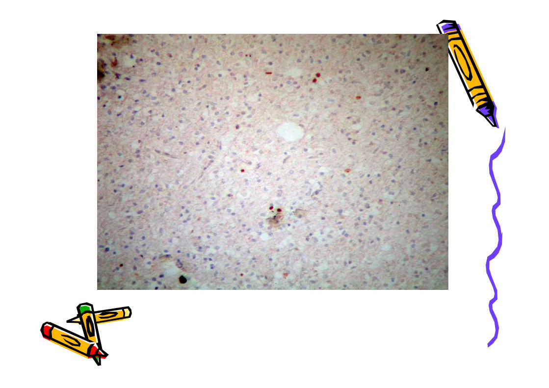
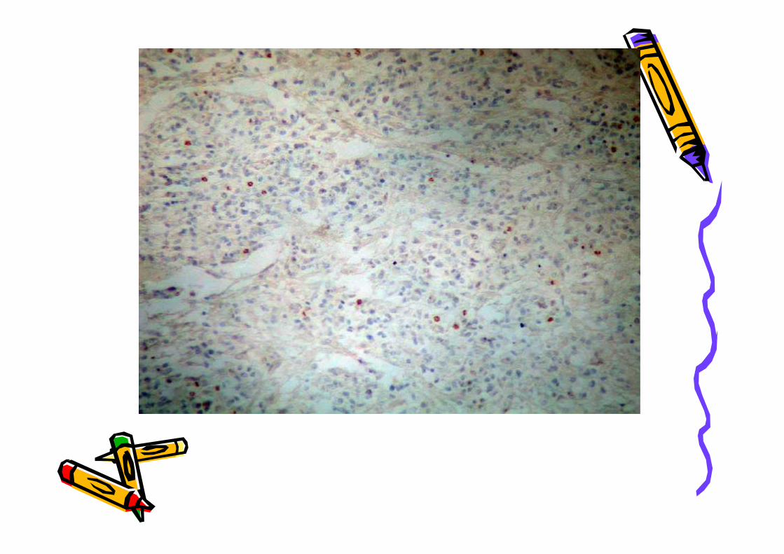
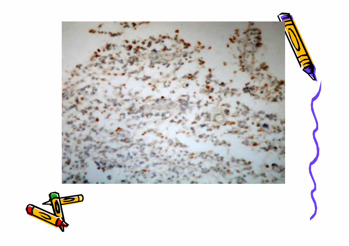
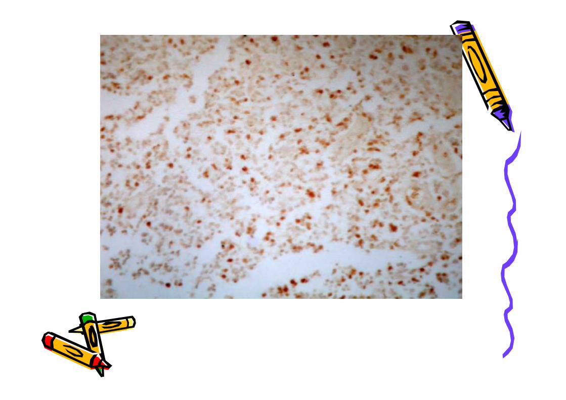
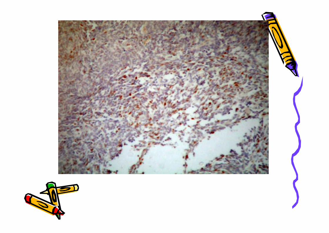
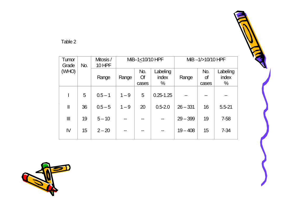
Table 2
Mitosis /10 HPF
MiB–1<10/10 HPF MiB –1/>10/10 HPFTumorGrade(WHO)
No.
Range RangeNo.Of
cases
Labelingindex
%Range
No. of
cases
Labelingindex
%
I
II
III
IV
5
36
19
15
0.5 – 1
0.5 – 5
5 – 10
2 – 20
1 – 9
1 – 9
--
--
5
20
--
--
0.25-1.25
0.5-2.0
--
--
--
26 – 331
29 – 399
19 – 408
--
16
19
15
--
5.5-21
7-58
7-34
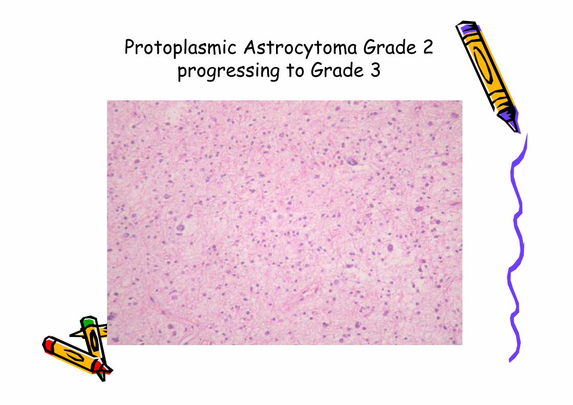
Protoplasmic Astrocytoma Grade 2progressing to Grade 3
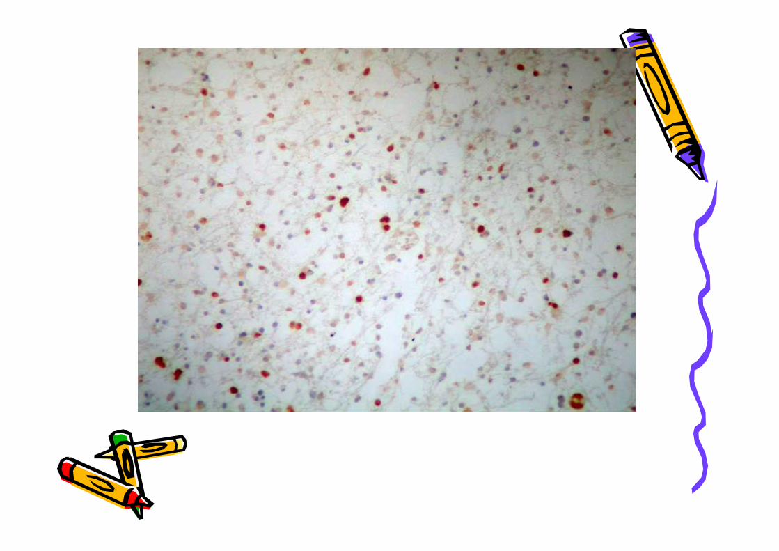
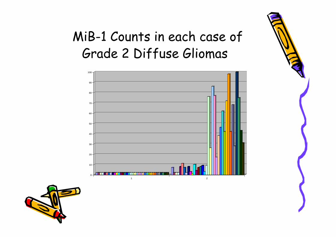
MiB-1 Counts in each case ofGrade 2 Diffuse Gliomas
0
10
20
30
40
50
60
70
80
90
100
1 20
10
20
30
40
50
60
70
80
90
100
1 2
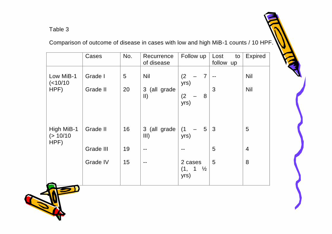
Table 3
Comparison of outcome of disease in cases with low and high MiB-1 counts / 10 HPF.
Cases No. Recurrenceof disease
Follow up Lost tofollow up
Expired
Low MiB-1(<10/10HPF)
High MiB-1(> 10/10HPF)
Grade I
Grade II
Grade II
Grade III
Grade IV
5
20
16
19
15
Nil
3 (all gradeII)
3 (all gradeIII)
--
--
(2 – 7yrs)
(2 – 8yrs)
(1 – 5yrs)
--
2 cases(1, 1 ½yrs)
--
3
3
5
5
Nil
Nil
5
4
8
• MiB-1 Study is simple, cost effective anddoes not need sophisticated Molecularbiology labs.
• The G1 Phase of cell cycle does not stainfor MiB-1, hence low false positive counts.
• Counting of stained cells in 10 consecutivefields is simple and counts give reliableinformation on the biological behavior ofthe Gliomas.
• MiB-1 counts do not throw any furtherlight on the prognosis of grade 3 and grade4(GBM) Gliomas.
• MiB-1 are most useful in Grade 2 Diffuse Gliomas.• MiB-1 counts below 10/10HPF especially in Grade 2
Diffuse Gliomas carry better prognosis.• The sub group of grade 2 Gliomas which are
strongly positive for MiB-1 (Counts >10/10hpf)appear to be potentially aggressive.
• The clinical outcome of the patients in thissubgroup of aggressive Grade2 Gliomas resemblesthe grade 3 tumors.
• Post surgical evaluation, aggressive therapy andclose follow up may be essential in these cases forbetter care of these patients
• Targeted therapies and biologicalmodifiers for Brain tumours
• Targeted therapies and biologicalmodifiers for Brain tumours
• Growth receptors:.1. EGFR family ie EGFR 1; EGFR 2 or her2,c erb 2; EGFR 3;EGFR4.2. Platelet derived growth factor3.Vascular endothelial growth factor 1, 2,
and 3 ( VEGFR)4.Insulin like growth factor5. Fibroblast growth factor receptor6. Nerve growth factor
• Growth receptors:.1. EGFR family ie EGFR 1; EGFR 2 or her2,c erb 2; EGFR 3;EGFR4.2. Platelet derived growth factor3.Vascular endothelial growth factor 1, 2,
and 3 ( VEGFR)4.Insulin like growth factor5. Fibroblast growth factor receptor6. Nerve growth factor
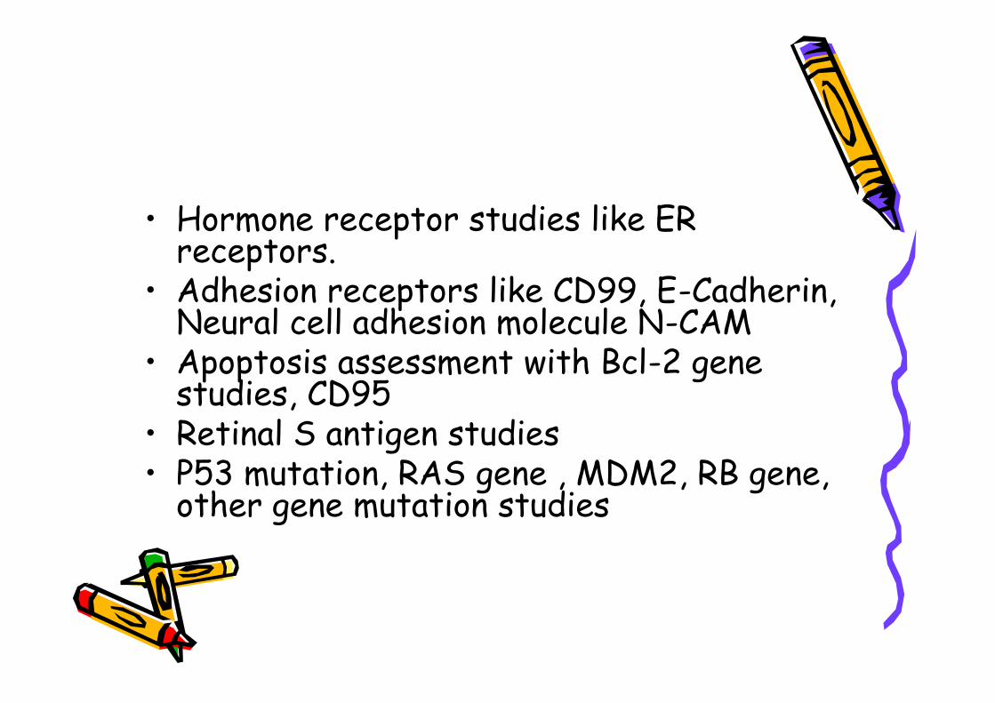
• Hormone receptor studies like ERreceptors.
• Adhesion receptors like CD99, E-Cadherin,Neural cell adhesion molecule N-CAM
• Apoptosis assessment with Bcl-2 genestudies, CD95
• Retinal S antigen studies• P53 mutation, RAS gene , MDM2, RB gene,
other gene mutation studies
• Hormone receptor studies like ERreceptors.
• Adhesion receptors like CD99, E-Cadherin,Neural cell adhesion molecule N-CAM
• Apoptosis assessment with Bcl-2 genestudies, CD95
• Retinal S antigen studies• P53 mutation, RAS gene , MDM2, RB gene,
other gene mutation studies
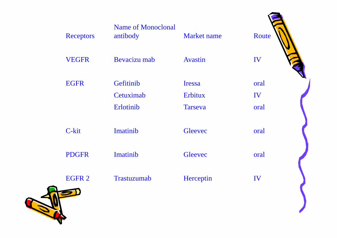
ReceptorsName of Monoclonalantibody Market name Route
VEGFR Bevacizu mab Avastin IV
EGFR Gefitinib Iressa oral
Cetuximab Erbitux IV
Erlotinib Tarseva oral
C-kit Imatinib Gleevec oral
PDGFR Imatinib Gleevec oral
EGFR 2 Trastuzumab Herceptin IV
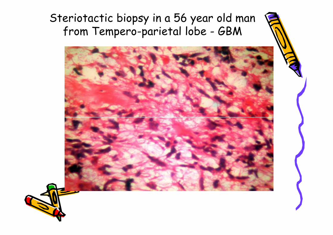
Steriotactic biopsy in a 56 year old manfrom Tempero-parietal lobe - GBM
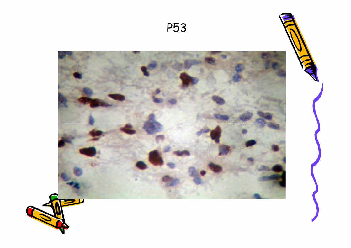
P53
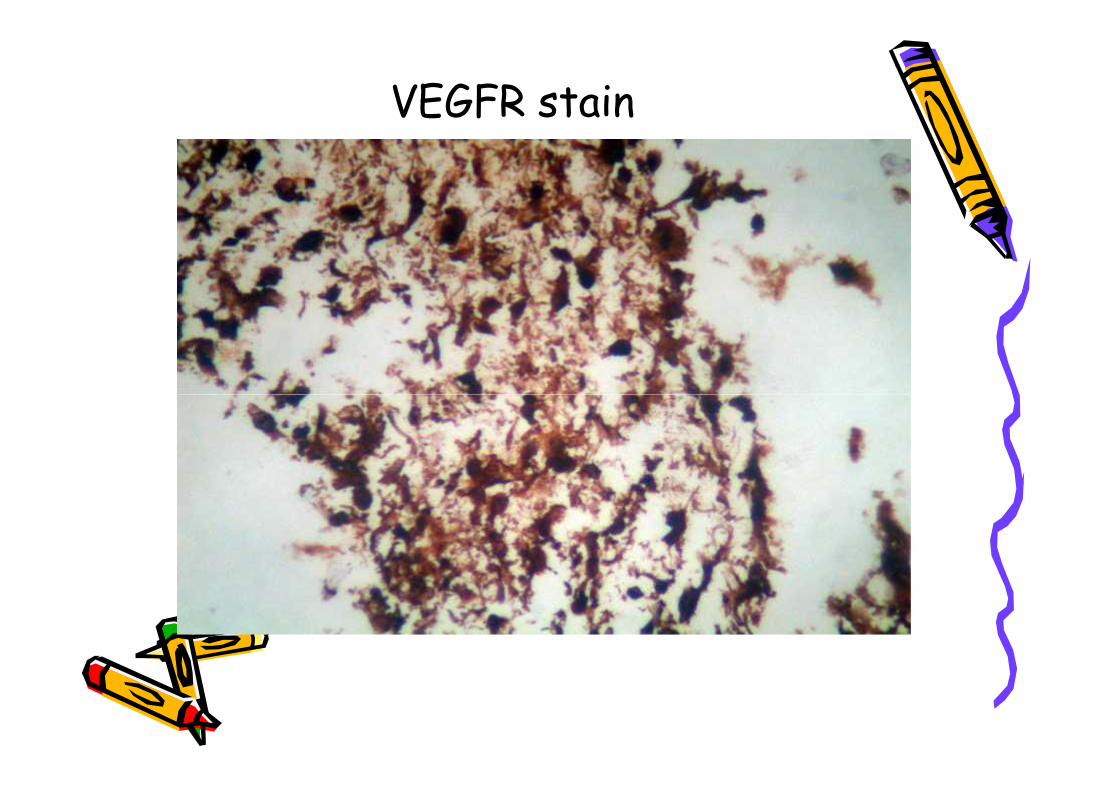
VEGFR stain
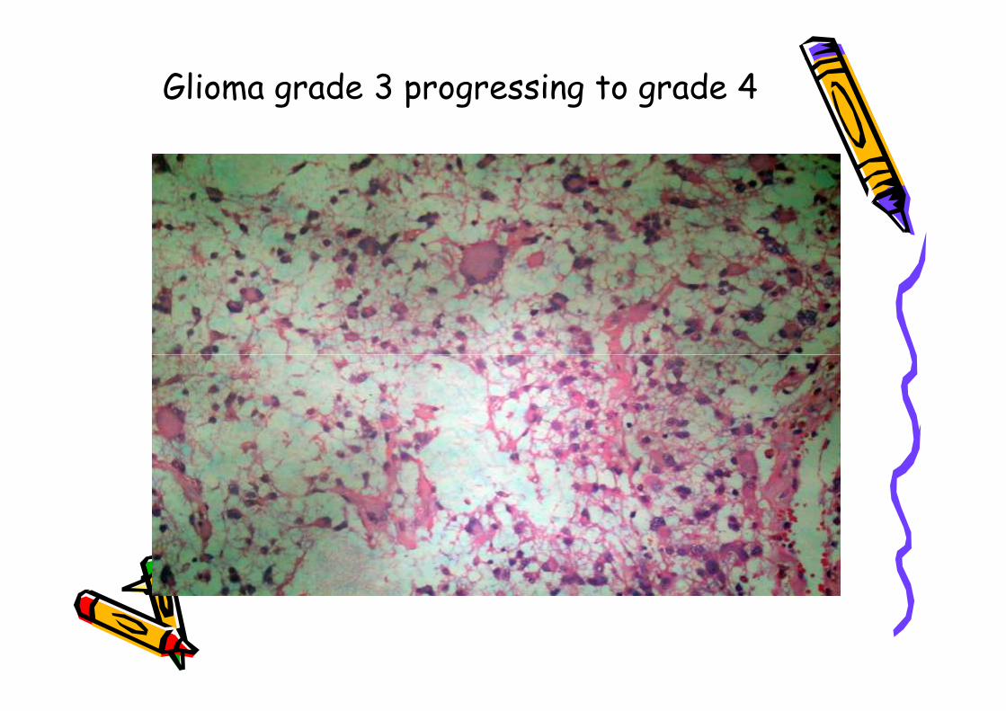
Glioma grade 3 progressing to grade 4
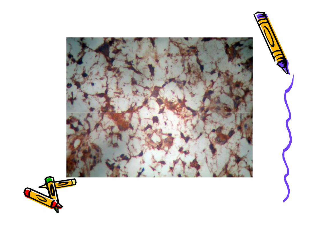
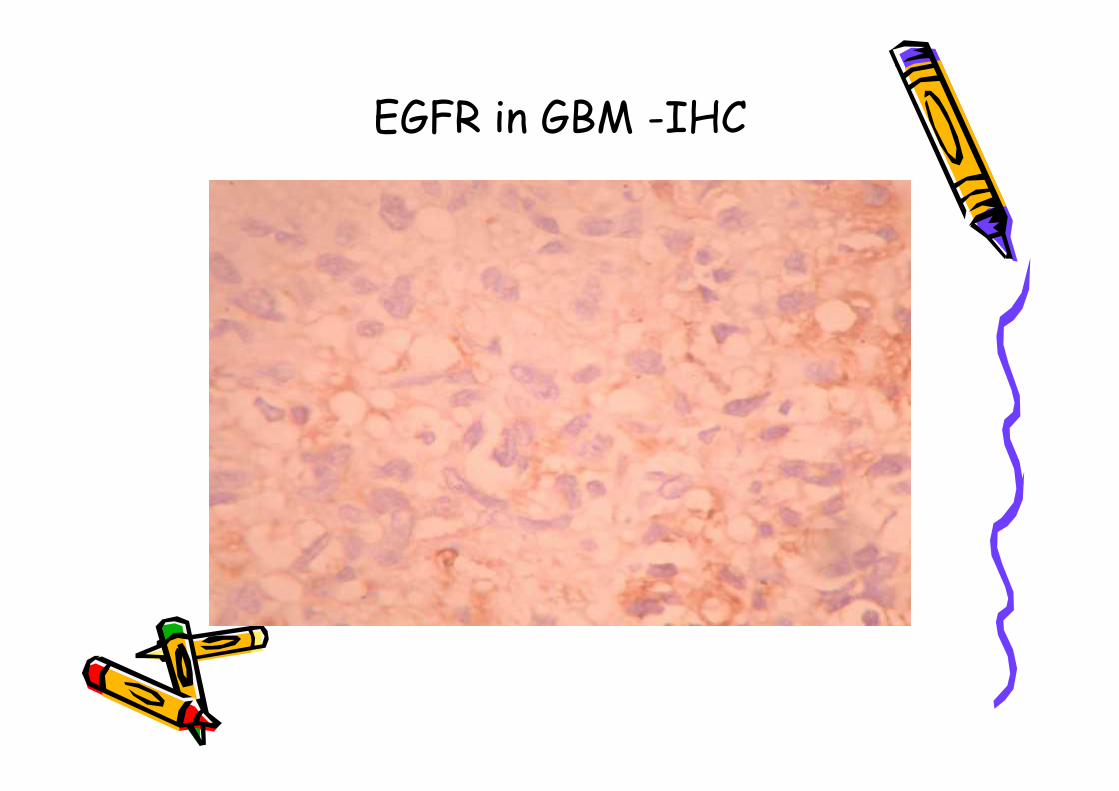
EGFR in GBM -IHC
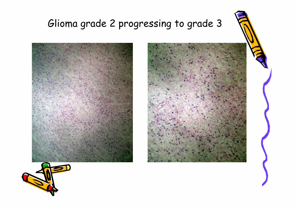
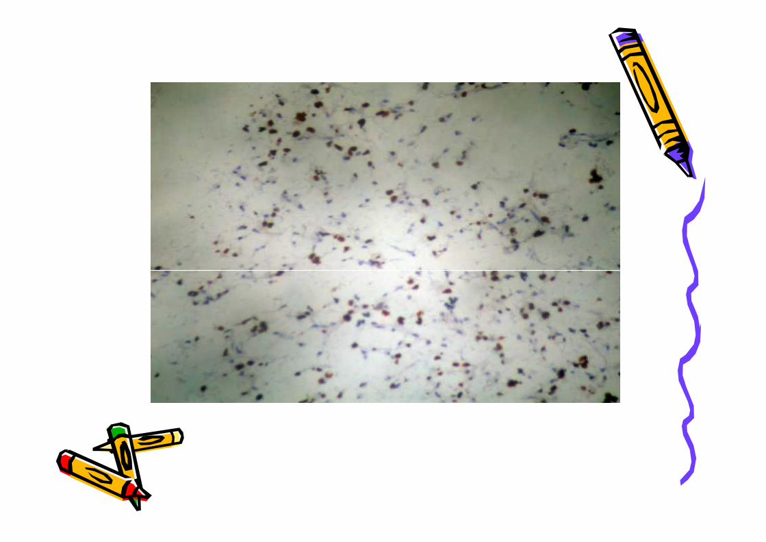
Glioma grade 2 progressing to grade 3
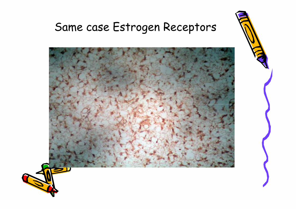
Same case Estrogen Receptors
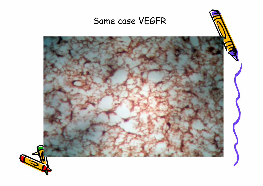
Same case VEGFR
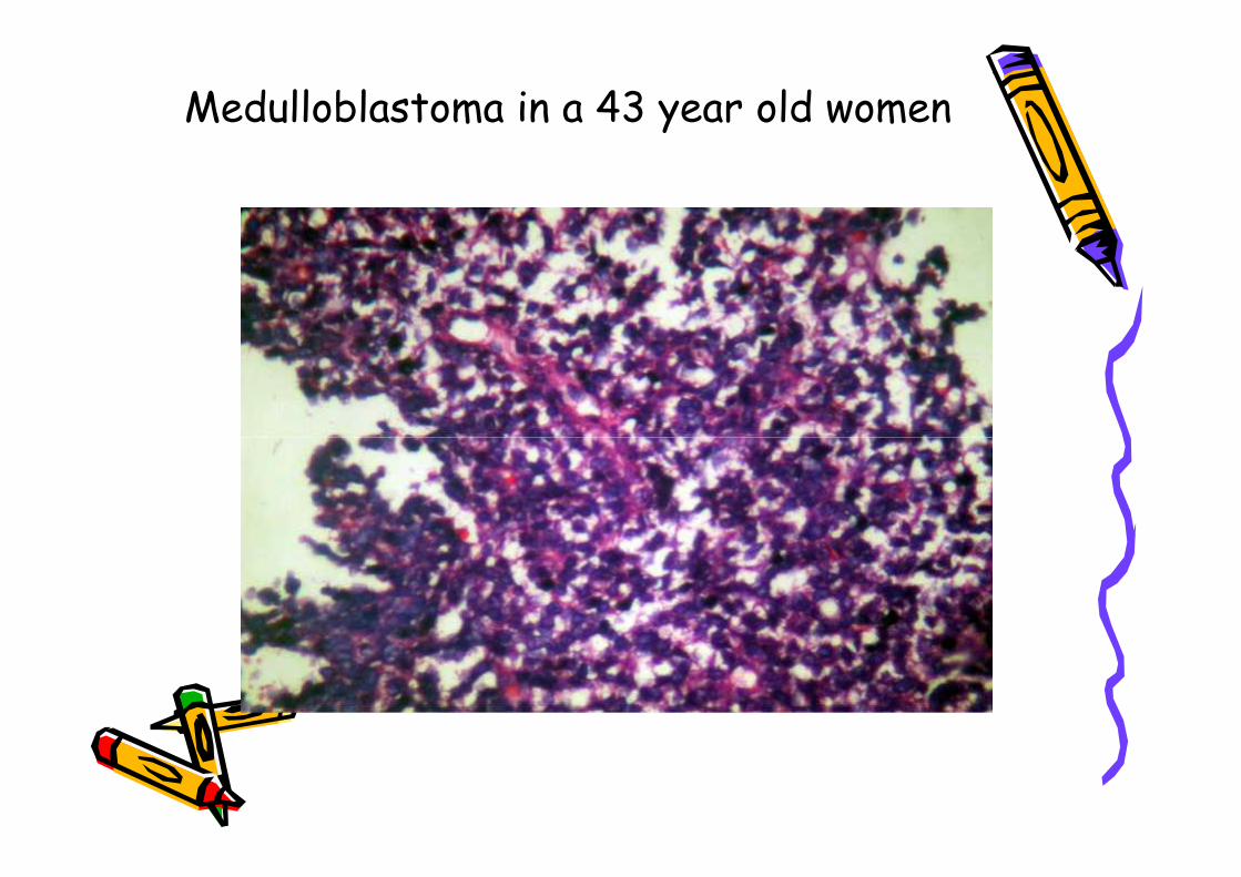
Medulloblastoma in a 43 year old women
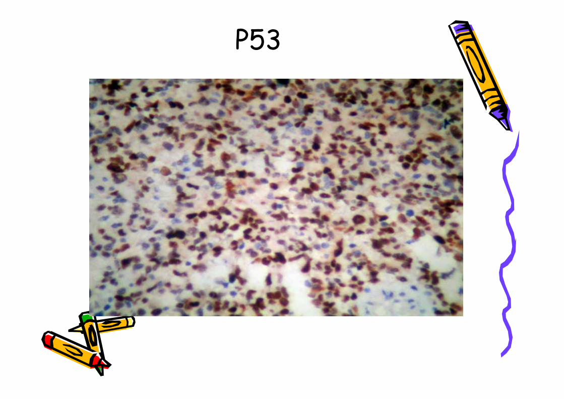
P53
Conclusion
• IHC is useful in diagnosis,understanding the biological
properties, predicting the responseto treatment and selection oftargeted therapies of brain
tumours.• Pathologists play a role in patient
care and management.
• IHC is useful in diagnosis,understanding the biological
properties, predicting the responseto treatment and selection oftargeted therapies of brain
tumours.• Pathologists play a role in patient
care and management.
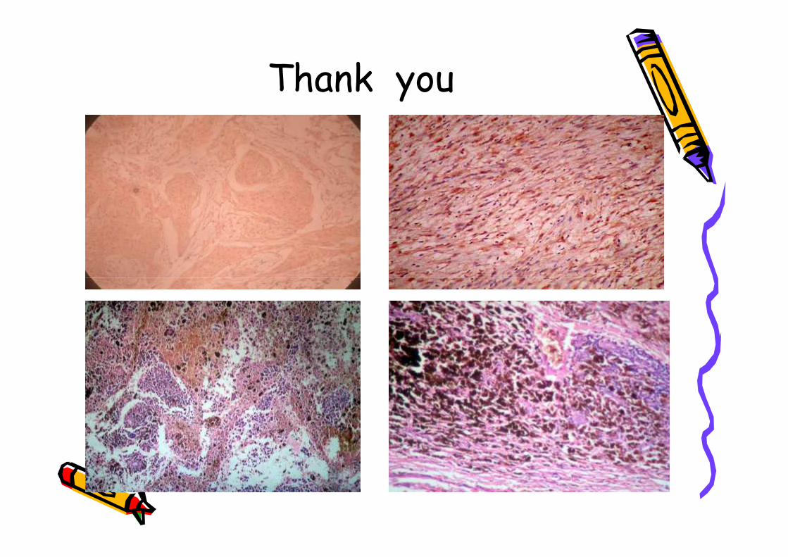
Thank you