Embed Size (px)
Citation preview
Unit J: Respiratory System
Program Area: Health Occupations Education
Course Title: Allied Health Sciences I Number: 7211
Unit Title: Respiratory System
SuggestedTime forInstruction: 5 class periods (90 minute classes)
11 class periods (55 minute classes)
Course Percent: 5 %
Unit Evaluation: 100 % Cognitive
-------------------------------------------------------------------------------
Competency: 1H10. Analyze the anatomy and physiology of the respiratory system.
Specific Objectives:
1H10.01 Describe the structure of the respiratory system.
1H10.02 Analyze the function of the respiratory system.
1H10.03 Identify characteristics and treatment of common respiratory disorders.
Summer 2005 J.1

Unit J Master Outline
J. Respiratory System
1H10.01 Describe the structure of the respiratory system.A. Nasal cavity (nose)
1. Nasal septum divides nose into R and L sides2. Cilia – hairs that trap dirt and particles
B. Sinuses1. Cavities in skull2. Connected to nasal cavity by ducts3. Lined with mucous membrane
C. Pharynx1. Throat2. 5” long
D. Larynx (voice box)1. Triangular chamber below pharynx2. Contains vocal cords3. Adam’s apple4. Epiglottis – covers larynx during swallowing
E. Trachea (windpipe)1. 4 ½” long2. Walls have bands of C-shaped cartilage3. Lined with ciliated mucous membrane
F. Bronchi and Bronchioles1. Lower end of trachea divides into R and L bronchus2. Become bronchial tubes and bronchioles as branches enter lungs
G. Alveoli1. Clusters of thin-walled sacs made of single layer epithelial tissue2. Inner surfaces covered with surfactant3. Each alveolus surrounded by capillaries
H. Lungs1. Fill thoracic cavity2. Upper part = apex3. Lower part = base4. Lung tissue porous and spongy, it floats5. R lung larger and shorter, 3 lobes6. L lung has 2 lobes
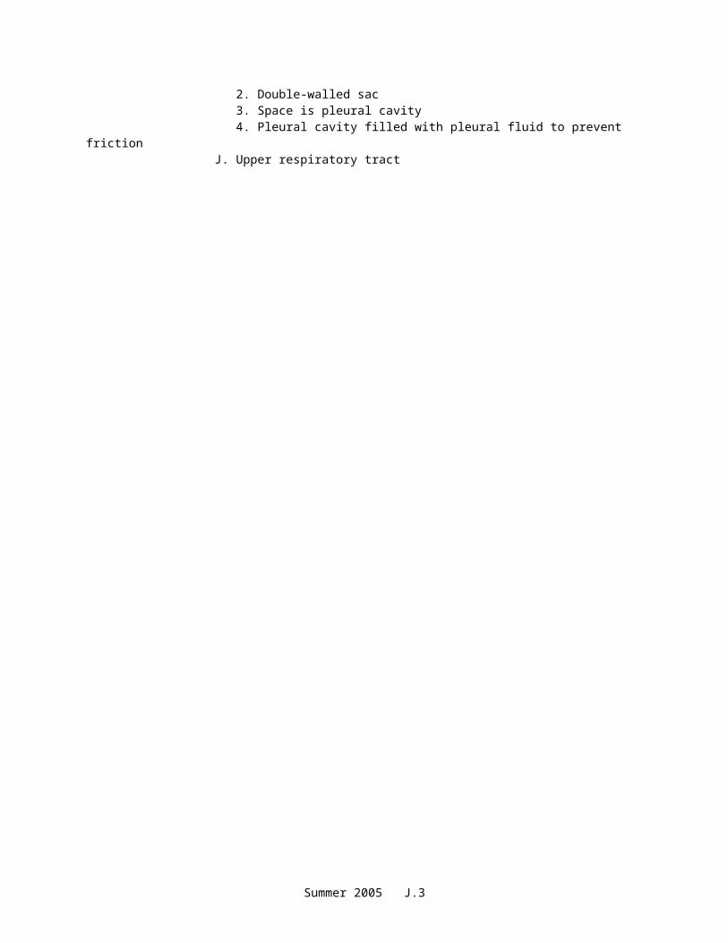
I. Pleura1. Membrane that covers lungs2. Double-walled sac3. Space is pleural cavity4. Pleural cavity filled with pleural fluid to prevent friction
J. Upper respiratory tract
Summer 2005 J.2
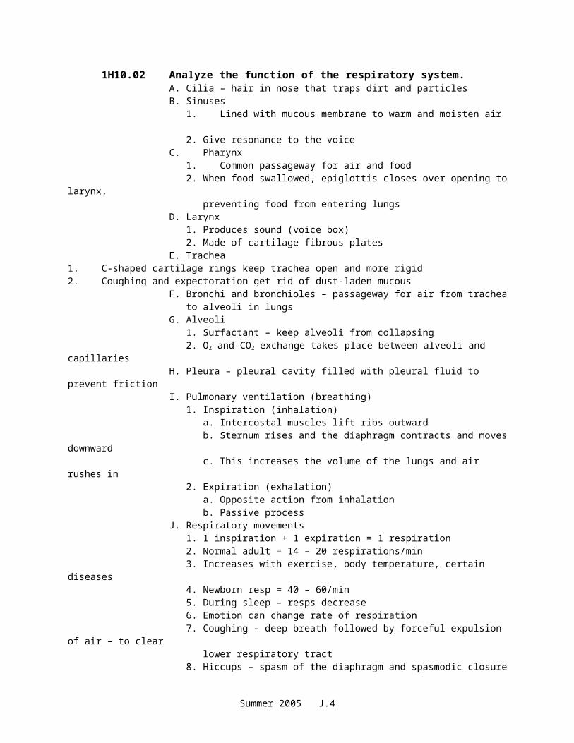
1H10.02 Analyze the function of the respiratory system.A. Cilia – hair in nose that traps dirt and particlesB. Sinuses
1. Lined with mucous membrane to warm and moisten air2. Give resonance to the voice
C. Pharynx1. Common passageway for air and food2. When food swallowed, epiglottis closes over opening to larynx,
preventing food from entering lungsD. Larynx
1. Produces sound (voice box)2. Made of cartilage fibrous plates
E. Trachea1. C-shaped cartilage rings keep trachea open and more rigid2. Coughing and expectoration get rid of dust-laden mucous
F. Bronchi and bronchioles – passageway for air from trachea to alveoli in lungsG. Alveoli
1. Surfactant – keep alveoli from collapsing2. O2 and CO2 exchange takes place between alveoli and capillaries
H. Pleura – pleural cavity filled with pleural fluid to prevent frictionI. Pulmonary ventilation (breathing)
1. Inspiration (inhalation)a. Intercostal muscles lift ribs outwardb. Sternum rises and the diaphragm contracts and moves downwardc. This increases the volume of the lungs and air rushes in
2. Expiration (exhalation)a. Opposite action from inhalationb. Passive process
J. Respiratory movements1. 1 inspiration + 1 expiration = 1 respiration2. Normal adult = 14 – 20 respirations/min3. Increases with exercise, body temperature, certain diseases4. Newborn resp = 40 – 60/min5. During sleep – resps decrease6. Emotion can change rate of respiration7. Coughing – deep breath followed by forceful expulsion of air – to clear
lower respiratory tract8. Hiccups – spasm of the diaphragm and spasmodic closure of the glottis9. Sneezing – air forced through nose to clear respiratory tract10. Yawning – deep prolonged breath that fills lungs, increases blood O2
K. Control of breathing1. Neural factors
a. Respiratory center located in medulla oblongatab. Increase or decrease of O2 or CO2 in the blood will trigger respiratory
centerc. Phrenic nerve – stimulates diaphragm
2. Chemical factorsa. Depends on level of blood CO2
b. Chemoreceptors in aorta and carotid arteries sensitive to the amount of blood O2
Summer 2005 J.3
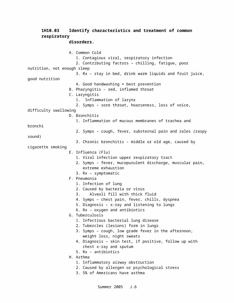
1H10.03 Identify characteristics and treatment of common respiratory disorders.
A. Common Cold
1. Contagious viral, respiratory infection2. Contributing factors – chilling, fatigue, poor nutrition, not enough sleep3. Rx – stay in bed, drink warm liquids and fruit juice, good nutrition4. Good handwashing = best prevention
B. Pharyngitis – red, inflamed throatC. Laryngitis
1. Inflammation of larynx2. Symps – sore throat, hoarseness, loss of voice, difficulty swallowing
D. Bronchitis1. Inflammation of mucous membranes of trachea and bronchi2. Symps – cough, fever, substernal pain and rales (raspy sound)3. Chronic bronchitis – middle or old age, caused by cigarette smoking
E. Influenza (Flu)1. Viral infection upper respiratory tract2. Symps – fever, mucopurulent discharge, muscular pain, extreme
exhaustion3. Rx – symptomatic
F. Pneumonia1. Infection of lung2. Caused by bacteria or virus3. Alveoli fill with thick fluid4. Symps – chest pain, fever, chills, dyspnea5. Diagnosis – x-ray and listening to lungs6. Rx – oxygen and antibiotics
G. Tuberculosis1. Infectious bacterial lung disease2. Tubercles (lesions) form in lungs3. Symps – cough, low grade fever in the afternoon, weight loss, night
sweats4. Diagnosis – skin test, if positive, follow up with chest x-ray and sputum5. Rx – antibiotics
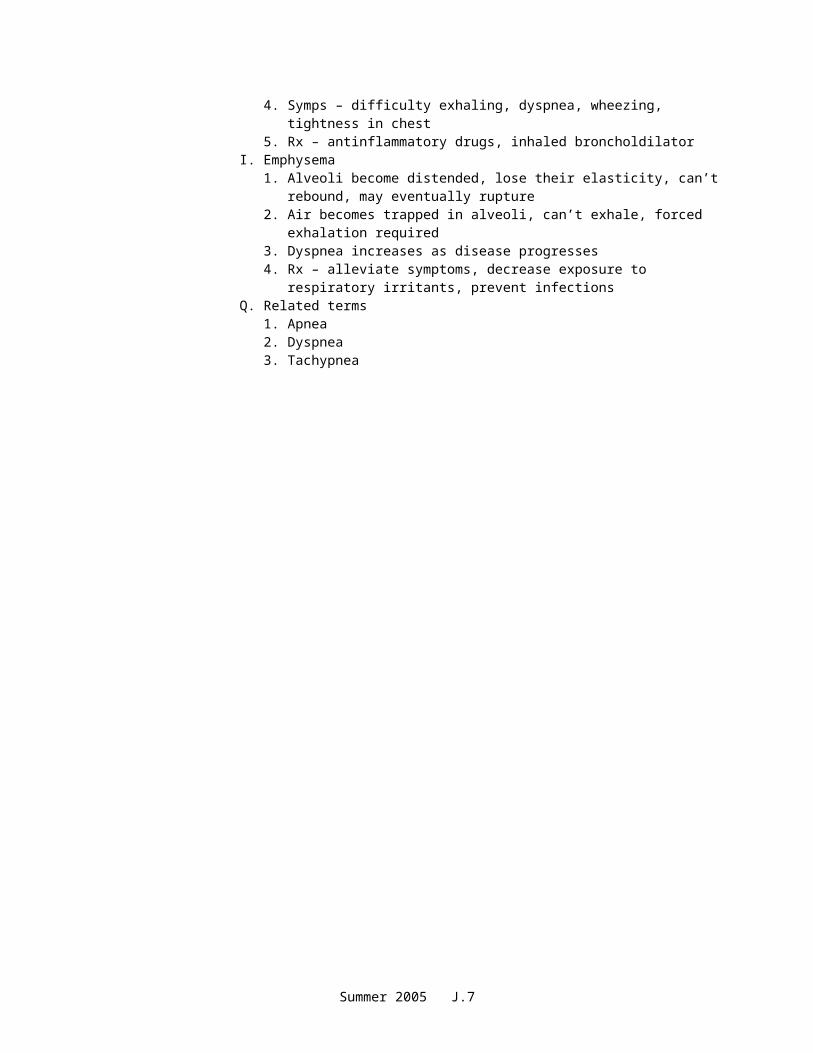
H. Asthma1. Inflammatory airway obstruction2. Caused by allergen or psychological stress3. 5% of Americans have asthma4. Symps – difficulty exhaling, dyspnea, wheezing, tightness in chest5. Rx – antinflammatory drugs, inhaled broncholdilator
I. Emphysema1. Alveoli become distended, lose their elasticity, can’t rebound, may
eventually rupture2. Air becomes trapped in alveoli, can’t exhale, forced exhalation required3. Dyspnea increases as disease progresses4. Rx – alleviate symptoms, decrease exposure to respiratory irritants,
prevent infectionsQ. Related terms
1. Apnea2. Dyspnea3. Tachypnea
Summer 2005 J.4
Unit J: Respiratory System
Competency 1H10: Describe the structure of the respiratory system.
Materials/Resources
Scott, Ann Senisi and Elizabeth Fong. Body Structures & Functions. Delmar Publishers, Latest Edition. www.DelmarAlliedHealth.com
National HOSA Handbook: Section B. Published by HOSA, Flower Mound, Texas. Current Edition. www.hosa.org
Teaching/Learning Indicators: The following letters are used to indicate specific skills/areas required in the instructional activity.
R Reading SS Social StudiesW Writing S ScienceM Math A The ArtsH Health professional/parent/community involvement
Summer 2005 J.5
Objective 1H10.01 Describe the structure of the respiratory system.
Teaching/Learning Activities
Cognitive S Have students label the diagram of the lung. (Appendix 1H10.01B)
Basic Skills S, RInstruct the students to read about the Respiratory System in Body Structures and Functions. Complete the “Respiratory Structure” Worksheet. (Appendix 1H10.01C)
Cognitive S Divide students into small groups. Give each group a ball (or orange) and an empty mesh bag. After a class discussion of alveoli, instruct the students to explain, in their own words, the relationship of alveoli to capillaries. Have them demonstrate the relationship with the ball and mesh bag. (The mesh bag should simulate the capillaries that surround the alveoli.) Allow one group to volunteer to present their understanding to the class.
Teamwork S, R Activity Title: As the Air GoesGive individual students one index card with the name of one structure of the Respiratory System. Have students read about the structure they’re been given in their textbook. On a signal given by the teacher, the students arrange themselves in a line in the order that air would pass through them.
Students will need to decide what to do with the mouth and nose as air comes in both, and the right and left bronchus as air goes thorough both. (See Transparency, Last page of Appendix) It is possible that you have more students than parts, in which case you could divide the class in half and let the two groups arrange themselves, or use multiple bronchiole and alveoli cards.
Technology S Take students to the computer lab and have them work through anatomy software such as A & P Challenge. Check with your media specialist or anatomy teacher for titles available. Students may work individually or in pairs.
HOSA SUsing HOSA Competitive Event Guidelines for Medical Spelling, hold a spelling bee using the terms in the Terminology List. If they spell the word correctly, allow them to go to an anatomical wall chart or model and point out the location of the structure they spelled.
Critical Thinking S, AHave students create “Model Lungs” using the directions provided. (Appendix 1H10.01D)
Special Needs
Each student will reach the highest level of mastery in the least restrictive environment as recommended in the student’s IEP.
Summer 2005 J.6
Objective 1H10.02 Analyze the function of the respiratory system.
Teaching/Learning Activities
Basic Skills S, RHave students read the section about respiratory function in Body Structures and Functions.
Have students create five matching questions. They can exchange papers and answer the peer-created questions
Critical Thinking S Give students a hoola hoop and ask them to demonstrate diffusion. Explain that half of them will be oxygen molecules and the other half will be carbon dioxide molecules. They are to use the hoop as the alveolus and must arrange the molecules to represent diffusion. (Students must “duck” underneath the hoop to move in and out.)
Have students brainstorm how they think the respiratory system responds to the need for oxygen. Then, introduce the section on “Control of Breathing” (Body Structures and Functions). Have them complete the worksheet provided. (Appendix 1H10.02A)
Technology/Employability S, HHave students use a pulse oxygen saturation monitor (pulse oximeter) to determine their own oxygen saturation level. If the HOE department does not have this piece of equipment, arrange to borrow one from a local hospital or other health facility.
You can also invite a respiratory therapist to speak with the class who can bring the monitor for the session. Have the students determine if their own oxygen saturation is within normal levels. Have the Respiratory Therapist discuss gas exchange and control of breathing.
Teamwork S. AHave students work in small groups to create a song that they will sing to the class. The lyrics must explain the production of sound. They may use the tunes from familiar songs such as Twinkle, Twinkle, Row Your Boat, etc.
Cognitive S, MIn small groups, have the students complete the “Respiratory Rate Comparison” worksheet. (Appendix 1H10.02B)
Special NeedsEach student will reach the highest level of mastery in the least restrictive environment as recommended in the student’s IEP.
Summer 2005 J.7
Objective 1H10.03 Identify characteristics and treatment of common respiratory disorders.
Teaching/Learning Activities
Basic Skills S, R, W, SSHave students read about respiratory disorders in their textbook and complete the “Respiratory Disease Fact Chart” provided. (Appendix 010.03A)
Call the American Lung Association to obtain material for the “Great American SmokeOut” which is usually held in November. Have students review the information, and plan activities for the student body and community. (Appendix 1H10.01B)
HOSA S, R, WHave students write an oral report on one of the following diseases: common cold, pharyngitis, laryngitis, bronchitis, tuberculosis, emphysema, pneumonia, asthma, or influenza. The report should include the etiology, signs and symptoms, treatment, and preventative measures.
Use the Prepared Speaking guidelines to evaluate the speeches.
Critical Thinking/Technology S, R, MHave students research the incidence of tuberculosis in their county, state, and country. Have them interview local health care providers to determine current treatment modalities, and any measures which will help reduce the incidence.
Have students present their findings using PowerPoint or Hyperstudio. Also, have students create pie charts or graphs to show the number of cases, age of patients, relation to other diseases, etc.
Teamwork S Have students create “Jeopardy” questions for respiratory diseases. Each question should include a clue, but should NOT contain a form of the answer. (Example: If the answer is laryngitis, the question should not contain the word larynx.) Gather all the questions, revise as necessary. Divide the class into teams and play “Jeopardy”.
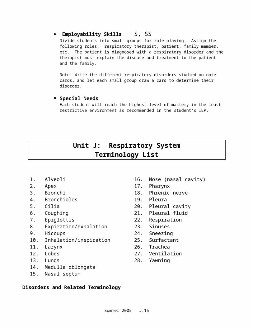
Employability Skills S, SSDivide students into small groups for role playing. Assign the following roles: respiratory therapist, patient, family member, etc. The patient is diagnosed with a respiratory disorder and the therapist must explain the disease and treatment to the patient and the family.
Note: Write the different respiratory disorders studied on note cards, and let each small group draw a card to determine their disorder.
Special NeedsEach student will reach the highest level of mastery in the least restrictive environment as recommended in the student’s IEP.
Summer 2005 J.8
Unit J: Respiratory SystemTerminology List
1. Alveoli2. Apex3. Bronchi4. Bronchioles5. Cilia6. Coughing7. Epiglottis8. Expiration/exhalation9. Hiccups10. Inhalation/inspiration11. Larynx12. Lobes13. Lungs14. Medulla oblongata15. Nasal septum
16. Nose (nasal cavity)17. Pharynx18. Phrenic nerve19. Pleura20. Pleural cavity21. Pleural fluid22. Respiration23. Sinuses24. Sneezing25. Surfactant26. Trachea27. Ventilation28. Yawning
Disorders and Related Terminology
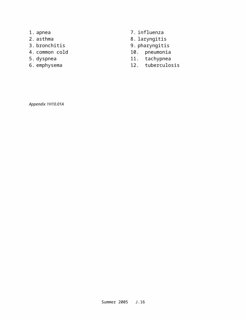
1. apnea2. asthma3. bronchitis4. common cold5. dyspnea6. emphysema
7. influenza8. laryngitis9. pharyngitis10.pneumonia11. tachypnea12. tuberculosis
Appendix 1H10.01A
Summer 2005 J.9

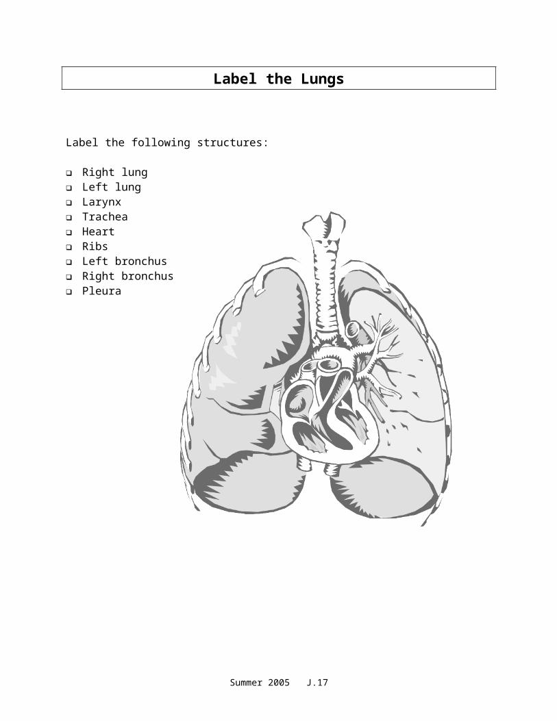
Label the Lungs
Label the following structures:
Right lung Left lung Larynx Trachea Heart Ribs Left bronchus Right bronchus Pleura
Appendix 1H10.01B
Summer 2005 J.10
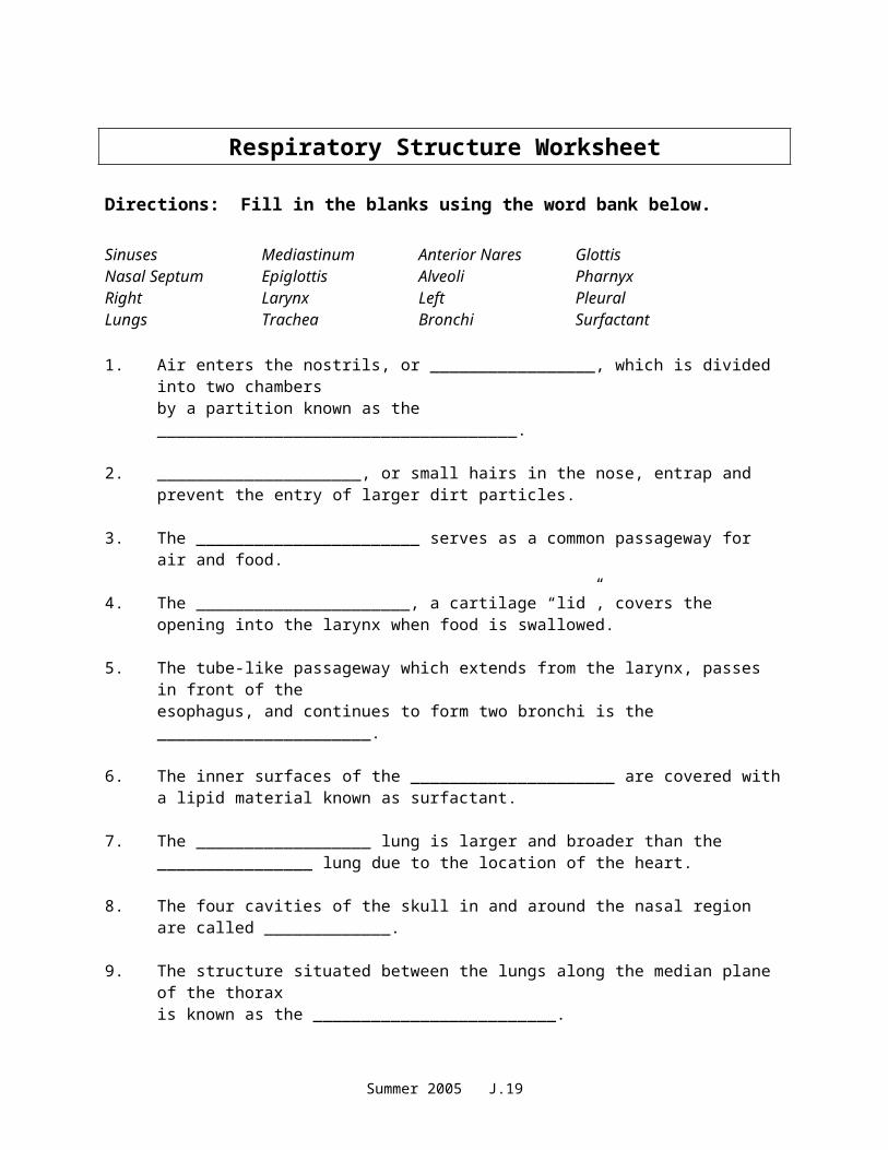
Respiratory Structure Worksheet
Directions: Fill in the blanks using the word bank below.
Sinuses Mediastinum Anterior Nares GlottisNasal Septum Epiglottis Alveoli PharnyxRight Larynx Left PleuralLungs Trachea Bronchi Surfactant
1. Air enters the nostrils, or _________________, which is divided into two chambersby a partition known as the _____________________________________.
2. _____________________, or small hairs in the nose, entrap and prevent the entry of larger dirt particles.
3. The _______________________ serves as a common passageway for air and food.
4. The ______________________, a cartilage “lid”, covers the opening into the larynx when food is swallowed.
5. The tube-like passageway which extends from the larynx, passes in front of the esophagus, and continues to form two bronchi is the ______________________.
6. The inner surfaces of the _____________________ are covered with a lipid material known as surfactant.
7. The __________________ lung is larger and broader than the ________________ lung due to the location of the heart.
8. The four cavities of the skull in and around the nasal region are called _____________.
9. The structure situated between the lungs along the median plane of the thoraxis known as the _________________________.
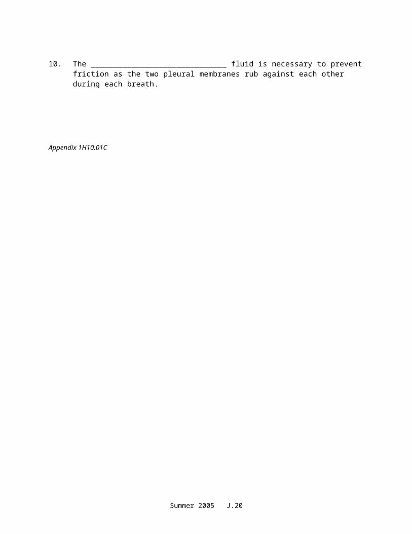
10. The ______________________________ fluid is necessary to prevent friction as the two pleural membranes rub against each other during each breath.
Appendix 1H10.01C
Summer 2005 J.11
Answer Key – Respiratory Structure Worksheet
1. anterior nares, nasal septum
2. cilia
3. pharynx
4. epiglottis
5. trachea
6. alveoli
7. right, left
8. sinuses
9. mediastinum
10. pleural
11. nasal cavity, pharynx, larynx, trachea, bronchial tree, bronchus, bronchiole, alveoli
Summer 2005 J.12
Model Lungs
When you inhale, muscles cause the chest to expand, making the lungs do the same. When this happens, air is sucked into the lungs. Make a model to demonstrate this. You will need:
Large clear, plastic bottle Three-way hose connector 2 rubber bands modeling clay plastic tube 3 small balloons scissors
Directions:
1. Push the plastic tube into one opening of the hose connector. Use modeling clay, if necessary, to make an airtight seal. Fix the balloons tightly onto the other opening with rubber bands, making sure that the joints between the connector and the balloons are airtight.
2. Carefully cut off the bottom 1 inch from the bottle, using the scissors. Make sure the cut edge of the bottle is smooth. Place the balloons and connector inside. Seal the plastic tube into the neck of the bottle with the modeling clay to make an airtight fit.
3. Tie a knot in the neck of the third balloon. Then carefully cut it in half, crossways. Gently stretch the knotted part of the balloon over the lower end of the bottle, and pull it around the sides. Make the balloon as taut as you can-like a drum skin. Now hold it by its knot.
4. The lower balloon represents the diaphragm, the main breathing muscle. Pull it down, As though you were inhaling. This lowers the air pressure in the bottle. Air from outside rushes in and makes the two balloons expand just like the real lungs in your chest.
Additional assignment:Read pages 261-262 in Body Structures and Functions. In your own words, explain the process of inspiration and expiration. Write your answer on the back of this handout.
Appendix 1H10.01D
Summer 2005 J.13
Breathing Control Worksheet
I. Directions: Read the situations below. If the situation is closely related to the NEURAL factors affecting respiration, write an “N” in the blank provided. If the situation is more closely related to the CHEMICAL factors, write a “C” in the blank.
_____1. Donnie jumps in a pool of cold water and “gasps” for air.
_____2. Maria is “out of breath” after running up four flights of stairs.
_____3. Emanuel, a health care assistant, determines the respiratory rate of his patient, Mr. Nguyen, to be 10 breaths per minute. Emanuel learns that his patient is on Morphine for post-operative pain.
_____4. Sally is allergic to pollen. When she works in her outdoor garden, she constantly sneezes.
_____5. Terrell has been taking diet pills and has experienced an increase inrespiratory rate.
II. Directions: Read the statements below and write the answer in the space provided.
1. Where is the body’s respiratory center located? What is the name of the structure?
2. Describe the major function of the phrenic nerves.
3. Explain how the level of oxygen and carbon dioxide in the blood will trigger the respiratory center.
4. Chemical regulators of respiration, or chemoreceptors are found in which major arteries in the body? Briefly explain their function.
5. Explain how sensory impulses are involved in changing the rhythm of breathing.
Appendix 010.02A
Summer 2005 J.14
Answer Key – Breathing Control Worksheet
I.
1. N2. C3. C4. N5. C
II.
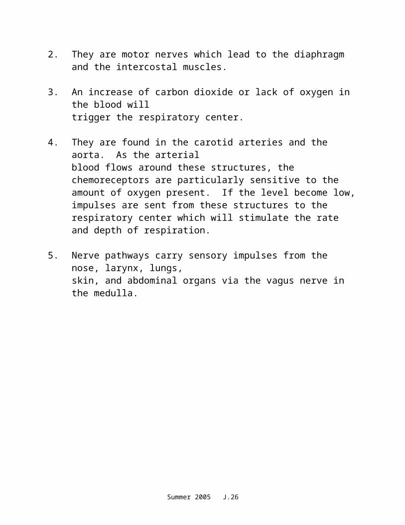
1. The respiratory center is located in the brain, the medulla oblongata.
2. They are motor nerves which lead to the diaphragm and the intercostal muscles.
3. An increase of carbon dioxide or lack of oxygen in the blood will trigger the respiratory center.
4. They are found in the carotid arteries and the aorta. As the arterialblood flows around these structures, the chemoreceptors are particularly sensitive to the amount of oxygen present. If the level become low, impulses are sent from these structures to the respiratory center which will stimulate the rate and depth of respiration.
5. Nerve pathways carry sensory impulses from the nose, larynx, lungs,skin, and abdominal organs via the vagus nerve in the medulla.
Summer 2005 J.15
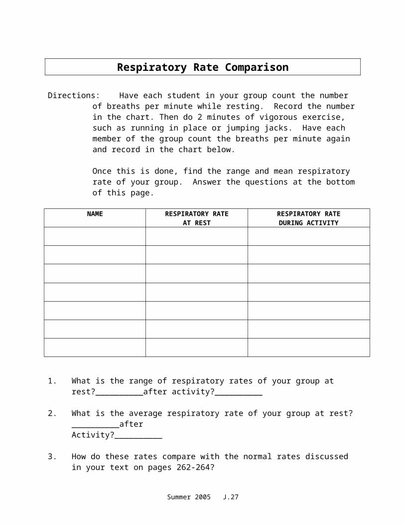
Respiratory Rate Comparison
Directions: Have each student in your group count the number of breaths per minute while resting. Record the number in the chart. Then do 2 minutes of vigorous exercise, such as running in place or jumping jacks. Have each member of the group count the breaths per minute again and record in the chart below.
Once this is done, find the range and mean respiratory rate of your group. Answer the questions at the bottom of this page.
NAME RESPIRATORY RATEAT REST
RESPIRATORY RATEDURING ACTIVITY
1. What is the range of respiratory rates of your group at rest?__________after activity?__________
2. What is the average respiratory rate of your group at rest?__________afterActivity?__________
3. How do these rates compare with the normal rates discussed in your text on pages 262-264?
4. After reviewing the pages listed above, describe other situations that can affect the respiratory rate.
Appendix 010.02A
Summer 2005 J.16
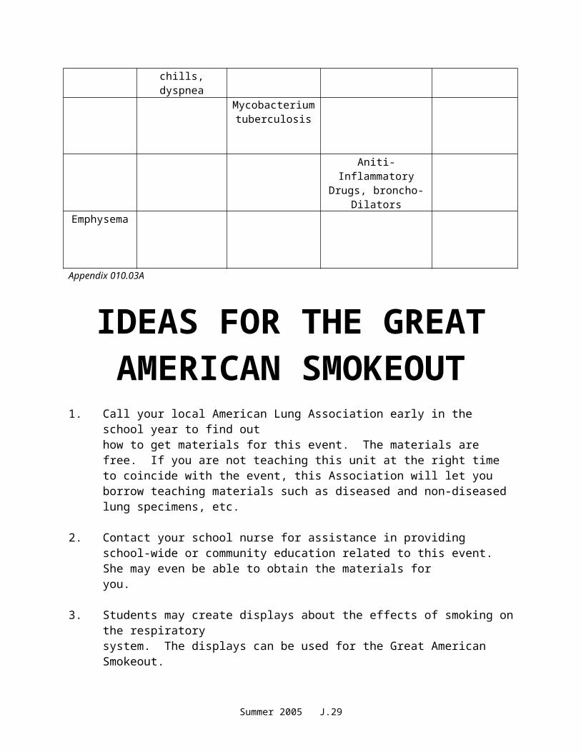
Respiratory Disease Fact Chart
Directions: Complete the chart by filling in the “missing” information.
DISEASE SIGNSSYMPTONS
ETIOLOGY TREATMENT PREVENTION
Common cold
Usually viral
Red, inflamed throat and
painful swallowing
Aimed at symptons-quit smoking
Laryngitis
Vaccine
Chest pain, fever, chills,
dyspnea
Mycobacterium tuberculosis
Aniti-Inflammatory
Drugs, broncho-Dilators
Emphysema
Appendix 010.03A
Summer 2005 J.17
IDEAS FOR THE GREAT AMERICAN SMOKEOUT
1. Call your local American Lung Association early in the school year to find out how to get materials for this event. The materials are free. If you are not teaching this unit at the right time to coincide with the event, this Association will let you borrow teaching materials such as diseased and non-diseased lung specimens, etc.
2. Contact your school nurse for assistance in providing school-wide or community education related to this event. She may even be able to obtain the materials for you.
3. Students may create displays about the effects of smoking on the respiratorysystem. The displays can be used for the Great American Smokeout.
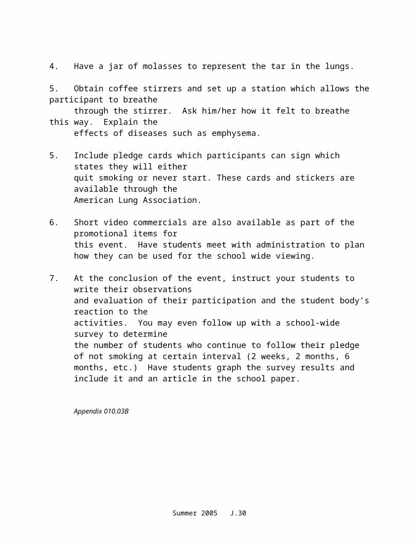
4. Have a jar of molasses to represent the tar in the lungs.
5. Obtain coffee stirrers and set up a station which allows the participant to breathethrough the stirrer. Ask him/her how it felt to breathe this way. Explain theeffects of diseases such as emphysema.
5. Include pledge cards which participants can sign which states they will eitherquit smoking or never start. These cards and stickers are available through theAmerican Lung Association.
6. Short video commercials are also available as part of the promotional items forthis event. Have students meet with administration to plan how they can be used for the school wide viewing.
7. At the conclusion of the event, instruct your students to write their observationsand evaluation of their participation and the student body’s reaction to the activities. You may even follow up with a school-wide survey to determinethe number of students who continue to follow their pledge of not smoking at certain interval (2 weeks, 2 months, 6 months, etc.) Have students graph the survey results and include it and an article in the school paper.
Appendix 010.03B
Summer 2005 J.18
Unit J: Respiratory System
OVERHEAD TRANSPARENCYMASTERS
Summer 2005 J.19
Includes the nasal cavity, pharynx, larynx, trachea, bronchi, bronchioles, alveoli, lungs, and pleura.
NASAL CAVITYNASAL SEPTUM = divides nasal cavities into R and L sides
Turbinates are bones that protrude into the nasal cavity – they increase surface area for filtering dust and dirt particles by the mucous membrane.
CILIA – the hairs in your nose, trap larger dirt particles
Summer 2005 J.20
SINUSES – cavities in the skull, ducts connect them to the nasal cavity, lined with mucous membrane to warm and moisten the air. Frontal Maxillary Ethmoid SphenoidSinuses give resonance to the voice.
PHARYNXPHARYNX
The throat Common passageway for air and food 5” long When food is swallowed, the EPIGLOTTIS closes
over the opening to the larynx, preventing food from entering the lungs.
Summer 2005 J.21

LARYNXLARYNX
Voice box Triangular chamber below pharynx Within the larynx are vocal cords (GLOTTIS) Adam’s Apple
TRACHEATRACHEA
Windpipe 4 ½ in. long walls are
alternate bands of membrane and C-shaped rings of hyaline cartilage – to keep trachea open
Lined with ciliated mucous membrane
Coughing and expectoration gets rid of dust-laden mucous
Summer 2005 J.22
BRONCHI and BRONCHIOLESBRONCHI and BRONCHIOLES
Lower end of trachea divides into R and L bronchus
As they enter lungs, subdivide into bronchial tubes and bronchioles
Bronchi – similar to trachea with ciliated mucous membrane and hyaline cartilage
Bronchial tubes – cartilaginous plates (instead of C-shaped rings)
Bronchioles – thinner walls of smooth muscle, lined with ciliated epithelium
At the end, alveolar duct and cluster of alveoli
ALVEOLIALVEOLI
Composed of a single layer of epithelial tissue
Inner surfaces covered with SURFACTANT – to keep alveoli from collapsing
Each alveolus surrounded by capillaries
O2 and CO2 exchange takes place between the alveoli and capillaries
Summer 2005 J.23
LUNGSLUNGS
Fill thoracic cavity Upper part = apex Lower part = base Base fits snugly over diaphragm Lung tissue porous and spongy – it floats R lung = larger and shorter (displaced by the liver)
and has 3 lobes L lung smaller (displaced by the heart) and has 2
lobes
PLEURAPLEURA
Thin, moist slippery membrane that covers lungs Double-walled sac Space is pleural cavity – filled with pleural fluid to
prevent friction
Summer 2005 J.24
FUNCTION OF THE RESPIRATORY SYSTEMFUNCTION OF THE RESPIRATORY SYSTEM
External respiration, internal respiration, and cellular respiration
Production of sound (vocal cords)
PULMONARY VENTILATION (Breathing)
INSPIRATION Intercostal
muscles lift ribs outward, sternum rises and the diaphragm contracts and moves downward – this increases the volume of the lungs and air rushes in.
EXPIRATION Opposite action
takes place Exhalation is a passive process
Summer 2005 J.25
Respiratory Movements 1 inspiration + 1 expiration = 1 respiration Normal adult = 14 - 20 respirations per minute Increases with exercise, body temperature, certain
diseases. Age - newborn = 40-60/min Sleep = respirations Emotion can or rate
Coughing – deep breath followed by forceful expulsion of air – to clear lower respiratory tract.
Hiccups – spasm of the diaphragm and spasmotic closure of the glottis – irritation to diaphragm or phrenic nerve
Sneezing – air forced through nose to clear respiratory tract
Yawning – deep prolonged breath that fills the lungs, increases oxygen within the blood
Summer 2005 J.26
Control of Breathing
Breathing controlled by neural and chemical factors.
Neural Factors
Respiratory center located in MEDULLA OBLONGATA
on CO2 or O2 in the blood will trigger respiratory center
PHRENIC NERVE – stimulates the diaphragm
Chemical Factors
Depends on the levels of CO2 in the blood (respiratory center in brain)
Chemoreceptors in aorta and carotid arteries sensitive to the amount of blood O2
Summer 2005 J.27

COMMON COLD COMMON COLD Contagious viral respiratory infection Indirect causes - chilling, fatigue, lack of proper
food, and not enough sleep Rx – stay in bed, drink warm liquids and fruit
juice, good nutrition Also called an Upper Respiratory Infection (URI) Handwashing – best preventative measure
LARYNGITIS Inflammation of larynx or voice box Often secondary to other respiratory infections Symptoms – sore throat, hoarseness or loss of
voice, dysphagia (difficulty swallowing)
SINUSITIS Infection of mucous membrane that lines sinus
cavities Caused by bacteria or virus Symptoms – headache or pressure, thick nasal
discharge, loss of voice resonance Rx – symptomatic, surgery for chronic sinusitisPHARYNGITIS – red, inflamed throat
Summer 2005 J.28
BRONCHITISBRONCHITIS Inflammation of the mucous membrane of the
trachea and bronchial tubes, producing excessive mucous
May be acute or chronic Acute bronchitis
characterized by cough, fever, substernal pain and RALES (raspy sound)
Chronic bronchitis – middle or old age, cigarette smoking most common cause
INFLUENZA (Flu) Viral infection causing inflammation of the
mucous membrane Fever, mucopurulent discharge, muscular pain,
extreme exhaustion Complications – pneumonia, neuritis, otitis media
and pleurisy Rx – treat the symptoms
Summer 2005 J.29
PNEUMONIA Infection of the lung Caused by bacteria or virus Alveoli fill with exudates (thick fluid) Symptoms – chest pain, fever, chills, dyspnea Rx – O2 and antibiotics
TUBERCULOSISTUBERCULOSIS Infectious bacterial lung disease Tubercles (lesions) form in the lungs Symptoms: cough, low grade fever in the
afternoon, weight loss, night sweats Diagnosis – TB skin test If skin test positive – follow up with chest x-ray
and sputum sample RX – antibiotic
ASTHMA Inflammatory airway obstruction Caused by allergen or psychological stress 5% of Americans have asthma Symptoms: difficulty exhaling, dyspnea,
wheezing, tightness in chest Rx: anti-inflammatory drugs, inhaled
bronchodilator
Summer 2005 J.30

EMPHYSEMA Alveoli become over-dilated, lose their elasticity,
can’t rebound, may eventually rupture Air becomes trapped, can’t exhale – forced
exhalation required Reduced exchange of O2 and CO2
Dyspnea increases as disease progresses
Rx – alleviate the symptoms, decrease exposure to respiratory irritants, prevent infections, restructure activities to prevent need for O2
Summer 2005 J.31