Embed Size (px)
Citation preview
THROMBOSIS
1
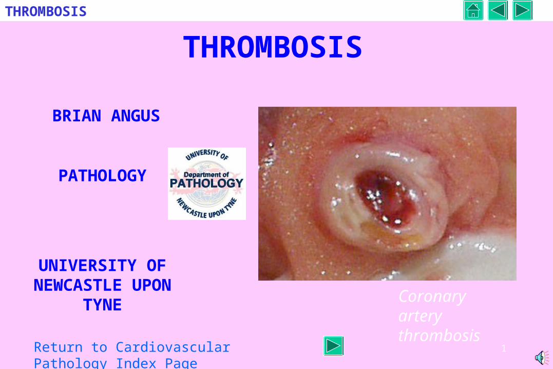
THROMBOSIS
BRIAN ANGUS
PATHOLOGY
UNIVERSITY OF NEWCASTLE UPON TYNE Coronary artery
thrombosis
Return to Cardiovascular Pathology Index Page
2
THROMBOSIS
CONTENTS
DEFINITIONS
COMPOSITION
PREDISPOSING FACTORS
TYPES
OUTCOME
ARTERIAL THROMBOSIS
CARDIAC (Valves and chambers)
VENOUS THROMBOSIS
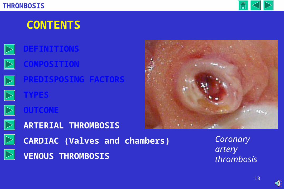
Coronary artery thrombosis
3
THROMBOSIS
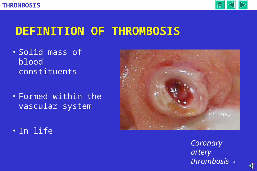
DEFINITION OF THROMBOSIS
• Solid mass of blood constituents
• Formed within the vascular system
• In lifeCoronary artery thrombosis
4
THROMBOSIS
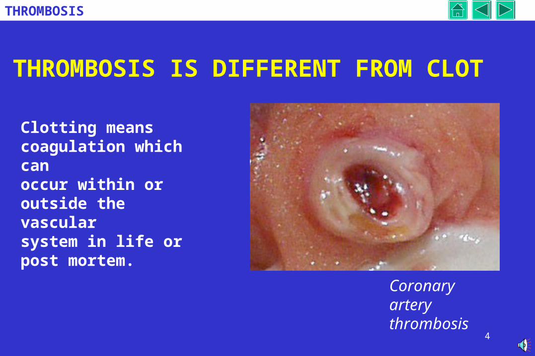
THROMBOSIS IS DIFFERENT FROM CLOT
Clotting means coagulation which canoccur within or outside the vascular system in life or post mortem.
Coronary artery thrombosis
5
THROMBOSIS
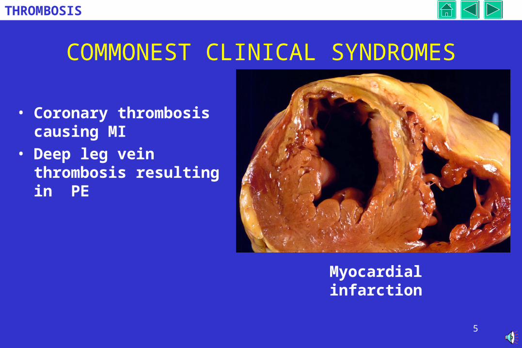
COMMONEST CLINICAL SYNDROMES
• Coronary thrombosis causing MI
• Deep leg vein thrombosis resulting in PE
Myocardial infarction
6
THROMBOSIS
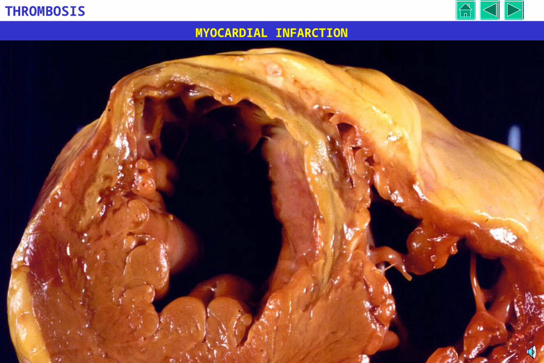
MYOCARDIAL INFARCTION
7
THROMBOSIS
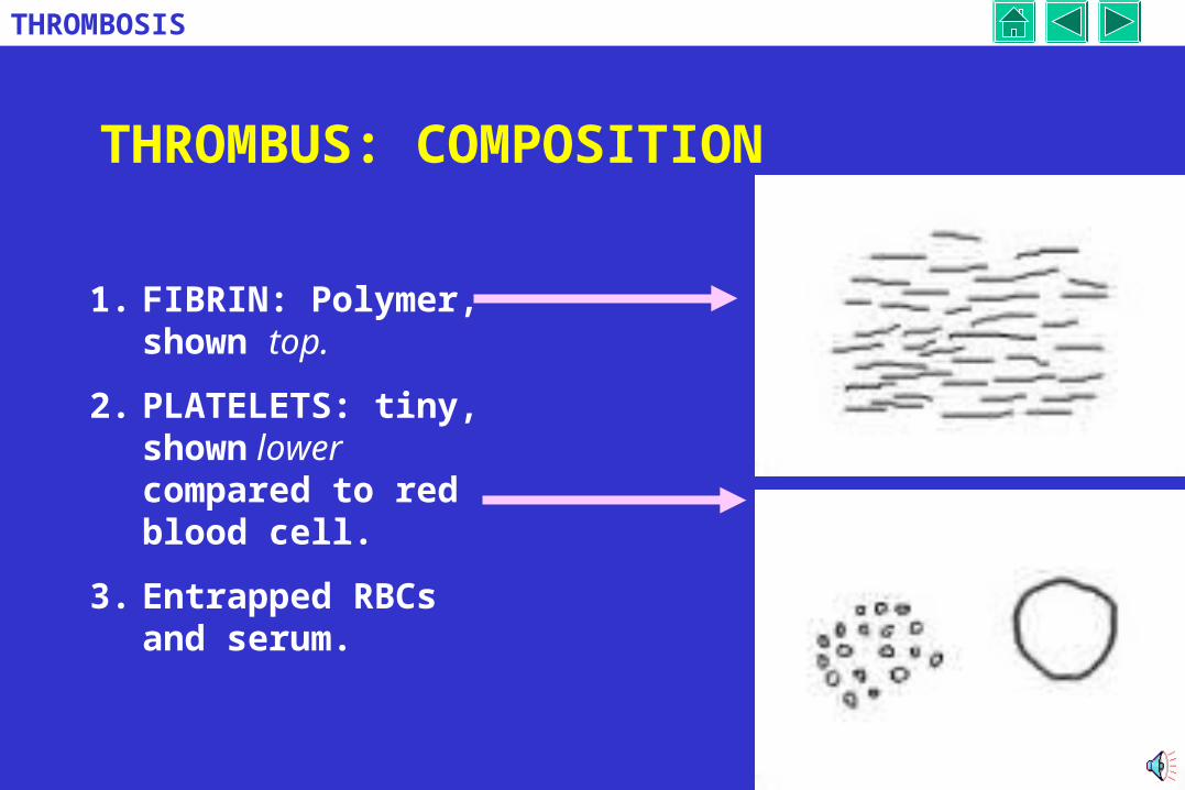
THROMBUS: COMPOSITION
1. FIBRIN: Polymer, shown top.
2. PLATELETS: tiny, shown lower compared to red blood cell.
3. Entrapped RBCs and serum.
8
THROMBOSIS
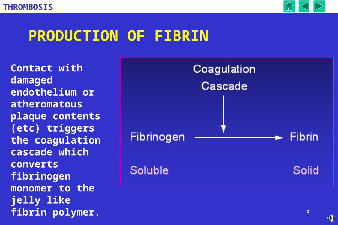
PRODUCTION OF FIBRIN
Contact with damaged endothelium or atheromatous plaque contents (etc) triggers the coagulation cascade which converts fibrinogen monomer to the jelly like fibrin polymer.
9
THROMBOSIS
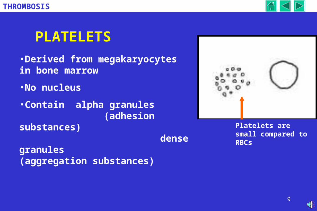
PLATELETS
Platelets are small compared to RBCs
•Derived from megakaryocytes in bone marrow
•No nucleus
•Contain alpha granules (adhesion substances)
dense granules (aggregation substances)
10
THROMBOSIS
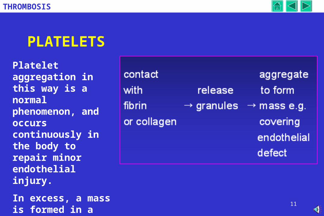
PLATELETS
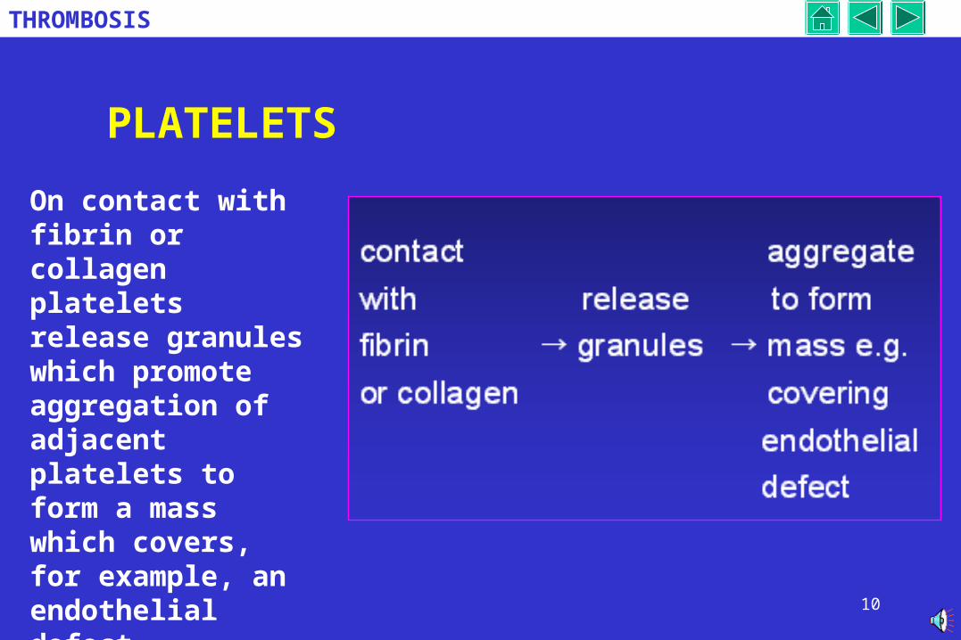
On contact with fibrin or collagen platelets release granules which promote aggregation of adjacent platelets to form a mass which covers, for example, an endothelial defect.
11
THROMBOSIS
PLATELETS
Platelet aggregation in this way is a normal phenomenon, and occurs continuously in the body to repair minor endothelial injury.
In excess, a mass is formed in a vessel: THROMBOSIS
12
THROMBOSIS
PREDISPOSING FACTORS FOR THROMBOSIS
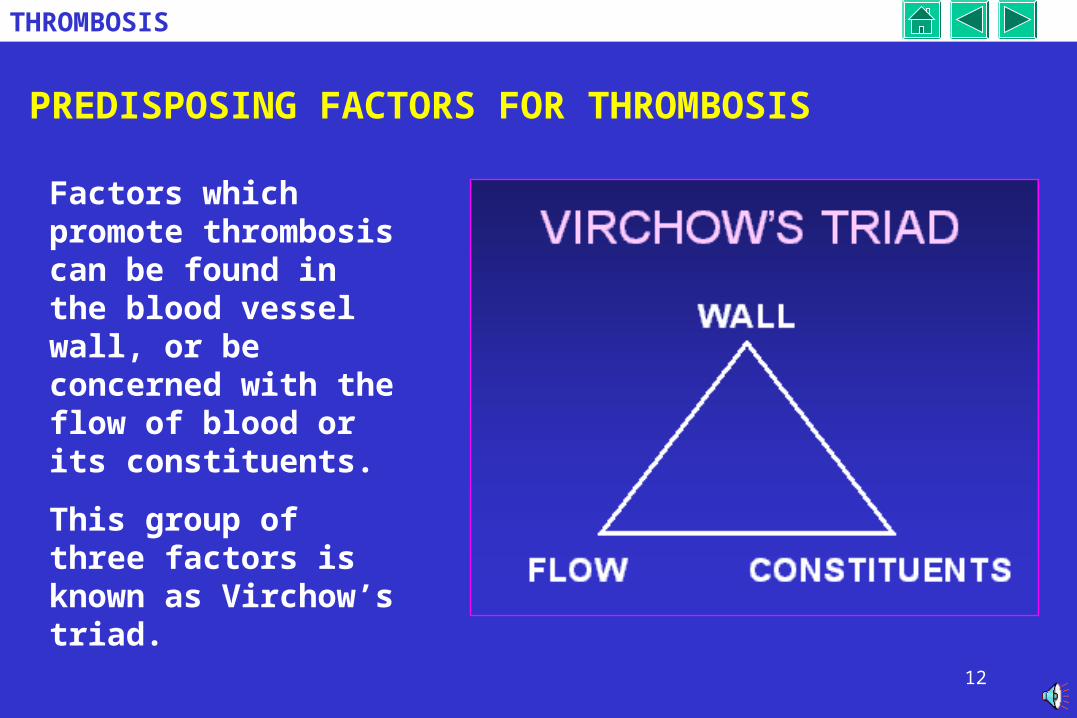
Factors which promote thrombosis can be found in the blood vessel wall, or be concerned with the flow of blood or its constituents.
This group of three factors is known as Virchow’s triad.
13
THROMBOSIS
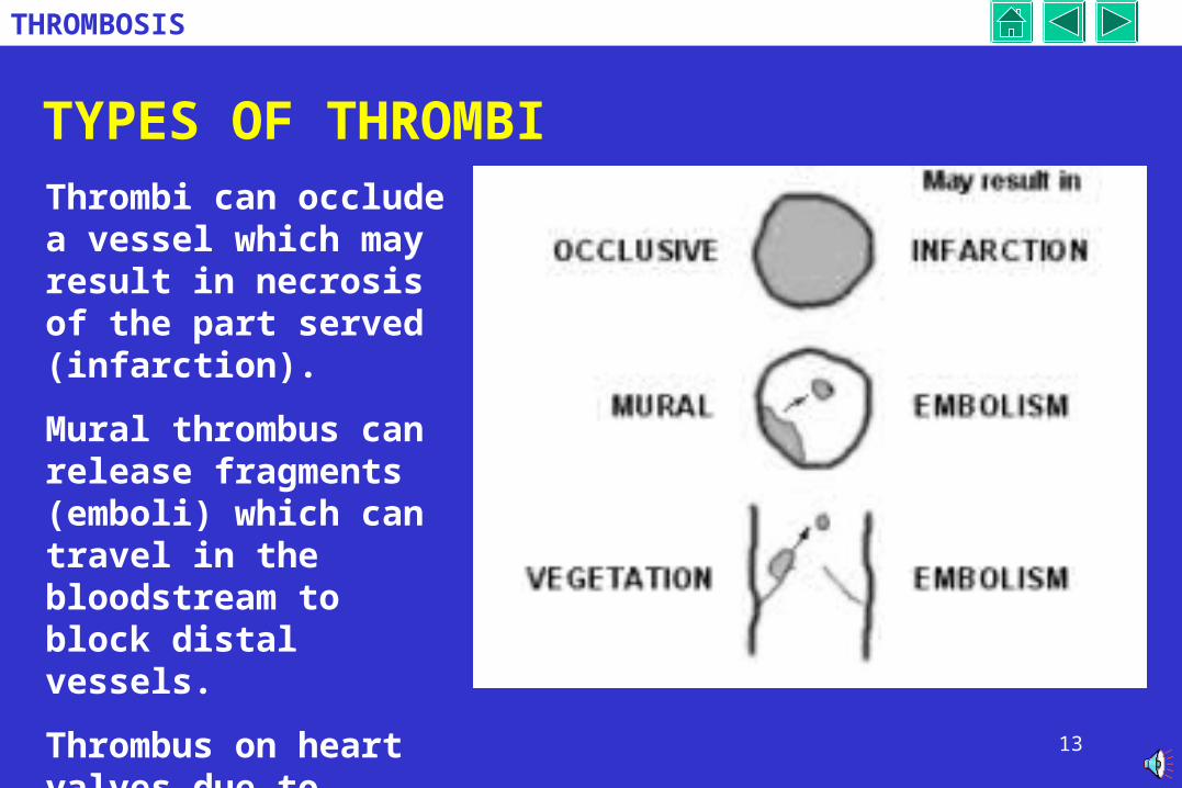
TYPES OF THROMBIThrombi can occlude a vessel which may result in necrosis of the part served (infarction).
Mural thrombus can release fragments (emboli) which can travel in the bloodstream to block distal vessels.
Thrombus on heart valves due to infection can also embolise.
14
THROMBOSIS
OUTCOMES OF THROMBOSIS 1
THROMBOLYSIS
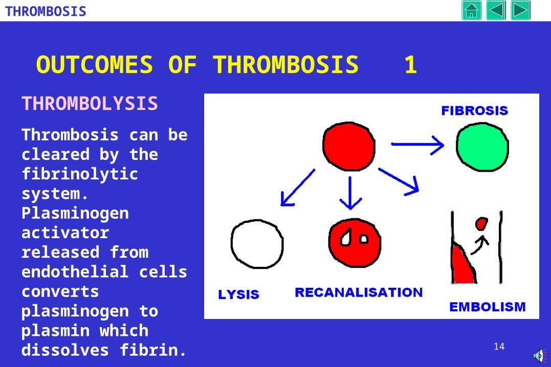
Thrombosis can be cleared by the fibrinolytic system. Plasminogen activator released from endothelial cells converts plasminogen to plasmin which dissolves fibrin.
15
THROMBOSIS
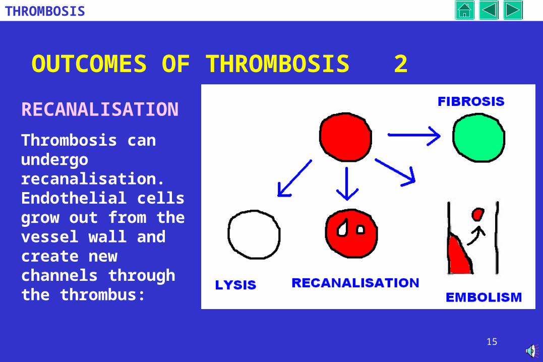
OUTCOMES OF THROMBOSIS 2
RECANALISATION
Thrombosis can undergo recanalisation. Endothelial cells grow out from the vessel wall and create new channels through the thrombus:
16
THROMBOSIS
OUTCOMES OF THROMBOSIS 3
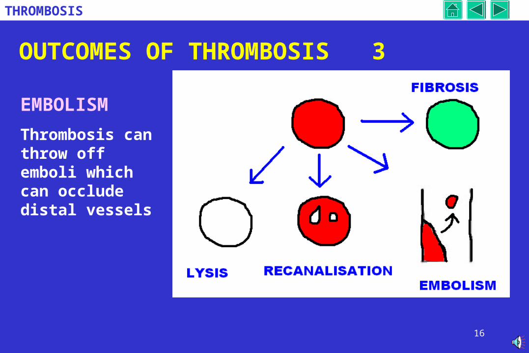
EMBOLISM
Thrombosis can throw off emboli which can occlude distal vessels
17
THROMBOSIS
OUTCOMES OF THROMBOSIS 4
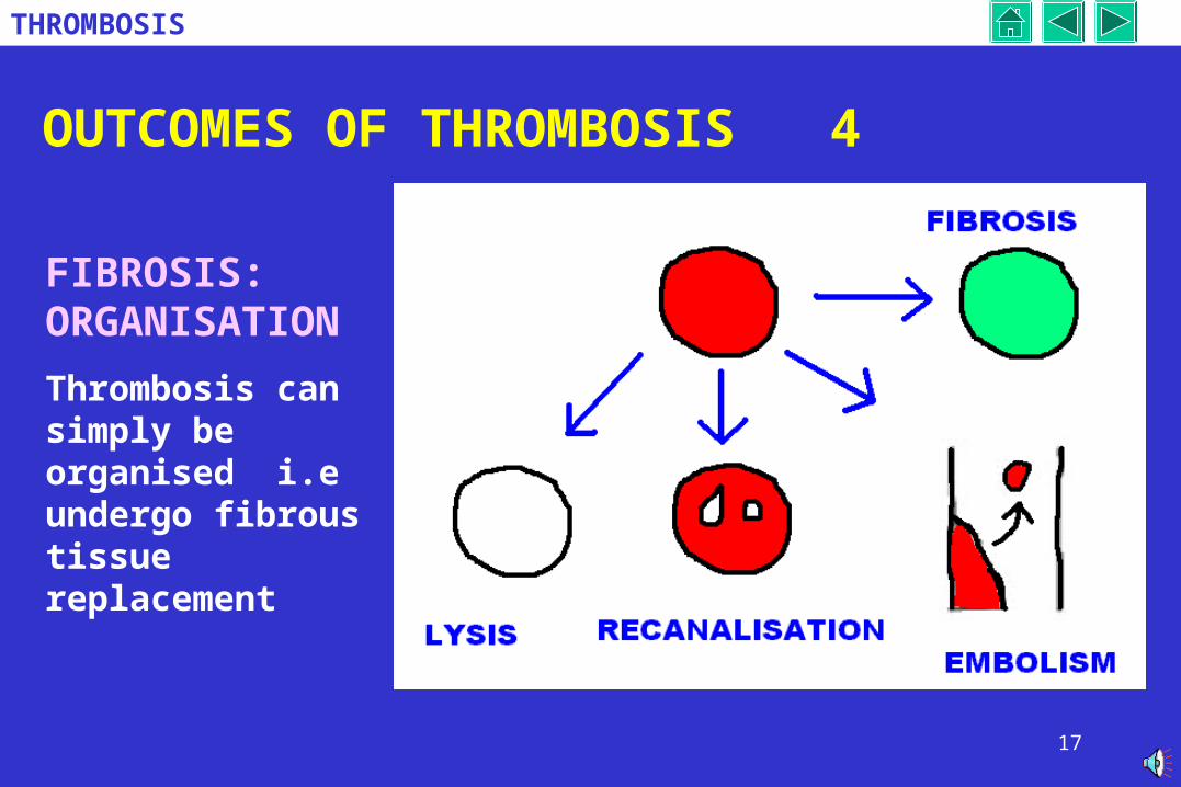
FIBROSIS: ORGANISATION
Thrombosis can simply be organised i.e undergo fibrous tissue replacement
18
THROMBOSIS
CONTENTS
DEFINITIONS
COMPOSITION
PREDISPOSING FACTORS
TYPES
OUTCOME
ARTERIAL THROMBOSIS
CARDIAC (Valves and chambers)
VENOUS THROMBOSIS
Coronary artery thrombosis
19
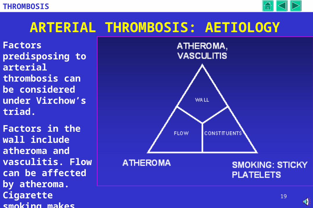
THROMBOSIS
ARTERIAL THROMBOSIS: AETIOLOGYFactors predisposing to arterial thrombosis can be considered under Virchow’s triad.
Factors in the wall include atheroma and vasculitis. Flow can be affected by atheroma. Cigarette smoking makes platelets sticky.
20
THROMBOSIS
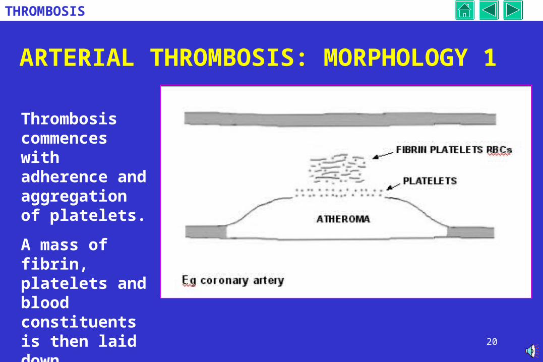
ARTERIAL THROMBOSIS: MORPHOLOGY 1
Thrombosis commences with adherence and aggregation of platelets.
A mass of fibrin, platelets and blood constituents is then laid down.
21
THROMBOSIS
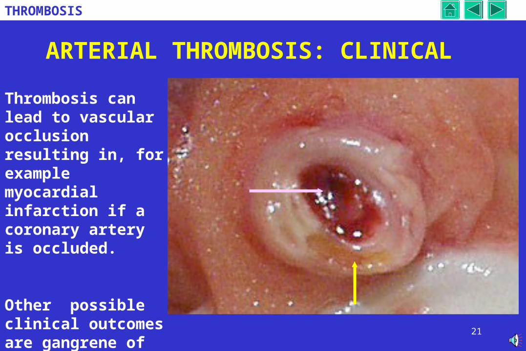
ARTERIAL THROMBOSIS: CLINICAL
Thrombosis can lead to vascular occlusion resulting in, for example myocardial infarction if a coronary artery is occluded.
Other possible clinical outcomes are gangrene of the leg and cerebral infarct.
22
THROMBOSIS
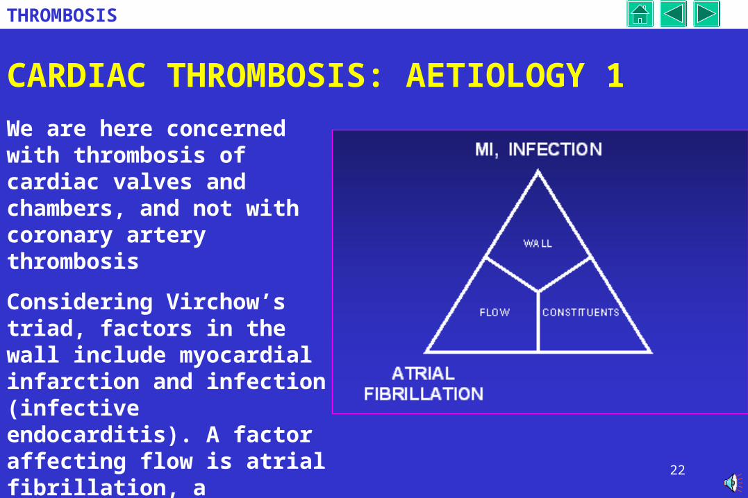
CARDIAC THROMBOSIS: AETIOLOGY 1
We are here concerned with thrombosis of cardiac valves and chambers, and not with coronary artery thrombosis
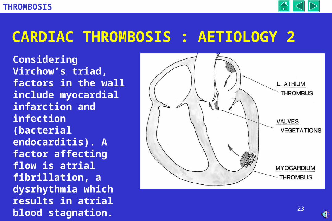
Considering Virchow’s triad, factors in the wall include myocardial infarction and infection (infective endocarditis). A factor affecting flow is atrial fibrillation, a dysrhythmia which results in atrial blood stagnation.
23
THROMBOSIS
CARDIAC THROMBOSIS : AETIOLOGY 2
Considering Virchow’s triad, factors in the wall include myocardial infarction and infection (bacterial endocarditis). A factor affecting flow is atrial fibrillation, a dysrhythmia which results in atrial blood stagnation.
Identify these factors on the diagram.
24
THROMBOSIS
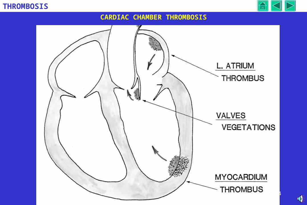
CARDIAC CHAMBER THROMBOSIS
25
THROMBOSIS
CARDIAC THROMBOSIS: CLINICAL
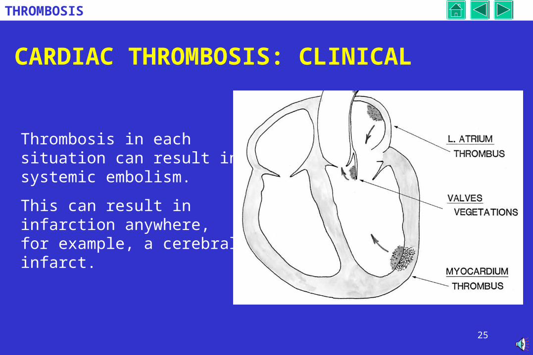
Thrombosis in each situation can result in systemic embolism.
This can result in infarction anywhere, for example, a cerebral infarct.
26
THROMBOSIS
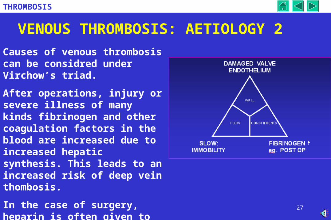
VENOUS THROMBOSIS: AETIOLOGY 1
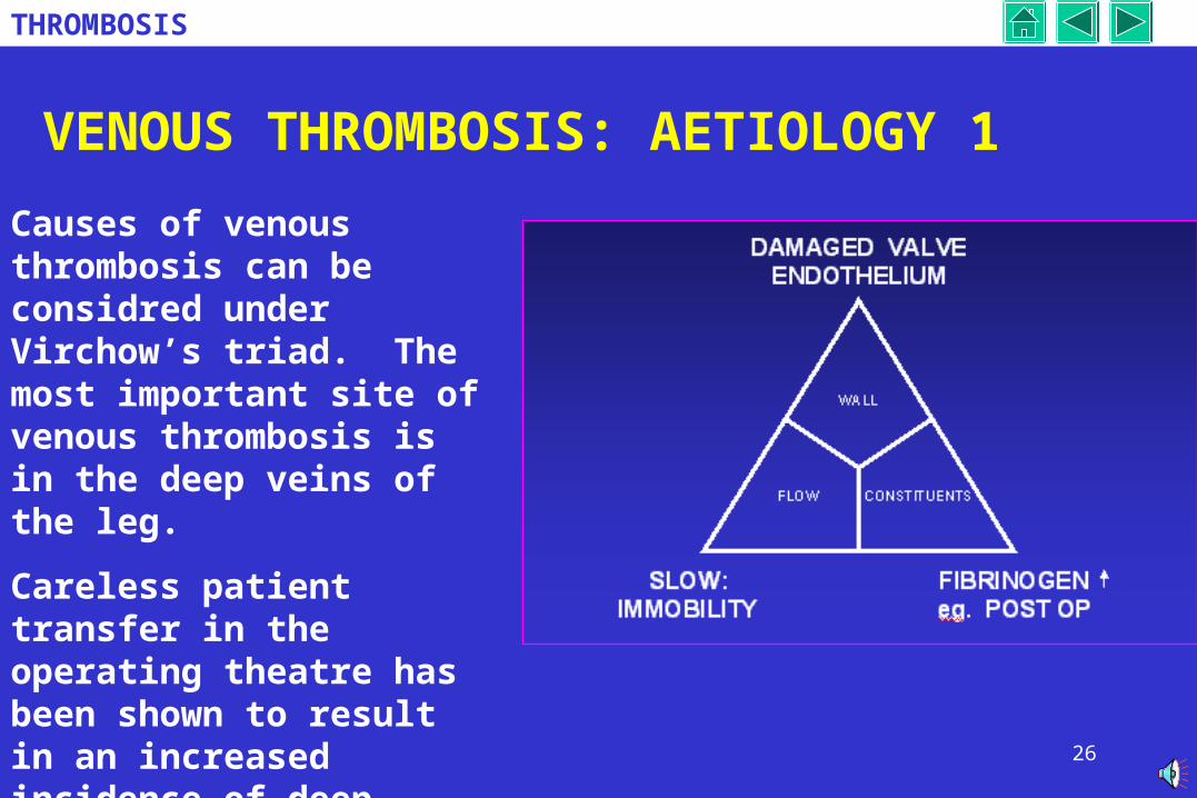
Causes of venous thrombosis can be considred under Virchow’s triad. The most important site of venous thrombosis is in the deep veins of the leg.
Careless patient transfer in the operating theatre has been shown to result in an increased incidence of deep venous thrombosis triggerd by damage to endothelium in calf veins.
27
THROMBOSIS
VENOUS THROMBOSIS: AETIOLOGY 2
Causes of venous thrombosis can be considred under Virchow’s triad.
After operations, injury or severe illness of many kinds fibrinogen and other coagulation factors in the blood are increased due to increased hepatic synthesis. This leads to an increased risk of deep vein thombosis.
In the case of surgery, heparin is often given to reduce the incidence of this potentially fatal complication
28
THROMBOSIS
VENOUS THROMBOSIS: AETIOLOGY 2
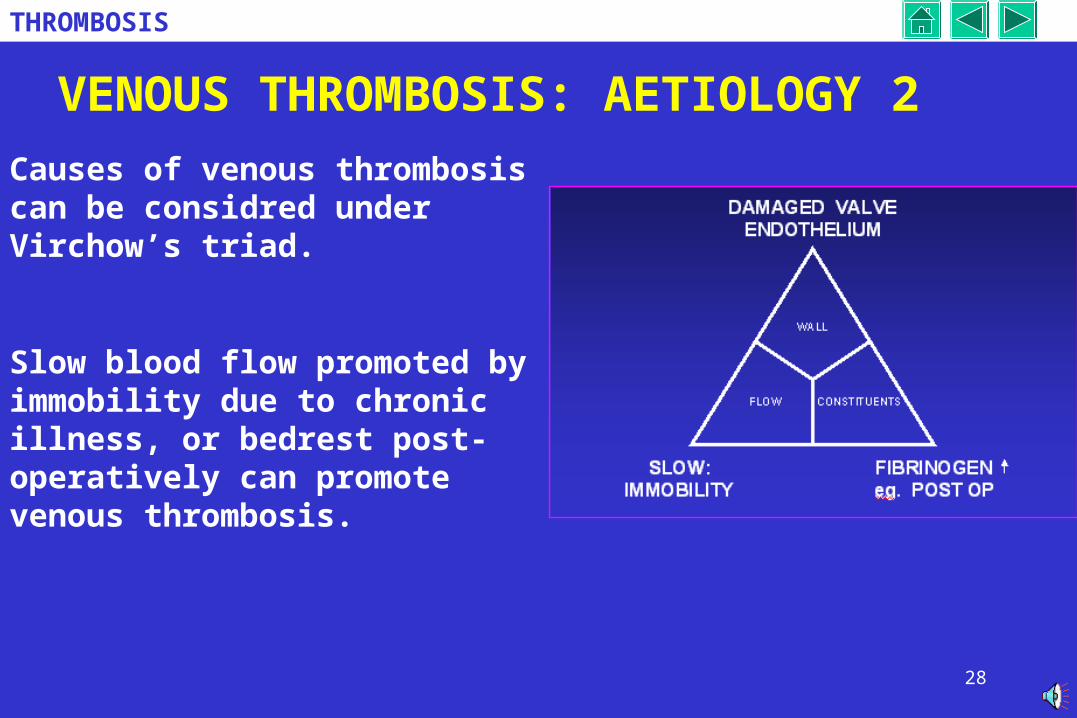
Causes of venous thrombosis can be considred under Virchow’s triad.
Slow blood flow promoted by immobility due to chronic illness, or bedrest post-operatively can promote venous thrombosis.
29
THROMBOSIS
VENOUS THROMBOSIS: AETIOLOGY 3
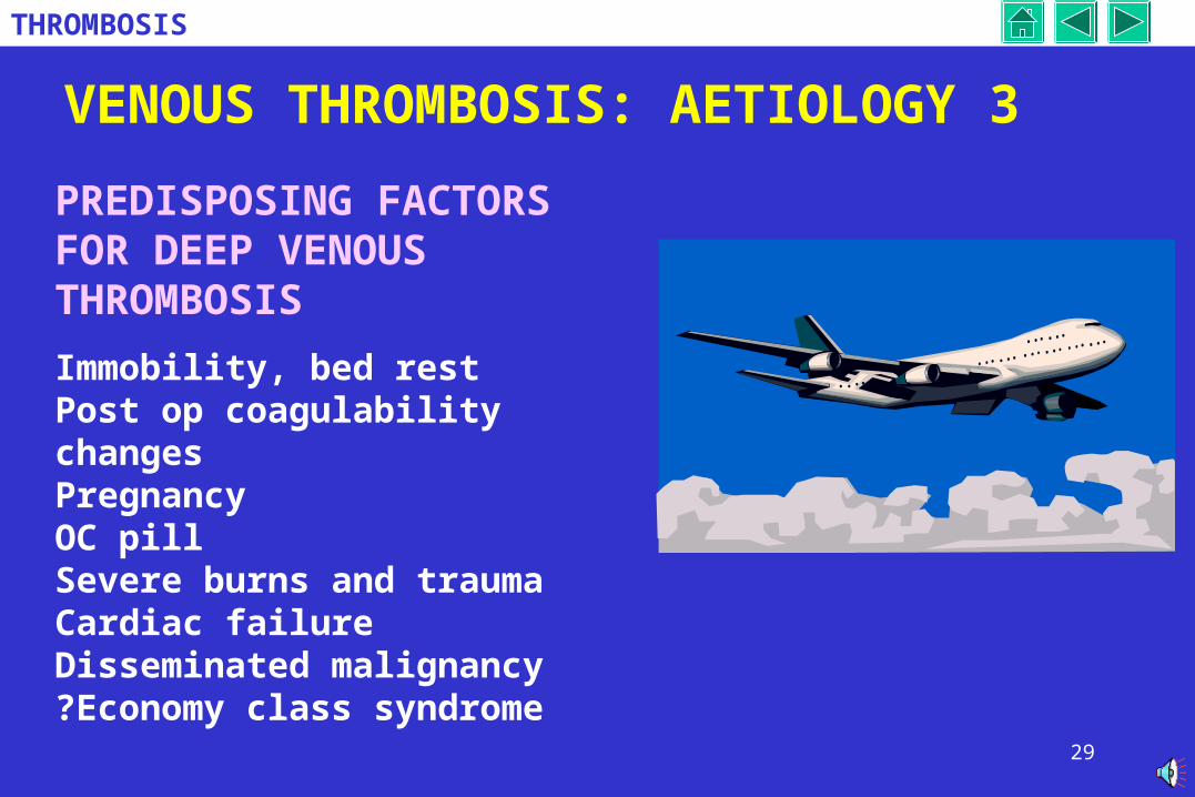
PREDISPOSING FACTORS FOR DEEP VENOUS THROMBOSIS
Immobility, bed restPost op coagulability changesPregnancyOC pillSevere burns and traumaCardiac failureDisseminated malignancy?Economy class syndrome
30
THROMBOSIS
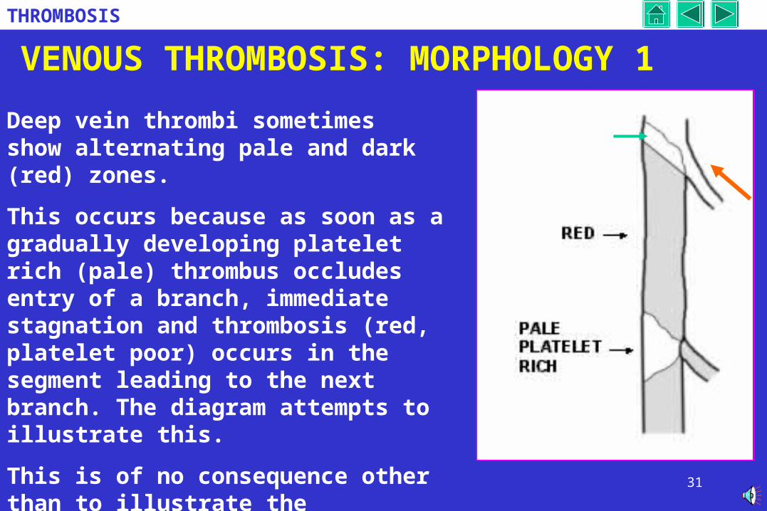
VENOUS THROMBOSIS: MORPHOLOGY 1
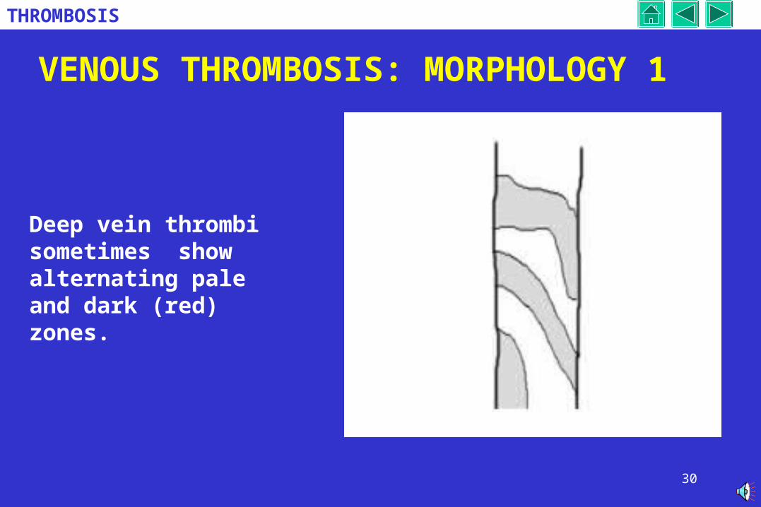
Deep vein thrombi sometimes show alternating pale and dark (red) zones.
31
THROMBOSIS
VENOUS THROMBOSIS: MORPHOLOGY 1
Deep vein thrombi sometimes show alternating pale and dark (red) zones.
This occurs because as soon as a gradually developing platelet rich (pale) thrombus occludes entry of a branch, immediate stagnation and thrombosis (red, platelet poor) occurs in the segment leading to the next branch. The diagram attempts to illustrate this.
This is of no consequence other than to illustrate the contribution of platelets (the initiator) and fibrin to thrombosis.
32
THROMBOSIS
DEEP VEIN THROMBOSIS: CLINICAL
The patient may be asymptomatic.
Often there is pain and swelling of the leg.
In the event of pulmonary embolism. A potentially fatal outcome, there nay be chest pain and perhaps haemoptysis due to pulmonary infarction.
.
MS Clipart
33
THROMBOSIS
DEEP VEIN THROMBOSIS: CLINICAL 2
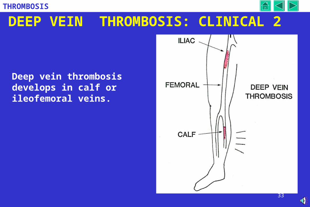
Deep vein thrombosis develops in calf or ileofemoral veins.
34
THROMBOSIS
DEEP VEIN THROMBOSIS: CLINICAL 3
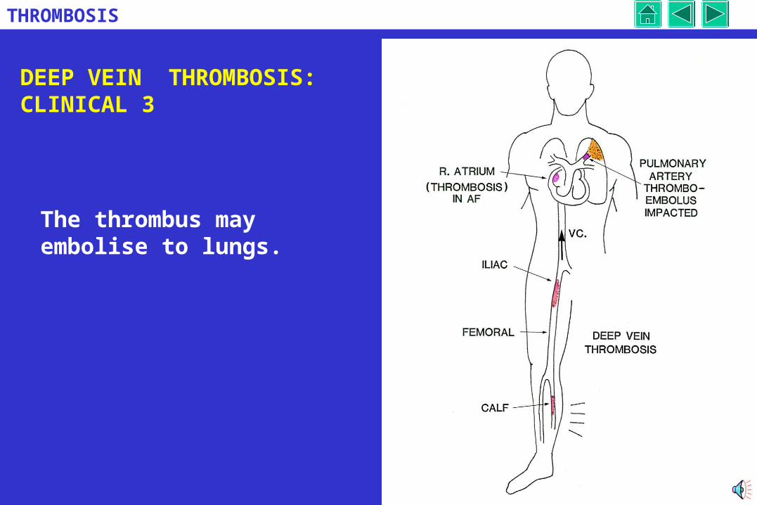
The thrombus may embolise to lungs.
35
THROMBOSIS
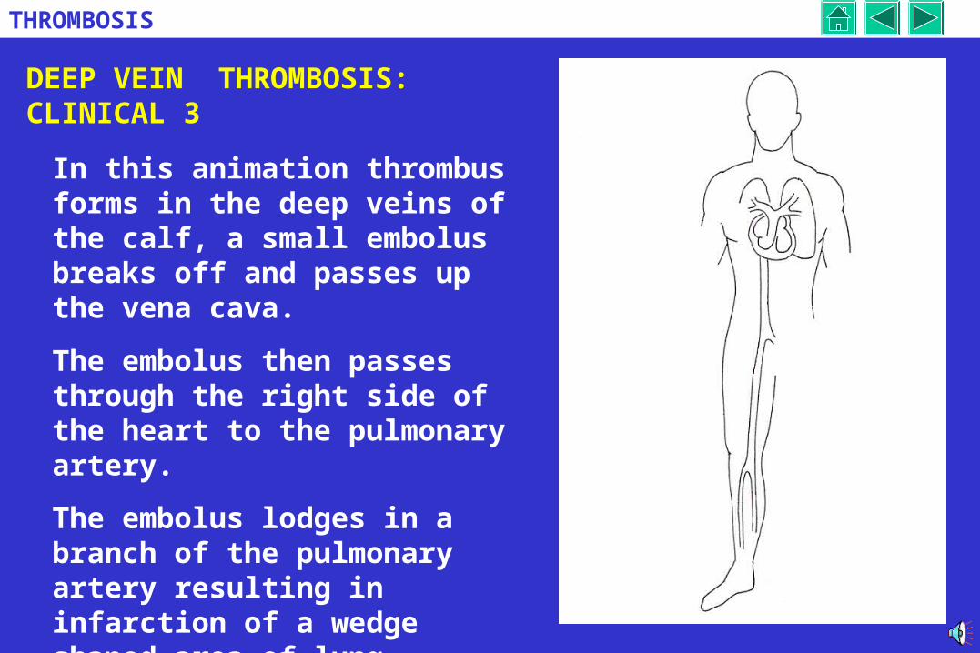
DEEP VEIN THROMBOSIS: CLINICAL 3
In this animation thrombus forms in the deep veins of the calf, a small embolus breaks off and passes up the vena cava.
The embolus then passes through the right side of the heart to the pulmonary artery.
The embolus lodges in a branch of the pulmonary artery resulting in infarction of a wedge shaped area of lung.
36
THROMBOSIS
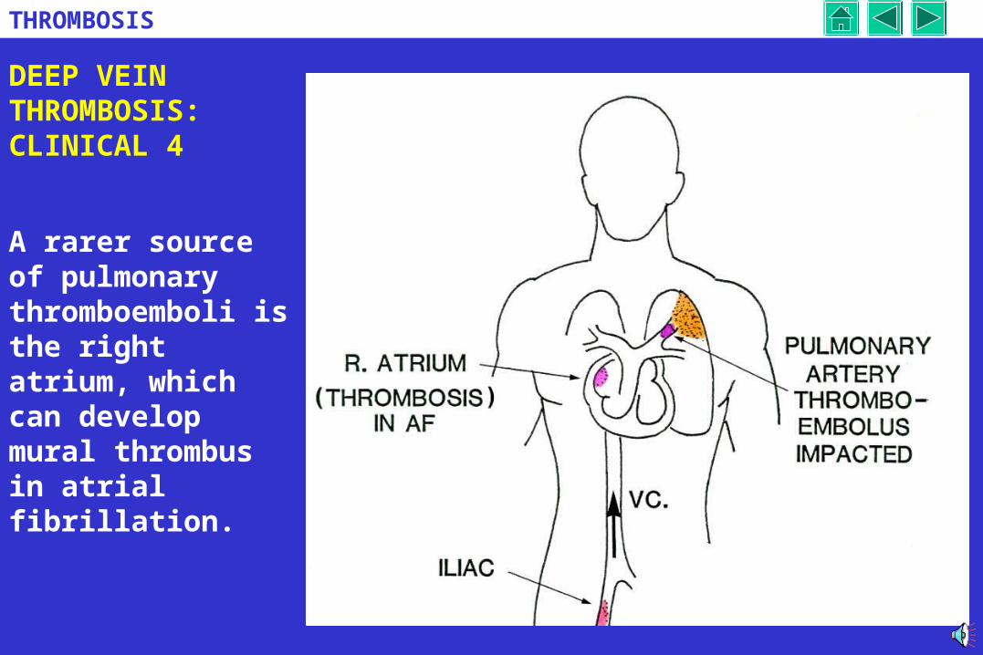
DEEP VEIN THROMBOSIS:CLINICAL 4
A rarer source of pulmonary thromboemboli is the right atrium, which can develop mural thrombus in atrial fibrillation.
37
THROMBOSIS
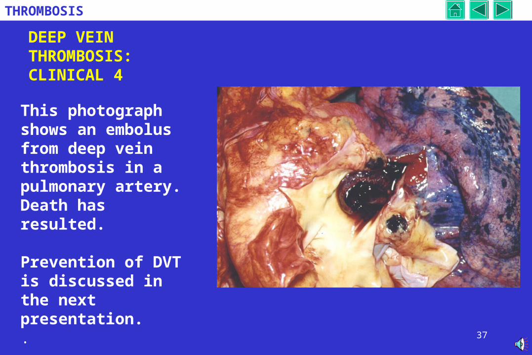
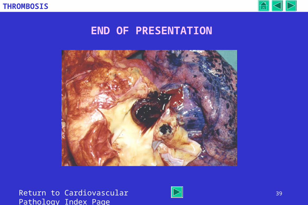
DEEP VEIN THROMBOSIS:CLINICAL 4
This photograph shows an embolus from deep vein thrombosis in a pulmonary artery. Death has resulted.
Prevention of DVT is discussed in the next presentation. .
38
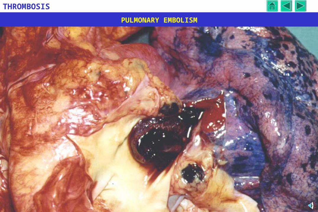
THROMBOSIS
PULMONARY EMBOLISM
39
THROMBOSIS
END OF PRESENTATION
Return to Cardiovascular Pathology Index Page