Embed Size (px)
Citation preview
Cell Pathology
1. Haemodynamic disorders• Describe the causes and consequences of
oedema at different sites• Define thrombosis and give the causes
and potential consequences of such an event.
• Define embolism and know about the importance of pulmonary embolism in clinical practice.
• Describe possible causes of haemorrhage and potential outcomes
• Define shock and identify the possible causes and mechanisms
• Define infarction and describe possible causes.
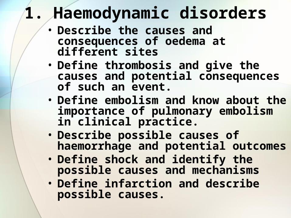
Oedema
Causes:
•Raised hydrostatic pressure•Reduced plasma osmotic pressure•Lymphatic obstruction•Sodium retention•Inflammation
Consequences:
•Cellulitits•Venous Eczema•Venous Ulcer•Pulmonary•Cerebral
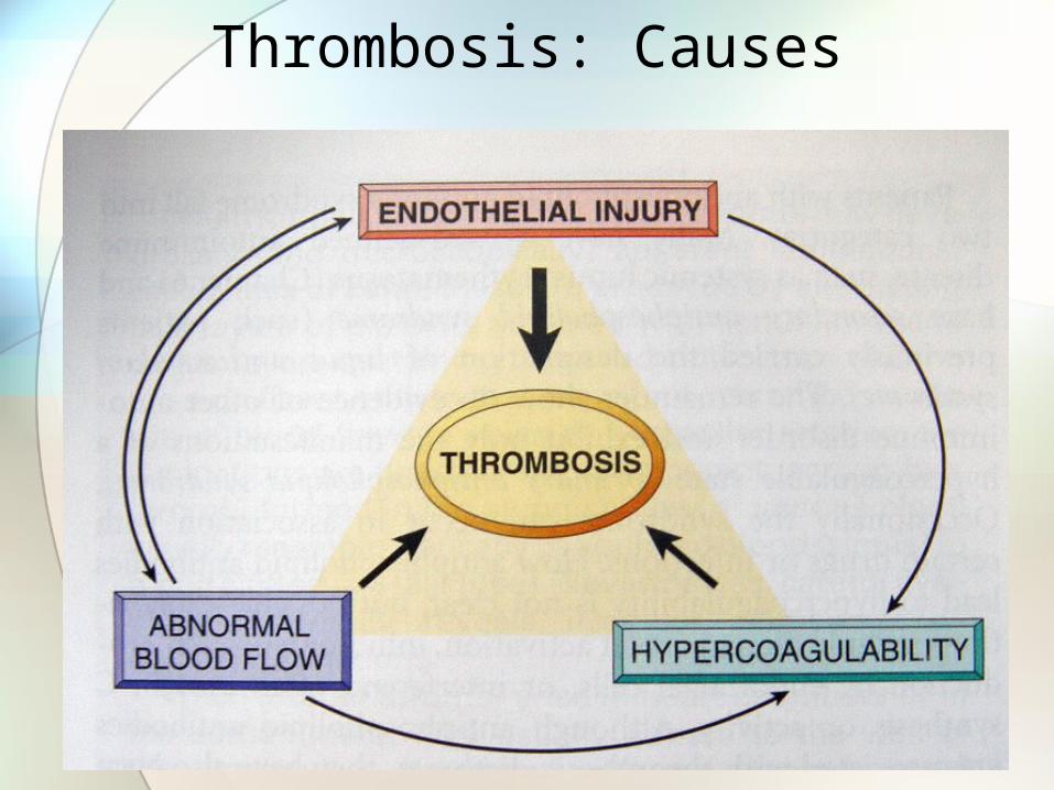
Thrombosis: Causes
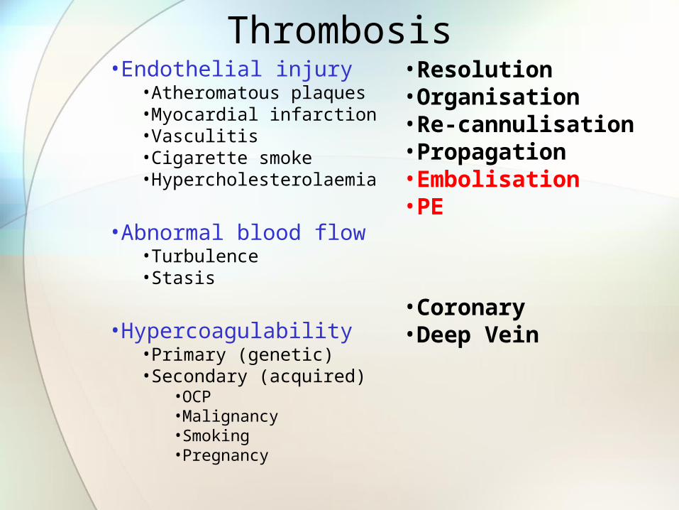
Thrombosis•Endothelial injury
•Atheromatous plaques•Myocardial infarction•Vasculitis•Cigarette smoke•Hypercholesterolaemia
•Abnormal blood flow•Turbulence•Stasis
•Hypercoagulability•Primary (genetic)•Secondary (acquired)
•OCP•Malignancy•Smoking•Pregnancy
•Resolution•Organisation•Re-cannulisation•Propagation•Embolisation•PE
•Coronary•Deep Vein
Haemorrhage
Causes•Trauma•Vasculitits•Vascular fragility
Consequences•None•Chronic anaemia•Hypovolaemic shock•Pressure

Shock• Cardiovascular collapse -> hypotension, impaired
tissue perfusion, cellular hypoxia
• Causes:• Hypovolaemic:
• Severe haemorrhage• Massive trauma• Burns• Sepsis• Anaphylactic: hypersensitivity
• Cardiogenic: pump failure• Myocardial infarction• Massive pulmonary embolism• Myocardial damage• Tamponade
• Neurogenic: spinal trauma
Shock - Consequences• Brain: ischaemic encephalopathy
• Heart: subendocardial ischaemia/necrosis
• Kidneys: acute tubular necrosis
• GI tract: haemorrhagic enteropathy
• Lungs: ARDS
2. Cell injury• List the causes of cell injury
• List the mechanisms of cell injury
• Define (and give examples of) hyperplasia, hypertrophy, atrophy, metasplasia and dysplasia
• Describe the morphological changes associated with reversible and irreversible injury
• Describe the differences between apoptosis and necrosis
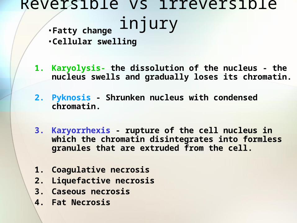
Reversible vs irreversible injury•Fatty change •Cellular swelling
1. Karyolysis- the dissolution of the nucleus - the nucleus swells and gradually loses its chromatin.
2. Pyknosis - Shrunken nucleus with condensed chromatin.
3. Karyorrhexis - rupture of the cell nucleus in which the chromatin disintegrates into formless granules that are extruded from the cell.
1. Coagulative necrosis2. Liquefactive necrosis3. Caseous necrosis4. Fat Necrosis
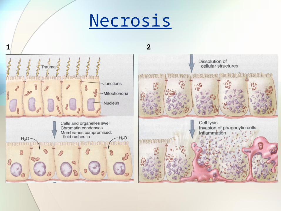
Necrosis1 2

1
2 3
Apoptosis
The differences between apoptosis and necrosis
1. Apoptosis may be physiological
2. Apoptosis is an active energy dependent process
3. Not associated with inflammation
Bruise • An extraversated collection of blood which has leaked from
damaged small arteries, venules and veins but not capillaries
• Fragility of vessels, coagulation state etc all effect bruising• May take hours or days to form
• May get patterned bruises (can see better with special light sources)
• Deep bruising may never be seen on the surface
• Blunt trauma
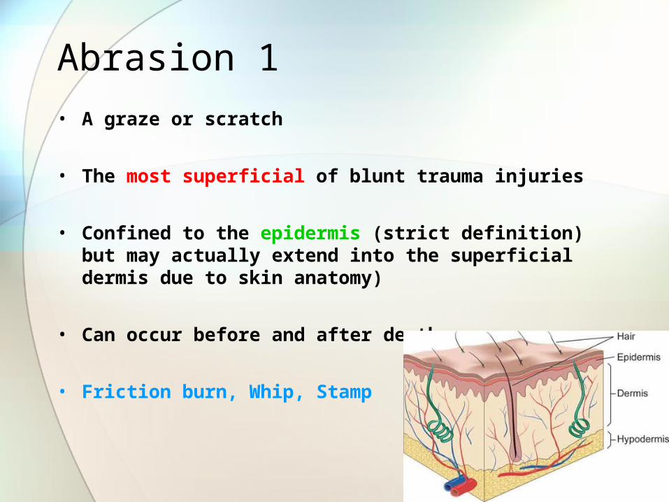
Abrasion 1• A graze or scratch
• The most superficial of blunt trauma injuries
• Confined to the epidermis (strict definition) but may actually extend into the superficial dermis due to skin anatomy)
• Can occur before and after death
• Friction burn, Whip, Stamp
Laceration 1• A split to the skin
• The result of blunt force overstretching the skin
• Usually pass through the full thickness of the skin
• They are deep and will bleed
• Margins ragged with crushing and bruising
• “Bridging fibres” arch across the skin defect
• Common where skin can be compressed between the force and underlying bone
• Eg Scalp, elbow, shin
• Rare over soft fleshy areas• Eg Buttocks, breasts
• Fall, Punch, Kick
Cut
(or slash)
The length of the injury is longer than its depth
Stab
(or penetrating injury)
The depth of the wound is greater than the width
Knife/metal
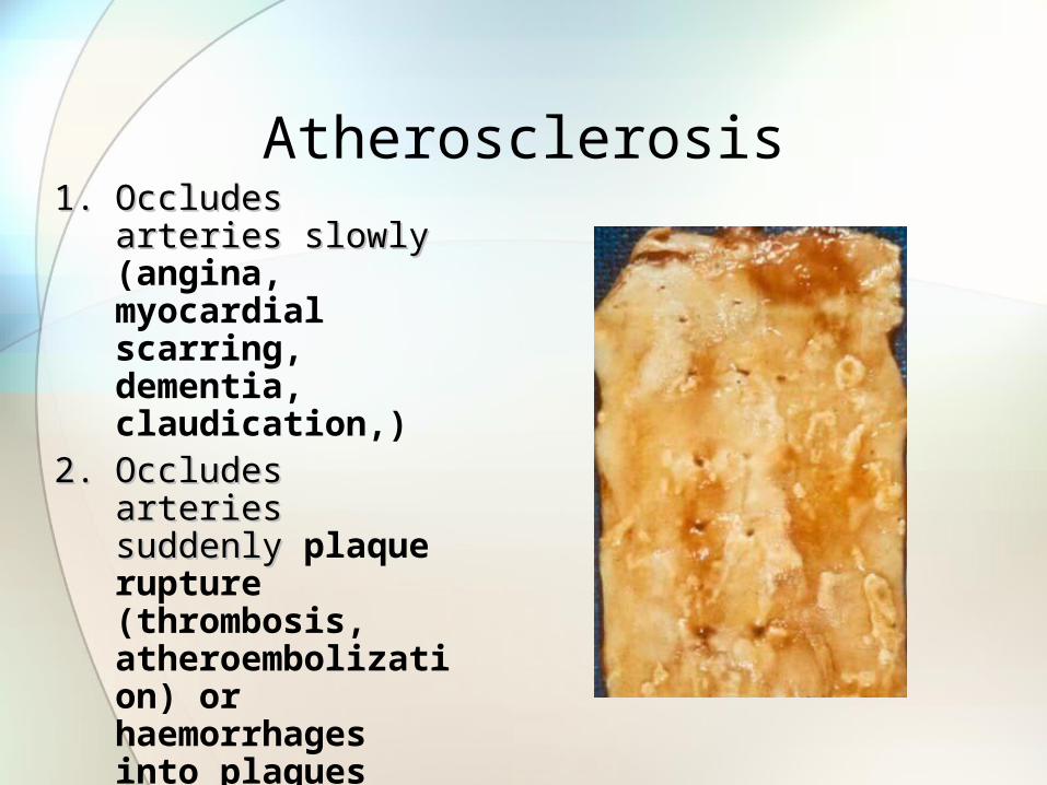
Atherosclerosis1.1. Occludes arteriesOccludes arteries
slowlyslowly (angina, myocardial scarring, dementia, claudication,)
2.2. Occludes arteriesOccludes arteries suddenlysuddenly plaque rupture (thrombosis, atheroembolization) or haemorrhages into plaques (MI, stroke, gangrene of the bowel)
3.3. WeakensWeakens artery wallsartery walls (aneurysms)