Embed Size (px)
Citation preview
UPDATE ON THE MANAGEMENT OF
NEONATAL THROMBOSIS Matthew A Saxonhouse, MD, FAAP
Attending Neonatologist Levine Children’s Hospital/
Jeff Gordon Children’s Hospital Carolinas Healthcare System
Charlotte, NC
Disclosure • The following relationships with commercial
interests related to this presentation existed during the past 12 months: – Nothing to disclose
• The following FDA disclosures related to this presentation exist: – Unfractionated heparin (UFH) - off-label use – Low molecular weight heparin (LWMH) - off-label use – Recombinant Tissue Plasminogen Activator (rTPA) -
off-label use – Nitroglycerin ointment – off-label use – Antithrombin 3 concentrate- off-label use
In other words, everything I recommend
Case Presentation
• Baby A is 3-day old, 39-week infant born by urgent cesarean section – Failure to progress, prolonged rupture of
membranes, and chorioamnionitis – G1 mother with no other prenatal abnormalities
• Developed tachypnea after birth and placed on nasal cannula
• Ampicillin and Gentamicin started but stopped at 48-hours due to negative blood cultures
Case Presentation
• Currently on room air and taking full PO breast milk feedings
• On exam, palpable mass in the RLQ • Last 3 diapers have been dry • History of admission platelet count of 95 x
103/L – Heel stick specimen – CBC ordered for following morning
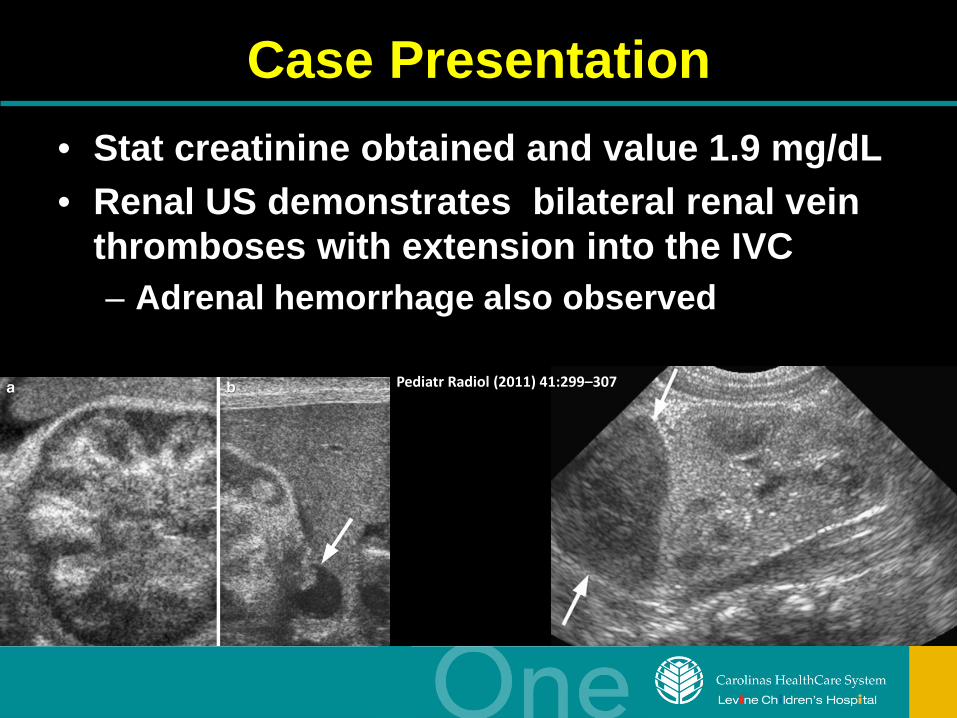
Case Presentation • Stat creatinine obtained and value 1.9 mg/dL • Renal US demonstrates bilateral renal vein
thromboses with extension into the IVC – Adrenal hemorrhage also observed
Pediatr Radiol (2011) 41:299–307
Dilemma
• Faced with acute renal failure with bilateral renal vein thromboses and an adrenal hemorrhage
• Not sure what to do as there are no evidence based protocols on how to treat
Objectives
• Briefly review neonatal hemostatic system • Types of neonatal thromboses • Perinatal and prothrombotic risk factors • Evaluation and management protocols
Important Points
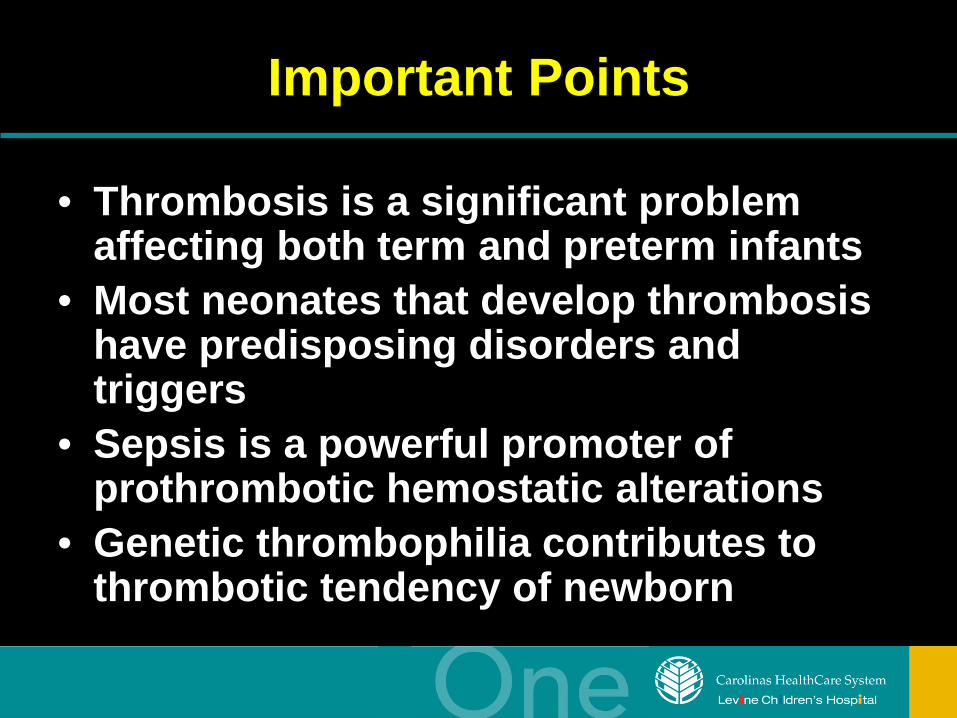
• Thrombosis is a significant problem affecting both term and preterm infants
• Most neonates that develop thrombosis have predisposing disorders and triggers
• Sepsis is a powerful promoter of prothrombotic hemostatic alterations
• Genetic thrombophilia contributes to thrombotic tendency of newborn
Fact
“Recommendations for neonatal treatment are based on extrapolation of principles of
therapy from adult guidelines, limited clinical information from registries, individual case studies, and knowledge of current common
clinical practice*”
*Monagle et al. Antithrombotic therapy in neonates and children: American College of Chest Physicians Evidence-Based Clinical Practice Guidelines (9th Edition). Chest. 2012;141(2)(Suppl):e737s-e801s.
Introduction
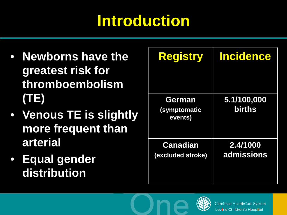
Registry Incidence
German (symptomatic
events)
5.1/100,000 births
Canadian (excluded stroke)
2.4/1000 admissions
• Newborns have the greatest risk for thromboembolism (TE)
• Venous TE is slightly more frequent than arterial
• Equal gender distribution
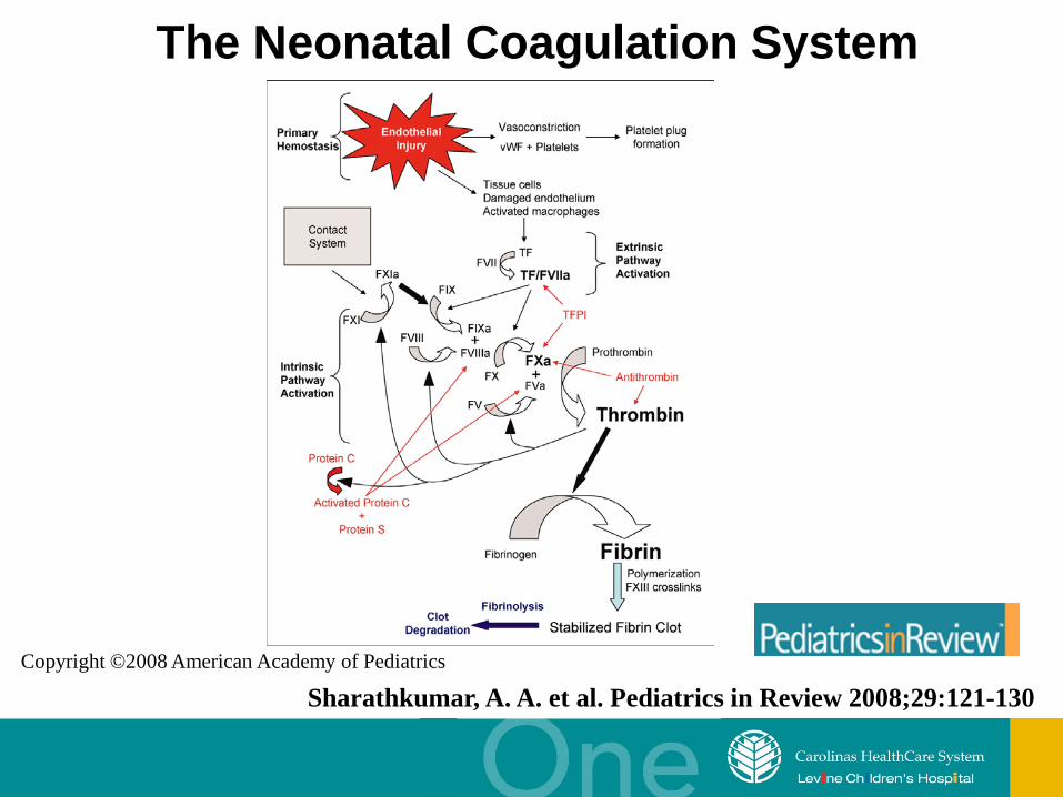
Copyright ©2008 American Academy of Pediatrics
Sharathkumar, A. A. et al. Pediatrics in Review 2008;29:121-130
The Neonatal Coagulation System
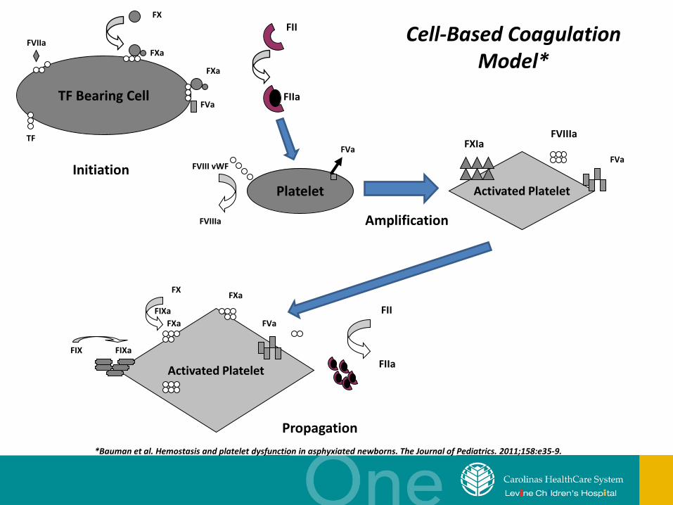
TF Bearing Cell
Platelet
Activated Platelet
Activated Platelet
FVIIa FXa
FX
FXa
FVa
TF
FII
FIIa
FVIII vWF
FVIIIa
FVa
Initiation
FXIa FVIIIa
FVa
FIX FIXa
FX
FIXa FXa
FXa
FVa FII
FIIa
Amplification
Propagation *Bauman et al. Hemostasis and platelet dysfunction in asphyxiated newborns. The Journal of Pediatrics. 2011;158:e35-9.
Cell-Based Coagulation Model*
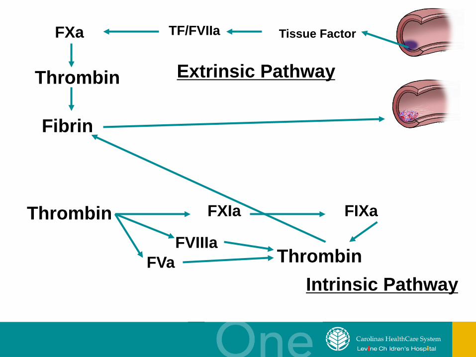
Tissue Factor TF/FVIIa FXa
Thrombin
Fibrin
Extrinsic Pathway
Thrombin FIXa FXIa
FVIIIa FVa Thrombin
Intrinsic Pathway
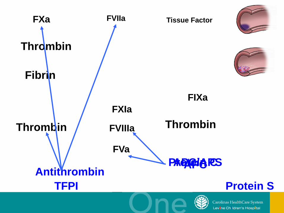
Tissue Factor FVIIa FXa
Thrombin
Fibrin
Thrombin
FIXa FXIa
FVIIIa
FVa
Thrombin
Protein C APC
Protein S
APC/APS
TFPI Antithrombin
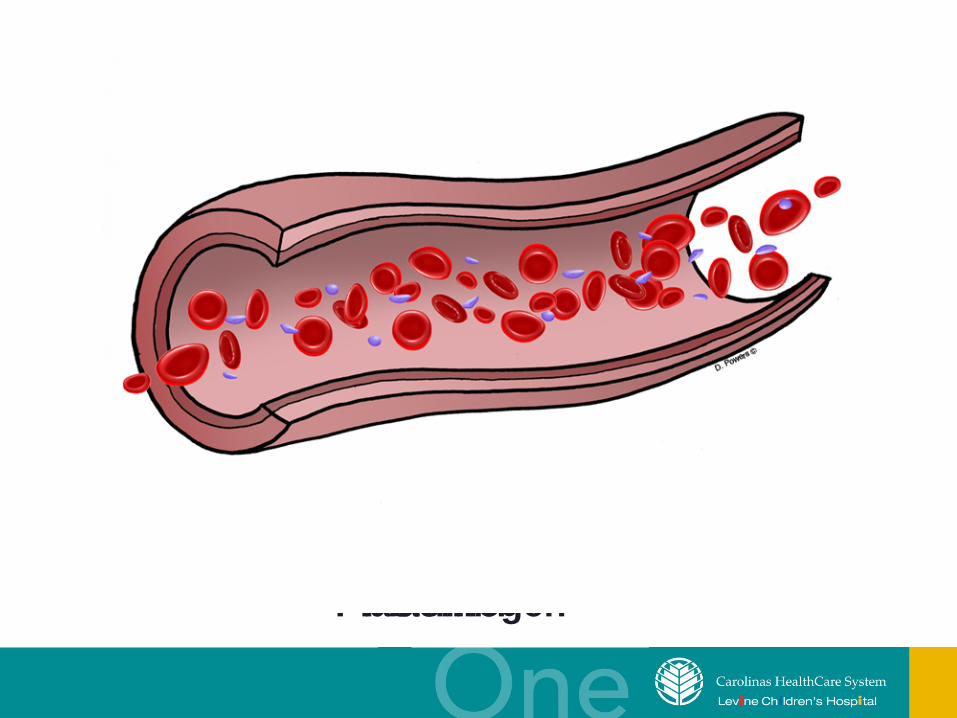
Plasminogen Plasmin
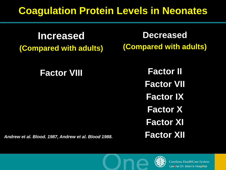
Coagulation Protein Levels in Neonates
Increased (Compared with adults)
Factor VIII
Decreased (Compared with adults)
Factor II
Factor VII Factor IX Factor X Factor XI Factor XII Andrew et al. Blood. 1987, Andrew et al. Blood 1988.
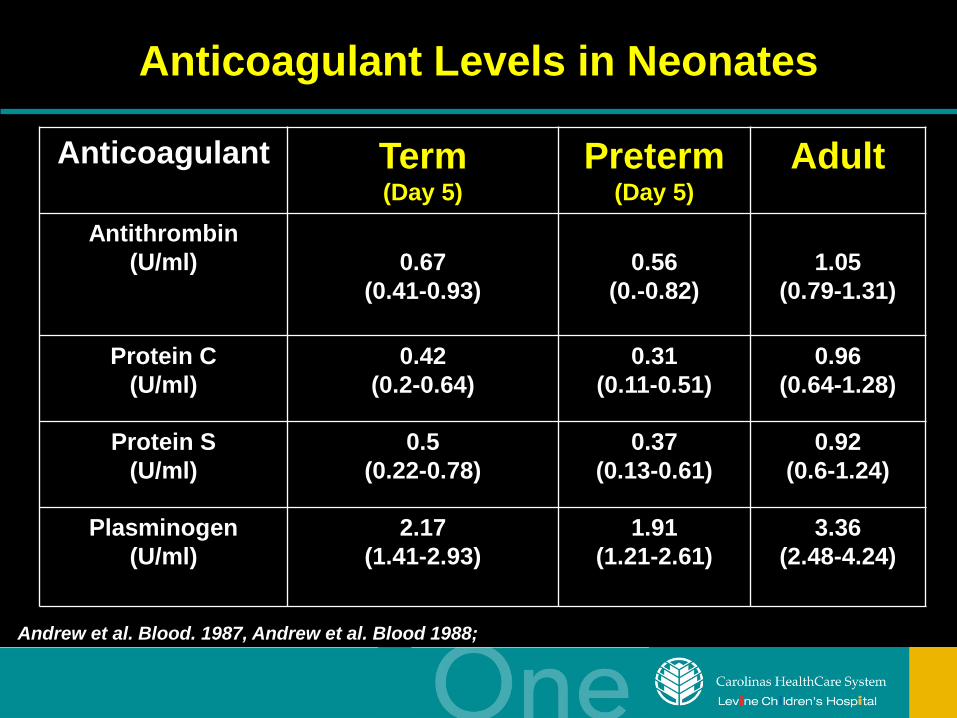
Anticoagulant Levels in Neonates
Anticoagulant Term (Day 5)
Preterm (Day 5)
Adult
Antithrombin (U/ml)
0.67
(0.41-0.93)
0.56
(0.-0.82)
1.05
(0.79-1.31)
Protein C (U/ml)
0.42 (0.2-0.64)
0.31 (0.11-0.51)
0.96 (0.64-1.28)
Protein S (U/ml)
0.5 (0.22-0.78)
0.37 (0.13-0.61)
0.92 (0.6-1.24)
Plasminogen (U/ml)
2.17 (1.41-2.93)
1.91 (1.21-2.61)
3.36 (2.48-4.24)
Andrew et al. Blood. 1987, Andrew et al. Blood 1988;

Thrombosis Hemorrhage
Neonatal Coagulation System At Birth
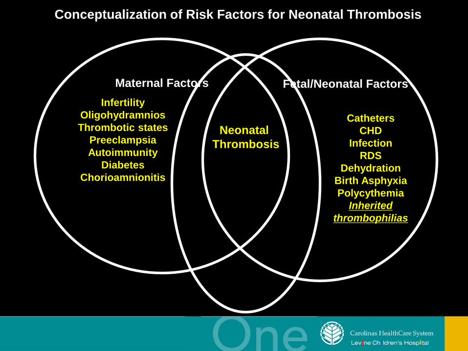
Maternal Factors
Conceptualization of Risk Factors for Neonatal Thrombosis
Infertility Oligohydramnios Thrombotic states
Preeclampsia Autoimmunity
Diabetes Chorioamnionitis
Fetal/Neonatal Factors
Catheters CHD
Infection RDS
Dehydration Birth Asphyxia Polycythemia
Inherited thrombophilias
Neonatal Thrombosis
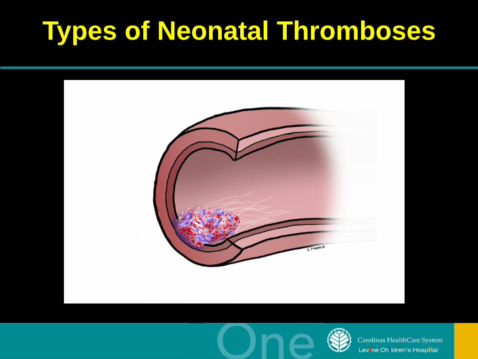
Types of Neonatal Thromboses
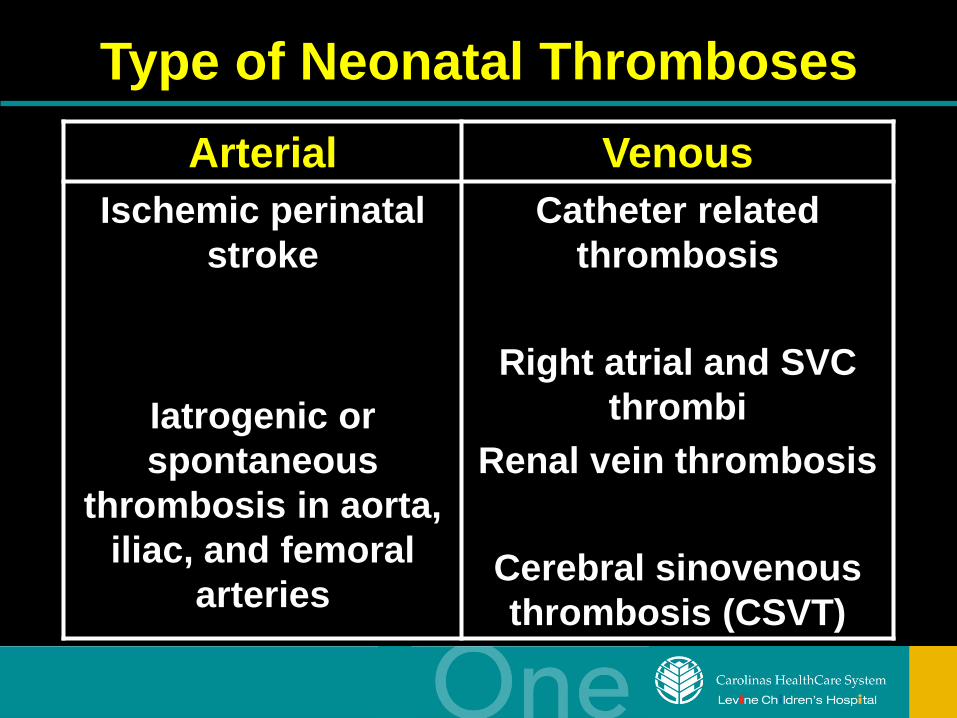
Type of Neonatal Thromboses Arterial Venous
Ischemic perinatal stroke
Iatrogenic or spontaneous
thrombosis in aorta, iliac, and femoral
arteries
Catheter related thrombosis
Right atrial and SVC
thrombi Renal vein thrombosis
Cerebral sinovenous thrombosis (CSVT)
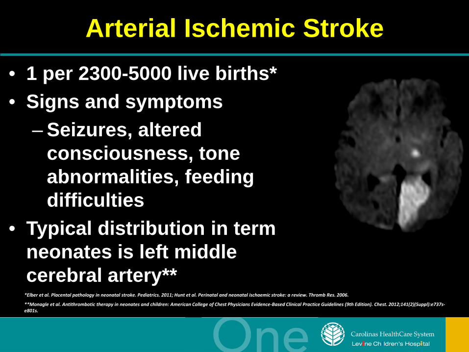
Arterial Ischemic Stroke • 1 per 2300-5000 live births* • Signs and symptoms
– Seizures, altered consciousness, tone abnormalities, feeding difficulties
• Typical distribution in term neonates is left middle cerebral artery**
*Elber et al. Placental pathology in neonatal stroke. Pediatrics. 2011; Hunt et al. Perinatal and neonatal ischaemic stroke: a review. Thromb Res. 2006.
**Monagle et al. Antithrombotic therapy in neonates and children: American College of Chest Physicians Evidence-Based Clinical Practice Guidelines (9th Edition). Chest. 2012;141(2)(Suppl):e737s-e801s.
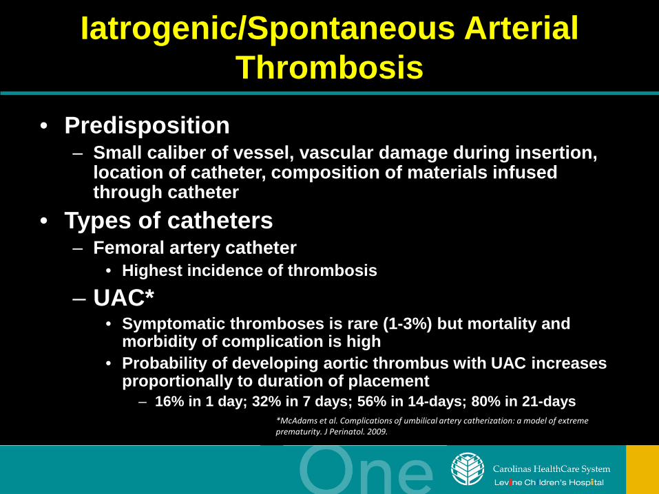
Iatrogenic/Spontaneous Arterial Thrombosis
• Predisposition – Small caliber of vessel, vascular damage during insertion,
location of catheter, composition of materials infused through catheter
• Types of catheters – Femoral artery catheter
• Highest incidence of thrombosis
– UAC* • Symptomatic thromboses is rare (1-3%) but mortality and
morbidity of complication is high • Probability of developing aortic thrombus with UAC increases
proportionally to duration of placement – 16% in 1 day; 32% in 7 days; 56% in 14-days; 80% in 21-days
*McAdams et al. Complications of umbilical artery catherization: a model of extreme prematurity. J Perinatol. 2009.
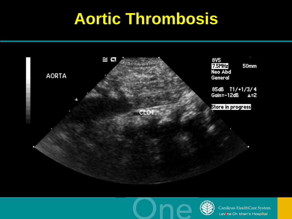
Aortic Thrombosis
Central Venous Catheter Related Thrombosis
• Signs and symptoms – Persistent infection/blood cultures – Thrombocytopenia – Line dysfunction – Swollen extremity – Chylothorax

Infection, Inflammation, and Thrombosis*
• Reciprocal relationship between catheter-related blood stream infection and thrombosis
• Fibrin sheaths formed around catheter tip serve as nidus for bacterial growth
• Inflammation from infection activates coagulation promoting thrombin formation on indwelling catheters
*Thornburg et al. 2008. Thromb Res.
Central Venous Catheter Related Thrombosis
• UVC*
– High rate of transient, asymptomatic thrombosis – Hematocrit > 55% risk factor in preterm infants – Portal venous thrombosis due to improper
placement/omphalitis • PICC lines*
– Use of 0.5 U/kg/hour of UFH prolongs patency but does not reduce risk for thrombosis or infection
*Monagle et al. Antithrombotic therapy in neonates and children: American College of Chest Physicians Evidence-Based Clinical Practice Guidelines (9th Edition). Chest. 2012;141(2)(Suppl):e737s-e801s.
Renal Vein Thrombosis
• Most common site of spontaneous thrombosis in newborn
• 25% bilateral – 52-60% extend into the IVC
• Presentation – Hematuria, palpable flank mass, thrombocytopenia
• Outcomes – 70% of affected kidneys irreversible atrophy – 20% hypertension; 3% chronic renal failure
*Monagle et al. Antithrombotic therapy in neonates and children: American College of Chest Physicians Evidence-Based Clinical Practice Guidelines (9th Edition). Chest. 2012;141(2)(Suppl):e737s-e801s.
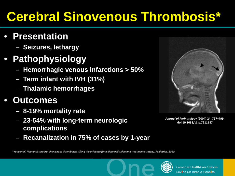
Cerebral Sinovenous Thrombosis* • Presentation
– Seizures, lethargy
• Pathophysiology – Hemorrhagic venous infarctions > 50% – Term infant with IVH (31%) – Thalamic hemorrhages
• Outcomes – 8-19% mortality rate – 23-54% with long-term neurologic
complications – Recanalization in 75% of cases by 1-year
*Yang et al. Neonatal cerebral sinovenous thrombosis: sifting the evidence for a diagnostic plan and treatment strategy. Pediatrics. 2010.
Journal of Perinatology (2004) 24, 797–799. doi:10.1038/sj.jp.7211187
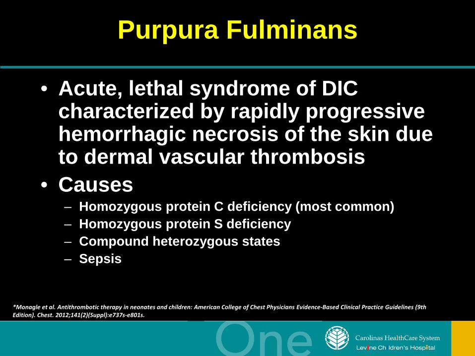
Purpura Fulminans
• Acute, lethal syndrome of DIC characterized by rapidly progressive hemorrhagic necrosis of the skin due to dermal vascular thrombosis
• Causes – Homozygous protein C deficiency (most common) – Homozygous protein S deficiency – Compound heterozygous states – Sepsis
*Monagle et al. Antithrombotic therapy in neonates and children: American College of Chest Physicians Evidence-Based Clinical Practice Guidelines (9th Edition). Chest. 2012;141(2)(Suppl):e737s-e801s.
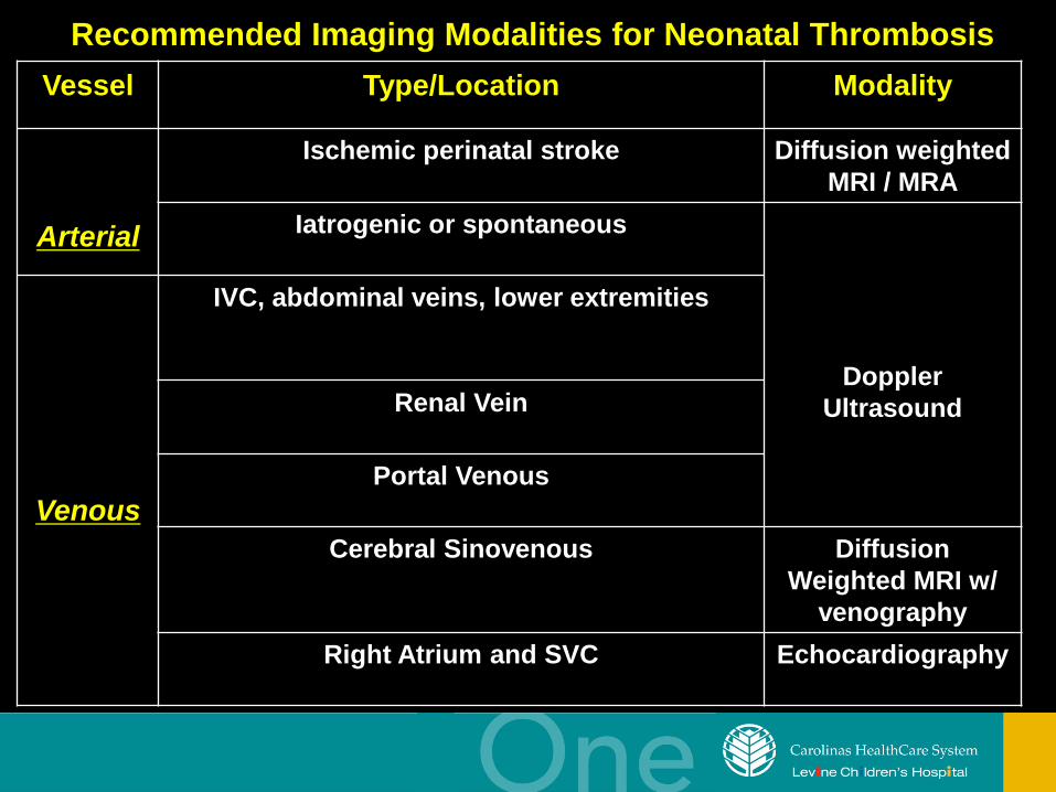
Vessel Type/Location Modality
Arterial
Ischemic perinatal stroke Diffusion weighted MRI / MRA
Iatrogenic or spontaneous
Doppler Ultrasound
Venous
IVC, abdominal veins, lower extremities
Renal Vein
Portal Venous
Cerebral Sinovenous Diffusion Weighted MRI w/
venography Right Atrium and SVC Echocardiography
Recommended Imaging Modalities for Neonatal Thrombosis
Genetic Thrombophilia • Genetic mutations resulting in
prothrombotic phenotype • Supporting evidence that genetic
thrombophilia is a risk factor for neonatal thrombosis and stroke (especially idiopathic) – Role in pathogenesis of neonatal
thrombosis is controversial and not completely understood
• Conflicting data in neonates with catheter-related thrombosis
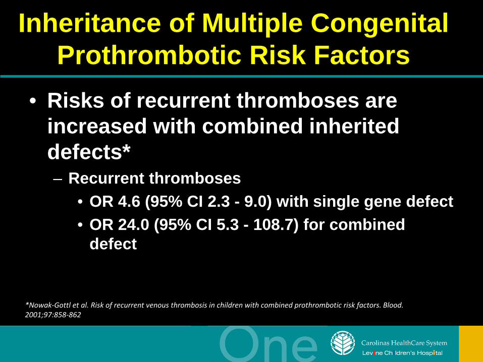
Inheritance of Multiple Congenital Prothrombotic Risk Factors
• Risks of recurrent thromboses are increased with combined inherited defects* – Recurrent thromboses
• OR 4.6 (95% CI 2.3 - 9.0) with single gene defect • OR 24.0 (95% CI 5.3 - 108.7) for combined
defect
*Nowak-Gottl et al. Risk of recurrent venous thrombosis in children with combined prothrombotic risk factors. Blood. 2001;97:858-862
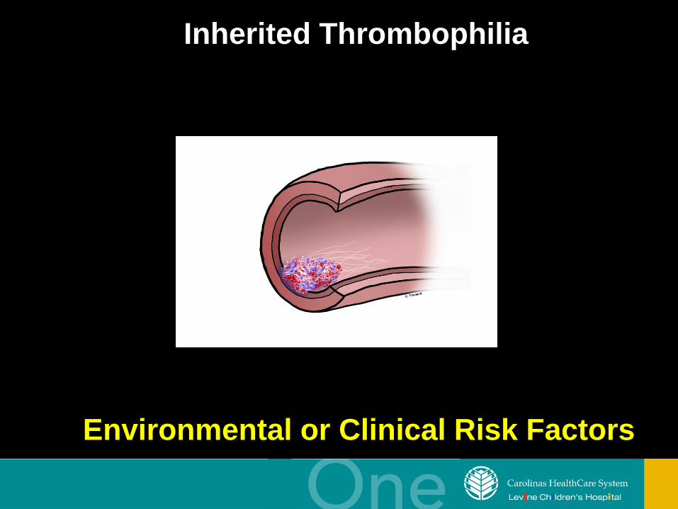
Inherited Thrombophilia
Environmental or Clinical Risk Factors
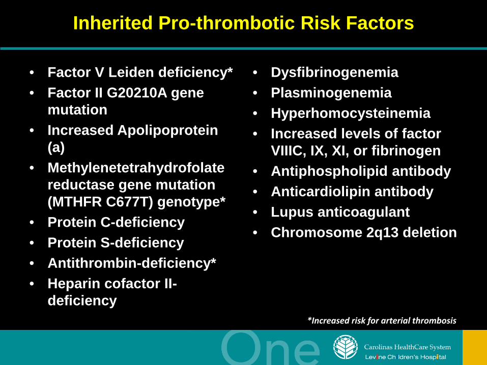
Inherited Pro-thrombotic Risk Factors
• Factor V Leiden deficiency* • Factor II G20210A gene
mutation • Increased Apolipoprotein
(a) • Methylenetetrahydrofolate
reductase gene mutation (MTHFR C677T) genotype*
• Protein C-deficiency • Protein S-deficiency • Antithrombin-deficiency* • Heparin cofactor II-
deficiency
• Dysfibrinogenemia • Plasminogenemia • Hyperhomocysteinemia • Increased levels of factor
VIIIC, IX, XI, or fibrinogen • Antiphospholipid antibody • Anticardiolipin antibody • Lupus anticoagulant • Chromosome 2q13 deletion
*Increased risk for arterial thrombosis
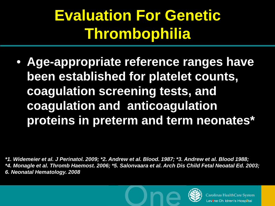
Evaluation For Genetic Thrombophilia
• Age-appropriate reference ranges have been established for platelet counts, coagulation screening tests, and coagulation and anticoagulation proteins in preterm and term neonates*
*1. Widemeier et al. J Perinatol. 2009; *2. Andrew et al. Blood. 1987; *3. Andrew et al. Blood 1988; *4. Monagle et al. Thromb Haemost. 2006; *5. Salonvaara et al. Arch Dis Child Fetal Neoatal Ed. 2003; 6. Neonatal Hematology. 2008
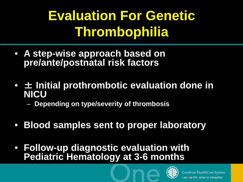
Evaluation For Genetic Thrombophilia
• A step-wise approach based on pre/ante/postnatal risk factors
• ± Initial prothrombotic evaluation done in NICU – Depending on type/severity of thrombosis
• Blood samples sent to proper laboratory
• Follow-up diagnostic evaluation with
Pediatric Hematology at 3-6 months
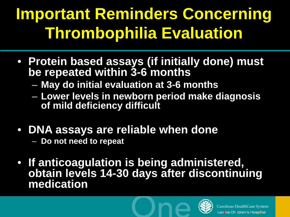
Important Reminders Concerning Thrombophilia Evaluation
• Protein based assays (if initially done) must be repeated within 3-6 months – May do initial evaluation at 3-6 months – Lower levels in newborn period make diagnosis
of mild deficiency difficult
• DNA assays are reliable when done – Do not need to repeat
• If anticoagulation is being administered,
obtain levels 14-30 days after discontinuing medication
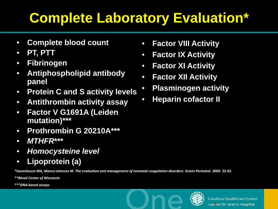
Complete Laboratory Evaluation*
• Complete blood count • PT, PTT • Fibrinogen • Antiphospholipid antibody
panel • Protein C and S activity levels • Antithrombin activity assay • Factor V G1691A (Leiden
mutation)*** • Prothrombin G 20210A*** • MTHFR*** • Homocysteine level • Lipoprotein (a)
• Factor VIII Activity • Factor IX Activity • Factor XI Activity • Factor XII Activity • Plasminogen activity • Heparin cofactor II
*Saxonhouse MA, Manco-Johnson M. The evaluation and management of neonatal coagulation disorders. Semin Perinatol. 2009. 52-65.
**Blood Center of Wisconsin
***DNA-based assays

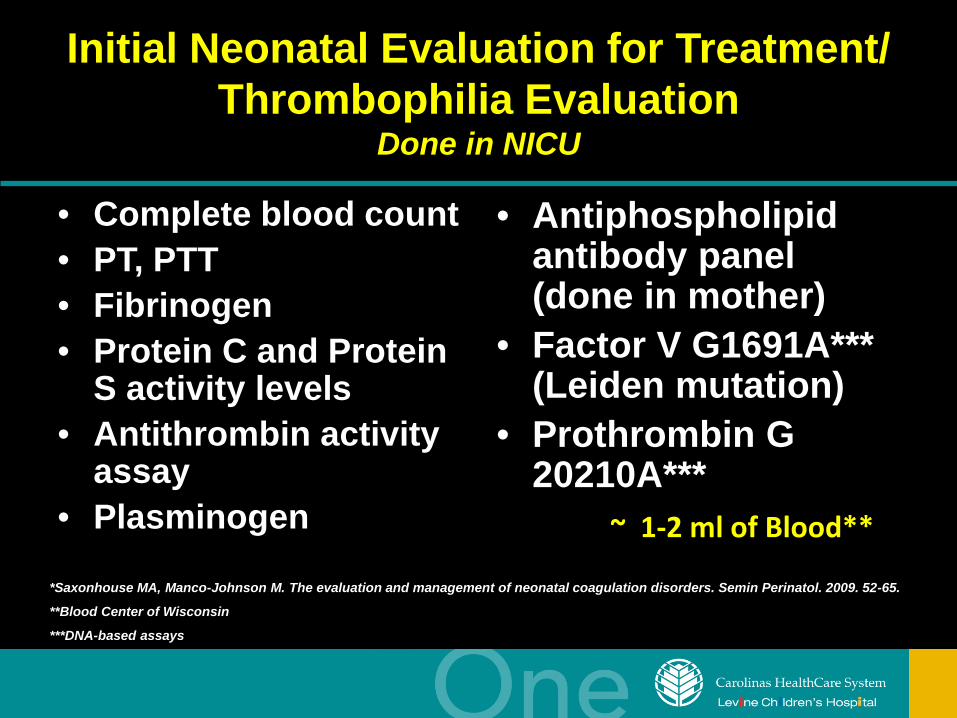
Initial Neonatal Evaluation for Treatment/ Thrombophilia Evaluation
Done in NICU
• Complete blood count • PT, PTT • Fibrinogen • Protein C and Protein
S activity levels • Antithrombin activity
assay • Plasminogen
• Antiphospholipid antibody panel (done in mother)
• Factor V G1691A*** (Leiden mutation)
• Prothrombin G 20210A*** ~ 1-2 ml of Blood**
*Saxonhouse MA, Manco-Johnson M. The evaluation and management of neonatal coagulation disorders. Semin Perinatol. 2009. 52-65.
**Blood Center of Wisconsin
***DNA-based assays
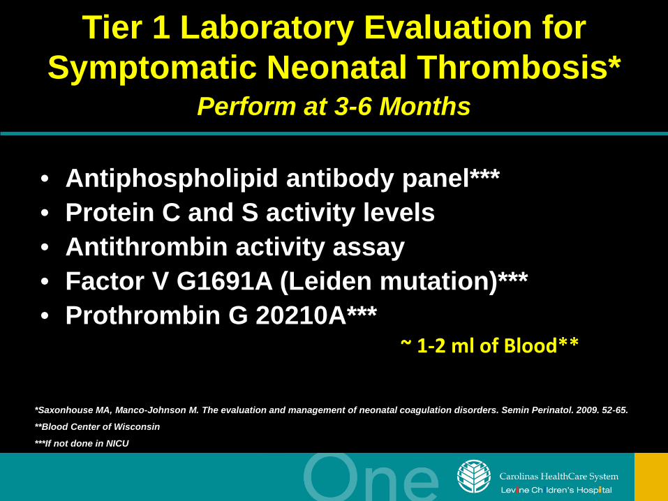
Tier 1 Laboratory Evaluation for Symptomatic Neonatal Thrombosis*
Perform at 3-6 Months
• Antiphospholipid antibody panel*** • Protein C and S activity levels • Antithrombin activity assay • Factor V G1691A (Leiden mutation)*** • Prothrombin G 20210A***
~ 1-2 ml of Blood**
*Saxonhouse MA, Manco-Johnson M. The evaluation and management of neonatal coagulation disorders. Semin Perinatol. 2009. 52-65.
**Blood Center of Wisconsin
***If not done in NICU
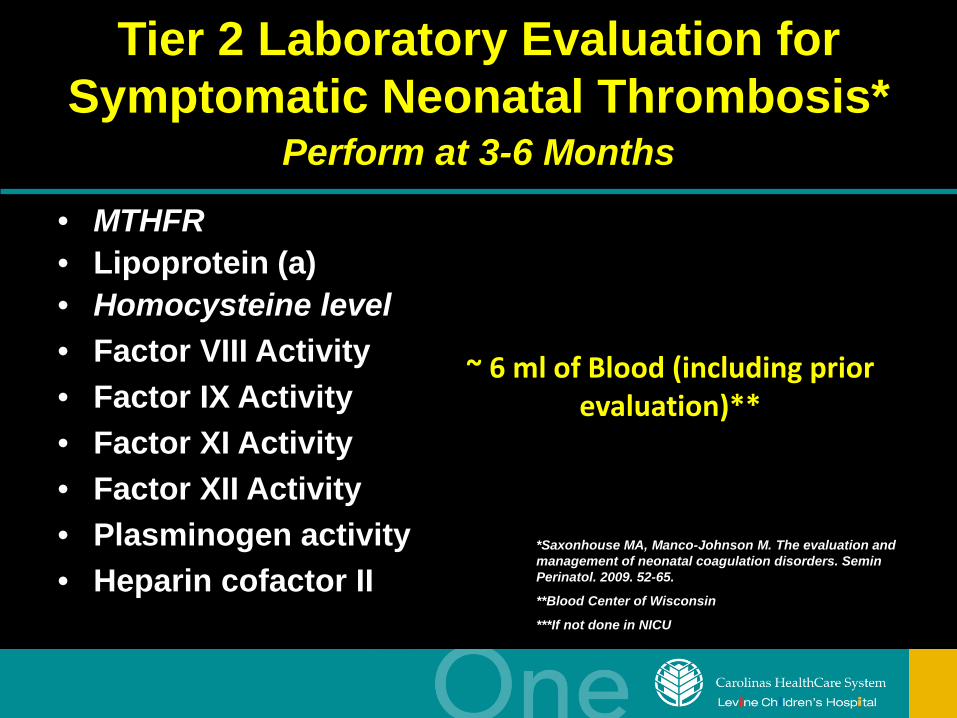
Tier 2 Laboratory Evaluation for Symptomatic Neonatal Thrombosis*
Perform at 3-6 Months • MTHFR • Lipoprotein (a) • Homocysteine level • Factor VIII Activity • Factor IX Activity • Factor XI Activity • Factor XII Activity • Plasminogen activity • Heparin cofactor II
~ 6 ml of Blood (including prior evaluation)**
*Saxonhouse MA, Manco-Johnson M. The evaluation and management of neonatal coagulation disorders. Semin Perinatol. 2009. 52-65.
**Blood Center of Wisconsin
***If not done in NICU
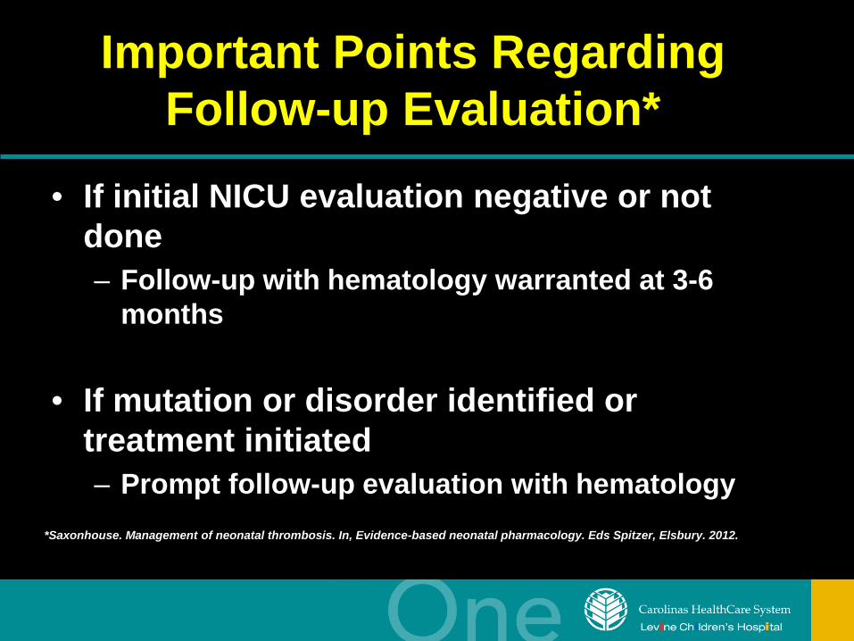
Important Points Regarding Follow-up Evaluation*
• If initial NICU evaluation negative or not done – Follow-up with hematology warranted at 3-6
months
• If mutation or disorder identified or treatment initiated – Prompt follow-up evaluation with hematology
*Saxonhouse. Management of neonatal thrombosis. In, Evidence-based neonatal pharmacology. Eds Spitzer, Elsbury. 2012.
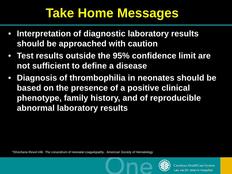
Take Home Messages • Interpretation of diagnostic laboratory results
should be approached with caution • Test results outside the 95% confidence limit are
not sufficient to define a disease • Diagnosis of thrombophilia in neonates should be
based on the presence of a positive clinical phenotype, family history, and of reproducible abnormal laboratory results
*Shoshana Revel-Vilk. The conundrum of neonatal coagulopathy.. American Society of Hematology
Take Home Messages
• Overdiagnosis and misdiagnosis may lead to administration of wrong and potentially harmful treatments for years
• Evaluation with hematologist will be tailored based on severity of thrombosis, type of thrombosis, family history, and clinical risk factors
*Shoshana Revel-Vilk. The conundrum of neonatal coagulopathy.. American Society of Hematology
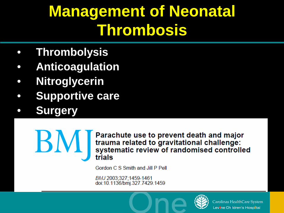
Management of Neonatal Thrombosis
• Thrombolysis • Anticoagulation • Nitroglycerin • Supportive care • Surgery
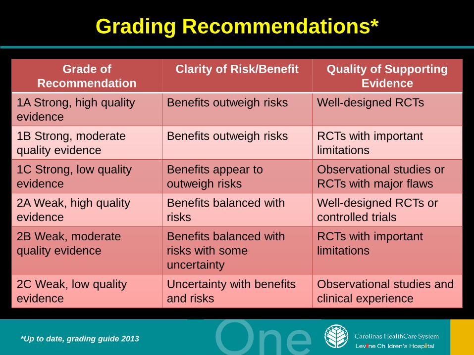
Grading Recommendations*
Grade of Recommendation
Clarity of Risk/Benefit Quality of Supporting Evidence
1A Strong, high quality evidence
Benefits outweigh risks Well-designed RCTs
1B Strong, moderate quality evidence
Benefits outweigh risks RCTs with important limitations
1C Strong, low quality evidence
Benefits appear to outweigh risks
Observational studies or RCTs with major flaws
2A Weak, high quality evidence
Benefits balanced with risks
Well-designed RCTs or controlled trials
2B Weak, moderate quality evidence
Benefits balanced with risks with some uncertainty
RCTs with important limitations
2C Weak, low quality evidence
Uncertainty with benefits and risks
Observational studies and clinical experience
*Up to date, grading guide 2013
Management of Neonatal Thrombosis
• Treatment should only occur at tertiary center that has proper neonatal, pediatric hematology, transfusion medicine, and pediatric surgical support*
• Pediatric hematologists with experience in thrombosis manage pediatric patients with thrombosis (Grade 2C)*
*Monagle et al. Antithrombotic therapy in neonates and children: American College of Chest Physicians Evidence-Based Clinical Practice Guidelines (9th Edition). Chest. 2012;141(2)(Suppl):e737s-e801s.
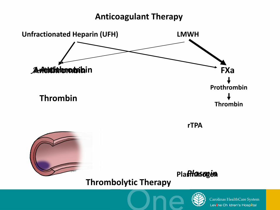
Thrombin
Antithrombin FXa
Plasminogen
Unfractionated Heparin (UFH)
rTPA
Plasmin Thrombolytic Therapy
LMWH
A-Antithrombin
Anticoagulant Therapy
Prothrombin
Thrombin
Thrombolytic Therapy
rTPA
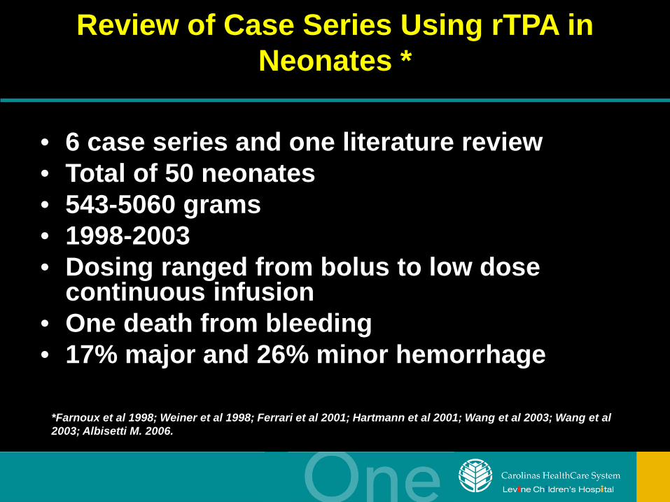
Review of Case Series Using rTPA in Neonates *
• 6 case series and one literature review • Total of 50 neonates • 543-5060 grams • 1998-2003 • Dosing ranged from bolus to low dose
continuous infusion • One death from bleeding • 17% major and 26% minor hemorrhage
*Farnoux et al 1998; Weiner et al 1998; Ferrari et al 2001; Hartmann et al 2001; Wang et al 2003; Wang et al 2003; Albisetti M. 2006.
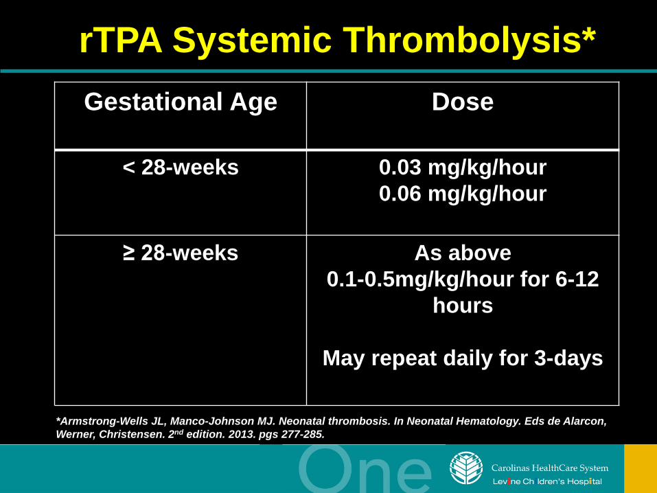
rTPA Systemic Thrombolysis*
*Armstrong-Wells JL, Manco-Johnson MJ. Neonatal thrombosis. In Neonatal Hematology. Eds de Alarcon, Werner, Christensen. 2nd edition. 2013. pgs 277-285.
Gestational Age Dose
< 28-weeks 0.03 mg/kg/hour 0.06 mg/kg/hour
≥ 28-weeks As above 0.1-0.5mg/kg/hour for 6-12
hours
May repeat daily for 3-days
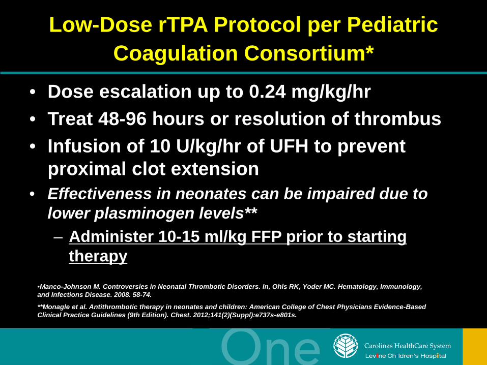
Low-Dose rTPA Protocol per Pediatric Coagulation Consortium*
• Dose escalation up to 0.24 mg/kg/hr • Treat 48-96 hours or resolution of thrombus • Infusion of 10 U/kg/hr of UFH to prevent
proximal clot extension • Effectiveness in neonates can be impaired due to
lower plasminogen levels** – Administer 10-15 ml/kg FFP prior to starting
therapy •Manco-Johnson M. Controversies in Neonatal Thrombotic Disorders. In, Ohls RK, Yoder MC. Hematology, Immunology,
and Infections Disease. 2008. 58-74.
**Monagle et al. Antithrombotic therapy in neonates and children: American College of Chest Physicians Evidence-Based Clinical Practice Guidelines (9th Edition). Chest. 2012;141(2)(Suppl):e737s-e801s.
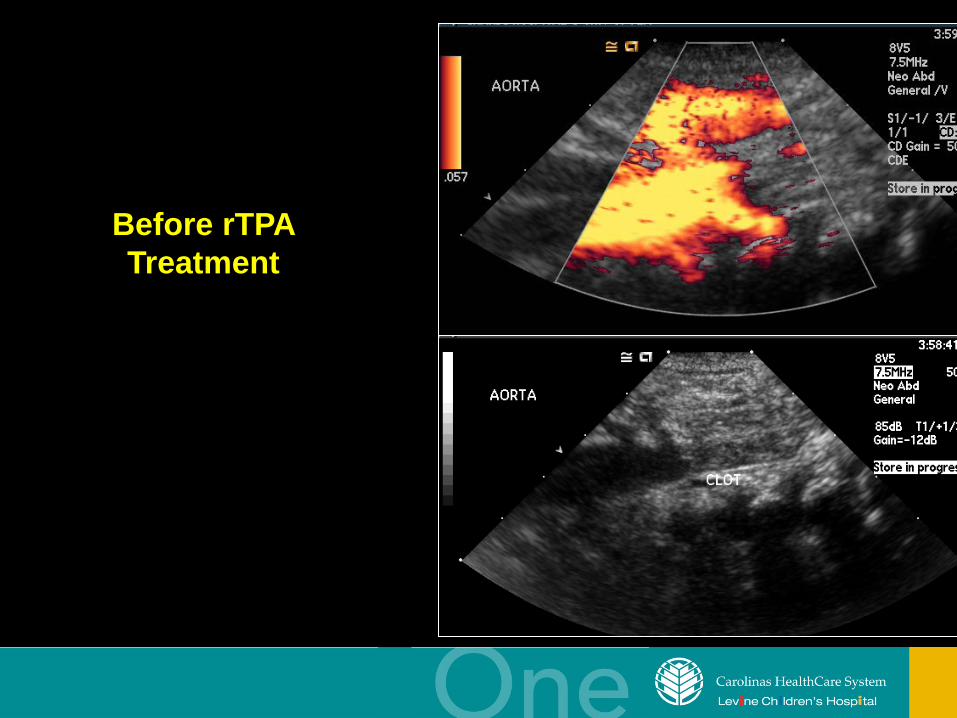
Before rTPA Treatment
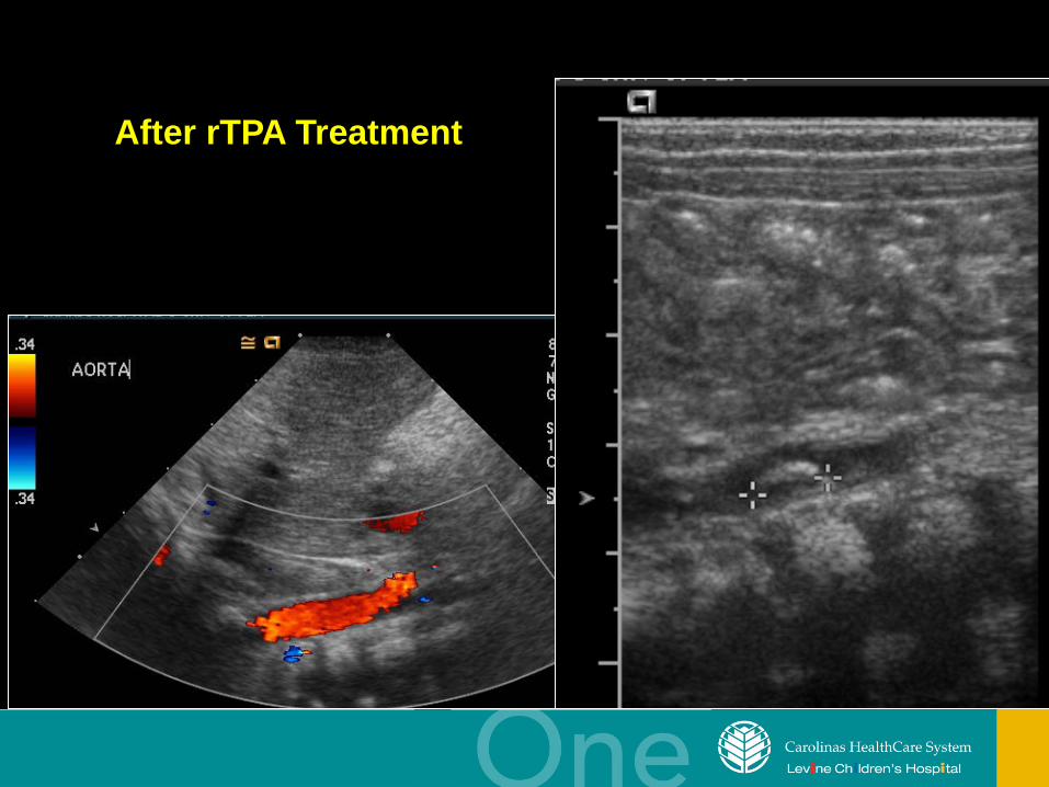
After rTPA Treatment
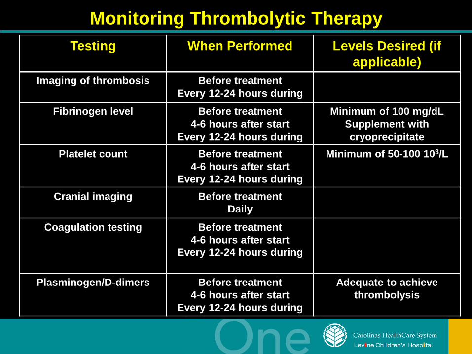
Monitoring Thrombolytic Therapy Testing When Performed Levels Desired (if
applicable) Imaging of thrombosis Before treatment
Every 12-24 hours during Fibrinogen level Before treatment
4-6 hours after start Every 12-24 hours during
Minimum of 100 mg/dL Supplement with cryoprecipitate
Platelet count Before treatment 4-6 hours after start
Every 12-24 hours during
Minimum of 50-100 103/L
Cranial imaging Before treatment Daily
Coagulation testing Before treatment 4-6 hours after start
Every 12-24 hours during
Plasminogen/D-dimers Before treatment 4-6 hours after start
Every 12-24 hours during
Adequate to achieve thrombolysis
Anticoagulation
UFH
LMWH
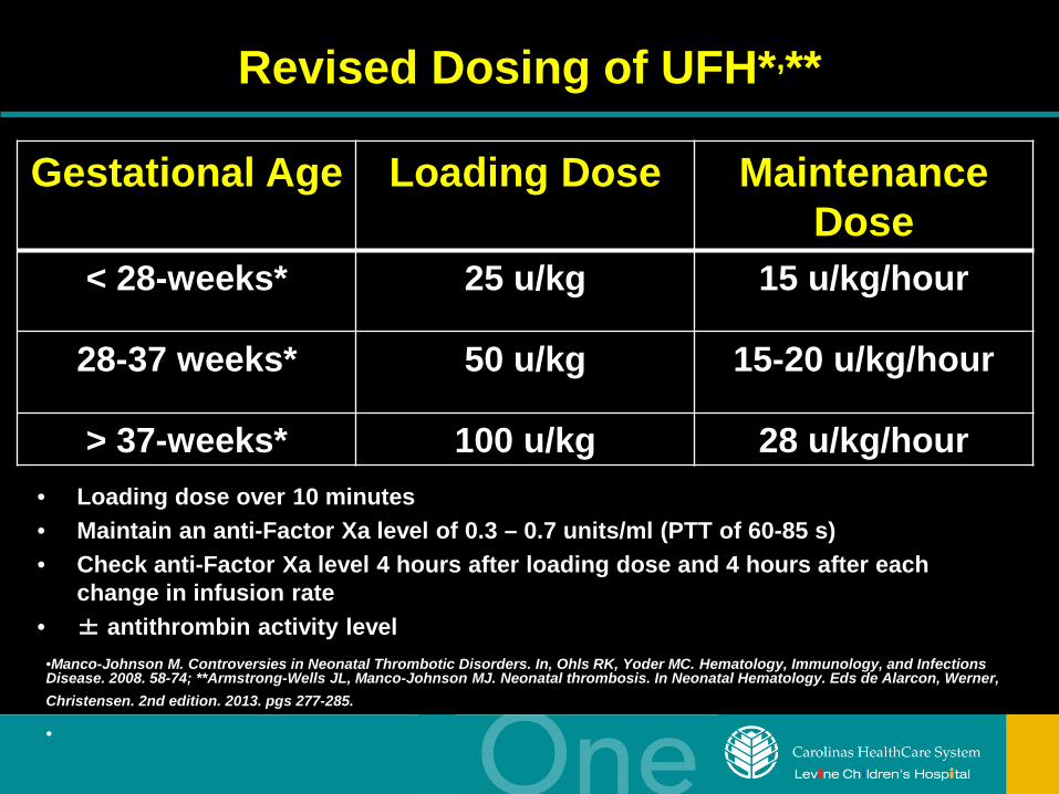
Revised Dosing of UFH*,**
• Loading dose over 10 minutes • Maintain an anti-Factor Xa level of 0.3 – 0.7 units/ml (PTT of 60-85 s) • Check anti-Factor Xa level 4 hours after loading dose and 4 hours after each
change in infusion rate • ± antithrombin activity level
•Manco-Johnson M. Controversies in Neonatal Thrombotic Disorders. In, Ohls RK, Yoder MC. Hematology, Immunology, and Infections Disease. 2008. 58-74; **Armstrong-Wells JL, Manco-Johnson MJ. Neonatal thrombosis. In Neonatal Hematology. Eds de Alarcon, Werner, Christensen. 2nd edition. 2013. pgs 277-285. •
Gestational Age Loading Dose Maintenance Dose
< 28-weeks* 25 u/kg 15 u/kg/hour
28-37 weeks* 50 u/kg 15-20 u/kg/hour
> 37-weeks* 100 u/kg 28 u/kg/hour
UFH and Antithrombin Treatment
• Effectiveness dependent on adequate antithrombin levels – Neonates have low levels of antithrombin
activity • Heparin potentiates effect of antithrombin • Inadequate antithrombin levels reduces
effectiveness of heparin therapy • Recommended doses reflect lower levels of
antithrombin
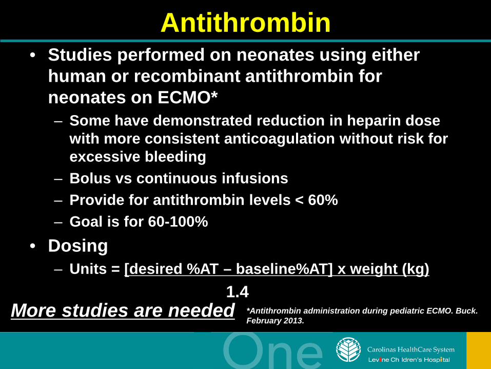
Antithrombin • Studies performed on neonates using either
human or recombinant antithrombin for neonates on ECMO* – Some have demonstrated reduction in heparin dose
with more consistent anticoagulation without risk for excessive bleeding
– Bolus vs continuous infusions – Provide for antithrombin levels < 60% – Goal is for 60-100%
• Dosing – Units = [desired %AT – baseline%AT] x weight (kg)
1.4
More studies are needed *Antithrombin administration during pediatric ECMO. Buck. February 2013.
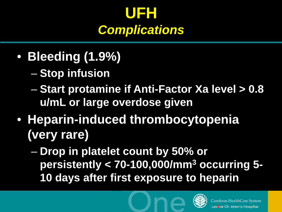
UFH Complications
• Bleeding (1.9%) – Stop infusion – Start protamine if Anti-Factor Xa level > 0.8
u/mL or large overdose given • Heparin-induced thrombocytopenia
(very rare) – Drop in platelet count by 50% or
persistently < 70-100,000/mm3 occurring 5-10 days after first exposure to heparin
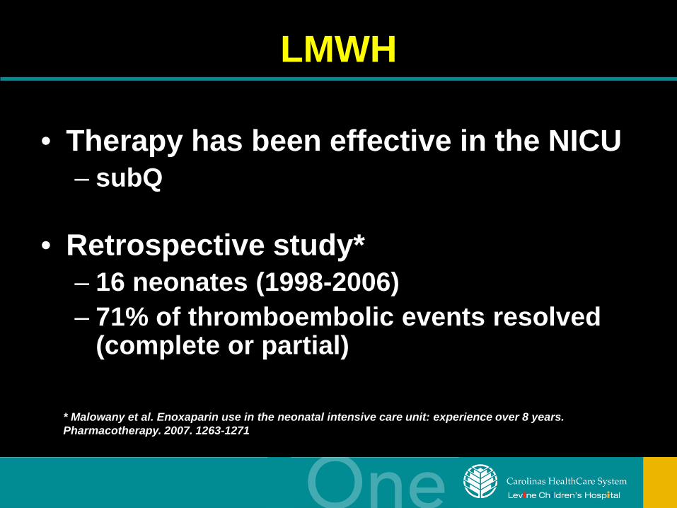
LMWH
• Therapy has been effective in the NICU – subQ
• Retrospective study*
– 16 neonates (1998-2006) – 71% of thromboembolic events resolved
(complete or partial) * Malowany et al. Enoxaparin use in the neonatal intensive care unit: experience over 8 years.
Pharmacotherapy. 2007. 1263-1271

LMWH (Enoxaparin)
• Goal of anti-FXa level of 0.5 to 1.0 U/ml when level checked 4-6 hours after 2nd dose (SQ injection)
• Goal of anti-FXA level of 0.5 – 0.8 U/ml when level checked 2-6 hours after SQ injection
• If infant with high hemorrhagic profile, use standard dosing • ± antithrombin activity level , platelet count
*Armstrong-Wells JL, Manco-Johnson MJ. Neonatal thrombosis. In Neonatal Hematology. Eds de Alarcon, Werner, Christensen. 2nd edition. 2013. pgs 277-285.
Gestational Age Dose*
< 28-weeks 1.25 mg/kg SQ q 12-hours
28-37-weeks 1.5 mg/kg SQ q 12-hours
> 37-weeks 1.625 mg/kg SQ q 12-hours
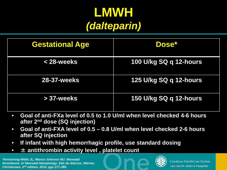
LMWH (dalteparin)
• Goal of anti-FXa level of 0.5 to 1.0 U/ml when level checked 4-6 hours after 2nd dose (SQ injection)
• Goal of anti-FXA level of 0.5 – 0.8 U/ml when level checked 2-6 hours after SQ injection
• If infant with high hemorrhagic profile, use standard dosing • ± antithrombin activity level , platelet count
*Armstrong-Wells JL, Manco-Johnson MJ. Neonatal thrombosis. In Neonatal Hematology. Eds de Alarcon, Werner, Christensen. 2nd edition. 2013. pgs 277-285.
Gestational Age Dose*
< 28-weeks 100 U/kg SQ q 12-hours
28-37-weeks 125 U/kg SQ q 12-hours
> 37-weeks 150 U/kg SQ q 12-hours
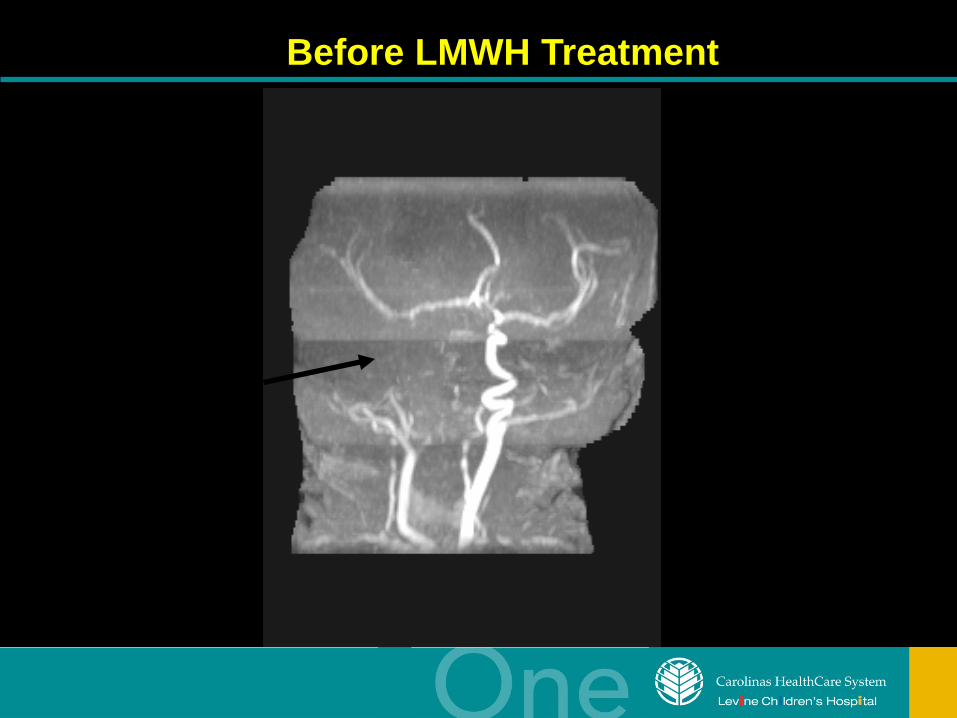
Before LMWH Treatment
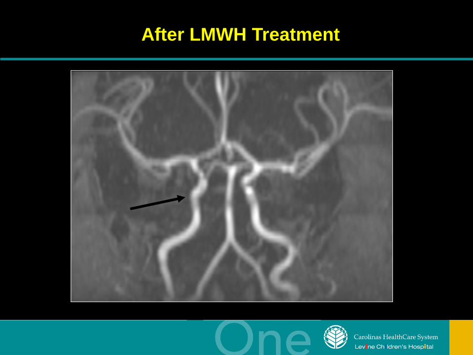
After LMWH Treatment
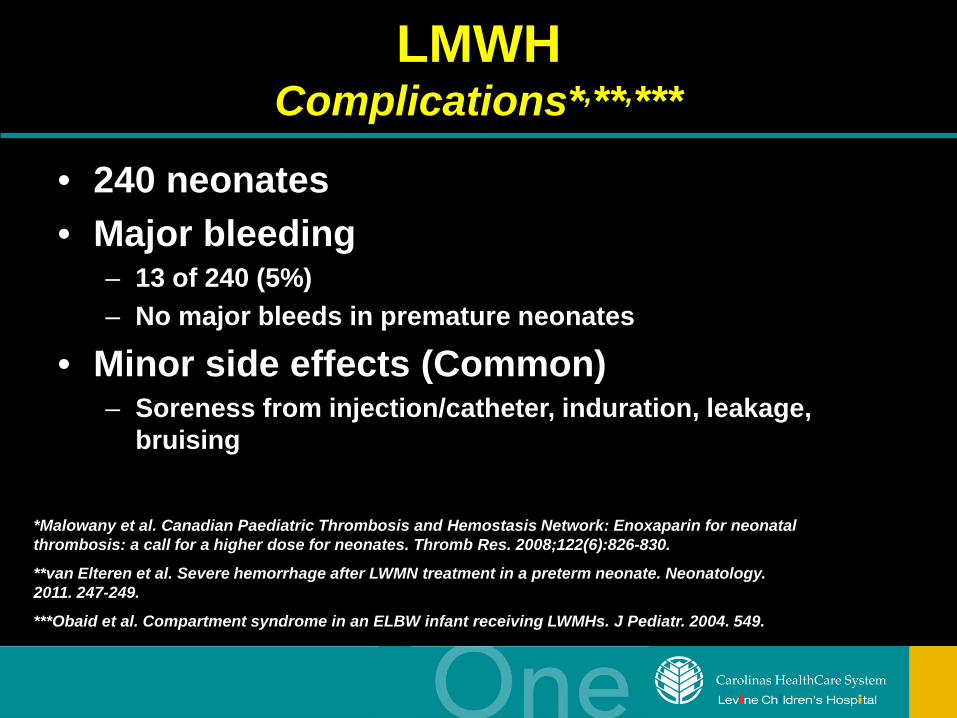
LMWH Complications*,**,***
• 240 neonates • Major bleeding
– 13 of 240 (5%) – No major bleeds in premature neonates
• Minor side effects (Common) – Soreness from injection/catheter, induration, leakage,
bruising
*Malowany et al. Canadian Paediatric Thrombosis and Hemostasis Network: Enoxaparin for neonatal thrombosis: a call for a higher dose for neonates. Thromb Res. 2008;122(6):826-830.
**van Elteren et al. Severe hemorrhage after LWMN treatment in a preterm neonate. Neonatology. 2011. 247-249.
***Obaid et al. Compartment syndrome in an ELBW infant receiving LWMHs. J Pediatr. 2004. 549.
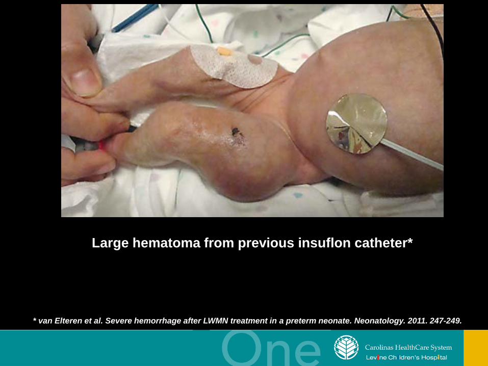
* van Elteren et al. Severe hemorrhage after LWMN treatment in a preterm neonate. Neonatology. 2011. 247-249.
Large hematoma from previous insuflon catheter*
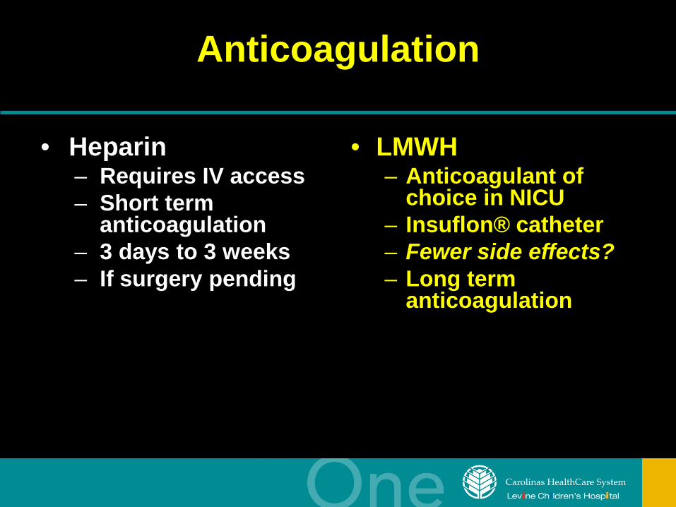
Anticoagulation
• Heparin – Requires IV access – Short term
anticoagulation – 3 days to 3 weeks – If surgery pending
• LMWH – Anticoagulant of
choice in NICU – Insuflon® catheter – Fewer side effects? – Long term
anticoagulation
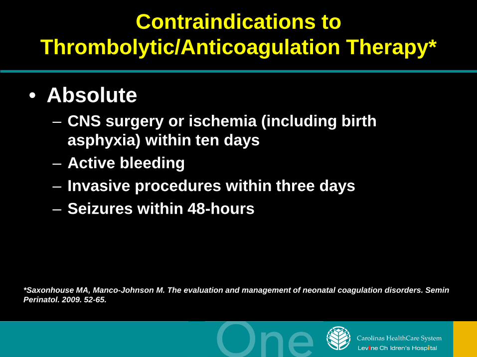
Contraindications to Thrombolytic/Anticoagulation Therapy*
• Absolute – CNS surgery or ischemia (including birth
asphyxia) within ten days – Active bleeding – Invasive procedures within three days – Seizures within 48-hours
*Saxonhouse MA, Manco-Johnson M. The evaluation and management of neonatal coagulation disorders. Semin Perinatol. 2009. 52-65.
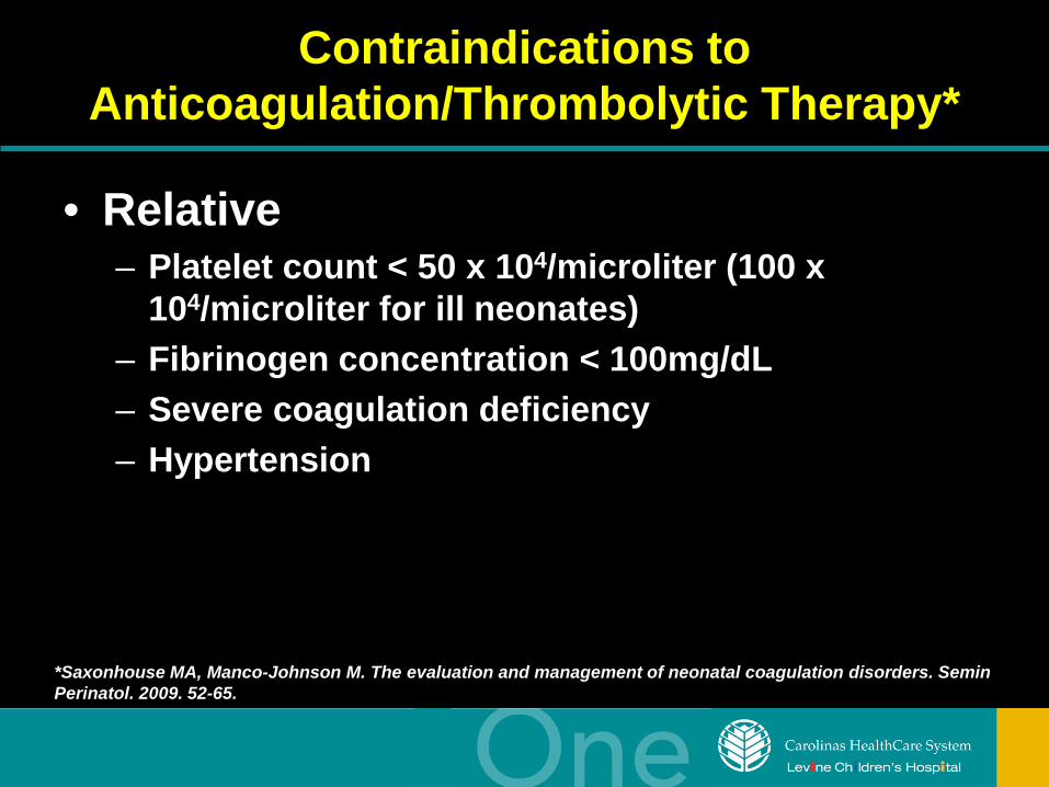
• Relative – Platelet count < 50 x 104/microliter (100 x
104/microliter for ill neonates) – Fibrinogen concentration < 100mg/dL – Severe coagulation deficiency – Hypertension
Contraindications to Anticoagulation/Thrombolytic Therapy*
*Saxonhouse MA, Manco-Johnson M. The evaluation and management of neonatal coagulation disorders. Semin Perinatol. 2009. 52-65.
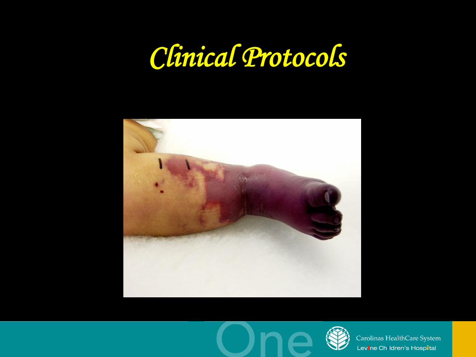
Clinical Protocols
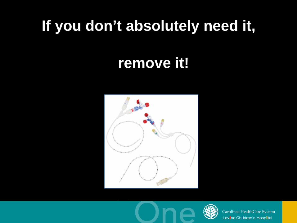
If you don’t absolutely need it,
remove it!
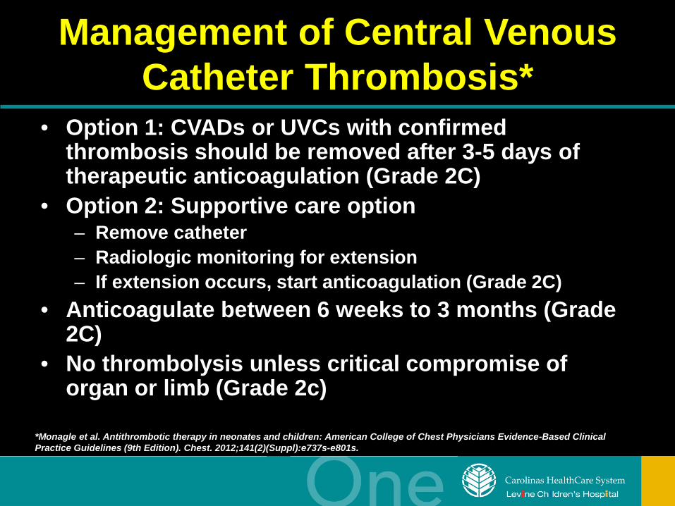
Management of Central Venous Catheter Thrombosis*
• Option 1: CVADs or UVCs with confirmed thrombosis should be removed after 3-5 days of therapeutic anticoagulation (Grade 2C)
• Option 2: Supportive care option – Remove catheter – Radiologic monitoring for extension – If extension occurs, start anticoagulation (Grade 2C)
• Anticoagulate between 6 weeks to 3 months (Grade 2C)
• No thrombolysis unless critical compromise of organ or limb (Grade 2c)
*Monagle et al. Antithrombotic therapy in neonates and children: American College of Chest Physicians Evidence-Based Clinical Practice Guidelines (9th Edition). Chest. 2012;141(2)(Suppl):e737s-e801s.
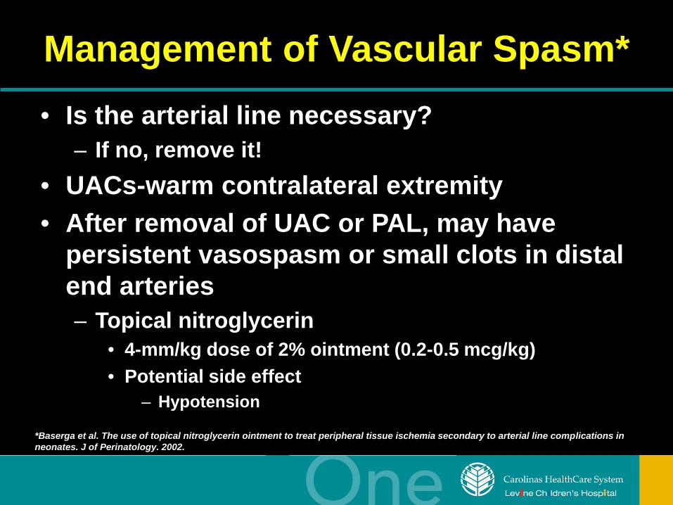
Management of Vascular Spasm* • Is the arterial line necessary?
– If no, remove it! • UACs-warm contralateral extremity • After removal of UAC or PAL, may have
persistent vasospasm or small clots in distal end arteries – Topical nitroglycerin
• 4-mm/kg dose of 2% ointment (0.2-0.5 mcg/kg) • Potential side effect
– Hypotension *Baserga et al. The use of topical nitroglycerin ointment to treat peripheral tissue ischemia secondary to arterial line complications in
neonates. J of Perinatology. 2002.

Management of Peripheral Arterial Catheter-Related TE
Event*,**
• Immediate removal of catheter (Grade 2B) – UFH with or without rTPA (Grade 2C) – Surgical thrombectomy and microvascular
repair with heparin therapy (Grade 2C)
*Monagle et al. Antithrombotic therapy in neonates and children: American College of Chest Physicians Evidence-Based Clinical Practice Guidelines (9th Edition). Chest. 2012;141(2)(Suppl):e737s-e801s.
**Albisetti et al. Arterial thromboembolic complications in critically ill children. J Crit Care. 2005;20(3):296-300.
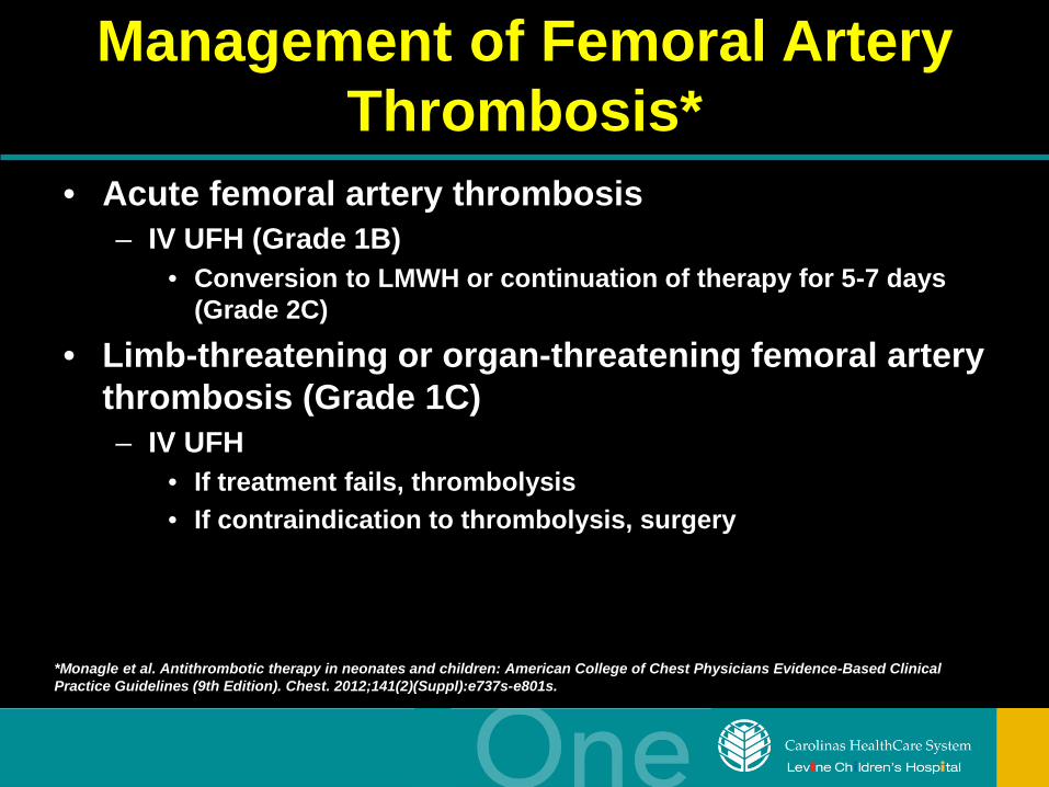
Management of Femoral Artery Thrombosis*
• Acute femoral artery thrombosis – IV UFH (Grade 1B)
• Conversion to LMWH or continuation of therapy for 5-7 days (Grade 2C)
• Limb-threatening or organ-threatening femoral artery thrombosis (Grade 1C) – IV UFH
• If treatment fails, thrombolysis • If contraindication to thrombolysis, surgery
*Monagle et al. Antithrombotic therapy in neonates and children: American College of Chest Physicians Evidence-Based Clinical Practice Guidelines (9th Edition). Chest. 2012;141(2)(Suppl):e737s-e801s.
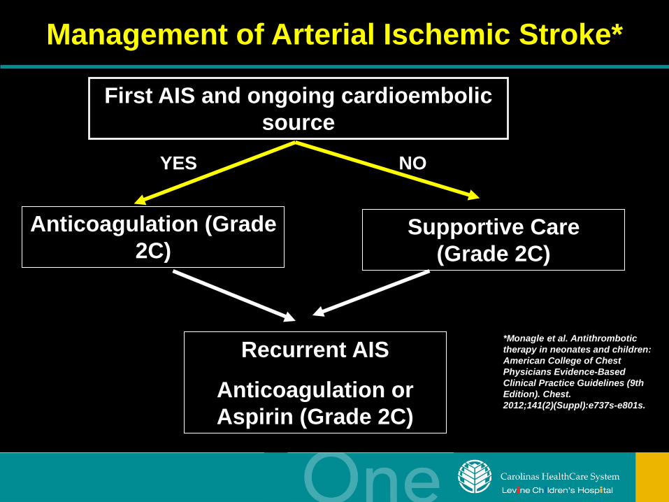
Management of Arterial Ischemic Stroke*
First AIS and ongoing cardioembolic source
Anticoagulation (Grade 2C)
YES NO
Supportive Care (Grade 2C)
Recurrent AIS
Anticoagulation or Aspirin (Grade 2C)
*Monagle et al. Antithrombotic therapy in neonates and children: American College of Chest Physicians Evidence-Based Clinical Practice Guidelines (9th Edition). Chest. 2012;141(2)(Suppl):e737s-e801s.
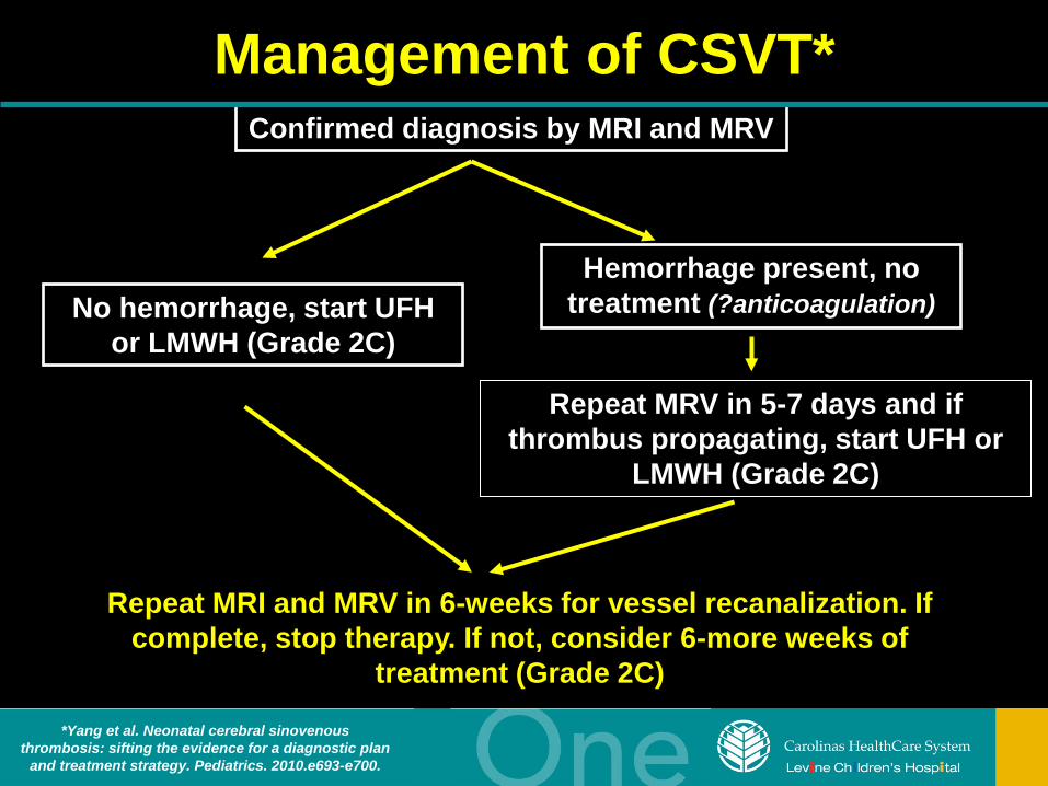
Management of CSVT* Confirmed diagnosis by MRI and MRV
No hemorrhage, start UFH or LMWH (Grade 2C)
Repeat MRV in 5-7 days and if thrombus propagating, start UFH or
LMWH (Grade 2C)
Repeat MRI and MRV in 6-weeks for vessel recanalization. If complete, stop therapy. If not, consider 6-more weeks of
treatment (Grade 2C)
*Yang et al. Neonatal cerebral sinovenous thrombosis: sifting the evidence for a diagnostic plan
and treatment strategy. Pediatrics. 2010.e693-e700.
Hemorrhage present, no treatment (?anticoagulation)
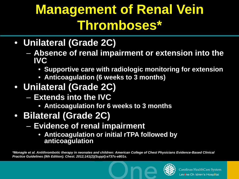
Management of Renal Vein Thromboses*
• Unilateral (Grade 2C) – Absence of renal impairment or extension into the
IVC • Supportive care with radiologic monitoring for extension • Anticoagulation (6 weeks to 3 months)
• Unilateral (Grade 2C) – Extends into the IVC
• Anticoagulation for 6 weeks to 3 months • Bilateral (Grade 2C)
– Evidence of renal impairment • Anticoagulation or initial rTPA followed by
anticoagulation *Monagle et al. Antithrombotic therapy in neonates and children: American College of Chest Physicians Evidence-Based Clinical Practice Guidelines (9th Edition). Chest. 2012;141(2)(Suppl):e737s-e801s.
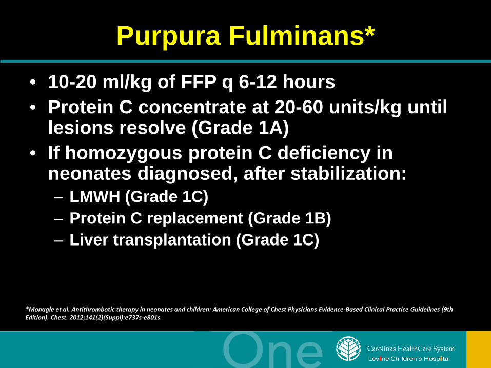
Purpura Fulminans* • 10-20 ml/kg of FFP q 6-12 hours • Protein C concentrate at 20-60 units/kg until
lesions resolve (Grade 1A) • If homozygous protein C deficiency in
neonates diagnosed, after stabilization: – LMWH (Grade 1C) – Protein C replacement (Grade 1B) – Liver transplantation (Grade 1C)
*Monagle et al. Antithrombotic therapy in neonates and children: American College of Chest Physicians Evidence-Based Clinical Practice Guidelines (9th Edition). Chest. 2012;141(2)(Suppl):e737s-e801s.
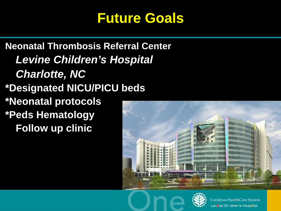
Future Goals
Neonatal Thrombosis Referral Center Levine Children’s Hospital Charlotte, NC *Designated NICU/PICU beds *Neonatal protocols *Peds Hematology Follow up clinic

Future Goals
• Safer (tested) and easier to administer neonatal anticoagulation
• Enhanced databases specific for types of
thromboses and patient populations
• Specific referral centers for neonatal coagulation disorders
Conclusions
• Lack of randomized trials addressing neonatal thromboses force neonatologists to base decisions on limited evidence
• Treat effectively without causing harm
Acknowledgements
• Neil Harris, PhD • Martha Sola-Visner, MD • Marilyn Manco-Johnson, MD • Joel Kaplan, DO • Damian Powers
– Damianpowers.com