Embed Size (px)
Citation preview
Hindawi Publishing CorporationUlcersVolume 2011, Article ID 340157, 23 pagesdoi:10.1155/2011/340157
Review Article
The Human Gastric Pathogen Helicobacter pylori andIts Association with Gastric Cancer and Ulcer Disease
Bianca Bauer and Thomas F. Meyer
Department of Molecular Biology, Max Planck Institute for Infection Biology, Campus Charite Mitte, Chariteplatz 1,10117 Berlin, Germany
Correspondence should be addressed to Bianca Bauer, [email protected]
Received 8 October 2010; Accepted 25 April 2011
Academic Editor: Hajime Kuwayama
Copyright © 2011 B. Bauer and T. F. Meyer. This is an open access article distributed under the Creative Commons AttributionLicense, which permits unrestricted use, distribution, and reproduction in any medium, provided the original work is properlycited.
With the momentous discovery in the 1980’s that a bacterium, Helicobacter pylori, can cause peptic ulcer disease and gastric cancer,antibiotic therapies and prophylactic measures have been successful, only in part, in reducing the global burden of these diseases. Todate, ∼700,000 deaths worldwide are still attributable annually to gastric cancer alone. Here, we review H. pylori’s contribution tothe epidemiology and histopathology of both gastric cancer and peptic ulcer disease. Furthermore, we examine the host-pathogenrelationship and H. pylori biology in context of these diseases, focusing on strain differences, virulence factors (CagA and VacA),immune activation and the challenges posed by resistance to existing therapies. We consider also the important role of host-genetic variants, for example, in inflammatory response genes, in determining infection outcome and the role of H. pylori in otherpathologies—some accepted, for example, MALT lymphoma, and others more controversial, for example, idiopathic thrombocyticpurpura. More recently, intriguing suggestions that H. pylori has protective effects in GERD and autoimmune diseases, such asasthma, have gained momentum. Therefore, we consider the basis for these suggestions and discuss the potential impact for futuretherapeutic rationales.
1. Introduction
After a long history of discoveries on the pathology andbacterial colonization of the gastric mucosa starting in thebeginning of the last century [1], the gastroenterologistBarry Marshall and the pathologist Robin Warren, in the1980’s, fulfilled Koch’s postulates for the association betweengastritis and the human gastric pathogen Helicobacter pylori[1–3]. This decisive demonstration substantially changedour views of the microbiology and pathology of the humanstomach and resulted in Marshall and Warren receiving the2005 Nobel Prize in Physiology and Medicine.
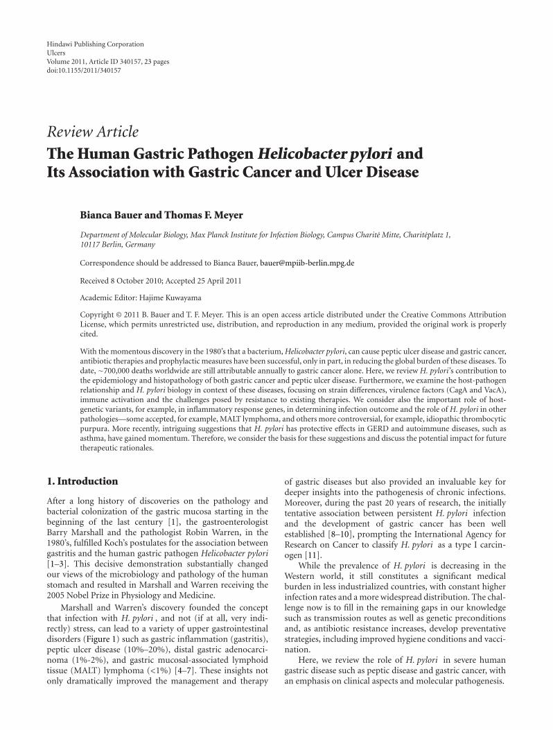
Marshall and Warren’s discovery founded the conceptthat infection with H. pylori , and not (if at all, very indi-rectly) stress, can lead to a variety of upper gastrointestinaldisorders (Figure 1) such as gastric inflammation (gastritis),peptic ulcer disease (10%–20%), distal gastric adenocarci-noma (1%-2%), and gastric mucosal-associated lymphoidtissue (MALT) lymphoma (<1%) [4–7]. These insights notonly dramatically improved the management and therapy
of gastric diseases but also provided an invaluable key fordeeper insights into the pathogenesis of chronic infections.Moreover, during the past 20 years of research, the initiallytentative association between persistent H. pylori infectionand the development of gastric cancer has been wellestablished [8–10], prompting the International Agency forResearch on Cancer to classify H. pylori as a type I carcin-ogen [11].
While the prevalence of H. pylori is decreasing in theWestern world, it still constitutes a significant medicalburden in less industrialized countries, with constant higherinfection rates and a more widespread distribution. The chal-lenge now is to fill in the remaining gaps in our knowledgesuch as transmission routes as well as genetic preconditionsand, as antibiotic resistance increases, develop preventativestrategies, including improved hygiene conditions and vacci-nation.
Here, we review the role of H. pylori in severe humangastric disease such as peptic disease and gastric cancer, withan emphasis on clinical aspects and molecular pathogenesis.
2 Ulcers
Gastritis (no overt disease)
Gastric cancer (1%–2%)MALT lymphoma (<1%)
Peptic ulcer (10%–20%)
Figure 1: Frequency of H. pylori-associated human disease. AllH. pylori-infected individuals develop gastric inflammation (gastri-tis). 10–20% develop peptic ulcers, whereas gastric cancer occurs in1-2% of cases. A minority develop MALT lymphoma (<1%).
2. Evolution and Epidemiology
The relationship between H. pylori and the human racebegan around 100,000 years ago. Phylogenetic simulationspredict that the bacterium spread from East Africa over thesame time scale as anatomically modern humans. The closeassociation is underlined by observations showing that keypatterns of bacterial genetic diversity are mirrored in themigration and ethnic origins of the human host [12]. Bygenotyping different strains collected from all over the world,the migration of humans into North America and the PacificArea has been tracked [13, 14]. Because a variety of gastricHelicobacter species can also be found in mammals besideshumans [15], it is speculated that Helicobacter species are,in general, ancestral in mammals, and we may have alreadybeen infected by ancestors of the present H. pylori strainsprior to our evolution towards modern mankind [16].
3. Prevalence
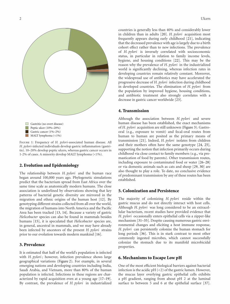
It is estimated that half of the world’s population is infectedwith H. pylori ; however, infection prevalence shows largegeographical variations (Figure 2). For example, in severalemerging nations and developing countries including India,Saudi Arabia, and Vietnam, more than 80% of the humanpopulation is infected. Infections in these regions are char-acterized by rapid acquisition even at a young age [17–19].By contrast, the prevalence of H. pylori in industrialized
countries is generally less than 40% and considerably lowerin children than in adults [20]. H. pylori acquisition mostfrequently appears during early childhood [21], indicatingthat the decreased prevalence with age is largely due to a birthcohort effect rather than to new infections. The prevalenceof H. pylori is inversely correlated with socioeconomicstatus, in particular in relation to family income levels,hygiene, and housing conditions [22]. This may be thereason why the prevalence of H. pylori in the industrializedworld is significantly declining, whereas infection rates indeveloping countries remain relatively constant. Moreover,the widespread use of antibiotics may have accelerated theprogressive decrease of H. pylori infection during childhoodin developed countries. The elimination of H. pylori fromthe population by improved hygiene, housing conditions,and antibiotic treatment also strongly correlates with adecrease in gastric cancer worldwide [23].
4. Transmission
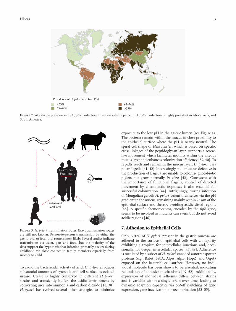
Although the association between H. pylori and severehuman disease has been established, the exact mechanismsof H. pylori acquisition are still unknown (Figure 3). Gastro-oral (e.g., exposure to vomit) and fecal-oral routes fromhuman to human are posited as the primary means oftransmission [21]. Indeed, H. pylori isolates from childrenand their mothers often have the same genotype [24, 25],supporting the notion that infection primarily occurs duringchildhood via close contact to family members (e.g., via pre-mastication of food by parents). Other transmission routes,including exposure to contaminated food or water [26–28]or via domestic animals such as cats and sheep [29, 30] arealso thought to play a role. To date, no conclusive evidenceof predominant transmission by any of these routes has beenestablished.
5. Colonization and Persistence
The majority of colonizing H. pylori reside within thegastric mucus and do not directly interact with host cells.Although H. pylori was long considered to be an extracel-lular bacterium, recent studies have provided evidence thatH. pylori occasionally enters epithelial cells via a zipper-likemechanism [31–35]. Despite causing numerous gastric envi-ronmental changes and eliciting a host immune response,H. pylori can persistently colonize the human stomach forlong periods [36]. This is in stark contrast to most othercommonly ingested microbes, which cannot successfullycolonize the stomach due to its manifold microbicidalproperties.
6. Mechanisms to Escape Low pH
One of the most efficient biological barriers against bacterialinfection is the acidic pH (<2) of the gastric lumen. However,the mucus layer overlying gastric epithelial cells exhibitsa pH gradient, ranging from about pH 2 at the luminalsurface to between 5 and 6 at the epithelial surface [37].
Ulcers 3
<55%55–64%
65–74%>75%
Prevalence of H. pylori infection (%)
Figure 2: Worldwide prevalence of H. pylori infection. Infection rates in percent. H. pylori infection is highly prevalent in Africa, Asia, andSouth America.
Gastro-oral
Gastro-oral
Fecal-oral
Fecal-oral
Figure 3: H. pylori transmission routes. Exact transmission routesare still not known. Person-to-person transmission by either thegastro-oral or fecal-oral route is most likely. Several studies indicatetransmission via water, pets and food, but the majority of thedata support the hypothesis that infection primarily occurs duringchildhood via close contact to family members especially frommother to child.
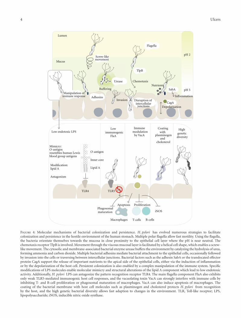
To avoid the bactericidal activity of acid, H. pylori producessubstantial amounts of cytosolic and cell surface-associatedurease. Urease is highly conserved in different H. pyloristrains and transiently buffers the acidic environment byconverting urea into ammonia and carbon dioxide [18, 38].H. pylori has evolved several other strategies to minimize
exposure to the low pH in the gastric lumen (see Figure 4).The bacteria remain within the mucus in close proximity tothe epithelial surface where the pH is nearly neutral. Thespiral cell shape of Helicobacter, which is based on specificcross-linkages of the peptidoglycan layer, supports a screw-like movement which facilitates motility within the viscousmucus layer and enhances colonization efficiency [39, 40]. Torapidly reach and remain in the mucus layer, H. pylori usespolar flagella [41, 42]. Interestingly, null mutants defective inthe production of flagella are unable to colonize gnotobioticpiglets but grow normally in vitro [43]. Consistent withthe importance of functional flagella, control of directedmovement by chemotactic responses is also essential forsuccessful colonization [44]. Intriguingly, during infectionof Mongolian gerbils H. pylori orient themselves via the pHgradient in the mucus, remaining mainly within 25 μm of theepithelial surface and thereby avoiding acidic distal regions[45]. A specific chemoreceptor, encoded by the tlpB gene,seems to be involved as mutants can swim but do not avoidacidic regions [46].
7. Adhesion to Epithelial Cells
Only ∼20% of H. pylori present in the gastric mucosa areadhered to the surface of epithelial cells with a majorityexhibiting a tropism for intercellular junctions and, occa-sionally, for deeper intercellular spaces [47, 48]. Adherenceis mediated by a subset of H. pylori-encoded autotransporterproteins (e.g., BabA, SabA, AlpA, AlpB, HopZ, and OipA)exposed on the bacterial cell surface. However, no indi-vidual molecule has been shown to be essential, indicatingredundancy of adhesive mechanisms [49–52]. Additionally,expression of individual adhesins differs between strainsand is variable within a single strain over time, leading todynamic adaption capacities via on/off switching of geneexpression, gene inactivation, or recombination [53–55].
4 Ulcers
Mucus
pH 2
pH 5
Lumen
Flagella
SabA
CagADepolarisation
Nutrients
Screw-likemovement
TlpB
Chemotaxis
Buffering
InvasionAdhesins Inflammation
Urease
Disruption ofintercellular
Inner core
Lipid A
Low endotoxic LPS
Mimicry:
TLR4
Antagonism
O-antigenO-antigenresembles human Lewis
blood group antigens
Modification:lipid A
TLR5
Lowimmunogenic
FlaA
T-cells B-cells
Proliferation
Macrophages
ApoptosisPhagosomalmaturation iNOS
Immunemodulation
by VacA
Coatingwith
plasminogenand
cholesterol
Highgeneticdiversity
Manipulation ofimmune response
junctions
Figure 4: Molecular mechanisms of bacterial colonization and persistence. H. pylori has evolved numerous strategies to facilitatecolonization and persistence in the hostile environment of the human stomach. Multiple polar flagella allow fast motility. Using the flagella,the bacteria orientate themselves towards the mucosa in close proximity to the epithelial cell layer where the pH is near neutral. Thechemotaxis receptor TlpB is involved. Movement through the viscous mucosal layer is facilitated by a helical cell shape, which enables a screw-like movement. The cytosolic and membrane-associated bacterial enzyme urease buffers the environment by catalyzing the hydrolysis of urea,forming ammonia and carbon dioxide. Multiple bacterial adhesins mediate bacterial attachment to the epithelial cells, occasionally followedby invasion into the cells or traversing between intercellular junctions. Bacterial factors such as the adhesin SabA or the translocated effectorprotein CagA support the release of important nutrients to the apical side of the epithelial cells, either via the induction of inflammationor by the depolarization of the host cell. Persistent colonization is also enabled by a complex manipulation of the immune system. Specificmodifications of LPS molecules enable molecular mimicry and structural alterations of the lipid A component which lead to low endotoxicactivity. Additionally, H. pylori LPS can antagonize the pattern recognition receptor TLR4. The main flagella component FlaA also exhibitsonly weak TLR5-mediated immunogenic host cell responses, and the vacuolating toxin VacA can strongly interfere with immune cells byinhibiting T- and B-cell proliferation or phagosomal maturation of macrophages. VacA can also induce apoptosis of macrophages. Thecoating of the bacterial membrane with host cell molecules such as plasminogen and cholesterol protects H. pylori from recognitionby the host, and the high genetic bacterial diversity allows fast adaption to changes in the environment. TLR, Toll-like receptor; LPS,lipopolysaccharide; iNOS, inducible nitric oxide synthase.
Ulcers 5
8. Manipulation of the Immune System
H. pylori can persist for long time periods within the hostwithout being extinguished by the immune system or bythe frequent gastric environmental changes. Interactions ofH. pylori with host epithelial surfaces are thought to eliciteffective escape mechanisms, often causing cellular damageand inflammation. For example, intimate adherence viaSabA has been shown to enhance inflammatory responses,which are assumed to facilitate access to essential nutrientsreleased from damaged host cells [56]. Although direct evi-dence is lacking that inflammation is beneficial for H. pylori ,the hypothesis still remains intriguing given that infectionalways elicits inflammation, regardless of symptoms. Inaddition, the delivery of cytotoxin associated antigen A(CagA), a key virulence factor, into the host cell via the typefour secretion system (T4SS), depolarizes the epithelial cellto exploit the apical cell surface for use as a replicative nicheand to obtain nutrients which are normally delivered to thebasolateral side [57].
In general terms, immunity does not seem to exert adecisive influence on the establishment of an infection, asimmune-compromised patients do not exhibit higher colo-nization rates [58]. Besides, adherence and the concomitantdelivery of toxins, H. pylori possess a range of mechanismsto attenuate and manipulate the immune response, forexample, the bacteria preferentially induce a T-helper cell 1[Th1] type-based response, classified as typical cell medi-ated immunity, essential for the fight against intracellularpathogens [59]. H. pylori also expresses lipopolysaccharide(LPS) with a very low endotoxic and immunobiologicalactivity compared to LPS of other Gram-negative bacteria[60] that can antagonize signaling mediated by the innateimmune receptor, toll-like receptor 4 (TLR4) [61]. Thisantagonistic feature is based on specific modifications of thelipid A component [62] and strain-dependent expression ofLPS O-antigens that are structurally related to Lewis bloodgroup antigens found on human cells [63]. This molecularmimicry is not only involved in autoimmune responsesbut could also allow H. pylori LPS to evade recognitionby the innate immune system [64]. The incorporation ofcholesterol and its subsequent glycosylation in the H. pylorimembrane [65] as well as the coating of the bacterium withhost molecules such as plasminogen [66] might representadditional mechanisms of antigenic camouflage. Moreover,H. pylori flagella, structures which are normally recognizedvia the innate immune receptor TLR5, evoke only a veryweak immune response due to a modified N-terminalTLR5 recognition site [67–69]. Other mechanisms by whichH. pylori triggers the host immune system are based onbacterial factors that directly target host immune cells;for example, the vacuolating cytotoxin (VacA), which isencoded in the genome of all H. pylori strains, inhibitsthe nuclear translocation of the transcription factor nuclearfactor of activated T-cells (NFAT), thereby blocking T-cellproliferation of CD4+ cells [70]. VacA also inhibits the pro-liferation of B-cells and CD8+cells [71] and is able to disruptthe normal function of macrophages by either inducingapoptosis [72] or interrupting phagosomal maturation [73].
Bacterial arginase and γ-glutamyl transferase show similarfunctions in altering the normal function of T-cells [74,75], while arginase and VacA additionally downregulate theexpression of inducible nitric oxide synthase [76, 77].
9. Genetic Diversity and Variation
H. pylori exhibits a remarkable allelic diversity and geneticvariability, typically involving endogenous (point) mutationsand recombination, which result in every infected personcarrying a distinct strain [78, 79]; however, differencesbetween relatives are minimal [80]. Allelic diversity ispromoted by a significantly higher mutation rate than foundin many other bacteria [81], which may also explain therapid development of high-level resistance to commonlyused antibiotics such as clarithromycin. High mutation ratesare most likely due to the lack of a complete DNA mismatchrepair system (mutS1/MutL/mutH) and several enzymesinvolved in base excision repair [82, 83]. A large repertoireof hyper-mutable genes frequently undergoes length changesas a consequence of slipped-strand mispairing-mediatedmutagenesis, leading to numerous subpopulations withinany large population [84]. Each of these subpopulationscarry a specific combination of active and inactive phase-variable genes; for instance, LPS biosynthesis and outermembrane encoding genes, which allow H. pylori to rapidlyadapt to environmental changes [85, 86]. High naturalcompetence for DNA uptake in combination with one of thehighest intergenomic recombination rates found amongstpathogenic bacteria [87] also contributes to the highgenomic variability between H. pylori isolates. Moreover,recent work has demonstrated that genetic exchange inducedby damage of bacterial DNA contributes to persistence ofH. pylori in its host [88]. Interestingly, a recent microarrayanalysis identified a large number of genes not previouslyassociated with infection as essential for colonization ofmice [89]. The majority of gene products were hypothetical,indicating that many other unknown bacterial factors may berequired for colonization.
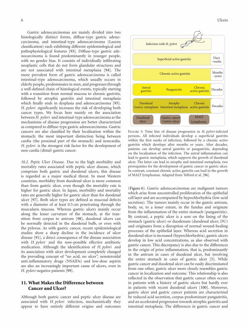
10. Gastric Cancer and Ulcers:Mutually Exclusive Pathologies?
10.1. Gastric Cancer. Although many H. pylori-associateddiseases including peptic ulcer, gastric cancer, and MALTlymphoma only develop decades after infection (Figure 5),their medical burden is tremendous. Gastric cancer is oneof the most common forms of cancer, with approximately700,000 to 900,000 new cases diagnosed every year, andthe second leading cause of cancer-related deaths worldwide[90]. Survival rates are very low, ranging from 15% ifdiagnosed during later stages of the disease to 65% ifdiagnosed early. Incidence rates vary widely geographically,and, in general, more males than females are affected [50%lower incidence]. Although high-risk areas in Japan, China,Eastern Europe, and certain Latin America countries stillremain [91], incidence rates worldwide have been decliningfor several decades [92].
6 Ulcers
Gastric adenocarcinomas are mainly divided into twohistologically distinct forms, diffuse-type gastric adeno-carcinoma, and intestinal-type adenocarcinoma (Laurenclassification) each exhibiting different epidemiological andpathophysiological features [93]. Diffuse-type gastric ade-nocarcinoma is found predominantly in younger people,with no gender bias. It consists of individually infiltratingneoplastic cells that do not form glandular structures andare not associated with intestinal metaplasia [94]. Themore prevalent form of gastric adenocarcinoma is calledintestinal-type adenocarcinoma, which usually occurs inelderly people, predominates in men, and progresses througha well-defined chain of histological events, typically startingwith a transition from normal mucosa to chronic gastritis,followed by atrophic gastritis and intestinal metaplasiawhich finally ends in dysplasia and adenocarcinoma [95].H. pylori significantly increases the risk of developing bothcancer types. We focus here mainly on the associationbetween H. pylori and intestinal-type adenocarcinoma as themechanisms of disease progression are better characterizedas compared to diffuse-type gastric adenocarcinoma. Gastriccancers are also classified by their localization within thestomach: the most important distinction being betweencardia (the proximal part of the stomach) and noncardia.H. pylori is the strongest risk factor for the development ofnon-cardia (distal) gastric cancer.
10.2. Peptic Ulcer Disease. Due to the high morbidity andmortality rates associated with peptic ulcer disease, whichcomprises both gastric and duodenal ulcers, this diseaseis regarded as a major medical threat. In most Westerncountries, morbidity from duodenal ulcer is more commonthan from gastric ulcer, even though the mortality rate ishigher for gastric ulcer. In Japan, morbidity and mortalityrates are generally higher for gastric ulcer than for duodenalulcer [97]. Both ulcer types are defined as mucosal defectswith a diameter of at least 0.5 cm penetrating through themuscularis mucosa. Whereas gastric ulcers mainly occuralong the lesser curvature of the stomach, at the tran-sition from corpus to antrum [98], duodenal ulcers canbe normally detected in the duodenal bulb, or even withthe pylorus. As with gastric cancer, recent epidemiologicalstudies show a sharp decline in the incidence of ulcerdisease [91], a direct consequence of the disease associationwith H. pylori and the now-possible effective antibioticmedication. Although the identification of H. pylori andits association with ulcer development completely changedthe prevailing concept of “no acid, no ulcer”, nonsteroidalanti-inflammatory drugs (NSAIDs) and low-dose aspirinare also an increasingly important cause of ulcers, even inH. pylori-negative patients [99].
11. What Makes the Difference betweenCancer and Ulcer?
Although both gastric cancer and peptic ulcer disease areassociated with H. pylori infection, mechanistically theyappear to have entirely different origins and outcomes
Infection with H. pylori
Superficial active gastritis
Chronic active gastritis
PangastritisAntral
gastritisChronic
active gastritis
DuodenalGastric metaplasia Intestinal metaplasia
Atrophyactive gastritis
Chronic
Duodenalulcer
Gastric cancerGastric ulcer
MALT
Lymphoma
–years
–weeksDays
Months
Decades
Decades
Figure 5: Time line of disease progression in H. pylori-infectedpersons. All infected individuals develop a superficial gastritiswithin the first weeks of infection, followed by a chronic activegastritis which develops after months or years. After decades,patients can develop antral gastritis or pangastritis, dependingon the localization of the infection. The antral inflammation canlead to gastric metaplasia, which supports the growth of duodenalulcer. The latter can lead to atrophy and intestinal metaplasia, twoprerequisites for the development of gastric cancer or gastric ulcer.In contrast, constant chronic active gastritis can lead to the growthof MALT lymphomas. Adapted from Telford et al. [96].
(Figure 6). Gastric adenocarcinomas are malignant tumorswhich arise from uncontrolled proliferation of the epithelialcell layer and are accompanied by hypochlorhydria (low-acidsecretion). The tumors mainly occur in the gastric antrum,body, or, to a lesser extent, in the fundus and originatefrom the inflammation of the entire stomach (pangastritis).By contrast, a peptic ulcer is a sore on the lining of thestomach (gastric ulcer) or duodenum (duodenal ulcer; DU)and originates from a disruption of normal wound-healingprocesses of the epithelial layer. Whereas acid secretion induodenal ulcer is increased (hyperchlorhydria), gastric ulcersdevelop in low acid concentrations, as also observed withgastric cancer. This discrepancy is also due to the differencesin the origin of prior inflammation, that is, predominantlyin the antrum in cases of duodenal ulcer, but involvingthe entire stomach in cases of gastric ulcer [5]. Whilegastric cancer and duodenal ulcer can be easily discriminatedfrom one other, gastric ulcer more closely resembles gastriccancer in localization and outcome. This relationship is alsoreflected in the observation that gastric cancer often occursin patients with a history of gastric ulcers but hardly everin patients with recent duodenal ulcers [100]. Moreover,gastric ulcer and gastric cancer patients are characterizedby reduced acid secretion, corpus-predominant pangastritis,and an accelerated progression towards atrophic gastritis andintestinal metaplasia. The differences in gastric cancer and
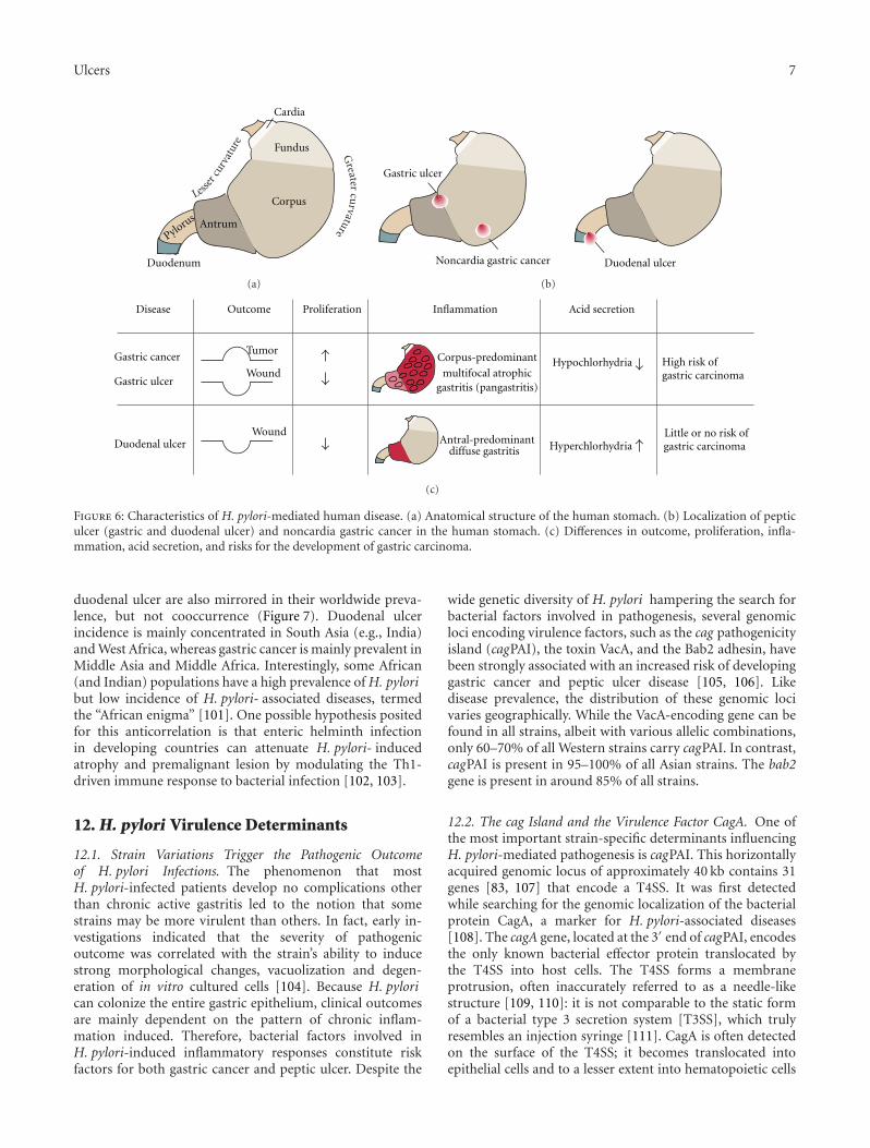
Ulcers 7
Fundus
Corpus
AntrumPyloru
s
Duodenum
Cardia
Lesser
curv
atur
e
Greater cu
rvature
(a)
Noncardia gastric cancer Duodenal ulcer
Gastric ulcer
(b)
Gastric cancer
Gastric ulcer
Duodenal ulcer
Corpus-predominant
multifocal atrophicgastritis (pangastritis)
Antral-predominantdiffuse gastritis
Inflammation
Tumor
Wound
Wound
ProliferationOutcome Acid secretion
Hyperchlorhydria
High risk ofgastric carcinoma
Little or no risk ofgastric carcinoma
Disease
Hypochlorhydria
(c)
Figure 6: Characteristics of H. pylori-mediated human disease. (a) Anatomical structure of the human stomach. (b) Localization of pepticulcer (gastric and duodenal ulcer) and noncardia gastric cancer in the human stomach. (c) Differences in outcome, proliferation, infla-mmation, acid secretion, and risks for the development of gastric carcinoma.
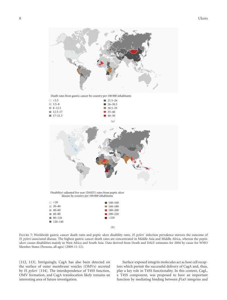
duodenal ulcer are also mirrored in their worldwide preva-lence, but not cooccurrence (Figure 7). Duodenal ulcerincidence is mainly concentrated in South Asia (e.g., India)and West Africa, whereas gastric cancer is mainly prevalent inMiddle Asia and Middle Africa. Interestingly, some African(and Indian) populations have a high prevalence of H. pyloribut low incidence of H. pylori- associated diseases, termedthe “African enigma” [101]. One possible hypothesis positedfor this anticorrelation is that enteric helminth infectionin developing countries can attenuate H. pylori- inducedatrophy and premalignant lesion by modulating the Th1-driven immune response to bacterial infection [102, 103].
12. H. pylori Virulence Determinants
12.1. Strain Variations Trigger the Pathogenic Outcomeof H. pylori Infections. The phenomenon that mostH. pylori-infected patients develop no complications otherthan chronic active gastritis led to the notion that somestrains may be more virulent than others. In fact, early in-vestigations indicated that the severity of pathogenicoutcome was correlated with the strain’s ability to inducestrong morphological changes, vacuolization and degen-eration of in vitro cultured cells [104]. Because H. pylorican colonize the entire gastric epithelium, clinical outcomesare mainly dependent on the pattern of chronic inflam-mation induced. Therefore, bacterial factors involved inH. pylori-induced inflammatory responses constitute riskfactors for both gastric cancer and peptic ulcer. Despite the
wide genetic diversity of H. pylori hampering the search forbacterial factors involved in pathogenesis, several genomicloci encoding virulence factors, such as the cag pathogenicityisland (cagPAI), the toxin VacA, and the Bab2 adhesin, havebeen strongly associated with an increased risk of developinggastric cancer and peptic ulcer disease [105, 106]. Likedisease prevalence, the distribution of these genomic locivaries geographically. While the VacA-encoding gene can befound in all strains, albeit with various allelic combinations,only 60–70% of all Western strains carry cagPAI. In contrast,cagPAI is present in 95–100% of all Asian strains. The bab2gene is present in around 85% of all strains.
12.2. The cag Island and the Virulence Factor CagA. One ofthe most important strain-specific determinants influencingH. pylori-mediated pathogenesis is cagPAI. This horizontallyacquired genomic locus of approximately 40 kb contains 31genes [83, 107] that encode a T4SS. It was first detectedwhile searching for the genomic localization of the bacterialprotein CagA, a marker for H. pylori-associated diseases[108]. The cagA gene, located at the 3′ end of cagPAI, encodesthe only known bacterial effector protein translocated bythe T4SS into host cells. The T4SS forms a membraneprotrusion, often inaccurately referred to as a needle-likestructure [109, 110]: it is not comparable to the static formof a bacterial type 3 secretion system [T3SS], which trulyresembles an injection syringe [111]. CagA is often detectedon the surface of the T4SS; it becomes translocated intoepithelial cells and to a lesser extent into hematopoietic cells
8 Ulcers
<3.53.5–88–12.512.5–1717–21.5
21.5–2626–30.530.5–3535–4040–50
Death rates from gastric cancer by country per 100 000 inhabitants
(a)
<2020–4040–6060–8080–120120–140
140–160160–180180–200200–220>220
Disability/-adjusted live year (DAILY) rates from peptic ulcerdisease by country per 100 000 inhabitants
(b)
Figure 7: Worldwide gastric cancer death rates and peptic ulcer disability rates. H. pylori infection prevalence mirrors the outcome ofH. pylori-associated disease. The highest gastric cancer death rates are concentrated in Middle Asia and Middle Africa, whereas the pepticulcer causes disabilities mainly in West Africa and South Asia. Data derived from Death and DALY estimates for 2004 by cause for WHOMember States (Persons, all ages) (2009-11-12).
[112, 113]. Intriguingly, CagA has also been detected onthe surface of outer membrane vesicles (OMVs) secretedby H. pylori [114]. The interdependence of T4SS function,OMV formation, and CagA translocation likely remains aninteresting area of future investigation.
Surface-exposed integrin molecules act as host cell recep-tors which permit the successful delivery of CagA and, thus,play a key role in T4SS functionality. In this context, CagL,a T4SS component, was proposed to have an importantfunction by mediating binding between β1α5 integrins and
Ulcers 9
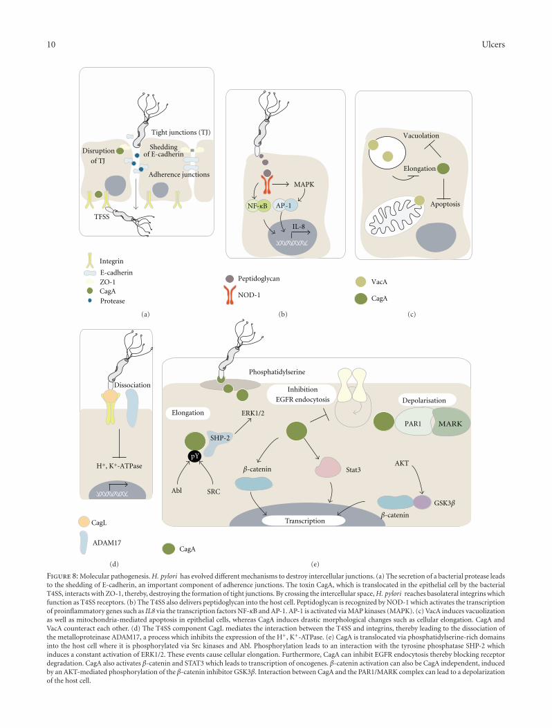
its RGD-domain [115]. Although contradictory data havebeen published [116], recent observations could supportthe CagL-integrin interaction concept by showing that CagLactivates the integrin-binding molecule ADAM17 via medi-ating its dissociation from β-1integrin [117]. Accordingly,ADAM17 inhibits the expression of the gastric H+, K+-ATPase α subunit via the transcription factor nuclear factorκB (NF-κB). Since the H+, K+-ATPase α subunit is involvedin acid secretion, these data demonstrate a T4SS-dependentmolecular mechanism for hypochlorhydria and indicate thatthe influence of cagPAI on the development of gastriccancer and gastric ulcer is more pronounced than on theinduction of duodenal ulcer (Figure 8). Additional cag-encoded proteins (CagA, CagI and CagY) have been shown tobind β1-integrin in order to induce conformational changesof integrin heterodimers which enable CagA translocation[116]. Since integrins are not located at the apical membranebut are expressed at the basolateral side, H. pylori wouldhave to reach the basolateral surface to efficiently activate theT4SS. Moreover, in addition to the normal expected apicalniche, viable bacteria have been observed within paracellularspaces and the gastric lamina propria [47]. Accordingly,H. pylori induces the shedding of E-cadherin to disruptadherence junction complexes in a CagA-independent man-ner [118], enabling bacteria to enter the basolateral side offormerly closed epithelial cell layers (Figure 8).
The status of CagA as a marker of pathogenic diseaseresulted from the observation that patients with elevatedantibody titers against CagA showed higher incidences ofboth peptic ulcers [119] and gastric adenocarcinoma [120,121]. The association of CagA with gastric cancer wascemented by subsequent work showing that the expressionof CagA in transgenic mice leads to gastric epithelial cellproliferation and the development of gastric adenocarci-noma [122]. Data showing CagA-dependent attenuation ofapoptosis in Mongolian gerbils [123] and that CagA-inducedblockage of endocytosis mediated the downregulation of theproliferation controlling epidermal growth factor receptor(EGFR) [124] further support the hypothesis that CagA hasoncogenic features. CagA can also provoke proinflammatoryresponses [125] and activate the signal transducer and acti-vator of transcription 3 (STAT3) pathway, thought to play arole in gastric cancerogenesis (Figure 8) [126]. Nevertheless,strains lacking cagPAI, which consequently do not expressCagA, have also been found in patients with peptic ulcers orgastric cancer, albeit at lower frequencies. The mechanismsby which CagA mediates gastric human disease are still notfully understood. Future work is required to unravel thecomplex but extremely efficient manipulation of importanthost cell signaling cascades facilitated by this multifunctionalprotein.
Recent investigations have shown that surface-exposedCagA interacts with externalized phosphatidylserine to initi-ate its entry into host cells [127]. Following its injection intoepithelial cells, CagA undergoes tyrosine phosphorylation[128, 129], mediated by the nonreceptor tyrosine kinasesSrc [130] and Abl [131], two well-known oncoproteins(Figure 8). Phosphorylation is tightly controlled by H. pyloriin a time-dependent manner; Src is only activated during
initial stages of infection (0.5–2 h) [132], whereas Abl isstrongly activated at late infection time points (2–8 h) [133].Phosphorylation takes place at specific C-terminal Glu-Pro-Ile-Tyr-Ala (EPIYA) sequence motifs of CagA which can varyin number in a strain-dependent manner [134, 135]. FourEPIYA-sites—A, B, C, D—have been identified. The EPIYA-Aand EPIYA-B motifs are distributed worldwide, whereas theEPIYA-C motif is more prominent in strains from Westerncountries (e.g., Europe, Australia, North America) and someAsian countries (Malaysia and India). EPIYA-D sequencemotifs are mainly present in East Asian CagA (Japan, China,and Korea) and are linked to the development of gastriccancer [136].
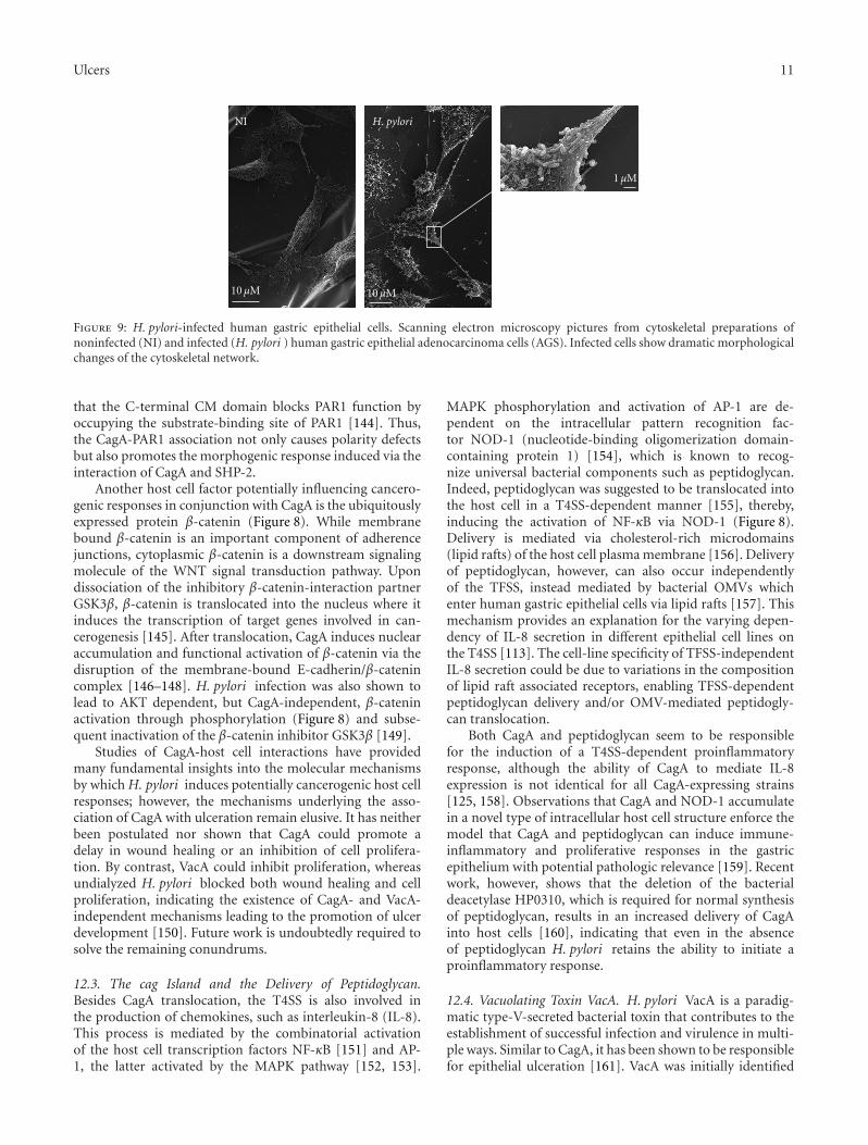
It is not only the sequence motif but also the numberand variation of phosphorylation sites that determine thepathogenic potential of CagA. Elevated phosphorylation,caused by an increase in number of EPIYA sites, leads toenhanced binding of another oncoprotein, the tyrosine phos-phatase SHP-2. After binding to CagA, SHP-2 is functionallyderegulated [137]. CagA recruits and activates SHP-2 ina phosphorylation-dependent manner and induces a Ras-independent response which leads to dramatic morpholog-ical changes of the host cell, reflected by a strong actinpolymerization and cellular elongation (Figure 9), termedthe “hummingbird” phenotype [138]. Expression of EPIYA-A and EPIYA-B sites correlate with the ability of CagAto bind the tyrosine kinase Csk, which in turn inhibitsSrc-dependent CagA-phosphorylation, thereby, attenuatingthe induction of cellular elongation by the CagA-SHP-2complex [139]. These contradictory functions of differentEPIYA sites highlight the complex and multifunctionalrole of CagA in H. pylori-mediated disease. That CagAinteracts with a surprisingly high number of host cellsignaling factors further complicates the elucidation of CagAfunction. Interactions can be phosphorylation dependent orindependent, and interaction partners range from kinases toadaptor molecules [140]. In addition to CagA-independentinteractions reported by Weydig and colleagues [118], aphosphorylation-independent interaction between Zonulaoccludens-1 (ZO-1), a junctional adhesion molecule, andCagA was shown to contribute to an alternative mecha-nism of epithelial layer disruption [141]. The CagA-ZO-1association was closely linked to an ectopic assembly oftight junction components at the site of bacterial attachmentwhich altered the composition and function of the apicaljunctional complex (see Figure 8).
Binding between CagA and the PAR1/MARK kinasecomplex, which has an essential role in the maintenance ofepithelial cell polarity (Figure 8), has also been shown to bephosphorylation independent [142]. Association with CagAinhibits PAR1 kinase activity leading to the dissociation ofPAR1 from the membrane causing junctional and polaritydefects. These effects are dependent on conserved aminoacid motifs at the C-terminus of CagA, termed the CagAmultimerization (CM) domain [143]. The binding of PAR1by the CM domain mediates the dimerization of CagA,thereby, strengthening the bond between CagA and SHP-2.The functional relevance of this association has been furthercorroborated by cocrystallography analysis of CagA, showing
10 Ulcers
Sheddingof E-cadherin
ZO-1
Tight junctions (TJ)
E-cadherin
Adherence junctions
Disruption
of TJ
TFSS
Integrin
Protease
CagA
(a)
MAPK
IL-8
Peptidoglycan
NOD-1
AP-1NF-κB
(b)
Vacuolation
Apoptosis
Elongation
VacA
CagA
(c)
H+, K+-ATPase
Dissociation
CagL
ADAM17
(d)
Phosphatidylserine
SHP-2
pY
SRCAbl
PAR1 MARK
Stat3
GSK3β
AKT
ERK1/2Elongation
Transcription
Depolarisation
Inhibition
EGFR endocytosis
CagA
β-catenin
β-catenin
(e)
Figure 8: Molecular pathogenesis. H. pylori has evolved different mechanisms to destroy intercellular junctions. (a) The secretion of a bacterial protease leadsto the shedding of E-cadherin, an important component of adherence junctions. The toxin CagA, which is translocated in the epithelial cell by the bacterialT4SS, interacts with ZO-1, thereby, destroying the formation of tight junctions. By crossing the intercellular space, H. pylori reaches basolateral integrins whichfunction as T4SS receptors. (b) The T4SS also delivers peptidoglycan into the host cell. Peptidoglycan is recognized by NOD-1 which activates the transcriptionof proinflammatory genes such as IL8 via the transcription factors NF-κB and AP-1. AP-1 is activated via MAP kinases (MAPK). (c) VacA induces vacuolizationas well as mitochondria-mediated apoptosis in epithelial cells, whereas CagA induces drastic morphological changes such as cellular elongation. CagA andVacA counteract each other. (d) The T4SS component CagL mediates the interaction between the T4SS and integrins, thereby leading to the dissociation ofthe metalloproteinase ADAM17, a process which inhibits the expression of the H+, K+-ATPase. (e) CagA is translocated via phosphatidylserine-rich domainsinto the host cell where it is phosphorylated via Src kinases and Abl. Phosphorylation leads to an interaction with the tyrosine phosphatase SHP-2 whichinduces a constant activation of ERK1/2. These events cause cellular elongation. Furthermore, CagA can inhibit EGFR endocytosis thereby blocking receptordegradation. CagA also activates β-catenin and STAT3 which leads to transcription of oncogenes. β-catenin activation can also be CagA independent, inducedby an AKT-mediated phosphorylation of the β-catenin inhibitor GSK3β. Interaction between CagA and the PAR1/MARK complex can lead to a depolarizationof the host cell.
Ulcers 11
NI H. pylori
10 μM 10 μM
1 μM
Figure 9: H. pylori-infected human gastric epithelial cells. Scanning electron microscopy pictures from cytoskeletal preparations ofnoninfected (NI) and infected (H. pylori ) human gastric epithelial adenocarcinoma cells (AGS). Infected cells show dramatic morphologicalchanges of the cytoskeletal network.
that the C-terminal CM domain blocks PAR1 function byoccupying the substrate-binding site of PAR1 [144]. Thus,the CagA-PAR1 association not only causes polarity defectsbut also promotes the morphogenic response induced via theinteraction of CagA and SHP-2.
Another host cell factor potentially influencing cancero-genic responses in conjunction with CagA is the ubiquitouslyexpressed protein β-catenin (Figure 8). While membranebound β-catenin is an important component of adherencejunctions, cytoplasmic β-catenin is a downstream signalingmolecule of the WNT signal transduction pathway. Upondissociation of the inhibitory β-catenin-interaction partnerGSK3β, β-catenin is translocated into the nucleus where itinduces the transcription of target genes involved in can-cerogenesis [145]. After translocation, CagA induces nuclearaccumulation and functional activation of β-catenin via thedisruption of the membrane-bound E-cadherin/β-catenincomplex [146–148]. H. pylori infection was also shown tolead to AKT dependent, but CagA-independent, β-cateninactivation through phosphorylation (Figure 8) and subse-quent inactivation of the β-catenin inhibitor GSK3β [149].
Studies of CagA-host cell interactions have providedmany fundamental insights into the molecular mechanismsby which H. pylori induces potentially cancerogenic host cellresponses; however, the mechanisms underlying the asso-ciation of CagA with ulceration remain elusive. It has neitherbeen postulated nor shown that CagA could promote adelay in wound healing or an inhibition of cell prolifera-tion. By contrast, VacA could inhibit proliferation, whereasundialyzed H. pylori blocked both wound healing and cellproliferation, indicating the existence of CagA- and VacA-independent mechanisms leading to the promotion of ulcerdevelopment [150]. Future work is undoubtedly required tosolve the remaining conundrums.
12.3. The cag Island and the Delivery of Peptidoglycan.Besides CagA translocation, the T4SS is also involved inthe production of chemokines, such as interleukin-8 (IL-8).This process is mediated by the combinatorial activationof the host cell transcription factors NF-κB [151] and AP-1, the latter activated by the MAPK pathway [152, 153].
MAPK phosphorylation and activation of AP-1 are de-pendent on the intracellular pattern recognition fac-tor NOD-1 (nucleotide-binding oligomerization domain-containing protein 1) [154], which is known to recog-nize universal bacterial components such as peptidoglycan.Indeed, peptidoglycan was suggested to be translocated intothe host cell in a T4SS-dependent manner [155], thereby,inducing the activation of NF-κB via NOD-1 (Figure 8).Delivery is mediated via cholesterol-rich microdomains(lipid rafts) of the host cell plasma membrane [156]. Deliveryof peptidoglycan, however, can also occur independentlyof the TFSS, instead mediated by bacterial OMVs whichenter human gastric epithelial cells via lipid rafts [157]. Thismechanism provides an explanation for the varying depen-dency of IL-8 secretion in different epithelial cell lines onthe T4SS [113]. The cell-line specificity of TFSS-independentIL-8 secretion could be due to variations in the compositionof lipid raft associated receptors, enabling TFSS-dependentpeptidoglycan delivery and/or OMV-mediated peptidogly-can translocation.
Both CagA and peptidoglycan seem to be responsiblefor the induction of a T4SS-dependent proinflammatoryresponse, although the ability of CagA to mediate IL-8expression is not identical for all CagA-expressing strains[125, 158]. Observations that CagA and NOD-1 accumulatein a novel type of intracellular host cell structure enforce themodel that CagA and peptidoglycan can induce immune-inflammatory and proliferative responses in the gastricepithelium with potential pathologic relevance [159]. Recentwork, however, shows that the deletion of the bacterialdeacetylase HP0310, which is required for normal synthesisof peptidoglycan, results in an increased delivery of CagAinto host cells [160], indicating that even in the absenceof peptidoglycan H. pylori retains the ability to initiate aproinflammatory response.
12.4. Vacuolating Toxin VacA. H. pylori VacA is a paradig-matic type-V-secreted bacterial toxin that contributes to theestablishment of successful infection and virulence in multi-ple ways. Similar to CagA, it has been shown to be responsiblefor epithelial ulceration [161]. VacA was initially identified
12 Ulcers
through its ability to cause vacuolation in cultured epithelialcells [104]. VacA-induced vacuoles are positive for markerproteins of the late endocytic compartment, including Rab7[162, 163], LAMP1, and Lgp110 [164]. It is supposedthat VacA induces the formation of large vacuoles after itsinternalization into endosomal structures, where it formsanion-selective channels, subsequently leading to swelling ofVacA-containing endosomal compartments [165]. Althoughvacuolation is readily observed in vitro, it does not seem tooccur in vivo [5].
The VacA amino acid sequence shares no similaritieswith other prokaryotic or eukaryotic proteins, and althoughall strains contain vacA, their sequences vary remarkably.vacA alleles are distinguished by differences in the 5′region(s-region) and midregion (m-region). Strains possessings1m1 type alleles are associated with an increased risk ofpeptic ulceration and gastric carcinoma as compared tostrains harboring other allelic forms, for example, s2m2[166, 167]. In countries with high rates of distal cancer, suchas Colombia and Japan, most H. pylori strains contain themore pathogenic vacA allele types [168]. In vivo experimentalstudies have corroborated the association, reporting mucosalinjury and gastric inflammation after the administration oflarge quantities of VacA into the stomachs of mice [169]and demonstrating, using isogenic wild-type and vacA-nullmutant strains that VacA contributes to severe gastritis ingerbils [170].
Monomeric 88 kDa VacA molecules are secreted into theextracellular space [171] via type V secretion [172] wherethey self-assemble into water-soluble oligomeric structuresto form anion-selective membrane channels. However, VacAcan also remain associated with the bacterial membrane as abiologically active molecule and can be taken up by the hostcell in a contact-dependent mechanism [173]. The matureform can undergo specific proteolytic cleavage to yield thefunctionally different subunits, p33 and p55 [174]. The p33domain contains a hydrophobic sequence that is involvedin pore formation [175, 176] and vacuolating cytotoxinactivity [177], whereas the p55 fragment contains cell-binding domains [178]. VacA can bind multiple epithelialcell surface molecules, including the transmembrane proteinreceptor-type tyrosine protein phosphatase-ζ (PTPRZ1)[179], fibronectin [180], EGFR [181], CD18 on T-cells [182]as well as various lipids [183] and sphingomyelin [184].
Besides its ability to induce the vacuolation of epithelialcells in vitro, VacA can also stimulate apoptosis (Figure 8),a process restricted to the more pathogenic allelic combi-nation s1m1 [185]. The p34 VacA subunit modulates mito-chondrial membrane permeability [186] by a mechanismdependent on toxin channel activity, ultimately resultingin cytochrome c release and the subsequent activation ofcaspase 3 [187]. Since VacA mutants that are defectivein forming membrane channels fail to elicit cytochrome crelease, it seems likely that VacA-mediated alterations ofmitochondria are based on the formation of VacA channelsin the mitochondrial membrane [188]. In stark contrast toVacA, CagA exhibits antiapoptotic features [123], leadingto the hypothesis that these two key virulence factors may
have antagonistic functions. Indeed, CagA has been shownto inhibit VacA-induced apoptosis [189] and reduce vacuola-tion of epithelial cells, while VacA can inhibit CagA-mediatedcellular elongation [190]. Within the latter scenario, activeVacA exerts its inhibitory impact by interfering with sig-naling pathways known to be crucial for cell scatteringand elongation, for example, the EGFR pathway [191]. Theopposing behavior of these two key virulence factors is evenmore fascinating because cagPAI-positive strains are mostlikely to possess the more toxic s1 forms of VacA, whereascagPAI-negative strains generally harbor nontoxic s2 forms.This is not due to genetic linkage as the gene loci are notclose to each other. The antagonistic properties of VacA andCagA could represent a strategy to protect the ecologicalniche of H. pylori against its own bacterial virulence factors,which are initially needed for colonization but later havedetrimental consequences for the human host. Moreover,the immunosuppressive properties of VacA may play animportant role in enabling H. pylori to persistently colonizethe human host.
12.5. Additional Virulence Attributes. Besides cagPAI, CagA,and VacA, other bacterial virulence effectors are knownto trigger H. pylori-mediated gastric disease. The duodenalulcer-promoting gene A (dupA), encoding a VirB4 ATPasehomolog, is associated with an increased risk of developingduodenal ulcer but a reduced risk of gastric atrophy andgastric cancer [192]. Recent work indicates this could be dueto dupA-mediated induction of proinflammatory cytokinesecretion by mononuclear cells [193].
Another gene implicated in peptic ulcer disease isiceA1, albeit with considerable geographic differences inexpression [194]. The gene encodes a CATG-recognizingrestriction endonuclease with significant sequence homologyto nlaIIIR, an endonuclease of Neisseria lactamica [195,196], and is induced by bacterial adherence to the gastricepithelium [197].
The Hop protein family member, bacterial outer mem-brane protein OipA (HopH) has also been identified asa potential disease-promoting factor. Like vacA, the oipAgene is present in all H. pylori strains, but its expression ismodified by phase variation, caused by variable numbers ofCT dinucleotide repeats in the 5′region. OipA was originallyidentified as a proinflammatory response-inducing protein,but may also serve as an adhesin [25]. Its expressioncorrelates with increased in vitro and in vivo productionof IL-8 [198] and, more recently, OipA has been shown tobe involved in the activation of the focal adhesion kinase(FAK) and cytoskeletal reorganization, resulting in an alteredmorphological host cell phenotype [199]. Other adhesinsof the Hop protein family such as SabA (HopP) and BabA(HopS) are also categorized as bacterial virulence factorssince they mediate bacterial adherence to the host and thusinfluence pathogenesis.
12.6. Host Cell Determinants of H. pylori Pathogenesis. It hasbecome apparent that not only the pathogen but also host
Ulcers 13
genetics play an important role in determining the clinicalmanifestation of H. pylori infections. Indeed, host geneticpolymorphisms affecting expression levels of importantgenes involved in pathogenicity have been demonstrated toinfluence susceptibility and severity of H. pylori infection. Ingeneral, genetic polymorphisms in proinflammatory genestend to increase the risk of gastric cancer, as demonstratedfor IL-1, a potent proinflammatory cytokine and the mostprominent inhibitor of gastric acid secretion [200]. IL-1is encoded by a gene cluster containing the polymorphicIL-1B (IL-1 cytokine-encoding gene) and IL-RN (IL-1receptor antagonist encoding gene) encoding genes. Severalpolymorphisms, such as IL-1B∗-31C, lead to the expressionof large quantities of IL-1β and a subsequent reduction inacid secretion [201]. Reduced acid secretion is linked tocorpus-predominant colonization by H. pylori , which resultsin pangastritis formation of atrophic gastritis and thus anincreased risk of gastric cancer and gastric ulcer disease [202–207]. Similar effects have been described for polymorphismsin other inflammation-associated genes, for example, thegenes encoding tumor necrosis factor alpha (TNF-α) andIL-10. Distinct TNF-α polymorphisms lead to increasedTNF-α expression, which influences, in concert withIL-1, gastrin production and thus acid production by parietalcells [208]. In addition to alterations in acid production,cancer patients carrying the IL1B-511T/T genotype showsignificantly higher methylation levels of specific genes thanpatients with other genotypes. This leads to the assumptionthat the L1B-511T/T allele is associated with enhancedhypermethylation of multiple CpG island loci, which mightcontribute to an increase in the risk of gastric cancerin H. pylori-infected individuals [209]. Similar to specificIL-1 genotypes, these TNF-α polymorphisms are, therefore,strongly linked to H. pylori infection and increased risk ofgastric cancer [202, 210, 211]. In addition, specific IL-10haplotypes lead to higher cytokine expression levels, thereby,shifting the balance towards an anti-inflammatory host cell[202, 212–214]; this is associated with the colonization ofmore virulent H. pylori strains [215]. By contrast, specificIL-10 haplotypes actually induce lower IL-10 expressionlevels, favoring proinflammatory responses and an associatedincreased risk of gastric cancer. Polymorphisms in othergene types may also influence H. pylori-induced disease; forinstance, a specific allelic variant of the TLR-9 promotergene sequence creates a potential NF-κB binding site thatincreases the transcriptional activity of the gene [216]. Sincealtered NF-κB activation is associated with premalignantgastric changes, this genotype could also be involved inH. pylori-mediated gastric cancer. Moreover, genes encodingIL-8 and NOD1 have also been shown to be associatedwith H. pylori-induced duodenal ulcer and gastritis [217].Interestingly, the number of polymorphisms seems to influ-ence the clinical outcome dramatically. Whereas single poly-morphisms of genes involved in proinflammatory responsesmay increase the risk of cancer development only two- tothreefold, the presence of multiple genotypes increases therisk further [205, 218].
13. Beyond Peptic Ulcerationand Gastric Cancer
13.1. MALT Lymphoma. All H. pylori-infected persons havea significantly increased risk for the development of gastricMALT lymphoma [7]. Accordingly, the majority of MALTlymphoma patients are H. pylori positive [219]. In veryrare cases, a monoclonal population of B-cells arises fromthis mucosal tissue and proliferates to form a MALT lym-phoma via chronic T-cell driven antigenic stimulation. Theincidence of MALT lymphoma in H. pylori-infected patientsis estimated to occur in less than 1% of H. pylori-positivesubjects [220], but due to diagnostic controversies, basedon difficult histological interpretations, and the rarity ofthis disorder, no exact figures are known. In contrast togastric cancer, where the “point of no return” often abolishessuccessful treatment of adenocarcinoma via the eradicationof H. pylori , the eradication of the bacteria in MALT patientscan lead to complete remission in 60%–80% of patientswith stage 1 low-grade gastric MALT lymphoma [221–224].However, 10–35% of patients in complete remission afterH. pylori eradication showed recurrent disease, necessitat-ing the implementation of mandatory long-term follow-up examinations [225]. Recurrence in specific patientscould be due to the presence of at [11; 18] (q21; q21)translocation, which is associated with API2-MALT1 fusion.API2 is involved in apoptosis, and the latter resembles acaspase-like protein. The gene fusion leads to the suppressionof apoptosis, and several studies have shown that MALT-lymphoma patients with this translocation do not or onlyrarely respond to H. pylori eradication [226, 227]. Despitethese caveats, H. pylori eradication has been designated asthe first choice treatment in the Maastricht III ConsensusReport [228].
13.2. GERD. The development of gastroesophageal refluxdisease (GERD) has long been considered to be inde-pendent of H. pylori as it occurred at the same fre-quency in H. pylori-positive and -negative patients [229].An intriguing, albeit controversial, role for H. pylori hasemerged from observations that the bacterium’s prevalencewas low in GERD patients [230], and that the incidenceof GERD increased after H. pylori eradication [231], sug-gesting that the bacteria play a protective role. Moreover,H. pylori-induced corpus gastritis has been shown to reduceacid secretion and thus prevent patients from contractingGERD [232]. Conflicting evidence exists, however, demon-strating that H. pylori eradication has no impact on eitherthe new cases of GERD [233] or the worsening of preexistingcases when treatment has been withdrawn during diseaseremission. This inverse correlation between H. pylori andGERD, if it exists, warrants further study before soundscientific conclusions can be made.
13.3. Extra-Gastrointestinal Disease. A putative role forH. pylori in the development of idiopathic thrombocyticpurpura (ITP) was first described by Gasbarrini and col-leagues [234]. Subsequent studies showed that platelet counts

14 Ulcers
in patients with ITP returned to normal levels after theeradication of H. pylori [235, 236]. In addition, anti-CagAantibody titers have been shown to be significantly decreasedin patients responsive to H. pylori eradication therapy incomparison to nonresponsive patients, implicating CagA inITP pathogenesis [237].
A number of studies have also demonstrated a linkbetween the pathogen and iron deficiency anaemia (IDA),another extragastrointestinal disease. The prevalence ofH. pylori was highly increased in patients with unexplainedIDA, and patients showed normal haemoglobin levelsafter H. pylori eradication therapies [238]. Interestingly,despite normal serum transferrin and iron levels, solubletransferring receptor (sTFR) was significantly elevated inH. pylori-infected children, suggesting that sTFR is a morereliable indicator of iron status than serum iron or ferritin.
Similar to GERD, a negative correlation betweenH. pylori and disorders such as asthma, allergy, and atopicdisease has been postulated. In general, H. pylori prevalenceand asthma as well as allergic diseases show an inverseassociation [239]; in children not predisposed to atopy,an inverse correlation between H. pylori and eczema hasbeen observed [240]. Furthermore, due to the observationsthat diminished exposure to microbes during childhoodleads to an increase of atopic disease [241], the debateregarding whether H. pylori should always be eradicatedupon diagnosis has been heightened.
14. Diagnosis and Treatment
Diagnostic tests for H. pylori are generally divided intotwo categories: invasive and noninvasive. Invasive testscomprise the histological examination of gastric specimens.Noninvasive tests are based on peripheral samples such asblood, breath, stools, urine, and saliva, in order to detectantibodies, bacterial antigens, or urease activity. The choiceof a specific test always depends on local experience andclinical settings, but usually a combination of two methodsis often recommended since, for example, the detectionof H. pylori-specific antibodies does not ultimately reflecta current infection.
Although H. pylori is sensitive to a wide range of antibi-otics in vitro, they all fail when applied as monother-apy in vivo. Therefore, a combined therapeutic strategyis used, usually including two antibiotics (clarithromycin,combined with amoxicillin or metronidazole) and eithera bismuth compound or a proton pump inhibitor (PPI).Rarely, quadruple therapies are used in which the bismuthcompound and PPI are used in combination with twoantibiotics. The use of these drugs has resulted in effectivetherapies, with eradication rates over 80%. During the pastseveral years, however, resistant bacteria have been detectedconstantly [242, 243], leading to the search for alternativedrugs and treatment strategies.
In the past decade, much effort has been devoted to thedevelopment of vaccination strategies. Based on the suc-cessful elimination of Helicobacter felis after mucosal immu-nization of mice with urease [244], the focus of much
research has been the induction of a humoral or Th2-drivenimmune response. To date, however, effective vaccinationhas only been observed in animal models and no humanvaccine trial has been successful [245]. The failure toreplicate the success of the vaccine in humans may be dueto differences in H. pylori-specific immune responses oranatomical differences of the stomach. For instance, one ofthe main surface bacterial virulence factors, cagPAI, is usuallyswitched off in mice.
Vaccines and antibiotics are not the only ways to preventand cure H. pylori infection or H. pylori-associated disease.H. pylori-positive individuals infected with helminthshave standard levels of H. pylori colonization rates andgastritis patterns, but they develop significantly lessH. pylori-associated disease [246, 247]. These are intriguingobservations that might result in low-dose administration ofimmunomodulating agents to H. pylori-positive patients,which have the same consequences as enteric helminthinfections.
Another approach is the application of probiotics. Thereis convincing evidence that H. pylori is killed by Lactobacilliboth in vitro and to a limited extent in vivo [248–250].Furthermore, Lactobacilli show a positive impact on someH. pylori therapy-related side effects, and recent studiessuggest that Lactobacilli supplements could be effective inincreasing eradication rates [251].
15. Closing Remarks
The discovery that the world’s most common bacterialinfection is clearly associated with the development of severehuman gastric disease signaled a medical revolution thathas already significantly reduced the incidence of one majorhuman disorder (duodenal ulcer disease) and also promisesto decrease a global lethal malignancy (gastric cancer). Thegreat leaps forward in understanding the mechanisms ofH. pylori pathogenesis are redefining our understanding ofbacterial ecology and homeostasis; however, we are still along way away from completely understanding how H. pyloriis associated with the host and the development of disease.Elucidating this conundrum faces a number of challengesthat require a combination of global and more focused, in-depth analyses. Monitoring the epigenetic changes occurringduring infection, alongside more detailed analyses of theH. pylori-induced adaptive and innate immune responsesmay help to decipher the reasons for the failure of cur-rent vaccines. Given the increased incidence of antibioticresistance, discovery of so far unknown bacterial virulencefactors may potentially facilitate the development of newdrugs. Moreover, in light of accumulating data showing thatH. pylori infection could be beneficial for humans, we mayneed to rethink the commonly used medical approaches totreat H. pylori infections.
References
[1] M. Kidd and I. M. Modlin, “A century of Helicobacter pylori:paradigms lost-paradigms regained,” Digestion, vol. 59, no. 1,pp. 1–15, 1998.
Ulcers 15
[2] J. R. Warren, “Unidentified curved bacilli on gastric epithe-lium in active chronic gastritis,” Lancet, vol. 1, no. 8336, pp.1273–1275, 1983.
[3] B. J. Marshall, J. A. Armstrong, D. B. McGechie, and R.J. Glancy, “Attempt to fulfil Koch’s postulates for pyloriccampylobacter,” Medical Journal of Australia, vol. 142, no. 8,pp. 436–439, 1985.
[4] A. Nomura, G. N. Stemmermann, P. H. Chyou, I. Kato,G. I. Perez-Perez, and M. J. Blaser, “Helicobacter pyloriinfection and gastric carcinoma among Japanese Americansin Hawaii,” New England Journal of Medicine, vol. 325, no. 16,pp. 1132–1136, 1991.
[5] J. G. Kusters, A. H. M. van Vliet, and E. J. Kuipers, “Pathogen-esis of Helicobacter pylori infection,” Clinical MicrobiologyReviews, vol. 19, no. 3, pp. 449–490, 2006.
[6] J. Parsonnet, G. D. Friedman, and D. P. Vandersteen, “Heli-cobacter pylori infection and the risk of gastric carcinoma,”New England Journal of Medicine, vol. 325, no. 16, pp. 1127–1131, 1991.
[7] J. Parsonnet, S. Hansen, and L. Rodriguez, “Helicobacterpylori infection and gastric lymphoma,” New England Journalof Medicine, vol. 330, no. 18, pp. 1267–1271, 1994.
[8] L. E. Hansson, L. Engstrand, and O. Nyren, “Helicobacterpylori infection: independent risk indicator of gastric adeno-carcinoma,” Gastroenterology, vol. 105, no. 4, pp. 1098–1103,1993.
[9] P. J. Hu, H. M. Mitchell, Y. Y. Li, M. H. Zhou, and S. L. Hazell,“Association of Helicobacter pylori with gastric cancer andobservations on the detection of this bacterium in gastriccancer cases,” American Journal of Gastroenterology, vol. 89,no. 10, pp. 1806–1810, 1994.
[10] N. Uemura, S. Okamoto, and S. Yamamoto, “Helicobacterpylori infection and the development of gastric cancer,” NewEngland Journal of Medicine, vol. 345, no. 11, pp. 784–789,2001.
[11] World Health Organisation, “Schistosomes, liver flukes andHelicobacter pylori. IARC Working Group on the Evaluationof Carcinogenic Risks to Humans. Lyon, 7–14 June 1994,”IARC Monographs on the Evaluation of Carcinogenic Risks toHumans / World Health Organization, International Agencyfor Research on Cancer, vol. 61, pp. 1–241, 1994.
[12] B. Linz, F. Balloux, and Y. Moodley, “An African origin forthe intimate association between humans and Helicobacterpylori,” Nature, vol. 445, no. 7130, pp. 915–918, 2007.
[13] D. Falush, T. Wirth, and B. Linz, “Traces of human migra-tions in Helicobacter pylori populations,” Science, vol. 299,no. 5612, pp. 1582–1585, 2003.
[14] Y. Moodley, B. Linz, Y. Yamaoka et al., “The peopling of thepacific from a bacterial perspective,” Science, vol. 323, no.5913, pp. 527–530, 2009.
[15] J. V. Solnick and D. B. Schauer, “Emergence of diverseHelicobacter species in the pathogenesis of gastric andenterohepatic diseases,” Clinical Microbiology Reviews, vol.14, no. 1, pp. 59–97, 2001.
[16] T. L. Cover and M. J. Blaser, “Helicobacter pylori in healthand disease,” Gastroenterology, vol. 136, no. 6, pp. 1863–1873,2009.
[17] M. A. Al-Moagel, D. G. Evans, and M. E. Abdulghani, “Preva-lence of Helicobacter (formerly Campylobacter) pylori infec-tion in Saudia Arabia, and comparison of those with andwithout upper gastrointestinal symptoms,” American Journalof Gastroenterology, vol. 85, no. 8, pp. 944–948, 1990.
[18] D. Y. Graham, M. F. Go, and D. J. Evans, “Review arti-cle: urease, gastric ammonium/ammonia, and Helicobacterpylori—the past, the present, and recommendations forfuture research,” Alimentary Pharmacology and Therapeutics,vol. 6, no. 6, pp. 659–669, 1992.
[19] H. M. Malaty, “Epidemiology of Helicobacter pylori infec-tion, best practice and research,” Clinical Gastroenterology,vol. 21, no. 2, pp. 205–214, 2007.
[20] K. D. Crew and A. I. Neugut, “Epidemiology of gastriccancer,” World Journal of Gastroenterology, vol. 12, no. 3, pp.354–362, 2006.
[21] R. A. Feldman, A. J. P. Eccersley, and J. M. Hardie, “Epi-demiology of Helicobacter pylori: acquisition, transmission,population prevalence and disease-to-infection ratio,” BritishMedical Bulletin, vol. 54, no. 1, pp. 39–53, 1998.
[22] H. Malaty, H. M. T. El-Zimaity, R. M. Genta, R. A. Cole,and D. Y. Graham, “High-dose proton pump inhibitor plusamoxycillin for the treatment or retreatment of Helicobacterpylori infection,” Alimentary Pharmacology and Therapeutics,vol. 10, no. 6, pp. 1001–1004, 1996.
[23] R. M. Peek and M. J. Blaser, “Helicobacter pylori and gas-trointestinal tract adenocarcinomas,” Nature Reviews Cancer,vol. 2, no. 1, pp. 28–37, 2002.
[24] M. Kurosawa, S. Kikuchi, Y. Inaba, T. Ishibashi, and F.Kobayashi, “Helicobacter pylori infection among Japanesechildren,” Journal of Gastroenterology and Hepatology, vol. 15,no. 12, pp. 1382–1385, 2000.
[25] Y. Yamaoka, D. H. Kwon, and D. Y. Graham, “A M(r)34000 proinflammatory outer membrane protein (OipA) ofHelicobacter pylori,” Proceedings of the National Academy ofSciences of the United States of America, vol. 97, no. 13, pp.7533–7538, 2000.
[26] K. J. Goodman, P. Correa, H. J. Tengana Aux et al.,“Helicobacter pylori infection in the Colombian Andes: apopulation-based study of transmission pathways,” AmericanJournal of Epidemiology, vol. 144, no. 3, pp. 290–299, 1996.
[27] R. J. Hopkins, P. A. Vial, and C. Ferreccio, “Seroprevalenceof Helicobacter pylori in chile: vegetables may serve as oneroute of transmission,” Journal of Infectious Diseases, vol. 168,no. 1, pp. 222–226, 1993.
[28] P. D. Klein, R. Gilman, and R. Leon-Barua, “Water sourceas risk factor for Helicobacter pylori infection in Peruvianchildren,” Lancet, vol. 337, no. 8756, pp. 1503–1506, 1991.
[29] M. P. Dore, A. R. Sepulveda, and H. El-Zimaity, “Isolation ofhelicobacter pylori from sheep-implications for transmissionto humans,” American Journal of Gastroenterology, vol. 96, no.5, pp. 1396–1401, 2001.
[30] J. G. Fox, “Non-human reservoirs of Helicobacter pylori,”Alimentary Pharmacology and Therapeutics, Supplement, vol.9, no. 2, pp. 93–103, 1995.
[31] T. Kwok, S. Backert, H. Schwarz, J. Berger, and T. F. Meyer,“Specific entry of Helicobacter pylori into cultured gastricepithelial cells via a zipper-like mechanism,” Infection andImmunity, vol. 70, no. 4, pp. 2108–2120, 2002.
[32] J. D. Oh, S. M. Karam, and J. I. Gordon, “Intracellular Heli-cobacter pylori in gastric epithelial progenitors,” Proceedingsof the National Academy of Sciences of the United States ofAmerica, vol. 102, no. 14, pp. 5186–5191, 2005.
[33] A. Ozbek, E. Ozbek, H. Dursun, Y. Kalkan, and T. Demirci,“Can Helicobacter pylori invade human gastric mucosa?:an in vivo study using electron microscopy, immunohisto-chemical methods, and real-time polymerase chain reaction,”
16 Ulcers
Journal of Clinical Gastroenterology, vol. 44, no. 6, pp. 416–422, 2010.
[34] A. M. Petersen, J. Blom, L. P. Andersen, and K. A. Krogfelt,“Role of strain type, AGS cells and fetal calf serum inHelicobacter pylori adhesion and invasion assays,” FEMSImmunology and Medical Microbiology, vol. 29, no. 1, pp. 59–67, 2000.
[35] M. R. Terebiznik, C. L. Vazquez, and K. Torbicki, “Helicobac-ter pylori VacA toxin promotes bacterial intracellular survivalin gastric epithelial cells,” Infection and Immunity, vol. 74, no.12, pp. 6599–6614, 2006.
[36] H. M. S. Algood and T. L. Cover, “Helicobacter pyloripersistence: an overview of interactions between H. pyloriand host immune defenses,” Clinical Microbiology Reviews,vol. 19, no. 4, pp. 597–613, 2006.
[37] N. J. Talley, J. E. Ormand, C. A. Frie, and A. R. Zinsmeister,“Stability of pH gradients in vivo across the stomach in Heli-cobacter pylori gastritis, dyspepsia, and health,” AmericanJournal of Gastroenterology, vol. 87, no. 5, pp. 590–594, 1992.
[38] J. V. Solnick, C. Josenhans, S. Suerbaum, L. S. Tompkins,and A. Labigne, “Construction and characterization of anisogenic urease-negative mutant of Helicobacter mustelae,”Infection and Immunity, vol. 63, no. 9, pp. 3718–3721, 1995.
[39] G. Geis, H. Leying, S. Suerbaum, U. Mai, and W. Opferkuch,“Ultrastructure and chemical analysis of Campylobacterpylori flagella,” Journal of Clinical Microbiology, vol. 27, no.3, pp. 436–441, 1989.
[40] L. K. Sycuro, Z. Pincus, K. D. Gutierrez et al., “Peptidoglycancrosslinking relaxation promotes Helicobacter pylori’s helicalshape and stomach colonization,” Cell, vol. 141, no. 5, pp.822–833, 2010.
[41] R. Haas, T. F. Meyer, and J. P. M. Van Putten, “Aflagellatedmutants of Helicobacter pylori generated by genetic trans-formation of naturalloy competent strains using transposonshuttle mutagenesis,” Molecular Microbiology, vol. 8, no. 4,pp. 753–760, 1993.
[42] S. Suerbaum, “The complex flagella of gastric Helicobacterspecies,” Trends in Microbiology, vol. 3, no. 5, pp. 168–170,1995.
[43] K. A. Eaton, S. Suerbaum, C. Josenhans, and S. Krakowka,“Colonization of gnotobiotic piglets by Helicobacter pylorideficient in two flagellin genes,” Infection and Immunity, vol.64, no. 7, pp. 2445–2448, 1996.
[44] D. J. McGee, M. L. Langford, E. L. Watson, J. E. Carter, Y.T. Chen, and K. M. Ottemann, “Colonization and inflamma-tion deficiencies in Mongolian gerbils infected by Helicobac-ter pylori chemotaxis mutants,” Infection and Immunity, vol.73, no. 3, pp. 1820–1827, 2005.
[45] S. R. Schreiber, M. Konradt, C. Groll et al., “The spatialorientation of Helicobacter pylori in the gastric mucus,”Proceedings of the National Academy of Sciences of the UnitedStates of America, vol. 101, no. 14, pp. 5024–5029, 2004.
[46] M. A. Croxen, G. Sisson, R. Melano, and P. S. Hoffman,“The Helicobacter pylori chemotaxis receptor tlpB (HP0103)is required for pH taxis and for colonization of the gastricmucosa,” Journal of Bacteriology, vol. 188, no. 7, pp. 2656–2665, 2006.
[47] V. Necchi, M. E. Candusso, and F. Tava, “Intracellular,intercellular, and stromal invasion of gastric mucosa, preneo-plastic lesions, and cancer by Helicobacter pylori,” Gastroen-terology, vol. 132, no. 3, pp. 1009–1023, 2007.
[48] L. A. Noach, T. M. Rolf, and G. N. J. Tytgat, “Electron micro-scopic study of association between Helicobacter pylori and
gastric and duodenal mucosa,” Journal of Clinical Pathology,vol. 47, no. 8, pp. 699–704, 1994.
[49] A. Dossumbekova, C. Prinz, J. Mages et al., “Helicobacterpylori HopH (OipA) and bacterial pathogenicity: genetic andfunctional genomic analysis of hopH gene polymorphisms,”Journal of Infectious Diseases, vol. 194, no. 10, pp. 1346–1355,2006.
[50] D. Ilver, A. Arnqvist, J. Ogren et al., “Helicobacter pyloriadhesin binding fucosylated histo-blood group antigensrevealed by retagging,” Science, vol. 279, no. 5349, pp. 373–377, 1998.
[51] J. Mahdavi, B. Sonden, M. Hurtig et al., “Helicobacterpylori SabA adhesin in persistent infection and chronicinflammation,” Science, vol. 297, no. 5581, pp. 573–578, 2002.
[52] B. Peck, M. Ortkamp, K. D. Diehl, E. Hundt, and B.Knapp, “Conservation, localization and expression of HopZ,a protein involved in adhesion of Helicobacter pylori,”Nucleic Acids Research, vol. 27, no. 16, pp. 3325–3333, 1999.
[53] M. Aspholm-Hurtig, G. Dailide, M. Lahmann et al., “Func-tional adaptation of BabA the H. pylori ABO blood groupantigen binding adhesin,” Science, vol. 305, no. 5683, pp. 519–522, 2004.
[54] A. Backstrom, C. Lundberg, D. Kersulyte, D. E. Berg, T.Boren, and A. Arnqvist, “Metastability of Helicobacter pyloribab adhesin genes and dynamics in Lewis b antigen binding,”Proceedings of the National Academy of Sciences of the UnitedStates of America, vol. 101, no. 48, pp. 16923–16928, 2004.
[55] J. V. Solnick, L. M. Hansen, N. R. Salama, J. K. Boonjakuakul,and M. Syvanen, “Modification of Helicobacter pylori outermembrane protein expression during experimental infectionof rhesus macaques,” Proceedings of the National Academy ofSciences of the United States of America, vol. 101, no. 7, pp.2106–2111, 2004.
[56] C. Petersson, M. Forsberg, M. Aspholm et al., “Helicobacterpylori SabA adhesin evokes a strong inflammatory responsein human neutrophils which is down-regulated by theneutrophil-activating protein,” Medical Microbiology andImmunology, vol. 195, no. 4, pp. 195–206, 2006.
[57] S. Tan, L. S. Tompkins, and M. R. Amieva, “Helicobacterpylori usurps cell polarity to turn the cell surface into areplicative niche,” PLoS Pathogens, vol. 5, no. 5, Article IDe1000407, 2009.
[58] R. Battan, M. C. Raviglione, A. Palagiano et al., “Helicobacterpylori infection in patients with acquired immune deficiencysyndrome,” American Journal of Gastroenterology, vol. 85, no.12, pp. 1576–1579, 1990.
[59] K. B. Bamford, X. Fan, S. E. Crowe et al., “Lymphocytes inthe human gastric mucosa during Helicobacter pylori have aT helper cell 1 phenotype,” Gastroenterology, vol. 114, no. 3,pp. 482–492, 1998.
[60] A. Muotiala, I. M. Helander, L. Pyhala, T. U. Kosunen, andA. P. Moran, “Low biological activity of Helicobacter pylorilipopolysaccharide,” Infection and Immunity, vol. 60, no. 4,pp. 1714–1716, 1992.
[61] P. M. Lepper, M. Triantafilou, C. Schumann, E. M. Schneider,and K. Triantafilou, “Lipopolysaccharides from Helicobacterpylori can act as antagonists for Toll-like receptor 4,” CellularMicrobiology, vol. 7, no. 4, pp. 519–528, 2005.
[62] A. P. Moran, B. Lindner, and E. J. Walsh, “Structural charac-terization of the lipid A component of Helicobacter pylorirough- and smooth-form lipopolysaccharides,” Journal ofBacteriology, vol. 179, no. 20, pp. 6453–6463, 1997.
Ulcers 17
[63] G. O. Aspinall and M. A. Monteiro, “Lipopolysaccharides ofHelicobacter pylori strains P466 and MO19: structures of theO antigen and core oligosaccharide regions,” Biochemistry,vol. 35, no. 7, pp. 2498–2504, 1996.
[64] B. J. Appelmelk, I. Simoons-Smit, R. Negrini et al., “Potentialrole of molecular mimicry between Helicobacter pylorilipopolysaccharide and host Lewis blood group antigens inautoimmunity,” Infection and Immunity, vol. 64, no. 6, pp.2031–2040, 1996.
[65] C. Wunder, Y. Churin, and F. Winau, “Cholesterol gluco-sylation promotes immune evasion by Helicobacter pylori,”Nature Medicine, vol. 12, no. 9, pp. 1030–1038, 2006.
[66] M. Ringner, K. H. Valkonen, and T. Wadstrom, “Binding ofvitronectin and plasminogen to Helicobacter pylori,” FEMSImmunology and Medical Microbiology, vol. 9, no. 1, pp. 29–34, 1994.
[67] E. Andersen-Nissen, K. D. Smith, K. L. Strobe et al., “Evasionof Toll-like receptor 5 by flagellated bacteria,” Proceedingsof the National Academy of Sciences of the United States ofAmerica, vol. 102, no. 26, pp. 9247–9252, 2005.
[68] A. T. Gewirtz, Y. Yu, U. S. Krishna, D. A. Israel, S. L. Lyons,and R. M. Peek, “Helicobacter pylori flagellin evades toll-likereceptor 5-mediated innate immunity,” Journal of InfectiousDiseases, vol. 189, no. 10, pp. 1914–1920, 2004.
[69] S. K. Lee, A. Stack, E. Katzowitsch, S. I. Aizawa, S. Suerbaum,and C. Josenhans, “Helicobacter pylori flagellins have verylow intrinsic activity to stimulate human gastric epithelialcells via TLR5,” Microbes and Infection, vol. 5, no. 15, pp.1345–1356, 2003.
[70] B. Gebert, W. Fischer, E. Weiss, R. Hoffmann, and R.Haas, “Helicobacter pylori vacuolating cytotoxin inhibits Tlymphocyte activation,” Science, vol. 301, no. 5636, pp. 1099–1102, 2003.
[71] V. J. Torres, S. E. VanCompernolle, M. S. Sundrud, D.Unutmaz, and T. L. Cover, “Helicobacter pylori vacuolatingcytotoxin inhibits activation-induced proliferation of humanT and B lymphocyte subsets,” Journal of Immunology, vol.179, no. 8, pp. 5433–5440, 2007.
[72] R. J. Menaker, P. J. M. Ceponis, and N. L. Jones, “Helicobacterpylori induces apoptosis of macrophages in association withalterations in the mitochondrial pathway,” Infection andImmunity, vol. 72, no. 5, pp. 2889–2898, 2004.
[73] P. Y. Zheng and N. L. Jones, “Helicobacter pylori strainsexpressing the vacuolating cytotoxin interrupt phagosomematuration in macrophages by recruiting and retainingTACO (coronin 1) protein,” Cellular Microbiology, vol. 5, no.1, pp. 25–40, 2003.
[74] C. Schmees, C. Prinz, T. Treptau et al., “Inhibition of T-cell proliferation by Helicobacter pylori gamma-glutamyltranspeptidase,” Gastroenterology, vol. 132, no. 5, pp. 1820–1833, 2007.
[75] J. Zabaleta, D. J. McGee, A. H. Zea et al., “Helicobacterpylori arginase inhibits T cell proliferation and reduces theexpression of the TCR zeta-chain (CD3zeta),” Journal ofImmunology, vol. 173, no. 1, pp. 586–593, 2004.
[76] A. P. Gobert, D. J. McGee, M. Akhtar et al., “Helicobacterpylori arginase inhibits nitric oxide production by eukaryoticcells: a strategy for bacterial survival,” Proceedings of theNational Academy of Sciences of the United States of America,vol. 98, no. 24, pp. 13844–13849, 2001.
[77] J. Yuan, P. Li, J. Tao et al., “H. pylori escape host immunore-action through inhibiting ILK expression by VacA,” Cellularand Molecular Immunology, vol. 6, no. 3, pp. 191–197, 2009.
[78] N. Akopyanz, N. O. Bukanov, T. U. Westblom, S. Kresovich,and D. E. Berg, “DNA diversity among clinical isolates ofhelicobacter pylori detected by PCR-based RAPD finger-printing,” Nucleic Acids Research, vol. 20, no. 19, pp. 5137–5142, 1992.
[79] I. Kansau, J. Raymond, and E. Bingen, “Genotyping ofHelicobacter pylori isolates by sequencing of PCR productsand comparison with the RAPD technique,” Research inMicrobiology, vol. 147, no. 8, pp. 661–669, 1996.
[80] S. Miehlke, R. M. Genta, D. Y. Graham, and M. F. Go,“Molecular relationships of Helicobacter pylori strains ina family with gastroduodenal disease,” American Journal ofGastroenterology, vol. 94, no. 2, pp. 364–368, 1999.
[81] B. Bjorkholm, M. Sjolund, P. G. Falk, O. G. Berg, L.Engstrand, and D. I. Andersson, “Mutation frequency andbiological cost of antibiotic resistance in Helicobacter pylori,”Proceedings of the National Academy of Sciences of the UnitedStates of America, vol. 98, no. 25, pp. 14607–14612, 2001.
[82] S. Suerbaum and C. Josenhans, “Helicobacter pylori evo-lution and phenotypic diversification in a changing host,”Nature Reviews Microbiology, vol. 5, no. 6, pp. 441–452, 2007.
[83] J. F. Tomb, O. White, A. R. Kerlavage et al., “The completegenome sequence of the gastric pathogen Helicobacterpylori,” Nature, vol. 388, no. 6642, pp. 539–547, 1997.
[84] B. D. Robertson and T. F. Meyer, “Genetic variation inpathogenic bacteria,” Trends in Genetics, vol. 8, no. 12, pp.422–427, 1992.
[85] L. Salaun, B. Linz, S. Suerbaum, and N. J. Saunders, “Thediversity within an expanded and redefined repertoire ofphase-variable genes in Helicobacter pylori,” Microbiology,vol. 150, no. 4, pp. 817–830, 2004.
[86] N. J. Saunders, J. F. Peden, D. W. Hood, and E. R. Moxon,“Simple sequence repeats in the Helicobacter pylori genome,”Molecular Microbiology, vol. 27, no. 6, pp. 1091–1098, 1998.
[87] M. Perez-Losada, E. B. Browne, A. Madsen, T. Wirth, R.P. Viscidi, and K. A. Crandall, “Population genetics ofmicrobial pathogens estimated from multilocus sequencetyping (MLST) data,” Infection, Genetics and Evolution, vol.6, no. 2, pp. 97–112, 2006.
[88] M. S. Dorer, J. Fero, and N. R. Salama, “DNA damage triggersgenetic exchange in Helicobacter pylori,” PLoS Pathogens, vol.6, no. 7, Article ID e1001026, pp. 1–10, 2010.
[89] D. N. Baldwin, B. Shepherd, and P. Kraemer, “Identificationof Helicobacter pylori genes that contribute to stomachcolonization,” Infection and Immunity, vol. 75, no. 2, pp.1005–1016, 2007.
[90] D. M. Parkin, F. Bray, J. Ferlay, and P. Pisani, “Global cancerstatistics, 2002,” CA: A Cancer Journal for Clinicians, vol. 55,no. 2, pp. 74–108, 2002.
[91] World Health Organization, “Disease and injury countryestimates,” 2009.
[92] P. Bertuccio, L. Chatenoud, and F. Levi, “Recent patterns ingastric cancer: a global overview,” International Journal ofCancer, vol. 125, no. 3, pp. 666–673, 2009.
[93] P. Lauren, “The two histological main types of gastriccarcinoma: diffuse and so-called intestinal-type carcinoma.An attempt at a histo-clinical classification,” Acta Pathologicaet Microbiologica Scandinavica, vol. 64, pp. 31–49, 1965.
[94] D. B. Polk and R. M. Peek, “Helicobacter pylori: gastriccancer and beyond,” Nature Reviews Cancer, vol. 10, no. 6,pp. 403–414, 2010.
[95] P. Correa, “Diet modification and gastric cancer prevention,”Journal of the National Cancer Institute. Monographs, vol. 12,no. 12, pp. 75–78, 1992.
18 Ulcers
[96] J. L. Telford, A. Covacci, R. Rappuoli, and P. Chiara,“Immunobiology of Helicobacter pylori infection,” CurrentOpinion in Immunology, vol. 9, no. 4, pp. 498–503, 1997.
[97] J. H. Kurata and B. M. Haile, “Epidemiology of peptic ulcerdisease,” Clinics in Gastroenterology, vol. 13, no. 2, pp. 289–307, 1984.
[98] S. J. van Zanten, M. F. Dixon, and A. Lee, “The gastrictransitional zones: neglected links between gastroduodenalpathology and Helicobacter ecology,” Gastroenterology, vol.116, no. 5, pp. 1217–1229, 1999.
[99] L. A. Garcıa Rodrıguez and L. Barreales Tolosa, “Risk ofupper gastrointestinal complications among users of tra-ditional NSAIDs and COXIBs in the general population,”Gastroenterology, vol. 132, no. 2, pp. 498–506, 2007.
[100] S. Take, M. Mizuno, K. Ishiki et al., “The effect of eradicatingHelicobacter pylori on the development of gastric cancerin patients with peptic ulcer disease,” American Journal ofGastroenterology, vol. 100, no. 5, pp. 1037–1042, 2005.
[101] C. Holcombe, “Helicobacter pylori: the African enigma,”Gut, vol. 33, no. 4, pp. 429–431, 1992.
[102] R. Ally, H. M. Mitchell, and I. Segal, “Differences in theimmune response to H. pylori infection in Sowetan subjectsmay relate to concurrent parasitic infections,” South AfricanMedical Journal, vol. 90, p. 642, 2000.
[103] J. G. Fox, P. Beck, C. A. Dangler et al., “Concurrent enterichelminth infection modulates inflammation and gastricimmune responses and reduces helicobacter-induced gastricatrophy,” Nature Medicine, vol. 6, no. 5, pp. 536–542, 2000.
[104] R. D. Leunk, P. T. Johnson, B. C. David, W. G. Kraft, andD. R. Morgan, “Cytotoxic activity in broth-culture filtrates ofCampylobacter pylori,” Journal of Medical Microbiology, vol.26, no. 2, pp. 93–99, 1988.
[105] M. Gerhard, N. Lehn, N. Neumayer et al., “Clinical relevanceof the Helicobacter pylori gene for blood-group antigen-binding adhesin,” Proceedings of the National Academy ofSciences of the United States of America, vol. 96, no. 22, pp.12778–12783, 1999.
[106] L. J. Van Doorn, C. Figueiredo, R. Sanna et al., “Clinicalrelevance of the cagA, vacA, and iceA status of Helicobacterpylori,” Gastroenterology, vol. 115, no. 1, pp. 58–66, 1998.
[107] R. A. Alm, L. S. L. Ling, D. T. Moir et al., “Genomic-sequencecomparison of two unrelated isolates of the human gastricpathogen Helicobacter pylori,” Nature, vol. 397, no. 6715, pp.176–180, 1999.
[108] J. C. Atherton, “H. pylori virulence factors,” British MedicalBulletin, vol. 54, no. 1, pp. 105–120, 1998.
[109] M. Rohde, J. Puls, R. Buhrdorf, W. Fischer, and R. Haas, “Anovel sheathed surface organelle of the Helicobacter pyloricag type IV secretion system,” Molecular Microbiology, vol.49, no. 1, pp. 219–234, 2003.
[110] J. Tanaka, T. Suzuki, H. Mimuro, and C. Sasakawa, “Struc-tural definition on the surface of Helicobacter pylori type IVsecretion apparatus,” Cellular Microbiology, vol. 5, no. 6, pp.395–404, 2003.
[111] T. Kubori, Y. Matsushima, and D. Nakamura, “Supramolecu-lar structure of the salmonella typhimurium type III proteinsecretion system,” Science, vol. 280, no. 5363, pp. 602–605,1998.
[112] S. Odenbreit, J. Puls, B. Sedlmaier, E. Gerland, W. Fischer,and R. Haas, “Translocation of Helicobacter pylori CagA intogastric epithelial cells by type IV secretion,” Science, vol. 287,no. 5457, pp. 1497–1500, 2000.
[113] B. Bauer, S. Moese, S. Bartfeld, T. F. Meyer, and M. Selbach,“Analysis of cell type-specific responses mediated by the type
IV secretion system of Helicobacter pylori,” Infection andImmunity, vol. 73, no. 8, pp. 4643–4652, 2005.
[114] A. Olofsson, A. Vallstrom, K. Petzold et al., “Biochemical andfunctional characterization of Helicobacter pylori vesicles,”Molecular Microbiology, vol. 77, no. 6, pp. 1539–1555, 2010.
[115] T. Kwok, D. Zabler, S. Urman et al., “Helicobacter exploitsintegrin for type IV secretion and kinase activation,” Nature,vol. 449, no. 7164, pp. 862–866, 2007.
[116] L. F. Jimenez-Soto, S. Kutter, and X. Sewald, “Helicobacterpylori type IV secretion apparatus exploits β1 integrin in anovel RGD-independent manner,” PLoS Pathogens, vol. 5, no.12, Article ID e1000684, 2009.
[117] A. Saha, S. Backert, C. E. Hammond, M. Gooz, and A.J. Smolka, “Helicobacter pylori CagL activates ADAM17 toinduce repression of the gastric H, K-ATPase α subunit,”Gastroenterology, vol. 139, no. 1, pp. 239–248, 2010.
[118] C. Weydig, A. Starzinski-Powitz, G. Carra, J. Lower, and S.Wessler, “CagA-independent disruption of adherence junc-tion complexes involves E-cadherin shedding and impliesmultiple steps in Helicobacter pylori pathogenicity,” Exper-imental Cell Research, vol. 313, no. 16, pp. 3459–3471, 2007.
[119] A. M. Y. Nomura, J. Lee, G. N. Stemmermann, R. Y. Nomura,G. I. Perez-Perez, and M. J. Blaser, “Helicobacter pyloriCagA seropositivity and gastric carcinoma risk in a JapaneseAmerican population,” Journal of Infectious Diseases, vol. 186,no. 8, pp. 1138–1144, 2002.
[120] M. J. Blaser, G. I. Perez-Perez, H. Kleanthous et al., “Infectionwith Helicobacter pylori strains possessing cagA is associatedwith an increased risk of developing adenocarcinoma of thestomach,” Cancer Research, vol. 55, no. 10, pp. 2111–2115,1995.
[121] A. H. Wu, J. E. Crabtree, L. Bernstein et al., “Role of Heli-cobacter pylori CagA+ strains and risk of adenocarcinoma ofthe stomach and esophagus,” International Journal of Cancer,vol. 103, no. 6, pp. 815–821, 2003.
[122] N. Ohnishi, H. Yuasa, S. Tanaka et al., “Transgenic expressionof Helicobacter pylori CagA induces gastrointestinal andhematopoietic neoplasms in mouse,” Proceedings of theNational Academy of Sciences of the United States of America,vol. 105, no. 3, pp. 1003–1008, 2008.
[123] H. Mimuro, T. Suzuki, S. Nagai et al., “Helicobacter pyloridampens gut epithelial self-renewal by inhibiting apoptosis,a bacterial strategy to enhance colonization of the stomach,”Cell Host and Microbe, vol. 2, no. 4, pp. 250–263, 2007.
[124] B. Bauer, S. Bartfeld, and T. F. Meyer, “H. pylori selectivelyblocks EGFR endocytosis via the non-receptor kinase c-Abland CagA,” Cellular Microbiology, vol. 11, no. 1, pp. 156–169,2009.
[125] S. Brandt, T. Kwok, R. Hartig, W. Konig, and S. Backert,“NF-kappaB activation and potentiation of proinflammatoryresponses by the Helicobacter pylori CagA protein,” Proceed-ings of the National Academy of Sciences of the United States ofAmerica, vol. 102, no. 26, pp. 9300–9305, 2005.
[126] D. M. Bronte-Tinkew, M. Terebiznik, and A. Franco, “Heli-cobacter pylori cytotoxin-associated gene a activates thesignal transducer and activator of transcription 3 pathway invitro and in vivo,” Cancer Research, vol. 69, no. 2, pp. 632–639, 2009.
[127] N. Murata-Kamiya, K. Kikuchi, T. Hayashi, H. Higashi,and M. Hatakeyama, “Helicobacter pylori exploits hostmembrane phosphatidylserine for delivery, localization, andpathophysiological action of the CagA oncoprotein,” CellHost and Microbe, vol. 7, no. 5, pp. 399–411, 2010.
Ulcers 19
[128] M. Stein, R. Rappuoli, and A. Covacci, “Tyrosine phospho-rylation of the Helicobacter pylori CagA antigen after cag-driven host cell translocation,” Proceedings of the NationalAcademy of Sciences of the United States of America, vol. 97,no. 3, pp. 1263–1268, 2000.
[129] M. Asahi, T. Azuma, and S. Ito, “Helicobacter pylori CagAprotein can be tyrosine phosphorylated in gastric epithelialcells,” Journal of Experimental Medicine, vol. 191, no. 4, pp.593–602, 2000.
[130] M. Selbach, S. Moese, C. R. Hauck, T. F. Meyer, and S.Backert, “Src is the kinase of the Helicobacter pylori CagAprotein in vitro and in vivo,” Journal of Biological Chemistry,vol. 277, no. 9, pp. 6775–6778, 2002.
[131] M. Poppe, S. M. Feller, G. Romer, and S. Wessler, “Phospho-rylation of Helicobacter pylori CagA by c-Abl leads to cellmotility,” Oncogene, vol. 26, no. 24, pp. 3462–3472, 2007.
[132] M. Selbach, S. Moese, R. Hurwitz, C. R. Hauck, T. F.Meyer, and S. Backert, “The Helicobacter pylori CagA proteininduces cortactin dephosphorylation and actin rearrange-ment by c-Src inactivation,” EMBO Journal, vol. 22, no. 3,pp. 515–528, 2003.
[133] I. Tammer, S. Brandt, R. Hartig, W. Konig, and S. Backert,“Activation of Abl by Helicobacter pylori: a novel kinasefor CagA and crucial mediator of host cell scattering,”Gastroenterology, vol. 132, no. 4, pp. 1309–1319, 2007.
[134] M. Hatakeyama, “SagA of CagA in Helicobacter pyloripathogenesis,” Current Opinion in Microbiology, vol. 11, no.1, pp. 30–37, 2008.
[135] M. Stein, F. Bagnoli, R. Halenbeck, R. Rappuoli, W. J. Fantl,and A. Covacci, “c-Src/Lyn kinases activate Helicobacterpylori CagA through tyrosine phosphorylation of the EPIYAmotifs,” Molecular Microbiology, vol. 43, no. 4, pp. 971–980,2002.
[136] K. R. Jones, Y. M. Joo, S. Jang et al., “Polymorphism in thecagA EPIYA motif impacts development of gastric cancer,”Journal of Clinical Microbiology, vol. 47, no. 4, pp. 959–968,2009.
[137] H. Higashi, R. Tsutsumi, S. Muto et al., “SHP-2 tyrosinephosphatase as an intracellular target of Helicobacter pyloriCagA protein,” Science, vol. 295, no. 5555, pp. 683–686, 2002.
[138] E. D. Segal, J. Cha, J. Lo, S. Falkow, and L. S. Tompkins,“Altered states: involvement of phosphorylated CagA in theinduction of host cellular growth changes by Helicobacterpylori,” Proceedings of the National Academy of Sciences of theUnited States of America, vol. 96, no. 25, pp. 14559–14564,1999.
[139] M. Naito, T. Yamazaki, R. Tsutsumi et al., “Influenceof EPIYA-repeat polymorphism on the phosphorylation-dependent biological activity of Helicobacter pylori CagA,”Gastroenterology, vol. 130, no. 4, pp. 1181–1190, 2006.
[140] S. Backert, N. Tegtmeyer, and M. Selbach, “The versatilityof helicobacter pylori Cag A effector protein functions: themaster key hypothesis,” Helicobacter, vol. 15, no. 3, pp. 163–176, 2010.
[141] M. R. Amieva, R. Vogetmann, A. Covacci, L. S. Tompkins, W.J. Nelson, and S. Falkow, “Disruption of the epithelial apical-junctional complex by Helicobacter pylori CagA,” Science,vol. 300, no. 5624, pp. 1430–1434, 2003.
[142] I. Saadat, H. Higashi, and C. Obuse, “Helicobacter pyloriCagA targets PAR1/MARK kinase to disrupt epithelial cellpolarity,” Nature, vol. 447, no. 7142, pp. 330–333, 2007.
[143] S. Ren, H. Higashi, H. Lu, T. Azuma, and M. Hatakeyama,“Structural basis and functional consequence of Helicobacter
pylori cagA multimerization in cells,” Journal of BiologicalChemistry, vol. 281, no. 43, pp. 32344–32352, 2006.
[144] D. Nesic, M. C. Miller, Z. T. Quinkert, M. Stein, B. T. Chait,and C. E. Stebbins, “Helicobacter pylori CagA inhibits PAR1-MARK family kinases by mimicking host substrates,” NatureStructural and Molecular Biology, vol. 17, no. 1, pp. 130–132,2010.
[145] D. Wu and W. Pan, “GSK3: a multifaceted kinase in Wntsignaling,” Trends in Biochemical Sciences, vol. 35, no. 3, pp.161–168, 2010.
[146] A. T. Franco, D. A. Israel, M. K. Washington et al., “Activationof β-catenin by carcinogenic Helicobacter pylori,” Proceed-ings of the National Academy of Sciences of the United States ofAmerica, vol. 102, no. 30, pp. 10646–10651, 2005.
[147] N. Murata-Kamiya, Y. Kurashima, Y. Teishikata et al.,“Helicobacter pylori CagA interacts with E-cadherin andderegulates the β-catenin signal that promotes intestinaltransdifferentiation in gastric epithelial cells,” Oncogene, vol.26, no. 32, pp. 4617–4626, 2007.
[148] M. Suzuki, H. Mimuro, T. Suzuki, M. Park, T. Yamamoto,and C. Sasakawa, “Interaction of CagA with Crk plays animportant role in Helicobacter pylori-induced loss of gastricepithelial cell adhesion,” Journal of Experimental Medicine,vol. 202, no. 9, pp. 1235–1247, 2005.
[149] O. Sokolova, P. M. Bozko, and M. Naumann, “Helicobacterpylori suppresses glycogen synthase kinase 3β to promote β-catenin activity,” Journal of Biological Chemistry, vol. 283, no.43, pp. 29367–29374, 2008.
[150] V. Ricci, C. Ciacci, R. Zarrilli et al., “Effect of Helicobacterpylori on gastric epithelial cell migration and proliferation invitro: role of VacA and CagA,” Infection and Immunity, vol.64, no. 7, pp. 2829–2833, 1996.
[151] S. A. Sharma, M. K. R. Tummuru, M. J. Blaser, and L. D. Kerr,“Activation of IL-8 gene expression by Helicobacter pylori isregulated by transcription factor nuclear factor-kappa B ingastric epithelial cells,” Journal of Immunology, vol. 160, no.5, pp. 2401–2407, 1998.
[152] A. Bhattacharyya, S. Pathak, S. Datta, S. Chattopadhyay, J.Basu, and M. Kundu, “Mitogen-activated protein kinasesand nuclear factor-κB regulate Helicobacter pylori-mediatedinterleukin-8 release from macrophages,” Biochemical Jour-nal, vol. 368, no. 1, pp. 121–129, 2002.
[153] M. Naumann, S. Wessler, C. Bartsch et al., “Activation ofactivator protein 1 and stress response kinases in epithelialcells colonized by Helicobacter pylori encoding the cagpathogenicity island,” Journal of Biological Chemistry, vol.274, no. 44, pp. 31655–31662, 1999.
[154] C. C. Allison, T. A. Kufer, E. Kremmer, M. Kaparakis,and R. L. Ferrero, “Helicobacter pylori induces MAPKphosphorylation and AP-1 activation via a NOD1-dependentmechanism,” Journal of Immunology, vol. 183, no. 12, pp.8099–8109, 2009.
[155] J. Viala, C. Chaput, I. G. Boneca et al., “Nod1 respondsto peptidoglycan delivered by the Helicobacter pylori cagpathogenicity island,” Nature Immunology, vol. 5, no. 11, pp.1166–1174, 2004.
[156] M. L. Hutton, M. Kaparakis-Liaskos, L. Turner, A. Cardona,T. Kwok, and R. L. Ferrero, “Helicobacter pylori exploitscholesterol-rich microdomains for induction of NF-κB-dependent responses and peptidoglycan delivery in epithelialcells,” Infection and Immunity, vol. 78, no. 11, pp. 4523–4531,2010.
20 Ulcers
[157] M. Kaparakis, L. Turnbull, L. Carneiro et al., “Bacterial mem-brane vesicles deliver peptidoglycan to NOD1 in epithelialcells,” Cellular Microbiology, vol. 12, no. 3, pp. 372–385, 2010.
[158] S.-Y. Kim, Y.-C. Lee, H. K. Kim, and M. J. Blaser, “Helicobac-ter pylori CagA transfection of gastric epithelial cells inducesinterleukin-8,” Cellular Microbiology, vol. 8, no. 1, pp. 97–106, 2006.
[159] V. Necchi, P. Sommi, V. Ricci, and E. Solcia, “In vivo accumu-lation of Helicobacter pylori products, NOD1, ubiquitinatedproteins and proteasome in a novel cytoplasmic structure,”PloS One, vol. 5, no. 3, article e9716, 2010.
[160] A. T. Franco, D. B. Friedman, T. A. Nagy et al., “Delineationof a carcinogenic Helicobacter pylori proteome,” Molecularand Cellular Proteomics, vol. 8, no. 8, pp. 1947–1958, 2009.
[161] A. Wada, E. Yamasaki, and T. Hirayama, “Helicobacterpylori vacuolating cytotoxin, VacA, is responsible for gastriculceration,” Journal of Biochemistry, vol. 136, no. 6, pp. 741–746, 2004.
[162] M. Molinari, C. Galli, N. Norais et al., “Vacuoles induced byHelicobacter pylori toxin contain both late endosomal andlysosomal markers,” Journal of Biological Chemistry, vol. 272,no. 40, pp. 25339–25344, 1997.
[163] E. Papini, M. De Bernard, E. Milia et al., “Cellular vacuolesinduced by Helicobacter pylori originate from late endoso-mal compartments,” Proceedings of the National Academy ofSciences of the United States of America, vol. 91, no. 21, pp.9720–9724, 1994.
[164] Y. Li, A. Wandinger-Ness, J. R. Goldenring, and T. L. Cover,“Clustering and redistribution of late endocytic compart-ments in response to Helicobacter pylori vacuolating toxin,”Molecular Biology of the Cell, vol. 15, no. 4, pp. 1946–1959,2004.
[165] T. L. Cover and S. R. Blanke, “Helicobacter pylori VacA,a paradigm for toxin multifunctionality,” Nature ReviewsMicrobiology, vol. 3, no. 4, pp. 320–332, 2005.
[166] J. C. Atherton, P. Cao, R. M. Peek, M. K. R. Tummuru,M. J. Blaser, and T. L. Cover, “Mosaicism in vacuolatingcytotoxin alleles of helicobacter pylori. Association of specificvacA types with cytotoxin production and peptic ulceration,”Journal of Biological Chemistry, vol. 270, no. 30, pp. 17771–17777, 1995.
[167] C. Figueiredo, J. C. Machado, P. Pharoah et al., “Helicobacterpylori and interleukin 1 genotyping: an opportunity toidentify high-risk individuals for gastric carcinoma,” Journalof the National Cancer Institute, vol. 94, no. 22, pp. 1680–1687, 2002.
[168] L. J. Van Doorn, C. Figueiredo, F. Megraud et al., “Geo-graphic distribution of vacA allelic types of Helicobacterpylori,” Gastroenterology, vol. 116, no. 4, pp. 823–830, 1999.
[169] J. L. Telford, P. Ghiara, M. Dell’Orco et al., “Gene structure ofthe Helicobacter pylori cytotoxin and evidence of its key rolein gastric disease,” Journal of Experimental Medicine, vol. 179,no. 5, pp. 1653–1658, 1994.
[170] K. Ogura, S. Maeda, M. Nakao et al., “Virulence factorsof Helicobacter pylori responsible for gastric diseases inMongolian gerbil,” Journal of Experimental Medicine, vol.192, no. 11, pp. 1601–1609, 2000.
[171] T. L. Cover and M. J. Blaser, “Purification and character-ization of the vacuolating toxin from Helicobacter pylori,”Journal of Biological Chemistry, vol. 267, no. 15, pp. 10570–10575, 1992.
[172] W. Fischer, R. Buhrdorf, E. Gerland, and R. Haas, “Outermembrane targeting of passenger proteins by the vacuolating
cytotoxin autotransporter of Helicobacter pylori,” Infectionand Immunity, vol. 69, no. 11, pp. 6769–6775, 2001.
[173] D. Ilver, S. Barone, D. Mercati, P. Lupetti, and J. L. Telford,“Helicobacter pylori toxin VacA is transferred to hostcells via a novel contact-dependent mechanism,” CellularMicrobiology, vol. 6, no. 2, pp. 167–174, 2004.
[174] P. Lupetti, J. E. Heuser, and R. Manetti, “Oligomeric andsubunit structure of the Helicobacter pylori vacuolatingcytotoxin,” Journal of Cell Biology, vol. 133, no. 4, pp. 801–807, 1996.
[175] M. S. McClain, H. Iwamoto, P. Cao et al., “Essential roleof a GXXXG motif for membrane channel formation byHelicobacter pylori vacuolating toxin,” Journal of BiologicalChemistry, vol. 278, no. 14, pp. 12101–12108, 2003.
[176] A. D. Vinion-Dubiel, M. S. McClain, D. M. Czajkowsky et al.,“A dominant negative mutant of Helicobacter pylori vacuo-lating toxin (VacA) inhibits VacA-induced cell vacuolation,”Journal of Biological Chemistry, vol. 274, no. 53, pp. 37736–37742, 1999.
[177] D. Ye, D. C. Willhite, and S. R. Blanke, “Identificationof the minimal intracellular vacuolating domain of theHelicobacter pylori vacuolating toxin,” Journal of BiologicalChemistry, vol. 274, no. 14, pp. 9277–9282, 1999.
[178] J. A. Garner and T. L. Cover, “Binding and internalizationof the Helicobacter pylori vacuolating cytotoxin by epithelialcells,” Infection and Immunity, vol. 64, no. 10, pp. 4197–4203,1996.
[179] K. Yahiro, T. Niidome, M. Kimura et al., “Activation ofHelicobacter pylori VacA toxin by alkaline or acid conditionsincreases its binding to a 250-kDa receptor protein-tyrosinephosphatase β,” Journal of Biological Chemistry, vol. 274, no.51, pp. 36693–36699, 1999.
[180] E. E. Hennig, M. M. Godlewski, E. Butruk, and J.Ostrowski, “Helicobacter pylori VacA cytotoxin interactswith fibronectin and alters HeLa cell adhesion and cytoskele-tal organization in vitro,” FEMS Immunology and MedicalMicrobiology, vol. 44, no. 2, pp. 143–150, 2005.
[181] K. Seto, Y. Hayashi-Kuwabara, T. Yoneta, H. Suda, and H.Tamaki, “Vacuolation induced by cytotoxin from Helicobac-ter pylori is mediated by the EGF receptor in HeLa cells,”FEBS Letters, vol. 431, no. 3, pp. 347–350, 1998.
[182] X. Sewald, B. Gebert-Vogl, S. Prassl et al., “Integrin subunitCD18 Is the T-lymphocyte receptor for the Helicobacterpylori vacuolating cytotoxin,” Cell Host and Microbe, vol. 3,no. 1, pp. 20–29, 2008.
[183] M. Molinari, C. Galli, M. De Bernard et al., “The acidactivation of Helicobacter pylori toxin VacA: structural andmembrane binding studies,” Biochemical and BiophysicalResearch Communications, vol. 248, no. 2, pp. 334–340, 1998.
[184] V. R. Gupta, H. K. Patel, S. S. Kostolansky, R. A. Ballivian,J. Eichberg, and S. R. Blanke, “Sphingomyelin functions as anovel receptor for Helicobacter pylori VacA,” PLoS Pathogens,vol. 4, no. 5, Article ID e1000073, 2008.
[185] T. L. Cover, U. S. Krishna, D. A. Israel, and R. M. Peek,“Induction of gastric epithelial cell apoptosis by Helicobacterpylori vacuolating cytotoxin,” Cancer Research, vol. 63, no. 5,pp. 951–957, 2003.
[186] D. C. Willhite and S. R. Blanke, “Helicobacter pylori vacuo-lating cytotoxin enters cells, localizes to the mitochondria,and induces mitochondrial membrane permeability changescorrelated to toxin channel activity,” Cellular Microbiology,vol. 6, no. 2, pp. 143–154, 2004.
Ulcers 21
[187] A. Galmiche, J. Rassow, A. Doye et al., “The N-terminal 34kDa fragment of Helicobacter pylori vacuolating cytotoxintargets mitochondria and induces cytochrome c release,”EMBO Journal, vol. 19, no. 23, pp. 6361–6370, 2000.
[188] D. C. Willhite, T. L. Cover, and S. R. Blanke, “Cellularvacuolation and mitochondrial cytochrome c release areindependent outcomes of Helicobacter pylori vacuolatingcytotoxin activity that are each dependent on membranechannel formation,” Journal of Biological Chemistry, vol. 278,no. 48, pp. 48204–48209, 2003.
[189] A. Oldani, M. Cormont, and V. Hofman, “Helicobacterpylori counteracts the apoptotic action of its VacA toxin byinjecting the CagA protein into gastric epithelial cells,” PLoSPathogens, vol. 5, no. 10, article e1000603, 2009.
[190] R. H. Argent, R. J. Thomas, D. P. Letley, M. G. Rittig, K. R.Hardie, and J. C. Atherton, “Functional association betweenthe Helicobacter pylori virulence factors VacA and CagA,”Journal of Medical Microbiology, vol. 57, no. 2, pp. 145–150,2008.
[191] N. Tegtmeyer, D. Zabler, D. Schmidt, R. Hartig, S. Brandt,and S. Backert, “Importance of EGF receptor, HER2/Neuand Erk1/2 kinase signalling for host cell elongation andscattering induced by the Helicobacter pylori CagA protein:antagonistic effects of the vacuolating cytotoxin VacA,”Cellular Microbiology, vol. 11, no. 3, pp. 488–505, 2009.
[192] H. Lu, P.-I. Hsu, D. Y. Graham, and Y. Yamaoka, “Duodenalulcer promoting gene of Helicobacter pylori,” Gastroenterol-ogy, vol. 128, no. 4, pp. 833–848, 2005.
[193] N. R. Hussein, R. H. Argent, C. K. Marx, S. R. Patel, K.Robinson, and J. C. Atherton, “Helicobacter pylori dupA ispolymorphic, and its active form induces proinflammatorycytokine secretion by mononuclear cells,” Journal of InfectiousDiseases, vol. 202, no. 2, pp. 261–269, 2010.
[194] Y. Yamaoka, T. Kodama, O. Gutierrez, J. G. Kim, K. Kashima,and D. Y. Graham, “Relationship between Helicobacter prioriiceA, cagA, and vacA status and clinical outcome: studies infour different countries,” Journal of Clinical Microbiology, vol.37, no. 7, pp. 2274–2279, 1999.
[195] C. Figueiredo, W. G. V. Quint, and R. Sanna, “Genetic orga-nization and heterogeneity of the iceA locus of Helicobacterpylori,” Gene, vol. 246, no. 1-2, pp. 59–68, 2000.
[196] Q. Xu, R. D. Morgan, R. J. Roberts et al., “Functional analysisof iceA1, a CATG-recognizing restriction endonuclease genein Helicobacter pylori,” Nucleic Acids Research, vol. 30, no. 17,pp. 3839–3847, 2002.
[197] R. M. Peek, S. A. Thompson, J. P. Donahue et al., “Adherenceto gastric epithelial cells induces expression of a Helicobacterpylori gene, iceA, that is associated with clinical outcome,”Proceedings of the Association of American Physicians, vol. 110,no. 6, pp. 531–544, 1998.
[198] Y. Yamaoka, S. Kikuchi, H. M. T. ElZimaity, O. Gutierrez, M.S. Osato, and D. Y. Graham, “Importance of Helicobacterpylori OipA in clinical presentation, gastric inflammation,and mucosal interleukin 8 production,” Gastroenterology, vol.123, no. 2, pp. 414–424, 2002.
[199] F. H. Tabassam, D. Y. Graham, and Y. Yamaoka, “OipAplays a role in Helicobacter pylori-induced focal adhesionkinase activation and cytoskeletal re-organization,” CellularMicrobiology, vol. 10, no. 4, pp. 1008–1020, 2008.
[200] J. Calam, “Helicobacter pylori modulation of gastric acid,”Yale Journal of Biology and Medicine, vol. 72, no. 2-3, pp. 195–202, 1999.
[201] E. M. El-Omar, M. Carrington, and W. H. Chow, “The role ofinterleukin-1 polymorphisms in the pathogenesis of gastriccancer.,” Nature, vol. 412, no. 6842, p. 99, 2001.
[202] E. M. El-Omar, C. S. Rabkin, M. D. Gammon et al.,“Increased risk of noncardia gastric cancer associated withproinflammatory cytokine gene polymorphisms,” Gastroen-terology, vol. 124, no. 5, pp. 1193–1201, 2003.
[203] T. Furuta, N. Shirai, F. Xiao et al., “Polymorphism ofinterleukin-1β affects the eradication rates of Helicobacterpylori by triple therapy,” Clinical Gastroenterology and Hepa-tology, vol. 2, no. 1, pp. 22–30, 2004.
[204] I. R. Hwang, P. I. Hsu, and L. E. Peterson, “Interleukin-6 genetic polymorphisms are not related to Helicobacterpylori-associated gastroduodenal diseases,” Helicobacter, vol.8, no. 2, pp. 142–148, 2003.
[205] J. C. Machado, C. Figueiredo, P. Canedo et al., “A proinflam-matory genetic profile increases the risk for chronic atrophicgastritis and gastric carcinoma,” Gastroenterology, vol. 125,no. 2, pp. 364–371, 2003.
[206] R. Rad, C. Prinz, B. Neu et al., “Synergistic effect of Heli-cobacter pylori virulence factors and interleukin-1 polymor-phisms for the development of severe histological changes inthe gastric mucosa,” Journal of Infectious Diseases, vol. 188,no. 2, pp. 272–281, 2003.
[207] C. F. Zambon, D. Basso, F. Navaglia et al., “Helicobacterpylori virulence genes and host IL-1RN and IL-1β genesinterplay in favouring the development of peptic ulcer andintestinal metaplasia,” Cytokine, vol. 18, no. 5, pp. 242–251,2002.
[208] T. Suzuki, E. Grand, and C. Bowman, “TNF-α and inter-leukin 1 activate gastrin gene expression via MAPK- andPKC-dependent mechanisms,” American Journal of Physiol-ogy Gastrointestinal and Liver Physiology, vol. 281, no. 6, pp.G1405–G1412, 2001.
[209] E. J. Yoo, S. Y. Park, and N. Y. Cho, “Influence of IL1B poly-morphism on CpG island hypermethylation in Helicobacterpylori-infected gastric cancer,” Virchows Archiv, vol. 456, no.6, pp. 647–652, 2010.
[210] I. Ohyama, N. Ohmiya, Y. Niwa et al., “The associationbetween tumour necrosis factor-α gene polymorphism andthe susceptibility to rugal hyperplastic gastritis and gastriccarcinoma,” European Journal of Gastroenterology and Hep-atology, vol. 16, no. 7, pp. 693–700, 2004.
[211] S. S. Yea, Y. I. Yang, W. H. Jang, Y. J. Lee, H. S. Bae, and K. H.Paik, “Association between TNF-α promoter polymorphismand Helicobacter pylori cagA subtype infection,” Journal ofClinical Pathology, vol. 54, no. 9, pp. 703–706, 2001.
[212] N. Hamajima, N. Katsuda, K. Matsuo et al., “High anti-Helicobacter pylori antibody seropositivity associated withthe combination of IL-8-251TT and IL-10-819TT geno-types,” Helicobacter, vol. 8, no. 2, pp. 105–110, 2003.
[213] S. Hellmig, J. Hampe, U. R. Folsch, and S. Schreiber, “Role ofIL-10 promoter haplotypes in Helicobacter pylori associatedgastric inflammation,” Gut, vol. 54, no. 6, p. 888, 2005.
[214] C. F. Zambon, D. Basso, F. Navaglia et al., “Pro- andanti-inflammatory cytokines gene polymorphisms and Heli-cobacter pylori infection: interactions influence outcome,”Cytokine, vol. 29, no. 4, pp. 141–152, 2005.
[215] R. Rad, A. Dossumbekova, and B. Neu, “Cytokine genepolymorphisms influence mucosal cytokine expression, gas-tric inflammation, and host specific colonisation duringHelicobacter pylori infection,” Gut, vol. 53, no. 8, pp. 1082–1089, 2004.
22 Ulcers
[216] M. T. H. Ng, R. Van’t Hof, and J. C. Crockett, “Increase inNF-κB binding affinity of the variant C allele of the toll-like receptor 9 -1237T/C polymorphism is associated withHelicobacter pylori-induced gastric disease,” Infection andImmunity, vol. 78, no. 3, pp. 1345–1352, 2010.
[217] P. Hofner, Z. Gyulai, Z. F. Kiss et al., “Genetic polymorphismsof NOD1 and IL-8, but not polymorphisms of TLR4 genes,are associated with Helicobacter pylori-induced duodenalulcer and gastritis,” Helicobacter, vol. 12, no. 2, pp. 124–131,2007.
[218] M. Macarthur, G. L. Hold, and E. M. El-Omar, “Inflam-mation and Cancer II. Role of chronic inflammation andcytokine gene polymorphisms in the pathogenesis of gas-trointestinal malignancy,” American Journal of PhysiologyGastrointestinal and Liver Physiology, vol. 286, no. 4, pp.G515–G520, 2004.
[219] S. Eidt, M. Stolte, and R. Fischer, “Helicobacter pylorigastritis and primary gastric non-Hodgkin’s lymphomas,”Journal of Clinical Pathology, vol. 47, no. 5, pp. 436–439, 1994.
[220] J. Parsonnet and P. G. Isaacson, “Bacterial infection andMALT lymphoma,” New England Journal of Medicine, vol.350, no. 3, pp. 213–215, 2004.
[221] A. De Mascarel, A. Ruskone-Fourmestraux, A. Lavergne-Slove, F. Megraud, P. Dubus, and J. P. Merlio, “Clinical, his-tological and molecular follow-up of 60 patients with gastricmarginal zone lymphoma of mucosa-associated lymphoidtissue,” Virchows Archiv, vol. 446, no. 3, pp. 219–224, 2005.
[222] W. Fischbach, M. E. Goebeler-Kolve, B. Dragosics, A. Greiner,and M. Stolte, “Long term outcome of patients with gastricmarginal zone B cell lymphoma of mucosa associated lym-phoid tissue (MALT) following exclusive Helicobacter pylorieradication therapy: experience from a large prospectiveseries,” Gut, vol. 53, no. 1, pp. 34–37, 2004.
[223] S. Nakamura, T. Matsumoto, H. Suekane et al., “Long-term clinical outcome of Helicobacter pylori eradication forgastric mucosa-associated lymphoid tissue lymphoma with areference to second-line treatment,” Cancer, vol. 104, no. 3,pp. 532–540, 2005.
[224] T. Wundisch, C. Thiede, A. Morgner et al., “Long-termfollow-up of gastric MALT lymphoma after Helicobacterpylori eradication,” Journal of Clinical Oncology, vol. 23, no.31, pp. 8018–8024, 2005.
[225] M. Raderer, B. Streubel, S. Woehrer et al., “High relapse ratein patients with MALT lymphoma warrants lifelong follow-up,” Clinical Cancer Research, vol. 11, no. 9, pp. 3349–3352,2005.
[226] H. Liu, H. Ye, and A. RuskoneFourmestraux, “T(11;18) is amarker for all stage gastric MALT lymphomas that will notrespond to H. pylori eradication,” Gastroenterology, vol. 122,no. 5, pp. 1286–1294, 2002.
[227] H. Inagaki, T. Nakamura, C. Li et al., “Gastric MALT lym-phomas are divided into three groups based on respon-siveness to Helicobacter pylori eradication and detection ofAPI2-MALT1 fusion,” American Journal of Surgical Pathology,vol. 28, no. 12, pp. 1560–1567, 2004.
[228] P. Malfertheiner, F. Megraud, C. O’Morain et al., “Currentconcepts in the management of Helicobacter pylori infection:the Maastricht III Consensus Report,” Gut, vol. 56, no. 6, pp.772–781, 2007.
[229] B. F. M. Werdmuller and R. J. L. F. Loffeld, “Helicobacterpylori infection has no role in the pathogenesis of refluxesophagitis,” Digestive Diseases and Sciences, vol. 42, no. 1, pp.103–105, 1997.
[230] C. A. Fallone, A. N. Barkun, M. U. Gottke et al., “Associationof Helicobacter pylori genotype with gastroesophageal refluxdisease and other upper gastrointestinal diseases,” AmericanJournal of Gastroenterology, vol. 95, no. 3, pp. 659–669, 2000.
[231] R. Befrits, S. Sjostedt, B. Odman, H. Sorngard, and G.Lindberg, “Curing Helicobacter pylori infection in patientswith duodenal ulcer does not provoke gastroesophagealreflux disease,” Helicobacter, vol. 5, no. 4, pp. 202–205, 2000.
[232] E. M. El-Omar, K. Oien, A. El-Nujumi et al., “Helicobacterpylori infection and chronic gastric acid hyposecretion,”Gastroenterology, vol. 113, no. 1, pp. 15–24, 1997.
[233] N. Vakil, B. Hahn, and D. McSorley, “Recurrent symp-toms and gastro-oesophageal reflux disease in patients withduodenal ulcer treated for Helicobacter pylori infection,”Alimentary Pharmacology and Therapeutics, vol. 14, no. 1, pp.45–51, 2000.
[234] A. Gasbarrini, F. Franceschi, R. Tartaglione, R. Landolfi,P. Pola, and G. Gasbarrini, “Regression of autoimmunethrombocytopenia after eradication of Helicobacter pylori,”Lancet, vol. 352, no. 9131, p. 878, 1998.
[235] G. Emilia, M. Luppi, P. Zucchini et al., “Helicobacter pyloriinfection and chronic immune thrombocytopenic purpura:long-term results of bacterium eradication and associationwith bacterium virulence profiles,” Blood, vol. 110, no. 12, pp.3833–3841, 2007.
[236] A. Asahi, M. Kuwana, H. Suzuki, T. Hibi, Y. Kawakami,and Y. Ikeda, “Effects of a Helicobacter pylori eradicationregimen on anti-platelet autoantibody response in infectedand uninfected patients with idiopathic thrombocytopenicpurpura,” Haematologica, vol. 91, no. 10, pp. 1436–1437,2006.
[237] M. Kodama, Y. Kitadai, M. Ito et al., “Immune response toCagA protein is associated with improved platelet count afterHelicobacter pylori eradication in patients with idiopathicthrombocytopenic purpura,” Helicobacter, vol. 12, no. 1, pp.36–42, 2007.
[238] C. Hershko, M. Ianculovich, and M. Souroujon, “A hema-tologist’s view of unexplained iron deficiency anemia inmales: impact of Helicobacter pylori eradication,” BloodCells, Molecules and Diseases, vol. 38, no. 1, pp. 45–53, 2007.
[239] Y. Chen and M. J. Blaser, “Inverse associations of Helicobacterpylori with asthma and allergy,” Archives of Internal Medicine,vol. 167, no. 8, pp. 821–827, 2007.
[240] O. Herbarth, M. Bauer, G. J. Fritz et al., “Helicobacterpylori colonisation and eczema,” Journal of Epidemiology andCommunity Health, vol. 61, no. 7, pp. 638–640, 2007.
[241] F. Cremonini and A. Gasbarrini, “Atopy, Helicobacter pyloriand the hygiene hypothesis,” European Journal of Gastroen-terology and Hepatology, vol. 15, no. 6, pp. 635–636, 2003.
[242] M. Kist, “Helicobacter pylori: primary antimicrobial resis-tance and first-line treatment strategies,” Eurosurveillance,vol. 12, no. 7, pp. E1–2, 2007.
[243] N. Wueppenhorst, H. P. Stueger, M. Kist, and E. Glocker,“Identification and molecular characterization of triple- andquadruple-resistant Helicobacter pylori clinical isolates inGermany,” Journal of Antimicrobial Chemotherapy, vol. 63,no. 4, pp. 648–653, 2009.
[244] P. F. Saldinger, N. Porta, P. Launois et al., “Immunization ofBALB/c mice with Helicobacter urease B induces a T helper 2response absent in Helicobacter infection,” Gastroenterology,vol. 115, no. 4, pp. 891–897, 1998.
[245] T. Aebischer, D. Bumann, H. J. Epple et al., “Correlation ofT cell response and bacterial clearance in human volunteerschallenged with Helicobacter pylori revealed by randomised
Ulcers 23
controlled vaccination with Ty21a-based Salmonella vac-cines,” Gut, vol. 57, no. 8, pp. 1065–1072, 2008.
[246] Y. Du, A. Agnew, X.-P. Ye, P. A. Robinson, D. Forman,and J. E. Crabtree, “Helicobacter pylori and Schistosomajaponicum co-infection in a Chinese population: helminthinfection alters humoral responses to H. pylori and serumpepsinogen I/II ratio,” Microbes and Infection, vol. 8, no. 1,pp. 52–60, 2006.
[247] M. F. Elshal, I. H. Elsayed, and I. M. El Kady, “Role ofconcurrent S. mansoni infection in H. pylori-associatedgastritis: a flow cytometric DNA-analysis and oxyradicalscorrelations,” Clinica Chimica Acta, vol. 346, no. 2, pp. 191–198, 2004.
[248] G. L. Lorca, T. Wadstrom, G. Font de Valdez, and A Ljungh,“Lactobacillus acidophilus autolysins inhibit Helicobacterpylori in vitro,” Current Microbiology, vol. 42, no. 1, pp. 39–44, 2001.
[249] P. D. Midolo, J. R. Lambert, R. Hull, F. Luo, and M. L.Grayson, “In vitro inhibition of Helicobacter pylori NCTC11637 by organic acids and lactic acid bacteria,” Journal ofApplied Bacteriology, vol. 79, no. 4, pp. 475–479, 1995.
[250] I. Sakamoto, M. Igarashi, K. Kimura, A. Takagi, T. Miwa, andY. Koga, “Suppressive effect of Lactobacillus gasseri OLL 2716(LG21) on Helicobacter pylori infection in humans,” Journalof Antimicrobial Chemotherapy, vol. 47, no. 5, pp. 709–710,2001.
[251] J. Zou, J. Dong, and X. Yu, “Meta-analysis: lactobacilluscontaining quadruple therapy versus standard triple first-linetherapy for Helicobacter pylori eradication,” Helicobacter,vol. 14, no. 5, pp. 97–107, 2009.

Submit your manuscripts athttp://www.hindawi.com
Hindawi Publishing Corporationhttp://www.hindawi.com Volume 2013
Oxidative Medicine and Cellular Longevity
Hindawi Publishing Corporation http://www.hindawi.com Volume 2013Hindawi Publishing Corporation http://www.hindawi.com Volume 2013
The Scientific World Journal
International Journal of
EndocrinologyHindawi Publishing Corporationhttp://www.hindawi.com
Volume 2013
ISRN Anesthesiology
Hindawi Publishing Corporationhttp://www.hindawi.com Volume 2013
Hindawi Publishing Corporationhttp://www.hindawi.com
OncologyJournal of
Volume 2013
PPARRe sea rch
Hindawi Publishing Corporationhttp://www.hindawi.com Volume 2013
OphthalmologyJournal of
Hindawi Publishing Corporationhttp://www.hindawi.com Volume 2013
ISRN Allergy
Hindawi Publishing Corporationhttp://www.hindawi.com Volume 2013
BioMed Research International
Hindawi Publishing Corporationhttp://www.hindawi.com Volume 2013
Hindawi Publishing Corporationhttp://www.hindawi.com Volume 2013
ObesityJournal of
ISRN Addiction
Hindawi Publishing Corporationhttp://www.hindawi.com Volume 2013
Hindawi Publishing Corporationhttp://www.hindawi.com Volume 2013
Computational and Mathematical Methods in Medicine
ISRN AIDS
Hindawi Publishing Corporationhttp://www.hindawi.com Volume 2013
Clinical &DevelopmentalImmunology
Hindawi Publishing Corporationhttp://www.hindawi.com
Volume 2013
Diabetes ResearchJournal of
Hindawi Publishing Corporationhttp://www.hindawi.com Volume 2013
Evidence-Based Complementary and Alternative Medicine
Volume 2013Hindawi Publishing Corporationhttp://www.hindawi.com
Hindawi Publishing Corporationhttp://www.hindawi.com Volume 2013
Gastroenterology Research and Practice
Hindawi Publishing Corporationhttp://www.hindawi.com Volume 2013
ISRN Biomarkers
Hindawi Publishing Corporationhttp://www.hindawi.com Volume 2013
MEDIATORSINFLAMMATION
of