Embed Size (px)
Citation preview

Helicobacter spp.-host interaction in the mucus niche
Médea Padra
Department of Medical Chemistry and Cell biology
Institute of Biomedicine
Sahlgrenska Academy, University of Gothenburg
Gothenburg 2018

Cover illustration: Helicobacter pylori aggregate by Médea Padra
Helicobacter spp.-host interaction in the mucus niche
© Médea Padra 2018
ISBN 978-91-7833-253-3 (PRINT)
ISBN 978-91-7833-254-0 (PDF)
The Swedish summary is translated from English by Sara Lindén.
Printed in Gothenburg, Sweden 2018
Printed by BrandFactory

ABSTRACT
Helicobacter pylori is the most common human gastric pathogen, colonizing
half of the world's population. Helicobacter suis colonizes the stomach of 60-
95% of pigs at slaughter age and it is the most prevalent non-Helicobacter
pylori Helicobacter species found in the human stomach causing severe gastric
disorders. The first barrier that gastric pathogens encounter is the mucus layer,
of which the main components are highly glycosylated mucin glycoproteins.
Mucins carry a high diversity of mucosal glycan chains terminating with
glycan structures that vary between species, individuals and tissue locations
and provides an extensive repertoire of interaction surfaces for bacteria.
In this thesis, we describe a constant dynamic interplay between Helicobacter
spp. and host gastric mucins. Helicobacter infection induces changes in host
gastric mucin composition and glycosylation, and these alterations affect the
binding avidity, growth and gene expression of the bacteria. The mucin
interaction with pathogens is mediated by its glycan composition and shows
high inter-individual difference. We show that H. pylori and H. suis bind to
human and pig gastric mucin glycans and glycolipids via different binding
modes and with different specificity. H. suis binding to gastric mucins and
glycolipids occurs via two modes of adhesion: to structures with terminal
galactose at both neutral and acidic pH, and to negatively charged structures at
acidic pH. These binding modes enable H. suis adhesion to mucins at lower
pH close to the gastric lumen and in parietal cells and a more intimate adhesion
to mucin glycans and glycolipids closer to the host epithelial cells.
We demonstrated that mucins play important role in host defense mechanism
against gastric pathogens. Mucins are able to limit bacterial growth by
adhesion and aggregation of H. pylori and they affect the adhesin gene
expression of the bacteria. Helicobacter infection changes host mucin
glycosylation in a way that decreases the amount of mucin glycan structures
targeted in binding and impairs the growth regulating effects of the mucins
maintaining a more inhabitable niche in the stomach.
Understanding the dynamic interplay between Helicobacters and host gastric
mucins and alleviating the impairments of the host defense by these pathogens
can contribute to the development of preventive strategies against Helicobacter
infection.
Keywords: Helicobacter, adhesion, mucin, glycosylation

SAMMANFATTNING PÅ SVENSKA
Helicobacter pylori (H. pylori) är den vanligaste sjukdomsalstrande
organismen i magen och halva jordens befolkning bär på denna bakterie.
Helicobacter suis (H. suis) är nära besläktad med H. pylori. H. suis koloniserar
grisens mage och är även den vanligaste icke-H. pylori Helicobacter-arten i
magen hos människa. Infektion med både H. pylori och H. suis orsakar
inflammation magen och kan leda till magsår och magcancer. Behandling av
dessa infektioner med antibiotika blir alltmer problematisk på grund av den
globala ökningen av förvärvad antimikrobiell resistans. Sjukdomsalstrande
organismer i lantbruksdjur utgör ett hot mot människors hälsa både på grund
av att zoonotiska bakterier såsom H. suis kan infektera både människor och
lantbruksdjur och på grund av att antibiotikabruk i djur ökar belastningen av
antibiotikaresistenta bakterier i omlopp. Den vanligaste vägen bakterier
kommer in i kroppen på är via slemhinnan. Denna yta är täckt av ett
kontinuerligt utsöndrat slem som tvättar bort bundna partiklar.
Huvudkomponenterna i detta slem är utsöndrade muciner. Mucinerna bär ett
stort antal kolhydratstrukturer, vilket ger många potentiella ställen för bakterier
att binda till.
I denna avhandling har vi visat att H. suis i likhet med H. pylori lever i
slemlagret som täcker magslemhinnan. Båda dessa bakterier binder till
muciner, fast till olika kolhydratstrukturer på mucinerna och därigenom binder
de till olika muciner. Infektion och inflammation kan orsaka kvalitativa och
kvantitativa förändringar i kolhydratstrukturerna som sitter på muciner.
Eftersom bakteriens bindning, tillväxt och genuttryck påverkas av mucinernas
kolhydratstrukturer förändrar detta relationen mellan bakterien och värden. Vi
visade att mucinerna, förutom att bära antimikrobiella glykanstrukturer, kan
begränsa bakteriell tillväxt genom att binda bakterierna. Helicobacter spp.
infektion kan minska mängden kolhydratstrukturer som binder till H. suis och
kan försämra de tillväxtreglerande effekterna av mucinerna, vilket kan leda till
en mer gästvänlig nisch för H. suis i magen. Vi observerade ett dynamiskt
samspel mellan Helicobacter spp. och värdmuciner. Vi tror att störning av
dessa värd-mikrob interaktioner och utveckling av strategier för att förändra
slemhinnans beteende vid infektion skulle kunna användas både förebyggande
och som ett alternativ till antibiotika i framtiden.

i
LIST OF PAPERS
This thesis is based on the following studies, referred to in the text by their Roman
numerals.
I. BabA dependent binding of Helicobacter pylori to human gastric mucins
cause aggregation that inhibits proliferation and is regulated via ArsS.
Skoog EC*, Padra M*, Åberg A, Gideonsson P, Obi I, Quintana-
Hayashi MP, Arnqvist A, Lindén SK
Sci Rep. 2017. 20;7:40656.* Equal contribution
II. Helicobacter suis binding to carbohydrates on human and porcine
gastric mucins and glycolipids occurs via two modes.
Padra M, Adamczyk B, Benktander J, Flahou B, Skoog EC, Padra JT,
Smet A, Jin C, Ducatelle R, Samuelsson T, Haesebrouck F, Karlsson
NG, Teneberg S, Lindén SK
Virulence. 2018. 31;9(1):898-918.
III. Helicobacter suis infection alters glycosylation and decreases the
pathogen growth inhibiting effect and binding avidity of gastric
mucins.
Padra M, Adamczyk B, Flahou B, Erhardsson M, Chahal G, Smet A, Jin
C, Thorell A, Ducatelle R, Haesebrouck F, Karlsson NG, Lindén SK
Manuscript
IV. Carbohydrate-dependent and antimicrobial peptide defense
mechanisms against Helicobacter pylori infections.
Médea Padra, John Benktander, Karen Robinson and Sara K. Lindén
Book chapter in “Current Topics in Microbiology and Immunology
(CTMI)” by Springer, volume "Molecular mechanisms of inflammation:
induction, resolution and escape by Helicobacter pylori"
Accepted

ii
CONTENT
ABBREVIATIONS ............................................................................................. IV
1 INTRODUCTION ........................................................................................... 1
1.1 Mucus .................................................................................................... 1
1.2 Mucins ................................................................................................... 1
1.3 Mucin glycosylation .............................................................................. 2
1.4 Pig gastric mucins ................................................................................. 3
1.5 Host-pathogen interactions in the mucus niche ..................................... 4
1.6 Helicobacter pylori ............................................................................... 5
1.7 H. pylori interaction with mucins .......................................................... 5
1.8 H. pylori adhesion to mucins ................................................................ 6
1.9 Effects of mucins on H. pylori growth .................................................. 8
1.10 H. pylori infection induced mucin glycosylation changes .................... 8
1.11 Helicobacter suis ................................................................................... 9
2 AIM ........................................................................................................... 10
3 METHODOLOGY ........................................................................................ 11
3.1 Bacterial growth detection .................................................................. 11
3.2 Bacterial binding detection ................................................................. 12
3.2.1 Binding assay using antibody detection (Paper I, II)................... 12
3.2.2 Binding inhibition assay (Paper II) .............................................. 13
3.2.3 Binding to purified mucins and glycoconjugates using biotinylated
bacteria (Paper II, III) ............................................................................ 13
3.2.4 Binding of Helicobacter spp. to glycosphingolipids on thin-layer
chromatograms (Paper II) ...................................................................... 13
4 RESULTS AND DISCUSSION ....................................................................... 15
4.1 BabA mediated binding of H. pylori affects the growth and gene
expression of the bacteria (Paper I) ............................................................ 15
4.2 ArsS affects H. pylori growth and BabA-dependent binding (Paper I)17
4.3 H. suis resides in the mucus layer and can also be found associated with
parietal cells (Paper II, III). ......................................................................... 17

iii
4.4 Gastric mucin glycosylation differs between pigs and humans as well as
between H. suis infected and non-infected pigs (Paper II, III) ................... 18
4.5 H. suis binding to gastric mucins in health and disease (Paper II, III).19
4.6 Helicobacter spp. infection induced mucin glycosylation changes
increase H. suis growth (Paper III). ............................................................ 22
5 CONCLUSIONS .......................................................................................... 24
6 ADDITIONAL BIBLIOGRAPHY ................................................................... 25
7 ACKNOWLEDGEMENT ............................................................................... 26
8 REFERENCES ............................................................................................. 28

iv
ABBREVIATIONS
Alp Adherence-associated lipoprotein
BabA Blood group antigen binding adhesin
CEACAM Carcinoembryonic antigen-related cell adhesion molecule
CFU Colony forming unit
ELISA Enzyme-linked immunosorbent assay
FBS Fetal bovine serum
GalNAc N-acetylgalacosamine
GlcNAc N-acetylglucosamin
GuHCl Guanidinium chloride
HpaA Helicobacter pylori adhesin A
H. pylori Helicobacter pylori
HSA Human serum albumin
H. suis Helicobacter suis
HRP Horseradish peroxidase
IL Interleukin
IFN Interferon
LabA LacdiNAc specific adhesin
Le Lewis
LNT Lacto-N-tetraose
LPS Lipopolysaccharide
MALT Mucosa-associated lymphoid tissue
NapA Neutrophil activating protein A
OD Optical density
OipA Outer inflammatory protein A
OMPs Outer membrane proteins
SabA Sialic acid binding adhesin
SLea Sialyl-Lea
SLex Sialyl-Lex
SP-D Surfactant binding protein D
TNF Tumor necrosis factor

Médea Padra
1 Introduction
1 INTRODUCTION
1.1 Mucus
An adherent mucus layer covers the epithelial surfaces of the eye and
gastrointestinal, respiratory and reproductive tracts in order to protect the
mucosa from mechanical damage or entrance of harmful chemicals, such as
drugs, toxins and heavy metals (1-3). In the stomach and duodenum, the mucus
contributes to surface neutralization of luminal acid by mucosal bicarbonate
secretion (4) and prevents access of luminal pepsin to the mucosal surface (5).
The viscous mucus layer is the first barrier in the gastrointestinal tract nutrients
and enteric drugs must interact with and diffuse through, in order to gain access
to their targeted organs through the circulatory system (6).
The thickness of the mucus layer varies greatly among organs, ranging
between 70-100 µm in the oral cavity (7), approximately 250 µm in the
stomach, 150-400 µm in the small intestine and reaching 800-900 µm in the
colon (8). In the gastrointestinal tract, the mucus is continuously cleared by the
peristaltic movement of luminal food and faecal material and can be rapidly
replaced from goblet cells by continuous secretion or by compound exocytosis
in response to chemical or physical irritation (8-10). Mucus consists
approximately 95% water but it also contains salts, lipids (11) and proteins
with protective function, such as lysozymes, immunoglobulins, defensins,
growth factors and trefoil factors (12-14). The main component of the mucus
is the gel-forming mucin glycoprotein, which is responsible for its viscous
properties (6, 15).
1.2 Mucins
Mucins are highly glycosylated glycoproteins with a molecular weight ranging
between 0.5 and 20 MDa (6). Mucins consist of a protein core with tandemly
repeating amino acids rich in serine and threonine, where O-linked
carbohydrate chains are added in the Golgi apparatus during biosynthesis (16).
The two major types of mucins are transmembrane and secretory mucins.
Transmembrane (cell-surface) mucins are located on the apical surface of the
mucosal epithelial cells. In the human gastrointestinal tract, cell surface mucins
include MUC1, MUC3, MUC4, MUC12, MUC13, MUC15, MUC16 and
MUC17 (17). They participate in mucosal defense translating external stimuli
to cellular responses (18). They can also play an important role under host-
pathogen interactions (19). The expression of MUC1 is upregulated in

Helicobacter spp.-host interaction in the mucus niche
Introduction 2
response to infection (20) and acts as a decoy to limit adhesion of the bacteria
to the cell surface (21). Secretory mucins are one of the major components of
the extracellular mucus barrier and they are characterized by high molecular
weight. They can be gel-forming (MUC2, MUC5AC, MUC5B, MUC6 and
MUC19) or non-gel-forming secreted mucins (MUC7). The gel-forming
mucins have cysteine-rich motifs that are important in the formation of
oligomers via inter-molecular disulphide bonds (22). In a healthy human
stomach, MUC5AC and MUC6 are the major gel-forming mucins located in
the surface and glandular region, respectively (23), whereas in the intestine the
main secreted mucin is MUC2. In the gastrointestinal tract, secretory mucins
are produced and secreted by mucous cells of glandular tissues and by goblet
cells which are specialized epithelial cells (24). The intestinal goblet cells
migrate from the bottom of the crypts to the villus tip where the mucus shed to
the lumen and they constantly get replaced (25). The migration along the crypt-
surface axis causes changes in morphology and mucin composition of the
goblet cells (26, 27). Mucin secretion can happen via vesicle secretion or
compound exocytosis and can be regulated by numerous environmental
stimuli, including cholinergic agonist, hormones, neurotransmitters and
intracellular messengers, such as Ca2+ and cAMP (28, 29). In response to
infection, both innate and adaptive immunity can regulate the expression of
mucins. Inflammatory cytokines, such as IL-1β, IL-4, IL-6, IL-9, IL-13, IFNγ,
tumor necrosis factor (TNF), nitric oxide and granulocyte proteases can
directly upregulate the expression of mucins (30).
1.3 Mucin glycosylation
Posttranslational modifications of proteins by glycosylation can occur in N-
linked and O-linked form. Mucin-type O-glycosylation takes place in the Golgi
complex and is initiated by the addition of α-N-acetylgalactosamine (GalNAc)
to the hydroxyl group of Ser/Thr side chains of the folded protein (31, 32). The
complex oligosaccharides on proteins have three regions: core region (core 1 -
core 8), backbone region (type 1 and type 2) and peripheral region. This latter
region can be terminated by fucose, galactose, GalNAc or sialic acid residues,
forming histo-blood group antigens such as A, B, H, Lewis a (Lea), Lewis b
(Leb), Lewis x (Lex), Lewis y (Ley), as well as sialyl-Lea and sialyl-Lex
structures (33) (Table 1). The structure of carbohydrates depends on
glycosyltransferases expressed in the cells (34). The mucin oligosaccharide
terminal structure varies between species (35), individuals (36) and between
tissue locations within one individual (37, 38). For instance, type 1 blood
group-related antigens are expressed in the cells of the surface epithelium,
whereas type 2 antigens are found mainly in the glandular region of the human

Médea Padra
3 Introduction
gastric mucosa (39). The majority of normal gastric mucin O-glycans are
neutral and fucosylated (36), increased sialylation and /or sulphation as well as
expression of Tn and T antigens can indicate aberrant or incomplete
glycosylation (40).
Mucin O-glycans contribute up to 80% of the molecular weight of mucins (6).
The high level of glycosylation enables mucins to function as a protective
barrier by lubricating the epithelium (41) and preventing the degradation of the
protein backbone by proteases (42). Mucin glycans can modulate cell adhesion
(43), serve as ligands for cell surface receptors (44) and take part in host-
pathogen interaction (20, 45, 46). Glycan structure alterations are associated
with many pathological conditions. Cancer associated mucin glycosylation
changes have been reported in tumor tissues from different organs (47, 48) and
these modifications can be potentially used as biomarkers of the development
or progression of tumors (49-51). Bacterial infection can also alter mucin
production and glycosylation quantitatively as well as qualitatively at both
cellular and subcellular level (52-54).
Histo bloodgroup antigens Glycan structure
Blood group H Fucα1-2Galβ1-
Blood group A Fucα1-2(GalNAcα1-3)Galβ1-
Blood group B Fucα1-2(Galα1-3)Galβ1-
Lewis a (Lea) Galβ1-3(Fucα1-4)GlcNAcβ1-
Lewis b (Leb) Fucα1-2Galβ1-3(Fucα1-4)GlcNAcβ1-
Sialyl-Lea NeuAc(α2-3)Galβ1-3(Fucα1-4)GlcNAcβ1-
Sialyl-Lex NeuAcα2-3 Galβ1-4(Fucα1-3)GlcNAcβ1-
Lewis x (Lex) Galβ1-4(Fucα1-3)GlcNAcβ1-
Lewis y (Ley) Fucα1-2Galβ1-4(Fucα1-3)GlcNAcβ1-
Table 1. Histo-blood group antigens carried by mucin O-glycans.
1.4 Pig gastric mucins
The stomach mucosa of pigs can be divided into two main parts: a glandular
part (containing cardiac gland zone, fundic gland zone and antrum with pyloric
glands) and a non-glandular part called pars esophagea that is covered by a
stratified squamous epithelium (55). The non-glandular region and the cardiac
gland zone have a pH around 5-7 due to the presence of saliva and cardiac
gland bicarbonate secretions (56), while the fundic and pyloric glands provide
lower pH in the distal part of the stomach with high inter-individual variability

Helicobacter spp.-host interaction in the mucus niche
Introduction 4
(57). The mucus secreted in the different regions of the pig stomach shows big
variations regarding density, size, viscosity and amino acid and glycan content
(58-60). Mucins produced by the surface epithelium also differs from the gland
mucins in apoprotein content and length of the glycosylated domains
indicating that the surface epithelium and the glands produce different mucins,
which mucins might represent the porcine equivalents of the human MUC5AC
and MUC6 mucins, respectively (61).
Alterations in pig mucins can be induced by certain environmental factors and
conditions, e.g., weaning associated mucin glycosylation changes play an
important role in the adaptation to new dietary constituents, physical
environment as well as commensal and pathogenic bacteria (62, 63). In weaned
pigs, increased mucin secretion occurs (64) and an elevated level of
fucosylated mucin glycans has also been reported (62). These glycosylation
changes can be further modified with dietary changes (63-66) and by microbial
activities (67).
1.5 Host-pathogen interactions in the mucus niche
The mucus layer serves as the first barrier between pathogens and host cells on
several organs providing a surface for host-pathogen interaction. Microbes
commonly interact with the glycan structures of the host glycocalyx to colonize
mucosal surfaces (68). The high variety of mucin oligosaccharides forms an
extensive repertoire of attachment sites for bacteria (69). Microorganisms
attach to mucosal glycans via adhesins with different carbohydrate specificities
(46, 70, 71) and the high diversity of mucin glycan chains can lead to region-
specific colonization by the bacteria (72). Binding of pathogens to the cell
surface mucins supports the barrier function of mucus by releasing the
extracellular domain together with the bound bacteria from the cell surface,
acting as a releasable decoy (21).
Mucus can serve as a reservoir for numerous pathogens (73-75), and be used
as a matrix for replication and colonization (76). Mucus can provide an
important source of nutrients for bacterial growth (77, 78). A number of
bacterial strains are able to degrade mucins by producing specific enzymes,
including glycosidases, sulphatases, sialidases and use the released glycans as
energy source (79-84). The microbiota inhabiting the mucus layer is able to
modulate the mucus niche in a way that it becomes beneficial for the bacteria.
Studies on germ-free mice have revealed that Bacteroides thetaiotaomicron,
by secreting signaling molecules, induces the expression of fucose on cell
surface glycoconjugates and these fucosylated glycans can be utilized by these

Médea Padra
5 Introduction
bacteria as carbon source (67). Some pathogens can also alter the pH of the
mucus in their microenvironment decreasing its viscoelasticity that can
facilitate bacterial motility (85). In response to infection, host mucins have the
ability to affect the behavior of the bacteria by regulating the growth and gene
expression of pathogens (86-88) which leads to a constant dynamic host-
pathogen interaction.
1.6 Helicobacter pylori
Helicobacter pylori (H. pylori) is a Gram-negative spiral-shaped
microaerophilic bacterium that colonizes the stomach of half the human
population. In 1983, Dr. J. Robin Warren and Dr. Barry Marshall reported the
hypothesis that peptic ulcers are caused by spiral shaped bacteria in the
stomach and they were the first who successfully isolated H. pylori from
gastric biopsies (89). H. pylori infection usually occurs at a very young age
and becomes persistent (90). Although the majority of H. pylori infections are
asymptomatic, infected patients may develop non-ulcer dyspepsia, peptic ulcer
disease, adenocarcinoma, and mucosa-associated lymphoid tissue (MALT)
lymphoma (91-93). The International Agency for Research on Cancer (IARC)
classified H. pylori as a class I carcinogenic agent based on epidemiologic
evidence (94). The clinical outcome of H. pylori infection can be determined
by the interplay of several bacterial, host or environmental factors, reviewed
in (95).
1.7 H. pylori interaction with mucins
H. pylori colonizes the human gastric mucosa and is predominantly located in
the mucus layer that covers the surface epithelial cells and only a small
percentage are found in close association to the epithelial cells (96). Because
of the rapid gastric mucus turnover, the bacteria need to develop strategies to
avoid being shed into the gastric lumen and to maintain a stable niche in the
stomach. The pH gradient across the gastric mucus layer ranges between 2 and
7, being very acidic close to the lumen and approximately neutral at the
epithelium. H. pylori can use the pH gradient in the mucus for chemotactic
orientation, which plays an important role in the persistence in the stomach
(97). H. pylori secretes urease enzyme, which is responsible for the tolerance
of the acidic environment and facilitates the motility of H. pylori in the gastric
mucus (98). Experiments using urease-negative H. pylori mutants
demonstrated that urease activity is crucial for gastric colonization and survival
of the bacteria (99). Urease level detection is a commonly used rapid diagnostic
tool for H. pylori infection in the stomach (100).
Helicobacter spp.-host interaction in the mucus niche
Introduction 6
1.8 H. pylori adhesion to mucins
H. pylori adhesion to gastric mucins allows the bacteria to gain access to
nutrients from host tissues (71, 101), and triggers host inflammatory responses
(102-104). Binding to membrane bound mucins protects the bacteria from
being shed by the passage of luminal content (21), which is crucial for
maintaining a stable niche in the mucus layer. The glycan environment that H.
pylori is exposed to constantly changes in response to different environmental
effects, such as bacterial infection and development of diseases (53, 105-107).
H. pylori requires a wide range of adhesive molecules to adapt to the dynamic
microenvironment in the stomach. The genome of H. pylori codes for
numerous outer membrane proteins (108) which allows adhesion to several
different carbohydrate structures on mucins. H. pylori binds to glycan
structures present on both glycolipids and mucins, the former providing a more
intimate adhesion to the host cells and the latter can serve as a decoy and be
part of the host defense system (21, 71, 109).
The blood group antigen binding adhesin (BabA) recognizes fucosylated
structures, such as Leb and H-type 1 antigen (46, 110) and mediates a high
affinity bacterial binding to these structures (71). H. pylori strains expressing
BabA have been considered more virulent, since they are more commonly
associated with the development of severe gastric diseases (110-113). BabA-
mediated binding can be influenced by certain environmental factors. It has
been shown that BabA-Leb adhesion is acid sensitive but fully reversible by
pH neutralization (114). This type of binding can also be reduced by treatment
with the redox-active pharmaceutical N-acetylcysteine that has been suggested
to be used in H. pylori eradication therapy development (115).
The sialic acid binding adhesin (SabA) mediates adhesion to α2,3-sialylated
structures, such as sialyl-Lea and sialyl-Lex (116). In a healthy human stomach,
sialyl-Lex antigen containing glycoconjugates are rarely expressed, whereas it
has been shown to be upregulated after H. pylori infection and inflammation
(116, 117). The acid responsiveness of sabA expression can be controlled by
the ArsRS two-component signal transduction system (118, 119). In vitro
studies demonstrated that sabA transcription is repressed by the acid-
responsive ArsS and the H. pylori J99 isogenic mutant lacking ArsS histidine
kinase locus (J99ΔarsS) had a 10 fold SabA-dependent binding to human
gastric cells compared to the wild-type strain (118).
Several other adhesion molecules have been described to mediate H. pylori
binding (Table 2). The lacdiNAc specific adhesin (LabA) has been suggested

Médea Padra
7 Introduction
to bind to the lacdiNAc structure (GalNAcβ1-4GlcNAc) on gastric mucins
(120). The neutrophil activating protein A (NapA) has been shown to mediate
binding to sulphated carbohydrate structures on high-molecular-mass salivary
mucins (121, 122) as well as to Lex blood group antigen (121) and to sialylated
glycans (122). Studies using isogenic mutants of H. pylori discovered a role of
adherence-associated lipoprotein A and B (AlpA and AlpB) in binding to the
host extracellular molecule laminin (123, 124) and a role of outer inflammatory
protein A (OipA) in binding to gastric epithelial cells (125), although, the
target receptor of the latter has not yet been identified. The Helicobacter pylori
adhesin A (HpaA) lipoprotein has been characterized as an N-
acetylneuraminyllactose-binding hemagglutinin (126). The HopZ membrane
protein of H. pylori has also been associated with adhesion to gastric cancer
cells (127). The type IV Cag secretion apparatus can also contribute to H.
pylori adhesion carrying the CagL adhesive protein that has been shown to
mediate α5β1 integrin binding on gastric epithelial cells (128).
Lipopolysaccharide (LPS) on the surface of most H. pylori strains express
Lewis blood group antigens with structural identity to the ones on host cells
(129, 130). This molecular mimicry helps the survival of H. pylori in the
stomach making the bacteria less recognizable by host immune cells (131).
The most likely mechanism of adhesion of H. pylori-expressed O-glycans to
host cells happens via the galactoside-binding lectine, galectin-3 (132). H.
pylori LPS is able to bind to surfactant binding protein D (SP-D) which is a C-
type lectin involved in antibody-independent pathogen recognition and
clearance (133).
Adhesion molecule Adhesion target Reference
BabA Leb, H-type-1 (46, 110)
SabA Sialyl-Lea, sialyl-Lex (116)
LabA LacdiNAc (120)
NapA Sulphated, sialylated glycans, Lex (121, 122)
AlpA, AlpB Laminin (123, 124)
OipA Unknown (125)
HpaA N-acetylneuraminyllactose (126)
HopZ Unknown (127)
HopQ CEACAM (134, 135)
CagL Integrin (128)
LPS Galectin-3, SP-D, E/L-selectin (132)
Table 2. Adhesion molecules and adhesion targets of H. pylori.

Helicobacter spp.-host interaction in the mucus niche
Introduction 8
Bacteria that penetrate the mucus layer get in contact with the large membrane-
bound mucins before they reach the host tissue (136, 137). In a healthy human
stomach, MUC1 is the most highly expressed cell surface mucin. MUC1 serves
as an adhesion target for H. pylori since it can carry ligands for the BabA and
SabA adhesins (21). After bacterial adherence, MUC1 can act as a releasable
decoy shedding together with the bound bacteria from the epithelial surface to
the gastric juice (19, 21).
1.9 Effects of mucins on H. pylori growth
Besides serving as attachment sites, mucins can be important for bacterial
colonization by providing energy source for the bacteria. Depending on the
origin and type of the mucin, it can have a stimulatory or inhibitory effect on
bacterial growth (86). In vitro proliferation assays revealed that culturing H.
pylori in the presence of purified human gastric mucins from tumor tissue and
from surface mucosa had a growth promoting effect, whereas mucins from the
glandular region tended to inhibit the growth of H. pylori (86). Glandular
mucins with α1,4-linked N-acetylglucosamine (α1,4-GlcNAc) terminating O-
glycans have been suggested to have an antimicrobial effect by inhibiting the
synthesis of a vital cell wall component, cholesteryl-α-D-glucopyranoside
(138, 139). The α1,4-GlcNAc glycan structure can be found on MUC6
produced by gland mucus cells which can explain H. pylori colonizing the
surface mucous layer and only rarely appear in deeper mucus (139).
1.10 H. pylori infection induced mucin glycosylation
changes
H. pylori infection can alter the expression of normal gastric mucins (53) and
impair mucin production and turnover rate (54). Prolonged infection with this
pathogen showed a decrease in fucosylation and an increase in sialylation of
mucin glycans in human stomach (140). Increased sialyl-Lewis antigen
expression have also been observed in H. pylori infected rhesus monkeys one
week after infection where the glycosylation returned to baseline level by 10
months post-infection (107). In addition, increased sialyl-Lewis antigen
expression upon H. pylori infection was observed in mice and mongolian
gerbils (141-143). These alterations in mucin glycosylation enable further H.
pylori infection by increasing receptors for the SabA adhesion molecule (144).
These mucin glycosylation changes can be explained by the ability of H. pylori
to alter the expression of several genes involved in glycan biosynthesis.
Infection experiments on human gastric cell lines demonstrated that H. pylori

Médea Padra
9 Introduction
infection alters the expression of GlcNAc-transferase (β3GnT5) gene that
drives the biosynthesis of sialyl-Lex (145).
1.11 Helicobacter suis
Tightly coiled spiral shaped bacteria in the pig stomach were first described by
Mendes et al. and Queiroz et al. (146, 147). The temporary name of this
bacterium was ‘Gastrospirillum suis’ because of its morphological similarity
to the bacterium ‘Gastrospirillum hominis’ (148). However, sequencing of the
16S rRNA gene of this bacterium revealed that it belongs to the Helicobacter
genus and the bacterium was renamed as ‘Candidatus Helicobacter suis’ (149).
Helicobacter suis is a Gram negative tightly coiled spiral-shaped bacterium
that requires highly enriched biphasic medium at pH 5 and a microaerobic
atmosphere for in vitro growth (150). In its main host, the pig, H. suis colonizes
mainly the antrum and the fundic gland zone of the stomach (151) and the
presence of H. suis DNA was also shown in the pars oesophagea (152). The
prevalence of H. suis in pigs gradually increases with age reaching up to 90%
at slaughter age (150, 153). In pigs, H. suis infection is associated with chronic
gastritis (55), decreased daily weight gain (154) and the presence of ulcers in
the pars oesophagea (155). H. suis infection may result in increased gastric
acid secretion, contributing to the contact of the non-glandular part of the
stomach with hydrochloric acid (55). H. suis can often be found in close
contact with the acid producing parietal cells (156, 157) and they are able to
affect the viability and function of these cells (158). The outcome of H. suis
infection in pigs was shown to be dependent on the phase of infection and the
age of the pigs (159).
H. suis is the most prevalent non-Helicobacter pylori Helicobacter (NHPH)
species in the human stomach, with a prevalence ranging between 0.2 and 6%,
which refers to its zoonotic importance (55). Pigs may serve as source of
infection for humans by direct contact or by consuming raw or undercooked
meat (160). Direct human-to-human transmission of H. suis has not yet been
reported. In the human host, H. suis can contribute to the development of peptic
ulcer disease, gastric mucosa-associated lymphoid tissue (MALT) lymphoma
and chronic gastritis (161). The risk of developing MALT lymphoma after
infection with NHPH species is higher than after H. pylori infection (162).
Apart from pigs and humans, H. suis infection has also been described in
rhesus monkeys and cynomolgus monkeys (163) where it caused relatively
mild gastric disorders (164, 165).

Helicobacter spp.-host interaction in the mucus niche
Aim 10
2 AIM
General aim:
The overall aim of the thesis was to investigate the dynamic interplay between
Helicobacter spp. and host gastric mucins. A better understanding of this host-
pathogen interaction can contribute to the development of therapeutic
strategies that can be used to manage Helicobacter infection.
Specific aims:
To investigate the relationship between H. pylori adhesin
mediated binding, aggregation, growth and adhesin gene
expression using purified human gastric mucins and synthetic
glycoconjugates.
To identify and characterize pig gastric mucins, investigate H.
suis binding to pig and human gastric mucins and glycolipids
and define bacterial binding-active structures on mucins.
To study the effect of experimental H. suis infection on pig
gastric mucin glycosylation.
To study how Helicobacter spp. infection related mucin
glycosylation changes affect H. suis binding and growth.

Médea Padra
11 Methodology
3 METHODOLOGY
The methods used in the thesis are described in details in the attached papers.
Here I discuss reasons behind the choice and required optimization and
adjustments of certain methods.
3.1 Bacterial growth detection
Mucins can promote or inhibit bacterial growth depending on the source and
the type of the mucin. To study the effect of purified gastric mucins on the
growth of different Helicobacter strains, we cultured the bacteria in the
presence or absence of mucins. Optical density (OD) is a widely used measure
of bacterial growth with the interpretation that the OD value of bacteria is
directly related to the bacterial cell count. Although this method appears
accurate in many cases, we have discovered that several H. pylori cultures with
strong binding to the added mucins resulted in high OD values despite low
colony forming unit (CFU) counts which can be explained by bacterial
aggregate formation (Paper I). If the aggregates are not fully dispersed prior to
the CFU plating, several bacteria can appear as one colony leading to an
enhancing error in OD measurements, and a diminishing error using the CFU
counting method. Another disadvantage of the bacterial growth detection with
OD measurement is that this method does not give information about the
viability of the bacteria (i.e. the ratio of live and dead cells in the bacterial
culture) that makes this method less informative about the growth response of
bacteria to different agents. Therefore, there was a need to develop a method
for the accurate assessment of H. pylori growth and viability. The metabolic
activity of bacteria can be measured by adding alamarBlue cell viability
reagent to the bacterial cultures (166). Our growth experiments demonstrated
that, the relationship between the alamarBlue signal and CFU counts is similar
to the relationship between OD measurement and CFU counts in the absence
of bacteria binding elements in the culture, thus metabolic activity
measurement with alamarBlue seems to be accurate in H. pylori growth
detection.
The same method, however, could not be used for H. suis growth detection,
since this pathogen requires a culture media with pH5 for in vitro growth which
is below the optimum pH for alamarBlue bioassays. Therefore, we needed to
develop a method that measures the metabolic activity of the bacteria in the
environment that is crucial for the bacterial growth. For this purpose, we used
the RealTime-Glo™ viability assay that is a luciferase reaction based assay,
where the detected light production is proportional to the number of live
Helicobacter spp.-host interaction in the mucus niche
Methodology 12
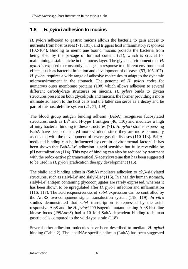
bacterial cells in the culture media (Paper III). With this method, we can
continually monitor the bacterial growth while maintaining an optimal
environment for the bacteria (Figure 1).
Figure 1. H. suis growth detection by RealTime-Glo™ viability assay. H. suis growth
curve when cultured in the absence (■) or presence of a growth promoting (●) or a growth
inhibiting (▲) mucin.
3.2 Bacterial binding detection
We used four different assays to study bacterial binding to purified mucins,
glycoconjugates and glycolipids.
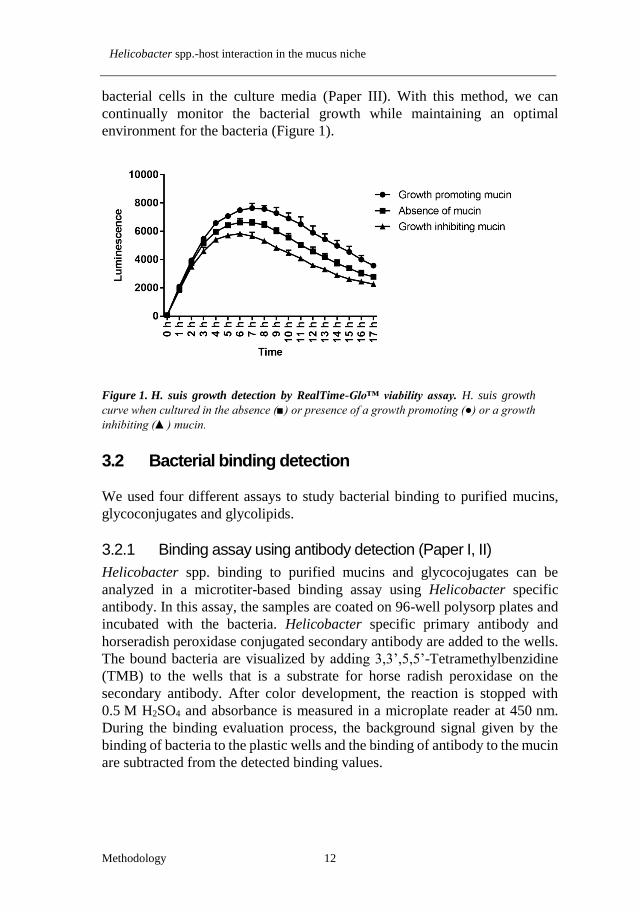
3.2.1 Binding assay using antibody detection (Paper I, II)
Helicobacter spp. binding to purified mucins and glycocojugates can be
analyzed in a microtiter-based binding assay using Helicobacter specific
antibody. In this assay, the samples are coated on 96-well polysorp plates and
incubated with the bacteria. Helicobacter specific primary antibody and
horseradish peroxidase conjugated secondary antibody are added to the wells.
The bound bacteria are visualized by adding 3,3’,5,5’-Tetramethylbenzidine
(TMB) to the wells that is a substrate for horse radish peroxidase on the
secondary antibody. After color development, the reaction is stopped with
0.5 M H2SO4 and absorbance is measured in a microplate reader at 450 nm.
During the binding evaluation process, the background signal given by the
binding of bacteria to the plastic wells and the binding of antibody to the mucin
are subtracted from the detected binding values.

Médea Padra
13 Methodology
3.2.2 Binding inhibition assay (Paper II)
To investigate the binding of bacteria to monosaccharides that are not
conjugated to carrier protein, hence cannot be coated on polysorp plates,
binding inhibition assay was performed. In this method, a mucin with strong
bacterial binding ability was coated on the 96-well plate and the bacteria were
pre-incubated with the sugars of interest prior to the incubation with mucins.
If the glycans that are used during pre-incubation are targets of the bacterial
adhesins, they are expected to inhibit binding of the bacteria to the mucin
sample by occupying the binding site of the adhesion molecules.
3.2.3 Binding to purified mucins and glycoconjugates using
biotinylated bacteria (Paper II, III)
The H. suis binding signal detected with the antibody detection method was
relatively weak compared to the level of adhesion found with H. pylori. The
low binding signal and high background signal due to the cross-reaction
between the antibody and mucins was technically challenging, thus we
performed an adhesion assay using biotinylated bacteria. To avoid damaging
the adhesins by biotinylation, mild biotinylation of bacteria was performed, as
previously described (167). Similarly to the binding detection with antibody,
the mucin and glycoconjugate samples were coated on 96-well polysorp plates,
and incubated with biotinylated bacteria. Bound bacteria were detected by the
reaction between biotin carried by the bacteria and horseradish peroxidase
conjugated streptavidin and the binding was visualized with TMB substrate.
The results obtained with this method were similar to the results obtained with
the antibody detection method, but with higher signal to noise ratio.
3.2.4 Binding of Helicobacter spp. to glycosphingolipids on thin-
layer chromatograms (Paper II)
Glycolipids can carry similar glycoepitopes as mucins that can serve as binding
sites for bacteria providing a more intimate bacterial adhesion to the host. The
use of glycolipids in binding studies simplifies the investigation of the binding
specificity, since these molecules carry only one glycan, contrary to the
multiple glycans carried by mucins. Binding detection to glycolipids was
performed by using 35S-methionine labeled bacteria. The labeling of
Helicobacter spp. was performed as described (168). The glycosphingolipids
were separated on aluminum-backed silica gel plates and the bound 35S-labeled
bacteria were detected by autoradiography. Due to that the low incorporation
of radioactive label into H. suis made the glycolipid binding results difficult to
reproduce, we also demonstrated binding of this pathogen to glycoconjugates

Helicobacter spp.-host interaction in the mucus niche
Methodology 14
terminating with the same epitopes using binding methods independent of
metabolic labeling of the bacteria (i.e. the above methods).

Médea Padra
15 Results and Discussion
4 RESULTS AND DISCUSSION
4.1 BabA mediated binding of H. pylori affects the growth
and gene expression of the bacteria (Paper I)
The presence of certain gastric mucins in bacterial culture has been shown to
promote or inhibit H. pylori growth, depending on the type and origin of the
mucin (86). Mucin molecules carry a vast array of different oligosaccharide
structures that can be utilized as nutrition source by mucin degrading bacteria,
hence can stimulate bacterial growth (83, 84, 169). Glandular mucins that carry
α1,4-GlcNAc terminating O-glycans have been suggested to have an
antimicrobial effect inhibiting the biosynthesis of a major H. pylori cell wall
component (138, 139), although, this cannot be the only explanation for the
growth inhibitory effect of mucins, since not all the mucin samples that inhibit
H. pylori growth carry this structure (86). To investigate the growth of H.
pylori in response to mucins and mucin glycans, we cultured the bacteria in the
presence of Leb and SLex glycoconjugates that are the adhesion targets of H.
pylori adhesin BabA and SabA, respectively, and in the presence of mucins
that carry these structures. In these experiments, we used strains with different
adhesin gene expression: strain J99 that carries both the babA and sabA gene
and strain P12 that carries the babA gene only. Both strains had stronger
binding avidity to Leb than to SLex and both strains bound better to the mucin
derived from healthy stomach that carries Leb and is lacking SLex, than to the
tumor-derived mucin that carries both Leb and SLex glycan structures. H. pylori
growth measurement by detecting alamarBlue reduction revealed that Leb-
conjugates decreased the metabolic activity of both H. pylori strains,
suggesting that adhesion to Leb and to mucins carrying these glycan structures
inhibit the growth of the bacteria. This hypothesis was supported by further
binding and growth experiments using adhesin deletion mutants. As expected,
babA deletion mutants of both strain J99 and P12 had lower binding avidity to
Leb glycoconjugate and to mucins that carry this structure than the isogenic wt
strains and they showed low or no binding to SLex. Deletion of babA reversed
the growth inhibiting effect on strain J99 and enhanced the growth of strain
P12 in the presence of the mucin and Leb, indicating that strain P12 has a
positive growth response to mucin glycans, which is suppressed by the BabA
mediated binding. These results together indicate that the interaction of both
H. pylori strains with mucin glycans is mediated by BabA and that the effect
of mucins on bacterial growth is highly dependent on the glycosylation of
mucins.

Helicobacter spp.-host interaction in the mucus niche
Results and Discussion 16
To visualize the morphology and viability of the bacteria in the presence of
mucin glycans, the bacterial culture of strain J99 wt and its isogenic mutants
after co-culturing with Leb and SLex glycoconjugates were stained with
Live/Dead double staining kit that stains viable and dead bacterial cells in the
culture media with different colors. This staining revealed aggregate formation
with J99ΔsabA and J99 wt in the presence of Leb-glycoconjugate, which was
accompanied by decreased metabolic activity as measured by alamarBlue
reduction. There was no aggregate formation when J99ΔbabA was cultured in
the presence of Leb, and it did not affect the metabolic activity of the bacteria.
Adhesive interaction between bacteria has been shown to be induced by
unfavorable growth conditions (170, 171), and the role of aggregate formation
in bacterial resistance towards antimicrobial agents has been also described
(172). These observations suggest that aggregate formation might be beneficial
for the bacteria as part of the bacterial survival strategy in response to
environmental factors. In this thesis, we describe the benefits of bacterial
aggregate formation for the host and we hypothesize that in addition to
facilitating washing away the bacteria from the stomach, mucin binding
controls the pathogen number in the stomach by the growth limiting effect of
aggregate formation. As it was revealed by Live/Dead staining, the majority of
the aggregate forming bacteria were alive, suggesting that there is no direct
antimicrobial effect of aggregate formation, the growth limiting effect can be
instead explained by the slow replication due to physical hindrance or inter-
bacterial communication.
Apart from affecting the growth of the bacteria, attachment to mucins can alter
the expression of H. pylori genes relevant to colonization. We have previously
shown that culturing H. pylori in the presence of mucins from different
individuals can affect the adhesin expression of the bacteria (86). Here we
demonstrated that H. pylori BabA and SabA adhesin expression in response to
mucins negatively correlates with the binding avidity of the bacteria to these
mucins. To verify the effect of adhesion on bacterial gene expression, we
analyzed adhesion gene expression level after co-culturing the bacteria with
SLex- and Leb-glycoconjugates and we observed that the presence of Leb
decreased babA expression, whereas the level of this gene was not affected by
the presence of SLex. The repression of babA gene expression in response to
Leb binding might be part of the bacterial defense mechanism avoiding
excessive binding to mucins that would lead to the removal of the bacteria
along with shedding mucus. Decreasing the amount of adhesins in response to
binding might serve as a negative feedback loop that can enable long-term
colonization by H. pylori.

Médea Padra
17 Results and Discussion
4.2 ArsS affects H. pylori growth and BabA-dependent
binding (Paper I)
H. pylori expresses the ArsS pH sensor histidine kinase protein that plays a
role in urease gene transcription and in urease protein delivery in order to
enhance acid acclimation (173). Increased of SabA expression level has been
previously shown in an H. pylori strain lacking the ArsS (118). In line with
these results, we detected increased binding of J99ΔarsS to SLex and to SLex
containing mucins compared to that of J99 wt and it was accompanied by
decreased binding to Leb and Leb containing mucins with the arsS deletion
mutants of both J99 and P12 strains. Deleting the arsS slightly increased BabA
protein expression in J99ΔarsS, whereas decreased in P12ΔarsS to a similar
degree. These results suggest that level of BabA-dependent binding is more
dependent on the topographical localization rather than the number of adhesins
present on the bacteria. Contrary to the growth inhibitory effect of mucin
glycans on the H. pylori wt strains, J99ΔarsS and P12ΔarsS growth did not
decrease in the presence of the mucin samples or Leb glycoconjugate, which
can be due to the lack of aggregate formation. The adhesion and growth
experiments using the H. pylori arsS deletion mutants further confirmed the
role of bacterial aggregate formation in the growth inhibitory effect of mucins.
4.3 H. suis resides in the mucus layer and can also be
found associated with parietal cells (Paper II, III).
In H. pylori infected human stomachs, the majority of H. pylori have been
detected in the surface mucus layer (96) that protects the bacteria from the low
acidity in the stomach and provides surface for host-pathogen interactions (70).
Here we analyzed the mucus layer of the pig stomach and the spatial
distribution of the pig gastric pathogen, H. suis. On pig gastric tissue sections,
we detected a thick mucus layer covering the epithelial cells. Antibody
detection on these sections as well as proteomic analysis on purified gastric
mucin samples revealed that MUC5AC is the predominant secreted mucin in
the pig stomach, similarly to the human stomach. We performed fluorescent in
situ hybridization on pig gastric tissue sections obtained from pigs
experimentally or naturally infected with H. suis and detected H. suis in the
mucus layer lining the surface epithelium and throughout the gastric pits. We
and others also detected this bacterium in close association with the acid
producing parietal cells (156). These findings suggest that H. suis is exposed
to neutral pH in the mucus layer closer to the epithelial cells and in the lamina

Helicobacter spp.-host interaction in the mucus niche
Results and Discussion 18
propria and exposed to acidic pH closer to the gastric lumen as well as when
inside the parietal cells.
4.4 Gastric mucin glycosylation differs between pigs and
humans as well as between H. suis infected and non-
infected pigs (Paper II, III)
H. suis is associated with the development of severe gastric disorders in its
main host, the pig and also in humans. Since H. suis resides in the gastric
mucus layer, interaction with mucin glycans can be crucial for colonization
and survival of the bacteria and can also support the host defense system
maintaining a dynamic host-pathogen interplay in the stomach. To get a better
understanding of this host-pathogen interaction, we analyzed the glycosylation
of purified pig gastric mucin samples by mass spectrometry analysis, and since
H. suis is of zoonotic importance, we studied the differences between pig and
human gastric mucin glycosylation. The glycan profile of both pig and human
mucins showed high inter-individual differences. We detected higher number
of different glycan structures in the human mucins than in the pig mucins,
which does not necessarily provide evidence for higher inter-individual
variability in humans than in pigs, since the pig samples analyzed here only
include non-infected pig mucins, whereas the human samples also include
pathological specimens. The length of mucin O-glycans varied between 2 and
14 residues in pig samples and from 2 to 12 residues in human mucins. Both
pig and human gastric mucin oligosaccharides were mainly extended core 1
and core 2 O-glycans, although, structures with core 3 and 4 were also detected.
The relative abundance of extended core 1 O-glycans was higher in pig than in
human mucins. The terminal residues on the mucin glycans are usually vital
parts in mucin-pathogen interaction, therefore we quantified the relative
abundance of these glycan epitopes on pig and human gastric mucins (Figure
2A). The most abundant terminal residue was galactose on pig mucin glycans
and fucose on human mucin glycans. The level of sialylation of gastric mucins
was low both in pigs and in humans. The relative abundance of mucin glycans
with terminal galactose were higher in pig mucins, whereas the relative
abundance of fucose terminating glycans were higher in human mucins. The
main difference between human and pig glycan terminal epitopes was the level
of sulphation which was around 50% in pig mucins and very low, around 0.6%
in the human samples.
Since H. pylori infection has been shown to trigger qualitative and quantitative
changes in host gastric mucins (53, 54, 141-143), we studied the alterations of

Médea Padra
19 Results and Discussion
pig gastric mucins in H. suis infection. With mass spectrometry analysis on
purified pig gastric mucins, we identified that MUC5AC is the major secreted
mucin in the pig stomach. A decreased level of MUC5AC was detected among
mucins from H. suis infected pigs compared to the samples from non-infected
pigs, similarly to the effect of H. pylori infection in the human stomach (53).
We detected in total 118 different oligosaccharides on the pig gastric mucin
samples, out of which 18 structures with low fucose and sialic acid content
were detected only in the non-infected samples and 7 structures containing
higher fucose and sialic acid level were only detected in the infected group.
The number of different oligosaccharide structures detected in the mucin
samples was lower in the infected group, implying a decrease in the number of
glycan structures on pig gastric mucins upon infection limiting the variety of
glycan epitopes for bacterial interactions. In mucins from the infected group,
we detected an increase in the relative abundance of acidic (mostly sulphated)
and fucosylated glycan structures and a decrease in glycan structures
terminating with galactose (Figure 2B). These terminal glycan structures play
important role in the binding and growth of H. suis in the presence of mucins,
as it is described later in the thesis.
Figure 2. Differences in the relative abundance of terminal glycan epitopes between pigs
and humans as well as between H. suis infected and non-infected pigs. A. Differences in
the relative abundance of terminal glycan structures between pig and human gastric mucins
B. Changes in pig gastric mucin glycosylation upon H. suis infection. Stars indicate
statistically significant difference between mucin glycans, *, ** and *** indicate p ≤ 0.05,
0.01 and 0.001, respectively, Two-way ANOVA.
4.5 H. suis binding to gastric mucins in health and disease
(Paper II, III).
We analyzed H. suis binding to purified pig and human gastric mucins at the
pH range present in the stomach and compared it with the binding of H. pylori

Helicobacter spp.-host interaction in the mucus niche
Results and Discussion 20
to the same human mucins. H. suis and H. pylori binding to purified mucins
and glycoconjugates showed different pattern both regarding their pH
preference and glycan specificity (Figure 3). H. suis binding level was highest
at pH 2 with a gradual decrease towards neutral pH, whereas H. pylori bound
better at neutral pH. H. suis and H. pylori bound to the same mucin samples
as well as glycoconjugates with different avidity, suggesting that the two
Helicobacter species use different modes of adhesion with different glycan
specificities. H. pylori binding to mucins has been described to happen via four
modes of adhesion (70), the most well characterized of which are the BabA
and SabA mediated binding to Leb and sialylated glycans, respectively. H. suis
genome analyses revealed that H. suis lacks homologs of BabA and SabA,
although, contains some OMPs similar to the major OMP families described
in H. pylori, which might be involved in binding to gastric mucins (161). Our
bacterial binding experiments showed that H. suis binding avidity differed
between the mucin samples investigated. Some samples bound the bacteria
only at acidic pH, whereas other samples bound also at neutral pH.
Furthermore, H. suis binding to GuHCl soluble mucins was more pronounced
than to insoluble ones. The differences in H. suis binding avidity to the
different mucin samples can be explained by the glycosylation differences
between the samples. Here we suggest that H. suis uses two ways of adhesion
to mucins: one binding mode that is dependent on acidic pH and one that is
functional also at neutral pH, and both binding modes depend on the glycan
structures carried by the mucins.
To find the glycan structures on the mucins that might serve as adhesion targets
for H. suis, we analyzed the relation between the bacterial binding amplitude
to the mucins and the abundance of the different glycan structures carried by
these mucins. We focused primarily on the terminal residues, since these
glycans are more exposed for bacterial binding. At pH 2, the relative
abundance of acidic (i.e. sulphated and/or sialylated) glycans correlated with
the level of H. suis binding, indicating that H. suis can bind to acidic structures
via charge dependent mode. This hypothesis was also confirmed by H. suis
binding to the highly charged DNA at pH 2, but not at pH 7. The stronger
binding avidity to mucins at lower pH cannot be the consequence of protein
denaturing because not all the mucin samples that were tested bound H. suis at
low pH. In addition, binding at low pH to DNA also suggests that binding at
acidic pH occurs to charged structures, not to denatured proteins (Figure 3A).

Médea Padra
21 Results and Discussion
Figure 3. H. suis and H. pylori binding differences regarding pH and glycan specificity.
A. H. suis binding to purified mucins, glycoconjugates and DNA at pH 2 and pH 7. B. H.
pylori binding to purified mucins, glycoconjugates and DNA at pH 2 and pH 7. The binding
values are shown after subtracting background signal (bacteria binding to plastic well).
Glycolipids can carry similar glycan structures as mucins and they represent a
more intimate adherence to the host by Helicobacter species. To further
investigate H. suis binding specificity, we examined H. suis binding to
glycosphingolipids isolated from porcine stomach, where binding to
lactotetraosylceramide (Galβ3GlcNAcβ3Galβ4Glcβ1Cer) was detected. To
confirm the binding specificity to this structure, H. suis binding to Lacto-N-
tetraose (LNT, Galβ3GlcNAcβ3Galβ4Glc) conjugated to human serum
albumin (HSA) was also tested. In line with the glycolipid binding results, H.
suis bound to LNT conjugated to HSA at pH 7 and the binding remained
functional at pH 2. We also demonstrated that binding to pig mucins can be
inhibited by pre-treating the bacteria with LNT or sialylated LNT. Together
these data indicate that H. suis can bind to mucins with terminal galactose and
that acidic modification may have beneficial effects on binding.
H. pylori induced gastric mucin glycosylation changes have been demonstrated
to influence the mucin binding avidity of the pathogen (107). The decreased
Leb level and increased sialylation in the gastric mucosa decrease BabA
mediated binding and increased the adhesion via SabA and via the charge-
dependent binding modes (70, 107) leading to an overall decreased adhesion,
since BabA mediated binding is generally higher than binding via SabA. To
study the effect of Helicobacter infection on the binding avidity of H. suis, we
used purified pig gastric mucins with or without H. suis infection as well as
human gastric mucins with and without Helicobacter spp. infection and tested
H. suis binding to these mucins at acidic and neutral pH. In line with previous

Helicobacter spp.-host interaction in the mucus niche
Results and Discussion 22
observations, H. suis binding to pig gastric mucins was higher at pH 2 than at
pH 7, regardless of the infection status of the pigs (Figure 4A, B), whereas pH
did not affect the binding avidity to human gastric mucins. Infection caused a
decreased H. suis binding avidity to pig gastric mucins at pH 7 (Figure 4B) but
not at pH 2 (Figure 4A). At neutral pH, the decreased binding after infection
can be explained by the infection-induced loss of adhesion targets by the
decreased terminal galactose on mucins and at acidic pH, H. suis also binds via
charge dependent mode to acidic structures, the abundance of which structures
increased during infection.
Bacterial adherence to gastric mucins can serve as important part of the host
defense mechanism protecting the epithelial cells from the invasion by
Helicobacters. This hypothesis has been supported by observations where H.
pylori-infected children and rhesus monkeys secreting mucins with less H.
pylori binding capacity, develop higher H. pylori density and more severe
gastritis (107, 174). Our results demonstrate that Helicobacter spp. infection
decreases the ability of mucins to bind H. suis, thereby avoiding the removal
of the pathogen from the gastric niche.
Figure 4. H. suis binding and growth in the presence of gastric mucins derived from H.
suis infected or non-infected pigs. A. H. suis binding to H. suis infected or non-infected pig
gastric mucin samples at pH 2. B. H. suis binding to H. suis infected or non-infected pig
gastric mucin samples at pH 7. C. H. suis growth in the presence of gastric mucin samples
derived from H. suis infected or non-infected pigs. (*p<0.05, ***p<0.001, Two-way
ANOVA).
4.6 Helicobacter spp. infection induced mucin
glycosylation changes increase H. suis growth (Paper
III).
The growth of H. pylori has been shown to have different response to mucins,
depending on the origin and the type of the mucin (86). For instance, when H.

Médea Padra
23 Results and Discussion
pylori is cultured in the presence of mucins that carry glycan structures that the
bacteria adheres to, the binding-induced aggregate formation slows down the
growth of the bacteria (Paper I). H. pylori growth can be also inhibited by
mucins containing α1,4GlcNAc-capped O-glycans (139), a structure that
primarily is associated with the glandular mucins in the stomach. In this thesis,
we demonstrated that the growth of H. suis is affected by both porcine and
human gastric mucins and the response of the bacteria to mucins is dependent
on the infection status of the individual the mucin was isolated from. Mucins
from non-infected individuals inhibited the growth of H. suis, whereas mucins
from infected individuals had a growth promoting effect (Figure 3C). To find
the possible glycan structures on the mucins that affect the bacterial growth,
we studied the relationship between the effect of mucin samples on H. suis
growth and the abundance of glycan structures carried by these mucins. The
growth inhibitory effect of mucins correlated with the abundance of galactose
terminating structures and was independent of α1,4GlcNAc-capped O-glycan
abundance. Positive correlations between growth and the abundance of acidic
and fucosylated structures on mucin glycans were observed. These results
suggest that Helicobacter spp. infection induced host mucin glycosylation
changes create a more stable and growth-promoting environment for H. suis,
and possibly for other Helicobacter species in the stomach that facilitates the
long term colonization by these pathogens.

Helicobacter spp.-host interaction in the mucus niche
Conclusions 24
5 CONCLUSIONS
In this thesis, we investigated host-pathogen interactions in the mucus niche
focusing on Helicobacter spp. and gastric mucins. We analyzed the mucus
alterations in infection and how these changes affect bacterial behavior. Based
on our results of mucin characterization as well as bacterial adhesion and
growth assays we can conclude that:
Helicobacter spp. infection induces a constant host-pathogen
adaptation and response process in the stomach. The mucin interaction
with pathogens is mediated by the mucin glycan composition, which
is able to inhibit H. pylori growth by adhesion and aggregation of
bacteria. Mucins also have the ability to influence H. pylori
pathogenicity by affecting adhesin gene expression.
H. pylori and H. suis binding to human and pig gastric mucins differ
in specificity and pH preference and show high inter-individual
variation, which can be explained by mucin glycosylation differences.
H. suis binding to gastric mucins and glycolipids occurs via two modes
of adhesion: to structures with terminal galactose at both neutral and
acidic pH, and to negatively charged structures at acidic pH. These
binding modes may enable bacterial adhesion at low pH close to the
gastric lumen and in parietal cells and a more intimate adhesion to
mucin glycans and glycolipids close to the epithelial cells.
Helicobacter spp. infection alters host mucin composition and
glycosylation in a way that decreases the amount of H. suis binding
glycan structures on gastric mucins and the H. suis growth regulating
effects of the mucins. By these alterations, Helicobacters create a more
stable and inhabitable niche in the stomach which may be crucial for
long-term colonization.

Médea Padra
25 Additional Bibliography
6 ADDITIONAL BIBLIOGRAPHY
BabA-mediated adherence of pediatric ulcerogenic H. pylori strains to
gastric mucins at neutral and acidic pH.
Quintana-Hayashi MP, Rocha R, Padra M, Thorell A, Jin C, Karlsson NG,
Roxo-Rosa M, Oleastro M, Lindén SK.
Virulence. 2018 Oct 9. doi: 10.1080/21505594.2018.1532243.
Influence of the viscosity of healthy and diseased human mucins on the
motility of Helicobacter pylori.
Su C, Padra M, Constantino MA, Sharba S, Thorell A, Lindén SK, Bansil R.
Sci Rep. 2018 Jun 26;8(1):9710.
Mucus-Pathogen Interactions in the Gastrointestinal Tract of Farmed
Animals.
Quintana-Hayashi MP, Padra M, Padra JT, Benktander J, Lindén SK.
Microorganisms. 2018 Jun 18;6(2). pii: E55. Review.
Structural Diversity of Human Gastric Mucin Glycans.
Jin C, Kenny DT, Skoog EC, Padra M, Adamczyk B, Vitizeva V, Thorell A,
Venkatakrishnan V, Lindén SK, Karlsson NG.
Mol Cell Proteomics. 2017 May;16(5):743-758.
The Helicobacter heilmannii hofE and hofF Genes are Essential for
Colonization of the Gastric Mucosa and Play a Role in IL-1β-Induced
Gastric MUC13 Expression.
Cheng L, Mirko R, Sara L, Medea P, Caroline B, Eva B, Myrthe J, Bram F,
Wim VD, Richard D, Freddy H, Annemieke S.
Helicobacter. 2016 Dec;21(6):504-522.
Divergence between the Highly Virulent Zoonotic Pathogen Helicobacter
heilmannii and Its Closest Relative, the Low-Virulence "Helicobacter
ailurogastricus" sp. nov.
Joosten M, Lindén S, Rossi M, Tay AC, Skoog E, Padra M, Peters F, Perkins
T, Vandamme P, Van Nieuwerburgh F, D'Herde K, Van den Broeck W, Flahou
B, Deforce D, Ducatelle R, Marshall B, Haesebrouck F, Smet A.
Infect Immun. 2015 Nov 2;84(1):293-306.

Helicobacter spp.-host interaction in the mucus niche
Acknowledgement 26
7 ACKNOWLEDGEMENT
This thesis could not have been written without all the support that I received
over the years. I am grateful to be surrounded by this many fantastic people
who contributed to my work both professionally and personally.
I express my special gratitude to my main supervisor, Sara Lindén. Thank you
for giving me the opportunity to be part of your research group. Thank you for
believing in me and always having full trust in me. You are a very kind and
supportive person who has always been there for me through the happy and
hard times. I have learnt a lot from you over the years and enjoyed our
discussions about science and life. Thank you for teaching, guiding and
motivating me.
My co-supervisors, Gunnar Hansson and Susann Teneberg, thank you for
the contribution to my PhD by great collaborations, sharing equipment and
creating a pleasant work environment.
I would like to thank all the past and present members of the Lindén group.
Special thanks to Sinan, for always being ready to help and consider my
scientific difficulties as your own. Your funny stories and impressions always
brightened my days in the dark basement. John, thank you for your input into
my thesis and also for the fun game nights. Gurdeep, keep up the hard work
and good luck with you PhD. Helen and Mattias, thank you for your work on
the human gastric mucins. I hope both of you will be great doctors one day.
Former group members, Harvey, Emma, Nazanin, Pushpa and Maca, thank
you all for being helpful when I joined the group and guiding my first steps in
the lab. Thanks for the nice conversations, friendly environment in the lab and
lots of fun outside work. I hope our roads will cross each other sometime in
the future. Vignesh, thank you for being a great friend and a huge support over
the last few years. Thank you for always motivating and encouraging me and
for improving my friendship with glycans, (south!)indian food and tamil
movies .
I would like to thank Niclas Karlsson`s group for the great collaboration.
Thank you Jin, for being always very quick and precise with Mass spec
analysis. Barbara, thank you for being a good friend and great co-worker.
Thank you for all your input into the H. suis project and for always being happy
to help, both in the lab and outside work. Varvara, thank you for the nice
conversations during coffee breaks and for the beautiful paintings. Shikha,

Médea Padra
27 Acknowledgement
thanks for the useful advices on organizing the defense. Yolanda, it is your
turn soon, I`m sure you will be great. Let`s beat LacdiNAc together .
I would like to thank all the members of the mucin biology groups. Special
thanks to George and Elizabeth for the nice chats, wonderful dinners and fun
game nights. I hope we keep up the good habit . Erik, thanks for making the
lunch breaks very entertaining. I´m impressed by your talent learning the
Hungarian language . Liisa, thanks for the nice talks and being always very
straightforward. I truly believe we are relatives . Hannah, it was nice sharing
funny stories about teaching. Karin, thanks a lot for your help in the course
lab, when you were around, I knew everything was under control.
I would like to thank Freddy Heasebrouck`s team at Ghent University for the
great collaboration, fruitful conversations at conferences and for providing us
with one of the leading characters of my thesis, H. suis.
My Hungarian friends in Sweden: Zsolt and Gábor, thanks for the nice
conversations over lunch. It was great sharing thoughts about work, politics,
culture and pálinka .
I would like to express my gratitude to my family: Anyu, Apu és Zsolti,
köszönöm, hogy mindig támogattatok és bíztattatok az évek során. Köszönöm,
hogy minding meleg szívvel vártok haza a “nagyvilágból”. Nagyon szeretlek
titeket!
Jani, köszönöm, hogy meggyőztél, hogy jöjjünk ki Svédországba, és hogy
bíztattál a PhD fokozat megszerzésére. Te voltál a legnagyobb támaszom a
laborban és otthon az elmúlt évek során. Köszönöm a sok segítséget, türelmet
és szeretetet, amit kaptam tőled. Köszönöm, hogy vagy nekem, hogy mellettem
állsz jóban, rosszban. Nagyon szeretlek!

Helicobacter spp.-host interaction in the mucus niche
References 28
8 REFERENCES
1. Allen A, Hutton DA, Leonard AJ, Pearson JP, & Sellers LA (1986)
The role of mucus in the protection of the gastroduodenal mucosa.
Scandinavian journal of gastroenterology. Supplement 125:71-78.
2. Walker WA (1985) Role of the mucosal barrier in toxin/microbial
attachment to the gastrointestinal tract. Ciba Foundation symposium
112:34-56.
3. Andrews GP, Laverty TP, & Jones DS (2009) Mucoadhesive
polymeric platforms for controlled drug delivery. European Journal
of Pharmaceutics and Biopharmaceutics 71(3):505-518.
4. Flemstrom G, Knutson L, & Kivilaakso E (1986) Gastroduodenal
mucosal secretion of bicarbonate and mucus: physiological control
and role in protection. Klinische Wochenschrift 64 Suppl 7:107-111.
5. Copeman M, et al. (1994) The gastroduodenal mucus barrier and its
role in protection against luminal pepsins: The effect of 16,16 dimethyl
prostaglandin E2, carbopol-polyacrylate, sucralfate and bismuth
subsalicylate. Journal of Gastroenterology and Hepatology
9(S1):S55-S59.
6. Bansil R & Turner BS (2006) Mucin structure, aggregation,
physiological functions and biomedical applications. Curr Opin
Colloid In 11(2-3):164-170.
7. Collins LM & Dawes C (1987) The surface area of the adult human
mouth and thickness of the salivary film covering the teeth and oral
mucosa. Journal of dental research 66(8):1300-1302.
8. Atuma C, Strugala V, Allen A, & Holm L (2001) The adherent
gastrointestinal mucus gel layer: thickness and physical state in vivo.
American journal of physiology. Gastrointestinal and liver physiology
280(5):G922-929.
9. Phillips TE, Phillips TH, & Neutra MR (1984) Regulation of intestinal
goblet cell secretion. III. Isolated intestinal epithelium. The American
journal of physiology 247(6 Pt 1):G674-681.
10. Grootjans J, Hundscheid IH, & Buurman WA (2013) Goblet cell
compound exocytosis in the defense against bacterial invasion in the
colon exposed to ischemia-reperfusion. Gut Microbes 4(3):232-235.
11. Allen A, Bell A, Mantle M, & Pearson JP (1982) The Structure and
Physiology of Gastrointestinal Mucus. Mucus in Health and Disease—
II, eds Chantler EN, Elder JB, & Elstein M (Springer US, Boston,
MA), pp 115-133.
12. Schumacher GFB, Kim MH, Hosseinian AH, & Dupon C
(Immunoglobulins, proteinase inhibitors, albumin, and lysozyme in

Médea Padra
29 References
human cervical mucus. American Journal of Obstetrics & Gynecology
129(6):629-636.
13. Jones DE & Bevins CL (1992) Paneth cells of the human small
intestine express an antimicrobial peptide gene. The Journal of
biological chemistry 267(32):23216-23225.
14. Mukherjee S, et al. (2014) Antibacterial membrane attack by a pore-
forming intestinal C-type lectin. Nature 505(7481):103-107.
15. Sellers LA, Allen A, Morris ER, & Ross-Murphy SB (1988) Mucus
glycoprotein gels. Role of glycoprotein polymeric structure and
carbohydrate side-chains in gel-formation. Carbohydrate research
178:93-110.
16. Strous GJ & Dekker J (1992) Mucin-Type Glycoproteins. Critical
Reviews in Biochemistry and Molecular Biology 27(1-2):57-92.
17. Hattrup CL & Gendler SJ (2008) Structure and function of the cell
surface (tethered) mucins. Annu Rev Physiol 70:431-457.
18. Singh PK & Hollingsworth MA (2006) Cell surface-associated mucins
in signal transduction. Trends in cell biology 16(9):467-476.
19. Mcguckin MA, et al. (2007) Muc1 mucin limits both Helicobacter
pylori colonization of the murine gastric mucosa and associated
gastritis. Gastroenterology 133(4):1210-1218.
20. Linden SK, Florin TH, & McGuckin MA (2008) Mucin dynamics in
intestinal bacterial infection. PloS one 3(12):e3952.
21. Linden SK, et al. (2009) MUC1 limits Helicobacter pylori infection
both by steric hindrance and by acting as a releasable decoy. PLoS
Pathog 5(10):e1000617.
22. Thornton DJ, Rousseau K, & McGuckin MA (2008) Structure and
function of the polymeric mucins in airways mucus. Annual review of
physiology 70:459-486.
23. Ho SB, et al. (1995) Expression cloning of gastric mucin
complementary DNA and localization of mucin gene expression.
Gastroenterology 109(3):735-747.
24. Verdugo P (1990) Goblet cells secretion and mucogenesis. Annual
review of physiology 52:157-176.
25. Merzel J & Leblond CP (1969) Origin and renewal of goblet cells in
the epithelium of the mouse small intestine. The American journal of
anatomy 124(3):281-305.
26. Radwan KA, Oliver MG, & Specian RD (1990) Cytoarchitectural
reorganization of rabbit colonic goblet cells during baseline secretion.
The American journal of anatomy 189(4):365-376.
27. Merzel J & Almeida ML (1973) Maturation of goblet cells of the
mouse small intestine as shown by the uptake of 35S-sodium sulfate.
The Anatomical record 177(4):519-524.

Helicobacter spp.-host interaction in the mucus niche
References 30
28. Laboisse C, et al. (1996) Recent aspects of the regulation of intestinal
mucus secretion. The Proceedings of the Nutrition Society 55(1b):259-
264.
29. Yang N, Garcia MA, & Quinton PM (2013) Normal mucus formation
requires cAMP-dependent HCO3- secretion and Ca2+-mediated
mucin exocytosis. The Journal of physiology 591(18):4581-4593.
30. Enss ML, et al. (2000) Proinflammatory cytokines trigger MUC gene
expression and mucin release in the intestinal cancer cell line LS180.
Inflammation research : official journal of the European Histamine
Research Society ... [et al.] 49(4):162-169.
31. Hanisch FG (2001) O-glycosylation of the mucin type. Biol Chem
382(2):143-149.
32. Bennett EP, et al. (2012) Control of mucin-type O-glycosylation: a
classification of the polypeptide GalNAc-transferase gene family.
Glycobiology 22(6):736-756.
33. Green C (1989) The ABO, Lewis and related blood group antigens; a
review of structure and biosynthesis. FEMS Microbiol Immunol 1(6-
7):321-330.
34. Rini J, Esko J, & Varki A (2009) Glycosyltransferases and Glycan-
processing Enzymes. Essentials of Glycobiology, eds nd, Varki A,
Cummings RD, Esko JD, Freeze HH, Stanley P, Bertozzi CR, Hart
GW, & Etzler MECold Spring Harbor (NY)).
35. Leonard BC, Yanez-Soto B, Raghunathan VK, Abbott NL, & Murphy
CJ (2016) Species variation and spatial differences in mucin
expression from corneal epithelial cells. Experimental eye research
152:43-48.
36. Jin C, et al. (2017) Structural Diversity of Human Gastric Mucin
Glycans. Molecular & cellular proteomics : MCP 16(5):743-758.
37. Medzihradszky KF, Kaasik K, & Chalkley RJ (2015) Tissue-Specific
Glycosylation at the Glycopeptide Level. Molecular & cellular
proteomics : MCP 14(8):2103-2110.
38. West MB, et al. (2010) Analysis of site-specific glycosylation of renal
and hepatic gamma-glutamyl transpeptidase from normal human
tissue. The Journal of biological chemistry 285(38):29511-29524.
39. Mollicone R, Bara J, Le Pendu J, & Oriol R (1985) Immunohistologic
pattern of type 1 (Lea, Leb) and type 2 (X, Y, H) blood group-related
antigens in the human pyloric and duodenal mucosae. Laboratory
investigation; a journal of technical methods and pathology
53(2):219-227.
40. Varki A, Kannagi R, & Toole BP (2009) Glycosylation Changes in
Cancer. Essentials of Glycobiology, eds nd, Varki A, Cummings RD,

Médea Padra
31 References
Esko JD, Freeze HH, Stanley P, Bertozzi CR, Hart GW, & Etzler ME
(Cold Spring Harbor Laboratory Press
The Consortium of Glycobiology Editors, La Jolla, California., Cold Spring
Harbor (NY)).
41. Crouzier T, et al. (2015) Modulating Mucin Hydration and Lubrication
by Deglycosylation and Polyethylene Glycol Binding. Advanced
Materials Interfaces 2(18):1500308-n/a.
42. Garner B, et al. (2001) Structural elucidation of the N- and O-glycans
of human apolipoprotein(a): role of o-glycans in conferring protease
resistance. The Journal of biological chemistry 276(25):22200-22208.
43. Fukuda M (2002) Roles of mucin-type O-glycans in cell adhesion.
Biochimica et biophysica acta 1573(3):394-405.
44. Yeaman C, et al. (1997) The O-glycosylated stalk domain is required
for apical sorting of neurotrophin receptors in polarized MDCK cells.
The Journal of cell biology 139(4):929-940.
45. Padra JT, et al. (2017) Aeromonas salmonicida Growth in Response
to Atlantic Salmon Mucins Differs between Epithelial Sites, Is
Governed by Sialylated and N-Acetylhexosamine-Containing O-
Glycans, and Is Affected by Ca(2). Infection and immunity 85(8).
46. Borén T, Falk P, Roth KA, Larson G, & Normark S (1993) Attachment
of Helicobacter pylori to human gastric epithelium mediated by blood
group antigens. Science 262(5141):1892-1895.
47. Veillon L, Fakih C, Abou-El-Hassan H, Kobeissy F, & Mechref Y
(2018) Glycosylation Changes in Brain Cancer. ACS chemical
neuroscience 9(1):51-72.
48. Sheta R, et al. (2017) Altered expression of different
GalNActransferases is associated with disease progression and poor
prognosis in women with high-grade serous ovarian cancer.
International journal of oncology 51(6):1887-1897.
49. Ricardo S, et al. (2015) Detection of glyco-mucin profiles improves
specificity of MUC16 and MUC1 biomarkers in ovarian serous
tumours. Molecular oncology 9(2):503-512.
50. Kirwan A, et al. (2015) Glycosylation-Based Serum Biomarkers for
Cancer Diagnostics and Prognostics. BioMed Research International
2015:16.
51. Aishima S, et al. (2006) Gastric mucin phenotype defines tumour
progression and prognosis of intrahepatic cholangiocarcinoma: gastric
foveolar type is associated with aggressive tumour behaviour.
Histopathology 49(1):35-44.
52. Freitas M, Axelsson LG, Cayuela C, Midtvedt T, & Trugnan G (2002)
Microbial-host interactions specifically control the glycosylation

Helicobacter spp.-host interaction in the mucus niche
References 32
pattern in intestinal mouse mucosa. Histochemistry and cell biology
118(2):149-161.
53. Byrd JC, Yan P, Yunker C, & Bresalier RS (1997) Aberrant expression
of gland-type gastric mucin in the surface epithelium of H-pylori-
infected patients. Gastroenterology 112(4):A80-A80.
54. Navabi N, Johansson ME, Raghavan S, & Linden SK (2013)
Helicobacter pylori infection impairs the mucin production rate and
turnover in the murine gastric mucosa. Infection and immunity
81(3):829-837.
55. Haesebrouck F, et al. (2009) Gastric Helicobacters in Domestic
Animals and Nonhuman Primates and Their Significance for Human
Health. Clin Microbiol Rev 22(2):202-+.
56. Yen J-T (2000) Swine Nutrition (CRC press Taylor and Francis group)
2nd Ed.
57. Merchant HA, et al. (2011) Assessment of gastrointestinal pH, fluid
and lymphoid tissue in the guinea pig, rabbit and pig, and implications
for their use in drug development. European journal of
pharmaceutical sciences : official journal of the European Federation
for Pharmaceutical Sciences 42(1-2):3-10.
58. Nordman H, et al. (1997) Mucus glycoproteins from pig gastric
mucose: Identification of different mucin populations from the surface
epithelium. Biochem J 326:903-910.
59. Ohara S, Ishihara K, & Hotta K (1993) Regional Differences in Pig
Gastric Mucins. Comp Biochem Phys B 106(1):153-158.
60. Karlsson NG, Nordman H, Karlsson H, Carlstedt I, & Hansson GC
(1997) Glycosylation differences between pig gastric mucin
populations: a comparative study of the neutral oligosaccharides using
mass spectrometry. Biochem J 326 ( Pt 3):911-917.
61. Nordman H, Davies JR, & Carlstedt I (1998) Mucus glycoproteins
from pig gastric mucosa: different mucins are produced by the surface
epithelium and the glands. Biochem J 331:687-694.
62. King TP, et al. (1995) Sialylation of intestinal microvillar membranes
in newborn, sucking and weaned pigs. Glycobiology 5(5):525-534.
63. Kelly D & King TP (1991) The influence of lactation products on the
temporal expression of histo-blood group antigens in the intestines of
suckling pigs: lectin histochemical and immunohistochemical
analysis. The Histochemical journal 23(1):55-60.
64. Pestova MI, Clift RE, Vickers RJ, Franklin MA, & Mathew AG (2000)
Effect of weaning and dietary galactose supplementation on digesta
glycoproteins in pigs. Journal of the Science of Food and Agriculture
80(13):1918-1924.

Médea Padra
33 References
65. Kelly D, King TP, McFadyen M, & Coutts AG (1993) Effect of
preclosure colostrum intake on the development of the intestinal
epithelium of artificially reared piglets. Biology of the neonate
64(4):235-244.
66. Liu P, et al. (2014) Effect of Dietary Zinc Oxide on Morphological
Characteristics, Mucin Composition and Gene Expression in the Colon
of Weaned Piglets. Plos One 9(3).
67. Bry L, Falk PG, Midtvedt T, & Gordon JI (1996) A model of host-
microbial interactions in an open mammalian ecosystem. Science
(New York, N.Y.) 273(5280):1380-1383.
68. Hooper LV & Gordon JI (2001) Glycans as legislators of host-
microbial interactions: spanning the spectrum from symbiosis to
pathogenicity. Glycobiology 11(2):1R-10R.
69. Karlsson KA (1989) Animal Glycosphingolipids as Membrane
Attachment Sites for Bacteria. Annu Rev Biochem 58:309-350.
70. Linden SK, Wickstrom C, Lindell G, Gilshenan K, & Carlstedt I
(2008) Four modes of adhesion are used during Helicobacter pylori
binding to human mucins in the oral and gastric niches. Helicobacter
13(2):81-93.
71. Aspholm-Hurtig M, et al. (2004) Functional adaptation of BabA, the
H. pylori ABO blood group antigen binding adhesin. Science
305(5683):519-522.
72. Robbe C, Capon C, Coddeville B, & Michalski JC (2004) Structural
diversity and specific distribution of 0-glycans in normal human
mucins along the intestinal tract. Biochem J 384:307-316.
73. Scharfman A, Lamblin G, & Roussel P (1995) Interactions between
human respiratory mucins and pathogens. Biochem Soc T 23(4):836-
839.
74. Ouwerkerk JP, de Vos WM, & Belzer C (2013) Glycobiome: bacteria
and mucus at the epithelial interface. Best practice & research.
Clinical gastroenterology 27(1):25-38.
75. Li H, et al. (2015) The outer mucus layer hosts a distinct intestinal
microbial niche. Nature communications 6:8292.
76. Laux D, P. Cohen, and T. Conway (2005) Role of the mucins layer in
bacterial colonization of the intestine. Colonization of Mucosal
Surfaces, ed J. P. Nataro PSC, H. L. T. Moblet, and J. Weisner (ASM
Press, Washington, DC. ), pp 199–206.
77. Miller RS & Hoskins LC (1981) Mucin degradation in human colon
ecosystems. Fecal population densities of mucin-degrading bacteria
estimated by a "most probable number" method. Gastroenterology
81(4):759-765.

Helicobacter spp.-host interaction in the mucus niche
References 34
78. Martens EC, Chiang HC, & Gordon JI (2008) Mucosal glycan
foraging enhances fitness and transmission of a saccharolytic human
gut bacterial symbiont. Cell host & microbe 4(5):447-457.
79. Anderson KM, et al. (2005) A clostridial endo-beta-galactosidase that
cleaves both blood group A and B glycotopes: the first member of a
new glycoside hydrolase family, GH98. The Journal of biological
chemistry 280(9):7720-7728.
80. Fujita M, et al. (2011) Glycoside hydrolase family 89 alpha-N-
acetylglucosaminidase from Clostridium perfringens specifically acts
on GlcNAc alpha1,4Gal beta1R at the non-reducing terminus of O-
glycans in gastric mucin. The Journal of biological chemistry
286(8):6479-6489.
81. Hoskins LC, et al. (1985) Mucin degradation in human colon
ecosystems. Isolation and properties of fecal strains that degrade ABH
blood group antigens and oligosaccharides from mucin glycoproteins.
The Journal of clinical investigation 75(3):944-953.
82. Corfield AP, et al. (1993) The roles of enteric bacterial sialidase,
sialate O-acetyl esterase and glycosulfatase in the degradation of
human colonic mucin. Glycoconjugate journal 10(1):72-81.
83. Png CW, et al. (2010) Mucolytic bacteria with increased prevalence in
IBD mucosa augment in vitro utilization of mucin by other bacteria.
The American journal of gastroenterology 105(11):2420-2428.
84. Crost EH, et al. (2013) Utilisation of mucin glycans by the human gut
symbiont Ruminococcus gnavus is strain-dependent. Plos One
8(10):e76341.
85. Celli JP, et al. (2009) Helicobacter pylori moves through mucus by
reducing mucin viscoelasticity. P Natl Acad Sci USA 106(34):14321-
14326.
86. Skoog EC, et al. (2012) Human gastric mucins differently regulate
Helicobacter pylori proliferation, gene expression and interactions
with host cells. PloS one 7(5):e36378.
87. Berry M, Harris A, Lumb R, & Powell K (2002) Commensal ocular
bacteria degrade mucins. The British journal of ophthalmology
86(12):1412-1416.
88. Mothey D, Buttaro BA, & Piggot PJ (2014) Mucin can enhance
growth, biofilm formation, and survival of Streptococcus mutans.
FEMS microbiology letters 350(2):161-167.
89. Warren JR & Marshall B (1983) Unidentified curved bacilli on gastric
epithelium in active chronic gastritis. Lancet (London, England)
1(8336):1273-1275.
90. Rowland M, et al. (2006) Age-Specific Incidence of Helicobacter
pylori. Gastroenterology 130(1):65-72.
Médea Padra
35 References
91. Lai LH & Sung JJ (2007) Helicobacter pylori and benign upper
digestive disease. Best practice & research. Clinical gastroenterology
21(2):261-279.
92. Atherton JC (2006) The pathogenesis of Helicobacter pylori-induced
gastro-duodenal diseases. Annu Rev Pathol 1:63-96.
93. Konturek PC, Konturek SJ, & Brzozowski T (2009) Helicobacter
pylori infection in gastric cancerogenesis. J Physiol Pharmacol
60(3):3-21.
94. Anonymous (1994) Schistosomes, liver flukes and Helicobacter
pylori. IARC Working Group on the Evaluation of Carcinogenic Risks
to Humans. Lyon, 7-14 June 1994. IARC monographs on the
evaluation of carcinogenic risks to humans 61:1-241.
95. Sgouros SN & Bergele C (2006) Clinical outcome of patients with
Helicobacter pylori infection: the bug, the host, or the environment?
Postgraduate Medical Journal 82(967):338-342.
96. Hessey SJ, et al. (1990) Bacterial adhesion and disease activity in
Helicobacter associated chronic gastritis. Gut 31(2):134-138.
97. Schreiber S, et al. (2004) The spatial orientation of Helicobacter pylori
in the gastric mucus. Proc Natl Acad Sci U S A 101(14):5024-5029.
98. Sidebotham RL, Worku ML, Karim QN, Dhir NK, & Baron JH (2003)
How Helicobacter pylori urease may affect external pH and influence
growth and motility in the mucus environment: evidence from in-vitro
studies. European journal of gastroenterology & hepatology
15(4):395-401.
99. Tsuda M, Karita M, Morshed MG, Okita K, & Nakazawa T (1994) A
urease-negative mutant of Helicobacter pylori constructed by allelic
exchange mutagenesis lacks the ability to colonize the nude mouse
stomach. Infection and immunity 62(8):3586-3589.
100. Uotani T & Graham DY (2015) Diagnosis of Helicobacter pylori using
the rapid urease test. Annals of translational medicine 3(1):9.
101. Kirschner DE & Blaser MJ (1995) The dynamics of Helicobacter
pylori infection of the human stomach. Journal of theoretical biology
176(2):281-290.
102. Alkout AM, Blackwell CC, & Weir DM (2000) Increased
inflammatory responses of persons of blood group O to Helicobacter
pylori. The Journal of infectious diseases 181(4):1364-1369.
103. Rieder G, et al. (1997) Role of adherence in interleukin-8 induction in
Helicobacter pylori-associated gastritis. Infection and immunity
65(9):3622-3630.
104. Ishijima N, et al. (2011) BabA-mediated adherence is a potentiator of
the Helicobacter pylori type IV secretion system activity. The Journal
of biological chemistry 286(28):25256-25264.

Helicobacter spp.-host interaction in the mucus niche
References 36
105. Ho SB, et al. (1995) Mucin Gene Expression in Normal, Preneoplastic,
and Neoplastic Human Gastric Epithelium. Cancer Research
55(12):2681-2690.
106. Sakamoto S, et al. (1989) Expression of Lewisa, Lewisb, Lewisx,
Lewisy, siayl-Lewisa, and sialyl-Lewisx blood group antigens in
human gastric carcinoma and in normal gastric tissue. Cancer Res
49(3):745-752.
107. Linden S, et al. (2008) Role of ABO secretor status in mucosal innate
immunity and H. pylori infection. PLoS Pathog 4(1):e2.
108. Tomb JF, et al. (1997) The complete genome sequence of the gastric
pathogen Helicobacter pylori. Nature 388(6642):539-547.
109. Benktander J, Angstrom J, Breimer ME, & Teneberg S (2012)
Redefinition of the carbohydrate binding specificity of Helicobacter
pylori BabA adhesin. The Journal of biological chemistry
287(38):31712-31724.
110. Ilver D, et al. (1998) Helicobacter pylori adhesin binding fucosylated
histo-blood group antigens revealed by retagging. Science (New York,
N.Y.) 279(5349):373-377.
111. Gerhard M, et al. (1999) Clinical relevance of the Helicobacter pylori
gene for blood-group antigen-binding adhesin. Proc Natl Acad Sci U
S A 96(22):12778-12783.
112. Prinz C, et al. (2001) Key importance of the Helicobacter pylori
adherence factor blood group antigen binding adhesin during chronic
gastric inflammation. Cancer Res 61(5):1903-1909.
113. Yamaoka Y, et al. (2006) Helicobacter pylori outer membrane proteins
and gastroduodenal disease. Gut 55(6):775-781.
114. Bugaytsova JA, et al. (2017) Helicobacter pylori Adapts to Chronic
Infection and Gastric Disease via pH-Responsive BabA-Mediated
Adherence. Cell host & microbe 21(3):376-389.
115. Moonens K, et al. (2016) Structural Insights into Polymorphic ABO
Glycan Binding by Helicobacter pylori. Cell host & microbe 19(1):55-
66.
116. Mahdavi J, et al. (2002) Helicobacter pylori SabA adhesin in persistent
infection and chronic inflammation. Science 297(5581):573-578.
117. Madrid JF, Ballesta J, Castells MT, & Hernandez F (1990)
Glycoconjugate distribution in the human fundic mucosa revealed by
lectin- and glycoprotein-gold cytochemistry. Histochemistry
95(2):179-187.
118. Goodwin AC, et al. (2008) Expression of the Helicobacter pylori
adhesin SabA is controlled via phase variation and the ArsRS signal
transduction system. Microbiology 154(Pt 8):2231-2240.

Médea Padra
37 References
119. Pflock M, Dietz P, Schar J, & Beier D (2004) Genetic evidence for
histidine kinase HP165 being an acid sensor of Helicobacter pylori.
FEMS microbiology letters 234(1):51-61.
120. Rossez Y, et al. (2014) The lacdiNAc-specific adhesin LabA mediates
adhesion of Helicobacter pylori to human gastric mucosa. The Journal
of infectious diseases 210(8):1286-1295.
121. Namavar F, Sparrius M, Veerman EC, Appelmelk BJ, &
Vandenbroucke-Grauls CM (1998) Neutrophil-activating protein
mediates adhesion of Helicobacter pylori to sulfated carbohydrates on
high-molecular-weight salivary mucin. Infection and immunity
66(2):444-447.
122. Teneberg S, et al. (1997) Carbohydrate binding specificity of the
neutrophil-activating protein of Helicobacter pylori. The Journal of
biological chemistry 272(30):19067-19071.
123. Odenbreit S, Till M, Hofreuter D, Faller G, & Haas R (1999) Genetic
and functional characterization of the alpAB gene locus essential for
the adhesion of Helicobacter pylori to human gastric tissue. Mol
Microbiol 31(5):1537-1548.
124. Senkovich OA, et al. (2011) Helicobacter pylori AlpA and AlpB bind
host laminin and influence gastric inflammation in gerbils. Infection
and immunity 79(8):3106-3116.
125. Dossumbekova A, et al. (2006) Helicobacter pylori HopH (OipA) and
bacterial pathogenicity: genetic and functional genomic analysis of
hopH gene polymorphisms. The Journal of infectious diseases
194(10):1346-1355.
126. Carlsohn E, Nyström J, Bölin I, Nilsson CL, & Svennerholm AM
(2006) HpaA Is Essential for Helicobacter pylori Colonization in
Mice. Infection and immunity 74(2):920-926.
127. Peck B, Ortkamp M, Diehl KD, Hundt E, & Knapp B (1999)
Conservation, localization and expression of HopZ, a protein involved
in adhesion of Helicobacter pylori. Nucleic acids research
27(16):3325-3333.
128. Kwok T, et al. (2007) Helicobacter exploits integrin for type IV
secretion and kinase activation. Nature 449(7164):862-866.
129. Moran AP (2008) Relevance of fucosylation and Lewis antigen
expression in the bacterial gastroduodenal pathogen Helicobacter
pylori. Carbohydrate research 343(12):1952-1965.
130. Moran AP (2009) Molecular mimicry of host glycosylated structures
by bacteria. Microbial Glycobiology – Structures, Relevance and
Applications, eds Moran AP, Holst O, Brennan PJ, & and von Itzstein
M (Academic Press, London), Vol in press.

Helicobacter spp.-host interaction in the mucus niche
References 38
131. Appelmelk BJ, Negrini R, Moran AP, & Kuipers EJ (1997) Molecular
mimicry between Helicobacter pylori and the host. Trends in
microbiology 5(2):70-73.
132. Fowler M, Thomas RJ, Atherton J, Roberts IS, & High NJ (2006)
Galectin-3 binds to Helicobacter pylori O-antigen: it is upregulated
and rapidly secreted by gastric epithelial cells in response to H. pylori
adhesion. Cellular microbiology 8(1):44-54.
133. Murray E, et al. (2002) Expression of surfactant protein D in the
human gastric mucosa and during Helicobacter pylori infection.
Infection and immunity 70(3):1481-1487.
134. Javaheri A, et al. (2016) Helicobacter pylori adhesin HopQ engages in
a virulence-enhancing interaction with human CEACAMs. Nature
microbiology 2:16189.
135. Moonens K, et al. (2018) Helicobacter pylori adhesin HopQ disrupts
trans dimerization in human CEACAMs. The EMBO journal 37(13).
136. McAuley JL (2007) MUC1 cell surface mucin is a critical element of
the mucosal barrier to infection. 117(8):2313-2324.
137. Gipson IK, Spurr-Michaud S, Tisdale A, & Menon BB (2014)
Comparison of the transmembrane mucins MUC1 and MUC16 in
epithelial barrier function. Plos One 9(6):e100393.
138. Lee H, et al. (2008) Alpha1,4GlcNAc-capped mucin-type O-glycan
inhibits cholesterol alpha-glucosyltransferase from Helicobacter
pylori and suppresses H. pylori growth. Glycobiology 18(7):549-558.
139. Kawakubo M, et al. (2004) Natural antibiotic function of a human
gastric mucin against Helicobacter pylori infection. Science (New
York, N.Y.) 305(5686):1003-1006.
140. Ota H, et al. (1998) Helicobacter pylori infection produces reversible
glycosylation changes to gastric mucins. Virchows Archiv 433(5):419-
426.
141. Ohno T, et al. (2011) Effects of blood group antigen-binding adhesin
expression during Helicobacter pylori infection of Mongolian gerbils.
The Journal of infectious diseases 203(5):726-735.
142. Magalhaes A, et al. (2015) Helicobacter pylori chronic infection and
mucosal inflammation switches the human gastric glycosylation
pathways. Biochimica et biophysica acta 1852(9):1928-1939.
143. Gomes J, et al. (2012) Glycophenotypic alterations induced by
Pteridium aquilinum in mice gastric mucosa: synergistic effect with
Helicobacter pylori infection. Plos One 7(6):e38353.
144. Kobayashi M, Lee H, Nakayama J, & Fukuda M (2009) Roles of
gastric mucin-type O-glycans in the pathogenesis of Helicobacter
pylori infection. Glycobiology 19(5):453-461.

Médea Padra
39 References
145. Marcos NT, et al. (2008) Helicobacter pylori induces beta3GnT5 in
human gastric cell lines, modulating expression of the SabA ligand
sialyl-Lewis x. The Journal of clinical investigation 118(6):2325-
2336.
146. Mendes EN, et al. (1990) Ultrastructure of a spiral micro-organism
from pig gastric mucosa ("Gastrospirillum suis"). Journal of Medical
Microbiology 33(1):61-66.
147. Queiroz DM, et al. (1990) A spiral microorganism in the stomach of
pigs. Veterinary Microbiology 24(2):199-204.
148. Mcnulty CAM, et al. (1989) New Spiral Bacterium in Gastric-Mucosa.
J Clin Pathol 42(6):585-591.
149. De Groote D, et al. (1999) 'Candidatus Helicobacter suis', a gastric
helicobacter from pigs, and its phylogenetic relatedness to other
gastrospirilla. Int J Syst Bacteriol 49 Pt 4:1769-1777.
150. Baele M, et al. (2008) Isolation and characterization of Helicobacter
suis sp. nov. from pig stomachs. International journal of systematic
and evolutionary microbiology 58(Pt 6):1350-1358.
151. Hellemans A, et al. (2007) Experimental infection of pigs with
'Candidatus Helicobacter suis'. Vet Res Commun 31(4):385-395.
152. Roosendaal R, et al. (2000) Slaughter pigs are commonly infected by
closely related but distinct gastric ulcerative lesion-inducing
gastrospirilla. J Clin Microbiol 38(7):2661-2664.
153. Hellemans A, et al. (2007) Prevalence of 'Candidatus Helicobacter
suis' in pigs of different ages. Vet Rec 161(6):189-192.
154. Kumar S, et al. (2010) An experimental Helicobacter suis infection
reduces daily weight gain in pigs. Helicobacter 15(4):324-324.
155. Barbosa AJ, Silva JC, Nogueira AM, Paulino Junior E, & Miranda CR
(1995) Higher incidence of Gastrospirillum sp. in swine with gastric
ulcer of the pars oesophagea. Veterinary pathology 32(2):134-139.
156. Hellemans A, et al. (2007) Experimental infection of pigs with
'Candidatus Helicobacter suis'. Veterinary research communications
31(4):385-395.
157. Flahou B, et al. (2010) Helicobacter suis causes severe gastric
pathology in mouse and mongolian gerbil models of human gastric
disease. Plos One 5(11):e14083.
158. Zhang G, et al. (2016) Helicobacter suis affects the health and function
of porcine gastric parietal cells. Veterinary research 47(1):101.
159. De Witte C, et al. (2017) Helicobacter suis induces changes in gastric
inflammation and acid secretion markers in pigs of different ages.
Veterinary research 48.

Helicobacter spp.-host interaction in the mucus niche
References 40
160. De Cooman LM, et al. (2011) Detection of Viable Helicobacter Suis
in Pork by a Combination of Ethidium Monoazide (Ema) and
Realtime-Pcr. Helicobacter 16:142-142.
161. Vermoote M, et al. (2011) Genome sequence of Helicobacter suis
supports its role in gastric pathology. Veterinary research 42.
162. Stolte M, et al. (2002) Helicobacter and gastric MALT lymphoma. Gut
50:19-24.
163. O'Rourke JL, et al. (2004) Description of 'Candidatus Helicobacter
heilmannii' based on DNA sequence analysis of 16S rRNA and urease
genes. Int J Syst Evol Microbiol 54(Pt 6):2203-2211.
164. Drevon-Gaillot E, Perron-Lepage MF, Clement C, & Burnett R (2006)
A review of background findings in cynomolgus monkeys (Macaca
fascicularis) from three different geographical origins. Experimental
and toxicologic pathology : official journal of the Gesellschaft fur
Toxikologische Pathologie 58(2-3):77-88.
165. Dubois A, et al. (1991) Gastric injury and invasion of parietal cells by
spiral bacteria in rhesus monkeys. Are gastritis and hyperchlorhydria
infectious diseases? Gastroenterology 100(4):884-891.
166. Ahmed SA, Gogal RM, Jr., & Walsh JE (1994) A new rapid and
simple non-radioactive assay to monitor and determine the
proliferation of lymphocytes: an alternative to [3H]thymidine
incorporation assay. Journal of immunological methods 170(2):211-
224.
167. Linden S, et al. (2002) Strain- and blood group-dependent binding of
Helicobacter pylori to human gastric MUC5AC glycoforms.
Gastroenterology 123(6):1923-1930.
168. Teneberg S, et al. (2002) Lactotetraosylceramide, a novel
glycosphingolipid receptor for Helicobacter pylori, present in human
gastric epithelium. The Journal of biological chemistry
277(22):19709-19719.
169. Hoskins LC, Boulding ET, Gerken TA, Harouny VR, & Kriaris MS
(1992) Mucin Glycoprotein Degradation by Mucin Oligosaccharide-
degrading Strains of Human Faecal Bacteria. Characterisation of
Saccharide Cleavage Products and their Potential Role in Nutritional
Support of Larger Faecal Bacterial Populations. Microbial Ecology in
Health and Disease 5(4):193-207.
170. Burdman S, Jurkevitch E, Schwartsburd B, Hampel M, & Okon Y
(1998) Aggregation in Azospirillum brasilense: effects of chemical
and physical factors and involvement of extracellular components.
Microbiology 144 ( Pt 7):1989-1999.

Médea Padra
41 References
171. Schleheck D, et al. (2009) Pseudomonas aeruginosa PAO1
preferentially grows as aggregates in liquid batch cultures and
disperses upon starvation. Plos One 4(5):e5513.
172. Drenkard E (2003) Antimicrobial resistance of Pseudomonas
aeruginosa biofilms. Microbes and infection 5(13):1213-1219.
173. Marcus EA, Sachs G, Wen Y, Feng J, & Scott DR (2012) Role of the
Helicobacter pylori Sensor Kinase ArsS in Protein Trafficking and
Acid Acclimation. Journal of Bacteriology 194(20):5545-5551.
174. Linden S, Semino-Mora C, Liu H, Rick J, & Dubois A (2010) Role of
mucin Lewis status in resistance to Helicobacter pylori infection in
pediatric patients. Helicobacter 15(4):251-258.