Embed Size (px)
Citation preview
02/04/2013
1
Learning Outcomes C3, C4, C5 & C6
Describe the interrelationships of the structures the heart
Students who have fully met this learning outcome are able to:
Identify and give functions (including where blood is coming from and going to, as applicable) for each of the following: left and right atria
left and right ventricles
coronary arteries and veins
anterior and posterior vena cava
aorta
pulmonary arteries and veins
pulmonary trunk
atrioventricular valves
chordae tendineae
semi-lunar valves
septum
Recognize heart structures using both internal and external diagram views
Analyse the relationship between heart rate and blood pressure
Students who have fully met this learning outcome are able to:
Describe the location and functions of the sinoatrial (SA) node, atrioventricular (AV) node, and Purkinje fibres
Describe how the autonomic nervous system increases and decreases heart rate and blood pressure
Differentiate between systolic and diastolic pressures
Describe hypertension and hypotension and their causes
Demonstrate the measurement of blood pressure
Analyse the functional interrelationships of the vessels of the circulatory system
02/04/2013
2
Students who have fully met this learning outcome are able to:
Identify and give the function (including where the vessel is carrying blood from and where it is carrying blood to) of each of the following:
subclavian arteries and veins
jugular veins
carotid arteries
mesenteric arteries
anterior and posterior vena cava
pulmonary veins and arteries
hepatic vein
hepatic portal vein
renal arteries and veins
iliac arteries and veins
coronary arteries and veins
aorta
Describe and differentiate among the five types of blood vessels with reference to characteristics such as structure and thickness of vessel walls presence of valves direction of blood flow (toward or away from the heart)
Describe the components of blood.
Students who have fully met this learning outcome are able to:
Describe the shape, function, and origin of red blood cells, white blood cells, and platelets
List the major components of plasma
Explain the roles of antigens and antibodies
The circulatory system is also often known as the cardiovascular system.
It has three type of blood vessels:
1. Arteries – carry blood away from the heart
Tiny branched arteries are known as arterioles
2. Capillaries – allows the exchange of materials within the blood with the tissues
3. Veins – return blood from the capillaries to the heart
Tiny branched veins are known as venules
Have three layers: 1. The inner layer which is composed of simple
squamous epithelium with a connective tissue basement membrane that contains elastic fibers
These elastic fibers give arteries their elastic properties.
2. The middle layer is the thickest layer and consists of smooth muscle
This smooth muscle can contract to regulate blood flow and blood pressure.
3. The outer layer is fibrous connective tissue near the middle layer become loose connective tissue near the outer edges.
Some arteries are so large they require their own blood vessels.
Arterioles are small arteries The middle layer of arteries has mostly elastic
tissue but is mostly composed of smooth muscle fibers.
When these muscle fibers are contracted the vessel has a small diameter and when these muscle fibers relax the vessel has a larger diameter.
Whether arterioles are constricted or dilated affects the blood pressure.
The greater the number of vessels dilated the lower the blood pressure.
02/04/2013
3
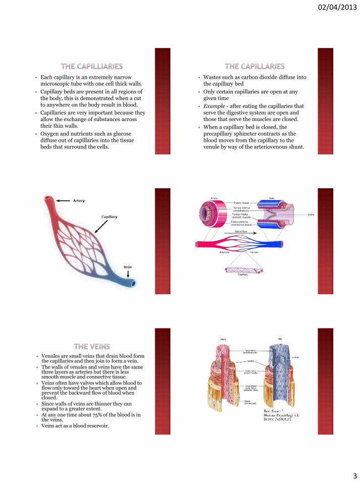
Each capillary is an extremely narrow microscopic tube with one cell thick walls.
Capillary beds are present in all regions of the body, this is demonstrated when a cut to anywhere on the body result in blood.
Capillaries are very important because they allow the exchange of substances across their thin walls.
Oxygen and nutrients such as glucose diffuse out of capillaries into the tissue beds that surround the cells.
Wastes such as carbon dioxide diffuse into the capillary bed
Only certain capillaries are open at any given time
Example - after eating the capillaries that serve the digestive system are open and those that serve the muscles are closed.
When a capillary bed is closed, the precapillary sphincter contracts as the blood moves from the capillary to the venule by way of the arteriovenous shunt.
Venules are small veins that drain blood form the capillaries and then join to form a vein.
The walls of venules and veins have the same three layers as arteries but there is less smooth muscle and connective tissue.
Veins often have valves which allow blood to flow only toward the heart when open and prevent the backward flow of blood when closed.
Since walls of veins are thinner they can expand to a greater extent.
At any one time about 75% of the blood is in the veins.
Veins act as a blood reservoir.
02/04/2013
4
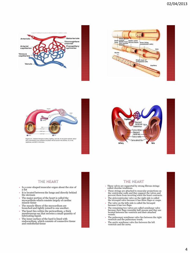
Is a cone-shaped muscular organ about the size of
a fist It is located between the lungs and directly behind
the sternum The major portion of the heart is called the
myocardium which consists largely of cardiac muscle tissue
The muscle fibers of the myocardium are branched and tightly joined to one another.
The heart lies within the pericardium, a thick membranous sac that secretes a small quantity of lubricating liquid.
The inner surface of the hard is lined with endocardium, which consists of connective tissue and endothelial tissue
These valves are supported by strong fibrous strings called chordae tendineae.
These strings are attached to muscular projections on the ventricular walls and they support the valves and prevent them from inverting when the heart contracts.
The atrioventricular valve on the right side is called the tricuspid valve because it has three flaps or cusps.
The valve on the lefts side is called the bicuspid because it has two flaps.
The remaining two valves are called semilunar valve because their flaps resemble half-moons and they are located between the ventricle and their attached vessels.
The pulmonary semilunar valve lies between the right ventricle and the pulmonary trunk.
The aortic semilunar valve lies between the left ventricle and the aorta.
02/04/2013
5
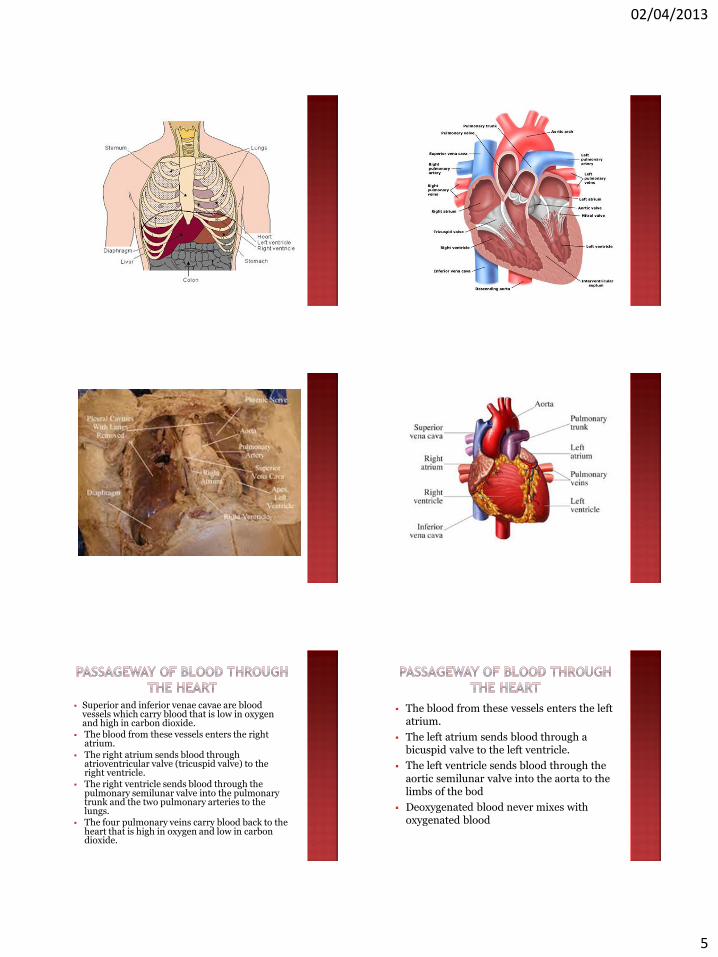
Superior and inferior venae cavae are blood vessels which carry blood that is low in oxygen and high in carbon dioxide.
The blood from these vessels enters the right atrium.
The right atrium sends blood through atrioventricular valve (tricuspid valve) to the right ventricle.
The right ventricle sends blood through the pulmonary semilunar valve into the pulmonary trunk and the two pulmonary arteries to the lungs.
The four pulmonary veins carry blood back to the heart that is high in oxygen and low in carbon dioxide.
The blood from these vessels enters the left atrium.
The left atrium sends blood through a bicuspid valve to the left ventricle.
The left ventricle sends blood through the aortic semilunar valve into the aorta to the limbs of the bod
Deoxygenated blood never mixes with oxygenated blood

02/04/2013
6
Blood must go through the lungs in order to pass from the right side to the left side of the heart.
The heart is a double pump because the right ventricles of the heart send blood
thorugh te lungs and the left ventricles send blood throughout the body.
Each heartbeat is called a cardiac cycle. When the heart beats, first the two atria
contract at the same time then the two ventricles contract at the same time
Then all four chambers relax The word systole refers to the contraction of
the heart muscle and the word diastole refers to the relaxation of the heart muscle.
The heat contracts or beat about 70 times a minute and each heart beast last about 0.85 seconds.
A normal adult rate at rest can vary from 60 to 80 beats per minute
When the heart beats it makes a “lub-dub” sound
The longer and lower pitched lob is causes by vibrations occurring when the AV valves close due to ventricular contractions.
The shorter sharper dub is heard when the semilunar valves close due to the pressure of blood in the arteries.
If a person has a heart murmur a slushy sound after the lub it is often heard due to an ineffective valve which allows blood to pass back into the atria.
02/04/2013
7
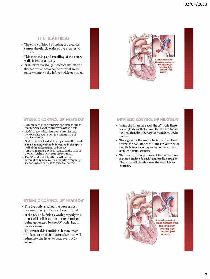
The surge of blood entering the arteries causes the elastic walls of the arteries to stretch.
This stretching and recoiling of the artery walls is felt as a pulse.
Pulse rates normally indicates the rate of the heartbeat because the arterial walls pulse whenever the left ventricle contracts
Contractions of the ventricle and atria is due to the intrinsic conduction system of the heart
Nodal tissue, which has both muscular and nervous characteristics, is a unique type of cardiac muscle.
Nodal tissue is located it two places in the heart:
The SA (sinoatrial) node is located in the upper wall of the right atrium and the AV (atrioventricular) node is located in the base of the right atrium very near the septum.
The SA node initiates the heartbeat and automatically sends out an impulse every 0.85 seconds which causes the atria to contract.
When the impulses reach the AV node there is a slight delay that allows the atria to finish their contractions before the ventricles begin theirs.
The signal for the ventricles to contract then travels the two branches of the atriventricular bundle before reaching many numerous and smaller purkinge fibers.
These ventricular portions of the conduction system consist of specialized cardiac muscle fibers that efficiently cause the ventricle to contract.
The SA node is called the pace maker because it keeps the heartbeat normal.
If the SA node fails to work properly the heart will still beat due to the impulses being generated by the AV node, but it beats slower.
To correct this condition doctors may
implant an artificial pacemaker that will stimulate the heart to beat every 0.85 second.
02/04/2013
8
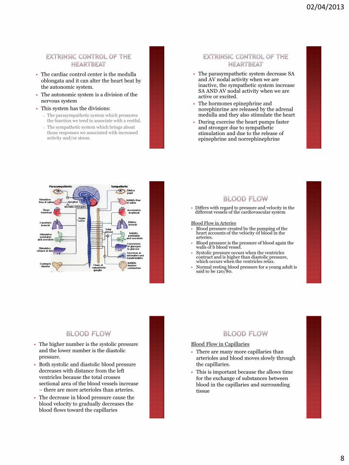
The cardiac control center is the medulla oblongata and it can alter the heart beat by the autonomic system.
The autonomic system is a division of the nervous system
This system has the divisions:
o The parasympathetic system which promotes the function we tend to associate with a restful.
o The sympathetic system which brings about those responses we associated with increased activity and/or stress.
The parasympathetic system decrease SA and AV nodal activity when we are inactive, the sympathetic system increase SA AND AV nodal activity when we are active or excited.
The hormones epinephrine and norephinrine are released by the adrenal medulla and they also stimulate the heart
During exercise the heart pumps faster and stronger due to sympathetic stimulation and due to the release of epinephrine and norrephinephrine
Differs with regard to pressure and velocity in the different vessels of the cardiovascular system
Blood Flow in Arteries Blood pressure created by the pumping of the
heart accounts of the velocity 0f blood in the arteries.
Blood pressure is the pressure of blood again the walls of b blood vessel.
Systolic pressure occurs when the ventricles contract and is higher than diastolic pressure, which occurs when the ventricles relax.
Normal resting blood pressure for a young adult is said to be 120/80.
The higher number is the systolic pressure and the lower number is the diastolic pressure.
Both systolic and diastolic blood pressure decreases with distance from the left
ventricles because the total crosses sectional area of the blood vessels increase – there are more arterioles than arteries.
The decrease in blood pressure cause the blood velocity to gradually decreases the blood flows toward the capillaries
Blood Flow in Capillaries
There are many more capillaries than arterioles and blood moves slowly through the capillaries.
This is important because the allows time for the exchange of substances between blood in the capillaries and surrounding tissue
02/04/2013
9
As the skeletal muscle contract they compress the weak walls of the veins, this causes blood to move past the next valve.
Once past the valve blood cannon flow backwards The importance of muscle contraction in moving blood
can be seen by forcing a person to stand absolutely still for an hour. Often the person will faint because blood collects in the limbs depriving the brain the blood it needs.
When inspiration occurs the thoracic pressure falls and the abdominal pressure rise as the chest expands.
This allows the flow of venous blood back to the heart because the blood is flowing in the direction of reduced pressure.
Blood velocity increases slightly in the venous vessels due to a progressive reduction in the cross sectional area as small venules joins to form veins.
Blood Flow in Veins
Blood pressure is minimal in venules and veins.
Instead the blood pressure, venous return is dependent upon three factors:
1. Skeletal muscle contraction
2. Presence of valves in veins
3. Respiratory movement
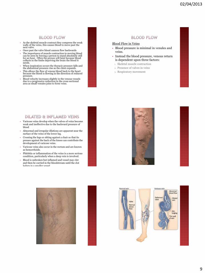
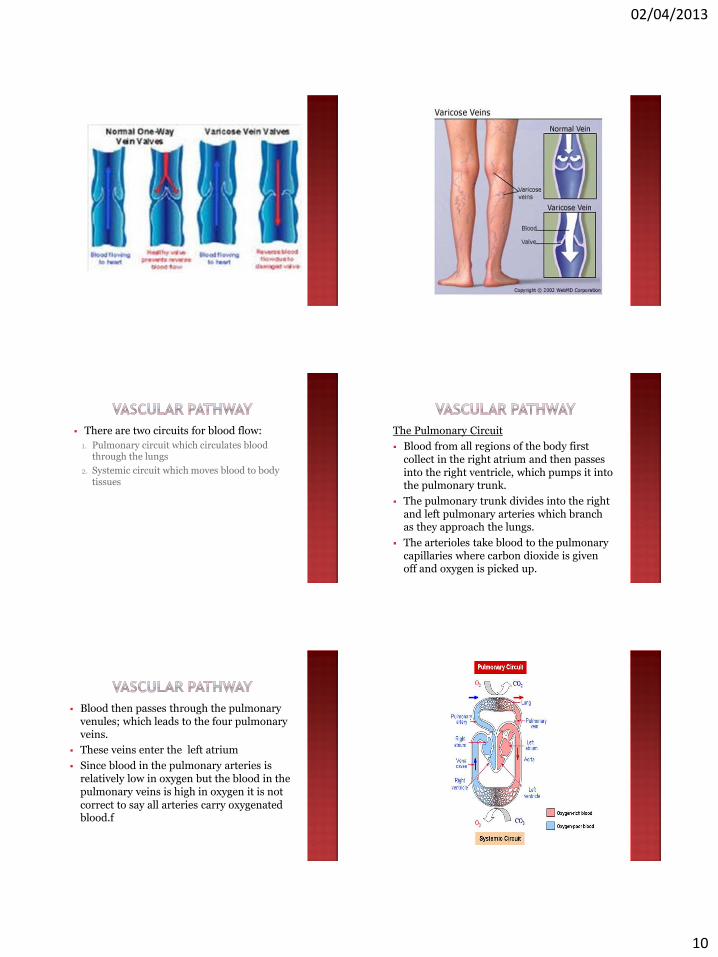
Varicose veins develop when the valves of veins become weak and ineffective due to the backward pressure of blood
Abnormal and irregular dilations are apparent near the surface of the veins of the lower leg.
Crossing the legs or sitting against a chair so that its presses against the back of the knees can contribute the development of varicose veins.
Varicose veins also occur in the rectum and are known as hemorrhoids.
Phlebitis or inflammation of the veins is a more serious condition, particularly when a deep vein is involved.
Blood is unbroken but inflamed and vessel may clot and then be carried in the bloodstream until the clot lodges in a smaller vessel.
If a blood clot blocks a pulmonary vessel death can occur.
02/04/2013
10
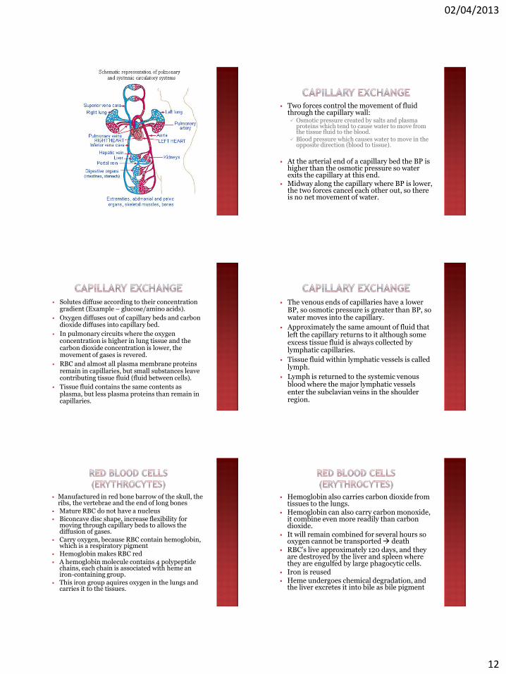
There are two circuits for blood flow:
1. Pulmonary circuit which circulates blood through the lungs
2. Systemic circuit which moves blood to body tissues
The Pulmonary Circuit
Blood from all regions of the body first collect in the right atrium and then passes into the right ventricle, which pumps it into the pulmonary trunk.
The pulmonary trunk divides into the right and left pulmonary arteries which branch as they approach the lungs.
The arterioles take blood to the pulmonary capillaries where carbon dioxide is given off and oxygen is picked up.
Blood then passes through the pulmonary venules; which leads to the four pulmonary veins.
These veins enter the left atrium
Since blood in the pulmonary arteries is relatively low in oxygen but the blood in the pulmonary veins is high in oxygen it is not
correct to say all arteries carry oxygenated blood.f
02/04/2013
11
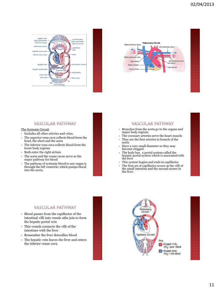
The Systemic Circuit
Includes all other arteries and veins.
The superior vena cava collects blood form the head, the chest and the arms
The inferior vena cava collects blood form the lower body regions
Both enter the right atrium
The aorta and the venae cavae serve as the major pathway for blood
The pathway of systemic blood to any organ is through the left ventricle; which pumps blood into the aorta.
Branches from the aorta go to the organs and major body regions.
The coronary arteries serve the heart muscle They are the first arteries to branch of the
aorta Have a very small diameter so they may
become clogged. The body has a portal system called the
hepatic portal system which is associated with the liver
This system begins and ends in capillaries The first set of capillaries occurs at the villi of
the small intestine and the second occurs in the liver.
Blood passes from the capillaries of the intestinal villi into venule athe join to form the hepatic portal vein
This vessels connects the villi of the intestines with the liver
Remember the liver detoxifies blood
The hepatic vein leaves the liver and enters the inferior venae cava.
02/04/2013
12
Two forces control the movement of fluid through the capillary wall: Osmotic pressure created by salts and plasma
proteins which tend to cause water to move from the tissue fluid to the blood.
Blood pressure which causes water to move in the opposite direction (blood to tissue).
At the arterial end of a capillary bed the BP is
higher than the osmotic pressure so water exits the capillary at this end.
Midway along the capillary where BP is lower, the two forces cancel each other out, so there is no net movement of water.
Solutes diffuse according to their concentration gradient (Example – glucose/amino acids).
Oxygen diffuses out of capillary beds and carbon dioxide diffuses into capillary bed.
In pulmonary circuits where the oxygen concentration is higher in lung tissue and the carbon dioxide concentration is lower, the movement of gases is revered.
RBC and almost all plasma membrane proteins remain in capillaries, but small substances leave contributing tissue fluid (fluid between cells).
Tissue fluid contains the same contents as plasma, but less plasma proteins than remain in capillaries.
The venous ends of capillaries have a lower BP, so osmotic pressure is greater than BP, so water moves into the capillary.
Approximately the same amount of fluid that left the capillary returns to it although some excess tissue fluid is always collected by lymphatic capillaries.
Tissue fluid within lymphatic vessels is called lymph.
Lymph is returned to the systemic venous blood where the major lymphatic vessels enter the subclavian veins in the shoulder region.
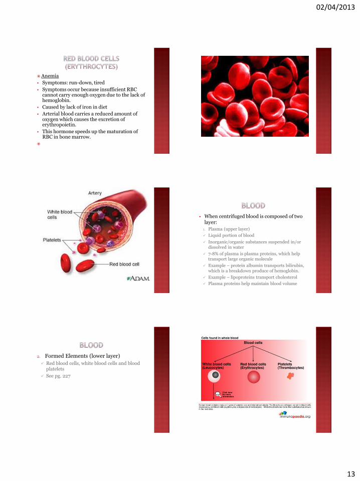
Manufactured in red bone barrow of the skull, the ribs, the vertebrae and the end of long bones
Mature RBC do not have a nucleus Biconcave disc shape, increase flexibility for
moving through capillary beds to allows the diffusion of gases.
Carry oxygen, because RBC contain hemoglobin, which is a respiratory pigment
Hemoglobin makes RBC red A hemoglobin molecule contains 4 polypeptide
chains, each chain is associated with heme an iron-containing group.
This iron group aquires oxygen in the lungs and carries it to the tissues.
Hemoglobin also carries carbon dioxide from tissues to the lungs.
Hemoglobin can also carry carbon monoxide, it combine even more readily than carbon dioxide.
It will remain combined for several hours so oxygen cannot be transported death
RBC’s live approximately 120 days, and they are destroyed by the liver and spleen where they are engulfed by large phagocytic cells.
Iron is reused Heme undergoes chemical degradation, and
the liver excretes it into bile as bile pigment
02/04/2013
13
Anemia
Symptoms: run-down, tired
Symptoms occur because insufficient RBC cannot carry enough oxygen due to the lack of hemoglobin.
Caused by lack of iron in diet
Arterial blood carries a reduced amount of oxygen which causes the excretion of erythropoietin.
This hormone speeds up the maturation of RBC in bone marrow.
When centrifuged blood is composed of two layer:
1. Plasma (upper layer)
Liquid portion of blood
Inorganic/organic substances suspended in/or dissolved in water
7-8% of plasma is plasma proteins, which help transport large organic molecule
Example – protein albumin transports bilirubin, which is a breakdown produce of hemoglobin.
Example – lipoproteins transport cholesterol
Plasma proteins help maintain blood volume
2. Formed Elements (lower layer)
Red blood cells, white blood cells and blood platelets
See pg. 227
02/04/2013
14
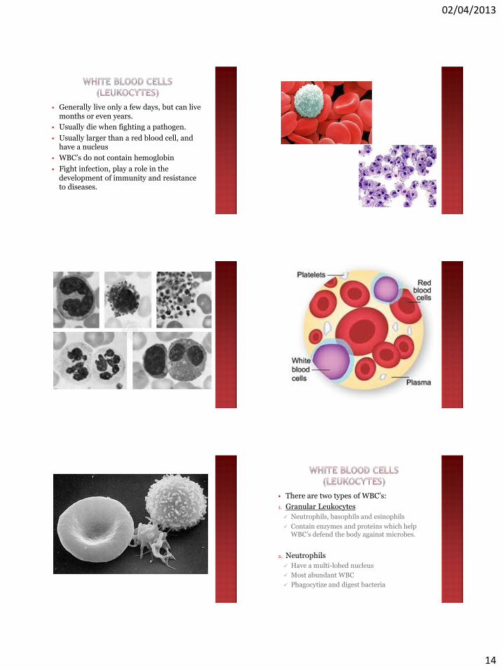
Generally live only a few days, but can live months or even years.
Usually die when fighting a pathogen.
Usually larger than a red blood cell, and have a nucleus
WBC’s do not contain hemoglobin
Fight infection, play a role in the development of immunity and resistance to diseases.
There are two types of WBC’s:
1. Granular Leukocytes
Neutrophils, basophils and esinophils
Contain enzymes and proteins which help WBC’s defend the body against microbes.
a. Neutrophils
Have a multi-lobed nucleus
Most abundant WBC
Phagocytize and digest bacteria
02/04/2013
15
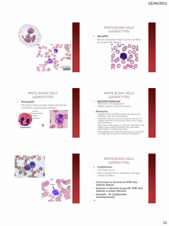
b. Basophils
Release histamines which can be a problem for people with allergies
c. Eosinophils
Thought to fight parasitic works and may be involved in creating some allergies
2. Agranular Leukocytes Monocytes and lymphocytes Kidney shaped or spherical nucleus
a. Monocytes Largest WBC and differentiate into phagocytic
dendritic cells and macrophages. Dendritic cells are present in tissues that are in
contact with the environment; skin, nose, lungs, and intestines.
Once these cells capture a microbe with their long spikey arms, called dendrites, they stimulate other WBC’s to defend the body.
Macrophages are a ferocious phagocyte, and have a similar role in the liver, kidneys and spleen.
b. Lymphocytes Two types: B & T
Play a specific role in immunity, and fight certain invaders
An increase or decrease in WBC may indicate disease
Increase or decrease in specific WBC may indicate a certain infection
Example – B- lymphocytes (mononucleosis)
02/04/2013
16
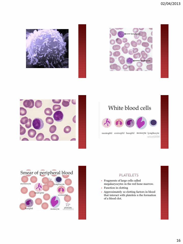
Fragments of large cells called megakaryocytes in the red bone marrow.
Function in clotting
Approximately 12 clotting factors in blood that interact with platelets n the formation of a blood clot.
02/04/2013
17
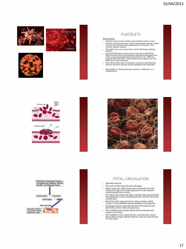
Blood Clotting
1. Platelets clump at sites of injury and partially seal the wound.
2. Platelets and damaged tissue release prothrombin activator which converts the plasma protein prothrombin to thrombin. This reaction requires calcium.
3. Thrombin acts as an enzyme that activates fibrinogen (plasma protein).
4. Activated fibrinogen proteins form a long chain called fibrin. Fibrin threads wind around platelets and plug the damaged area of the vessel and provides the framework for a clot. RBC are trapped within the fibrin, which makes the clot appear red. The fibrin clot is only temporary.
5. Once blood vessel repair is initiated, an enzyme called plasmin destroys the fibrin network and the epithelial cells regenerate.
Hemophilia is a clotting disorder caused by a deficiency in a clotting factor.
Specialty features
Does not use the lungs for gas exchanges
Blood enters the right atrium and is shunted to the left atrium through an oval opening known as the foramen ovale between the two atria.
Any blood that enters the right ventricle and is pumped into the pulmonary trunk is shunted into the aorta by way of the arterial duct.
Blood from the aorta travels to various arteries which connect to the umbilical arteries leading to the placenta
Exchange of gases and nutrients between maternal and fetal blood occurs within the placenta.
The umbilical vein carriers blood rich in nutrients and oxygen to the fetus.
The umbilical veins enters the liver and joins the venous ducts which merges with the inferior vena cava and returns it to the heart.