Embed Size (px)
Citation preview
CIRCULATORY SYSTEM
CHAPTER NO.7
10TH ICSE
INTRODUCTION
• In all organisms there are fluids which circulate throughout the body
• These fluids transport substances to various parts of body and collect substances from various parts of body.
• These are the body fluids
NEED FOR TRANSPORT INSIDE THE BODY• The digestive system digests and absorbs
nutrients which have to be transported to every cell of the body
• Respiratory system takes in air containing O2 which goes to the lungs from there O2 has to be transported to every body cell and the CO2 from every body cell has to be transported to the lungs so that it can be given out during expiration.
• All the extra water , excess salts and urea have to be transported from different parts of body to the excretory system so that they can be thrown out of the body.
• Hormones secreted by endocrine glands have to be transported throughout the body to act wherever they are required
All such transportation is carried out by 2 circulating fluids
a)Blood b)Lymph
BLOOD• Colour – bright red or dark red
• Volume – An average adult person has 5-6 litres of blood
• Taste – salty since it is alkaline it has a pH of 7.3-7.45
• Study of blood is haematology
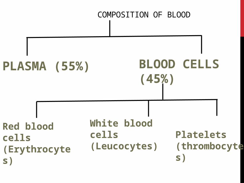
COMPOSITION OF BLOOD
PLASMA (55%)
BLOOD CELLS(45%)
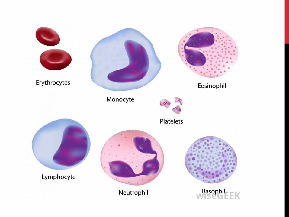
Red blood cells (Erythrocytes)
White blood cells (Leucocytes)
Platelets (thrombocytes)
PLASMA• It s light yellow in colour slightly alkaline
• It contains
a)Water - 90-92%
b) Proteins – 7-8%
c) Inorganic salts (NaCl,NaHCO3)- 1%
d) Other substances (Glucose , a.a , fibrinogen , urea , hormones etc) –Traces
Serum is Plasma - Fibrinogen
BLOOD CELLSA) R.B.CS
• Round-Biconcave in shape because of the shape they can easily pass through capillaries
• They are oxygen carriers and are also called erythrocytes
• Size is small hence large surface area so they can absorb oxygen efficiently (7 micron in diameter)
• Adult human male-5 million RBC per cubic mm of blood and Adult human Female – 4.5 million RBCS
• RBCs are called oxygen carriers because they have haemoglobin(Hb) which is a respiratory pigment
Oxygen + Haemoglobin Oxyhaemoglobin
Carbon dioxide + Haemoglobin Carbaminohaemoglobin
• Haemoglobin has a strong affinity for carbon monoxide
Hb+CO HbCO (Carboxyhaemoglobin)
This property of Hb results in CO poisoning
• RBCs do not have nucleus , E.R , Mitochondria and makes them more efficient in transporting oxygen i.e Because they do not have nucleus their surface area increases , Because of no mitochondria O2 is not used up , Because of no E.R they do not use up glucose in blood plasma.
• Life span of RBCs is 120 days
• Polycythaemia Increase in no. of RBCs
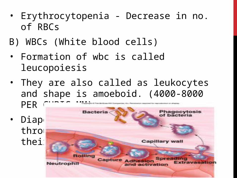
• Erythrocytopenia - Decrease in no. of RBCs
B) WBCs (White blood cells)
• Formation of wbc is called leucopoiesis
• They are also called as leukocytes and shape is amoeboid. (4000-8000 PER CUBIC MM)
• Diapedesis-WBCs can squeeze out through the capillaries because of their shape
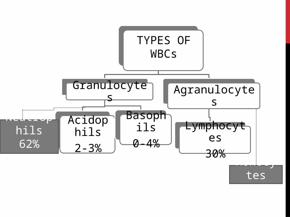
TYPES OF WBCs
Granulocytes
Acidophils
2-3%
Basophils
0-4%
Agranulocytes
Lymphocytes
30%
Neutrophils
62%
Monocytes
5-3%
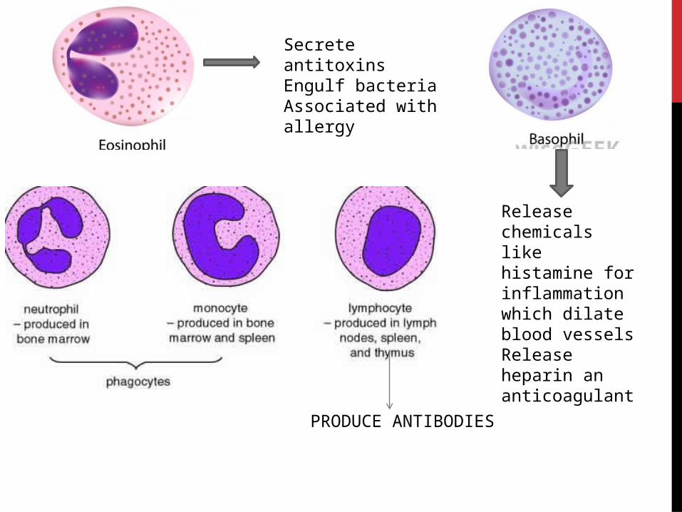
PRODUCE ANTIBODIES
Secrete antitoxinsEngulf bacteriaAssociated with allergy
Release chemicals like histamine for inflammation which dilate blood vesselsRelease heparin an anticoagulant
The average life span of wbcs is 2 weeks .
Leucopenia is abnormal decrease in no. of wbcs
Leucocytosis is increase in no. of wbcs
Abnormal increase in WBC i.e about 50000 or more indicates infection in body
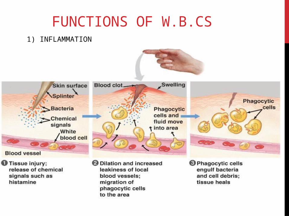
FUNCTIONS OF W.B.CS1) INFLAMMATION
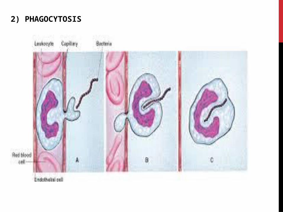
2) PHAGOCYTOSIS
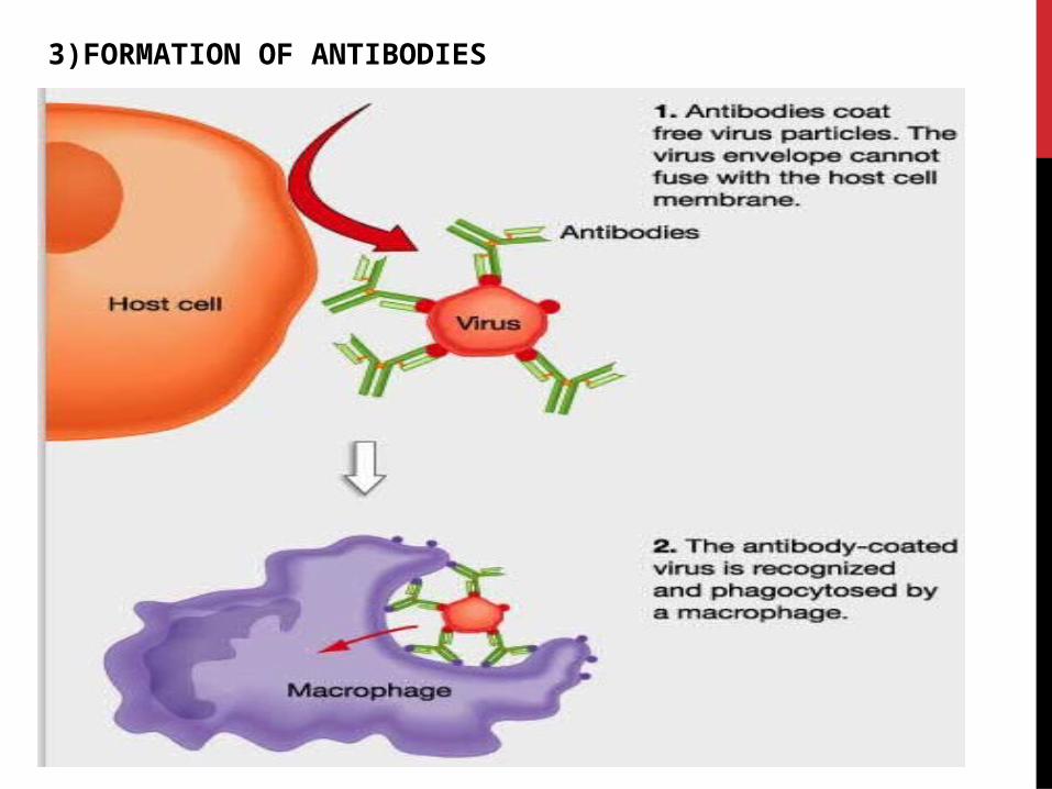
3)FORMATION OF ANTIBODIES

3)PLATELETS
They are formed from megakaryocytes
2 lac -4lac per cubic mm of blood in adults
Life span is 3-5 days
They help in process of clotting of blood
Formation is thrombopoiesis
Increase-thrombocytosis,decrease-thrombocytopenia
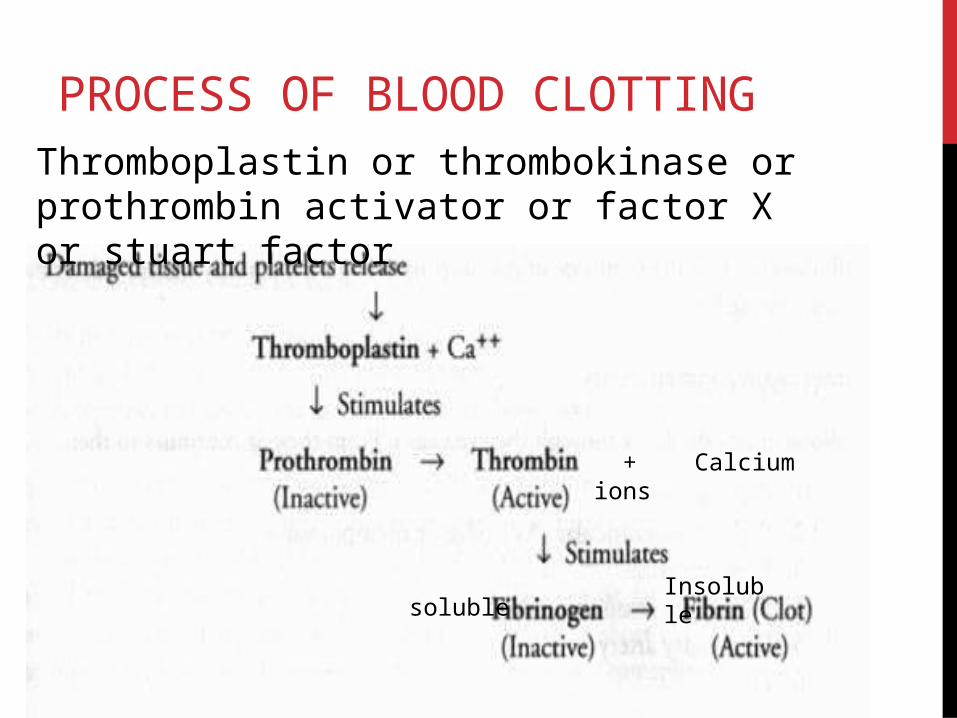
PROCESS OF BLOOD CLOTTINGThromboplastin or thrombokinase or prothrombin activator or factor X or stuart factor
+ Calcium ions
solubleInsoluble
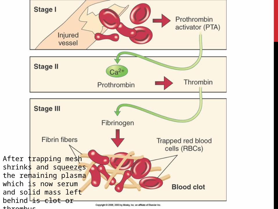
After trapping mesh shrinks and squeezes the remaining plasma which is now serum and solid mass left behind is clot or thrombus
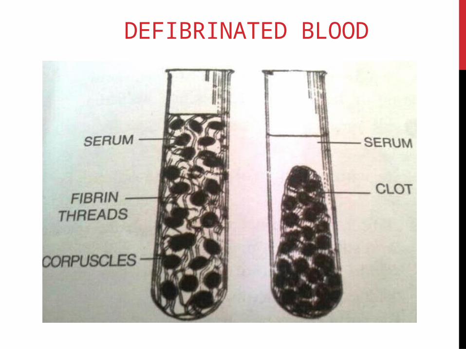
DEFIBRINATED BLOOD
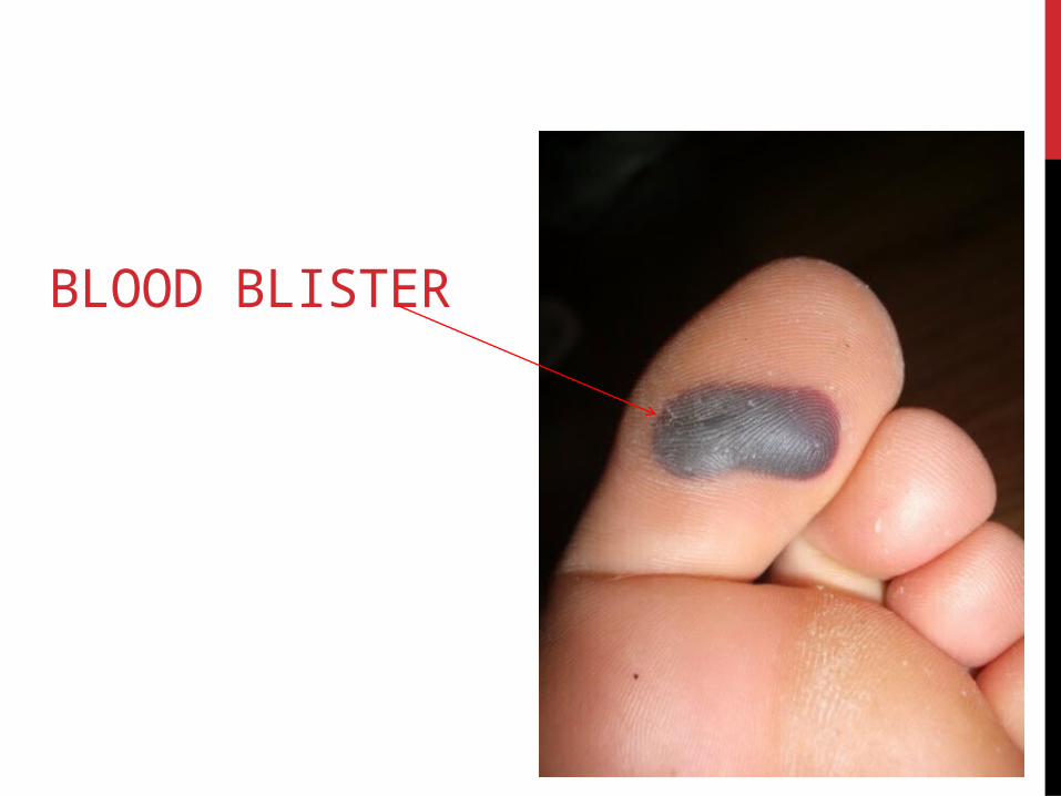
BLOOD BLISTER
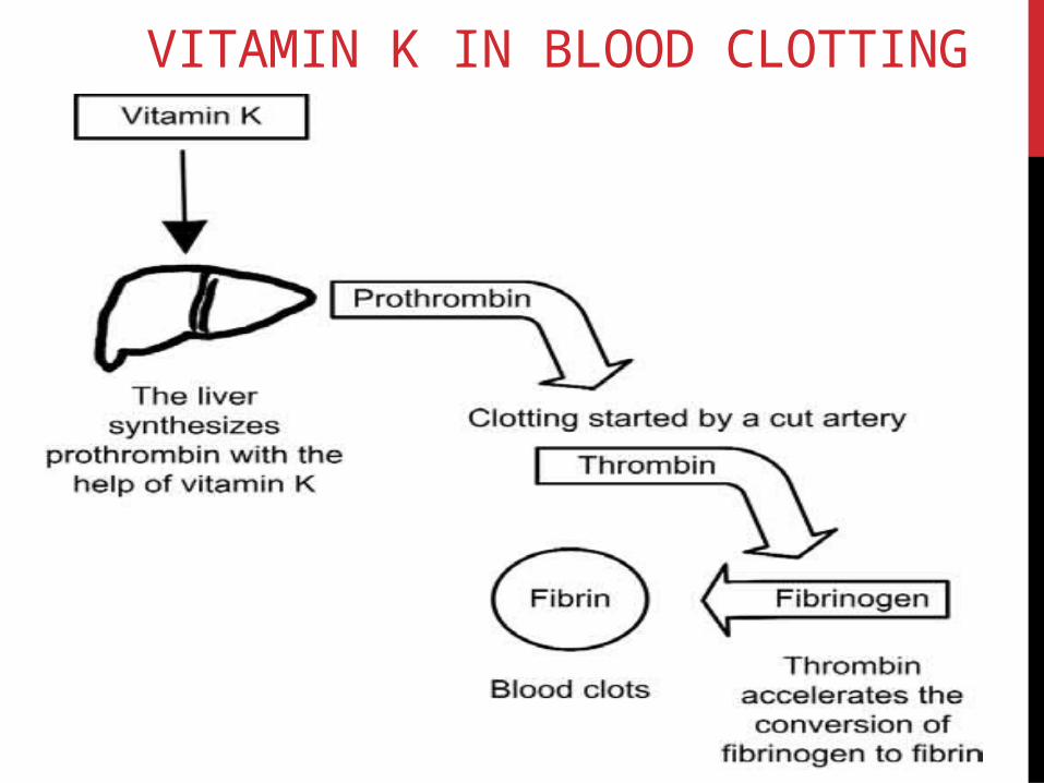
VITAMIN K IN BLOOD CLOTTING

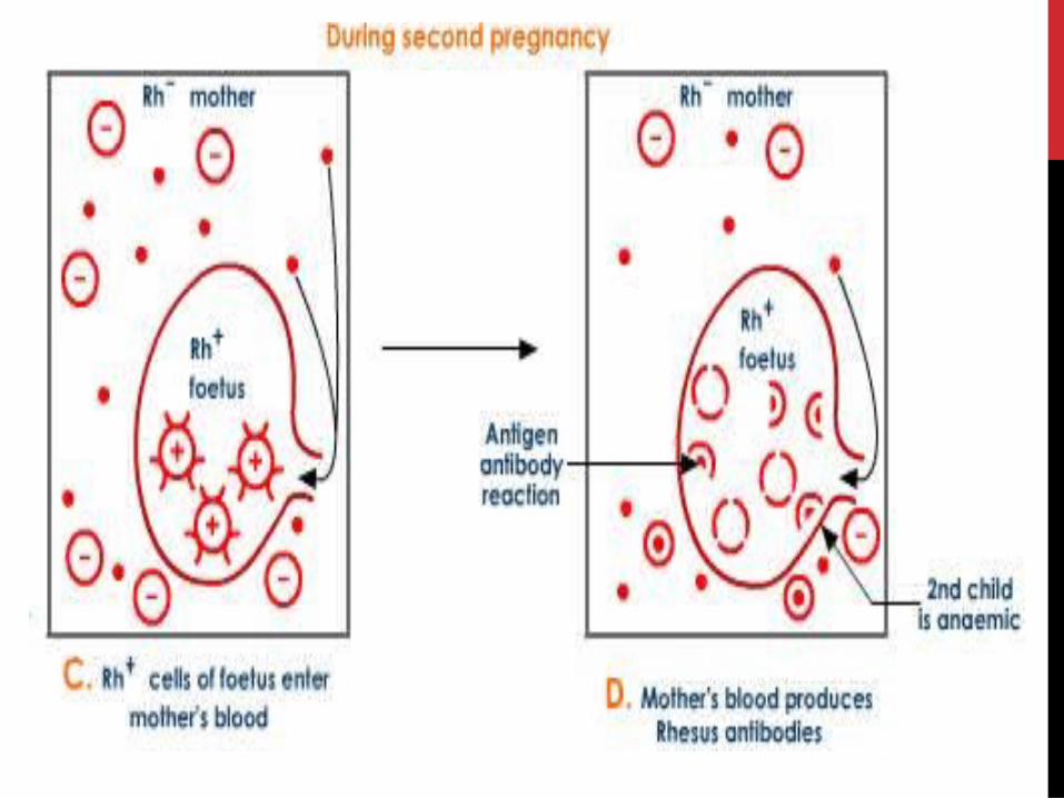
RH FACTOR DURING PREGNANCY
TRANSPORTATION IN HUMAN BEINGSThe process of transportation is called as circulation
3 distinct components
Blood - fluid circulates in our body and carries
out the function of transport of various
materials in our body
Blood vessels- tubes that help the blood to
circulate.
Heart - pumping organ that circulates the
blood around the body.
HEART-THE INVOLUNTARY PUMPMuscular organPericardial membraneWeight-360 gms 12cm in length and 9cm in breadth.Thoracic cavity between the lungs in a space called mediastinum
Pericardium –fibrous and serous
Serous-parietal layer and visceral layer
Pericardial fluid
Heart wall-it has 3 layers
Epicardium , myocardium , endocardium
Vertical septum divides heart into right and left compartments4 chambers- upper ones are atria and lower ones are ventricles thus rt.atrium and lt.atrium ,rt.ventricle and lt. ventricle The heart has different chambers to prevent the oxygen-rich blood(oxygenated blood) from mixing with the blood containing carbon dioxide (Deoxygenated blood).
The carbon dioxide-rich blood has to reach the lungs for the carbon dioxide to be removed, and the oxygenated blood from the lungs has to be brought back to the heart. This oxygen-rich blood is then pumped to the rest of the body.Right side of heart-oxygenated blood and left side-Deoxygenated blood this kind of separation of blood makes the oxygen supply to the entire body highly efficient.Valves are present between atria and ventricles to prevent backflow of blood.Blood vessels- Veins carry blood towards heart from diff. parts of body and Arteries carry blood away from heartVenacava-large veins and Aorta-largest artery.Pulmonary artery and pulmonary veins.Ventricles have thick walls since they have to pump blood to long distances
Atria have thinner walls because they have to receive blood and pump it to the next ventricle.
At the base of aorta there are 2 coronary arteries that supply heart muscles if there are blockages in these arteries then it will lead to heart attack (myocardial infarction)
Cardiac veins that collect blood from heart walls and pour it to the right auricle.
VALVES IN THE HEARTThere are 4 valves in the heart and they are as follows:
a)Right atrioventricular valves/Tricuspid valve
Present at the opening of r.a to r.v and it has 3 flaps and it is held in position by chordae tendinae which arises from muscular projections of ventricle wall and is known as papillary muscles
b)Left atrioventricular valves/Bicuspid/Mitral valves-
Present at the opening of l.a to l.v. It has 2 flaps
c)Pulmonary semilunar valves- Present at the opening of r.v to p.a..3 in no. and are pocket shaped
d)Aortic semilunar valves-Present at the opening of l.v to aorta 3 in no. and are pocket shaped.
The opening of inferior venacava is guarded by Eustachian valve while the opening of coronary sinus is guarded by thebesian valve
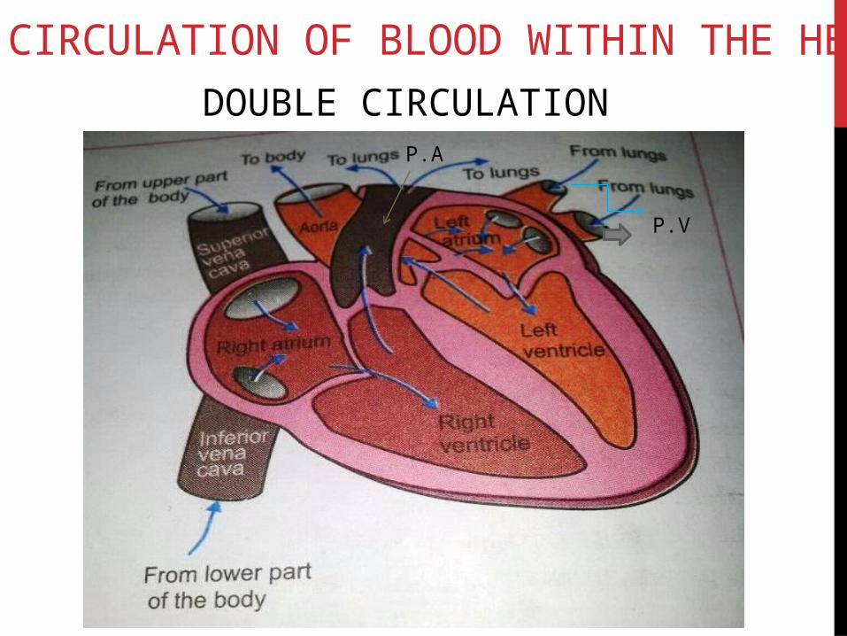
CIRCULATION OF BLOOD WITHIN THE HEART
P.A
P.V
DOUBLE CIRCULATION
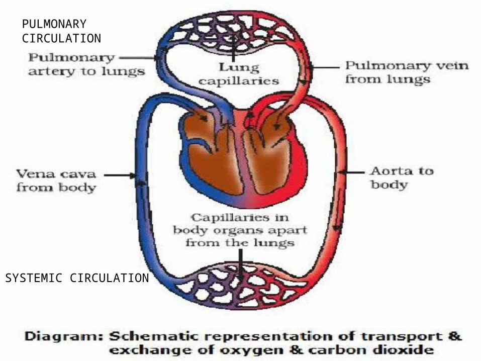
PULMONARY CIRCULATION
SYSTEMIC CIRCULATION
CORONARY CIRCULATIONCardiac muscles of heart receive oxygenated blood through coronary arteries . deoxygenated blood is collected by coronary veins which join to form coronary sinus which opens into right atrium

CARDIAC CYCLE
The steps of heart beat includes :
a)Atrial Systole-0.15 sec
b)Ventricular systole- 0.30 sec
c)Joint diastole- 0.40 sec
Atrial Systole-
• Atria contract and blood enters the ventricles
• Openings of pulmonary vein and venacava close
• Tricuspid and bicuspid open and semilunar valves at the roots of pulmonary artery and aorta are closed
Ventricular Systole
• Ventricles contract
• Tricuspid and Bicuspid valves close
• Semilunar valves open and blood enters pulmonary artery and aorta
• Chordae tendinae hold the valves in position preventing upturning due to pressure exerted by contracting ventricles
Joint Diastole
• Both atria and ventricles relax
• Right atrium receives deoxygenated blood from all parts of the body through venacava
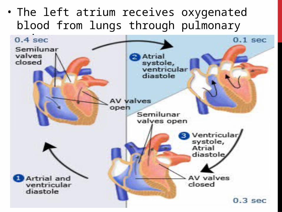
• The left atrium receives oxygenated blood from lungs through pulmonary veins
The sequence of events in a heart beat is called as cardiac cycle.
THE HEART SOUNDS
LUBB-At the start of ventricular systole the atrio-ventricular valves close and produce a sound LUBB
DUP-At the beginning of ventricular diastole the semilunar valves at the roots of aorta and pulmonary artery get closed and produce a sound called as DUP
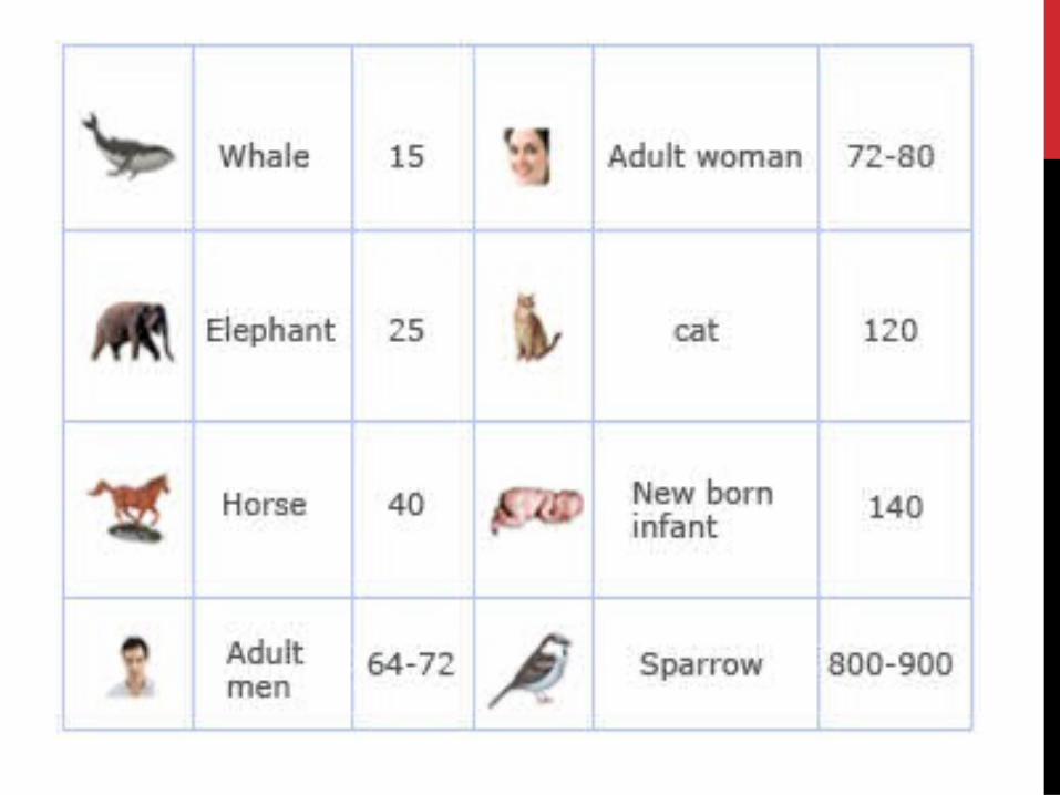
The rate of heart beat is different among diff. species and it is seen that larger the size of the org. slower is the rate of heart beat or heart rate because heart has to pump more volumes of blood through narrow capillaries
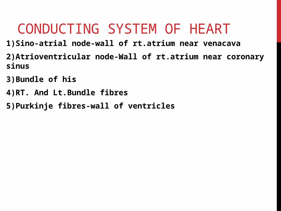
CONDUCTING SYSTEM OF HEART1)Sino-atrial node-wall of rt.atrium near venacava
2)Atrioventricular node-Wall of rt.atrium near coronary sinus
3)Bundle of his
4)RT. And Lt.Bundle fibres
5)Purkinje fibres-wall of ventricles
BLOOD PRESSUREForce exerted by blood on the walls of the blood
vessels.
It is greater in arteries than in veins.
The Pressure of blood inside the artery during
ventricular systole (contraction) is called systolic
pressure.
Pressure in artery during ventricular diastole
(relaxation) is called diastolic pressure.
The normal systolic pressure -120mm Hg.
The normal diastolic pressure-80 mmHg.
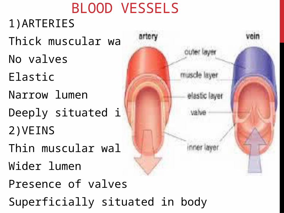
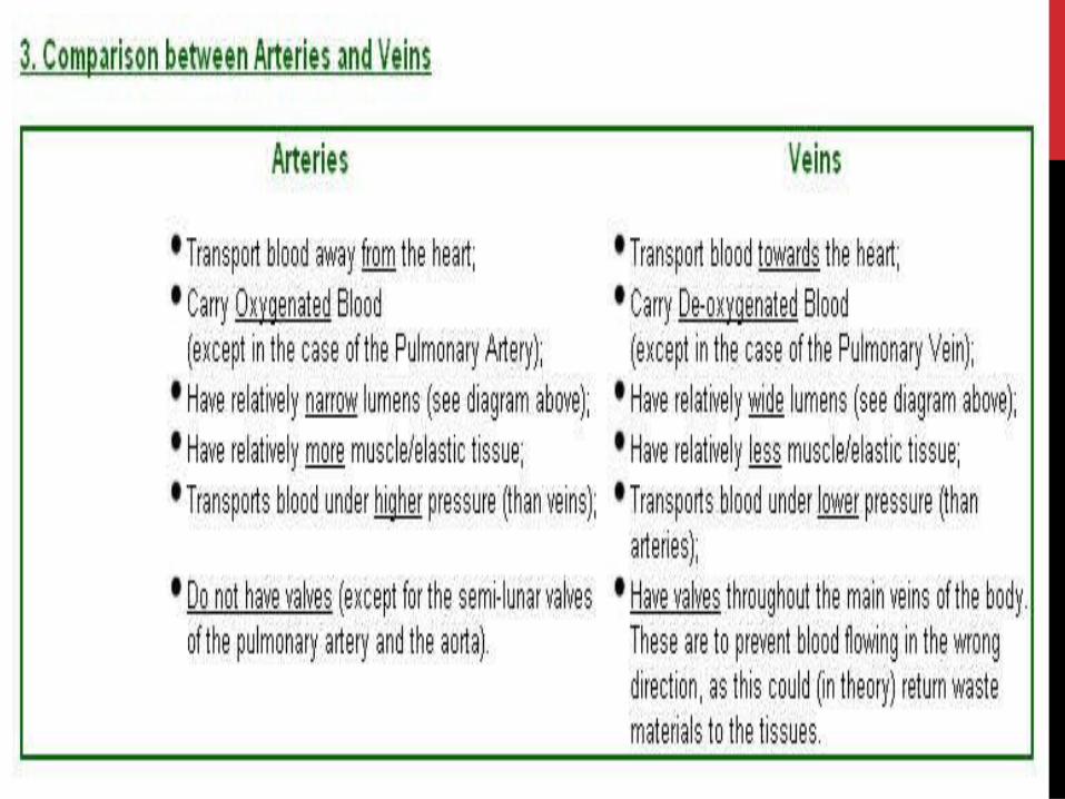
BLOOD VESSELS1)ARTERIES
Thick muscular walls
No valves
Elastic
Narrow lumen
Deeply situated in body
2)VEINS
Thin muscular walls
Wider lumen
Presence of valves
Superficially situated in body
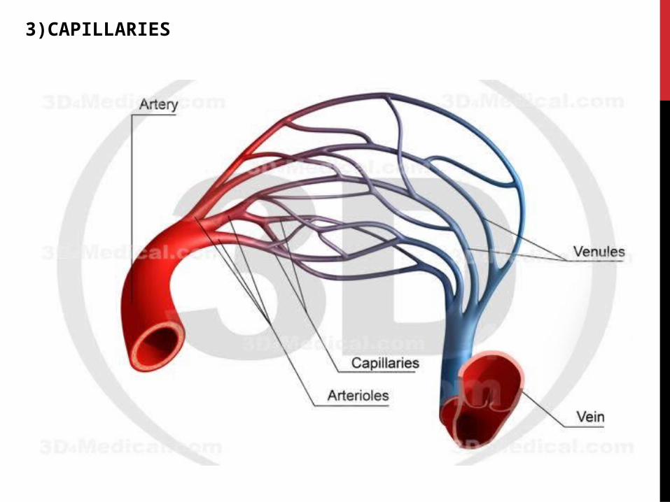
3)CAPILLARIES
The study of blood vessels is called angiology
Heart beat is the rhythmic contraction and relaxation of heart.
The heart beats 72 times per min.this is called heart rate
PULSE it is the alternate expansion and elastic recoil of the wall of artery during ventricular systole
Tachycardia means fast heart rate
Bradycardia means slow heart rate
BLOOD RELATED DISORDERS
1)Hypertension-Rupture blood vessels of eyes ,kidneys,brain
2)Coronary artery disease (CAD)-atherosclerosis (deposition of fatty substances in the lining of arteries).It causes narrowing of coronary arteries so blood flow to heart is reduced
3)Angina pectoris-It is chest pain because of narrowing and hardening of coronary arteries. heaviness and severe pain in the chest.It occurs during exercise when heart demands more oxygen.
4)Heart failure-heart muscles become weak and heart fails to pump effectively which results in heart failure
MAIN BLOOD VESSELSHEART :
1)Blood vessels entering heart
• Superior venacava (formed by union of jugular vein and subclavian vein)
• Inferior venacava
• Pulmonary vein
2)Blood vessels leaving heart
• Pulmonary artery
• Aorta

LIVER:
1)Blood vessels entering liver
• Hepatic artery (from aorta into liver)
• Hepatic portal vein(From stomach and intestine into liver)
2)Blood vessels leaving liver
• Hepatic vein (from liver to inferior venacava)
KIDNEY
1)Blood vessels entering kidney
• Renal artery (from aorta to kidney)
2)Blood vessels leaving kidney
• Renal vein (from kidney to inferior venacava)
HEPATIC PORTAL SYSTEM
• The veins starting from stomach and intestines do not directly transport the blood to inferior venacava
• Instead they first enter liver as hepatic portal vein and inside the liver it breaks into capillaries and these capillaries join to form vein which joins the inferior venacava
• A portal vein is the one which starts with capillaries and also ends in capillaries.
USE OF HPS
Detoxification regulate the quantity of nutrients

LYMPHCOMPOSITION
A)Cellular part
• Only leukocytes (lymphocytes)
B)Non-cellular part
• Water-94%
• Proteins , fats , carbs , antibodies , enzs etc)-6%
FUNCTION
• NUTRITIVE-Supplies nutrition and oxygen to those parts where blood cannot reach
• ABSORPTION-Fats from intestines are obsorbed by lacteals
• DEFENCE-Lymphocytes and monocytes of lymph defend body
LYMPHATIC SYSTEMI)LYMPH
2)LYMPHATIC CAPILLARIES
3)LYMPHATIC VESSELS
3)LYMPH NODES
SPLEEN
It is present behind the stomach and above left kidney
FUNCTIONS
• Blood reservoir
• Produces lymphocytes
• Destroys worn out RBCS
• In embryo it produces RBCS