Embed Size (px)
Citation preview

1
SPECIAL SENSESSPECIAL SENSES
(INDERA KHUSUS)(INDERA KHUSUS)
Dr.Milahayati DaulayDr.Milahayati Daulay
Departemen Fisiologi Departemen Fisiologi
FK USUFK USU
Eye and Associated Eye and Associated
StructuresStructures
�� 70% of all sensory receptors are 70% of all sensory receptors are
in the eyein the eye
�� Most of the eye is protected by a Most of the eye is protected by a
cushion of fat and the bony orbitcushion of fat and the bony orbit
�� Accessory structures include Accessory structures include
eyebrows, eyelids, conjunctiva, eyebrows, eyelids, conjunctiva,
lacrimal apparatus, and extrinsic lacrimal apparatus, and extrinsic
eye muscleseye muscles

2
Eyebrows
� Coarse hairs that overlie the supraorbital margins
� Functions include:
� Shading the eye
� Preventing perspiration from reaching the eye
� Orbicularis muscle – depresses the eyebrows
� Corrugator muscles – move the eyebrows medially
Palpebrae (Eyelids)
� Protect the eye anteriorly
� Palpebral fissure – separates eyelids
� Canthi – medial and lateral angles
(commissures)

3
Palpebrae (Eyelids)Palpebrae (Eyelids)
�� Lacrimal caruncle Lacrimal caruncle –– contains glands that contains glands that
secrete a whitish, oily secretion (Sandman’s secrete a whitish, oily secretion (Sandman’s
eye sand)eye sand)
�� Tarsal plates of connective tissue support the Tarsal plates of connective tissue support the
eyelids internallyeyelids internally
�� Levator palpebrae superioris Levator palpebrae superioris –– gives the upper gives the upper
eyelid mobilityeyelid mobility
Palpebrae (Eyelids)
Eyelashes
Project from the free margin of each eyelid
Initiate reflex blinking
Lubricating glands associated with the
eyelids
Meibomian glands and sebaceous glands
Ciliary glands lie between the hair follicles

4
Palpebrae (Eyelids)
Figure 15.5b
ConjunctivaConjunctiva
�� Transparent membrane that:Transparent membrane that:
�� Lines the eyelids as the palpebral conjunctivaLines the eyelids as the palpebral conjunctiva
�� Covers the whites of the eyes as the ocular Covers the whites of the eyes as the ocular conjunctivaconjunctiva
�� Lubricates and protects the eyeLubricates and protects the eye

5
Lacrimal ApparatusLacrimal Apparatus
•• Consists of the lacrimal gland and associated Consists of the lacrimal gland and associated
ductsducts
•• Lacrimal glands secrete tears Lacrimal glands secrete tears
•• TearsTears
–– Contain mucus, antibodies, and lysozymeContain mucus, antibodies, and lysozyme
–– Enter the eye via superolateral excretory ducts Enter the eye via superolateral excretory ducts
–– Exit the eye medially via the lacrimal punctumExit the eye medially via the lacrimal punctum
–– Drain into the nasolacrimal ductDrain into the nasolacrimal duct
Lacrimal Apparatus
Figure 15.6

6
Extrinsic Eye MusclesExtrinsic Eye Muscles
�� Six straplike extrinsic eye musclesSix straplike extrinsic eye muscles
�� Enable the eye to follow moving objectsEnable the eye to follow moving objects
�� Maintain the shape of the eyeballMaintain the shape of the eyeball
�� Four rectus muscles originate from the Four rectus muscles originate from the annular ringannular ring
�� Two oblique muscles move the eye in the Two oblique muscles move the eye in the vertical planevertical plane
Extrinsic Eye Muscles
Figure 15.7a, b

7
Summary of Cranial Nerves and
Muscle Actions• Names, actions, and cranial nerve
innervation of the extrinsic eye muscles
Figure 15.7c
Structure of the Eyeball
�A slightly irregular hollow sphere with
anterior and posterior poles
�The wall is composed of three tunics –
fibrous, vascular, and sensory
�The internal cavity is filled with fluids
called humors
�The lens separates the internal cavity into
anterior and posterior segments

8
Structure of the Eyeball
Figure 15.8a
Fibrous Tunic
� Forms the outermost coat of the eye and is composed of:
– Opaque sclera (posteriorly)
– Clear cornea (anteriorly)
� The sclera protects the eye and anchors extrinsic muscles
� The cornea lets light enter the eye

9
Vascular Tunic (Uvea): Choroid
Region
� Has three regions: choroid, ciliary body,
and iris
� Choroid region
• A dark brown membrane that forms the
posterior portion of the uvea
• Supplies blood to all eye tunics
Vascular Tunic: Ciliary Body
� A thickened ring of tissue surrounding the lens
� Composed of smooth muscle bundles (ciliary muscles)
� Anchors the suspensory ligament that holds the lens in place

10
Vascular Tunic: Iris
� The colored part of the eye
� Pupil – central opening of the iris
�Regulates the amount of light entering the eye during:
� Close vision and bright light – pupils constrict
� Distant vision and dim light – pupils dilate
� Changes in emotional state – pupils dilate when
the subject matter is appealing or requires
problem-solving skills
Pupil Dilation and Constriction
Figure 15.9

11
Sensory Tunic: Retina
� A delicate two-layered membrane
� Pigmented layer – the outer layer that absorbs light and prevents its scattering
� Neural layer, which contains:
� Photoreceptors that transduce light energy
� Bipolar cells and ganglion cells
� Amacrine and horizontal cells
Sensory Tunic: Retina
Figure 15.10a

12
The Retina: Ganglion Cells and the Optic Disc
Ganglion cell axons:
Run along the inner surface of the retina
Leave the eye as the optic nerve
The optic disc:
Is the site where the optic nerve leaves the eye
Lacks photoreceptors (the blind spot)
The Retina: Ganglion Cells and the Optic Disc
Figure 15.10b
13
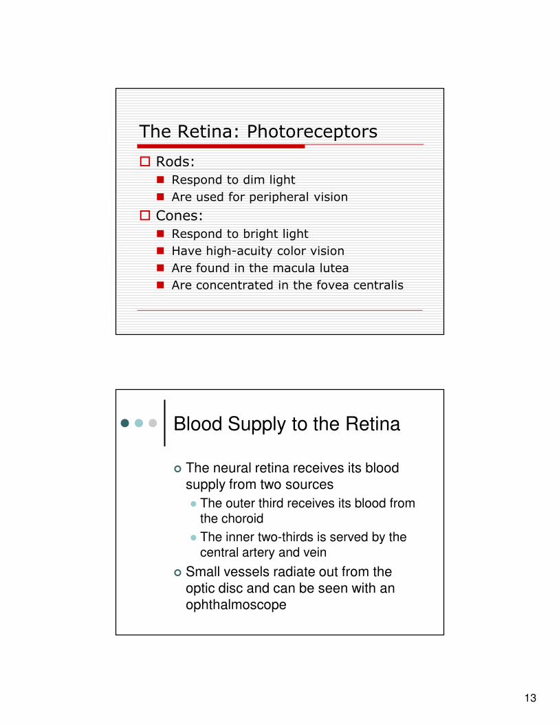
The Retina: Photoreceptors
� Rods:
� Respond to dim light
� Are used for peripheral vision
� Cones:
� Respond to bright light
� Have high-acuity color vision
� Are found in the macula lutea
� Are concentrated in the fovea centralis
Blood Supply to the Retina
� The neural retina receives its blood
supply from two sources
� The outer third receives its blood from the choroid
� The inner two-thirds is served by the central artery and vein
� Small vessels radiate out from the
optic disc and can be seen with an
ophthalmoscope

14
Inner Chambers and Fluids
�The lens separates the internal eye into
anterior and posterior segments
�The posterior segment is filled with a clear
gel called vitreous humor that:
�Transmits light
�Supports the posterior surface of the lens
�Holds the neural retina firmly against the pigmented layer
�Contributes to intraocular pressure
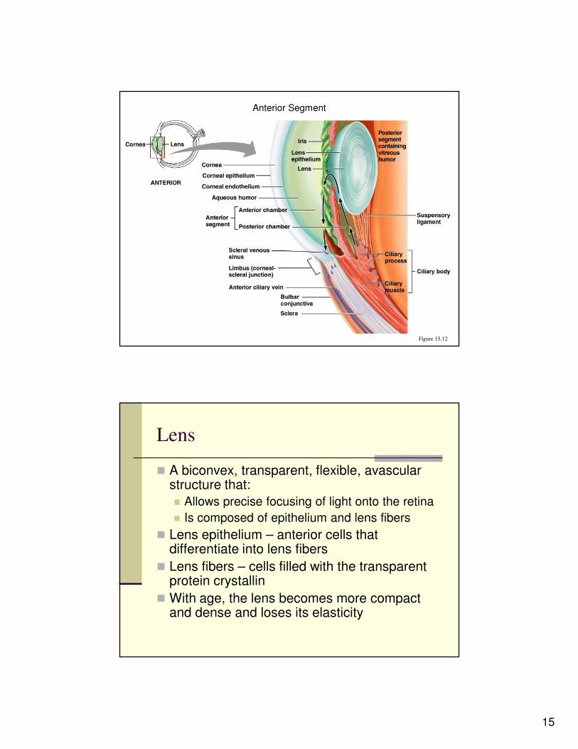
Anterior Segment
� Composed of two chambers
– Anterior – between the cornea and the iris
– Posterior – between the iris and the lens
� Aqueous humor
– A plasmalike fluid that fills the anterior segment
– Drains via the canal of Schlemm
� Supports, nourishes, and removes wastes
15
Anterior Segment
Figure 15.12
Lens
� A biconvex, transparent, flexible, avascular structure that:
� Allows precise focusing of light onto the retina
� Is composed of epithelium and lens fibers
� Lens epithelium – anterior cells that differentiate into lens fibers
� Lens fibers – cells filled with the transparent protein crystallin
� With age, the lens becomes more compact and dense and loses its elasticity

16
LightLight
•• Electromagnetic radiation Electromagnetic radiation –– all energy waves all energy waves
from short gamma rays to long radio wavesfrom short gamma rays to long radio waves
•• Our eyes respond to a small portion of this Our eyes respond to a small portion of this
spectrum called thespectrum called the visible spectrumvisible spectrum
•• Different cones in the retina respond to Different cones in the retina respond to
different wavelengths of the visible spectrumdifferent wavelengths of the visible spectrum
Light
Figure 15.14

17
Refraction and LensesRefraction and Lenses
�� When light passes from one transparent When light passes from one transparent medium to another its speed changes and medium to another its speed changes and it refracts (bends)it refracts (bends)
�� Light passing through a convex lens (as in Light passing through a convex lens (as in the eye) is bent so that the rays converge the eye) is bent so that the rays converge to a focal pointto a focal point
�� When a convex lens forms an image, the When a convex lens forms an image, the image is upside down and reversed right image is upside down and reversed right to leftto left
Refraction and Lenses
Figure 15.16
18
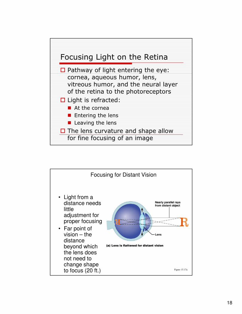
Focusing Light on the Retina
� Pathway of light entering the eye: cornea, aqueous humor, lens, vitreous humor, and the neural layer of the retina to the photoreceptors
� Light is refracted:
� At the cornea
� Entering the lens
� Leaving the lens
� The lens curvature and shape allow for fine focusing of an image
Focusing for Distant Vision
• Light from a distance needs little adjustment for proper focusing
• Far point of vision – the distance beyond which the lens does not need to change shape to focus (20 ft.) Figure 15.17a

19
Focusing for Close VisionFocusing for Close Vision
�� Close vision requires:Close vision requires:
�� Accommodation Accommodation –– changing the lens shape by changing the lens shape by ciliary muscles to increase refractory powerciliary muscles to increase refractory power
�� Constriction Constriction –– the pupillary reflex constricts the pupillary reflex constricts the pupils to prevent divergent light rays from the pupils to prevent divergent light rays from entering the eyeentering the eye
�� Convergence Convergence –– medial rotation of the eyeballs medial rotation of the eyeballs toward the object being viewedtoward the object being viewed
Focusing for Close Vision
Figure 15.7b

20
Problems of Refraction
Emmetropic eye – normal eye with
light focused properly
Myopic eye (nearsighted) – the focal
point is in front of the retina
Corrected with a concave lens
Hyperopic eye (farsighted) – the focal
point is behind the retina
Corrected with a convex lens
Problems of Refraction
Figure 15.18

21
RodsRods
�� Functional characteristicsFunctional characteristics
�� Sensitive to dim light and best suited for night Sensitive to dim light and best suited for night visionvision
�� Absorb all wavelengths of visible lightAbsorb all wavelengths of visible light
�� Perceived input is in gray tones onlyPerceived input is in gray tones only
�� Sum of visual input from many rods feeds into Sum of visual input from many rods feeds into a single ganglion cell a single ganglion cell
�� Results in fuzzy and indistinct imagesResults in fuzzy and indistinct images
ConesCones
►►Functional characteristics Functional characteristics
�� Need bright light for activation (have low Need bright light for activation (have low sensitivity)sensitivity)
�� Have pigments that furnish a vividly colored Have pigments that furnish a vividly colored viewview
�� Each cone synapses with a single ganglion cellEach cone synapses with a single ganglion cell
�� Vision is detailed and has high resolutionVision is detailed and has high resolution

22
Cones and Rods
Figure 15.10a

23
Visual PathwaysVisual Pathways
�� Axons of retinal ganglion cells form the Axons of retinal ganglion cells form the optic nerve optic nerve
�� Medial fibers of the optic nerve decussate Medial fibers of the optic nerve decussate at the optic chiasmat the optic chiasm
�� Most fibers of the optic tracts continue to Most fibers of the optic tracts continue to the lateral geniculate body of the thalamusthe lateral geniculate body of the thalamus
�� Other optic tract fibers end in superior Other optic tract fibers end in superior colliculi (initiating visual reflexes) and colliculi (initiating visual reflexes) and pretectal nuclei (involved with pupillary pretectal nuclei (involved with pupillary reflexes)reflexes)
�� Optic radiations travel from the thalamus Optic radiations travel from the thalamus to the visual cortexto the visual cortex
Visual Pathways
Figure 15.23