Embed Size (px)
Citation preview
2530 Mol. BioSyst., 2013, 9, 2530--2540 This journal is c The Royal Society of Chemistry 2013
Cite this: Mol. BioSyst.,2013,9, 2530
In silico modeling and evaluation of Gordoniaalkanivorans for biodesulfurization†
Shilpi Aggarwal, I. A. Karimi* and Gregorius Reinaldi Ivan
The genus Gordonia is well known for its catabolic diversity and ability to transform several compounds
including the various recalcitrant polyaromatic sulfur heterocycles (PASHs) found in the fossil fuels.
In fact, some strains offer the unique ability to desulfurize even benzothiophene (BT) and other
thiophenic compounds, which most of the commonly studied rhodococci strains cannot. In this work,
we present the first genome scale metabolic model for G. alkanivorans, a desulfurizing strain, to enable
a holistic study of its metabolism and comparison with R. erythropolis. Our model consists of 881
unique metabolites and 922 reactions associated with 568 ORFs/genes and 544 unique enzymes.
It successfully predicts the growth rates from experimental studies and quantitatively elucidates the
pathways for the desulfurization of the commonly studied sulfur compounds, namely
dibenzothiophene (DBT) and benzothiophene (BT). Using our model, we identify the minimal media for
G. alkanivorans, and show the significant effect of carbon sources on desulfurization with ethanol as
the best source. Our model shows that the sulfur-containing amino acids such as cysteine and
methionine decrease desulfurization activity, and G. alkanivorans prefers BT over DBT as a sulfur source.
It also suggests that this preference may be driven by the lower NADH requirements for BT metabolism
rather than the higher affinity of the transport system for BT. Our in silico comparison of R. erythropolis
and G. alkanivorans suggests the latter to be a better desulfurizing strain due to its versatility for both
BT and DBT, higher desulfurization activity, and higher growth rate.
Introduction
The combustion of fossil fuels releases various pollutants suchas the oxides of carbon (COx), nitrogen (NOx), and sulfur (SOx).SOx, in particular, have attracted increasingly stringent regula-tions due to their harmful effects on the environment andhuman health.1 Desulfurization is a key step in the pre-processingof fossil fuels to achieve compliance with these regulations.At present, hydrodesulfurization is the most common methodfor desulfurization. In addition to being expensive and energy-intensive, it requires extreme conditions of temperature andpressure to remove sulfur from certain recalcitrant compoundssuch as dibenzothiophene (DBT), benzothiophene (BT), andtheir derivatives.2 Furthermore, being non-specific in action, itleads to the undesirable hydrogenation of certain non-sulfuraromatic compounds that contribute to the lubricating propertyand thermal stability of the fuels. Therefore, there is a need for
developing efficient, specific, and economical desulfurizationmethods. To this end, biodesulfurization is considered a pro-spective alternative.
Biodesulfurization employs whole microbes or theirenzymes as catalysts to remove the sulfur atom selectively fromthe various recalcitrant compounds present in the fossil fuels.1
Several strains of Pseudomonas, Rhodococcus, Mycobacterium,Gordonia, etc. have been studied for their ability to metabolizevarious polyaromatic sulfur heterocycles (PASHs) present infossil fuels. Most desulfurization studies in the literaturehave used DBT as the model compound. While several rhodo-cocci strains exhibit non-destructive desulfurization of DBT,R. erythropolis IGTS8 was the first to be identified3 and hasreceived the most attention. However, most rhodococci areunable to show high activity for the alkyl derivatives of DBTand show no activity for BT and other thiophenic compounds.Furthermore, there are only limited biochemical and geneticstudies of bacteria that exhibit desulfurization of both DBT andBT.4,5 Since fossil fuels do contain these compounds in signi-ficant amounts, it is critical to study microbes that possessactivity for compounds other than DBT. Furthermore, becausedesulfurizing these compounds requires distinct pathways,bacterial strains that possess the associated genes for all these
Department of Chemical and Biomolecular Engineering, National University of
Singapore, 4 Engineering Drive 4, 117576, Singapore. E-mail: [email protected];
Fax: +65-6779-1936; Tel: +65-6516-6359
† Electronic supplementary information (ESI) available: Details of metabolites,reactions, and dead-end metabolites. See DOI: 10.1039/c3mb70132h
Received 2nd April 2013,Accepted 2nd July 2013
DOI: 10.1039/c3mb70132h
www.rsc.org/molecularbiosystems
MolecularBioSystems
PAPER
Ope
n A
cces
s A
rtic
le. P
ublis
hed
on 0
3 Ju
ly 2
013.
Dow
nloa
ded
on 2
/16/
2022
6:0
6:08
PM
. T
his
artic
le is
lice
nsed
und
er a
Cre
ativ
e C
omm
ons
Attr
ibut
ion
3.0
Unp
orte
d L
icen
ce.
View Article OnlineView Journal | View Issue
This journal is c The Royal Society of Chemistry 2013 Mol. BioSyst., 2013, 9, 2530--2540 2531
pathways are clearly desirable. Gordonia is an attractive genusin this regard, because its members exhibit much metabolicversatility.6
Numerous Gordonia strains exhibit higher desulfurizationactivities5,6 than the rhodococci and for a broader range ofPASHs.5,7–11 Of them, G. alkanivorans5,7,12,13 desulfurizes DBTvia the well-known 4S pathway14 that non-destructively elim-inates the sulfur atom from DBT with the concomitant releaseof 2-hydroxybiphenyl (HBP), the sulfur free compound. The 4Spathway in G. alkanivorans is conferred by three genes namelydszA, dszB, and dszC.7 The dszABC genes of G. alkanivorans arehighly similar to those from R. erythropolis. However, besidesDBT, it can also specifically cleave the C–S bond in BT andother thiophenes. Because of its ability to desulfurize a widerrange of PASHs, G. alkanivorans appears to offer some advan-tage over R. erythropolis for biodesulfurization. Moreover,G. alkanivorans strains are reported15 to show nearly 2–10 timeshigher desulfurization activities than the desulfurizingR. erythropolis strains. In other words, it has the greater abilityto reduce the overall sulfur content of the fossil fuels.
In spite of its promise, desulfurization studies withG. alkanivorans are far more limited than those with R. erythropolis.Although it does offer higher desulfurizing activity thanR. erythropolis, the activity levels are still not acceptable forcommercial application. Thus, there is a need to identify andstudy the factors and host functions that may play key roles incontrolling the extent of desulfurization by G. alkanivorans.However, the complexity of metabolic networks makes it diffi-cult to predict or identify such host functions intuitively orusing a trial-and-error experimental approach. Since cellularactivities are invariably and intricately coupled, a holisticstudy of the various metabolic functions occurring withinG. alkanivorans besides the desulfurization of PASHs is essentialto understand the interactions between the various compo-nents of its metabolic network. Such a study would also allowone to compare Gordonia strains with rhodococci in a theore-tical and comprehensive manner. However, no such holisticstudy on Gordonia exists in the literature.
Constraint-based metabolic models16 have successfully beenused to perform such holistic studies to elucidate relationshipsamong various metabolic activities both qualitatively and quanti-tatively. These models, constructed based on the genomic andbiochemical information of an organism, clearly establish thecorrespondence between its gene(s), protein(s) and metabolicfunction(s). They are easier to build, as they require onlystoichiometric rather than kinetic information about variousmetabolic reactions. Nevertheless, they provide an effectiveframework for studying genotype–phenotype relationships,interactions among various metabolic activities, and internalflux distributions associated with various metabolic activitiesunder given environmental conditions. Such constraint-basedgenome-scale metabolic (GSM) models have been reconstructedand analyzed widely for several industrially important bacterialstrains such as Escherichia coli,17,18 R. erythropolis,19,20 Saccharo-myces cerevisae,21,22 and Zymomonas mobilis,23 and even mam-malian cells such as mouse hydridoma.24,25 Once constructed,
these models can be very useful in exploring the possible statesand properties of the metabolic network of an organism.
This work reports the first in silico GSM model forG. alkanivorans. It covers the key metabolic pathways such ascentral metabolism, amino acids biosyntheses, nucleotidemetabolism, and sulfur metabolism that describes the assimi-lation of sulfur into biomass. It can help in understandingthe metabolic architecture of G. alkanivorans, and its hostfunctions related to desulfurization. We validate the modelusing the available desulfurization and growth data in theliterature,11 and use it to study the effects of various mediumcomponents such as carbon sources, amino acids, and vitaminson the desulfurization activity of G. alkanivorans. We assess theproperties of its metabolic network such as flexibility androbustness using flux variability26 and gene essentialityanalyses. Finally, we use flux sum analyses27 to study qualitativelyand quantitatively the effect of intracellular metabolites ongrowth and desulfurization activity and propose several experi-mentally testable conditions and modifications that may helpenhance the desulfurizing activity of G. alkanivorans.
Results and discussionReconstructed GSM of G. alkanivorans
Our initial draft model comprised 739 reactions. However, itfailed to show any cell growth on substrates known to beutilized by G. alkanivorans. We found that nearly 60% of allprecursor metabolites could not be synthesized by the draftmodel. For instance, the draft model was unable to produceamino acids such as methionine and histidine, althoughG. alkanivornas is known5 to synthesize them and survivewithout any external supply. Our draft model also showed zerogrowth with DBT and BT as sole sulfur sources, althoughG. alkanivorans is known5 to metabolize them. Therefore, weadded relevant pathways and reactions based on the informationavailable in the literature and databases.5,14,28,29 As mentionedearlier, G. alkanivorans offers the advantage of desulfurizing awider range of PASHs such as the alkyl derivatives of BT, DBT,and thiophene. However, we could not include such alkylderivatives in our model, as their metabolic pathways andintermediates are still unknown. Thus, there is a clear needfor obtaining genetic and biochemical data for the associatedmetabolic pathways in G. alkanivorans.
GapFind revealed 279 DEMs that could not be produced inthe initial draft of our model, as they were disconnected fromthe rest of the network either upstream or downstream. Gapfillcould identify the possible candidate reactions to restore theconnectivity for only 140 of the 279 DEMs. We performedBLASTp analyses for assigning putative ORFs to the enzymesassociated with these reactions. To ensure that we do not addreactions indiscriminately to our model, we used a high ecut-off of 10�30 to include only the reactions with a strongevidence of ORFs. We could locate ORFs for only 62 (B22%)DEMs, thus we did not include other reactions.
Our final curated model consists of 881 unique metabolitesand 922 reactions associated with 568 ORFs/genes and 544
Paper Molecular BioSystems
Ope
n A
cces
s A
rtic
le. P
ublis
hed
on 0
3 Ju
ly 2
013.
Dow
nloa
ded
on 2
/16/
2022
6:0
6:08
PM
. T
his
artic
le is
lice
nsed
und
er a
Cre
ativ
e C
omm
ons
Attr
ibut
ion
3.0
Unp
orte
d L
icen
ce.
View Article Online
2532 Mol. BioSyst., 2013, 9, 2530--2540 This journal is c The Royal Society of Chemistry 2013
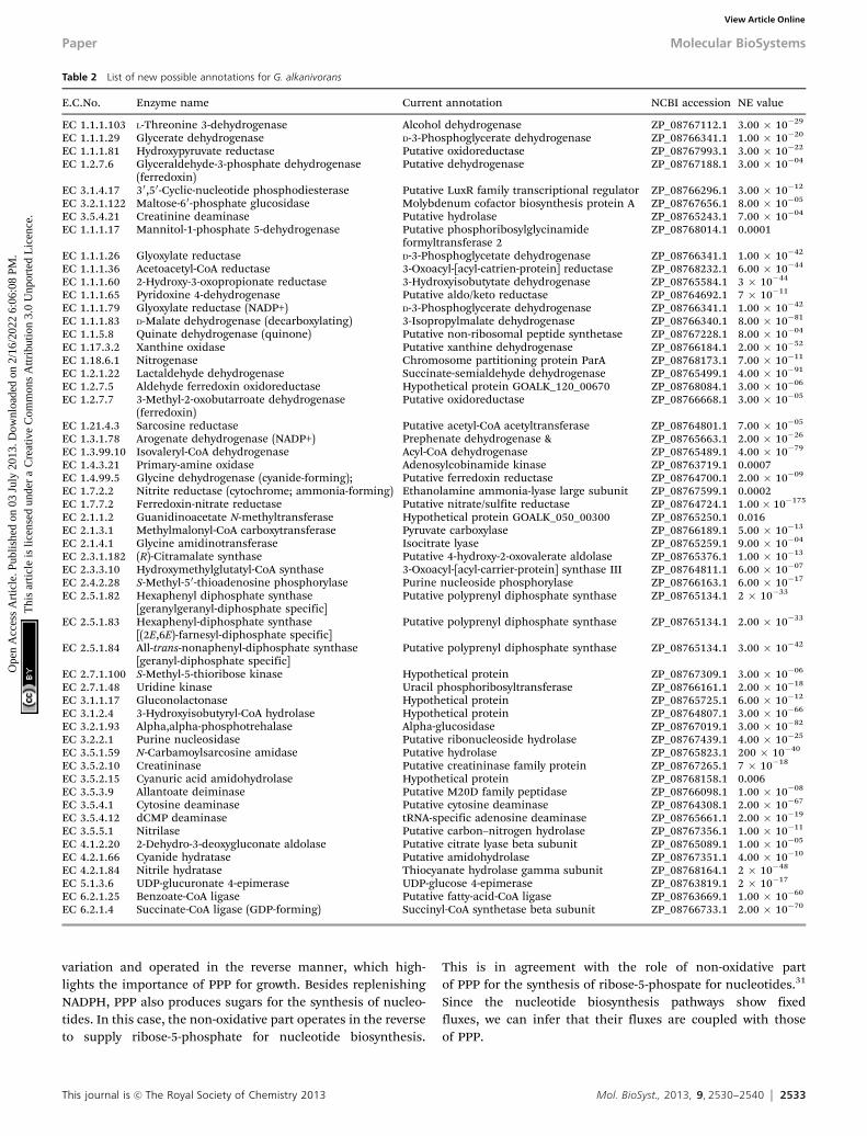
unique enzymes. Of these 922 reactions, 67 account for thetransport of various metabolites across the membrane, whilethe rest (855) account for intracellular metabolic activities.Table 1 lists the features of our GSM model. The BLASTpanalyses identified possible annotations for 55 ORFs inG. alkanovirans, which are given in Table 2. However, the modelstill has 217 DEMs, which warrants further biochemistry studies.
Model validation
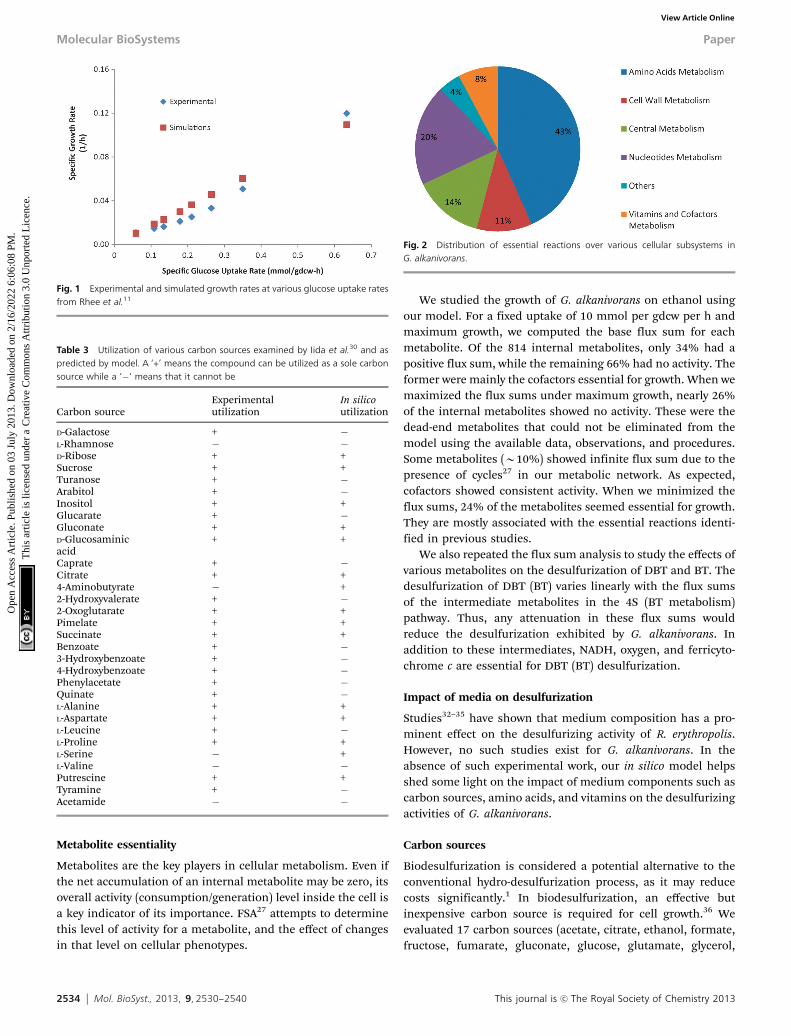
We mimicked the experiments of Rhee et al.11 and comparedthe growth rates predicted by our model with their experi-mental values. From their experiments, we inferred glucose tobe limiting, as its complete depletion from the mediumtriggered the stationary phase. So we computed and usedspecific uptake rates for glucose at several time points toconstrain our model. We assumed unlimited supply for othermedium components, as they were in excess. As seen in Fig. 1,our predicted biomass growth rates are in close agreement withthe experimental data of Rhee et al.11
Minimal media and growth
Gordonia strains are known for their metabolic versatility6 anddemand no special nutrients to the best of our knowledge.Thus, we infer that they can synthesize all the necessaryprecursor metabolites from simple sources in a medium. How-ever, it is desirable to identify the essential nutrients and theiralternatives based on our model.
Rhee et al.11 used a rich medium (as described in Materialsand methods) to study the growth of G. alkanivorans with DBTas the sole sulfur source. Using our model, we simulatedgrowth by removing one nutrient at a time from the richmedium of Rhee et al.11 From that, we identified glucose,oxygen, an ammonium salt, a phosphorus source, and DBT tocomprise the minimal medium. As alternatives, we identifiedBT, cysteine, and sulfate for sulfur, and glutamate for bothcarbon and nitrogen.
Iida et al.30 experimented with 31 carbon substrates. Weused our in silico model to simulate their experiments anddetect cell growth on these 31 sources. In each simulation, wespecified 1 mmol per gdcw per h uptake of a different substrateas the sole carbon source along with the minimal media andmaximized cell growth. Table 3 compares our model predictionswith the observations of Iida et al.30 Our model predicts growthcorrectly for 16 of the 31 substrates. We observe both false positiveand false negative results for the remaining 15 substrates.
In the former, our model shows false growth, while in thelatter, it fails to show growth. These errors arise, because thebiochemical information on G. alkanivorans is still incomplete.Further work in this regard is warranted. Since our model lacksregulatory mechanisms, this is another source of error. How-ever, model predictions in this case may be improved byincorporating regulatory information.
Gene essentiality
Most cells can withstand disturbances at the genetic as well asmetabolic levels by utilizing alternative genes, enzymes, andpathways depending on their environment. However, the non-functionality of certain reactions and genes may be lethal for acell. We studied the robustness of G. alkanivorans metabolismby assessing its ability to exhibit in silico growth in the case ofgene knockouts or mutations. The utilization of a pathway, andthus the essentiality of its reactions and genes, will in generaldepend on medium components. We used five minimal mediawith alternative carbon sources of ethanol, fumarate, oxoglutarate,pyruvate, and glutamate, and evaluated the essentialities ofgenes and reactions for each medium. For reactions, weremoved one reaction at a time by setting its flux as zero, andmaximized cell growth on minimal medium. If the model couldnot produce cell mass, then we classified the reaction asessential, and vice versa. Similarly, for genes, if the removal ofa gene prevented cell growth, then we classified that gene asessential, and vice versa. To remove a gene, we set the fluxes ofall its associated reactions to zero in our in silico model.However, if a reaction was controlled by two or more isozymes,then the reaction was kept active in the absence of any one ofthe associated genes.
We identified 116 reactions and 75 genes to be essentialirrespective of the medium. As seen in Fig. 2, most essentialreactions belong to the amino acids metabolism followed bynucleotides metabolism, central metabolism, and cell wallmetabolism. Any reduction in their activity levels may reducegrowth or prove lethal for G. alkanivorans. The difference in thenumbers of essential reactions and essential genes is due toisozymes, as several reactions are catalyzed by enzymes thatmultiple genes encode. These reactions are essential at themetabolic level, but not the genetic level.
Flux variability
Metabolic network models normally exhibit multiple alternativesolutions for flux distributions. Flux variability analysis (FVA26)allows us to study the range over which a given flux can varyin alternative solutions. We performed an FVA on our modelfor an ethanol uptake of 10 mmol per gdcw per h and unrest-ricted supplies of other essential nutrients. For the maximumcell growth, we computed the minimum and maximumpossible fluxes for each reaction. Nearly 85% of the reactionsshowed significant flux variations. The remaining 15%represent the inflexible parts of the metabolism for growth,which mainly include the biosynthesis pathways for aminoacids and nucleotides. We also observed that the non-oxidativepart of the pentose phosphate pathway (PPP) showed no flux
Table 1 Features of the reconstructed genome scale model of G. alkanivorans
Features Properties
Reactions in genome scale mode 922No. of ORFs included 568No. of enzymes included 544Intracellular reactions 855Transport reactions 67Metabolites in genome scale model 881Internal metabolites 814External metabolites 67
Molecular BioSystems Paper
Ope
n A
cces
s A
rtic
le. P
ublis
hed
on 0
3 Ju
ly 2
013.
Dow
nloa
ded
on 2
/16/
2022
6:0
6:08
PM
. T
his
artic
le is
lice
nsed
und
er a
Cre
ativ
e C
omm
ons
Attr
ibut
ion
3.0
Unp
orte
d L
icen
ce.
View Article Online
This journal is c The Royal Society of Chemistry 2013 Mol. BioSyst., 2013, 9, 2530--2540 2533
variation and operated in the reverse manner, which high-lights the importance of PPP for growth. Besides replenishingNADPH, PPP also produces sugars for the synthesis of nucleo-tides. In this case, the non-oxidative part operates in the reverseto supply ribose-5-phosphate for nucleotide biosynthesis.
This is in agreement with the role of non-oxidative partof PPP for the synthesis of ribose-5-phospate for nucleotides.31
Since the nucleotide biosynthesis pathways show fixedfluxes, we can infer that their fluxes are coupled with thoseof PPP.
Table 2 List of new possible annotations for G. alkanivorans
E.C.No. Enzyme name Current annotation NCBI accession NE value
EC 1.1.1.103 L-Threonine 3-dehydrogenase Alcohol dehydrogenase ZP_08767112.1 3.00 � 10�29
EC 1.1.1.29 Glycerate dehydrogenase D-3-Phosphoglycerate dehydrogenase ZP_08766341.1 1.00 � 10�20
EC 1.1.1.81 Hydroxypyruvate reductase Putative oxidoreductase ZP_08767993.1 3.00 � 10�22
EC 1.2.7.6 Glyceraldehyde-3-phosphate dehydrogenase(ferredoxin)
Putative dehydrogenase ZP_08767188.1 3.00 � 10�04
EC 3.1.4.17 30,50-Cyclic-nucleotide phosphodiesterase Putative LuxR family transcriptional regulator ZP_08766296.1 3.00 � 10�12
EC 3.2.1.122 Maltose-60-phosphate glucosidase Molybdenum cofactor biosynthesis protein A ZP_08767656.1 8.00 � 10�05
EC 3.5.4.21 Creatinine deaminase Putative hydrolase ZP_08765243.1 7.00 � 10�04
EC 1.1.1.17 Mannitol-1-phosphate 5-dehydrogenase Putative phosphoribosylglycinamideformyltransferase 2
ZP_08768014.1 0.0001
EC 1.1.1.26 Glyoxylate reductase D-3-Phosphoglycetate dehydrogenase ZP_08766341.1 1.00 � 10�42
EC 1.1.1.36 Acetoacetyl-CoA reductase 3-Oxoacyl-[acyl-catrien-protein] reductase ZP_08768232.1 6.00 � 10�44
EC 1.1.1.60 2-Hydroxy-3-oxopropionate reductase 3-Hydroxyisobutytate dehydrogenase ZP_08765584.1 3 � 10�44
EC 1.1.1.65 Pyridoxine 4-dehydrogenase Putative aldo/keto reductase ZP_08764692.1 7 � 10�11
EC 1.1.1.79 Glyoxylate reductase (NADP+) D-3-Phosphoglycerate dehydrogenase ZP_08766341.1 1.00 � 10�42
EC 1.1.1.83 D-Malate dehydrogenase (decarboxylating) 3-Isopropylmalate dehydrogenase ZP_08766340.1 8.00 � 10�81
EC 1.1.5.8 Quinate dehydrogenase (quinone) Putative non-ribosomal peptide synthetase ZP_08767228.1 8.00 � 10�04
EC 1.17.3.2 Xanthine oxidase Putative xanthine dehydrogenase ZP_08766184.1 2.00 � 10�52
EC 1.18.6.1 Nitrogenase Chromosome partitioning protein ParA ZP_08768173.1 7.00 � 10�11
EC 1.2.1.22 Lactaldehyde dehydrogenase Succinate-semialdehyde dehydrogenase ZP_08765499.1 4.00 � 10�91
EC 1.2.7.5 Aldehyde ferredoxin oxidoreductase Hypothetical protein GOALK_120_00670 ZP_08768084.1 3.00 � 10�06
EC 1.2.7.7 3-Methyl-2-oxobutarroate dehydrogenase(ferredoxin)
Putative oxidoreductase ZP_08766668.1 3.00 � 10�05
EC 1.21.4.3 Sarcosine reductase Putative acetyl-CoA acetyltransferase ZP_08764801.1 7.00 � 10�05
EC 1.3.1.78 Arogenate dehydrogenase (NADP+) Prephenate dehydrogenase & ZP_08765663.1 2.00 � 10�26
EC 1.3.99.10 Isovaleryl-CoA dehydrogenase Acyl-CoA dehydrogenase ZP_08765489.1 4.00 � 10�79
EC 1.4.3.21 Primary-amine oxidase Adenosylcobinamide kinase ZP_08763719.1 0.0007EC 1.4.99.5 Glycine dehydrogenase (cyanide-forming); Putative ferredoxin reductase ZP_08764700.1 2.00 � 10�09
EC 1.7.2.2 Nitrite reductase (cytochrome; ammonia-forming) Ethanolamine ammonia-lyase large subunit ZP_08767599.1 0.0002EC 1.7.7.2 Ferredoxin-nitrate reductase Putative nitrate/sulfite reductase ZP_08764724.1 1.00� 10�175
EC 2.1.1.2 Guanidinoacetate N-methyltransferase Hypothetical protein GOALK_050_00300 ZP_08765250.1 0.016EC 2.1.3.1 Methylmalonyl-CoA carboxytransferase Pyruvate carboxylase ZP_08766189.1 5.00 � 10�13
EC 2.1.4.1 Glycine amidinotransferase Isocitrate lyase ZP_08765259.1 9.00 � 10�04
EC 2.3.1.182 (R)-Citramalate synthase Putative 4-hydroxy-2-oxovalerate aldolase ZP_08765376.1 1.00 � 10�13
EC 2.3.3.10 Hydroxymethylglutatyl-CoA synthase 3-Oxoacyl-[acyl-carrier-protein] synthase III ZP_08764811.1 6.00 � 10�07
EC 2.4.2.28 S-Methyl-50-thioadenosine phosphorylase Purine nucleoside phosphorylase ZP_08766163.1 6.00 � 10�17
EC 2.5.1.82 Hexaphenyl diphosphate synthase[geranylgeranyl-diphosphate specific]
Putative polyprenyl diphosphate synthase ZP_08765134.1 2 � 10�33
EC 2.5.1.83 Hexaphenyl-diphosphate synthase[(2E,6E)-farnesyl-diphosphate specific]
Putative polyprenyl diphosphate synthase ZP_08765134.1 2.00 � 10�33
EC 2.5.1.84 All-trans-nonaphenyl-diphosphate synthase[geranyl-diphosphate specific]
Putative polyprenyl diphosphate synthase ZP_08765134.1 3.00 � 10�42
EC 2.7.1.100 S-Methyl-5-thioribose kinase Hypothetical protein ZP_08767309.1 3.00 � 10�06
EC 2.7.1.48 Uridine kinase Uracil phosphoribosyltransferase ZP_08766161.1 2.00 � 10�18
EC 3.1.1.17 Gluconolactonase Hypothetical protein ZP_08765725.1 6.00 � 10�12
EC 3.1.2.4 3-Hydroxyisobutyryl-CoA hydrolase Hypothetical protein ZP_08764807.1 3.00 � 10�66
EC 3.2.1.93 Alpha,alpha-phosphotrehalase Alpha-glucosidase ZP_08767019.1 3.00 � 10�82
EC 3.2.2.1 Purine nucleosidase Putative ribonucleoside hydrolase ZP_08767439.1 4.00 � 10�25
EC 3.5.1.59 N-Carbamoylsarcosine amidase Putative hydrolase ZP_08765823.1 200 � 10�40
EC 3.5.2.10 Creatininase Putative creatininase family protein ZP_08767265.1 7 � 10�18
EC 3.5.2.15 Cyanuric acid amidohydrolase Hypothetical protein ZP_08768158.1 0.006EC 3.5.3.9 Allantoate deiminase Putative M20D family peptidase ZP_08766098.1 1.00 � 10�08
EC 3.5.4.1 Cytosine deaminase Putative cytosine deaminase ZP_08764308.1 2.00 � 10�67
EC 3.5.4.12 dCMP deaminase tRNA-specific adenosine deaminase ZP_08765661.1 2.00 � 10�19
EC 3.5.5.1 Nitrilase Putative carbon–nitrogen hydrolase ZP_08767356.1 1.00 � 10�11
EC 4.1.2.20 2-Dehydro-3-deoxygluconate aldolase Putative citrate lyase beta subunit ZP_08765089.1 1.00 � 10�05
EC 4.2.1.66 Cyanide hydratase Putative amidohydrolase ZP_08767351.1 4.00 � 10�10
EC 4.2.1.84 Nitrile hydratase Thiocyanate hydrolase gamma subunit ZP_08768164.1 2 � 10�48
EC 5.1.3.6 UDP-glucuronate 4-epimerase UDP-glucose 4-epimerase ZP_08763819.1 2 � 10�17
EC 6.2.1.25 Benzoate-CoA ligase Putative fatty-acid-CoA ligase ZP_08763669.1 1.00 � 10�60
EC 6.2.1.4 Succinate-CoA ligase (GDP-forming) Succinyl-CoA synthetase beta subunit ZP_08766733.1 2.00 � 10�70
Paper Molecular BioSystems
Ope
n A
cces
s A
rtic
le. P
ublis
hed
on 0
3 Ju
ly 2
013.
Dow
nloa
ded
on 2
/16/
2022
6:0
6:08
PM
. T
his
artic
le is
lice
nsed
und
er a
Cre
ativ
e C
omm
ons
Attr
ibut
ion
3.0
Unp
orte
d L
icen
ce.
View Article Online
2534 Mol. BioSyst., 2013, 9, 2530--2540 This journal is c The Royal Society of Chemistry 2013
Metabolite essentiality
Metabolites are the key players in cellular metabolism. Even ifthe net accumulation of an internal metabolite may be zero, itsoverall activity (consumption/generation) level inside the cell isa key indicator of its importance. FSA27 attempts to determinethis level of activity for a metabolite, and the effect of changesin that level on cellular phenotypes.
We studied the growth of G. alkanivorans on ethanol usingour model. For a fixed uptake of 10 mmol per gdcw per h andmaximum growth, we computed the base flux sum for eachmetabolite. Of the 814 internal metabolites, only 34% had apositive flux sum, while the remaining 66% had no activity. Theformer were mainly the cofactors essential for growth. When wemaximized the flux sums under maximum growth, nearly 26%of the internal metabolites showed no activity. These were thedead-end metabolites that could not be eliminated from themodel using the available data, observations, and procedures.Some metabolites (B10%) showed infinite flux sum due to thepresence of cycles27 in our metabolic network. As expected,cofactors showed consistent activity. When we minimized theflux sums, 24% of the metabolites seemed essential for growth.They are mostly associated with the essential reactions identi-fied in previous studies.
We also repeated the flux sum analysis to study the effects ofvarious metabolites on the desulfurization of DBT and BT. Thedesulfurization of DBT (BT) varies linearly with the flux sumsof the intermediate metabolites in the 4S (BT metabolism)pathway. Thus, any attenuation in these flux sums wouldreduce the desulfurization exhibited by G. alkanivorans. Inaddition to these intermediates, NADH, oxygen, and ferricyto-chrome c are essential for DBT (BT) desulfurization.
Impact of media on desulfurization
Studies32–35 have shown that medium composition has a pro-minent effect on the desulfurizing activity of R. erythropolis.However, no such studies exist for G. alkanivorans. In theabsence of such experimental work, our in silico model helpsshed some light on the impact of medium components such ascarbon sources, amino acids, and vitamins on the desulfurizingactivities of G. alkanivorans.
Carbon sources
Biodesulfurization is considered a potential alternative to theconventional hydro-desulfurization process, as it may reducecosts significantly.1 In biodesulfurization, an effective butinexpensive carbon source is required for cell growth.36 Weevaluated 17 carbon sources (acetate, citrate, ethanol, formate,fructose, fumarate, gluconate, glucose, glutamate, glycerol,
Fig. 1 Experimental and simulated growth rates at various glucose uptake ratesfrom Rhee et al.11
Table 3 Utilization of various carbon sources examined by Iida et al.30 and aspredicted by model. A ‘+’ means the compound can be utilized as a sole carbonsource while a ‘�‘ means that it cannot be
Carbon sourceExperimentalutilization
In silicoutilization
D-Galactose + �L-Rhamnose � �D-Ribose + +Sucrose + +Turanose + �Arabitol + �Inositol + +Glucarate + �Gluconate + +D-Glucosaminicacid
+ +
Caprate + �Citrate + +4-Aminobutyrate � +2-Hydroxyvalerate + �2-Oxoglutarate + +Pimelate + +Succinate + +Benzoate + �3-Hydroxybenzoate + �4-Hydroxybenzoate + �Phenylacetate + �Quinate + �L-Alanine + +L-Aspartate + +L-Leucine + �L-Proline + +L-Serine � +L-Valine � �Putrescine + +Tyramine + �Acetamide � �
Fig. 2 Distribution of essential reactions over various cellular subsystems inG. alkanivorans.
Molecular BioSystems Paper
Ope
n A
cces
s A
rtic
le. P
ublis
hed
on 0
3 Ju
ly 2
013.
Dow
nloa
ded
on 2
/16/
2022
6:0
6:08
PM
. T
his
artic
le is
lice
nsed
und
er a
Cre
ativ
e C
omm
ons
Attr
ibut
ion
3.0
Unp
orte
d L
icen
ce.
View Article Online
This journal is c The Royal Society of Chemistry 2013 Mol. BioSyst., 2013, 9, 2530--2540 2535
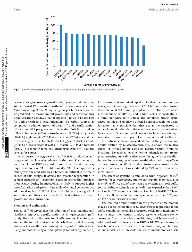
lactate, malate, oxaloacetate, oxoglutarate, pyruvate, and succinate).We performed 17 simulations with one carbon source at a time.Assuming an uptake of 20 mg per gdcw per h for each source,we predicted the maximum cell growth rate and correspondingdesulfurization activity. Ethanol appears (Fig. 3) to be the bestfor both growth and desulfurization. The carbon sources ascompared to ethanol (growth of 0.027 h�1 and desulfurizationof 3.3 mmol HBP per gdcw per h) have the 100% basis rank asfollows: fumarate (80%) > oxoglutarate (78.79%) > pyruvate(78.43%) > glutamate (78.24%) > succinate (78%) > acetate Efructose E glucose E lactate (76.86%) > glycerol (75%) > citrate(71.88%) > oxaloacetate (69.70%) > malate (69.11%) > formate(50%). This ranking remained unchanged even for BT as thesole sulfur source.
As discussed by Aggarwal et al.,20 NADH production andusage could explain why ethanol is the best. For the cell toconsume 1 mol DBT as a sulfur source via the 4S pathwayrequires 4 moles of NADH. Additionally, NADH is required forother growth related activities. The carbon nutrient is the mainsource of this energy. It affects the cofactor regeneration incellular metabolism. Therefore, a carbon source that providesmore NADH during its metabolism is likely to support higherdesulfurization and growth. One mole of ethanol generates twoadditional moles of NADH. This is the highest among all 17substrates, and thus it seems to be the best substrate for bothgrowth and desulfurization.
Vitamins and amino acids
Yan et al.35 observed that the addition of nicotinamide andriboflavin improved desulfurization by R. erythropolis signifi-cantly. No such studies exist for G. alkanivorans. Therefore, westudied the impact of nicotinamide, riboflavin, and the twentyamino acids on the desulfurizing activity of G. alkanivoransusing our model. Using a fixed uptake (1 mmol per gdcw per h)
for glucose and unlimited uptake of other medium compo-nents, we obtained a growth rate of 0.17 h�1 and a desulfuriza-tion rate of 0.021 mmol per gdcw per h. Then, we addednicotinamide, riboflavin, and amino acids individually at1 mmol per gdcw per h uptake and simulated growth again.Nicotinamide and riboflavin affected neither growth nor desul-furization. It is possible that they act at the regulatory ortranscriptional rather than the metabolic level as hypothesizedby Yan et al.35 Since our model does not include these effects, itis unable to show the impact of nicotinamide and riboflavin.
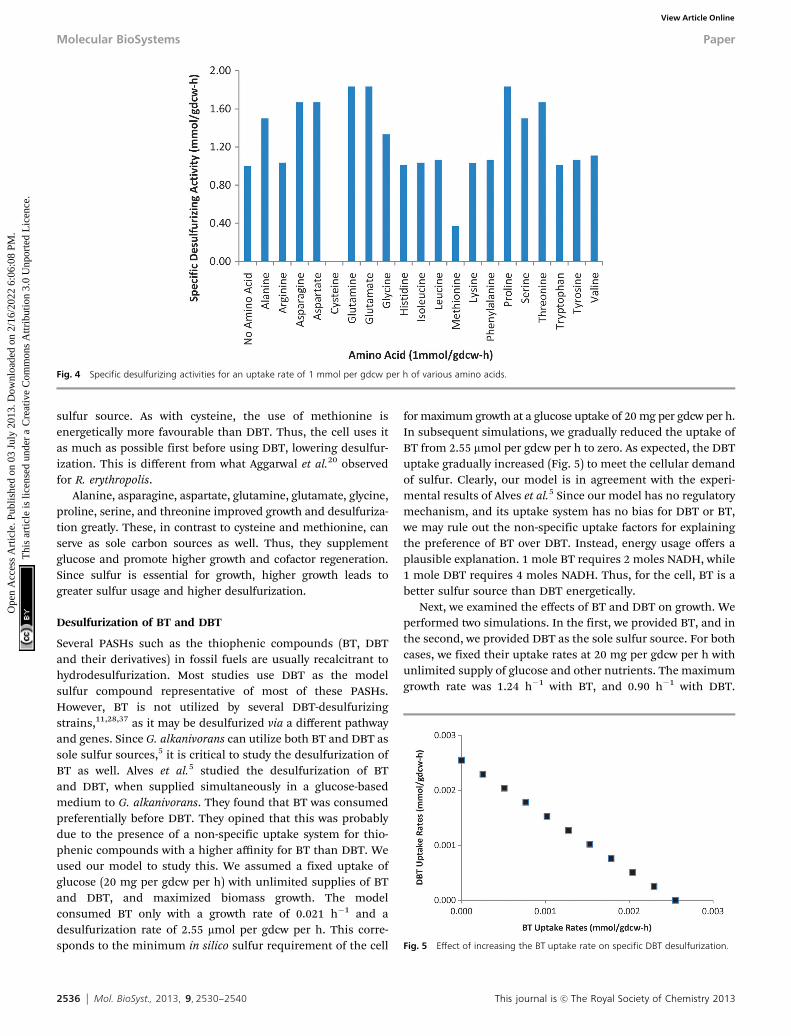
In contrast, some amino acids did affect the growth of anddesulfurization by G. alkanivorans. Fig. 4 shows the relativeeffects of various amino acids on desulfurization. Arginine,histidine, isoleucine, leucine, lysine, phenylalanine, trypto-phan, tyrosine, and valine affected neither growth nor desulfur-ization. In contrast, cysteine and methionine had strong effectson desulfurization. While no desulfurization occurred in thepresence of cysteine, it was reduced by 63% in the presence ofmethionine.
The effect of cysteine is similar to what Aggarwal et al.20
showed for R. erythropolis, and we can explain as follows. LikeR. erythropolis, G. alkanivorans can use cysteine as a sole sulfursource. Using cysteine is energetically less expensive than DBT,as 1 mole DBT requires additional 4 moles of NADH.20 There-fore, the cell prefers to consume cysteine rather than DBT, andno DBT desulfurization occurs.
The reduced desulfurization in the presence of methioninemay be due to the inability of G. alkanivorans to produce all thesulfur-containing metabolic precursors solely from methionine.For instance, they cannot produce cysteine, L-homocysteine,coenzyme A, etc. solely from methionine, and hence need anadditional sulfur source such as DBT or BT. While this may bereal, but no evidence exists in the literature, it may well be a gapin our model, which prevents the use of methionine as a sole
Fig. 3 Specific desulfurizing activities for an uptake rate of 20 mg per gdcw per h of various carbon sources.
Paper Molecular BioSystems
Ope
n A
cces
s A
rtic
le. P
ublis
hed
on 0
3 Ju
ly 2
013.
Dow
nloa
ded
on 2
/16/
2022
6:0
6:08
PM
. T
his
artic
le is
lice
nsed
und
er a
Cre
ativ
e C
omm
ons
Attr
ibut
ion
3.0
Unp
orte
d L
icen
ce.
View Article Online
2536 Mol. BioSyst., 2013, 9, 2530--2540 This journal is c The Royal Society of Chemistry 2013
sulfur source. As with cysteine, the use of methionine isenergetically more favourable than DBT. Thus, the cell uses itas much as possible first before using DBT, lowering desulfur-ization. This is different from what Aggarwal et al.20 observedfor R. erythropolis.
Alanine, asparagine, aspartate, glutamine, glutamate, glycine,proline, serine, and threonine improved growth and desulfuriza-tion greatly. These, in contrast to cysteine and methionine, canserve as sole carbon sources as well. Thus, they supplementglucose and promote higher growth and cofactor regeneration.Since sulfur is essential for growth, higher growth leads togreater sulfur usage and higher desulfurization.
Desulfurization of BT and DBT
Several PASHs such as the thiophenic compounds (BT, DBTand their derivatives) in fossil fuels are usually recalcitrant tohydrodesulfurization. Most studies use DBT as the modelsulfur compound representative of most of these PASHs.However, BT is not utilized by several DBT-desulfurizingstrains,11,28,37 as it may be desulfurized via a different pathwayand genes. Since G. alkanivorans can utilize both BT and DBT assole sulfur sources,5 it is critical to study the desulfurization ofBT as well. Alves et al.5 studied the desulfurization of BTand DBT, when supplied simultaneously in a glucose-basedmedium to G. alkanivorans. They found that BT was consumedpreferentially before DBT. They opined that this was probablydue to the presence of a non-specific uptake system for thio-phenic compounds with a higher affinity for BT than DBT. Weused our model to study this. We assumed a fixed uptake ofglucose (20 mg per gdcw per h) with unlimited supplies of BTand DBT, and maximized biomass growth. The modelconsumed BT only with a growth rate of 0.021 h�1 and adesulfurization rate of 2.55 mmol per gdcw per h. This corre-sponds to the minimum in silico sulfur requirement of the cell
for maximum growth at a glucose uptake of 20 mg per gdcw per h.In subsequent simulations, we gradually reduced the uptake ofBT from 2.55 mmol per gdcw per h to zero. As expected, the DBTuptake gradually increased (Fig. 5) to meet the cellular demandof sulfur. Clearly, our model is in agreement with the experi-mental results of Alves et al.5 Since our model has no regulatorymechanism, and its uptake system has no bias for DBT or BT,we may rule out the non-specific uptake factors for explainingthe preference of BT over DBT. Instead, energy usage offers aplausible explanation. 1 mole BT requires 2 moles NADH, while1 mole DBT requires 4 moles NADH. Thus, for the cell, BT is abetter sulfur source than DBT energetically.
Next, we examined the effects of BT and DBT on growth. Weperformed two simulations. In the first, we provided BT, and inthe second, we provided DBT as the sole sulfur source. For bothcases, we fixed their uptake rates at 20 mg per gdcw per h withunlimited supply of glucose and other nutrients. The maximumgrowth rate was 1.24 h�1 with BT, and 0.90 h�1 with DBT.
Fig. 4 Specific desulfurizing activities for an uptake rate of 1 mmol per gdcw per h of various amino acids.
Fig. 5 Effect of increasing the BT uptake rate on specific DBT desulfurization.
Molecular BioSystems Paper
Ope
n A
cces
s A
rtic
le. P
ublis
hed
on 0
3 Ju
ly 2
013.
Dow
nloa
ded
on 2
/16/
2022
6:0
6:08
PM
. T
his
artic
le is
lice
nsed
und
er a
Cre
ativ
e C
omm
ons
Attr
ibut
ion
3.0
Unp
orte
d L
icen
ce.
View Article Online
This journal is c The Royal Society of Chemistry 2013 Mol. BioSyst., 2013, 9, 2530--2540 2537
Thus, BT promotes higher growth than DBT. This can also beexplained by the lower energy requirements of BT as mentionedin the previous paragraph.
Comparison with R. erythropolis model
As mentioned earlier, R. erythropolis is the most widely studiedbacteria for desulfurization, but G. alkanivorans shows desul-furization activity for a wider range of PASHs. However, it wouldbe desirable to compare these two bacteria under the sameconditions. For this, we studied the desulfurization of DBT andBT individually by the existing genome scale metabolic modelof R. erythropolis20 and our reconstructed model of G. alkani-vorans. We maximized cell growth for a fixed glucose uptake of1 mmol per gdcw per h with unlimited supplies of DBT/BT andother minimal nutrients. Both R. erythropolis and G. alkanivoranscan utilize DBT as the sole sulfur source and give out HBPas the desulfurized product in the medium. However,G. alkanivorans can utilize BT, but R. erythropolis cannot. Thissuggests that G. alkanivorans can indeed be a more suitablebiocatalyst for desulfurizing the fossil fuels, which normallyhave a spectrum of DBT, BT, and their derivatives.
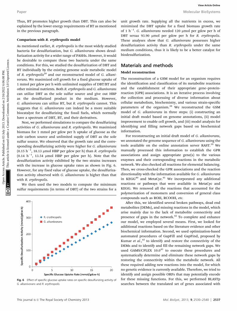
Next, we performed simulations to compare the desulfurizingactivities of G. alkanivorans and R. erythropolis. We maximizedbiomass for 1 mmol per gdcw per h uptake of glucose as thesole carbon source and unlimited supply of DBT as the solesulfur source. We observed that the growth rate and the corre-sponding desulfurizing activity were higher for G. alkanivorans(0.15 h�1, 18.13 mmol HBP per gdcw per h) than R. erythropolis(0.14 h�1, 13.54 mmol HBP per gdcw per h). Note that thedesulfurization activity exhibited by the two strains increaseswith the increase in glucose uptake rates as shown in Fig. 6.However, for any fixed value of glucose uptake, the desulfuriza-tion activity observed with G. alkanivorans is higher than thatwith R. erythropolis.
We then used the two models to compute the minimumsulfur requirements (in terms of DBT) of the two strains for a
unit growth rate. Supplying all the nutrients in excess, weminimized the DBT uptake for a fixed biomass growth rateof 1 h�1. G. alkanivorans needed 120 mmol per gdcw per h ofDBT versus 93.90 mmol per gdcw per h for R. erythropolis.These analyses show that G. alkanivorans possesses higherdesulfurization activity than R. erythropolis under the samemedium conditions, thus it is likely to be a better catalyst forbiodesulfurization.
Materials and methodsModel reconstruction
The reconstruction of a GSM model for an organism requiresthe identification and classification of its metabolite reactionsand the establishment of their appropriate gene–protein-reaction (GPR) associations. It is an iterative process involvingthe collection and processing of diverse information aboutcellular metabolism, biochemistry, and various strain-specificparameters of the organism.38 We reconstructed the GSMmodel of G. alkanivorans in three steps: (i) constructing aninitial draft model based on genome annotations, (ii) modelimprovement to enable cell growth, and (iii) model analysis foridentifying and filling network gaps based on biochemicalinformation.
For reconstructing an initial draft model of G. alkanivorans,we annotated the genome sequence of G. alkanivorans using thetools available on the online annotation server RAST.39 Wemanually processed this information to establish the GPRassociations and assign appropriate gene(s) to the variousenzymes and their corresponding reactions in the metabolicnetwork. We also checked all reactions for elemental balancing.Then, we cross-checked the GPR associations and the reactiondirectionality with the information available for G. alkanivoransin KEGG40 and MetaCyc.41 We incorporated any additionalreactions or pathways that were available in MetaCyc andKEGG. We removed all the reactions that accounted for thepolymerization of monomers and conversion of general classcompounds such as ROH, RCOOH, etc.
After this, we identified several broken pathways, dead endmetabolites (DEMs), and missing reactions in the model, whicharise mainly due to the lack of metabolite connectivity andpresence of gaps in the network.42 To complete and enhanceour model, we employed several means. First, we looked foradditional reactions based on the literature evidence and otherbiochemical information. Second, we used optimization-basedautomated procedures of GapFill and GapFind, proposed byKumar et al.,42 to identify and restore the connectivity of theDEMs and to identify and fill the remaining network gaps. Weused GAMS/CPLEX 10.043 to execute these procedures andsystematically determine and eliminate these network gaps byrestoring the connectivity within the metabolic network. Allthese required adding new reactions into the model, for whichno genetic evidence is currently available. Therefore, we tried toidentify and assign possible ORFs that may potentially encodefor these missing functions. For this, we performed BLASTpsearches between the translated set of genes associated with
Fig. 6 Effect of specific glucose uptake rates on specific desulfurizing activity ofG. alkanivorans and R. erythropolis.
Paper Molecular BioSystems
Ope
n A
cces
s A
rtic
le. P
ublis
hed
on 0
3 Ju
ly 2
013.
Dow
nloa
ded
on 2
/16/
2022
6:0
6:08
PM
. T
his
artic
le is
lice
nsed
und
er a
Cre
ativ
e C
omm
ons
Attr
ibut
ion
3.0
Unp
orte
d L
icen
ce.
View Article Online
2538 Mol. BioSyst., 2013, 9, 2530--2540 This journal is c The Royal Society of Chemistry 2013
these additional reactions in various databases and the genomeof G. alkanivorans. While we used a high e cut-off of 10�30 formost network improvement reactions, we used a low cut-offof 10�5 for some reactions to enable the essential activity ofbiomass generation.
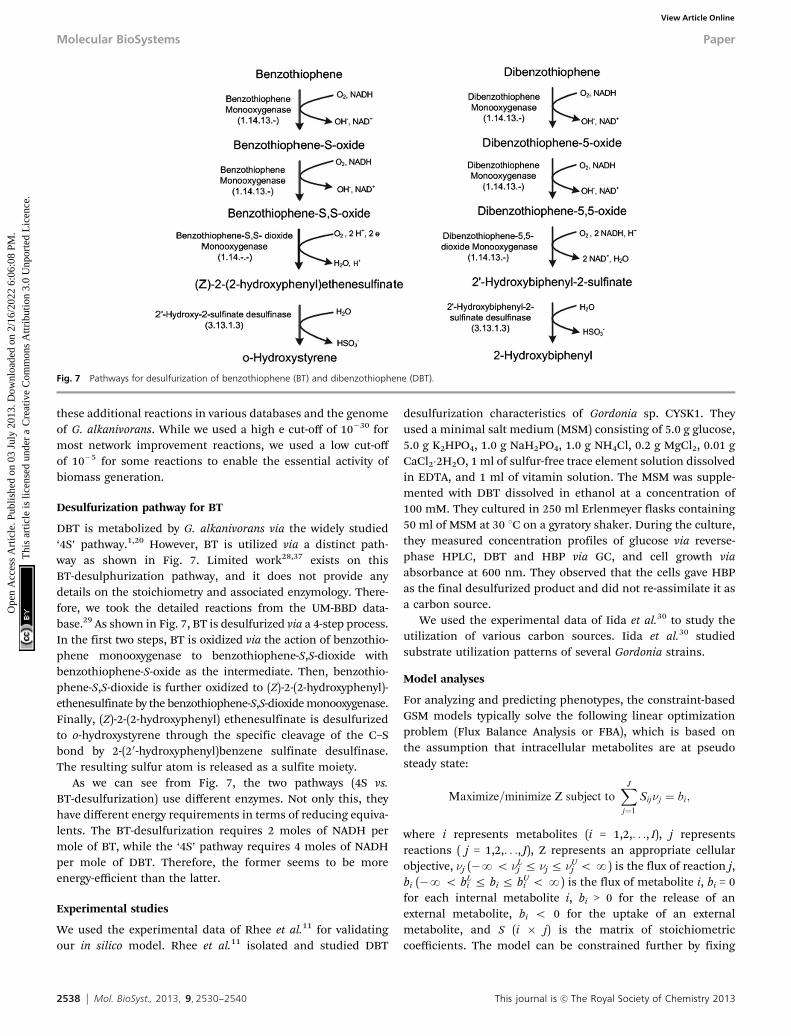
Desulfurization pathway for BT
DBT is metabolized by G. alkanivorans via the widely studied‘4S’ pathway.1,20 However, BT is utilized via a distinct path-way as shown in Fig. 7. Limited work28,37 exists on thisBT-desulphurization pathway, and it does not provide anydetails on the stoichiometry and associated enzymology. There-fore, we took the detailed reactions from the UM-BBD data-base.29 As shown in Fig. 7, BT is desulfurized via a 4-step process.In the first two steps, BT is oxidized via the action of benzothio-phene monooxygenase to benzothiophene-S,S-dioxide withbenzothiophene-S-oxide as the intermediate. Then, benzothio-phene-S,S-dioxide is further oxidized to (Z)-2-(2-hydroxyphenyl)-ethenesulfinate by the benzothiophene-S,S-dioxide monooxygenase.Finally, (Z)-2-(2-hydroxyphenyl) ethenesulfinate is desulfurizedto o-hydroxystyrene through the specific cleavage of the C–Sbond by 2-(20-hydroxyphenyl)benzene sulfinate desulfinase.The resulting sulfur atom is released as a sulfite moiety.
As we can see from Fig. 7, the two pathways (4S vs.BT-desulfurization) use different enzymes. Not only this, theyhave different energy requirements in terms of reducing equiva-lents. The BT-desulfurization requires 2 moles of NADH permole of BT, while the ‘4S’ pathway requires 4 moles of NADHper mole of DBT. Therefore, the former seems to be moreenergy-efficient than the latter.
Experimental studies
We used the experimental data of Rhee et al.11 for validatingour in silico model. Rhee et al.11 isolated and studied DBT
desulfurization characteristics of Gordonia sp. CYSK1. Theyused a minimal salt medium (MSM) consisting of 5.0 g glucose,5.0 g K2HPO4, 1.0 g NaH2PO4, 1.0 g NH4Cl, 0.2 g MgCl2, 0.01 gCaCl2�2H2O, 1 ml of sulfur-free trace element solution dissolvedin EDTA, and 1 ml of vitamin solution. The MSM was supple-mented with DBT dissolved in ethanol at a concentration of100 mM. They cultured in 250 ml Erlenmeyer flasks containing50 ml of MSM at 30 1C on a gyratory shaker. During the culture,they measured concentration profiles of glucose via reverse-phase HPLC, DBT and HBP via GC, and cell growth viaabsorbance at 600 nm. They observed that the cells gave HBPas the final desulfurized product and did not re-assimilate it asa carbon source.
We used the experimental data of Iida et al.30 to study theutilization of various carbon sources. Iida et al.30 studiedsubstrate utilization patterns of several Gordonia strains.
Model analyses
For analyzing and predicting phenotypes, the constraint-basedGSM models typically solve the following linear optimizationproblem (Flux Balance Analysis or FBA), which is based onthe assumption that intracellular metabolites are at pseudosteady state:
Maximize=minimize Z subject toXJ
j¼1Sijnj ¼ bi;
where i represents metabolites (i = 1,2,. . ., I), j representsreactions ( j = 1,2,. . ., J), Z represents an appropriate cellularobjective, nj (�No nL
j r nj r nUj oN) is the flux of reaction j,
bi (�N o bLi r bi r bU
i o N) is the flux of metabolite i, bi = 0for each internal metabolite i, bi > 0 for the release of anexternal metabolite, bi o 0 for the uptake of an externalmetabolite, and S (i � j) is the matrix of stoichiometriccoefficients. The model can be constrained further by fixing
Fig. 7 Pathways for desulfurization of benzothiophene (BT) and dibenzothiophene (DBT).
Molecular BioSystems Paper
Ope
n A
cces
s A
rtic
le. P
ublis
hed
on 0
3 Ju
ly 2
013.
Dow
nloa
ded
on 2
/16/
2022
6:0
6:08
PM
. T
his
artic
le is
lice
nsed
und
er a
Cre
ativ
e C
omm
ons
Attr
ibut
ion
3.0
Unp
orte
d L
icen
ce.
View Article Online
This journal is c The Royal Society of Chemistry 2013 Mol. BioSyst., 2013, 9, 2530--2540 2539
the fluxes (bi) of one or more extracellular metabolites based ontheir experimentally measured uptake/release rates, settingrealistic bounds on various fluxes, or demanding a knownmaintenance energy for the organism. The solution of theFBA model gives the possible flux distributions that mayrepresent the metabolic state of a cell under given environ-mental conditions.
To solve the FBA model, we need a cellular objective (Z).Several cellular objectives such as maximum cell growth, mini-mum substrate utilization, minimum maintenance energy,etc.44 have been used in the literature. Cell growth is the mostcommon, as microbial cells have evolved to maximize growth. Itcan be expressed as a synthetic reaction consuming multiplebiomass precursor metabolites in some ratios, which can bedetermined from cell composition. In the absence of any datain the literature on the cellular composition of G. alkanivorans,we adapted information from the metabolic models of a relatedorganism, Corynebacterium glutamicum.45,46 Such adaptationfrom related organisms is an established practice in the recon-struction of metabolic models.16 We used MetaFluxNet47 andGAMS/CPLEX 10.043 to solve and analyze our FBA model.
Conclusion
We have presented the first genome scale metabolic model forG. alkanivorans. We have identified 55 new genome annotationsto explain some functionalities missing from its current gen-ome annotations. Our model successfully predicts and explainsthe limited experimental observations reported in the litera-ture. It suggests that ethanol may be the best carbon sourceamong the sixteen studied in this work. Also, the modelanalyses show that the presence of sulfur containing aminoacids, cysteine and methionine in the medium, can reduce thedesulfurization activity of G. alkanivorans. Our model alsoconfirms the experimental observations that BT is preferen-tially desulfurized over DBT by G. alkanivorans. However, itsuggests that BT’s lower energy requirements in terms of NADHrather than specific uptake mechanisms may be a betterexplanation for this preference. Further, our analyses show thatNADH plays an important role in desulfurization, thusre-engineering G. alkanivorans for improved supply/regenerationof NADH is likely to increase desulfurization. Our modelappropriately captures the inter-relationships between the var-ious metabolic activities occurring within G. alkanivorans andcan be used to study other properties of its metabolic networkand devise metabolic engineering strategies for obtainingimproved strains. Our comparative study of R. erythropolisand G. alkanivorans suggests that G. alkanivorans can be abetter desulfurizing strain as it can desulfurize both BT andDBT, and also exhibits higher desulfurization activity.
References
1 M. Soleimani, A. Bassi and A. Margaritis, Biotechnol. Adv.,2007, 25, 570–596.
2 C. Song, Catal. Today, 2003, 86, 211–263.
3 J. J. Kilbane II and K. Jackowski, Biotechnol. Bioeng., 1992,40, 1107–1114.
4 K. J. Kayser, L. Cleveland, H. S. Park, J. H. Kwak,A. Kolhatkar and J. J. Kilbane II, Appl. Microbiol. Biotechnol.,2002, 59, 737–745.
5 L. Alves, R. Salgueiro, C. Rodrigues, E. Mesquita, J. Matosand F. M. Gırio, Appl. Biochem. Biotechnol., 2005, 120,199–208.
6 M. Arenskotter, D. Broker and A. Steinbuchel, Appl. Environ.Microbiol., 2004, 70, 3195–3204.
7 L. Alves, M. Melo, D. Mendonça, F. Simoes, J. Matos,R. Tenreiro and F. M. Gırio, Enzyme Microb. Technol.,2007, 40, 1598–1603.
8 J. J. Kilbane II and J. Robbins, Appl. Microbiol. Biotechnol.,2007, 75, 843–851.
9 S. C. C. Santos, D. S. Alviano, C. S. Alviano, M. Padula,A. C. Leitao, O. B. Martins, C. M. S. Ribeiro, M. Y. M.Sassaki, C. P. S. Matta, J. Bevilaqua, G. V. Sebastian andL. Seldin, Appl. Microbiol. Biotechnol., 2006, 71, 355–362.
10 G. Q. Li, S. S. Li, S. W. Qu, Q. K. Liu, T. Ma, L. Zhu, F. L.Liang and R. L. Liu, Biotechnol. Lett., 2008, 30, 1759–1764.
11 S. K. Rhee, J. H. Chang, Y. K. Chang and H. N. Chang, Appl.Environ. Microbiol., 1998, 64, 2327–2331.
12 M. Shavandi, M. Sadeghizadeh, A. Zomorodipour andK. Khajeh, Bioresour. Technol., 2008, 100, 475–479.
13 L. Alves and S. M. Paixao, Bioresour. Technol., 2011, 102,9162–9166.
14 K. A. Gray, O. S. Pogrebinsky, G. T. Mrachko, L. Xi,D. J. Monticello and C. H. Squires, Nat. Biotechnol., 1996,14, 1705–1708.
15 G. Mohebali, A. S. Ball, B. Rasekh and A. Kaytash, EnzymeMicrob. Technol., 2007, 40, 578–584.
16 M. Durot, P. Y. Bourguignon and V. Schachter, FEMSMicrobiol. Rev., 2009, 33, 164–190.
17 A. M. Feist, C. S. Henry, J. L. Reed, M. Krummenacker,A. R. Joyce, P. D. Karp, L. J. Broadbelt, V. Hatzimanikatisand B. Ø. Palsson, Mol. Syst. Biol., 2007, 3, 121.
18 J. S. Edwards and B. O. Palsson, Proc. Natl. Acad. Sci. U. S. A.,2000, 97, 5528–5533.
19 S. Aggarwal, I. A. Karimi and D. Y. Lee, FEMS Microbiol. Lett.,2011, 315, 115–121.
20 S. Aggarwal, I. A. Karimi and D. Y. Lee, Mol. BioSyst., 2011, 7,3122–3131.
21 I. Famili, J. Forster, J. Nielsen and B. O. Palsson, Proc. Natl.Acad. Sci. U. S. A., 2003, 100, 13134–13139.
22 N. C. Duarte, M. J. Herrgård and B. Ø. Palsson, Genome Res.,2004, 14, 1298–1309.
23 H. Widiastuti, J. Y. Kim, S. Selvarasu, I. A. Karimi, H. Kim,J. S. Seo and D. Y. Lee, Biotechnol. Bioeng., 2011, 108,655–665.
24 S. Selvarasu, I. A. Karimi, G. H. Ghim and D. Y. Lee, Mol.BioSyst., 2009, 6, 152–161.
25 S. Selvarasu, V. V. T. Wong, I. A. Karimi and D. Y. Lee,Biotechnol. Bioeng., 2009, 102, 1494–1504.
26 R. Mahadevan and C. H. Schilling, Metab. Eng., 2003, 5,264–276.
Paper Molecular BioSystems
Ope
n A
cces
s A
rtic
le. P
ublis
hed
on 0
3 Ju
ly 2
013.
Dow
nloa
ded
on 2
/16/
2022
6:0
6:08
PM
. T
his
artic
le is
lice
nsed
und
er a
Cre
ativ
e C
omm
ons
Attr
ibut
ion
3.0
Unp
orte
d L
icen
ce.
View Article Online
2540 Mol. BioSyst., 2013, 9, 2530--2540 This journal is c The Royal Society of Chemistry 2013
27 B. K. S. Chung and D. Y. Lee, BMC Syst. Biol., 2009, 3, 117.28 T. Matsui, T. Onaka, K. Maruhashi and R. Kurane, Appl.
Microbiol. Biotechnol., 2001, 57, 212–215.29 J. Gao, L. B. M. Ellis and L. P. Wackett, Nucleic Acids Res.,
2010, 38, D488–D491.30 S. Iida, H. Taniguchi, A. Kageyama, K. Yazawa, H. Chibana,
S. Murata, F. Nomura, R. M. Kroppenstedt and Y. Mikami,Int. J. Syst. Evol. Microbiol., 2005, 55, 1871–1876.
31 R. K. Murray, D. K. Granner and P. A. R. V. W. Mayes,Harper’s Biochemistry, Prentice Hall International, London,1996.
32 C. H. Del Olmo, A. Alcon, V. E. Santos and F. Garcia-Ochoa,Enzyme Microb. Technol., 2005, 37, 157–166.
33 H. Honda, H. Sugiyama, I. Saito and T. Kobayashi,J. Ferment. Bioeng., 1998, 85, 334–338.
34 H. Yan, M. Kishimoto, T. Omasa, Y. Katakura, K. I. Suga,K. Okumura and O. Yoshikawa, J. Biosci. Bioeng., 2000, 89,361–366.
35 H. Yan, X. Sun, Q. Xu, Z. Ma, C. Xiao and N. Jun, J. Environ.Sci., 2008, 20, 613–618.
36 E. Franchi, F. Rodriguez, L. Serbolisca and F. de Ferra, OilGas Sci. Technol., 2003, 58, 515–520.
37 S. C. Gilbert, J. Morton, S. Buchanan, C. Oldfield andA. McRoberts, Microbiology, 1998, 144, 2545–2553.
38 R. Saha, P. F. Suthers and C. D. Maranas, PLoS One, 2011,6, e21784.
39 R. K. Aziz, D. Bartels, A. Best, M. DeJongh, T. Disz,R. A. Edwards, K. Formsma, S. Gerdes, E. M. Glass,M. Kubal, F. Meyer, G. J. Olsen, R. Olson, A. L. Osterman,R. A. Overbeek, L. K. McNeil, D. Paarmann, T. Paczian,B. Parrello, G. D. Pusch, C. Reich, R. Stevens, O. Vassieva,V. Vonstein, A. Wilke and O. Zagnitko, BMC Genomics, 2008,9, 75.
40 M. Kanehisa and S. Goto, Nucleic Acids Res., 2000, 28,27–30.
41 R. Caspi, H. Foerster, C. A. Fulcher, P. Kaipa,M. Krummenacker, M. Latendresse, S. Paley, S. Y. Rhee,A. G. Shearer, C. Tissier, T. C. Walk, P. Zhang andP. D. Karp, Nucleic Acids Res., 2008, 36, D623–D631.
42 V. Satish Kumar, M. S. Dasika and C. D. Maranas, BMCBioinf., 2007, 8, 212.
43 A. Brooke, D. Kendrick, A. Meeraus and R. Raman, GAMS aUser’s Guide, GAMS Development Corporation, Washington,DC, 1998.
44 A. P. Burgard and C. D. Maranas, Biotechnol. Bioeng., 2003,82, 670–677.
45 K. R. Kjeldsen and J. Nielsen, Biotechnol. Bioeng., 2009, 102,583–597.
46 Y. Shinfuku, N. Sorpitiporn, M. Sono, C. Furusawa,T. Hirasawa and H. Shimizu, Microb. Cell Fact., 2009, 8, 43.
47 D. Y. Lee, H. Yun, S. Park and S. Y. Lee, Bioinformatics, 2003,19, 2144–2146.
Molecular BioSystems Paper
Ope
n A
cces
s A
rtic
le. P
ublis
hed
on 0
3 Ju
ly 2
013.
Dow
nloa
ded
on 2
/16/
2022
6:0
6:08
PM
. T
his
artic
le is
lice
nsed
und
er a
Cre
ativ
e C
omm
ons
Attr
ibut
ion
3.0
Unp
orte
d L
icen
ce.
View Article Online