Embed Size (px)
Citation preview
Clinicopathological correlates of adrenalCushing’s syndromeKai Duan,1,2 Karen Gomez Hernandez,3,4 Ozgur Mete1,2,4
1Department of Pathology,University Health Network,Toronto, Ontario, Canada2Department of LaboratoryMedicine and Pathobiology,University of Toronto, Ontario,Canada3Department of Medicine,University Health Network,Toronto, Ontario, Canada4Endocrine Oncology SiteGroup, Princess MargaretCancer Centre, Toronto,Ontario, Canada
Correspondence toDr Ozgur Mete,200 Elizabeth Street, 11thfloor, University HealthNetwork, Department ofPathology, Toronto, Ontario,Canada M5G 2C4;[email protected]
Received 25 September 2014Revised 25 October 2014Accepted 2 November 2014Published Online First25 November 2014
To cite: Duan K, GomezHernandez K, Mete O. J ClinPathol 2015;68:175–186.
ABSTRACTEndogenous Cushing’s syndrome is a rare endocrinedisorder that incurs significant cardiovascular morbidityand mortality, due to glucocorticoid excess. It comprisesadrenal (20%) and non-adrenal (80%) aetiologies. Whilethe majority of cases are attributed to pituitary or ectopiccorticotropin (ACTH) overproduction, primary cortisol-producing adrenal cortical lesions are increasinglyrecognised in the pathophysiology of Cushing’ssyndrome. Our understanding of this disease hasprogressed substantially over the past decade. Recently,important mechanisms underlying the pathogenesis ofadrenal hypercortisolism have been elucidated with thediscovery of mutations in cyclic AMP signalling (PRKACA,PRKAR1A, GNAS, PDE11A, PDE8B), armadillo repeatcontaining 5 gene (ARMC5) a putative tumoursuppressor gene, aberrant G-protein-coupled receptors,and intra-adrenal secretion of ACTH. Accurate subtypingof Cushing’s syndrome is crucial for treatment decision-making and requires a complete integration of clinical,biochemical, imaging and pathology findings.Pathological correlates in the adrenal glands includehyperplasia, adenoma and carcinoma. While the mostcommon presentation is diffuse adrenocorticalhyperplasia secondary to excess ACTH production, thisentity is usually treated with pituitary or ectopic tumourresection. Therefore, when confronted withadrenalectomy specimens in the setting of Cushing’ssyndrome, surgical pathologists are most commonlyexposed to adrenocortical adenomas, carcinomas andprimary macronodular or micronodular hyperplasia.This review provides an update on the rapidly evolvingknowledge of adrenal Cushing’s syndrome and discussesthe clinicopathological correlations of this importantdisease.
INTRODUCTIONFirst described in 1932, Cushing’s syndrome is awell known clinical entity that reflects chronicexposure of the body to excess glucocorticoids,from either endogenous or more commonlyexogenous sources.1–5 Accurate epidemiologicaldata on this topic is currently lacking.5 Thereported incidence of endogenous cases rangesfrom 2 per million to 3 per million per year, withapproximately 10% of cases occurring in chil-dren.1–6 Recognition of Cushing’s syndrome is crit-ical because the overall mortality frommoderate-to-severe hypercortisolism is increasedtwofold due to macrovascular (myocardial infarc-tion, stroke) and infectious complications.7 Withmodern-day treatments, mortality rates after nor-malisation of cortisol have been reported to besimilar to that of an age-matched population.1–5 7
Endogenous Cushing’s syndrome is classicallydivided into corticotropin (ACTH)-dependent andACTH-independent hypercortisolism.1–12 Themajority of cases of the ACTH-dependent subtypeare caused by a pituitary ACTH-producing adenoma,also known as Cushing’s disease (70% of endogenousCushing’s syndrome; table 1).1–4 Other cases arecaused by ectopic ACTH secretion (10%) from avariety of neuroendocrine neoplasms including para-ganglioma, phaeochromocytoma and neuroendo-crine tumours of various sites (lung, thyroid, thymus,appendix and pancreas).1–6 Rare neuroendocrinetumours (<1%) can also present with Cushing’s syn-drome when they secrete corticotropin-releasinghormone (CRH).3 10 In contrast with the diverseaetiologies of ACTH-dependent Cushing’s syndrome,ACTH-independent cases are attributed to primarycortisol-producing adrenocortical lesions (20%),which may arise sporadically or in the setting of raregenetic syndromes.8 11–13
Accurate subtyping of Cushing’s syndrome isessential for treatment decision-making and requiresa complete integration of clinical, biochemical,imaging and pathology findings. Pathological corre-lates in the adrenal glands include hyperplasia,adenoma and carcinoma.11–13 While ACTH-dependent adrenal hyperplasia is the most commonclinical manifestation of endogenous Cushing’s syn-drome, this entity rarely presents itself in surgicalpathology because most cases are treated with trans-sphenoidal pituitary adenomectomy, ectopic ACTH-producing neuroendocrine tumour resection,radiation or medical therapy.2 3 14 15 Therefore, insurgical series of Cushing’s syndrome, the mostcommon adrenal pathologies relate to ACTH-independent hypercortisolism (adrenal Cushing’ssyndrome).16 Cortisol-producing adrenocorticaladenoma and carcinoma account for, respectively,55% and 35% of cases of adrenal Cushing’s syn-drome, while primary adrenocortical macronodularand micronodular hyperplasia have been attributedto the remainder 10% of cases (table 2).2 3 11–13
CLINICAL AND BIOCHEMICAL ASPECTS OFCUSHING’S SYNDROMEIn clinical practice, Cushing’s syndrome can presentat all ages, with a female predominance.1–5 Whilethe overt presentation is clinically unmistakable(central ‘truncal’ obesity, ‘moon facies’ and ‘buffalohumps’), milder cases are more difficult to diagnose,due to the wide spectrum of manifestations.1–4
None of its symptoms are pathognomonic andmany of its features (obesity, diabetes, hypertension,fatigue, irritability, depression, decreased concentra-tion and menstrual irregularity) are commonly seenin the general population.1–4 A clue to the presence
Editor’s choiceScan to access more
free content
Duan K, et al. J Clin Pathol 2015;68:175–186. doi:10.1136/jclinpath-2014-202612 175
Review on 10 July 2018 by guest. P
rotected by copyright.http://jcp.bm
j.com/
J Clin P
athol: first published as 10.1136/jclinpath-2014-202612 on 25 Novem
ber 2014. Dow
nloaded from
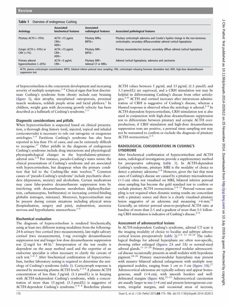
of hypercortisolism is the concurrent development and increasingseverity of multiple symptoms.1–4 Clinical signs that best discrim-inate Cushing’s syndrome from obesity include easy bruising(figure 1), skin atrophy, unexplained osteoporosis, proximalmuscle weakness, reddish purple striae and facial plethora.1 Inchildren, weight gain with decreasing growth velocity has beendescribed as a hallmark of Cushing’s syndrome.1 6
Diagnostic considerations and pitfallsWhen hypercortisolism is suspected based on clinical presenta-tion, a thorough drug history (oral, injected, topical and inhaledcorticosteroids) is necessary to rule out iatrogenic or exogenousaetiologies.1–4 Factitious Cushing’s syndrome has also beenreported in less than 1% of cases, and can be extremely difficultto recognise.17 Other pitfalls in the diagnosis of endogenousCushing’s syndrome include drug interactions and physiological/physiopathological changes in the hypothalamic-pituitary-adrenal axis.1–4 For instance, pseudo-Cushing’s states mimic theclinical presentations of Cushing’s syndrome and are associatedwith hypercortisolism, but disappear when the primary condi-tion that led to the Cushing-like state resolves.18 Commoncauses of ‘pseudo-Cushing’s syndrome’ include psychiatric disor-ders (depression, anxiety) and alcoholism. Certain medicationsmay cause false-positive dexamethasone suppression tests byinterfering with dexamethasone metabolism (diphenylhydan-toin, carbamazepine, barbiturates) or increasing cortisol-bindingglobulin (estrogen, mitotane). Moreover, hypercortisolism maybe present during certain situations including physical stress(hospitalisation, surgery and pain), malnutrition, anorexianervosa and hypothalamic amenorrhoea.1–4
Biochemical evaluationThe diagnosis of hypercortisolism is rendered biochemically,using at least two different testing modalities from the following:24-h urinary free cortisol (two measurements), late-night salivarycortisol (two measurements), 1-mg overnight dexamethasonesuppression test and longer low dose dexamethasone suppressiontest (2 mg/d for 48 h).1 Interpretation of the test results isdependent on the assay method used and the expertise of anendocrine specialist is often necessary to clarify the caveats ofeach test.1–4 8 After biochemical confirmation of hypercortiso-lism, further laboratory testing is required to determine the aeti-ology of Cushing’s syndrome (table 1). Corticotroph response isassessed by measuring plasma ACTH levels.2–4 8 A plasma ACTHconcentration of less than 5 pg/mL (1.1 pmol/L) is in keepingwith ACTH-independent Cushing’s syndrome, while a concen-tration of more than 15 pg/mL (3.3 pmol/L) is suggestive ofACTH-dependent Cushing’s syndrome.2–4 8 Borderline plasma
ACTH values between 5 pg/mL and 15 pg/mL (1.1 pmol/L and3.3 pmol/L) are equivocal, and a CRH stimulation test may behelpful in differentiating Cushing’s disease from other aetiolo-gies.2–4 ACTH and cortisol increases after intravenous adminis-tration of CRH is suggestive of Cushing’s disease, whereas ablunted response is observed when the aetiology is adrenal.2–4 InACTH-dependent hypercortisolism, CRH stimulation test is alsoused in conjunction with high-dose dexamethasone suppressiontest to differentiate between pituitary and ectopic ACTH over-production; if CRH stimulation and high-dose dexamethasonesuppression tests are positive, a petrosal sinus sampling test maynot be warranted to confirm or exclude the diagnosis of pituitaryACTH oversecretion.2–4
RADIOLOGICAL CONSIDERATIONS IN CUSHING’SSYNDROMEAfter biochemical confirmation of hypercortisolism and ACTHstatus, radiological investigations provide a supplementary methodfor preoperative subtyping (table 1). In ACTH-dependentCushing’s syndrome, pituitary MRI is the modality of choice todetect a pituitary adenoma.2–4 However, given the fact that mostcases of Cushing’s disease are caused by a pituitary microadenoma(<1 cm) often not visualised on MRI, bilateral inferior petrosalsinus sampling has become the gold standard test to confirm orexclude pituitary ACTH oversecretion.2–4 15 Petrosal venous sam-pling is not required when dynamic testing results are concordantwith a pituitary source and there is a clearly visualised pituitarylesion suggestive of an adenoma and measuring >6 mm.19
Generally, an inferior petrosal sinus-to-peripheral ACTH ratio atbaseline of more than 2:1 and a gradient of more than 3:1 follow-ing CRH stimulation is indicative of Cushing’s disease.2–4
Assessment of adrenocortical lesionsIn ACTH-independent Cushing’s syndrome, adrenal CT scan isthe imaging modality of choice to localise and subtype adreno-cortical lesions preoperatively (table 2).2–4 11 20–24 The radio-logical findings for adrenal hyperplasia are often non-specific,showing either enlarged (figures 2A and 3A) or normal-sizedadrenal glands.11 21–24 Primary pigmented nodular adrenocorti-cal disease occasionally presents as small nodules with hypodensepigment.24–26 Primary macronodular hyperplasia may presentwith massive bilateral adrenal enlargement with multiple non-pigmented nodules, ranging from 1 cm to 5 cm (figure 3A).27
Adrenocortical adenomas are typically solitary and appear homo-geneous, small (<4 cm), with smooth borders and well-delineated margins (figure 4A).21–23 Adrenocortical carcinomasare usually larger in size (>4 cm) and present heterogeneous con-tents, irregular margins, and occasional areas of necrosis,
Table 1 Overview of endogenous Cushing
AetiologyAssociatedbiochemical features
Associatedradiological features Associated pathological features
Pituitary ACTH (∼70%) ACTH >15 pg/mLCRH+HDD+
Pituitary MRI±BIPSS+
Pituitary corticotroph adenoma and Crooke’s hyaline change in the non-tumorouscorticotrophs; secondary diffuse±nodular adrenal cortical hyperplasia
Ectopic ACTH (∼10%) orCRH (<1%)
ACTH >15 pg/mLCRH−HDD−
Pituitary MRI−BIPSS−
Primary neuroendocrine tumour; secondary diffuse adrenal cortical hyperplasia
Primary adrenalhypercortisolism (∼20%)
ACTH <5 pg/mLCRH−
Pituitary MRI−Adrenal CT or MRI+
Adrenal cortical hyperplasia, adenoma and carcinoma
ACTH, plasma corticotropin concentration; BIPSS, bilateral inferior petrosal sinus sampling; CRH, corticotropin releasing hormone stimulation test; HDD, high-dose dexamethasonesuppression test.
176 Duan K, et al. J Clin Pathol 2015;68:175–186. doi:10.1136/jclinpath-2014-202612
Review on 10 July 2018 by guest. P
rotected by copyright.http://jcp.bm
j.com/
J Clin P
athol: first published as 10.1136/jclinpath-2014-202612 on 25 Novem
ber 2014. Dow
nloaded from
haemorrhage and calcification (figure 5A; table 2).21–23 Giventhe fact that many adenomas have intracytoplasmic fat resultingin lower attenuation, measurement of the attenuation value ishelpful in distinguishing adenomas from carcinoma; tumours
with attenuation values below 10 Hounsfield units on non-contrast CT are indicative of adrenocortical adenomas.21–23 28
Additionally, on contrast-enhanced CT, an absolute contrastwashout of more than 60% is suggestive of adenoma.21
Adrenal incidentalomasWith the advent of widespread imaging studies, primary hyper-cortisolism is increasingly diagnosed as part of the recom-mended workup for adrenal incidentalomas, which are detectedfor reasons unrelated to adrenal diseases.1 29–32 The reportedincidence of adrenal incidentalomas is approaching the 8.7%incidence reported in autopsy series, with up to 10% of casescausing hypercortisolism.29 The most common presentation issubclinical Cushing’s syndrome, although undiagnosed overtdisease may also occur.29–32 While the management of subclin-ical Cushing’s syndrome remains an area of controversy,29–32
the most recent guidelines suggest that adrenalectomy beoffered to patients with worsening hypertension, abnormalglucose tolerance, dyslipidaemia or osteoporosis.29
Measurement of fractionated metanephrines and catecholaminesis recommended in all cases of adrenal incidentalomas to ruleout phaeochromocytoma.29 If hypertension is present, analdosterone-to-renin ratio is also performed to exclude primaryaldosteronism.28 29 33
HISTOPATHOLOGICAL CORRELATES OF CUSHING’SSYNDROMEThe normal adult adrenal gland weighs approximately 4 g atsurgical excision, with an average cortical thickness of2 mm.16 34–38 In Cushing’s syndrome, histopathological corre-lates in the adrenal glands include hyperplasia, adenoma andcarcinoma.11–13 34–39 While ACTH-dependent adrenal hyper-plasia is the most common clinical manifestation of Cushing’ssyndrome, this entity rarely presents itself in surgical pathologybecause most cases are treated with transsphenoidal pituitaryresection, ectopic tumour resection, radiation or medicaltherapy.2 3 14–16 Therefore, when confronted with adrenalect-omy specimens in the setting of Cushing’s syndrome, patholo-gists are most commonly exposed to cortisol-producingneoplasms and ACTH-independent macronodular and micro-nodular hyperplasia.11–13 16 34–38
Table 2 Clinical, biochemical and radiological features of adrenal Cushing
Primary adrenocortical hyperplasia
Macronodular hyperplasia Micronodular hyperplasia
Adrenocortical adenoma Adrenocortical carcinoma BMAH c-BMAH PPNAD MAD
Frequency ∼55% ∼35% Estimated 10%Age All ages All ages; familial cases in childhood Fifth to sixth
decadeEarly childhood Childhood or early adulthood
Clinicalpresentation
Mild-to-severe Cushing Moderate-to-severe Cushing withrapid onset and possible virilisation
Mild Cushingwith insidiousonset
Moderate-to-severeCushing
Moderate-to-severe Cushing
Biochemicalfeatures
Negative Liddle’s test Negative Liddle’s test NegativeLiddle’s test
Negative Liddle’s test Paradoxical cortisolresponse inLiddle’s test
NegativeLiddle’s test
Radiologicalfeatures
Solitary mass, often <4 cm,defined margins, homogeneous,<10 HFU, absolute washout>60%
Solitary mass, often >4 cm, irregularmargins, heterogenous, >10 HFU,absolute washout <60%
Marked bilateral adrenal enlargementwith multiple large non-pigmentednodules (1–5 cm)
Normal or small size adrenalglands with occasional smallnodules (<1 cm)
BMAH, primary bilateral macronodular adrenocortical hyperplasia; c-BMAH, childhood BMAH; HFU, Hounsfield unit; MAD, non-pigmented micronodular adrenocortical disease; PPNAD,primary pigmented nodular adrenocortical disease.
Figure 1 Clinical features of Cushing’s syndrome. The overtpresentation is clinically unmistakable (central ‘truncal’ obesity, ‘moonfacies’ and ‘buffalo humps’), milder cases are more difficult todiagnose, due to the wide spectrum of manifestations. None of itssymptoms are pathognomonic and many of its features are commonlyseen in the general population. Clinical signs that have been reportedto best discriminate Cushing’s syndrome from obesity include easybruising, skin atrophy, unexplained osteoporosis, proximal muscleweakness, reddish purple striae and facial plethora.
Duan K, et al. J Clin Pathol 2015;68:175–186. doi:10.1136/jclinpath-2014-202612 177
Review on 10 July 2018 by guest. P
rotected by copyright.http://jcp.bm
j.com/
J Clin P
athol: first published as 10.1136/jclinpath-2014-202612 on 25 Novem
ber 2014. Dow
nloaded from
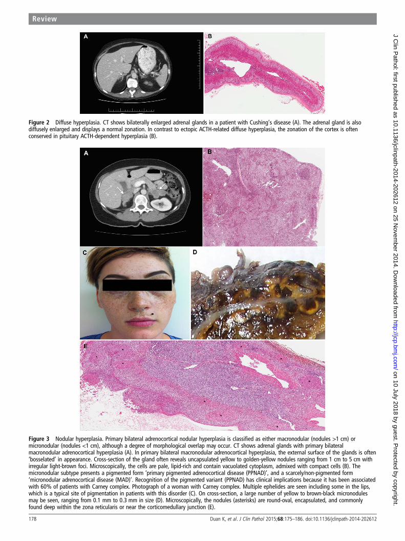
Figure 2 Diffuse hyperplasia. CT shows bilaterally enlarged adrenal glands in a patient with Cushing’s disease (A). The adrenal gland is alsodiffusely enlarged and displays a normal zonation. In contrast to ectopic ACTH-related diffuse hyperplasia, the zonation of the cortex is oftenconserved in pituitary ACTH-dependent hyperplasia (B).
Figure 3 Nodular hyperplasia. Primary bilateral adrenocortical nodular hyperplasia is classified as either macronodular (nodules >1 cm) ormicronodular (nodules <1 cm), although a degree of morphological overlap may occur. CT shows adrenal glands with primary bilateralmacronodular adrenocortical hyperplasia (A). In primary bilateral macronodular adrenocortical hyperplasia, the external surface of the glands is often‘bosselated’ in appearance. Cross-section of the gland often reveals uncapsulated yellow to golden-yellow nodules ranging from 1 cm to 5 cm withirregular light-brown foci. Microscopically, the cells are pale, lipid-rich and contain vacuolated cytoplasm, admixed with compact cells (B). Themicronodular subtype presents a pigmented form ‘primary pigmented adrenocortical disease (PPNAD)’, and a scarcely/non-pigmented form‘micronodular adrenocortical disease (MAD)’. Recognition of the pigmented variant (PPNAD) has clinical implications because it has been associatedwith 60% of patients with Carney complex. Photograph of a woman with Carney complex. Multiple ephelides are seen including some in the lips,which is a typical site of pigmentation in patients with this disorder (C). On cross-section, a large number of yellow to brown-black micronodulesmay be seen, ranging from 0.1 mm to 0.3 mm in size (D). Microscopically, the nodules (asterisks) are round-oval, encapsulated, and commonlyfound deep within the zona reticularis or near the corticomedullary junction (E).
178 Duan K, et al. J Clin Pathol 2015;68:175–186. doi:10.1136/jclinpath-2014-202612
Review on 10 July 2018 by guest. P
rotected by copyright.http://jcp.bm
j.com/
J Clin P
athol: first published as 10.1136/jclinpath-2014-202612 on 25 Novem
ber 2014. Dow
nloaded from
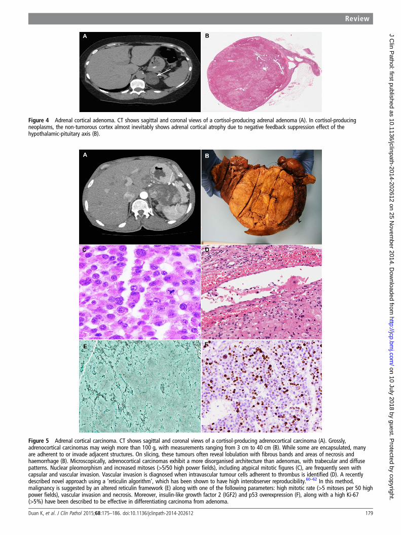
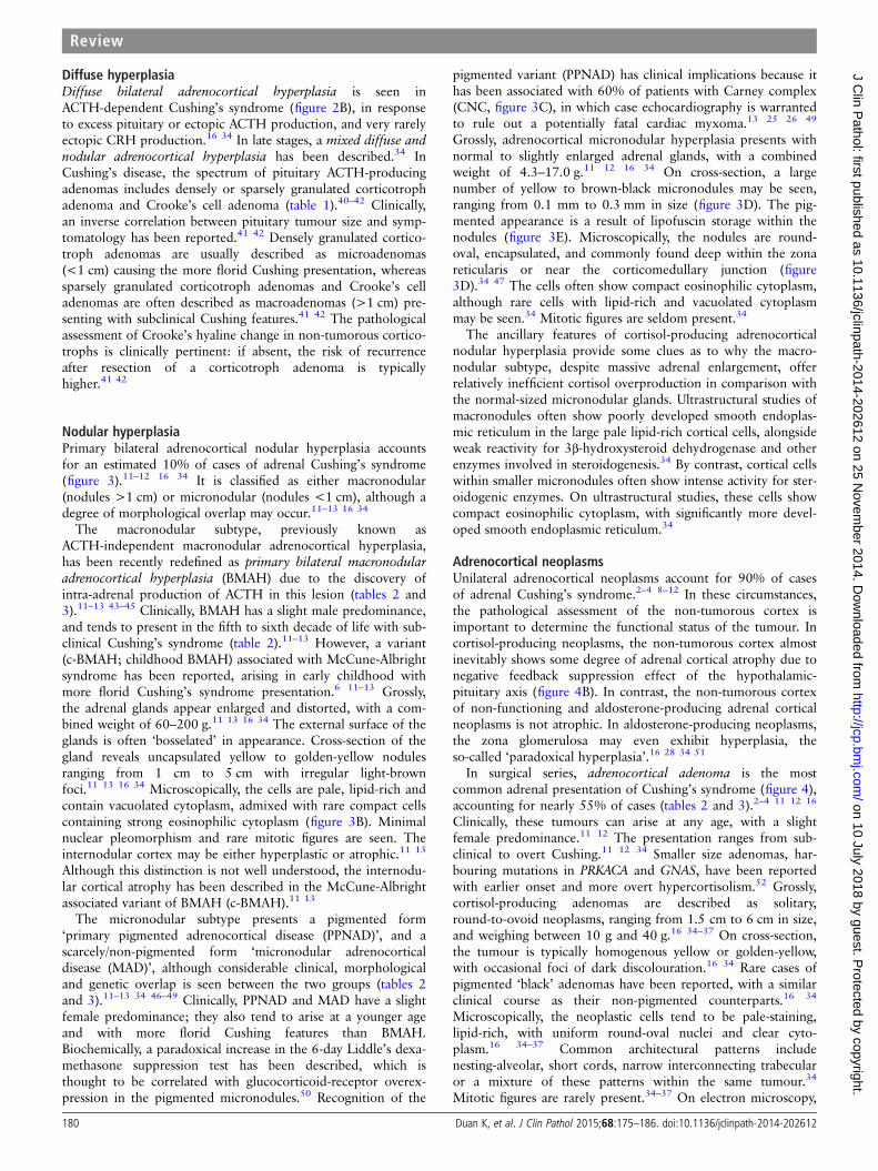
Figure 4 Adrenal cortical adenoma. CT shows sagittal and coronal views of a cortisol-producing adrenal adenoma (A). In cortisol-producingneoplasms, the non-tumorous cortex almost inevitably shows adrenal cortical atrophy due to negative feedback suppression effect of thehypothalamic-pituitary axis (B).
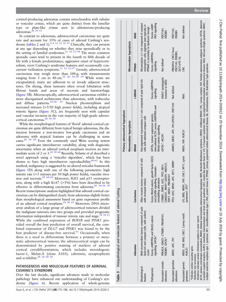
Figure 5 Adrenal cortical carcinoma. CT shows sagittal and coronal views of a cortisol-producing adrenocortical carcinoma (A). Grossly,adrenocortical carcinomas may weigh more than 100 g, with measurements ranging from 3 cm to 40 cm (B). While some are encapsulated, manyare adherent to or invade adjacent structures. On slicing, these tumours often reveal lobulation with fibrous bands and areas of necrosis andhaemorrhage (B). Microscopically, adrenocortical carcinomas exhibit a more disorganised architecture than adenomas, with trabecular and diffusepatterns. Nuclear pleomorphism and increased mitoses (>5/50 high power fields), including atypical mitotic figures (C), are frequently seen withcapsular and vascular invasion. Vascular invasion is diagnosed when intravascular tumour cells adherent to thrombus is identified (D). A recentlydescribed novel approach using a ‘reticulin algorithm’, which has been shown to have high interobserver reproducibility.60–62 In this method,malignancy is suggested by an altered reticulin framework (E) along with one of the following parameters: high mitotic rate (>5 mitoses per 50 highpower fields), vascular invasion and necrosis. Moreover, insulin-like growth factor 2 (IGF2) and p53 overexpression (F), along with a high Ki-67(>5%) have been described to be effective in differentiating carcinoma from adenoma.
Duan K, et al. J Clin Pathol 2015;68:175–186. doi:10.1136/jclinpath-2014-202612 179
Review on 10 July 2018 by guest. P
rotected by copyright.http://jcp.bm
j.com/
J Clin P
athol: first published as 10.1136/jclinpath-2014-202612 on 25 Novem
ber 2014. Dow
nloaded from
Diffuse hyperplasiaDiffuse bilateral adrenocortical hyperplasia is seen inACTH-dependent Cushing’s syndrome (figure 2B), in responseto excess pituitary or ectopic ACTH production, and very rarelyectopic CRH production.16 34 In late stages, a mixed diffuse andnodular adrenocortical hyperplasia has been described.34 InCushing’s disease, the spectrum of pituitary ACTH-producingadenomas includes densely or sparsely granulated corticotrophadenoma and Crooke’s cell adenoma (table 1).40–42 Clinically,an inverse correlation between pituitary tumour size and symp-tomatology has been reported.41 42 Densely granulated cortico-troph adenomas are usually described as microadenomas(<1 cm) causing the more florid Cushing presentation, whereassparsely granulated corticotroph adenomas and Crooke’s celladenomas are often described as macroadenomas (>1 cm) pre-senting with subclinical Cushing features.41 42 The pathologicalassessment of Crooke’s hyaline change in non-tumorous cortico-trophs is clinically pertinent: if absent, the risk of recurrenceafter resection of a corticotroph adenoma is typicallyhigher.41 42
Nodular hyperplasiaPrimary bilateral adrenocortical nodular hyperplasia accountsfor an estimated 10% of cases of adrenal Cushing’s syndrome(figure 3).11–12 16 34 It is classified as either macronodular(nodules >1 cm) or micronodular (nodules <1 cm), although adegree of morphological overlap may occur.11–13 16 34
The macronodular subtype, previously known asACTH-independent macronodular adrenocortical hyperplasia,has been recently redefined as primary bilateral macronodularadrenocortical hyperplasia (BMAH) due to the discovery ofintra-adrenal production of ACTH in this lesion (tables 2 and3).11–13 43–45 Clinically, BMAH has a slight male predominance,and tends to present in the fifth to sixth decade of life with sub-clinical Cushing’s syndrome (table 2).11–13 However, a variant(c-BMAH; childhood BMAH) associated with McCune-Albrightsyndrome has been reported, arising in early childhood withmore florid Cushing’s syndrome presentation.6 11–13 Grossly,the adrenal glands appear enlarged and distorted, with a com-bined weight of 60–200 g.11 13 16 34 The external surface of theglands is often ‘bosselated’ in appearance. Cross-section of thegland reveals uncapsulated yellow to golden-yellow nodulesranging from 1 cm to 5 cm with irregular light-brownfoci.11 13 16 34 Microscopically, the cells are pale, lipid-rich andcontain vacuolated cytoplasm, admixed with rare compact cellscontaining strong eosinophilic cytoplasm (figure 3B). Minimalnuclear pleomorphism and rare mitotic figures are seen. Theinternodular cortex may be either hyperplastic or atrophic.11 13
Although this distinction is not well understood, the internodu-lar cortical atrophy has been described in the McCune-Albrightassociated variant of BMAH (c-BMAH).11 13
The micronodular subtype presents a pigmented form‘primary pigmented adrenocortical disease (PPNAD)’, and ascarcely/non-pigmented form ‘micronodular adrenocorticaldisease (MAD)’, although considerable clinical, morphologicaland genetic overlap is seen between the two groups (tables 2and 3).11–13 34 46–49 Clinically, PPNAD and MAD have a slightfemale predominance; they also tend to arise at a younger ageand with more florid Cushing features than BMAH.Biochemically, a paradoxical increase in the 6-day Liddle’s dexa-methasone suppression test has been described, which isthought to be correlated with glucocorticoid-receptor overex-pression in the pigmented micronodules.50 Recognition of the
pigmented variant (PPNAD) has clinical implications because ithas been associated with 60% of patients with Carney complex(CNC, figure 3C), in which case echocardiography is warrantedto rule out a potentially fatal cardiac myxoma.13 25 26 49
Grossly, adrenocortical micronodular hyperplasia presents withnormal to slightly enlarged adrenal glands, with a combinedweight of 4.3–17.0 g.11 12 16 34 On cross-section, a largenumber of yellow to brown-black micronodules may be seen,ranging from 0.1 mm to 0.3 mm in size (figure 3D). The pig-mented appearance is a result of lipofuscin storage within thenodules (figure 3E). Microscopically, the nodules are round-oval, encapsulated, and commonly found deep within the zonareticularis or near the corticomedullary junction (figure3D).34 47 The cells often show compact eosinophilic cytoplasm,although rare cells with lipid-rich and vacuolated cytoplasmmay be seen.34 Mitotic figures are seldom present.34
The ancillary features of cortisol-producing adrenocorticalnodular hyperplasia provide some clues as to why the macro-nodular subtype, despite massive adrenal enlargement, offerrelatively inefficient cortisol overproduction in comparison withthe normal-sized micronodular glands. Ultrastructural studies ofmacronodules often show poorly developed smooth endoplas-mic reticulum in the large pale lipid-rich cortical cells, alongsideweak reactivity for 3β-hydroxysteroid dehydrogenase and otherenzymes involved in steroidogenesis.34 By contrast, cortical cellswithin smaller micronodules often show intense activity for ster-oidogenic enzymes. On ultrastructural studies, these cells showcompact eosinophilic cytoplasm, with significantly more devel-oped smooth endoplasmic reticulum.34
Adrenocortical neoplasmsUnilateral adrenocortical neoplasms account for 90% of casesof adrenal Cushing’s syndrome.2–4 8–12 In these circumstances,the pathological assessment of the non-tumorous cortex isimportant to determine the functional status of the tumour. Incortisol-producing neoplasms, the non-tumorous cortex almostinevitably shows some degree of adrenal cortical atrophy due tonegative feedback suppression effect of the hypothalamic-pituitary axis (figure 4B). In contrast, the non-tumorous cortexof non-functioning and aldosterone-producing adrenal corticalneoplasms is not atrophic. In aldosterone-producing neoplasms,the zona glomerulosa may even exhibit hyperplasia, theso-called ‘paradoxical hyperplasia’.16 28 34 51
In surgical series, adrenocortical adenoma is the mostcommon adrenal presentation of Cushing’s syndrome (figure 4),accounting for nearly 55% of cases (tables 2 and 3).2–4 11 12 16
Clinically, these tumours can arise at any age, with a slightfemale predominance.11 12 The presentation ranges from sub-clinical to overt Cushing.11 12 34 Smaller size adenomas, har-bouring mutations in PRKACA and GNAS, have been reportedwith earlier onset and more overt hypercortisolism.52 Grossly,cortisol-producing adenomas are described as solitary,round-to-ovoid neoplasms, ranging from 1.5 cm to 6 cm in size,and weighing between 10 g and 40 g.16 34–37 On cross-section,the tumour is typically homogenous yellow or golden-yellow,with occasional foci of dark discolouration.16 34 Rare cases ofpigmented ‘black’ adenomas have been reported, with a similarclinical course as their non-pigmented counterparts.16 34
Microscopically, the neoplastic cells tend to be pale-staining,lipid-rich, with uniform round-oval nuclei and clear cyto-plasm.16 34–37 Common architectural patterns includenesting-alveolar, short cords, narrow interconnecting trabecularor a mixture of these patterns within the same tumour.34
Mitotic figures are rarely present.34–37 On electron microscopy,
180 Duan K, et al. J Clin Pathol 2015;68:175–186. doi:10.1136/jclinpath-2014-202612
Review on 10 July 2018 by guest. P
rotected by copyright.http://jcp.bm
j.com/
J Clin P
athol: first published as 10.1136/jclinpath-2014-202612 on 25 Novem
ber 2014. Dow
nloaded from
cortisol-producing adenomas contain mitochondria with tubularor vesicular cristae, which are quite distinct from the lamellartype or plate-like cristae seen in aldosterone-producingadenomas.28 34 51
In contrast to adenomas, adrenocortical carcinomas are quiterare and account for 35% of cases of adrenal Cushing’s syn-drome (tables 2 and 3).2–4 11 12 16 47 Clinically, they can presentat any age depending on whether they arise sporadically or inthe setting of familial syndromes.11 12 53–58 The more commonsporadic cases tend to present in the fourth to fifth decade oflife with a female predominance, aggressive onset of hypercorti-solism, overt Cushing’s syndrome features and occasionally con-current virilisation symptoms.11 12 53–57 Grossly, adrenocorticalcarcinomas may weigh more than 100 g, with measurementsranging from 3 cm to 40 cm.16 28 34–38 59 While some areencapsulated, many are adherent to or invade adjacent struc-tures. On slicing, these tumours often reveal lobulation withfibrous bands and areas of necrosis and haemorrhage(figure 5B). Microscopically, adrenocortical carcinomas exhibit amore disorganised architecture than adenomas, with trabecularand diffuse patterns.34–38 59 Nuclear pleomorphism andincreased mitoses (>5/50 high power fields), including atypicalmitotic figures (figure 5C), are frequently seen with capsularand vascular invasion in the vast majority of high-grade adreno-cortical carcinomas.28 34–38
While the morphological features of ‘florid’ adrenal cortical car-cinomas are quite different from typical benign adenomas, the dis-tinction between a non-invasive low-grade carcinoma and anadenoma with atypical features can be challenging in somecases.28 58 60 Even the commonly used Weiss scoring systemcarries significant interobserver variability, along with diagnosticuncertainty when an adrenal cortical neoplasm receives an inter-mediate score of 2 or 3.28 58 60 Recently, Volante et al described anovel approach using a ‘reticulin algorithm’, which has beenshown to have high interobserver reproducibility.60–62 In thismethod, malignancy is suggested by an altered reticulin framework(figure 5D) along with one of the following parameters: highmitotic rate (>5 mitoses per 50 high power fields), vascular inva-sion and necrosis.58 60–62 Moreover, IGF2 and p53 overexpres-sion, along with a high Ki-67 (>5%) have been described to beeffective in differentiating carcinoma from adenoma.28 34–38 58
Recent transcriptome analysis highlighted that adrenal cortical car-cinomas can be distinguished clearly from adenomas slightly betterthan morphological assessment based on gene expression profileof an adrenal cortical neoplasm.54 58 63 Moreover, DNA micro-array analysis of a large group of adrenocortical tumours dividedthe malignant tumours into two groups and provided prognosticinformation independent of tumour mitotic rate and stage.54 58 63
While the combined expression of BUB1B and PINK1 pro-vided overall the best prediction of overall survival, the com-bined expression of DLG7 and PINK1 was found to be thebest predictor of disease-free survival.63 Occasionally, whenthere is a need to differentiate between a primary or meta-static adrenocortical tumour, the adrenocortical origin can bedemonstrated by positive staining of markers of adrenalcortical cytodifferentiation, which includes steroidogenicfactor-1, Melan-A (clone A103), calretinin, synaptophysinand α-inhibin.28 34–38 58
PATHOGENESIS AND MOLECULAR FEATURES OF ADRENALCUSHING’S SYNDROMEOver the last decade, significant advances made in molecularpathology have enhanced our understanding of Cushing’s syn-drome (figure 6). Recent application of whole-genome
Table3
Histopathologicaland
molecular
features
ofadrenalC
ushing
Prim
aryad
reno
cortical
hype
rplasia
Macrono
dularhype
rplasia
Microno
dularhype
rplasia
Ade
noma
Carcinom
aBM
AH
c-BM
AH
PPNAD
MAD
Morphological
features
Well-circum
scribed
tumourcom
posedof
pale
lipid-rich
andcompactcells,w
ithuniform
nuclei
Ofteninvasivetumourcom
posedof
cells
with
increasedmitosis
Non-pigmentedmacronodules(>1cm
)com
posedof
palelipid-rich
cells
with
admixed
compactcells
Micronodules(<1cm
)com
posedof
compact
eosin
ophilic
cells;p
igmentseen
typically
inPPNAD
issecondaryto
lipofuscinstorage
Pathogenesis
Excess
cAMP/PKAandWnt/β-catenin
signalling;G
PCRs*
Excess
Wnt/β-catenin,g
rowth
factor
(IGF2)
andp53/Rb
signalling;chrom
atin
remodellingalterations
GPC
Rs,tum
oursuppressor
gene(s),
intra
-adrenalAC
TH;excesscAMP/PKAand
Wnt/β-catenin
signalling
Excess
cAMP/PKA
signalling
Excess
cAMP/PKA
signalling
Excess
cAMP/
PKAsig
nalling
Associated
somatic
alterations
PRKA
CA,G
NAS
1,CTNBB
1,PRKA
R1A†
,PD
E8B†,G
PCRs*
CTNBB
1,ZN
RF3,
IGF2,TP53,
RB1†,
CDKN
2A†,M
EN1†
DAXX
†,M
ED12†,
TERT†,INHA
†,P
RKAR
1A†
GPC
Rs*,
MC2
R†GNAS
1(postzygotic
mutationinMAS
)Not
defined
PDE11A
‡,
PDE8B‡
Associated
germ
line
alterations
MEN
1,AP
C,FH†
TP53,IGF2,M
EN1,
APC,
NF1†,M
MR†
ARMC5
,MEN
1,AP
C,FH†,GPC
Rs†,P
DE11A‡
Not
defined
PRKA
R1A,
CNC2
locus
PDE11A
‡,P
DE8B‡,
2p12-p16;5
q
PDE11A
‡,
PDE8B‡
Associated
genetic
syndromes
MEN
1,FAP,MAS
,CNC†
,Carneytriad†,
HLRC
S†LFS,BW
S,MEN
1,FAP,CN
C†,N
F1†,Lynch
syndrome†,R
TS*
MEN
1,FAP,HLRC
S†MAS
CNC
Not
defined
*Indicates
unclearcausativegene.
†Indicatesrare
occurre
nce.
‡Evidence
indicatesunclearinheritance
patte
rnin
somereports.
ACTH
,plasm
acorticotro
pinconcentra
tion;
BMAH
,bilateralm
acronodulara
drenocorticalhyperplasia
;BWS,Beckwith-W
iedemannsyndrome;c-BM
AH,childhood
BMAH
;CNC,
Carney
complex;FAP
,fam
ilialadenom
atouspolyposis
syndrome;GPC
Rs,aberra
ntG-protein-coupled
receptors;HLRC
S,hereditary
leiomyomatosisandrenalcancersyndrome;IGF2,insulin-like
grow
thfactor
2;LFS,Li-Fraum
enisyndrom
e;MAD
,non-pigmentedmicronodulara
drenocorticaldisease;MAS
,McCune-Alright
syndrome;MEN
1,multipleendocrineneoplasia
type
1syndrome;NF1,n
eurofibromatosistype
I;PKA,
proteinkinase
A;PPNAD
,prim
arypigm
entednodulara
drenocorticaldisease;Rb,retinoblastom
a;RTS,Rubinstein-Taybisyndrome.
Duan K, et al. J Clin Pathol 2015;68:175–186. doi:10.1136/jclinpath-2014-202612 181
Review on 10 July 2018 by guest. P
rotected by copyright.http://jcp.bm
j.com/
J Clin P
athol: first published as 10.1136/jclinpath-2014-202612 on 25 Novem
ber 2014. Dow
nloaded from
sequencing techniques have allowed the discovery of somaticand germline mutations implicated in the pathogenesis ofprimary cortisol-producing adrenocortical lesions.52 64–72 Mostof these mutations (PRKACA, PRKAR1A, GNAS, PDE11A,PDE8B) cause aberrant activation of the cyclic AMP(cAMP)-signalling pathway, resulting in hormone overproduc-tion and cellular proliferation (figure 6).11–13 52 64–81 Othermutations involve pathways associated with ‘neoplastic trans-formation’, favouring tumour growth over hormone overpro-duction.70 These include mutations in armadillo repeatcontaining 5 gene (ARMC5) a putative tumour suppressorgene, Wnt/β-catenin pathway (CTNNB1, ZNRF3), growthfactor overexpression (IGF2), p53/retinoblastoma proteinpathway (TP53, CDKN2A, RB1) and chromatin remodelling(MEN1, DAXX).11–13 39 56 58 82–91
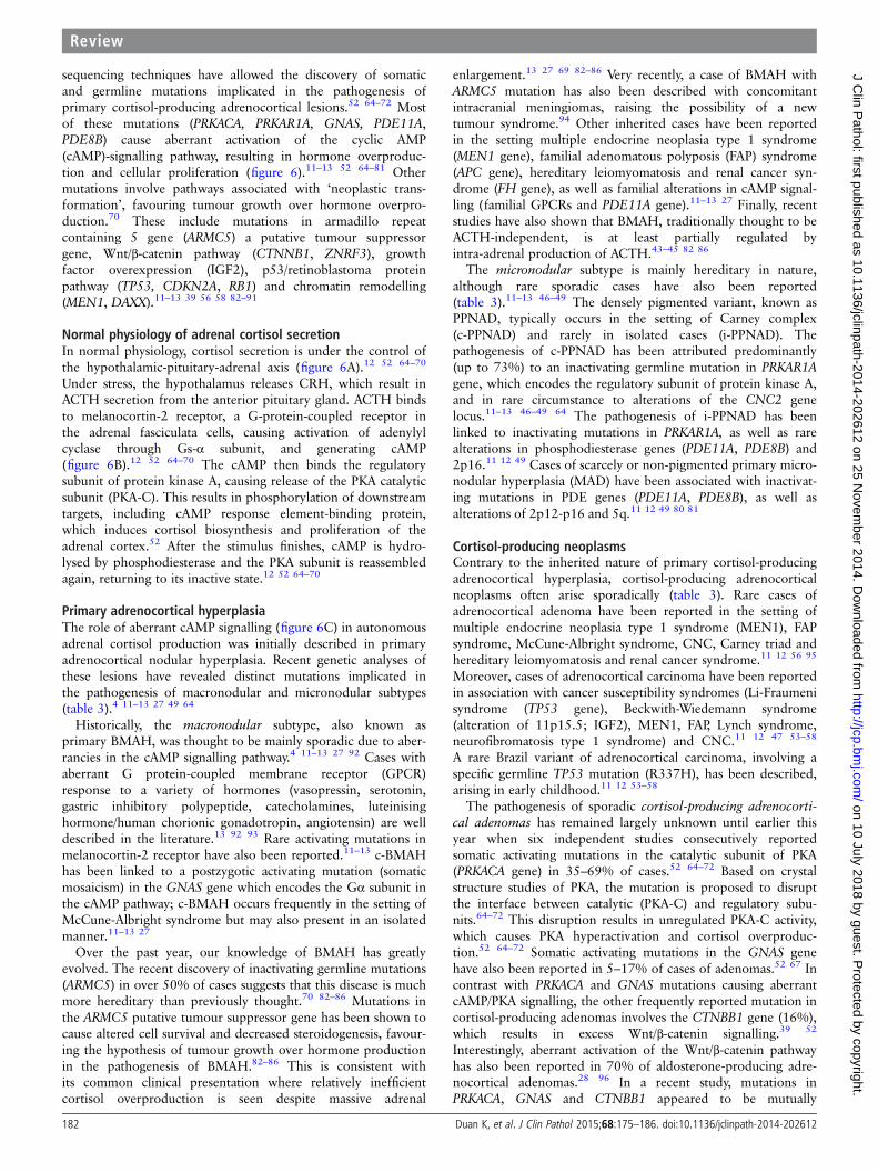
Normal physiology of adrenal cortisol secretionIn normal physiology, cortisol secretion is under the control ofthe hypothalamic-pituitary-adrenal axis (figure 6A).12 52 64–70
Under stress, the hypothalamus releases CRH, which result inACTH secretion from the anterior pituitary gland. ACTH bindsto melanocortin-2 receptor, a G-protein-coupled receptor inthe adrenal fasciculata cells, causing activation of adenylylcyclase through Gs-α subunit, and generating cAMP(figure 6B).12 52 64–70 The cAMP then binds the regulatorysubunit of protein kinase A, causing release of the PKA catalyticsubunit (PKA-C). This results in phosphorylation of downstreamtargets, including cAMP response element-binding protein,which induces cortisol biosynthesis and proliferation of theadrenal cortex.52 After the stimulus finishes, cAMP is hydro-lysed by phosphodiesterase and the PKA subunit is reassembledagain, returning to its inactive state.12 52 64–70
Primary adrenocortical hyperplasiaThe role of aberrant cAMP signalling (figure 6C) in autonomousadrenal cortisol production was initially described in primaryadrenocortical nodular hyperplasia. Recent genetic analyses ofthese lesions have revealed distinct mutations implicated inthe pathogenesis of macronodular and micronodular subtypes(table 3).4 11–13 27 49 64
Historically, the macronodular subtype, also known asprimary BMAH, was thought to be mainly sporadic due to aber-rancies in the cAMP signalling pathway.4 11–13 27 92 Cases withaberrant G protein-coupled membrane receptor (GPCR)response to a variety of hormones (vasopressin, serotonin,gastric inhibitory polypeptide, catecholamines, luteinisinghormone/human chorionic gonadotropin, angiotensin) are welldescribed in the literature.13 92 93 Rare activating mutations inmelanocortin-2 receptor have also been reported.11–13 c-BMAHhas been linked to a postzygotic activating mutation (somaticmosaicism) in the GNAS gene which encodes the Gα subunit inthe cAMP pathway; c-BMAH occurs frequently in the setting ofMcCune-Albright syndrome but may also present in an isolatedmanner.11–13 27
Over the past year, our knowledge of BMAH has greatlyevolved. The recent discovery of inactivating germline mutations(ARMC5) in over 50% of cases suggests that this disease is muchmore hereditary than previously thought.70 82–86 Mutations inthe ARMC5 putative tumour suppressor gene has been shown tocause altered cell survival and decreased steroidogenesis, favour-ing the hypothesis of tumour growth over hormone productionin the pathogenesis of BMAH.82–86 This is consistent withits common clinical presentation where relatively inefficientcortisol overproduction is seen despite massive adrenal
enlargement.13 27 69 82–86 Very recently, a case of BMAH withARMC5 mutation has also been described with concomitantintracranial meningiomas, raising the possibility of a newtumour syndrome.94 Other inherited cases have been reportedin the setting multiple endocrine neoplasia type 1 syndrome(MEN1 gene), familial adenomatous polyposis (FAP) syndrome(APC gene), hereditary leiomyomatosis and renal cancer syn-drome (FH gene), as well as familial alterations in cAMP signal-ling (familial GPCRs and PDE11A gene).11–13 27 Finally, recentstudies have also shown that BMAH, traditionally thought to beACTH-independent, is at least partially regulated byintra-adrenal production of ACTH.43–45 82 86
The micronodular subtype is mainly hereditary in nature,although rare sporadic cases have also been reported(table 3).11–13 46–49 The densely pigmented variant, known asPPNAD, typically occurs in the setting of Carney complex(c-PPNAD) and rarely in isolated cases (i-PPNAD). Thepathogenesis of c-PPNAD has been attributed predominantly(up to 73%) to an inactivating germline mutation in PRKAR1Agene, which encodes the regulatory subunit of protein kinase A,and in rare circumstance to alterations of the CNC2 genelocus.11–13 46–49 64 The pathogenesis of i-PPNAD has beenlinked to inactivating mutations in PRKAR1A, as well as rarealterations in phosphodiesterase genes (PDE11A, PDE8B) and2p16.11 12 49 Cases of scarcely or non-pigmented primary micro-nodular hyperplasia (MAD) have been associated with inactivat-ing mutations in PDE genes (PDE11A, PDE8B), as well asalterations of 2p12-p16 and 5q.11 12 49 80 81
Cortisol-producing neoplasmsContrary to the inherited nature of primary cortisol-producingadrenocortical hyperplasia, cortisol-producing adrenocorticalneoplasms often arise sporadically (table 3). Rare cases ofadrenocortical adenoma have been reported in the setting ofmultiple endocrine neoplasia type 1 syndrome (MEN1), FAPsyndrome, McCune-Albright syndrome, CNC, Carney triad andhereditary leiomyomatosis and renal cancer syndrome.11 12 56 95
Moreover, cases of adrenocortical carcinoma have been reportedin association with cancer susceptibility syndromes (Li-Fraumenisyndrome (TP53 gene), Beckwith-Wiedemann syndrome(alteration of 11p15.5; IGF2), MEN1, FAP, Lynch syndrome,neurofibromatosis type 1 syndrome) and CNC.11 12 47 53–58
A rare Brazil variant of adrenocortical carcinoma, involving aspecific germline TP53 mutation (R337H), has been described,arising in early childhood.11 12 53–58
The pathogenesis of sporadic cortisol-producing adrenocorti-cal adenomas has remained largely unknown until earlier thisyear when six independent studies consecutively reportedsomatic activating mutations in the catalytic subunit of PKA(PRKACA gene) in 35–69% of cases.52 64–72 Based on crystalstructure studies of PKA, the mutation is proposed to disruptthe interface between catalytic (PKA-C) and regulatory subu-nits.64–72 This disruption results in unregulated PKA-C activity,which causes PKA hyperactivation and cortisol overproduc-tion.52 64–72 Somatic activating mutations in the GNAS genehave also been reported in 5–17% of cases of adenomas.52 67 Incontrast with PRKACA and GNAS mutations causing aberrantcAMP/PKA signalling, the other frequently reported mutation incortisol-producing adenomas involves the CTNBB1 gene (16%),which results in excess Wnt/β-catenin signalling.39 52
Interestingly, aberrant activation of the Wnt/β-catenin pathwayhas also been reported in 70% of aldosterone-producing adre-nocortical adenomas.28 96 In a recent study, mutations inPRKACA, GNAS and CTNBB1 appeared to be mutually
182 Duan K, et al. J Clin Pathol 2015;68:175–186. doi:10.1136/jclinpath-2014-202612
Review on 10 July 2018 by guest. P
rotected by copyright.http://jcp.bm
j.com/
J Clin P
athol: first published as 10.1136/jclinpath-2014-202612 on 25 Novem
ber 2014. Dow
nloaded from
exclusive, which might explain the distinct genotype-phenotypecorrelations seen in cortisol-producing adenomas.52 Adenomasharbouring mutations in the cAMP pathway (PRKACA andGNAS) are generally smaller in size and present with more overthypercortisolism than those harbouring mutations in the Wnt/B-catenin pathway (CTNBB1).52 Rare cases of sporadic cortisol-producing adenomas have also been reported with alterations inPRKAR1A, PDE8B, and G protein-coupled receptors.11 12
In contrast with sporadic cortisol-producing adenomas, altera-tions in the cAMP pathway are scarcely reported in sporadiccortisol-producing adrenocortical carcinoma (table 3).11 12 Themost frequently reported molecular alterations in adrenocortical
carcinoma relate to ‘tumorigenesis’ pathways involving cellularproliferation, differentiation, survival and/or apoptosis.39 70
These include aberrant Wnt/β-catenin signalling (CTNNB1 andZNRF3 gene mutations), insulin-like growth factor 2 (IGF2),p53/retinoblastoma protein signalling (TP53, CDKN2A and RB1gene mutations), chromatin remodelling (MEN1 and DAXX) aswell as alterations in MED12 and TERT. Rare sporadiccarcinomas have also been reported with alterations inPRKAR1A.11 12 53–58 87 90 97
The topic of precursor lesions in adrenal neoplasms remainsan area of controversy.46 98 In contrast with other solid tumourssuch as in colon cancer, the frequently seen hyperplasia-
Figure 6 Molecular biologicalfeatures of adrenal Cushing. In normalphysiology, cortisol secretion in adrenalzona fasciculata (ZF) cells is mediatedby the cAMP/protein kinase A (PKA)signalling pathway. In the resting state,protein kinase A exists as an inactivetetramer, with the catalytic subunits(PKA-C) bound to regulatory subunits(PKA-R) (A). Under stress, the pituitarygland secretes corticotropin (ACTH),which binds to melanocortin receptor 2(MC2R) on the ZF cells, causingactivation of adenylyl cyclase throughstimulatory G-protein α subunit (Gsα),generating cyclic AMP (cAMP) fromATP (B). The cAMP then binds PKA-R,causing release of PKA-C. This resultsin phosphorylation of downstreamtargets, including cAMP responseelement-binding protein (CREB), whichinduces cortisol biosynthesis andproliferation of ZF cells. After thestimulus finishes, cAMP is hydrolysedby phosphodiesterase (PDE) and thePKA subunit is reassembled again,returning to its inactive state. Inadrenal Cushing, somatic and germlinemutations may arise at various steps ofthe cAMP/PKA pathway, causingexcess signalling and resultant cortisoloverproduction and secretion. Theseinclude activating mutations in PKA-C(PRKACA), PKA-R (PRKA1A), Gsα(GNAS), MC2R (MCR2), inactivatingmutations in PDEs (PDE11A andPDE8B) and ectopic/aberrantG-protein-coupled receptors (C).
Duan K, et al. J Clin Pathol 2015;68:175–186. doi:10.1136/jclinpath-2014-202612 183
Review on 10 July 2018 by guest. P
rotected by copyright.http://jcp.bm
j.com/
J Clin P
athol: first published as 10.1136/jclinpath-2014-202612 on 25 Novem
ber 2014. Dow
nloaded from
adenoma-carcinoma sequence has not been clarified in theadrenal cortex.58 98 99 Cases of adrenocortical adenomas andcarcinomas arising in a background of hyperplasia have beenfrequently described in genetic syndromes (CNC, McCune-Albright syndrome, MEN1, FAP).46 47 Moreover, aberrancies inWnt/β-catenin signalling are increasingly reported in the devel-opment of hyperplasia and subsequent adrenocortical adenomasand carcinomas.28 39 46 58 99 Recently, Ronchi et al99 providedthe first genome-wide high-resolution study of chromosomalchanges in a large series of adrenocortical tumours.58 They dis-covered that malignant tumours carry more genetic aberrationsthan benign ones, and over 70% of the most frequent geneticalterations (small isolated copy number gains) found in aden-omas were also present in carcinoma, supporting the hypothesisof a common early molecular signature. These data suggest thatthere is likely a subset of adrenocortical carcinoma that arises inthe background of adrenocortical adenoma and hyperplasia.
TREATMENT AND PROGNOSISThe management of endogenous Cushing’s syndrome is mainlysurgical and tailored to its primary aetiology.2–4 11–15 InACTH-dependent Cushing’s syndrome, first-line treatmentmodalities include transsphenoidal pituitary adenomectomy andectopic corticotroph tumour resection.14 15 Second-line treat-ment is necessary when tumour resection is not possible or notcurative. The pituitary gland may be addressed with radiationtherapy and/or medical therapy using a dopamine agonist(cabergoline), or a somatostatin receptor agonist (pasireo-tide).100 101 Other options for controlling endogenous gluco-corticoid excess include targeting of the adrenal gland viabilateral adrenalectomy or the use of steroidogenesis inhibitors(ketoconazole, metyrapone, mitotane, etomidate). Finally, theperipheral action of cortisol may be antagonised with mifepris-tone.102 Adrenalectomy is seldom used in ACTH-dependentCushing because of the risk of Nelson’s syndrome and lifelongadrenal replacement therapy.14 15 In contrast, the treatment ofchoice for ACTH-independent Cushing’s syndrome is laparo-scopic adrenalectomy.2–4 11 13 20 Bilateral adrenalectomy isrecommended for adrenocortical micronodular and macronodu-lar hyperplasia, while solitary cortisol-producing adrenocorticalneoplasms are typically treated with unilateral adrenalect-omy.3 11 25 Perioperative adrenal supplementation is oftennecessary to avoid Addisonian crisis, a potentially fatal compli-cation.3 20 In BMAH, medical therapy may be attempted priorto surgery using steroidogenesis inhibitors or specific GPCRtherapy (β-blocker, GnRH analogue, somatostatin agonist), andunilateral adrenalectomy has been proposed in milder cases.13
The use of laparoscopic techniques remains controversial in sus-pected cases of adrenocortical carcinoma, and many institutionsrecommend open surgery.11 54 55 20 There is ongoing debateregarding the indications for adjuvant mitotane therapy. Mostinvestigators recommend it after incomplete resection of theadrenal carcinoma or in cases in which the carcinoma has ahigh proliferation (Ki67>10% or mitotic rate>20 per50 high-power fields).54 55 103 Germline testing of p53 mutationis currently recommended in all patients diagnosed with adreno-cortical carcinoma.47 54 104
CONCLUSIONSOver the past year, important milestones in the understandingof endocrine disorders and tumorigenesis have been achievedthrough genomic sequencing of adrenal cortical tumours.Concurrent to the discovery of mutations (KCNJ5, ATP1A1,ATP2B3, CACNA1D) in the calcium/calmodulin kinase pathway
causing primary aldosteronism,28 105 somatic and germlinemutations (PRKACA, PRKAR1A, GNAS, PDE11A, PDE8B) dis-covered in the cAMP/PKApathway is now known to cause mostcases of primary adrenal hypercortisolism. Despite emerging evi-dence of aberrant Wnt/β-catenin signalling in the developmentof adrenocortical hyperplasia and neoplasms, the frequentlyseen hyperplasia-adenoma-carcinoma progression sequenceremains to be determined in the adrenal cortex. Further clarifi-cation of these molecular biological features is critical to betterclassify and understand the genotype-phenotype correlates inadrenal Cushing’s syndrome. Pathological correlates includeadrenocortical hyperplasia, adenoma and carcinoma. In con-junction with careful consideration of clinical, biochemical andradiological findings, a thorough pathological assessment is cur-rently the gold standard for subtyping and management ofCushing’s syndrome.
Take home messages
▸ Accurate subtyping of Cushing’s syndrome is essential fortreatment decision-making and requires a completeintegration of clinical, biochemical, radiological andpathological findings.
▸ Pathological correlations of Cushing’s syndrome in theadrenal glands include adrenal cortical hyperplasia (diffuseand nodular), adenoma and carcinoma.
▸ Somatic and germline alterations in the cAMP/PKApathwayis now known to cause most cases of primary adrenalhypercortisolism.
▸ In cortisol-producing adrenal cortical neoplasms, the non-tumorous cortex almost inevitably shows adrenal corticalatrophy due to negative feedback suppression effect of thehypothalamic-pituitary axis.
▸ The diagnosis of adrenal cortical carcinoma is suggested byan altered reticulin framework along with one of thefollowing parameters: high mitotic rate (>5 mitoses per 50high power fields), vascular invasion and necrosis.
▸ IGF2 and p53 overexpression, along with a high Ki-67(>5%) have also been described to be effective indifferentiating adrenal cortical carcinoma from adenoma.
▸ Transcriptome profile of an adrenal cortical neoplasmdistinguishes carcinoma from adenoma and providesprognostic information in a definite carcinoma.
Contributors OM: substantial contributions to the conception or design of thework; or the acquisition, analysis, or interpretation of data for the work; finalapproval of the version to be published; agreement to be accountable for all aspectsof the work in ensuring that questions related to the accuracy or integrity of anypart of the work are appropriately investigated and resolved. KD, KGH and OM:drafting the work or revising it critically for important intellectual content.
Competing interests None.
Patient consent Obtained.
Provenance and peer review Not commissioned; externally peer reviewed.
REFERENCES1 Nieman LK, Biller BM, Findling JW, et al. The diagnosis of Cushing’s syndrome:
an Endocrine Society Clinical Practice Guideline. J Clin Endocrinol Metab2008;93:1526–40.
2 Bertagna X, Guignat L, Groussin L, et al. Cushing’s disease. Best Pract Res ClinEndocrinol Metab 2009;23:607–23.
3 Hatipoglu BA. Cushing’s syndrome. J Surg Oncol 2012;106:565–71.
184 Duan K, et al. J Clin Pathol 2015;68:175–186. doi:10.1136/jclinpath-2014-202612
Review on 10 July 2018 by guest. P
rotected by copyright.http://jcp.bm
j.com/
J Clin P
athol: first published as 10.1136/jclinpath-2014-202612 on 25 Novem
ber 2014. Dow
nloaded from
4 Newell-price J, Bertagna X, Grossman AB, et al. Cushing’s syndrome. Lancet2006;367:1605–17.
5 Tabarin A, Perez P. Pros and cons of screening for occult Cushing syndrome.Nat Rev Endocrinol 2011;7:445–55.
6 Stratakis CA. Cushing syndrome in pediatrics. Endocrinol Metab Clin North Am2012;41:793–803.
7 Clayton RN, Raskauskiene D, Reulen RC, et al. Mortality and morbidity inCushing’s disease over 50 years in Stoke-on-Trent, UK: audit and meta-analysis ofliterature. J Clin Endocrinol Metab 2011;96:632–42.
8 Carroll TB, Findling JW. Cushing’s syndrome of nonpituitary causes. Curr OpinEndocrinol Diabetes Obes 2009;16:308–15.
9 Bourdeau I, Lampron A, Costa MH, et al. Adrenocorticotropichormone-independent Cushing’s syndrome. Curr Opin Endocrinol Diabetes Obes2007;14:219–25.
10 Shahani S, Nudelman RJ, Nalini R, et al. Ectopic corticotropin-releasing hormone(CRH) syndrome from metastatic small cell carcinoma: a case report and review ofthe literature. Diagn Pathol 2010;5:56.
11 Stratakis CA. Cushing syndrome caused by adrenocortical tumors and hyperplasias(corticotropin-independent Cushing syndrome). Endocr Dev 2008;13:117–32.
12 Stratakis CA, Boikos SA. Genetics of adrenal tumors associated with Cushing’ssyndrome: a new classification for bilateral adrenocortical hyperplasias. Nat ClinPract Endocrinol Metab 2007;3:748–57.
13 De Venanzi A, Alencar GA, Bourdeau I, et al. Primary bilateral macronodularadrenal hyperplasia. Curr Opin Endocrinol Diabetes Obes 2014;21:177–84.
14 Biller BM, Grossman AB, Stewart PM, et al. Treatment ofadrenocorticotropin-dependent Cushing’s syndrome: a consensus statement.J Clin Endocrinol Metab 2008;93:2454–62.
15 Tritos NA, Biller BM, Swearingen B. Management of Cushing disease. Nat RevEndocrinol 2011;7:279–89.
16 Sasano H. The adrenal cortex. In: Stefaneanu L, Sasano H, Kovacs K, eds.Molecular and cellular endocrine pathology. London: Arnold, 2000:221–52.
17 Cizza G, Nieman LK, Doppman JL, et al. Factitious Cushing syndrome. J ClinEndocrinol Metab 1996;81:3573–7.
18 Newell-price J, Trainer P, Besser M, et al. The diagnosis and differential diagnosisof Cushing’s syndrome and pseudo-Cushing’s states. Endocr Rev1998;19:647–72.
19 Boscaro M, Arnaldi G. Approach to the patient with possible Cushing’s syndrome.J Clin Endocrinol Metab 2009;94:3121–31.
20 Miller BS, Doherty GM. Surgical management of adrenocortical tumours. Nat RevEndocrinol 2014;10:282–92.
21 Ilias I, Sahdev A, Reznek RH, et al. The optimal imaging of adrenal tumours:a comparison of different methods. Endocr Relat Cancer 2007;14:587–99.
22 Lattin GE, Sturgill ED, Tujo CA, et al. From the radiologic pathology archives:Adrenal tumors and tumor-like conditions in the adult: radiologic-pathologiccorrelation. Radiographics 2014;34:805–29.
23 Young WF. Clinical practice. The incidentally discovered adrenal mass. N Engl JMed 2007;356:601–10.
24 Rockall AG, Babar SA, Sohaib SA, et al. CT and MR imaging of the adrenalglands in ACTH-independent cushing syndrome. Radiographics 2004;24:435–52.
25 Powell AC, Stratakis CA, Patronas NJ, et al. Operative management of Cushingsyndrome secondary to micronodular adrenal hyperplasia. Surgery2008;143:750–8.
26 Courcoutsakis N, Prassopoulos P, Stratakis CA. CT findings of primary pigmentednodular adrenocortical disease: rare cause of ACTH-independent Cushingsyndrome. AJR Am J Roentgenol 2010;194:W541.
27 Lacroix A. ACTH-independent macronodular adrenal hyperplasia. Best Pract ResClin Endocrinol Metab 2009;23:245–59.
28 Duan K, Mete O. Clinicopathologic correlates of primary aldosteronism. ArchPathol Lab Med. In press.
29 Zeiger MA, Thompson GB, Duh QY, et al. American Association of ClinicalEndocrinologists and American Association of Endocrine Surgeons MedicalGuidelines for the Management of Adrenal Incidentalomas: executive summary ofrecommendations. Endocr Pract 2009;15:450–3.
30 Terzolo M, Stigliano A, Chiodini I, et al. AME position statement on adrenalincidentaloma. Eur J Endocrinol 2011;164:851–70.
31 Kannan S, Remer EM, Hamrahian AH. Evaluation of patients with adrenalincidentalomas. Curr Opin Endocrinol Diabetes Obes 2013;20:161–9.
32 Arnaldi G, Boscaro M. Adrenal incidentaloma. Best Pract Res Clin EndocrinolMetab 2012;26:405–19.
33 Funder J, Carey R, Fardella C, et al. Case detection, diagnosis, and treatment ofpatients with primary aldosteronism: an Endocrine Society clinical practiceguideline. J Clin Endocrinol Metab 2008;93:3266–81.
34 Lack EE. AFIP atlas of tumor pathology, fourth series, fascicle 8. Tumors of theadrenal glands and extraadrenal paraganglia. Washington, DC: ARP Press, 2007.
35 Mcnicol AM. Diagnostic and molecular aspects of adrenal cortical tumors. SeminDiagn Pathol 2013;30:197–206.
36 Mcnicol AM. Update on tumours of the adrenal cortex, phaeochromocytoma andextra-adrenal paraganglioma. Histopathology 2011;58:155–68.
37 Mcnicol AM. Lesions of the adrenal cortex. Arch Pathol Lab Med 2008;132:1263–71.38 Mcnicol AM. A diagnostic approach to adrenal cortical lesions. Endocr Pathol
2008;19:241–51.39 Berthon A, Martinez A, Bertherat J, et al. Wnt/β-catenin signalling in adrenal
physiology and tumour development. Mol Cell Endocrinol 2012;351:87–95.40 Xekouki P, Azevedo M, Stratakis CA. Anterior pituitary adenomas: inherited
syndromes, novel genes and molecular pathways. Expert Rev Endocrinol Metab2010;5:697–709.
41 Mete O, Asa SL. Clinicopathological correlations in pituitary adenomas. BrainPathol 2012;22:443–53.
42 Mete O, Asa SL. Therapeutic implications of accurate classification of pituitaryadenomas. Semin Diagn Pathol 2013;30:158–64.
43 Louiset E, Duparc C, Young J, et al. Intraadrenal corticotropin in bilateralmacronodular adrenal hyperplasia. N Engl J Med 2013;369:2115–25.
44 Nishikawa T, Iwata M, Sasano H. Intraadrenal corticotropin in bilateralmacronodular adrenal hyperplasia. N Engl J Med 2014;370:1071.
45 Lefebvre H, Duparc C, Chartrel N, et al. Intraadrenal adrenocorticotropinproduction in a case of bilateral macronodular adrenal hyperplasia causingCushing’s syndrome. J Clin Endocrinol Metab 2003;88:3035–42.
46 Mete O, Asa SL. Precursor lesions of endocrine system neoplasms. Pathology2013;45:316–30.
47 Morin E, Mete O, Wasserman JD, et al. Carney complex with adrenal corticalcarcinoma. J Clin Endocrinol Metab 2012;97:E202–6.
48 Carney JA, Libé R, Bertherat J, et al. Primary pigmented nodular adrenocorticaldisease: the original 4 cases revisited after 30 years for follow-up, newinvestigations, and molecular genetic findings. Am J Surg Pathol2014;38:1266–73.
49 Almeida MQ, Stratakis CA. Carney complex and other conditions associated withmicronodular adrenal hyperplasias. Best Pract Res Clin Endocrinol Metab2010;24:907–14.
50 Louiset E, Stratakis CA, Perraudin V, et al. The paradoxical increase in cortisolsecretion induced by dexamethasone in primary pigmented nodular adrenocorticaldisease involves a glucocorticoid receptor-mediated effect of dexamethasone onprotein kinase A catalytic subunits. J Clin Endocrinol Metab 2009;94:2406–13.
51 Mete O, Asa SL. Morphological distinction of cortisol-producing andaldosterone-producing adrenal cortical adenomas: not only possible but a criticalclinical responsibility. Histopathology 2012;60:1015–16.
52 Goh G, Scholl UI, Healy JM, et al. Recurrent activating mutation in PRKACA incortisol-producing adrenal tumors. Nat Genet 2014;46:613–17.
53 De Martino MC, Al Ghuzlan A, Aubert S, et al. Molecular screening for apersonalized treatment approach in advanced adrenocortical cancer. J ClinEndocrinol Metab 2013;98:4080–108.
54 Bourdeau I, Mackenzie-feder J, Lacroix A. Recent advances in adrenocorticalcarcinoma in adults. Curr Opin Endocrinol Diabetes Obes 2013;20:192–7.
55 Fassnacht M, Libé R, Kroiss M, et al. Adrenocortical carcinoma: a clinician’supdate. Nat Rev Endocrinol 2011;7:323–35.
56 Lerario AM, Moraitis A, Hammer GD. Genetics and epigenetics of adrenocorticaltumors. Mol Cell Endocrinol 2014;386:67–84.
57 Lacroix A. Approach to the patient with adrenocortical carcinoma. J ClinEndocrinol Metab 2010;95:4812–22.
58 Papotti M, Duregon E, Volante M, et al. Pathology of the adrenal cortex:a reappraisal of the past 25 years focusing on adrenal cortical tumors. EndocrPathol 2014;25:35–48.
59 DeLellis RA, ed. Pathology & genetics: tumours of endocrine organs. Vol. 8. IARC,2004.
60 Papotti M, Libè R, Duregon E, et al. The Weiss score and beyond—histopathologyfor adrenocortical carcinoma. Horm Cancer 2011;2:333–40.
61 Volante M, Bollito E, Sperone P, et al. Clinicopathological study of a series of 92adrenocortical carcinomas: from a proposal of simplified diagnostic algorithm toprognostic stratification. Histopathology 2009;55:535–43.
62 Duregon E, Fassina A, Volante M, et al. The reticulin algorithm for adrenocorticaltumor diagnosis: a multicentric validation study on 245 unpublished cases. Am JSurg Pathol 2013;37:1433–40.
63 De Reyniès A, Assié G, Rickman DS, et al. Gene expression profiling reveals a newclassification of adrenocortical tumors and identifies molecular predictors ofmalignancy and survival. J Clin Oncol 2009;27:1108–15.
64 Espiard S, Ragazzon B, Bertherat J. Protein kinase A alterations in adrenocorticaltumors. Horm Metab Res 2014.
65 Di Dalmazi G, Kisker C, Calebiro D, et al. Novel somatic mutations in thecatalytic subunit of the protein kinase A as a cause of adrenal Cushing’s syndrome:a European multicentric study. J Clin Endocrinol Metab 2014;99:E2093–100.
66 Nakajima Y, Okamura T, Gohko T, et al. Somatic mutations of the catalytic subunitof cyclic AMP-dependent protein kinase (PRKACA) gene in Japanese patients withseveral adrenal adenomas secreting cortisol [Rapid Communication]. Endocr J 2014.
67 Sato Y, Maekawa S, Ishii R, et al. Recurrent somatic mutations underliecorticotropin-independent Cushing’s syndrome. Science 2014;344:917–20.
68 Cao Y, He M, Gao Z, et al. Activating hotspot L205R mutation in PRKACA andadrenal Cushing’s syndrome. Science 2014;344:913–17.
Duan K, et al. J Clin Pathol 2015;68:175–186. doi:10.1136/jclinpath-2014-202612 185
Review on 10 July 2018 by guest. P
rotected by copyright.http://jcp.bm
j.com/
J Clin P
athol: first published as 10.1136/jclinpath-2014-202612 on 25 Novem
ber 2014. Dow
nloaded from
69 Beuschlein F, Fassnacht M, Assié G, et al. Constitutive activation of PKA catalyticsubunit in adrenal Cushing’s syndrome. N Engl J Med 2014;370:1019–28.
70 Kirschner LS. Medicine. A unified cause for adrenal Cushing’s syndrome. Science2014;344:804–5.
71 Giordano TJ. Genetics: Pinpointing a hotspot in adrenal Cushing syndrome.Nat Rev Endocrinol 2014;10:447–8.
72 Sargent J. Neuroendocrine cancer. An activating hotspot mutation in PRKACAprovides clues for adrenal Cushing syndrome therapeutics. Nat Rev Endocrinol2014;10:311.
73 De Joussineau C, Sahut-Barnola I, Levy I, et al. The cAMP pathway and thecontrol of adrenocortical development and growth. Mol Cell Endocrinol2012;351:28–36.
74 Yu B, Ragazzon B, Rizk-Rabin M, et al. Protein kinase A alterations in endocrinetumors. Horm Metab Res 2012;44:741–8.
75 Wilmot Roussel H, Vezzosi D, Rizk-Rabin M, et al. Identification of geneexpression profiles associated with cortisol secretion in adrenocortical adenomas.J Clin Endocrinol Metab 2013;98:E1109–21.
76 Kirschner LS, Carney JA, Pack SD, et al. Mutations of the gene encoding theprotein kinase A type I-alpha regulatory subunit in patients with the Carneycomplex. Nat Genet 2000;26:89–92.
77 Azevedo MF, Faucz FR, Bimpaki E, et al. Clinical and molecular genetics of thephosphodiesterases (PDEs). Endocr Rev 2014;35:195–233.
78 Vezzosi D, Libé R, Baudry C, et al. Phosphodiesterase 11A (PDE11A) gene defectsin patients with ACTH-independent macronodular adrenal hyperplasia (AIMAH):functional variants may contribute to genetic susceptibility of bilateral adrenaltumors. J Clin Endocrinol Metab 2012;97:E2063–9.
79 Rothenbuhler A, Horvath A, Libé R, et al. Identification of novel genetic variants inphosphodiesterase 8B (PDE8B), a cAMP-specific phosphodiesterase highlyexpressed in the adrenal cortex, in a cohort of patients with adrenal tumours.Clin Endocrinol (Oxf ) 2012;77:195–9.
80 Horvath A, Stratakis CA. Unravelling the molecular basis of micronodular adrenalhyperplasia. Curr Opin Endocrinol Diabetes Obes 2008;15:227–33.
81 Horvath A, Boikos S, Giatzakis C, et al. A genome-wide scan identifies mutationsin the gene encoding phosphodiesterase 11A4 (PDE11A) in individuals withadrenocortical hyperplasia. Nat Genet 2006;38:794–800.
82 Gagliardi L, Schreiber AW, Hahn CN, et al. ARMC5 mutations are common infamilial bilateral macronodular adrenal hyperplasia. J Clin Endocrinol Metab2014;99:E1784–92.
83 Alencar GA, Lerario AM, Nishi MY, et al. ARMC5 mutations are a frequent causeof primary macronodular adrenal hyperplasia. J Clin Endocrinol Metab 2014;99:E1501–9.
84 Faucz FR, Zilbermint M, Lodish MB, et al. Macronodular adrenal hyperplasia dueto mutations in an armadillo repeat containing 5 (ARMC5) gene: a clinical andgenetic investigation. J Clin Endocrinol Metab 2014;99:E1113–19.
85 Assié G, Libé R, Espiard S, et al. ARMC5 mutations in macronodular adrenalhyperplasia with Cushing’s syndrome. N Engl J Med 2013;369:2105–14.
86 Lacroix A. Heredity and cortisol regulation in bilateral macronodular adrenalhyperplasia. N Engl J Med 2013;369:2147–9.
87 Assié G, Letouzé E, Fassnacht M, et al. Integrated genomic characterization ofadrenocortical carcinoma. Nat Genet 2014;46:607–12.
88 Bonnet S, Gaujoux S, Launay P, et al. Wnt/β-catenin pathway activation inadrenocortical adenomas is frequently due to somatic CTNNB1-activating mutations,which are associated with larger and nonsecreting tumors: a study in cortisol-secretingand -nonsecreting tumors. J Clin Endocrinol Metab 2011;96:E419–26.
89 Assié G, Jouinot A, Bertherat J. The ‘omics’ of adrenocortical tumours forpersonalized medicine. Nat Rev Endocrinol 2014;10:215–28.
90 Berthon A, Sahut-barnola I, Lambert-langlais S, et al. Constitutive beta-cateninactivation induces adrenal hyperplasia and promotes adrenal cancer development.Hum Mol Genet 2010;19:1561–76.
91 Drelon C, Berthon A, Val P. Adrenocortical cancer and IGF2: is the gameover or our experimental models limited? J Clin Endocrinol Metab 2013;98:505–7.
92 Lacroix A, Bourdeau I, Lampron A, et al. Aberrant G-protein coupled receptorexpression in relation to adrenocortical overfunction. Clin Endocrinol (Oxf )2010;73:1–15.
93 Schteingart DE. The clinical spectrum of adrenocortical hyperplasia. Curr OpinEndocrinol Diabetes Obes 2012;19:176–82.
94 Elbelt U, Trovato A, Kloth M, et al. Molecular and clinical evidence for an ARMC5tumor syndrome: concurrent inactivating germline and somatic mutations areassociated with both primary macronodular adrenal hyperplasia and meningioma.J Clin Endocrinol Metab 2014:jc20142648.
95 Carney JA, Stratakis CA, Young WF. Adrenal cortical adenoma: the fourthcomponent of the Carney triad and an association with subclinical Cushingsyndrome. Am J Surg Pathol 2013;37:1140–9.
96 Berthon A, Drelon C, Ragazzon B, et al. WNT/β-catenin signalling is activated inaldosterone-producing adenomas and controls aldosterone production. Hum MolGenet 2014;23:889–905.
97 Ragazzon B, Libé R, Assié G, et al. Mass-array screening of frequent mutations incancers reveals RB1 alterations in aggressive adrenocortical carcinomas. Eur JEndocrinol 2014;170:385–91.
98 Stratakis CA. Adrenal cancer in 2013: Time to individualize treatment foradrenocortical cancer? Nat Rev Endocrinol 2014;10:76–8.
99 Ronchi CL, Sbiera S, Leich E, et al. Single nucleotide polymorphism array profilingof adrenocortical tumors—evidence for an adenoma carcinoma sequence?.PLoS ONE 2013;8:e73959.
100 Lila AR, Gopal RA, Acharya SV, et al. Efficacy of cabergoline in uncured (persistentor recurrent) Cushing disease after pituitary surgical treatment with or withoutradiotherapy. Endocr Pract 2010;16:968–76.
101 Colao A, Petersenn S, Newell-price J, et al. A 12-month phase 3 study ofpasireotide in Cushing’s disease. N Engl J Med 2012;366:914–24.
102 Fleseriu M, Biller BM, Findling JW, et al. Mifepristone, a glucocorticoid receptorantagonist, produces clinical and metabolic benefits in patients with Cushing’ssyndrome. J Clin Endocrinol Metab 2012;97:2039–49.
103 Terzolo M, Zaggia B, Allasino B, et al. Practical treatment using mitotanefor adrenocortical carcinoma. Curr Opin Endocrinol Diabetes Obes2014;21:159–65.
104 Tinat J, Bougeard G, Baert-desurmont S, et al. 2009 version of the Chompretcriteria for Li Fraumeni syndrome. J Clin Oncol 2009;27:e108–9.
105 Gomez-Sanchez CE. Channels and pumps in aldosterone-producing adenomas.J Clin Endocrinol Metab 2014;99:1152–6.
186 Duan K, et al. J Clin Pathol 2015;68:175–186. doi:10.1136/jclinpath-2014-202612
Review on 10 July 2018 by guest. P
rotected by copyright.http://jcp.bm
j.com/
J Clin P
athol: first published as 10.1136/jclinpath-2014-202612 on 25 Novem
ber 2014. Dow
nloaded from