Embed Size (px)
Citation preview
RESEARCH ARTICLE Open Access
Immunological and clinicopathologicalcharacteristics of C1RL in 2120 gliomapatientsJunyou Wang1†, Luqing Tong2,3†, Gaojun Lin1, Hui Wang1, Liang Zhang2,4 and Xuejun Yang2*
Abstract
Background: Glioma is a deadly and immunosuppressive brain tumour. Complement C1r subcomponent like(C1RL), a prognostic biomarker in several kinds of tumours, has attracted increasing attention from oncologists.However, the role of C1RL in glioma remains unclear.
Methods: Through analysis of 2120 glioma patients from 5 public datasets, the relationships between C1RLexpression and clinicopathological characteristics were evaluated. Furthermore, the C1RL-associated genes werescreened, and Gene Ontology (GO) analysis was conducted to investigate biological process enrichment. Inaddition, tumour purity, leukocyte infiltration and overall survival were evaluated based on C1RL expression.
Results: We found that C1RL expression was upregulated in glioblastoma (GBM), especially mesenchymal GBM andprimary GBM. Increased C1RL expression accompanied the IDH1-wt phenotype in both lower grade glioma (LGG)and GBM. C1RL- associated genes were mainly enriched in biological processes related to the immune response.C1RL expression was also correlated with reduced tumour purity and increased M2 macrophage infiltration. HigherC1RL expression predicted unfavourable survival in patients with glioma and therapeutic resistance in GBM.
Conclusions: Our results imply that C1RL is involved in immunological activities and is an independentunfavourable prognostic biomarker in patients with glioma. C1RL is a potential clinical immunotherapeutic targetfor glioma treatment in the future.
Keywords: Glioma, C1RL, Immunosuppression, Unfavourable survival, Therapeutic resistance
BackgroundGlioblastoma (GBM; WHO grade IV) and lower gradeglioma (LGG; WHO grade II and III) are incurable braintumour. Existing therapeutic strategies only prolong thesurvival of glioma patients to a limited extent. Patientswith glioma eventually die from tumour recurrence, evenwith aggressive treatment. Novel therapies that havebeen successful in other tumours, such as PD-1 inhib-ition [1] and bevacizumab administration [2, 3], have
failed to extend the overall survival time of patients withglioma. Tumour treating fields (TTF), a novel therapythat was recently approved for GBM treatment by theFood and Drug Administration (FDA), is not widely usedin clinical practice because of its high price and difficultprocess [4, 5]. The current poor situation pushes us toexplore the mechanism of glioma development and iden-tify novel therapies.The immunosuppressive microenvironment significantly
contributes to the progression and therapeutic resistance ofglioma. On the one hand, glioma cells induce a relativelyweak immune response and enhance immunosuppression.Compared to other malignancies, glioma exhibits a lower
© The Author(s). 2020 Open Access This article is licensed under a Creative Commons Attribution 4.0 International License,which permits use, sharing, adaptation, distribution and reproduction in any medium or format, as long as you giveappropriate credit to the original author(s) and the source, provide a link to the Creative Commons licence, and indicate ifchanges were made. The images or other third party material in this article are included in the article's Creative Commonslicence, unless indicated otherwise in a credit line to the material. If material is not included in the article's Creative Commonslicence and your intended use is not permitted by statutory regulation or exceeds the permitted use, you will need to obtainpermission directly from the copyright holder. To view a copy of this licence, visit http://creativecommons.org/licenses/by/4.0/.The Creative Commons Public Domain Dedication waiver (http://creativecommons.org/publicdomain/zero/1.0/) applies to thedata made available in this article, unless otherwise stated in a credit line to the data.
* Correspondence: [email protected]†Junyou Wang and Luqing Tong contributed equally to this work.2Department of Neurosurgery, Tianjin Medical University General Hospital,Tianjin 300052, ChinaFull list of author information is available at the end of the article
Wang et al. BMC Cancer (2020) 20:931 https://doi.org/10.1186/s12885-020-07436-6
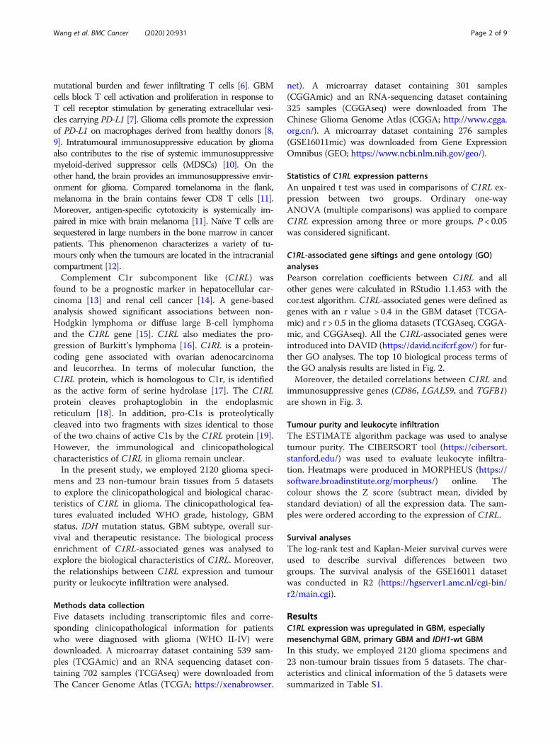
mutational burden and fewer infiltrating T cells [6]. GBMcells block T cell activation and proliferation in response toT cell receptor stimulation by generating extracellular vesi-cles carrying PD-L1 [7]. Glioma cells promote the expressionof PD-L1 on macrophages derived from healthy donors [8,9]. Intratumoural immunosuppressive education by gliomaalso contributes to the rise of systemic immunosuppressivemyeloid-derived suppressor cells (MDSCs) [10]. On theother hand, the brain provides an immunosuppressive envir-onment for glioma. Compared tomelanoma in the flank,melanoma in the brain contains fewer CD8 T cells [11].Moreover, antigen-specific cytotoxicity is systemically im-paired in mice with brain melanoma [11]. Naïve T cells aresequestered in large numbers in the bone marrow in cancerpatients. This phenomenon characterizes a variety of tu-mours only when the tumours are located in the intracranialcompartment [12].Complement C1r subcomponent like (C1RL) was
found to be a prognostic marker in hepatocellular car-cinoma [13] and renal cell cancer [14]. A gene-basedanalysis showed significant associations between non-Hodgkin lymphoma or diffuse large B-cell lymphomaand the C1RL gene [15]. C1RL also mediates the pro-gression of Burkitt’s lymphoma [16]. C1RL is a protein-coding gene associated with ovarian adenocarcinomaand leucorrhea. In terms of molecular function, theC1RL protein, which is homologous to C1r, is identifiedas the active form of serine hydrolase [17]. The C1RLprotein cleaves prohaptoglobin in the endoplasmicreticulum [18]. In addition, pro-C1s is proteolyticallycleaved into two fragments with sizes identical to thoseof the two chains of active C1s by the C1RL protein [19].However, the immunological and clinicopathologicalcharacteristics of C1RL in glioma remain unclear.In the present study, we employed 2120 glioma speci-
mens and 23 non-tumour brain tissues from 5 datasetsto explore the clinicopathological and biological charac-teristics of C1RL in glioma. The clinicopathological fea-tures evaluated included WHO grade, histology, GBMstatus, IDH mutation status, GBM subtype, overall sur-vival and therapeutic resistance. The biological processenrichment of C1RL-associated genes was analysed toexplore the biological characteristics of C1RL. Moreover,the relationships between C1RL expression and tumourpurity or leukocyte infiltration were analysed.
Methods data collectionFive datasets including transcriptomic files and corre-sponding clinicopathological information for patientswho were diagnosed with glioma (WHO II-IV) weredownloaded. A microarray dataset containing 539 sam-ples (TCGAmic) and an RNA sequencing dataset con-taining 702 samples (TCGAseq) were downloaded fromThe Cancer Genome Atlas (TCGA; https://xenabrowser.
net). A microarray dataset containing 301 samples(CGGAmic) and an RNA-sequencing dataset containing325 samples (CGGAseq) were downloaded from TheChinese Glioma Genome Atlas (CGGA; http://www.cgga.org.cn/). A microarray dataset containing 276 samples(GSE16011mic) was downloaded from Gene ExpressionOmnibus (GEO; https://www.ncbi.nlm.nih.gov/geo/).
Statistics of C1RL expression patternsAn unpaired t test was used in comparisons of C1RL ex-pression between two groups. Ordinary one-wayANOVA (multiple comparisons) was applied to compareC1RL expression among three or more groups. P < 0.05was considered significant.
C1RL-associated gene siftings and gene ontology (GO)analysesPearson correlation coefficients between C1RL and allother genes were calculated in RStudio 1.1.453 with thecor.test algorithm. C1RL-associated genes were defined asgenes with an r value > 0.4 in the GBM dataset (TCGA-mic) and r > 0.5 in the glioma datasets (TCGAseq, CGGA-mic, and CGGAseq). All the C1RL-associated genes wereintroduced into DAVID (https://david.ncifcrf.gov/) for fur-ther GO analyses. The top 10 biological process terms ofthe GO analysis results are listed in Fig. 2.Moreover, the detailed correlations between C1RL and
immunosuppressive genes (CD86, LGALS9, and TGFB1)are shown in Fig. 3.
Tumour purity and leukocyte infiltrationThe ESTIMATE algorithm package was used to analysetumour purity. The CIBERSORT tool (https://cibersort.stanford.edu/) was used to evaluate leukocyte infiltra-tion. Heatmaps were produced in MORPHEUS (https://software.broadinstitute.org/morpheus/) online. Thecolour shows the Z score (subtract mean, divided bystandard deviation) of all the expression data. The sam-ples were ordered according to the expression of C1RL.
Survival analysesThe log-rank test and Kaplan-Meier survival curves wereused to describe survival differences between twogroups. The survival analysis of the GSE16011 datasetwas conducted in R2 (https://hgserver1.amc.nl/cgi-bin/r2/main.cgi).
ResultsC1RL expression was upregulated in GBM, especiallymesenchymal GBM, primary GBM and IDH1-wt GBMIn this study, we employed 2120 glioma specimens and23 non-tumour brain tissues from 5 datasets. The char-acteristics and clinical information of the 5 datasets weresummarized in Table S1.
Wang et al. BMC Cancer (2020) 20:931 Page 2 of 9
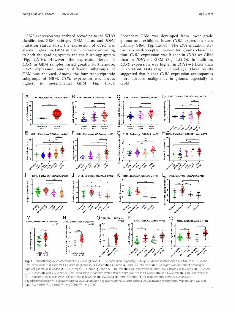
C1RL expression was analysed according to the WHOclassification, GBM subtype, GBM status and IDH1mutation status. First, the expression of C1RL wasalways highest in GBM in the 5 datasets accordingto both the grading system and the histology system(Fig. 1.A-H). However, the expression levels ofC1RL in GBM samples varied greatly. Furthermore,C1RL expression among different subgroups ofGBM was analysed. Among the four transcriptomicsubgroups of GBM, C1RL expression was alwayshighest in mesenchymal GBM (Fig. 1.I-L).
Secondary GBM was developed from lower gradeglioma and exhibited lower C1RL expression thanprimary GBM (Fig. 1.M-N). The IDH mutation sta-tus is a well-accepted marker for glioma classifica-tion. C1RL expression was higher in IDH1-wt GBMthan in IDH1-mt GBM (Fig. 1.O-Q). In addition,C1RL expression was higher in IDH1-wt LGG thanin IDH1-mt LGG (Fig. 1 P and Q). These resultssuggested that higher C1RL expression accompaniesmore advaced malignancy in glioma, especially inGBM.
Fig. 1 Histopathological characteristics of C1RL in glioma. a. C1RL expression in primary GBM (p-GBM) and nontumour brain tissues in TCGAmic.C1RL expression in distinct WHO grades of glioma in CGGAseq (b), CGGAmic (c), and GSE16011mic (d). C1RL expression in distinct histologicaltypes of glioma in TCGAseq (e), CGGAseq (f), CGGAmic (g), and GSE16011mic (h). C1RL expression in four GBM subtypes in TCGAmic (i), TCGAseq(j), CGGAseq (k), and CGGAmic (l). C1RL expression in samples with different GBM statuses in CGGAseq (m) and CGGAmic (n). C1RL expression inIDH1-mutant or IDH1-wild-type LGG or GBM in TCGAmic (o), CGGAseq (p), and CGGAmic (q). O, oligodendroglioma; AO, anaplasticoligodendroglioma; OA, oligoastrocytoma; AOA, anaplastic oligoastrocytoma; A, astrocytoma; AA, anaplastic astrocytoma. Mut, mutant; wt, wild-type. *, p < 0.05; **, p < 0.01; ***, p < 0.001; ****, p < 0.0001
Wang et al. BMC Cancer (2020) 20:931 Page 3 of 9
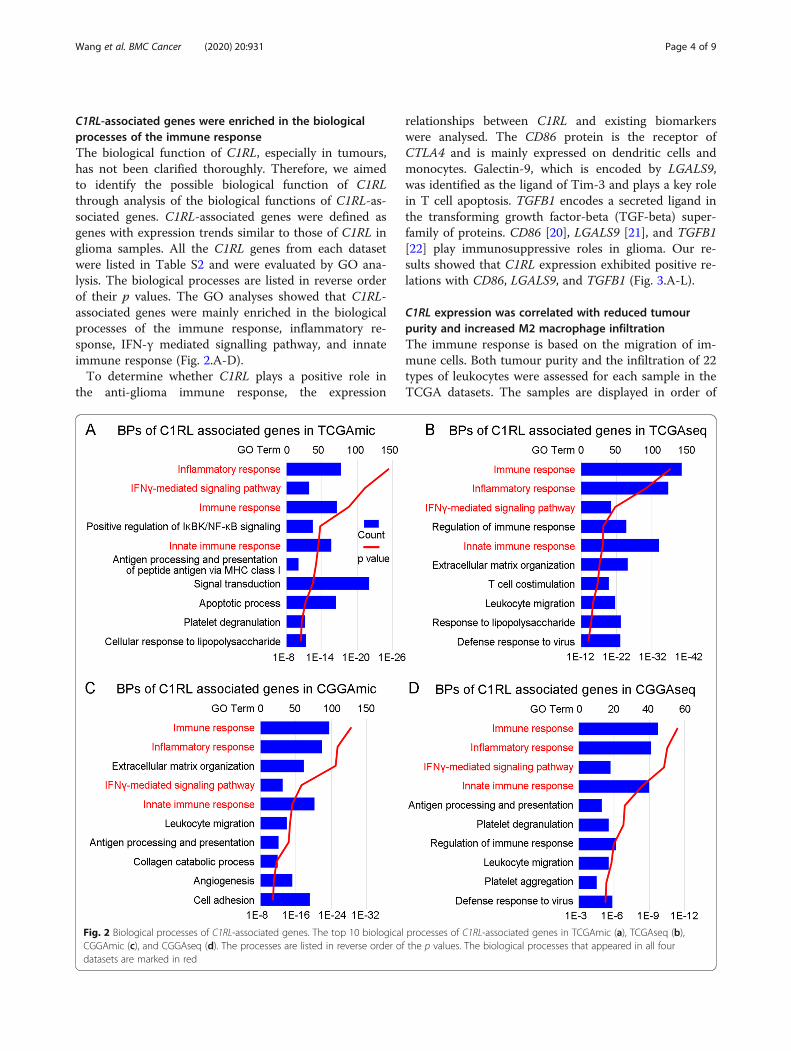
C1RL-associated genes were enriched in the biologicalprocesses of the immune responseThe biological function of C1RL, especially in tumours,has not been clarified thoroughly. Therefore, we aimedto identify the possible biological function of C1RLthrough analysis of the biological functions of C1RL-as-sociated genes. C1RL-associated genes were defined asgenes with expression trends similar to those of C1RL inglioma samples. All the C1RL genes from each datasetwere listed in Table S2 and were evaluated by GO ana-lysis. The biological processes are listed in reverse orderof their p values. The GO analyses showed that C1RL-associated genes were mainly enriched in the biologicalprocesses of the immune response, inflammatory re-sponse, IFN-γ mediated signalling pathway, and innateimmune response (Fig. 2.A-D).To determine whether C1RL plays a positive role in
the anti-glioma immune response, the expression
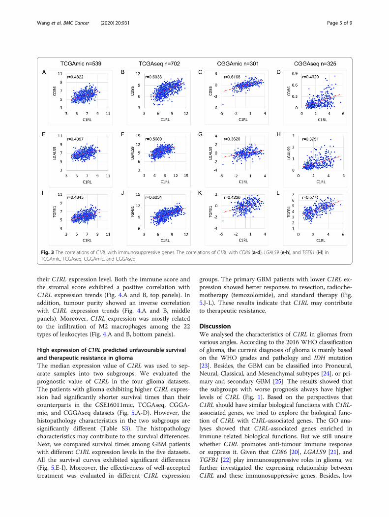
relationships between C1RL and existing biomarkerswere analysed. The CD86 protein is the receptor ofCTLA4 and is mainly expressed on dendritic cells andmonocytes. Galectin-9, which is encoded by LGALS9,was identified as the ligand of Tim-3 and plays a key rolein T cell apoptosis. TGFB1 encodes a secreted ligand inthe transforming growth factor-beta (TGF-beta) super-family of proteins. CD86 [20], LGALS9 [21], and TGFB1[22] play immunosuppressive roles in glioma. Our re-sults showed that C1RL expression exhibited positive re-lations with CD86, LGALS9, and TGFB1 (Fig. 3.A-L).
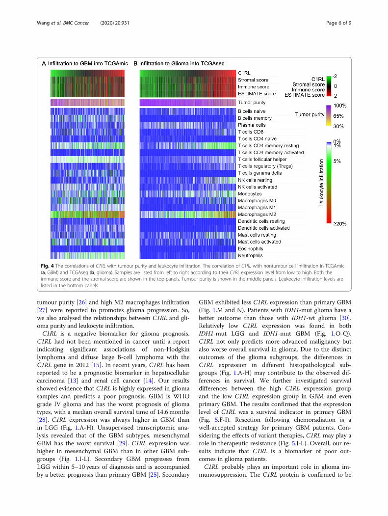
C1RL expression was correlated with reduced tumourpurity and increased M2 macrophage infiltrationThe immune response is based on the migration of im-mune cells. Both tumour purity and the infiltration of 22types of leukocytes were assessed for each sample in theTCGA datasets. The samples are displayed in order of
Fig. 2 Biological processes of C1RL-associated genes. The top 10 biological processes of C1RL-associated genes in TCGAmic (a), TCGAseq (b),CGGAmic (c), and CGGAseq (d). The processes are listed in reverse order of the p values. The biological processes that appeared in all fourdatasets are marked in red
Wang et al. BMC Cancer (2020) 20:931 Page 4 of 9
their C1RL expression level. Both the immune score andthe stromal score exhibited a positive correlation withC1RL expression trends (Fig. 4.A and B, top panels). Inaddition, tumour purity showed an inverse correlationwith C1RL expression trends (Fig. 4.A and B, middlepanels). Moreover, C1RL expression was mostly relatedto the infiltration of M2 macrophages among the 22types of leukocytes (Fig. 4.A and B, bottom panels).
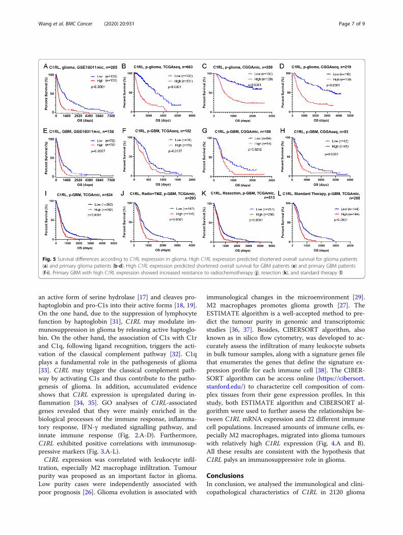
High expression of C1RL predicted unfavourable survivaland therapeutic resistance in gliomaThe median expression value of C1RL was used to sep-arate samples into two subgroups. We evaluated theprognostic value of C1RL in the four glioma datasets.The patients with glioma exhibiting higher C1RL expres-sion had significantly shorter survival times than theircounterparts in the GSE16011mic, TCGAseq, CGGA-mic, and CGGAseq datasets (Fig. 5.A-D). However, thehistopathology characteristics in the two subgroups aresignificantly different (Table S3). The histopathologycharacteristics may contribute to the survival differences.Next, we compared survival times among GBM patientswith different C1RL expression levels in the five datasets.All the survival curves exhibited significant differences(Fig. 5.E-I). Moreover, the effectiveness of well-acceptedtreatment was evaluated in different C1RL expression
groups. The primary GBM patients with lower C1RL ex-pression showed better responses to resection, radioche-motherapy (temozolomide), and standard therapy (Fig.5.J-L). These results indicate that C1RL may contributeto therapeutic resistance.
DiscussionWe analysed the characteristics of C1RL in gliomas fromvarious angles. According to the 2016 WHO classificationof glioma, the current diagnosis of glioma is mainly basedon the WHO grades and pathology and IDH mutation[23]. Besides, the GBM can be classified into Proneural,Neural, Classical, and Mesenchymal subtypes [24], or pri-mary and secondary GBM [25]. The results showed thatthe subgroups with worse prognosis always have higherlevels of C1RL (Fig. 1). Based on the perspectives thatC1RL should have similar biological functions with C1RL-associated genes, we tried to explore the biological func-tion of C1RL with C1RL-associated genes. The GO ana-lyses showed that C1RL-associated genes enriched inimmune related biological functions. But we still unsurewhether C1RL promotes anti-tumour immune responseor suppress it. Given that CD86 [20], LGALS9 [21], andTGFB1 [22] play immunosuppressive roles in glioma, wefurther investigated the expressing relationship betweenC1RL and these immunosuppressive genes. Besides, low
Fig. 3 The correlations of C1RL with immunosuppressive genes. The correlations of C1RL with CD86 (a-d), LGALS9 (e-h), and TGFB1 (i-l) inTCGAmic, TCGAseq, CGGAmic, and CGGAseq
Wang et al. BMC Cancer (2020) 20:931 Page 5 of 9
tumour purity [26] and high M2 macrophages infiltration[27] were reported to promotes glioma progression. So,we also analysed the relationships between C1RL and gli-oma purity and leukocyte infiltration.C1RL is a negative biomarker for glioma prognosis.
C1RL had not been mentioned in cancer until a reportindicating significant associations of non-Hodgkinlymphoma and diffuse large B-cell lymphoma with theC1RL gene in 2012 [15]. In recent years, C1RL has beenreported to be a prognostic biomarker in hepatocellularcarcinoma [13] and renal cell cancer [14]. Our resultsshowed evidence that C1RL is highly expressed in gliomasamples and predicts a poor prognosis. GBM is WHOgrade IV glioma and has the worst prognosis of gliomatypes, with a median overall survival time of 14.6 months[28]. C1RL expression was always higher in GBM thanin LGG (Fig. 1.A-H). Unsupervised transcriptomic ana-lysis revealed that of the GBM subtypes, mesenchymalGBM has the worst survival [29]. C1RL expression washigher in mesenchymal GBM than in other GBM sub-groups (Fig. 1.I-L). Secondary GBM progresses fromLGG within 5–10 years of diagnosis and is accompaniedby a better prognosis than primary GBM [25]. Secondary
GBM exhibited less C1RL expression than primary GBM(Fig. 1.M and N). Patients with IDH1-mut glioma have abetter outcome than those with IDH1-wt glioma [30].Relatively low C1RL expression was found in bothIDH1-mut LGG and IDH1-mut GBM (Fig. 1.O-Q).C1RL not only predicts more advanced malignancy butalso worse overall survival in glioma. Due to the distinctoutcomes of the glioma subgroups, the differences inC1RL expression in different histopathological sub-groups (Fig. 1.A-H) may contribute to the observed dif-ferences in survival. We further investigated survivaldifferences between the high C1RL expression groupand the low C1RL expression group in GBM and evenprimary GBM. The results confirmed that the expressionlevel of C1RL was a survival indicator in primary GBM(Fig. 5.F-I). Resection following chemoradiation is awell-accepted strategy for primary GBM patients. Con-sidering the effects of variant therapies, C1RL may play arole in therapeutic resistance (Fig. 5.J-L). Overall, our re-sults indicate that C1RL is a biomarker of poor out-comes in glioma patients.C1RL probably plays an important role in glioma im-
munosuppression. The C1RL protein is confirmed to be
Fig. 4 The correlations of C1RL with tumour purity and leukocyte infiltration. The correlation of C1RL with nontumour cell infiltration in TCGAmic(a, GBM) and TCGAseq (b, glioma). Samples are listed from left to right according to their C1RL expression level from low to high. Both theimmune score and the stromal score are shown in the top panels. Tumour purity is shown in the middle panels. Leukocyte infiltration levels arelisted in the bottom panels
Wang et al. BMC Cancer (2020) 20:931 Page 6 of 9
an active form of serine hydrolase [17] and cleaves pro-haptoglobin and pro-C1s into their active forms [18, 19].On the one hand, due to the suppression of lymphocytefunction by haptoglobin [31], C1RL may modulate im-munosuppression in glioma by releasing active haptoglo-bin. On the other hand, the association of C1s with C1rand C1q, following ligand recognition, triggers the acti-vation of the classical complement pathway [32]. C1qplays a fundamental role in the pathogenesis of glioma[33]. C1RL may trigger the classical complement path-way by activating C1s and thus contribute to the patho-genesis of glioma. In addition, accumulated evidenceshows that C1RL expression is upregulated during in-flammation [34, 35]. GO analyses of C1RL-associatedgenes revealed that they were mainly enriched in thebiological processes of the immune response, inflamma-tory response, IFN-γ mediated signalling pathway, andinnate immune response (Fig. 2.A-D). Furthermore,C1RL exhibited positive correlations with immunosup-pressive markers (Fig. 3.A-L).C1RL expression was correlated with leukocyte infil-
tration, especially M2 macrophage infiltration. Tumourpurity was proposed as an important factor in glioma.Low purity cases were independently associated withpoor prognosis [26]. Glioma evolution is associated with
immunological changes in the microenvironment [29].M2 macrophages promotes glioma growth [27]. TheESTIMATE algorithm is a well-accepted method to pre-dict the tumour purity in genomic and transcriptomicstudies [36, 37]. Besides, CIBERSORT algorithm, alsoknown as in silico flow cytometry, was developed to ac-curately assess the infiltration of many leukocyte subsetsin bulk tumour samples, along with a signature genes filethat enumerates the genes that define the signature ex-pression profile for each immune cell [38]. The CIBER-SORT algorithm can be access online (https://cibersort.stanford.edu/) to characterize cell composition of com-plex tissues from their gene expression profiles. In thisstudy, both ESTIMATE algorithm and CIBERSORT al-gorithm were used to further assess the relationships be-tween C1RL mRNA expression and 22 different immunecell populations. Increased amounts of immune cells, es-pecially M2 macrophages, migrated into glioma tumourswith relatively high C1RL expression (Fig. 4.A and B).All these results are consistent with the hypothesis thatC1RL palys an immunosuppressive role in glioma.
ConclusionsIn conclusion, we analysed the immunological and clini-copathological characteristics of C1RL in 2120 glioma
Fig. 5 Survival differences according to C1RL expression in glioma. High C1RL expression predicted shortened overall survival for glioma patients(a) and primary glioma patients (b-d). High C1RL expression predicted shortened overall survival for GBM patients (e) and primary GBM patients(f-i). Primary GBM with high C1RL expression showed increased resistance to radiochemotherapy (j), resection (k), and standard therapy (l)
Wang et al. BMC Cancer (2020) 20:931 Page 7 of 9
patients from five datasets. The results indicate thatC1RL is a negative biomarker for the patients with gli-oma. Furthermore, C1RL probably plays an immunosup-pressive role in the pathogenesis of glioma by triggeringthe activation of haptoglobin and C1s.
Supplementary informationSupplementary information accompanies this paper at https://doi.org/10.1186/s12885-020-07436-6.
Additional file 1 Table S1. Clinical information of 2143 patients fromthe different datasets.
Additional file 2 Table S2. C1RL associated genes.
Additional file 3 Table S3. C1RL and WHO grade and gliomahistopathology.
AbbreviationsC1RL: Complement C1r subcomponent like; CGGA: The Chinese GliomaGenome Atlas; FDA: The Food and Drug Administration; GBM: Glioblastoma;GO: Gene Ontology; LGG: Lower grade glioma; MDSCs: Myeloid-derivedsuppressor cells; TCGA: The Cancer Genome Atlas; TGF-beta: Thetransforming growth factor-beta; TTF: Tumour treating fields
AcknowledgementsNot Applicable.
Authors’ contributionsXY and JW participated the design of this study. JW and LT performed thestatistical analysis and drafted the manuscript. GL and HW carried out dataacquisition. LZ edited the tables, the figures, and the manuscript. All authorshave read and approved the final manuscript.
FundingThis work was supported by grants from the Beijing-Tianjin-Hebei Basic Re-search Cooperation Project (No. 19JCZDJC64200) and the State ScholarshipFund from China Scholarship Council (No. 201806940031).
Availability of data and materialsThe microarray dataset of 539 samples (TCGAmic) and the RNA sequencingdataset of 702 samples (TCGAseq) were downloaded from The CancerGenome Atlas (TCGA, https://xenabrowser.net). The microarray dataset of 301samples (CGGAmic) and the RNA sequencing dataset of 325 samples(CGGAseq) were downloaded from The Chinese Glioma Genome Atlas(CGGA, http://www.cgga.org.cn/). The microarray dataset of 276 samples(GSE16011mic) was downloaded from Gene Expression Omnibus (GEO,https://www.ncbi.nlm.nih.gov/geo/). The survival analysis of GSE16011dataset was conducted in R2 (https://hgserver1.amc.nl/cgi-bin/r2/main.cgi).
Ethics approval and consent to participateThis study was approved by the Ethics Committee of Tianjin MedicalUniversity General Hospital.
Consent for publicationInformed consent was obtained from all participants for publication.
Competing interestsThe authors declare no competing interests.
Author details1Department of Neurosurgery, The First People’s Hospital of Wenling,Wenling 317500, China. 2Department of Neurosurgery, Tianjin MedicalUniversity General Hospital, Tianjin 300052, China. 3Department ofNeurosurgery, The First Affiliated Hospital of Medical School of ZhejiangUniversity, Hangzhou 310003, China. 4Department of Neurosurgery, TheJohns Hopkins University School of Medicine, Baltimore 21287, USA.
Received: 29 April 2020 Accepted: 17 September 2020
References1. Filley AC, Henriquez M, Dey M. Recurrent glioma clinical trial, CheckMate-
143: the game is not over yet. Oncotarget. 2017;8:91779–94.2. Wick W, Gorlia T, Bendszus M, Taphoorn M, Sahm F, Harting I, Brandes AA,
Taal W, Domont J, Idbaih A, Campone M, Clement PM, Stupp R, Fabbro M,Le RE, Dubois F, Weller M. von DA, Golfinopoulos V, Bromberg JC, PlattenM, Klein M, van den Bent MJ. Lomustine and Bevacizumab in ProgressiveGlioblastoma. N Engl J Med. 2017;377:1954–63.
3. Gilbert MR, Dignam JJ, Armstrong TS, Wefel JS, Blumenthal DT, Vogelbaum MA,Colman H, Chakravarti A, Pugh S, Won M, Jeraj R, Brown PD, Jaeckle KA, Schiff D,Stieber VW, Brachman DG, Werner-Wasik M, Tremont-Lukats IW, Sulman EP, AldapeKD, Curran WJ, Mehta MP. A randomized trial of bevacizumab for newly diagnosedglioblastoma. N Engl J Med. 2014;370:699–708.
4. Stupp R, Taillibert S, Kanner A, Read W, Steinberg D, Lhermitte B, Toms S,Idbaih A, Ahluwalia MS, Fink K, Di MF, Lieberman F, Zhu JJ, Stragliotto G,Tran D, Brem S, Hottinger A, Kirson ED, Lavy-Shahaf G, Weinberg U, Kim CY,Paek SH, Nicholas G, Bruna J, Hirte H, Weller M, Palti Y, Hegi ME, Ram Z.Effect of tumor-treating fields plus maintenance Temozolomide vsmaintenance Temozolomide alone on survival in patients withGlioblastoma: a randomized clinical trial. JAMA. 2017;318:2306–16.
5. Palmer JD, Bhamidipati D, Mehta M, Williams NL, Dicker AP, Werner-WasikM, Shi W. Treatment recommendations for elderly patients with newlydiagnosed glioblastoma lack worldwide consensus. J Neuro-Oncol. 2018;140:421–6.
6. Li B, Severson E, Pignon JC, Zhao H, Li T, Novak J, Jiang P, Shen H, Aster JC,Rodig S, Signoretti S, Liu JS, Liu XS. Comprehensive analyses of tumorimmunity: implications for cancer immunotherapy. Genome Biol. 2016;17:174.
7. Ricklefs FL, Alayo Q, Krenzlin H, Mahmoud AB, Speranza MC, Nakashima H,Hayes JL, Lee K, Balaj L, Passaro C, Rooj AK, Krasemann S, Carter BS, ChenCC, Steed T, Treiber J, Rodig S, Yang K, Nakano I, Lee H, Weissleder R,Breakefield XO, Godlewski J, Westphal M, Lamszus K, Freeman GJ, Bronisz A,Lawler SE, Chiocca EA. Immune evasion mediated by PD-L1 onglioblastoma-derived extracellular vesicles. Sci Adv. 2018;4:eaar2766.
8. Tong L, Li J, Choi J, Pant A, Xia Y, Jackson C, Liu P, Yi L, Boussouf E, Lim M,Yang X. CLEC5A expressed on myeloid cells as a M2 biomarker relates toimmunosuppression and decreased survival in patients with glioma. CancerGene Ther. 2019.
9. Bloch O, Crane CA, Kaur R, Safaee M, Rutkowski MJ, Parsa AT. Gliomaspromote immunosuppression through induction of B7-H1 expression intumor-associated macrophages. Clin Cancer Res. 2013;19:3165–75.
10. Chae M, Peterson TE, Balgeman A, Chen S, Zhang L, Renner DN, JohnsonAJ, Parney IF. Increasing glioma-associated monocytes leads to increasedintratumoral and systemic myeloid-derived suppressor cells in a murinemodel. Neuro-Oncology. 2015;17:978–91.
11. Jackson CM, Kochel CM, Nirschl CJ, Durham NM, Ruzevick J, Alme A,Francica BJ, Elias J, Daniels A, Dubensky TW, Lauer P, Brockstedt DG, Baxi EG,Calabresi PA, Taube JM, Pardo CA, Brem H, Pardoll DM, Lim M, Drake CG.Systemic tolerance mediated by melanoma brain tumors is reversible byradiotherapy and vaccination. Clin Cancer Res. 2016;22:1161–72.
12. Chongsathidkiet P, Jackson C, Koyama S, Loebel F, Cui X, Farber SH,Woroniecka K, Elsamadicy AA, Dechant CA, Kemeny HR, Sanchez-Perez L,Cheema TA, Souders NC, Herndon JE, Coumans JV, Everitt JI, Nahed BV,Sampson JH, Gunn MD, Martuza RL, Dranoff G, Curry WT, Fecci PE.Sequestration of T cells in bone marrow in the setting of glioblastoma andother intracranial tumors. Nat Med. 2018;24:1459–68.
13. Xu B, Lv W, Li X, Zhang L, Lin J. Prognostic genes of hepatocellular carcinomabased on gene coexpression network analysis. J Cell Biochem. 2019.
14. Chinello C, Cazzaniga M, De Sio G, Smith AJ, Grasso A, Rocco B, Signorini S,Grasso M, Bosari S, Zoppis I, Mauri G, Magni F. Tumor size, stage and gradealterations of urinary peptidome in RCC. J Transl Med. 2015;13:332.
15. Bassig BA, Zheng T, Zhang Y, Berndt SI, Holford TR, Hosgood HD, Hu W,Leaderer B, Yeager M, Menashe I, Boyle P, Xu J, Zou K, Zhu Y, Chanock S,Rothman N, Lan Q. Polymorphisms in complement system genes and riskof non-Hodgkin lymphoma. Environ Mol Mutagen. 2012;53:145–51.
16. Han B, Wang S, Zhao H. MicroRNA-21 and microRNA-155 promote theprogression of Burkitt's lymphoma by the PI3K/AKT signaling pathway. Int JClin Exp Pathol. 2020;13:89–98.
Wang et al. BMC Cancer (2020) 20:931 Page 8 of 9
17. Navarrete M, Ho J, Krokhin O, Ezzati P, Rigatto C, Reslerova M, Rush DN,Nickerson P, Wilkins JA. Proteomic characterization of serine hydrolaseactivity and composition in normal urine. Clin Proteomics. 2013;10:17.
18. Wicher KB, Fries E. Prohaptoglobin is proteolytically cleaved in theendoplasmic reticulum by the complement C1r-like protein. Proc Natl AcadSci U S A. 2004;101:14390–5.
19. Ligoudistianou C, Xu Y, Garnier G, Circolo A, Volanakis JE. A novel humancomplement-related protein, C1r-like protease (C1r-LP), specifically cleavespro-C1s. Biochem J. 2005;387:165–73.
20. Tumangelova-Yuzeir K, Naydenov E, Ivanova-Todorova E, Krasimirova E,Vasilev G, Nachev S, Kyurkchiev D. Mesenchymal stem cells derived andcultured from Glioblastoma Multiforme increase Tregs, Downregulate Th17,and induce the Tolerogenic phenotype of monocyte-derived cells. StemCells Int. 2019;2019:6904638.
21. Yuan F, Ming H, Wang Y, Yang Y, Yi L, Li T, Ma H, Tong L, Zhang L, Liu P, Li J,Lin Y, Yu S, Ren B, Yang X. Molecular and clinical characterization of Galectin-9in glioma through 1,027 samples. J Cell Physiol. 2020;235:4326–34.
22. Han J, Chen X, Chu J, Xu B, Meisen WH, Chen L, Zhang L, Zhang J, He X,Wang QE, Chiocca EA, Kaur B, Caligiuri MA, Yu J. TGFβ treatment enhancesGlioblastoma Virotherapy by inhibiting the innate immune response. CancerRes. 2015;75:5273–82.
23. Louis DN, Perry A, Reifenberger G, von DA F-BD, Cavenee WK, Ohgaki H,Wiestler OD, Kleihues P, Ellison DW. The 2016 World Health Organizationclassification of tumors of the central nervous system: a summary. ActaNeuropathol. 2016;131:803–20.
24. Verhaak RG, Hoadley KA, Purdom E, Wang V, Qi Y, Wilkerson MD, Miller CR,Ding L, Golub T, Mesirov JP, Alexe G, Lawrence M, O'Kelly M, Tamayo P, WeirBA, Gabriel S, Winckler W, Gupta S, Jakkula L, Feiler HS, Hodgson JG, James CD,Sarkaria JN, Brennan C, Kahn A, Spellman PT, Wilson RK, Speed TP, Gray JW,Meyerson M, Getz G, Perou CM, Hayes DN. Integrated genomic analysisidentifies clinically relevant subtypes of glioblastoma characterized byabnormalities in PDGFRA, IDH1, EGFR, and NF1. Cancer Cell. 2010;17:98–110.
25. Hu H, Mu Q, Bao Z, Chen Y, Liu Y, Chen J, Wang K, Wang Z, Nam Y, Jiang B,Sa JK, Cho HJ, Her NG, Zhang C, Zhao Z, Zhang Y, Zeng F, Wu F, Kang X,Liu Y, Qian Z, Wang Z, Huang R, Wang Q, Zhang W, Qiu X, Li W, Nam DH,Fan X, Wang J, Jiang T. Mutational Landscape of Secondary GlioblastomaGuides MET-Targeted Trial in Brain Tumor. Cell. 2018;175:1665–78 e18.
26. Zhang C, Cheng W, Ren X, Wang Z, Liu X, Li G, Han S, Jiang T, Wu A. Tumorpurity as an underlying key factor in Glioma. Clin Cancer Res. 2017;23:6279–91.
27. Zhou W, Ke SQ, Huang Z, Flavahan W, Fang X, Paul J, Wu L, Sloan AE,McLendon RE, Li X, Rich JN, Bao S. Periostin secreted by glioblastoma stemcells recruits M2 tumour-associated macrophages and promotes malignantgrowth. Nat Cell Biol. 2015;17:170–82.
28. Stupp R, Mason WP, van den Bent MJ, Weller M, Fisher B, Taphoorn MJ,Belanger K, Brandes AA, Marosi C, Bogdahn U, Curschmann J, Janzer RC,Ludwin SK, Gorlia T, Allgeier A, Lacombe D, Cairncross JG, Eisenhauer E,Mirimanoff RO. Radiotherapy plus concomitant and adjuvant temozolomidefor glioblastoma. N Engl J Med. 2005;352:987–96.
29. Wang Q, Hu B, Hu X, Kim H, Squatrito M, Scarpace L, de Carvalho AC, Lyu S,Li P, Li Y, Barthel F, Cho HJ, Lin YH, Satani N, Martinez-Ledesma E, Zheng S,Chang E, SCE G, Olar A, Lan ZD, Finocchiaro G, Phillips JJ, Berger MS,Gabrusiewicz KR, Wang G, Eskilsson E, Hu J, Mikkelsen T, RA DP, Muller F,Heimberger AB, Sulman EP, Nam DH, RGW V. Tumor Evolution of Glioma-Intrinsic Gene Expression Subtypes Associates with Immunological Changesin the Microenvironment. Cancer Cell. 2018;33:152.
30. Yan H, Parsons DW, Jin G, McLendon R, Rasheed BA, Yuan W, Kos I, Batinic-Haberle I, Jones S, Riggins GJ, Friedman H, Friedman A, Reardon D, HerndonJ, Kinzler KW, Velculescu VE, Vogelstein B, Bigner DD. IDH1 and IDH2mutations in gliomas. N Engl J Med. 2009;360:765–73.
31. Sadrzadeh SM, Bozorgmehr J. Haptoglobin phenotypes in health anddisorders. Am J Clin Pathol. 2004;121(Suppl):S97–104.
32. Lu J, Kishore U. C1 complex: an adaptable Proteolytic module forcomplement and non-complement functions. Front Immunol. 2017;8:592.
33. Mangogna A, Belmonte B, Agostinis C, Zacchi P, Iacopino DG, Martorana A,Rodolico V, Bonazza D, Zanconati F, Kishore U, Bulla R. Prognostic implicationsof the complement protein C1q in Gliomas. Front Immunol. 2019;10:2366.
34. Severino P, Ariga SK, Barbeiro HV, de Lima TM, de Paula SE, Barbeiro DF,MCC M, Nizet V, dSF P, et al. J Mol Med (Berl). 2017;95:995–1003.
35. Shi L, Zhu B, Xu M, Wang X. Selection of AECOPD-specificimmunomodulatory biomarkers by integrating genomics and proteomicswith clinical informatics. Cell Biol Toxicol. 2018;34:109–23.
36. Yoshihara K, Shahmoradgoli M, Martínez E, Vegesna R, Kim H, Torres-GarciaW, Treviño V, Shen H, Laird PW, Levine DA, Carter SL, Getz G, Stemke-Hale K,Mills GB, Verhaak RG. Inferring tumour purity and stromal and immune celladmixture from expression data. Nat Commun. 2013;4:2612.
37. Cao JY, Guo Q, Guan GF, Zhu C, Zou CY, Zhang LY, Cheng W, Wang GL,Cheng P, Wu AH, Li GY. Elevated lymphocyte specific protein 1 expressionis involved in the regulation of leukocyte migration andimmunosuppressive microenvironment in glioblastoma. Aging (Albany NY).2020;12:1656–84.
38. Newman AM, Liu CL, Green MR, Gentles AJ, Feng W, Xu Y, Hoang CD,Diehn M, Alizadeh AA. Robust enumeration of cell subsets from tissueexpression profiles. Nat Methods. 2015;12:453–7.
Publisher’s NoteSpringer Nature remains neutral with regard to jurisdictional claims inpublished maps and institutional affiliations.
Wang et al. BMC Cancer (2020) 20:931 Page 9 of 9