Embed Size (px)
Citation preview
Harshal et al. European Journal of Biomedical and Pharmaceutical Sciences
www.ejbps.com 446
CLINICOPATHOLOGICAL STUDY AND MANAGEMENT OF PRIMARY HEPATIC
SPACE OCCUPYING LESIONS
1Dr. Niranjan Jadhav, *
2Dr. Harshal Ramteke and
3Dr. Dhirendra Wagh
1Post Graduate in Department of General Surgery, AVBRH, Sawangi (Meghe), Wardha.
2,3Professor in Department of General Surgery, AVBRH, Sawangi (Meghe), Wardha.
Article Received on 16/06/2018 Article Revised on 06/072018 Article Accepted on 27/07/2018
INTRODUCTION
The liver confounds the surgeon’s dependence on
anatomy. With its smooth, soft, shiny exterior the liver
leaves a shadowly harmless first impression. Yet as those
well acquainted with its ways could assure, it is one of the most difficult organ a surgeon can handle.[1] Bismuth
wrote in 1982, ― The time when liver surgery was
confined to atypical hepatectomies or wedge resections,
according to location or volume of a lesion, belongs to
the past. At present, liver resections are based upon the
precise knowledge of natural lines of division of the liver
which define the anatomical surgery of the liver‖.[1]
Complete understanding of the surgical anatomy is essential for any practitioner who intervenes in the liver
and biliary tract, which ever be the mode of intervention.
The liver is the largest organ in the abdomen, weighing
SJIF Impact Factor 4.918 Research Article
ejbps, 2018, Volume 5, Issue 8, 446-464.
European Journal of Biomedical AND Pharmaceutical sciences
http://www.ejbps.com
ISSN 2349-8870
Volume: 5
Issue: 8
446-464
Year: 2018
*Corresponding Author: Dr. Harshal Ramteke
Professor in Department of General Surgery, AVBRH, Sawangi (Meghe), Wardha.
ABSTRACT The liver is the largest organ in the abdomen, weighing about 1800 g in men and slightly less in women. A space
occupying lesion by definition is a discrete abnormality arising within the liver. Space occupying lesions of the
liver can be classified into developmental, neoplastic, inflammatory and miscellaneous. Although in some cases, it
is difficult to distinguish these entities with imaging criteria alone, certain focal liver lesions have classic
ultrasonic, computed tomographic (CT) and magnetic resonance (MR) imaging feature. The majority of these
lesions are detected incidentally in asymptomatic patients.[2] [3] An accurate history and physical examination are
essential for the diagnosis and treatment of solid liver masses. For example, the use of oral contraceptives or
anabolic steroids might be related to hepatic adenoma (HA), alcohol use and occupational exposure are associated
with angiosarcoma and primary sclerosing cholangitis, liver fluke, Caroli’s disease, and choledochal cysts are
associat ed with cholangiocarcinoma.[4] Physical examination should look for liver tenderness, stigmata of chronic
liver disease, or general deterioration signs (fever, we ight loss). High alkaline phosphatase, high lactate dehydrogenase (LDH), low albumin, high prothrombin time, and iron overload are non-specific but might suggest
an underlying chronic hepatitis, cirrhosis or an infiltrative process.[4] Hence, this study mainly intends to know the
incidence, various modes of presentation, different modalities of diagnosis, treatment and prognosis in a rural
setup, studied to identify factors which can help in better management of these cases thus helping to improve the
prognosis and management care in primary hepatic space occupying lesions. Aim: To do the clinicopathological
study and management of primary hepatic space occupying lesions. Conclusion: This study was carried out at
Jawaharlal Nehru Medical College and Acharya Vinoba Bhave Rural Hospital, Sawangi(M), Wardha,
Maharashtra, between July 2015 TO October 2017. Out of 45 primary hepatic space occupying lesions in this study, most common lesion were liver abscess and hydatid cyst of liver and the most common neoplastic lesion
was hepatocelullar carcinoma. USG was the imaging modality of choice and was highly effective in diagnosis
with 100% sensitivity and specificity, although this is operator dependant. Percutaenous drainage proved to be the best treatment modality in case of liver abscess with supportive therapy and was associated with less
complications, which is suggestive that minimal invasive treatment is the need of the hour. Partial cystectomy
with ometoplasty was the treatment of choice in case of hydatid cyst of liver with better outcome. Management in case of malignant lesions such as hepatocellular carcinoma and intrahepatic cholangiocarcinoma who presented
in advanced stages was done by palliation and chemotherapy. Cholangitis is the most common complication in our study, which is associated to the direct consequence of the disease or due to the iatrogenic causes such as
instrumention during surgery or any image guided procedure.
KEYWORDS: Liver, primary hepatic space occupying lesions, hydatid cyst, cholangiocarcinoma, liver abscess.
Harshal et al. European Journal of Biomedical and Pharmaceutical Sciences
www.ejbps.com 447
about 1800 g in men and slightly less in women. A space
occupying lesion by definition is a discrete abnormality
arising within the liver. Space occupying lesions of the
liver can be classified into developmental, neoplastic,
inflammatory and miscellaneous. Although in some
cases, it is difficult to distinguish these entities with imaging criteria alone, certain focal liver lesions have
classic ultrasonic, computed tomographic (CT) and
magnetic resonance (MR) imaging feature. The majority
of these lesions are detected incidentally in
asymptomatic patients.[2,3] An accurate history and
physical examination are essential for the diagnosis and
treatment of solid liver masses. For example, the use of
oral contraceptives or anabolic steroids might be related
to hepatic adenoma (HA), alcohol use and occupational
exposure are associated with angiosarcoma and primary
sclerosing cholangitis, liver fluke, Caroli’s disease, and
choledochal cysts are associat ed with cholangiocarcinoma.[4] Physical examination should look
for liver tenderness, stigmata of chronic liver disease, or
general deterioration signs (fever, we ight loss). High
alkaline phosphatase, high lactate dehydrogenase (LDH),
low albumin, high prothrombin time, and iron overload
are non-specific but might suggest an underlying chronic
hepatitis, cirrhosis or an infiltrative process.[4] Liver
abscesses, both amebic and pyogenic, continue to be an
important cause of morbidity and mortality in tropical
countries. The primary mode of treatment of amebic liver
abscess is medical; however, as many as 15% of amebic abscesses may be refractory to medical therapy. Also,
secondary bacterial infection may complicat e 20% of
amebic liver abscesses. In recent years, imaging guided
percutaneous drainage has been increasingly used to treat
liver abscesses with reported success rates ranging from
70% to 100%.[5] Cystic diseases of Liver constitute a
large percentage of the liver space occupying lesion
Hydatid disease of the liver is still endemic in certain
parts of the world. The diagnosis of noncomplicated
hydatid cyst of the liver depends on clinical suspicion.
Ultrasonography and computed tomography, the most
important diagnostic tools, are helpful for determining the complications and planning treatment. The modern
treatment of hydatid cyst of the liver varies from surgical
intervention to percutaneous drainage or medical
therapy. Surgery is still the treatment of choice and can
be performed by the conventional or laparoscopic
approach. Percutaneous drainage and treatment of the
cyst with hypertonic saline or alcohol seems to be a good
alternative to surgery in selected cases. Currently, we
treat types I and II by ultrasound-guided percutaneous
drainage and types IV and V (excluding totally calcified
cysts) surgically. Type III cysts can be managed either way depending on the presence of drainable content.[6]
Modern imaging techniques have led to the recognition
of some incidental lesions that usually have no clinical
relevance, e.g. simple liver cysts, liver capillary
haemangiomas.. Simple hepatic cysts are a congenital
abnormality that are usually asymptomatic, but, as with
all lesions that may have either no clinical importance or
may be part of a defined condition or syndrome (e.g.
polycystic kidneys, von HippelLindau disease), the
prevalence and characteristics of these lesions within the
population should be known.[7] Hepatocellular carcinoma
(HCC) is the most frequent primary liver malignancy and
the third cause of cancer-related death in the Western
Countries. The well-established causes of HCC are chronic liver infections such as hepatitis B virus or
chronic hepatitis C virus, nonalcoholic fatty liver disease,
consumption of aflatoxins and tobacco smocking.
Clinical presentation varies widely; patients can be
asymptomatic while symptomatology extends from right
upper abdominal quadrant paint and weight loss to
obstructive jaundice and lethargy. Imaging is the first
key and one of the most important aspects at all stages of
diagnosis, therapy and followup of patients with HCC.
The Barcelona Clinic Liver Cancer Staging System
remains the most widely classification system used for
HCC management guidelines. Up until now, HCC remains a challenge to early diagnose, and treat
effectively; treating management is focused on hepatic
resection, orthotopic liver transplantation, ablative
therapies, chemoembolization and systemic therapies
with cytotocix drugs, and targeted agents.[8] Biliary tract
cancer accounts for Cholangiocarcinoma (intrahepatic,
hilar and distal) and gallbladder cancer, are distinguished
with different pathogenesis and prognosis. The treatment
is based on surgery, radiotherapy in selected cases, and
chemotherapy. The standard cytotoxic treatment for
advanced/metastatic disease is represented by the combination of gemcitabine and cisplatin, whereas
fluoropyrimidines are generally administered in second
line setting. At the present time, no biologic drug
demonstrated a clear efficacy in this cancer, although the
molecular characterisation could provide a promising
basis for experimental treatments. A good supportive c
are and an early palliative care are warranted in most
patients and should be delivered as a part of a global
approach.[9] Hence, this study mainly intends to know the
incidence, various modes of presentation, different
modalities of diagnosis, treatment and prognosis in a
rural setup, studied to identify factors which can help in better management of these cases thus helping to
improve the prognosis and management care in primary
hepatic space occupying lesions.
AIM: To do the clinicopathological study and
management of primary hepatic space occupying lesions.
OBJECTIVES
To study the clinical features and mode of presentation
of different types of primary hepatic SOL in different age
groups & sex.
To study different modalities of investigations available
to diagnose various types of primary hepatic SOL.
To study various modes of management including
complications, morbidity & mortality.
Harshal et al. European Journal of Biomedical and Pharmaceutical Sciences
www.ejbps.com 448
MATERIALS AND METHODS
This Present study was carried out at Acharya Vinoba
Bhave Rural Hospital (AVBRH) of Jawaharlal Nehru
Medical College between July 2015 to October 2017 in
which total 45 cases of primary hepatic space occupying
lesion which were studied prospectively. Type of Study: prospective study. Sample size : 45 cases. Sample Size
Formula: N= 2א p(1-p) + Np(1-p) C2(N-1) +p(1-p)
Where 2א is the chi-square value at 5% level of
significance =3.84 N=Total no of cases of primary
hepatic space occupying lesions in three years in
AVBRH =108 C= Desired confidence interval =0.05
P=Proportion =0.50 N= 3.84*(0.5*0.5) +108*0.5*0.5
=27.96 In present study N =45 total patients Inclusion
criteria: All patients more than 18 yrs having primary
hepatic space occupying lesions. Exclusion criteria:
Patients with suspected secondaries in liver Patient of
bleeding diasthesis. Patient on anticoagulant medication. Noncooperative patient who refuses to undergo the
study.
Mode of study
After detailed history, complete general and systemic
examinations, provisional diagnosis was done. Diagnosis
was confirmed with the help of various diagnostic
modalities like USG, CT scan & FNAC. USG was done
in all cases but CT scan & FNAC were done in selected
patients where the diagnosis was in doubt or more
detailed information about the pathology was needed. Along with various diagnostic investigations, patients
were subjected to routine investigations such as
haemogram, liver function test, PTINR, kidney function
test etc. to know the status of the disease, condition of
the patient & feasibility for surgery/procedure. After
complete evaluation, mode of management was selected
depending on extent of disease.
Statistical analysis
Statistical analysis was done by using descriptive and
inferential statistics using chisquare test and software used in the analysis were SPSS 22.0 version and
GraphPad Prism 6.0 version and p.
OBSERVATION AND RESULTS
Demographics
In present study, it was observed that the youngest
patient was 22 yrs who presented with amoebic liver
abscess & the oldest was of 86 yrs diagnosed as
pyogenic liver abscess & mean age of presenting PHSOL
was 49.22±15.69 yrs.
Liver abscess manifested most commonly in 7th Decade & mean age is 50.42 yrs. hydatid cyst of liver manifested
most commonly in 4th decade & mean age is 45.85 yrs.
hemangioma of liver manifested most commonly in 4th
& 5th decade & mean age is 43.20 yrs. hepatocelullar
carcinoma manifested most commonly in 7th decade &
mean age is 61.85 yrs. cholangiocarnima manifested
most commonly in 3 rd decade & mean age is 43.60 yrs.
out of 45 cases, there were 33 males & 12 females
showing male: female sex ratio of 2.75 :1.
In present study, liver abscess and hydatid cyst were more common in males.(92.86% & 71.43%). HCC was
found in 5(71.43%) males & 2(28.57%) females.
Intrahepatic cholangiocarcinoma was found in 3(60%)
males & 2(40%) females. Hemangioma was found in
2(40%) males & 3 (60%) females.
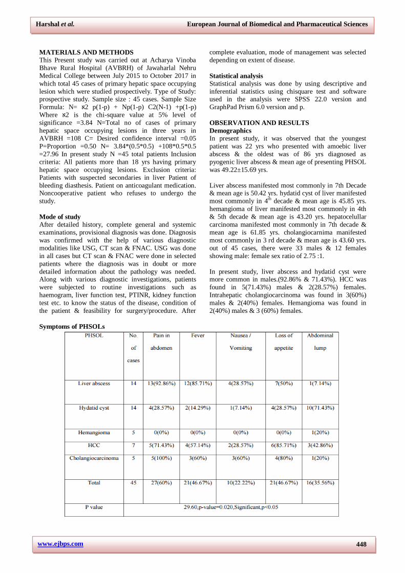
Symptoms of PHSOLs
Harshal et al. European Journal of Biomedical and Pharmaceutical Sciences
www.ejbps.com 449
Clinical Signs of PHSOLs
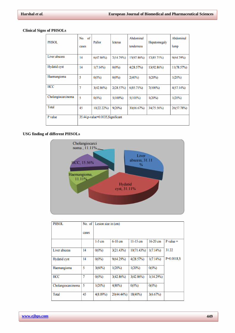
USG finding of different PHSOLs
Harshal et al. European Journal of Biomedical and Pharmaceutical Sciences
www.ejbps.com 450
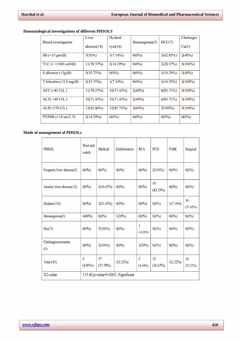
Haematological investigations of different PHSOLS
Mode of management of PHSOLs
Harshal et al. European Journal of Biomedical and Pharmaceutical Sciences
www.ejbps.com 451
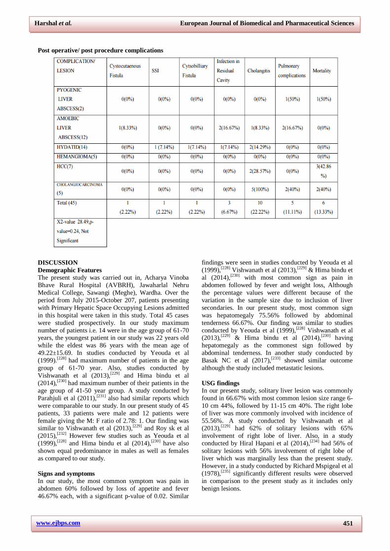
Post operative/ post procedure complications
DISCUSSION
Demographic Features
The present study was carried out in, Acharya Vinoba
Bhave Rural Hospital (AVBRH), Jawaharlal Nehru
Medical College, Sawangi (Meghe), Wardha. Over the
period from July 2015-October 207, patients presenting
with Primary Hepatic Space Occupying Lesions admitted
in this hospital were taken in this study. Total 45 cases
were studied prospectively. In our study maximum
number of patients i.e. 14 were in the age group of 61-70 years, the youngest patient in our study was 22 years old
while the eldest was 86 years with the mean age of
49.22±15.69. In studies conducted by Yeouda et al
(1999).[228] had maximum number of patients in the age
group of 61-70 year. Also, studies conducted by
Vishwanath et al (2013),[229] and Hima bindu et al
(2014),[230] had maximum number of their patients in the
age group of 41-50 year group. A study conducted by
Parahjuli et al (2011),[231] also had similar reports which
were comparable to our study. In our present study of 45
patients, 33 patients were male and 12 patients were
female giving the M: F ratio of 2.78: 1. Our finding was similar to Vishwanath et al (2013),[229] and Roy sk et al
(2015),[232] However few studies such as Yeouda et al
(1999),[228] and Hima bindu et al (2014),[230] have also
shown equal predominance in males as well as females
as compared to our study.
Signs and symptoms
In our study, the most common symptom was pain in
abdomen 60% followed by loss of appetite and fever
46.67% each, with a significant p-value of 0.02. Similar
findings were seen in studies conducted by Yeouda et al
(1999),[228] Vishwanath et al (2013),[229] & Hima bindu et
al (2014),[230] with most common sign as pain in
abdomen followed by fever and weight loss, Although
the percentage values were different because of the
variation in the sample size due to inclusion of liver
secondaries. In our present study, most common sign
was hepatomegaly 75.56% followed by abdominal
tenderness 66.67%. Our finding was similar to studies
conducted by Yeouda et al (1999),[228] Vishwanath et al (2013),[229] & Hima bindu et al (2014),[230] having
hepatomegaly as the commonest sign followed by
abdominal tenderness. In another study conducted by
Basak NC et al (2017),[233] showed similar outcome
although the study included metastatic lesions.
USG findings
In our present study, solitary liver lesion was commonly
found in 66.67% with most common lesion size range 6-
10 cm 44%, followed by 11-15 cm 40%. The right lobe
of liver was more commonly involved with incidence of
55.56%. A study conducted by Vishwanath et al (2013),[229] had 62% of solitary lesions with 65%
involvement of right lobe of liver. Also, in a study
conducted by Hiral Hapani et al (2014),[234] had 56% of
solitary lesions with 56% involvement of right lobe of
liver which was marginally less than the present study.
However, in a study conducted by Richard Mspigeal et al
(1978),[235] significantly different results were observed
in comparison to the present study as it includes only
benign lesions.
Harshal et al. European Journal of Biomedical and Pharmaceutical Sciences
www.ejbps.com 452
Liver abscess
In our present study out of the 45 PHSOLs, 14 were
found to be liver abscess with the mean age of
50.42±17.65 years, most common incidence was in 7th
decade & the M: F ratio was found to be 13:1. In the
study conducted by Soumik et al (2014),[236] had mean age of 41yrs with similar sex ratio of 13.3: 1. Also,
studies conducted by A H Mohsen et al (2014),[237] &
Hyo Min et al (1993),[238] had similar observations in
view of mean age but were significantly different in sex
ratio. A study conducted by Lodhi et al (2004),[239] had
similar observations and is comparable to our present
study.
Signs and symptoms
Our present study, pain in abdomen 92.86% was the
most common symptom followed by fever 85.71% &
loss of appetite accounting for 50% of cases. In study conducted by Hyo min et al (1993)[238] 85% cases had
pain in abdomen & 71% cases had fever. Also studies
conducted by A H Mohsen et al (2002),[237]
Lodhi et al,
(2004),[239] S singh et al,(2013),[240] had similar
observations as our present study. However, in a study
conducted by Jonathan et al(2015),[241] observed
significantly different findings with 40% having pain in
abdomen and 47% having fever. In our present study,
abdominal tenderness 92.86% was the most common
sign followed by hepatomegaly 85.71 %. In studies
conducted by Huang et al (1996)[242] had 65% abdominal tenderness & 48% had hepatomegaly. However, study
conducted by Lodhi et al (2004),[239] had similar
observations as our present study with 87% abdominal
tenderness and 71% having hepatomegaly. Also, studies
conducted by Mangukiya et al (2012),[243] S singh et al
(2013),[82] had similar observation as our present study
and were comparable.
Haeatological investigations
In our present study, Alakaline phophtase levels were
elevated most commonly (92.86%) followed by
leucocytosis in 78.57% of the patients. In studies conducted by Lok et al (2008) (44), low albumin levels
were found in 92.8% of patients Leukocytosis (74.8%),
increased alkaline phosphatase (72.1%), and elevated
alanine aminotransferase levels (ALT; 58.6%). Also
studies conducted by Hyo min et al(1993)(238), Haung
et al(1996)(242), S. Singh et al(2013)(82) showed similar
results and were comparable to our present study.
USG finding
In our study out of 14 cases of liver abscess, 78.57%
cases involved the right lobe, 21.43% cases involved left lobe & none of the cases involved both the lobes. In the
study conducted by S. Singh et al (2013), 80% cases of
liver abscess involved the right lobe while 15% involved
left lobe & 7% involved both the lobes. Also, the studies
conducted by Hyo Min et al (1993),[238] Lodhi et al
(2004),[239] Mangukiya et al (2012)[243] had similar results
and were comparable to ourpresent study. A study
conducted by Jonathan et al (2015) [241] involved right
lobe in 59.5% cases, left lobe in 16.7% cases and both
lobes in 23.8% cases which were found to be different as
compared to our present study. The more common
involvement of right lobe could be explained on the basis
of a) There are blood channels divided into two halves
and the major portion supplies the right lobe of the liver.
b) The bulk of right lobe is larger as compared to left
lobe. In the present study, out of 14 liver abscess the
maximum volume was found to be 900cc while the
minimum was 160cc. In 3(21.43%) cases the volume
was found to be between 100 -300 cc, in 4(28.57%)
cases the volume was found to be between 301 -600 cc
and in 7(50%) cases the volume was found to be between
601 -900 cc. In a study conducted by Singh et al
(2013),[82] observed that volume of abscess cavity was
between 150 cc – 350 cc & only 2 cases were found to
have volume between 600 – 900 cc.
Treatment
In our present study, 16.67% cases underwent only
medical management & 85.71% cases underwent
percutaneous drainage with supportive therapy. In a
study conducted by A Mohsen et al (2002),[237] 17.39%
cases were treated by medical management alone, 18.9%
underwent percutaneous drainage with supportive
therapy & 14.5% underwent surgical management. Also
studies conducted by Soumik et al (2014),[236] showed
comparable results to the current study. However, Hyo Min et al (1993)[238] observed 21% surgical management
and none of the patient underwent percutaneous drainage
due to the lack of technique at that time. In our present
study, the mean time of 50% reduction of abscess
volume was 4±1.6 days & most commonly the pigtail
was removed between 6-9 days after insertion. The
reduction in volume was checked by doing USG on 3rd
day after pigtail insertion In a study conducted by S.
Singh et al (2013)[82] the mean time for reduction in
volume was 4.9±1.6 days & the pigtail was removed
between 6 – 9 days after insertion. Rajak et al (1997)[5]
observed the mean time in reduction of abscess volume of 5 days and pigtail removal in a period of 7- 9 days.
However, study conducted by Jonathan et al (2015)[241]
the pigtail was kept in situ for 14.4 ± 17.6 days which
was different as compare to the present study.
Complications
In our present study out of 14 cases of liver abscess,
1(2.22%) developed cystocutaenous fistula, 2(16.67%)
developed infected residual cavity, 1(8.33%) developed
cholangitis, 3(21.42%) developed pulmonary
complications & 1 (7.14%) died post operatively/ procedure. Complications such as hemorrhage, pleural
effusion/ empyema, persistent bile drainage, catheter
displacement, sepsis etc., have been reported with PCD
12% in the studies of Lambiase et al(1991).[244] Baek et
al (1993).[245] described the much lower incidence of
complications with PNA than with PCD as one of the
major advantages of needle aspiration over catheter
drainage. However, in our study and some studies like of
Harshal et al. European Journal of Biomedical and Pharmaceutical Sciences
www.ejbps.com 453
Rajak et al (1998),[5] Yu et al,(2004),[246] the procedures
were found to be safe if performed properly with
minimal complications. There was no mortality in either
of the study groups.
Hydatid cyst of liver
Demographics
In our present study of the 45 PHSOLs, 14 were of
hydatid cyst of liver and they manifested most
commonly in 4th decade & mean age was 45.85 yrs, with
M: F sex ratio of 2.5: 1.
On comparing the various studies with our present study,
it was observed that the studies conducted by Palanivelu
et al (2006),[249] & Mergen et al (2007),[251] had mean age
similar & had male preponderance. On the contrary, the
studies conducted by Ahmet et al (1999)[247] Yorganci et
al (2002),[248] & Daradekh et al (2006)[250] had similarities in mean age with our study but had female
preponderance.
Signs and symptoms
In our study out of 14 cases of hydatid cyst, 71.43% had
complaint of lump in abdomen & 28.57% had complaints
of pain in abdomen. the commonest sign was observed as
heaptomegaly in 85.71% cases. In studies conducted by
Ahmet et al (1999),[247] Yorganci et al (2002),[248]
Palanivelu et a(2006)[249] Mergen et al (2007)[251] had
contrasting observations as compared to our present study in view of symptoms, as they had abdominal pain
as their most common symptom, which is suggestive that
the maximum patients in their study came in the infective
stage Although hepatomegaly was seen as the most
common sign in these studies which is similar to our
present study. In a recent study conducted by Ankit kayal
et al (2014),[252] had similar results as our present study
and lump in abdomen was the most common symptom,
even if the percentage is different but the more amount
of patients presented with lump in abdomen and
commonest sign was hepatomegaly.
Imaging studies
In our present study out of 14 cases of hydatid cyst,
7(50%) had solitary lesion, 7(50%) had multiple lesions,
6(42.86%) cases involved the right lobe, 4(28.57%)
cases involved left lobe & 4(28.57%) cases involved
both the lobes. In studies conducted by Yorganci et al
(2002),[248] Yagci et al (2005)[253] & Mergen et al
(2007)[251] in turkey, observed similar findings with right
lobe of liver with 72.6%, 64.78%, 73.1% respectively
predominantly involving right lobe, but differed in the
perecentage of lesions having more number of solitary lesion 71%, 68%, 69% respectively. Also studies
conducted by El tahir et al (1992)[132] & another study
conducted by Palanivelu et al (2006)[249] had similar
observations which can be compared with our study.
Treatment
In our present study out of 14 cases of hydatid cyst,
21.43% underwent medical management, 71.43%
underwent surgical management & 7.14% patient
underwent puncture aspiration injection and re-aspiration
(PAIR). The management was decided by considering
factors like patient’s age, general condition, systemic
disease, American Society of Anesthesiologists (ASA)
values, viral hepatitis markers, radiological images, liver function tests. Medical management included, oral
Albendazole 400mg BD over a period of 21 days
followed by 7 days withholding time, followed by
another cycle. It was given in patients with multiple cysts
in two or more organs, or peritoneal cysts.[128]
The facilities for radical liver surgeries were not
available at our centre, hence the patients mainly
underwent conservative surgery in the form of Partial
Cyctectomy with Omentoplasty. Yagci et al (2005),[253]
observed similar outcome in his study, with 39.4%
patients undergoing PAIR, 56.61% patients undergoing conservative surgery & only 4% underwent radical
surgery. None of the patients in this study underwent
solely medical treatment. Also Elzer et al (2004),[255]
&
Yorganci, et al (2002)[248] observed similar findings
which have been compared in table above In a recent
sudy, conducted by Sami et al (2010)[256] it was observed
that 69.5% patient underwent conservative surgery,
while 30.5% underwent radical surgery.
Complications
In our present study out of 14 cases of hydatid cyst, 7.14% developed surgical site infection, 7.14%
developed cystobiliary fistula, 7.14% developed infected
residual cavity & 14.29% developed cholangitis. In a
study conducted by Ahmet et al (1999),[247] most
common complication was infected residual cavity with
29.5% in PAIR patients & 4.5% in omentoplasty
patients, 6.6% developed cholangitis, 8.9% developed
cystobilliary fistula which resolved spontaneously.
Daradekh et al (2006)[250] observed most common
complication as cystobilliary fistula 15.6%, infected
residual cavity in 11% & pulmonary complications in
7% patients. The overall morbidity rate of this series was 53.8%, for which the author gave an explanation that
37.8% of these presented with pre-exsisting
comorbidities.
Hemangioma
Demographics
In our present study, there were 5 patients of
hemangioma. The mean age was 45 years and the male:
female sex ratio was 1:1.5.
On comparison with the other three studies done by Gandolfi et al (1991)[257] & Gedaly et al(1999)[258] there
was similarity in the mean age. Recent studies done by
Arash et al (2011)[259] showed similar demographic data
& increased female preponderance.
Harshal et al. European Journal of Biomedical and Pharmaceutical Sciences
www.ejbps.com 454
Signs and Symptoms of hemangioma
In this study lump per abdomen was the most common
symptom (25%). Most common signs were
Hepatomegaly and Abdominal tenderness 20%.
Investigations In the present study, 5 cases were diagnosed as
hemagioma of liver in which hematological and liver
function tests were within normal limits. In various
studies chronic anemia is not uncommon in patients
requiring surgical excision. 12 of 24 (severe in two
cases) in the series of Belli et al,[260] and 6 of 49 in the
series of Trastek et al.[261] The only study with a low
incidence of anemia was in that of Kato et al,[262] with 1
of 55 patients who had anemia. Imaging studies in 4 out
of 5 patients, the hemagioma was an incidental finding,
CECT abdomen was done in one of the patient
presenting with giant hemangioma and clinically had abdominal lump along with tenderness and hepatoegaly.
Anaemia was also present in this patient.
Treatment
In our study maximum patients were treated by wait and
watch technique, only one patient underwent
Transarterial embolisation as the size of the hemagioma
was large and the patient was symptomatic. This
procedure was done under interventional radiology,
transarterial embolisation after which the patient was
referred to higher centre for surgery as the facility for liver resection and haemostasis were not available in our
hospital.
Hepatocellular carcinoma
Demographics
In our present study of the 45 PHSOLs, 7 were found to
be hepatocelullar carcinoma with the mean age of 61.85
years, most common incidence was in 7th decade & the
M: F ratio was found to be 2.5:1. On comparison of our
present study with studies conducted by Nelly et al
(2000),[263] & Thomasset et al (2015)[268] had their
demographic distribution similar with mean age being 56 yrs, 58yrs and sex ratio of 2.8:1, 3.5 :1 respectively. Also
in the other studies conducted by Saini et al (2006),[264] R
kumar et al (2008)[265]
& yin et al(2012)[267]
had similar
observations as shown in table above, and mainly had
more number of male cases which has been mainly
linked with increased alcohol and HBV infection. In
contrast with our present study, the observations made by
Shashi bhalla et al (2011),[266] had lower mean age of
incidence (45 years).
Signs and symtoms In our present study, the commonest symptom was loss
of appetite/weight 85.71% and the most common sign
was hepatomegaly 100%.
Liver function
In our present study out of the 7 cases of HCC, 14.29%
cases had S albumin2.0 mg/dl, 85.71% cases had AST
>40 U/L, 85.71% cases had ALT >40 U/L & 100% cases
had ALP >170 U/L. There was no significant
derangement seen in the serum bilirubin & serum
albumin level, although the sample size of our study is
small and the results may vary on comparison. On
comparing our current study with Llovet et al(2008)[269]
who studied the effect of sorafenib in advanced HCC, observed similar findings with the mean serum albumin
& serum bilirubin levels 3.9 g/dl & 0.7 g/dl respectively.
The cases in this study mainly consisted of child pugh
score A in view of liver function test.
USG finding
In our present study, USG assessment was done
regarding size, site & no. of lesions. Out of the 7 cases of
HCC, 3(42.86%) cases had lesion size 6-10 cm,
3(42.86%) cases had lesion size 10 -15 cm, 1(14.29%)
cases had lesion size 16-20 cm, 5(71.43%) cases
involved the right lobe, none involved left lobe, 2(28.57%) cases involved both the lobes, 4(57.14%) had
solitary lesion & 3(42.86%) had multiple lesions. In
studies conducted by Gellati et al (2003),[270]
observed
that 69% patients had multiple lesions & 55.1% patients
had tumor size between 2-5 cm. Also studies conducted
by R kumar et al (2008),[265] Shashi bhalla et al, 2011[266]
Nelly et al (2000)[263] observed predominant involvement
of right lobe of liver, similar tumor size ranging
maximally from 1- 10 cm & more no. of solitary lesion.
Dubbins et al (1991)[271] in his study over USG findings
and diagnosis via USG of hepatocellular carcinoma observed similar results in terms of site of tumor as both
the lobes of liver were involved in 63 % cases and 63%
cases had multiple lesions which was contrasting to our
present study.
Imaging studies & Cytopathological relations In our
present study, USG was done in all the PHSOLs and
CECT abdomen was done in all the malignancy patients
which was then followed by FNAC/FNAB. Out of the 7
patients of HCC, the FNAC was positive in all the
patients with a result of primary hepatocellular
carcinoma.
Treatment
Out of 7 cases of HCC, 7(100%) underwent medical
management in the form of chemotherapy, 1(14.29%)
underwent radiofrequency ablation. On comparing the
present study with the various studies in the above table,
Nelly et al (2000)[263] Gellati et al(2003)[270] Saini et al
(2006)[264] & Borie et al (2008)[273] had similar
management with chemotherapy being the most common
choice of treatment, only few underwent Radiofrequency
ablation and curative surgery with R0 resection. However, in a recent study conducted by Takayoshi et al
(2016)[272] at a centre in Osaka, Japan had most common
line of treatment as major and minor hepatectomies
(92.5%). This was possible because of early screening of
the patients & early diagnosis followed by due follow up,
yet out of the 30 patients 1 died and 6 survived for 8
years and the other 23 developed reccurence which were
treated with chemotherapy and radiofrequency ablation.
Harshal et al. European Journal of Biomedical and Pharmaceutical Sciences
www.ejbps.com 455
In our present study, plenty of limitations are present in
view of treatment modality of HCC. Patients usually
presented in advanced stage of the disease & lack of
facilities for liver surgeries and non availability of
various newer advancement in management of HCC such
as TACE etc.
Intrahepatic cholangiocarcinoma
Demographics
In our present study, 5 patients were of intrahepatic
cholangiocarcinoma. Patients manifested most
commonly in 3rd decade & mean age is 43.60 yrs. It was
found in 3(60%) males & 2(40%) females.
On comparison of our present study, mean age in
intrahepatic cholangiocarcinoma 44yrs and a M : F sex
ratio of 1.5 :1 showing male preponderance with various
studies mentioned in table above, it was observed that study by Weber et al(2001)(204), Gugliemi et
al(2009)(275), Ahmad Ramzi et al(2012)(276) & Yan
Ming Zhou et al(2014)(277) had equivocality with our
study in terms of sex ratio but slightly differed in the
mean age of the patients.
Signs and symptoms
In our present study, the most commonest symptom is
pain in abdomen 100% followed by loss of appetite 80%
while the most common sign seen in the patient was
icterus 100%, although the sample size is small but these findings were found to be significant. The studies
conducted by R T Schlinkert et al(1991),[274] Roayeie et
al(1998)[198] & Endo et al(2008)[199] have commomest
symptom of presentation as pain in abdomen and sign as
icterus, but they differ in the percentage with the present
study which is mainly due to proper and early screening
techniques in patients presenting in premalignant states
and large amount of sample size.
Haematological study
In intrahepatic cholangiocarcinoma out of the 5 cases,
40% cases had Hb 11000 cell/dl, 40% cases had S albumin 2.0 mg/dl, 100% cases had AST >40 U/L, 100%
cases had ALT >40 U/L & 100% cases had ALP >170
U/L. The raised bilirubin levels (70% & mean = 15.7) is
further indicative of billiary obstruction and it is also an
indicator for unresectability of the tumor, Roayaie and
colleagues (1998)[198] concluded that jaundice as a
presenting symptom is predictive of unresectable disease
because of significant involvement of inflow structures
bilaterally or the presence of massive parenchymal
replacement.
Treatment
In our present study, the patients mainly presented in
advanced stage of the disease, so the main domain of
treatment was by chemotherapy and palliation 100%. As
most of our cases presented with jaundice and elevated
serum bilirubin levels, they were first subjected to
percutaenous transhepatic billiary drainage and internal
stenting (PTBD) which was followed by systemic
chemotherapy in the form of Gemcitabine and
Carboplatin, patients also presented with pre exsisting
co-morbidities which were detrimental in the final
outcome of the patient.
Complications In our study, Out of 5 cases of intrahepatic
cholangiocarcinoma, 5 (100%) developed cholangitis,
2(40%) developed pulmonary complications & 2 (40%)
died. The late presentation of the patients with aadvanced
stage of diseas was very much responsible for the
outcome. The complications found in our study were
commonly found in various studies mentioned in table
above, but there was marked difference in the percentage
of patients involved. This difference was seen due to the
difference in the sample size of various studies when
compared to our present study. One of the commonest
complications which were seen in all the studies with surgical dominance was reccurence which was then
followed by cholangitis and mortality.
CONCLUSION
This study was carried out at Jawaharlal Nehru Medical
College and Acharya Vinoba Bhave Rural Hospital,
Sawangi(M), Wardha, Maharashtra, between July 2015
TO October 2017. Out of 45 primary hepatic space occupying lesions in this study, most common lesion
were liver abscess and hydatid cyst of liver and the most
common neoplastic lesion was hepatocelullar carcinoma.
USG was the imaging modality of choice and was highly effective in diagnosis with 100% sensitivity and
specificity, although this is operator dependant. Percutaenous drainage proved to be the best treatment
modality in case of liver abscess with supportive therapy and was associated with less complications, which is
suggestive that minimal invasive treatment is the need of
the hour. Partial cystectomy with ometoplasty was the treatment of choice in case of hydatid cyst of liver with
better outcome. Management in case of malignant lesions such as hepatocellular carcinoma and intrahepatic
cholangiocarcinoma who presented in advanced stages
was done by palliation and chemotherapy. Cholangitis is the most common complication in our study, which is
associated to the direct consequence of the disease or due
to the iatrogenic causes such as instrumention during
surgery or any image guided procedure.
RECOMMENDATIONS
Detailed history, thorough clinical examination is very
important to reach correct clinical diagnosis of primary
hepatic space occupying lesions. USG abdomen should be considered as first investigation of choice for
diagnosing PHSOLs. In patients of liver abscess with size of abscess cavity.
REFERENCE
1. Martins AC de A, Martins C. History of liver
anatomy: Mesopotamian liver clay models. HPB.,
2013; 15(4): 322–3.
Harshal et al. European Journal of Biomedical and Pharmaceutical Sciences
www.ejbps.com 456
2. Premashis kar et al – imaging of space occupying
lesions of liver –medicine Update, 2011.
3. Cong W-M, Dong H, Tan L, Sun X-X, Wu M-C.
Surgicopathological classification of hepatic space-
occupying lesions: A single-center experience with
literature review. World J Gastroenterol WJG., 2011 May 21; 17(19): 2372–8.
4. Assy N, Nasser G, Djibre A, Beniashvili Z, Elias S,
Zidan J. Characteristics of common solid liver
lesions and recommendations for diagnostic workup.
World J Gastroenterol WJG., 2009 Jul 14; 15(26):
3217–27.
5. 5. Rajak. Percutaneous treatment of liver abscesses:
needle aspiration versus catheter drainage. :
American Journal of Roentgenology: Vol. 170, No.
4 (AJR) [Internet]. [cited 2017 Oct 17]. Available
from: http://www.ajronline.org/doi/abs/
10.2214/ajr.170.4.9530055. 6. Sayek I, Onat D. Diagnosis and treatment of
uncomplicated hydatid cyst of the liver. World J
Surg., 2001 Jan; 25(1): 21–7.
7. Gnarra M, Behr G, Kitajewski A, Wu JK, Anupindi
SA, Shawber CJ, et al. History of the infantile
hepatic hemangioma: From imaging to generating a
differential diagnosis. World J Clin Pediatr., 2016
Aug. 8; 5(3): 273–80.
8. Dimitroulis D, Damaskos C, Valsami S, Davakis S,
Garmpis N, Spartalis E, et al. From diagnosis to
treatment of hepatocellular carcinoma: An epidemic problem for both developed and developing world.
World J Gastroenterol, 2017 Aug. 7; 23(29):
5282–94.
9. Squadroni M, Tondulli L, Gatta G, Mosconi S,
Beretta G, Labianca R. Cholangiocarcinoma. Crit
Rev Oncol Hematol, 2017 Aug.; 116: 11–31.
10. Celsus AC, Spencer WG. De medicina [Internet].
Cambridge, Mass.: Harvard university press ;
London : W. Heinemann, ltd, 1935. [cited 2017 Oct
17].
11. Androulakis J, Colborn GL, Skandalakis PN,
Skandalakis LJ, Skandalakis JE. EMBRYOLOGIC AND ANATOMIC BASIS OF DUODENAL
SURGERY. Surg Clin., 2000 Feb. 1; 80(1): 171–99.
12. Biliary tract disease since antiquity. [Internet]. [cited
2017 Oct 17]. Available from:
https://www.ncbi.nlm.nih.gov/pmc/articles/PMC174
9880/.
13. Chakrovorty RC, Wanebo HJ. Historic preamble:
liver and biliary cancer. Hepatic Biliary Cancer
Marcel Dekker N Y., 1987; 13.
14. Fortner JG, Blumgart LH. A historic perspective of
liver surgery for tumors at the end of the millennium. J Am Coll Surg., 2001; 193(2):
210–222.
15. IV. Surgical Treatment of Tumor of the Liver, with
the Report of a Case, [cited 2017 Oct 17].
16. Kousnetzoff M, Pensky J. Sur la resection partielle
du foie. Rev Chir., 1896; 16(501): 954.
17. Garrè C. On resection of the liver. Surgical
Publishing Company of Chicago, 1906.
18. Pringle JH. V. Notes on the arrest of hepatic
hemorrhage due to trauma. Ann Surg., 1908; 48(4):
541.
19. Fong Y, Blumgart LH: Useful stapling techniques in
liver surgery, J Am Coll Surg, 1997; 185: 93-100.
20. Jarnagin WR, Gonen M, Fong Y, DeMatteo RP, Ben-Porat L, Little S, et al. Improvement in
perioperative outcome after hepatic resection:
analysis of 1,803 consecutive cases over the past
decade. Ann Surg., 2002; 236(4): 397.
21. Pack GT, Islami AH, Hubbard JC, Brasfield RD.
Regeneration of human liver after major
hepatectomy. Surgery., 1962; 52(4): 617–623.
22. Blumgart LH, Leach KG, Karran SJ. Observations
on liver regeneration after right hepatic lobectomy.
Gut., 1971; 12(11): 922–928.
23. Quattlebaum JK. Massive resection of the liver. Ann
Surg., 1953; 137(6): 787. 24. Serrea P, Brunschwig A. Freezing of liver
parenchyma withliquid nitrogen for hemostasis in
excisional liver surgery: anexperimental study.
Cancer, 1955; 8: 1234–1238.
25. Fortner JG, Kim DK, Maclean BJ, Barrett MK,
Iwatsuki S, Turnbull AD, et al. Major hepatic
resection for neoplasia: personal experience in 108
patients. Ann Surg., 1978; 188(3): 363.
26. Tabuse K. A new operative procedure of hepatic
surgery using a microwave tissue coagulator, 1979.
27. Papachristou DN, Barters R. Resection of the liver with a water jet. Br J Surg., 1982; 69(2): 93–94.
28. Hodgson WJ, DelGuercio LR. Preliminary
experience in liver surgery using the ultrasonic
scalpel. Surgery, 1984; 95(2): 230–234.
29. Hochwald SN, Blumgart LH. Giant hepatic
hemangioma with Kasabach–Merritt syndrome: is
the appropriate treatment enucleation or liver
transplantation? HPB Surg., 2000; 11(6): 413–419.
30. Cardinale V, Wang Y, Carpino G, Alvaro D, Reid L,
Gaudio E. Multipotent stem cells in the biliary tree.
Ital J Anat Embryol, 2010; 115(1/2): 85.
31. Claude Couinaud | Gastroenterology | JAMA Surgery | The JAMA Network [Internet]. [cited 2017
Oct 17].
32. Clinically Oriented Anatomy - Keith L. Moore,
Arthur F. Dalley, A. M. R. Agur. [cited 2017 Oct
17].
33. Bismuth H. Surgical anatomy and anatomical
surgery of the liver. World J Surg., 1982; 6(1): 3–9.
34. Abdel-Misih SRZ, Bloomston M. Liver Anatomy.
Surg Clin North Am., 2010 Aug; 90(4): 643–53.
35. Jamieson GG. The anatomy of general surgical
operations. Elsevier Health Sciences, 2006. 36. Kogure K, Ishizaki M, Nemoto M, Kuwano H,
Yorifuji H, Ishikawa H, et al. Close relation between
the inferior vena cava ligament and the caudate lobe
in the human liver. J Hepato-Biliary-Pancreat Sci.,
2007; 14(3): 297–301.
37. Skandalakis JE, Skandalakis LJ, Skandalakis PN,
Mirilas P. Hepatic surgical anatomy. Surg Clin.,
2004; 84(2): 413–435.
Harshal et al. European Journal of Biomedical and Pharmaceutical Sciences
www.ejbps.com 457
38. Blumgart LH. Surgery of the liver, biliary tract, and
pancreas, 2007.
39. Ger R. Surgical anatomy of the liver. Surg Clin
North Am., 1989; 69(2): 179–192.
40. Van Gulik TM, de Graaf W, Dinant S, Busch OR,
Gouma DJ. Vascular occlusion techniques during liver resection. Dig Surg., 2007; 24(4): 274–281.
41. Fraser R, Dobbs BR, Rogers GW. Lipoproteins and
the liver sieve: the role of the fenestrated sinusoidal
endothelium in lipoprotein metabolism,
atherosclerosis, and cirrhosis. Hepatology, 1995;
21(3): 863–874.
42. Chunyue Yin,1 Kimberley J. Evason,2 Kinji
Asahina, 3 and Didier Y.R. Stainier1 Hepatic stellate
cells in liver development, regeneration, and cancer
J Clin Invest, 2013 May 1; 123(5): 1902–1910.
43. Meddings L, Myers RP, Hubbard J, Shaheen AA,
Laupland KB, Dixon E, et al. A population-based study of pyogenic liver abscesses in the United
States: incidence, mortality, and temporal trends.
Am J Gastroenterol, 2010; 105(1): 117.
44. Lok K-H, Li K-F, Li K-K, Szeto M-L. Pyogenic
liver abscess: clinical profile, microbiological
characteristics, and management in a Hong Kong
hospital. J Microbiol Immunol Infect, 2008; 41(6):
483–490.
45. Lai H-C, Lin C-C, Cheng K-S, Kao J-T, Chou J-W,
Peng C-Y, et al. Increased incidence of
gastrointestinal cancers among patients with pyogenic liver abscess: a populationbased cohort
study. Gastroenterology, 2014; 146(1): 129–137.
46. Chou F-F, Sheen-Chen S-M, Chen Y-S, Chen M-C.
Single and multiple pyogenic liver abscesses:
clinical course, etiology, and results of treatment.
World J Surg., 1997; 21(4): 384–389.
47. Seeto RK, Rockey DC. Pyogenic liver abscess
changes in etiology, management, and outcome.
Medicine (Baltimore)., 1996; 75(2): 99–113.
48. Chang FY, Chou MY. Comparison of pyogenic liver
abscesses caused by Klebsiella pneumoniae and
non-K. pneumoniae pathogens. J Formos Med Assoc Taiwan Yi Zhi., 1995; 94(5): 232–237.
49. Cosme A, Ojeda E, Zamarreño I, Bujanda L,
Garmendia G, Echeverría MJ, et al. Pyogenic versus
amoebic liver abscesses. A comparative clinical
study in a series of 58 patients. Rev Esp Enferm
Dig., 2010; 102(2): 90.
50. Chen W, Tsai W, Chen C, Hsu W, Shih C. Outcome
and Prognostic Factors of Patients with Pyogenic
Liver Abscess Requiring Intensive Care.
Respirology, 2006; 11: A209.
51. Yang DM, Kim HN, Kang JH, Seo TS, Park CH, Kim HS. Complications of pyogenic hepatic
abscess: computed tomography and clinical features.
J Comput Assist Tomogr., 2004; 28(3): 311–317.
52. Jeffrey Jr RB, Tolentino CS, Chang FC, Federle MP.
CT of small pyogenic hepatic abscesses: the cluster
sign. Am J Roentgenol, 1988; 151(3): 487–489.
53. Ng FH, Wong WM, Wong BCY, Kng C, Wong SY,
Lai KC, et al. Sequential intravenous/oral antibiotic
vs. continuous intravenous antibiotic in the
treatment of pyogenic liver abscess. Aliment
Pharmacol Ther., 2002; 16(6): 1083–1090.
54. Johannsen EC, Sifri CD, Madoff LC. Pyogenic liver
abscesses. Infect Dis Clin North Am., 2000; 14(3):
547–563. 55. Bowers ED, Robison DJ, Doberneck RC. Pyogenic
liver abscess. World J Surg., 1990; 14(1): 128–132.
56. Chung YF. Pyogenic liver abscess–predicting failure
to improve outcome. Neth J Med., 2008; 66(5):
183–184.
57. Mølle I, Thulstrup AM, Vilstrup H, Sørensen HT.
Increased risk and case fatality rate of pyogenic liver
abscess in patients with liver cirrhosis: a nationwide
study in Denmark. Gut., 2001; 48(2): 260–263.
58. Li E, Stanley SL. Protozoa. Gastroenterol Clin.,
1996; 25(3): 471–492.
59. Ximénez C, Morán P, Rojas L, Valadez A, Gómez A. Reassessment of the epidemiology of amebiasis:
state of the art. Infect Genet Evol., 2009; 9(6):
1023–1032.
60. Guerrant RL. The global problem of amebiasis:
current status, research needs, and opportunities for
progress. Rev Infect Dis., 1986; 8: 218–227.
61. Tannich E, Horstmann RD, Knobloch J, Arnold HH.
Genomic DNA differences between pathogenic and
nonpathogenic Entamoeba histolytica. Proc Natl
Acad Sci., 1989; 86(13): 5118–5122.
62. Stanley SL. Vaccines for amoebiasis: barriers and opportunities. Parasitology, 2006; 133(S2):
S81–S86.
63. Irusen EM, Jackson T, Simjee AE. Asymptomatic
intestinal colonization by pathogenic Entamoeba
histolytica in amebic liver abscess: prevalence,
response to therapy, and pathogenic potential. Clin
Infect Dis., 1992; 14(4): 889–893.
64. Thomas PG, Ravindra KV. Amoebiasis and biliary
infection. Surg Liver Ad Biliary Tract 3rd Ed N Y
Saunders WB Impr-Elsevier Sci., 2000; 916–918.
65. Agarwal DK, Baijal SS, Roy S, Mittal BR, Gupta R,
Choudhuri G. Percutaneous catheter drainage of amebic liver abscesses with and with out
intrahepatic biliary communication: a comparative
study. Eur J Radiol., 1995; 20(1): 61–64.
66. Loulergue P, Mir O. Pleural empyema secondary to
amebic liver abscess. Int J Infect Dis., 2009; 13(3):
e135–e136.
67. Kapoor OP, Joshi VR. Multiple amoebic liver
abscesses. A study of 56 cases. J Trop Med Hyg.,
1972; 75(1): 4–6.
68. Baxt LA. Characterization of a rhomboid protease in
Entamoeba histolytica. Stanford University, 2010. 69. DeBAKEY ME, Ochsner A. Hepatic amebiasis; a 20
year experience and analysis of 263 cases. Surg
Gynecol Obstet., 1951; 92(3): 209.
70. Martinez-Palomo A. In Martinez-Palomo A.
ed:―Amebiasis: Human Parasitic Diseases.‖
Elsevier Biomed. Div. Amsterdam, 1986.
Harshal et al. European Journal of Biomedical and Pharmaceutical Sciences
www.ejbps.com 458
71. Ralls PW, Colletti PM, Quinn MF, Halls J.
Sonographic findings in hepatic amebic abscess.
Radiology, 1982; 145(1): 123–126.
72. Sukov RJ, Cohen LJ, Sample WF. Sonography of
hepatic amebic abscesses. Am J Roentgenol, 1980;
134(5): 911–915. 73. Kurland JE, Brann OS. Pyogenic and amebic liver
abscesses. Curr Gastroenterol Rep., 2004; 6(4): 273.
74. Malik AA, Bari SU, Rouf KA, Wani KA. Pyogenic
liver abscess: Changing patterns in approach. World
J Gastrointest Surg., 2010; 2(12): 395.
75. Kimura K, Stoopen M, Reeder MM, Moncada R.
Amebiasis: modern diagnostic imaging with
pathological and clinical correlation. In: Seminars in
roentgenology. Elsevier, 1997; 250–275.
76. Radin DR, Ralls PW, Colletti PM, Halls JM. CT of
amebic liver abscess. Am J Roentgenol, 1988;
150(6): 1297–1301. 77. Restrepo MI, Restrepo Z, Villareal CLE, Aguirre A,
Restrepo M. Diagnostic tests for amoebic liver
abscess: comparison of enzyme-linked
immunosorbent assay (ELISA) and
counterimmunoelectrophoresis (CIE). Rev Soc Bras
Med Trop., 1996; 29(1): 27–32.
78. Hira PR, Iqbal J, Al-Ali F, Philip R, Grover S,
D’Almeida E, et al. Invasive amebiasis: challenges
in diagnosis in a non-endemic country (Kuwait). Am
J Trop Med Hyg., 2001; 65(4): 341–345.
79. Ormaechea TG, de Fuentes Corripio I. Amebiasis En España: Diagnóstico Molecular Y Estudio
Epidemiológico De Una Parasitosis Emergente.
80. Chavez-Tapia NC, Hernandez-Calleros J, Tellez-
Avila FI, Torre A, Uribe M. Imageguided
percutaneous procedure plus metronidazole versus
metronidazole alone for uncomplicated amoebic
liver abscess. Cochrane Libr. 2009.
81. Khanna S, Chaudhary D, Kumar A, Vij JC.
Experience with aspiration in cases of amebic liver
abscess in an endemic area. Eur J Clin Microbiol
Infect Dis., 2005; 24(6): 428–430.
82. Singh O, Gupta S, Moses S, Jain DK. Comparative study of catheter drainage and needle aspiration in
management of large liver abscesses. Indian J
Gastroenterol, 2009; 28(3): 88–92.
83. Jain NK, Madan A, Sharma TN, Sharma DK,
Mandhana RG. Hepatopulmonary amoebiasis.
Efficacy of various treatment regimens containing
dehydroemetine and/or metronidazole. J Assoc
Physicians India, 1990; 38(4): 269–271.
84. Stanley SL. Amoebiasis. The Lancet., 2003;
361(9362): 1025–1034.
85. Thompson Jr JE, Forlenza S, Verma R. Amebic liver abscess: a therapeutic approach. Rev Infect Dis.,
1985; 7(2): 171–179.
86. Khan R, Hamid S, Abid S, Jafri W, Abbas Z, Islam
M, et al. Predictive factors for early aspiration in
liver abscess. World J Gastroenterol WJG., 2008;
14(13): 2089.
87. Sharma MP, Dasarathy S, Verma N, Saksena S,
Shukla DK. Prognostic markers in amebic liver
abscess: a prospective study. Am J Gastroenterol,
1996; 91(12).
88. Wells, Christopher D., and Miguel Arguedas.
"Amebic liver abscess." Southern Medical Journal,
July 2004, p. 673+. Academic OneFile, Accessed 1
Nov, 2017. 89. Balasegaram M: Management of hepatic abscess,
Current Problems in Surgery, May, 1981; 18(5):
281-340.
90. Brunetti E, Kern P, Vuitton DA. Expert consensus
for the diagnosis and treatment of cystic and alveolar
echinococcosis in humans. Acta Trop., 2010;
114(1): 1–16.
91. Thompson RCA, Jenkins DJ. Echinococcus as a
model system: biology and epidemiology. Int J
Parasitol, 2014; 44(12): 865–877.
92. Larrieu EJ, Frider B. Human cystic echinococcosis:
contributions to the natural history of the disease. Ann Trop Med Parasitol, 2001; 95(7): 679–687.
93. Romig T. Epidemiology of echinococcosis.
Langenbecks Arch Surg., 2003; 388(4): 209–217.
94. Derbel F, Mabrouk MB, Hamida MBH, Mazhoud J,
Youssef S, Ali AB, et al. Hydatid Cysts of the Liver
- Diagnosis, Complications and Treatment. 2012.
[cited 2017 Oct 19].
95. Bourée P. Hydatidosis: dynamics of transmission.
World J Surg., 2001; 25(1): 4–9.
96. Krige JEJ, Beckingham IJ. Liver abscesses and
hydatid disease. BMJ., 2001; 322(7285): 537. 97. Krige JEJ, Beckingham IJ. ABC of diseases of liver,
pancreas, and biliary system: liver abscesses and
hydatid disease. BMJ., 2001; 322(7285): 537.
98. Vuitton DA. Echinococcosis and allergy. Clin Rev
Allergy Immunol, 2004; 26(2): 93–104.
99. Pitt HA, Korzellus J, Tompkins RK. Management of
hepatic echinococcosis in Southern California. Am J
Surg., 1986; 152(1): 110–115.
100. Gharbi HA, Hassine W, Brauner MW, Dupuch K.
Ultrasound examination of the hydatic liver.
Radiology., 1981; 139(2): 459–463.
101. Beggs I. The radiological appearances of hydatid disease of the liver. Clin Radiol., 1983; 34(5):
555–563.
102. McCorkell SJ. Unintended percutaneous aspiration
of pulmonary echinococcal cysts. Am J Roentgenol,
1984; 143(1): 123–126.
103. Rinaldi F, De Silvestri A, Tamarozzi F, Cattaneo F,
Lissandrin R, Brunetti E. Medical treatment versus
―Watch and Wait‖ in the clinical management of
CE3b echinococcal cysts of the liver. BMC Infect
Dis., 2014; 14(1): 492.
104. Eckert J. Geographic distribution and prevalence Echinococcosis. WHOOIE Man Echinococcosis
Hum Anim Public Health Probl Glob Concern.,
2001; 100–142.
105. Şahin E, Enön S, Cangır AK, Kutlay H, Kavukçu Ş,
Akay H, et al. Single-stage transthoracic approach
for right lung and liver hydatid disease. J Thorac
Cardiovasc Surg., 2003; 126(3): 769–773.
Harshal et al. European Journal of Biomedical and Pharmaceutical Sciences
www.ejbps.com 459
106. Baskaran V, Patnaik PK. Feasibility and safety of
laparoscopic management of hydatid disease of the
liver. JSLS., 2004; 8(4): 359.
107. Bickel A, Eitan A. The use of a large, transparent
cannula, with a beveled tip, for safe laparoscopic
management of hydatid cysts of liver. Surg Endosc., 1995; 9(12): 1304–1305.
108. Krige JEJ, Millar AJW, Rode H, Knobel D. Fatal
hypernatraemia after hypertonic saline irrigation of
hepatic hydatid cysts. Pediatr Surg Int., 2002; 18(1):
64–65.
109. Belghiti J, Benhamou J-P, Houry S, Grenier P,
Huguier M, Fékété F. Caustic sclerosing cholangitis:
a complication of the surgical treatment of hydatid
disease of the liver. Arch Surg., 1986; 121(10):
1162–1165.
110. ÇOKER A. The optimal treatment of hydatid cyst of
the liver: radical surgery with a significant reduced risk of recurrence. Turk J Gastroenterol., 2008;
19(1): 33–39.
111. Abu ZM, El-Eibiedy G, Abu-El-Einien A, Gad E-
HN, Abd E-WM, Azzat F. Surgical treatment of
hepatic hydatid cysts. Hepatogastroenterology,
1998; 45(23): 1802–1806.
112. Özaslan E, Bayraktar Y. Endoscopic therapy in the
management of hepatobiliary hydatid disease. J Clin
Gastroenterol, 2002; 35(2): 160–174.
113. Sielaff TD, Taylor B, Langer B. Recurrence of
hydatid disease. World J Surg., 2001; 25(1): 83–86. 114. Akyıldız HY, Akcan A, Karahan İ, Kucuk C, Sözüer
E, Esin H. Recurrent liver hydatid disease: when
does it become symptomatic and how does one
diagnose it? Clin Imaging, 2009; 33(1): 55–58.
115. Jerraya H, Khalfallah M, Osman SB, Nouira R,
Dziri C. Predictive factors of recurrence after
surgical treatment for liver hydatid cyst. Surg
Endosc., 2015; 29(1): 86–93.
116. Bedioui H, Ayari H, Bouslama K, Maghrebi H,
Hsairi H, Jouini M, et al. Recurrence of hydatid cyst
of liver: predictive factors: tunisian experience. Bull
Soc Pathol Exot., 1990, 2012; 105(4): 265–269. 117. El Malki HO, El Mejdoubi Y, Souadka A, Zakri B,
Mohsine R, Ifrine L, et al. Does primary surgical
management of liver hydatid cyst influence
recurrence? J Gastrointest Surg., 2010; 14(7):
1121–1127.
118. Moro P, Schantz PM. Echinococcosis: a review. Int
J Infect Dis., 2009; 13(2): 125–133.
119. Nari GA, Palacios RO, Russo N, López BS, Albiol
M, Falgueras L, et al. Liver resections as radical
surgery for hepatic hydatidosis: results in 50
patients. Acta Gastroenterol Latinoam, 2014; 44(1): 39–44.
120. Dervenis C, Delis S, Avgerinos C, Madariaga J,
Milicevic M. Changing concepts in the management
of liver hydatid disease. J Gastrointest Surg., 2005;
9(6): 869–877.
121. Mueller PR, Dawson SL, Ferrucci Jr JT, Nardi GL.
Hepatic echinococcal cyst: successful percutaneous
drainage. Radiology., 1985; 155(3): 627–628.
122. Akhan O, Özmen MN. Percutaneous treatment of
liver hydatid cysts. Eur J Radiol., 1999; 32(1):
76–85.
123. Ben AN, Gargouri M, Gharbi HA, Golvan YJ,
Ayachi K, Kchouck H. Trial therapy of inoperable
abdominal hydatid cysts by puncture. Ann Parasitol Hum Comp., 1986; 61(6): 689–692.
124. Khuroo MS, Dar MY, Yattoo GN, Zargar SA,
Javaid G, Khan BA, et al. Percutaneous drainage
versus albendazole therapy in hepatic hydatidosis: a
prospective, randomized study. Gastroenterology,
1993; 104(5): 1452–1459.
125. Saremi F, McNamara TO. Hydatid cysts of the liver:
long-term results of percutaneous treatment using a
cutting instrument. AJR Am J Roentgenol, 1995;
165(5): 1163–1167.
126. Schipper HG, Lameris JS, Van Delden OM, Rauws
EA, Kager PA. Percutaneous evacuation (PEVAC) of multivesicular echinococcal cysts with or without
cystobiliary fistulas which contain non-drainable
material: first results of a modified PAIR method.
Gut., 2002; 50(5): 718–723.
127. Doğru D, Kiper N, Özçelik U, Yalçın E, Göçmen A.
Medical treatment of pulmonary hydatid disease: for
which child? Parasitol Int., 2005; 54(2): 135–138.
128. Arif SH, Wani NA, Zargar SA, Wani MA,
Tabassum R, Hussain Z, et al. Albendazole as an
adjuvant to the standard surgical management of
hydatid cyst liver. Int J Surg., 2008; 6(6): 448–451. 129. Bradley M, Horton J. Assessing the risk of
benzimidazole therapy during pregnancy. Trans R
Soc Trop Med Hyg., 2001; 95(1): 72–73.
130. Saimot AG. Medical treatment of liver hydatidosis.
World J Surg., 2001; 25(1): 15–20.
131. Franchi C, Di Vico B, Teggi A. Long-term
evaluation of patients with hydatidosis treated with
benzimidazole carbamates. Clin Infect Dis., 1999;
29(2): 304–309.
132. Keshmiri M, Baharvahdat H, Fattahi SH, Davachi B,
Dabiri RH, Baradaran H, et al. Albendazole versus
placebo in treatment of echinococcosis. Trans R Soc Trop Med Hyg., 2001; 95(2): 190–194.
133. Hao W, Pei-Fan Z, Wen-Guang Y, Jian L, Yun-Hai
W, Jing-Hui Z, et al. Albendazole chemotherapy for
human cystic and alveolar echinococcosis in north-
western China. Trans R Soc Trop Med Hyg., 1994;
88(3): 340–343.
134. Choi BY, Nguyen MH. The diagnosis and
management of benign hepatic tumors. J Clin
Gastroenterol, 2005; 39(5): 401–412.
135. Toro A, Mahfouz A-E, Ardiri A, Malaguarnera M,
Malaguarnera G, Loria F, et al. What is changing in indications and treatment of hepatic hemangiomas.
A review. Ann Hepatol off J Mex Assoc Hepatol.
2014; 13(4).
136. Biecker E, Fischer HP, Strunk H, Sauerbruch T.
Benign hepatic tumours. Z Für Gastroenterol, 2003;
41(02): 191–200.
137. Kim DY, Pantelic MV, Yoshida A, Jerius J,
Abouljoud MS. Cavernous hemangioma presenting
Harshal et al. European Journal of Biomedical and Pharmaceutical Sciences
www.ejbps.com 460
as Budd-Chiari syndrome. J Am Coll Surg., 2005;
200(3): 470–471.
138. Cobey FC, Salem RR. A review of liver masses in
pregnancy and a proposed algorithm for their
diagnosis and management. Am J Surg., 2004;
187(2): 181–191. 139. Von Herbay A, Vogt C, Willers R, Häussinger D.
Real-time Imaging With the Sonographic Contrast
Agent SonoVue. J Ultrasound Med., 2004; 23(12):
1557–1568.
140. Perkins AB, Imam K, Smith WJ, Cronan JJ. Color
and power Doppler sonography of liver
hemangiomas: a dream unfulfilled? J Clin
Ultrasound, 2000; 28(4): 159–165.
141. Kim T, Federle MP, Baron RL, Peterson MS,
Kawamori Y. Discrimination of small hepatic
hemangiomas from hypervascular malignant tumors
smaller than 3 cm with three-phase helical CT. Radiology, 2001; 219(3): 699–706.
142. Semelka RC, Martin DR, Balci C, Lance T. Focal
liver lesions: comparison of dualphase CT and
multisequence multiplanar MR imaging including
dynamic gadolinium enhancement. J Magn Reson
Imaging., 2001; 13(3): 397–401.
143. Yamamoto T, Kawarada Y, Yano T, Noguchi T,
Mizumoto R. Spontaneous rupture of hemangioma
of the liver: treatment with transcatheter hepatic
arterial embolization. Am J Gastroenterol, 1991;
86(11). 144. Charny CK, Jarnagin WR, Schwartz LH,
Frommeyer HS, DeMatteo RP, Fong Y, et al.
Management of 155 patients with benign liver
tumours. Br J Surg., 2001; 88(6): 808–813.
145. Wahab MA, Elghwalby N, Fathy O, Bisuony N,
Wahab KA, El Sorogy M. Enucleation of a five-
kilograms hemangioma of the caudate lobe of the
liver: A case report. Int J Case Rep Images IJCRI.,
2013; 4(7): 358–362.
146. Deutsch GS, Yeh KA, Bates III WB, Tannehill WB.
Embolization for management of hepatic
hemangiomas. Am Surg., 2001; 67(2): 159. 147. Gaspar L, Mascarenhas F, Costa MS, Dias JS,
Afonso JG, Silvestre ME. Radiation therapy in the
unresectable cavernous hemangioma of the liver.
Radiother Oncol., 1993; 29(1): 45–50.
148. Ferlay J, Soerjomataram I, Dikshit R, Eser S,
Mathers C, Rebelo M, et al. Cancer incidence and
mortality worldwide: sources, methods and major
patterns in GLOBOCAN 2012. Int J Cancer., 2015;
136(5).
149. Chang M-H, Chen C-J, Lai M-S, Hsu H-M, Wu T-
C, Kong M-S, et al. Universal hepatitis B vaccination in Taiwan and the incidence of
hepatocellular carcinoma in children. N Engl J Med.,
1997; 336(26): 1855–1859.
150. El-Serag HB. Epidemiology of viral hepatitis and
hepatocellular carcinoma. Gastroenterology, 2012;
142(6): 1264–1273.
151. Morgan TR, Mandayam S, Jamal MM. Alcohol and
hepatocellular carcinoma. Gastroenterology, 2004;
127(5): S87–S96.
152. Kuper H, Tzonou A, Kaklamani E, Hsieh C-C,
Lagiou P, Adami H-O, et al. Tobacco smoking,
alcohol consumption and their interaction in the causation of hepatocellular carcinoma. Int J Cancer.,
2000; 85(4): 498–502.
153. Arase Y, Kobayashi M, Suzuki F, Suzuki Y,
Kawamura Y, Akuta N, et al. Effect of type 2
diabetes on risk for malignancies includes
hepatocellular carcinoma in chronic hepatitis C.
Hepatology, 2013; 57(3): 964–973.
154. Hoshida Y, Toffanin S, Lachenmayer A, Villanueva
A, Minguez B, Llovet JM. Molecular classification
and novel targets in hepatocellular carcinoma: recent
advancements. In: Seminars in liver disease.
\copyright Thieme Medical Publishers, 2010; 035–051.
155. Yachida S, Jones S, Bozic I, Antal T, Leary R, Fu B,
et al. Distant metastasis occurs late during the
genetic evolution of pancreatic cancer. Nature.,
2010; 467(7319): 1114.
156. Yap TA, Gerlinger M, Futreal PA, Pusztai L,
Swanton C. Intratumor heterogeneity: seeing the
wood for the trees. Sci Transl Med., 2012; 4(127):
127ps10–127ps10.
157. Cornellà H, Alsinet C, Villanueva A. Molecular
pathogenesis of hepatocellular carcinoma. Alcohol Clin Exp Res., 2011; 35(5): 821–825.
158. Liaw Y-F, Sung JJ, Chow WC, Farrell G, Lee C-Z,
Yuen H, et al. Lamivudine for patients with chronic
hepatitis B and advanced liver disease. N Engl J
Med., 2004; 351(15): 1521–1531.
159. Liver EAFTSOT. EASL–EORTC clinical practice
guidelines: management of hepatocellular
carcinoma. J Hepatol, 2012; 56(4): 908–943.
160. Sangiovanni A, Del Ninno E, Fasani P, De Fazio C,
Ronchi G, Romeo R, et al. Increased survival of
cirrhotic patients with a hepatocellular carcinoma
detected during surveillance. Gastroenterology, 2004; 126(4): 1005–1014.
161. Bruix J, Sherman M. Management of hepatocellular
carcinoma: an update. Hepatology, 2011; 53(3):
1020–1022.
162. Chen L-D, xu h-x, xie x-y, xie x-h, xu z-f, liu g-j, et
al. Intrahepatic cholangiocarcinoma and
hepatocellular carcinoma: differential diagnosis with
contrast-enhanced ultrasound. Eur Radiol., 2010;
20(3): 743–753.
163. Patel T. Cholangiocarcinoma—controversies and
challenges. Nat Rev Gastroenterol Hepatol., 2011; 8(4): 189–200.
164. de Lope CR, Tremosini S, Forner A, Reig M, Bruix
J. Management of HCC. J Hepatol, 2012; 56:
S75–S87.
165. Vauthey J-N, Lauwers GY, Esnaola NF, Do K-A,
Belghiti J, Mirza N, et al. Simplified staging for
hepatocellular carcinoma. J Clin Oncol., 2002;
20(6): 1527–1536.
Harshal et al. European Journal of Biomedical and Pharmaceutical Sciences
www.ejbps.com 461
166. Pugh R, Murray-Lyon IM, Dawson JL, Pietroni MC,
Williams R. Transection of the oesophagus for
bleeding oesophageal varices. Br J Surg., 1973;
60(8): 646–649.
167. Okuda K, Ohtsuki T, Obata H, Tomimatsu M,
Okazaki N, Hasegawa H, et al. Natural history of hepatocellular carcinoma and prognosis in relation
to treatment study of 850 patients. Cancer., 1985;
56(4): 918–928.
168. Reig M, Darnell A, Forner A, Rimola J, Ayuso C,
Bruix J. Systemic therapy for hepatocellular
carcinoma: the issue of treatment stage migration
and registration of progression using the BCLC-
refined RECIST. In: Seminars in liver disease.
Thieme Medical Publishers, 2014; 444–455.
169. Llovet JM, Bruix J. Systematic review of
randomized trials for unresectable hepatocellular
carcinoma: chemoembolization improves survival. Hepatology, 2003; 37(2): 429–442.
170. Burrel M, Reig M, Forner A, Barrufet M, de Lope
CR, Tremosini S, et al. Survival of patients with
hepatocellular carcinoma treated by transarterial
chemoembolization (TACE) using Drug Eluting
Beads. Implications for clinical practice and trial
design. J Hepatol., 2012; 56(6): 1330–1335.
171. Cheng A-L, Kang Y-K, Chen Z, Tsao C-J, Qin S,
Kim JS, et al. Efficacy and safety of sorafenib in
patients in the Asia-Pacific region with advanced
hepatocellular carcinoma. 172. Llop E, Berzigotti A, Reig M, Erice E, Reverter E,
Seijo S, et al. Assessment of portal hypertension by
transient elastography in patients with compensated
cirrhosis and potentially resectable liver tumors. J
Hepatol, 2012; 56(1): 103–108.
173. Mazzaferro V, Regalia E, Doci R, Andreola S,
Pulvirenti A, Bozzetti F, et al. Liver transplantation
for the treatment of small hepatocellular carcinomas
in patients with cirrhosis. N Engl J Med., 1996;
334(11): 693–700.
174. Martin P, DiMartini A, Feng S, Brown R, Fallon M.
Evaluation for liver transplantation in adults: 2013 practice guideline by the American Association for
the Study of Liver Diseases and the American
Society of Transplantation. Hepatology, 2014;
59(3): 1144–1165.
175. Olnes MJ, Erlich R. A review and update on
cholangiocarcinoma. Oncology, 2004; 66(3):
167–179.
176. Lazaridis KN, Gores GJ. Cholangiocarcinoma.
Gastroenterology, 2005; 128(6): 1655–1667.
177. Shaib Y, El-Serag HB. The epidemiology of
cholangiocarcinoma. In: Seminars in liver disease. Copyright\ copyright 2004 by Thieme Medical
Publishers, Inc., 333 Seventh Avenue, New York,
NY 10001, USA, 2004; 115–125.
178. Farrant JM, Hayllar KM, Wilkinson ML, Karani J,
Portmann BC, Westaby D, et al. Natural history and
prognostic variables in primary sclerosing
cholangitis. Gastroenterology, 1991; 100(6):
1710–1717.
179. Pitt HA, Yeo CJ, Dooley WC, Cameron JL.
Malignancies of the biliary tree. Curr Probl Surg,
1995; 32(1): 13–90.
180. Harewood GC. Endoscopic tissue diagnosis of
cholangiocarcinoma. Curr Opin Gastroenterol, 2008;
24(5): 627–630. 181. Haswell-Elkins MR, Mairiang E, Mairiang P,
Chaiyakum J, Chamadol N, Loapaiboon V, et al.
Cross-sectional study of Opisthorchis viverrini
infection and cholangiocarcinoma in communities
within a high-risk area in northeast thailand. Int J
Cancer, 1994; 59(4): 505–509.
182. Sripa B, Kaewkes S, Sithithaworn P, Mairiang E,
Laha T, Smout M, et al. Liver fluke induces
cholangiocarcinoma. PLoS Med, 2007; 4(7): e201.
183. Chen, M.-F., Jan, Y.-Y., Wang, C.-S., Hwang, T.-L.,
Jeng, L.-B., Chen, S.-C. and Chen, T.-J. A
reappraisal of cholangiocarcinoma in patient with hepatolithiasis. Cancer, 1993; 71: 2461–2465.
doi:10.1002/1097-0142(19930415)
184. Liu Z-Y, Zhou Y-M, Shi L-H, Yin Z-F. Risk factors
of intrahepatic cholangiocarcinoma in patients with
hepatolithiasis: a case-control study. Hepatobiliary
Pancreat Dis Int., 2011; 10(6): 626–631.
185. Kim YT, Byun JS, Kim J, Jang YH, Lee WJ, Ryu
JK, et al. Factors predicting concurrent
cholangiocarcinomas associated with hepatolithiasis.
Hepatogastroenterology, 2003; 50(49): 8–12.
186. Ohtsuka T, Inoue K, Ohuchida J, Nabae T, Takahata S, Niiyama H, et al. Carcinoma arising in
choledochocele. Endoscopy, 2001; 33(07): 614–619.
187. Goto N, Yasuda I, Uematsu T, Kanemura N, Takai
S, Ando K, et al. Intrahepatic cholangiocarcinoma
arising 10 years after the excision of congenital
extrahepatic biliary dilation. J Gastroenterol, 2001;
36(12): 856–862.
188. Shaib YH, El-Serag HB, Davila JA, Morgan R,
McGlynn KA. Risk factors of intrahepatic
cholangiocarcinoma in the United States: a case-
control study. Gastroenterology, 2005; 128(3):
620–626. 189. Buetow PC, Buck JL, Pantongrag-Brown L, Ros PR,
Devaney K, Goodman ZD, et al. Biliary
cystadenoma and cystadenocarcinoma: clinical-
imaging-pathologic correlations with emphasis on
the importance of ovarian stroma. Radiology, 1995;
196(3): 805–810.
190. Lipshutz GS, Brennan TV, Warren RS. Thorotrast-
induced liver neoplasia: a collective review. J Am
Coll Surg, 2002; 195(5): 713–718.
191. Wise C, Pilanthananond M, Perry BF, Alpini G,
McNeal M, Glaser SS. Mechanisms of biliary carcinogenesis and growth. World J Gastroenterol
WJG., 2008; 14(19): 2986.
192. Sia D, Hoshida Y, Villanueva A, Roayaie S, Ferrer
J, Tabak B, et al. Integrative molecular analysis of
intrahepatic cholangiocarcinoma reveals 2 classes
that have different outcomes. Gastroenterology,
2013; 144(4): 829–840.
Harshal et al. European Journal of Biomedical and Pharmaceutical Sciences
www.ejbps.com 462
193. Jaiswal M, LaRusso NF, Burgart LJ, Gores GJ.
Inflammatory cytokines induce DNA damage and
inhibit DNA repair in cholangiocarcinoma cells by a
nitric oxide-dependent mechanism. Cancer Res.,
2000; 60(1): 184–190.
194. Schottenfeld D, Beebe-Dimmer J. Chronic inflammation: a common and important factor in the
pathogenesis of neoplasia. CA Cancer J Clin., 2006;
56(2): 69–83.
195. Berthiaume EP, Wands J. The molecular
pathogenesis of cholangiocarcinoma. In: Seminars in
liver disease. Copyright\copyright 2004 by Thieme
Medical Publishers, Inc., 333 Seventh Avenue, New
York, NY 10001, USA., 2004; 127–137.
196. De Oliveira ML et al: Cholangiocarcinoma: thirty-
one-year experience with 564 patients at a single
institution Ann Surg, 2007 May; 245(5): 755–762.
197. Weber SM, Jarnagin WR, Klimstra D, DeMatteo RP, Fong Y, Blumgart LH. Intrahepatic
cholangiocarcinoma: resectability, recurrence
pattern, and outcomes. J Am Coll Surg, 2001;
193(4): 384–391.
198. Roayaie S, Guarrera JV, Michele QY, Thung SN,
Emre S, Fishbein TM, et al. Aggressive surgical
treatment of intrahepatic cholangiocarcinoma:
predictors of outcomes. J Am Coll Surg, 1998;
187(4): 365–372.
199. Endo I, Gonen M, Yopp AC, Dalal KM, Zhou Q,
Klimstra D, et al. Intrahepatic cholangiocarcinoma: rising frequency, improved survival and
determinants of outcome after resection. Ann Surg,
2008; 248(1): 84–96.
200. D’angelica M, Fong Y, Weber S, Gonen M,
DeMatteo RP, Conlon K, et al. The role of staging
laparoscopy in hepatobiliary malignancy:
prospective analysis of 401 cases. Ann Surg Oncol,
2003; 10(2): 183–189.
201. Edge SB, Byrd DR, Compton CC, Fritz AG, Greene
FL, Trotti A. American joint committee on cancer
staging manual. 7. New York: Springer, 2009.
202. Nathan H, Aloia TA, Vauthey J-N, Abdalla EK, Zhu AX, Schulick RD, et al. A proposed staging system
for intrahepatic cholangiocarcinoma. Ann Surg
Oncol, 2009; 16(1): 14–22.
203. Edge SB, Byrd DR, Compton CC, Fritz AG, Greene
FL, Trotti A. AJCC cancer staging handbook. 7.
Chic Am Jt Comm Cancer, 2010.
204. Weber SM, Ribero D, O’reilly EM, Kokudo N,
Miyazaki M, Pawlik TM. Intrahepatic
cholangiocarcinoma: expert consensus statement.
HPB, 2015; 17(8): 669–680.
205. Ebata T, Yokoyama Y, Igami T, Sugawara G, Takahashi Y, Nagino M. Portal vein embolization
before extended hepatectomy for biliary cancer:
current technique and review of 494 consecutive
embolizations. Dig Surg, 2012; 29(1): 23–29.
206. Chu K-M, Fan S-T. Intrahepatic
cholangiocarcinoma in Hong Kong. J Hepato-Biliary
Pancreat Sci., 1999; 6(2): 149–153.
207. Shirabe K, Shimada M, Harimoto N, Sugimachi K,
Yamashita Y, Tsujita E, et al. Intrahepatic
cholangiocarcinoma: its mode of spreading and
therapeutic modalities. Surgery, 2002; 131(1):
S159–S164.
208. Nozaki Y, Yamamoto M, Ikai I, Yamamoto Y, Ozaki N, Fujii H, et al. Reconsideration of the
lymph node metastasis pattern (N factor) from
intrahepatic cholangiocarcinoma using the
International Union Against Cancer TNM staging
system for primary liver carcinoma. Cancer, 1998;
83(9): 1923–1929.
209. Pichlmayl R, Lamesch P, Weimann A, Tusch G,
Ringe B. Surgical treatment of cholangiocellular
carcinoma. World J Surg, 1995; 19(1): 83–88.
210. Cherqui D, Tantawi B, Alon R, Piedbois P,
Rahmouni A, Dhumeaux D, et al. Intrahepatic
cholangiocarcinoma: results of aggressive surgical management. Arch Surg, 1995; 130(10): 1073–1078.
211. Fu B-S, Zhang T, Li H, Yi S-H, Wang G-S, Xu C, et
al. The role of liver transplantation for intrahepatic
cholangiocarcinoma: a single-center experience. Eur
Surg Res., 2011; 47(4): 218–221.
212. Schwartz JJ, Hutson WR, Gayowski TJ, Sorensen
JB. Liver transplantation for cholangiocarcinoma.
Transplantation, 2009; 88(3): 295–298.
213. Rai R, Manas D, Rose J. Radiofrequency ablation of
recurrent cholangiocarcinoma after orthotopic liver
transplantation-a case report. World J Gastroenterol WJG., 2005; 11(4): 612.
214. Kim JH, Won HJ, Shin YM, Kim K-A, Kim PN.
Radiofrequency ablation for the treatment of
primary intrahepatic cholangiocarcinoma. Am J
Roentgenol, 2011; 196(2): W205–W209.
215. Yu M-A, Liang P, Yu X-L, Cheng Z-G, Han Z-Y,
Liu F-Y, et al. Sonography-guided percutaneous
microwave ablation of intrahepatic primary
cholangiocarcinoma. Eur J Radiol, 2011; 80(2):
548–552.
216. Burger I, Hong K, Schulick R, Georgiades C,
Thuluvath P, Choti M, et al. Transcatheter arterial chemoembolization in unresectable
cholangiocarcinoma: initial experience in a single
institution. J Vasc Interv Radiol, 2005; 16(3):
353–361.
217. Vogl TJ, Naguib NN, Nour-Eldin N-EA, Bechstein
WO, Zeuzem S, Trojan J, et al. Transarterial
chemoembolization in the treatment of patients with
unresectable cholangiocarcinoma: results and
prognostic factors governing treatment success. Int J
Cancer, 2012; 131(3): 733–740.
218. Aliberti C, Benea G, Tilli M, Fiorentini G. Chemoembolization (TACE) of unresectable
intrahepatic cholangiocarcinoma with slow-release
doxorubicin-eluting beads: preliminary results.
Cardiovasc Intervent Radiol, 2008; 31(5): 883–888.
219. Kuhlmann JB, Euringer W, Spangenberg HC,
Breidert M, Blum HE, Harder J, et al. Treatment of
unresectable cholangiocarcinoma: conventional
transarterial chemoembolization compared with drug
Harshal et al. European Journal of Biomedical and Pharmaceutical Sciences
www.ejbps.com 463
eluting bead-transarterial chemoembolization and
systemic chemotherapy. Eur J Gastroenterol
Hepatol, 2012; 24(4): 437–443.
220. Glazer ES, Liu P, Abdalla EK, Vauthey J-N, Curley
SA. Neither neoadjuvant nor adjuvant therapy
increases survival after biliary tract cancer resection with wide negative margins. J Gastrointest Surg,
2012; 16(9): 1666–1671.
221. Valle J, Wasan H, Palmer DH, Cunningham D,
Anthoney A, Maraveyas A, et al. Cisplatin plus
gemcitabine versus gemcitabine for biliary tract
cancer. N Engl J Med, 2010; 362(14): 1273–1281.
222. Yamanaka K, Hatano E, Kanai M, Tanaka S,
Yamamoto K, Narita M, et al. A singlecenter
analysis of the survival benefits of adjuvant
gemcitabine chemotherapy for biliary tract cancer.
Int J Clin Oncol, 2014; 19(3): 485–489.
223. Miller SJ. The National Comprehensive Cancer Network (NCCN) guidelines of care for
nonmelanoma skin cancers. Dermatol Surg, 2000;
26(3): 289–292.
224. Yi SW, Kang DR, Kim KS, Park MS, Seong J, Park
JY, et al. Efficacy of concurrent chemoradiotherapy
with 5-fluorouracil or gemcitabine in locally
advanced biliary tract cancer. Cancer Chemother
Pharmacol, 2014; 73(1): 191–198.
225. Jarnagin WR, Schwartz LH, Gultekin DH, Gönen
M, Haviland D, Shia J, et al. Regional chemotherapy
for unresectable primary liver cancer: results of a phase II clinical trial and assessment of DCE-MRI
as a biomarker of survival. Ann Oncol, 2009; 20(9):
1589–1595.
226. Stein DE, Heron DE, Rosato EL, Anné PR, Topham
AK. Positive microscopic margins alter outcome in
lymph node-negative cholangiocarcinoma when
resection is combined with adjuvant radiotherapy.
Am J Clin Oncol, 2005; 28(1): 21–23.
227. Ben-Josef E, Normolle D, Ensminger WD, Walker
S, Tatro D, Ten Haken RK, et al. Phase II trial of
high-dose conformal radiation therapy with
concurrent hepatic artery floxuridine for unresectable intrahepatic malignancies. J Clin
Oncol, 2005; 23(34): 8739–8747.
228. Non-imaging-guided fine-needle aspiration of liver
lesions: a retrospective study of 279 patients
[Internet]. [cited 2017 Oct 30]. Available from:
https://www.ncbi.nlm.nih.gov/pmc/articles/PMC468
8542/.
229. Thimmaiah VT. Evaluation of focal liver lesions by
ultrasound as a prime imaging modality. Sch J App
Med Sci., 2013; 1(6): 1041–59.
230. Hima Bindu P, Shukla AK, Krishnappa N. Sonographic evaluation of focal and diffuse hepatic
lesions.
231. Parahjuli S, Tuladhar A, Basnet RB. Ultrasound and
computed tomography guided fine needle aspiration
cytology in diagnosing intra-abdominal and intra-
thoracic lesions. J Pathol Nepal., 2011; 1(1): 17–21.
232. Roy SK, Sultana S, Mollah NU, Yasmin T, Sarker
A, Jahan M uz. Role of ultrasonography in diagnosis
of solid space occupying lesion in the liver
correlation with FNAC. Bangladesh Med Res Counc
Bull., 2016 Oct 18; 41(2): 81–8.
233. Basak NC et al, A Study of 200 Cases of Space
Occupying Lesion in Liver; Incidental Detection of
Two Cases of Hepatic Tuberculosis with Uncommon Presentation [Dinajpur Med Col J, 2017
Jan; 10 (1): 66-69].
234. Hapani N, Kalola J, Trivedi A, Chawla A.
Ultrasound evaluation of focal hepatic lesions. J
Dent Med Sci., 2014; 16(12): 40–45.
235. Spiegel RM, King DL, Green WM. Ultrasonography
of primary cysts of the liver. Am J Roentgenol,
1978; 131(2): 235–238.
236. Ghosh S, Sharma S, Gadpayle AK, Gupta HK,
Mahajan RK, Sahoo R, et al. Clinical, laboratory,
and management profile in patients of liver abscess
from northern India. J Trop Med., 2014; 2014. 237. Mohsen AH, Green ST, Read RC, McKendrick
MW. Liver abscess in adults: ten year’s experience
in a UK centre. Qjm., 2002; 95(12): 797–802.
238. Yoo HM, Kim SKS, Chun WH, Kang JK, Park IS.
The Changing Patterns of Liver Abscess. Yonsei
Med J., 1993; 34(4).
239. Lodhi S, Sarwari AR, Muzammil M, Salam A,
Smego RA. Features distinguishing amoebic from
pyogenic liver abscess: a review of 577 adult cases.
Trop Med Int Health., 2004; 9(6): 718–723.
240. Singh S, Chaudhary P, Saxena N, Khandelwal S, Poddar DD, Biswal UC. Treatment of liver abscess:
prospective randomized comparison of catheter
drainage and needle aspiration Ann Gastroenterol Q
Publ Hell Soc Gastroenterol., 2013; 26(4): 332.
241. Dulku G, Mohan G, Samuelson S, Ferguson J,
Tibballs J. Percutaneous aspiration versus catheter
drainage of liver abscess: A retrospective review.
Australas Med J., 2015; 8(1): 7.
242. Huang C-J, Pitt HA, Lipsett PA, Osterman Jr FA,
Lillemoe KD, Cameron JL, et al. Pyogenic hepatic
abscess. Changing trends over 42 years. Ann Surg.,
1996; 223(5): 600. 243. Mangukiya DO, Darshan JR, Kanani VK, Gupta ST.
A prospective series case study of pyogenic liver
abscess: recent trands in etiology and management.
Indian J Surg., 2012; 74(5): 385–390.
244. Do H, Lambiase RE, Deyoe L, Cronan JJ, Dorfman
GS. Percutaneous drainage of hepatic abscesses:
comparison of results in abscesses with and without
intrahepatic biliary communication. AJR Am J
Roentgenol., 1991; 157(6): 1209–1212.
245. Baek SY, Lee MG, Cho KS, Lee SC, Sung KB, Auh
YH. Therapeutic percutaneous aspiration of hepatic abscesses: effectiveness in 25 patients. AJR Am J
Roentgenol., 1993; 160(4): 799–802.
246. Yu SC, Ho SS, Lau WY, Yeung DT, Yuen EH, Lee
PS, et al. Treatment of pyogenic liver abscess:
prospective randomized comparison of catheter
drainage and needle aspiration. Hepatology, 2004;
39(4): 932–938.
Harshal et al. European Journal of Biomedical and Pharmaceutical Sciences
www.ejbps.com 464
247. Balik AA, Başoğlu M, Çelebi F, Ören D, Polat KY,
Atamanalp SS, et al. Surgical treatment of hydatid
disease of the liver: review of 304 cases. Arch Surg.,
1999; 134(2): 166–169.
248. Yorganci K, Sayek I. Surgical treatment of hydatid
cysts of the liver in the era of percutaneous treatment. Am J Surg., 2002; 184(1): 63–69.
249. Palanivelu C, Senthilkumar R, Jani K, Rajan PS,
Sendhilkumar K, Parthasarthi R, et al. Palanivelu
hydatid system for safe and efficacious laparoscopic
management of hepatic hydatid disease. Surg
Endosc Interv Tech., 2006; 20(12): 1909–1913.
250. Daradkeh S, Husam E-M, Farah G, Sroujieh AS,
Abu-Khalaf M. Predictors of morbidity and
mortality in the surgical management of hydatid cyst
of the liver. Langenbecks Arch Surg., 2007; 392(1):
35–39.
251. Mergen H, Genç H, Tavusbay C. Assessment of liver hydatid cyst cases-10 years’ experience
inTurkey. Trop Doct., 2007; 37(1): 54–56.
252. Kayal A, Hussain A. A Comprehensive Prospective
Clinical Study of Hydatid Disease. ISRN
Gastroenterol., 2014; 2014.
253. Yagci G, Ustunsoz B, Kaymakcioglu N, Bozlar U,
Gorgulu S, Simsek A, et al. Results of surgical,
laparoscopic, and percutaneous treatment for hydatid
disease of the liver: 10 years experience with 355
patients. World J Surg., 2005; 29(12): 1670–1679.
254. Cirenei A, Bertoldi I. Evolution of surgery for liver hydatidosis from 1950 to today: analysis of a
personal experience. World J Surg., 2001; 25(1):
87–92.
255. Ezer A, Zafer Nursal T, Moray G, Yildirim S,
Karakayali F, Noyan T, et al. Surgical treatment of
liver hydatid cysts. HPB., 2006; 8(1): 38–42.
256. Akbulut S, Senol A, Sezgin A, Cakabay B, Dursun
M, Satici O. Radical vs conservative surgery for
hydatid liver cysts: experience from single center.
World J Gastroenterol WJG., 2010; 16(8): 953.
257. Gandolfi L, Leo P, Solmi L, Vitelli E, Verros G,
Colecchia A. Natural history of hepatic haemangiomas: clinical and ultrasound study. Gut.,
1991; 32(6): 677–680.
258. Gedaly R, Pomposelli JJ, Pomfret EA, Lewis WD,
Jenkins RL. Cavernous hemangioma of the liver:
anatomic resection vs enucleation. Arch Surg., 1999;
134(4): 407–411.
259. Arash Etemadi,, 1,2 Asieh Golozar,, 1,2 Akhgar
Ghassabian,, 1,3 Mahsa Zarei, 1 Amir Pejman
Hashemi Taheri,, 4,* Sanford M. Dawsey, 2 and
Reza Malekzadeh,. Cavernous hemangioma of the
liver: factors affecting disease progression in general hepatology practiceEur J Gastroenterol Hepatol,
2011 Apr; 23(4): 354–358.
260. Wesson RN, Cameron AM. Differential diagnosis of
focal hepatic lesions. In: Primary Liver Cancer.
Springer, 2012; 79–126.
261. Trastek VF, van Heerden JA, Sheedy PF, Adson
MA. Cavernous hemangiomas of the liver: resect or
observe? Am J Surg., 1983; 145(1): 49–53.
262. Kato M, Sugawara I, Okada A, Kuwata K, Satani M,
Okamoto E, et al. Hemangioma of the liver:
diagnosis with combined use of laparoscopy and
hepatic arteriography. Am J Surg., 1975; 129(6):
698–704.
263. Mohmad NH, EL-ZAWAHRY HM, Mokhtar NM, Faisal SS, Gad El-Mawla N. Review of
Epidemiologic and Clinico-pathologic features of
403 Hepatocellular Carcinoma (HCC) Patients. J
Egypt Nat Cancer Inst, 2000; 12: 87–93.
264. Saini N, Bhagat A, Sharma S, Duseja A, Chawla Y.
Evaluation of clinical and biochemical parameters in
hepatocellular carcinoma: experience from an Indian
center. Clin Chim Acta., 2006; 371(1): 183–186.
265. Kumar R, Saraswat MK, Sharma BC, Sakhuja P,
Sarin SK. Characteristics of hepatocellular
carcinoma in India: a retrospective analysis of 191
cases. QJM Int J Med., 2008; 101(6): 479–485. 266. Paul SB, Gamanagatti S, Sreenivas V,
Chandrashekhara SH, Mukund A, Gulati MS, et al.
Trans-arterial chemoembolization (TACE) in
patients with unresectable Hepatocellular carcinoma:
Experience from a tertiary care centre in India.
Indian J Radiol Imaging, 2011; 21(2): 113.
267. Yin X, Zhang B-H, Qiu S-J, Ren Z-G, Zhou J, Chen
X-H, et al. Combined hepatocellular carcinoma and
cholangiocarcinoma: clinical features, treatment
modalities, and prognosis. Ann Surg Oncol, 2012;
19(9): 2869–2876. 268. Thomasset SC, Dennison AR, Garcea G. Ablation
for recurrent hepatocellular carcinoma: a systematic
review of clinical efficacy and prognostic factors.
World J Surg., 2015; 39(5): 1150–1160.
269. Llovet JM, Ricci S, Mazzaferro V, Hilgard P, Gane
E, Blanc J-F, et al. Sorafenib in advanced
hepatocellular carcinoma. N Engl J Med., 2008;
359(4): 378–390.
270. Gelatti U, Donato F, Tagger A, Fantoni C, Portolani
N, Ribero ML, et al. Etiology of hepatocellular
carcinoma influences clinical and pathologic
features but not patient survival. Am J Gastroenterol, 2003; 98(4): 907–914.
271. Dubbins PA, O’Riordan D, Melia WM. Ultrasound
in hepatoma—can specific diagnosis be made? Br J
Radiol., 1981; 54(640): 307–311.