Embed Size (px)
Citation preview
1468 DEC. 26, 1959 BRONCHIECTASIS WITH OSTEOPOROSIS
Clinicopathological Conference
A CASE OF BRONCHIECTASIS WITH OSTEOPOROSISDEM6NSTRATED AT THE
POSTGRADUATE MEDICAL SCHOOL OF LONDON
This is the case of a man with bronchiectasis whosedeath was accelerated by a rapidly progressive kyphosis(Case No. 161,617; P.M. No. 8,429).
Clinical History
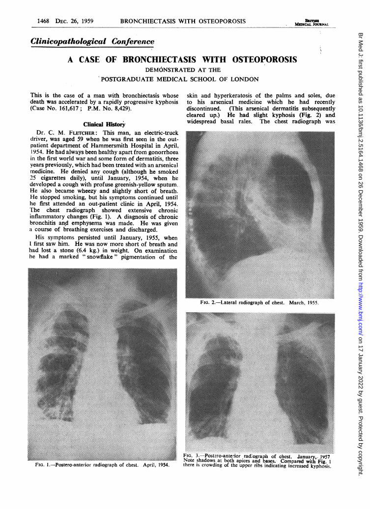
Dr. C. M. FLETCHER: This man, an electric-truckdriver, was aged 59 when he was first seen in the out-patient department of Hammersmith Hospital in April,1954. He had always been healthy apart from gonorrhoeain the first world war and some form of dermatitis, threeyears previously, which had-been treated with an arsenicalmedicine. He denied any cough (although he smoked25 cigarettes daily), until January, 1954, when hedeveloped a cough with profuse greenish-yellow sputum.He also became wheezy and slightly short of breath.He stopped smoking, but his symptoms continued untilhe first attended an out-patient clinic in April, 1954.The chest radiograph showed extensive chronicinflammatory changes (Fig. 1). A diagnosis of chronicbronchitis and emphysema was made. He was givena course of breathing exercises and discharged.His symptoms persisted until January, 1955, when
I first saw him. He was now more short of breath andhad lost a stone (6.4 kg.) in weight. On examinationhe had a marked "snowflake" pigmentation of the
skin and hyperkeratosis of the palms and soles, dueto his arsenical medicine which he had recentlydiscontinued. (This arsenical dermatitis subsequentlycleared up.) He had slight kyphosis (Fig. 2) andwidespread basal rales. The chest radiograph was
FIG. 2.-Lateral radiograph of chest. March, 1955.
FIG. 1.-Postero-anterior radiograph of chest. April, 1954.
FIG. 3.-Postzro-ante.-ior rad.ograph of chest, lanuary, 1957.Note shadows at both apices and bases. Compared with Fig. 1
there is crowding of the upper ribs indicating increased kyphosis.
BRMaMEDCAL JOURNAL
on 17 January 2022 by guest. Protected by copyright.
http://ww
w.bm
j.com/
Br M
ed J: first published as 10.1136/bmj.2.5164.1468 on 26 D
ecember 1959. D
ownloaded from
DEC. 26, 1959 BRONCHIECTASIS WITH OSTEOPOROSIS BRmsH 1469MEDICAL JOURNAL
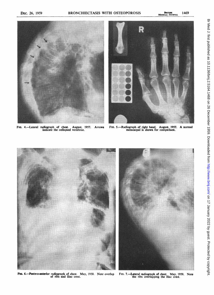
FIG. 4.-Lateral radiograph of chest. August, 1957. Arrows Fia. 5.-Radiograph of right hand. August, 1957. A normalindicate the collapsed vertebrae. metacarpal is shown for comparison.
Fio. 6.-Postero-anterior radiograph of chest. May, 1958. Note overlapof ribs and iliac crest.
...................................sS;S ............F.s,
|||gSLt' 'M} mm1|-We ......... !-SF!.r- } :' ............... ,4S ..... NX .'i.: '. . . . :__ ......... _!:: . : ^,: Q _ }: . : : : . : .' : : . : :F9; i .. ;;. EC_, ......................... , .. e ....... .. syg .
? F w!.. : .. :Ffis8xtt ..tww . : e
si, ?::?1 ::i;.sss_: :w'':Z . }R. st':. ::.?.X'.as. i:,: ......?xls.::'s' ::
'':7S'i ;:' :. :: :r?: ::: .. ::| L.> . ,X,@ ; ;.j . ' ;"!
isM.2> Y;}.:: ::: . . ...- lywn: nto.iS i. :- 2 :.::?': :: :.: .E D;:.':'.::...: :.::
|S '^.':'..: :.:l LY'° ............. .ggi ........................................................Ew::...:-. :.Sa::::: .:::sfi S! ';:.i::e'' .::.ISB ?.Xlvs :: :.* .. ::: .: :- ,8:':.e .: :. ::?.:'li' :: ::.: ::i::w, .:: ::
::.S::.i :.::?Sew SiilE "5 j:: .. .-isH |:: :1.e
-1|E"'' ii:-: 1
:. .:..OMs,.;.* :.: ::j: ;.: .i:;: a
FIG. 7.-LEateral radiograph of chest. May, 1958. Notethe ribs overlapping the iliac crest.
on 17 January 2022 by guest. Protected by copyright.
http://ww
w.bm
j.com/
Br M
ed J: first published as 10.1136/bmj.2.5164.1468 on 26 D
ecember 1959. D
ownloaded from
1470 DEC. 26, 1959 BRONCHIECTASIS WITH OSTEOPOROSIS BRsIISHMEDICAL JOURNAL
on 17 January 2022 by guest. Protected by copyright.
http://ww
w.bm
j.com/
Br M
ed J: first published as 10.1136/bmj.2.5164.1468 on 26 D
ecember 1959. D
ownloaded from
BRONCHIECTASIS WITH OSTEOPOROSIS BRITISH 1471MEDICAL JOURNAL
unaltered and a diagnosis of bronchiectasis was made.He was instructed in postural drainage, and given afortnight's course of penicillin injections. He improvedslightly, but Haemophlilus influenzae was cultured fromthe sputum and tetracycline (I g. daily) was given fora week. His sputum became mucoid and reduced involume and his shortness of breath decreased. Thex-ray shadows cleared considerably and he returned towork. He remained fairly well but his sputumrepeatedly became purulent, but became mucoid afterfurther short courses of tetracycline in March and June,1955. From September, 1955, to April, 1956, he wasgiven 0.25 g. tetracycline twice daily. lt was thenstopped because a resistant Klebsiella pneumoniaepredominated in his sputum. He remained at workeven in foggy weather, but his sputum, though smallerin amount, reniained purulent. An estimation of hisindirect maximum breathing capacity (M.B.C.) inSeptember, 1955, was 60 litres/min. (normal 64-122).He was able to walk as far as he liked on the level,but was breathless on hurrying.
In January, 1957, he had an episode of acutebreathlessness with increased sputum. His own doctorgave him tetracycline and he improved. After this,apical shadows were noted on his chest radiograph(Fig. 3) in both posterior segments. These shadowsincreased in intensity over the next six months, but sevensputa were negative for acid-fast bacilli. The shadowswere probably due to bronchiectasis developing in theupper lobes. Continuous tetracycline therapy was givenagain, the dose being restricted to 0.25 g. twice dailyas before because a larger dose caused diarrhoea.
In May, 1957, the patient was more breathless andhe noticed he was getting more round-shouldered. Thiswas confirmed clinically. He developed pain in hisshoulders and hypochondrium which was worse onbreathing or coughing. He had lost 4 lb. (1.8 kg.) inone month. He was much more brpathless and unableto work. A radiograph (Fig. 4) showed very thinvertebrae with collapse of D5, 6, 7, and 9. He wasadmitted to the Metabolic Unit under Professor T.Russell Fraser.
INVESTIGATIONSSkeletal.-Further skeletal radiograph (Fig. 5) showed
widespread osteoporosis.Cardiovascular.-Electrocardiograph tracing normal.
Serum.-Calcium, 5.3; phosphorus, 2.1 mEq/litre;alkaline phosphatase, 8.0 K.-A. units; acid formolstable, 1.4 units.Calcium Balance.-Diet. 15.2; faeces, 12.2; urine
10.8 mEq/24 hours.Iliac Crest Biopsy.-Osteoporosis. Seams up to 5[k
thick.Strontium Test of Bone Uptake.-CaE, 12.7; CaB.
2.15 (high normal).Ventilatory Function*.-F.E.V., 1.04 litres (normal
8-3.5). V.C., 1.61 litres (normal 2.9-3.9). IndirectM.B.C. 36 litres/min. (normal 64-122). F.E.V.%V.C.64% (normal). No response to isoprenaline. Thesetests show severe restriction of ventilatory capacity ofthe restrictive rather than obstructive type, since 64% ofthe ventilatory capacity was exhaled in 1 second.
SUBSEQUENT COURSEOn discharge, a 600-mg. testosterone implant was
given together with 95 mE calcium, 1 mg. stilboestrol,and 0.5 g. tetracycline daily. A spinal brace was fittedbut the patient found it too heavy to wear. Duringthe winter the tetracycline was changed to chloram-phenicol, 0.25 mg. twice daily, and this kept his sputummucoid. His breathlessness remained severe and waspresent even at rest. Oedema of the ankles developedbut was thought to be due to varicose veins. Ondiscontinuing chloramphenicol in March, 1958, thesputum soon became purulent and he had great difficultyin coughing because of increasing kyphosis.
In May, 1958, he was readmitted. He was verybreathless but not definitely cyanosed. The kyphosiswas more extreme, his lower ribs resting on his iliaccrest (Figs. 6 and 7). The jugular venous pulse wasdifficult to see because of his kyphosis, but did notappear to be raised. There was no clubbing. The dayafter admission he suddenly became cyanosed andcomatose. The head of the bed was lowered, a largequantity of purulent sputum was aspirated, but thepatient died in 10 minutes.
Clinical Diagnosis(1) The persistent and profuse purulent sputum was
attributed to bronchiectasis affecting both lower lobesat first, and later the upper lobes on both sides.
(2) The kyphosis was due to an unexplainedosteoporosis.A diagnosis of emphysema was made in two radio-
logical reports and it appears in several letters to doctorsand on the case notes after his first admission. I do notaccept the radiological diagnosis, and the ventilatoryfunction tests were not characteristic of emphysema.I do not believe there was significant anatomicalemphysema. The breathlessness was due partly tobronchial obstruction by excessive secretion, but chieflyto the restrictive effect of the kyphosis, and his deathwas due to suffocation from bronchial secretion whichhe could not expectorate.The only evidence of right heart failure was ankle
oedema, but he had varicose veins. He was nevergrossly cyanosed and his E.C.G. was normal. I do notdiagnose significant abnormality of the heart.
*F.E.V.,0O=Forced expiratory volume. V.C.=Vital capacity.F.E.V.%V.C.=Forced expiratory volume as percentage of vitalcapacity. M.B.C.=Maximum breathing capacity. (See Thomson,W. B., and Hugh-Jones. P., Brit. med. J., 1958, 1, 1093.)
D
LEGENDS TO SPECIAL PLATE
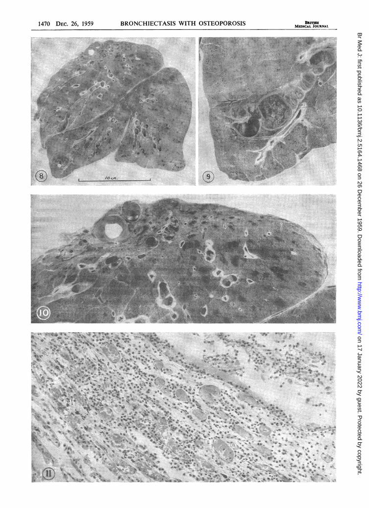
FIG. 8.-An an'ero-posterior slice of the right lung. Theupper lobe is flattened by the chest-wall deformity.There is bronchectasis in many parts, most obvious inthe middle lobe and the fasal segments of the lower lobe.(Prepared by pressure-fixation and barium sulphate
impregnation.)FIG. 9.-A higher magnification of the middle lobe to
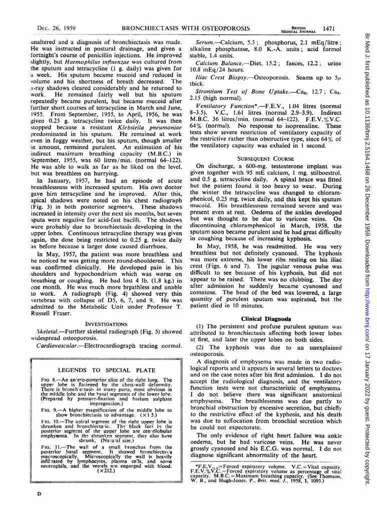
show bronchiectasis to advantage. (x 1 5.)FIG. 10.-The apical segment of the right upper lobe isshrunken and hronchiecta ic. The black foci in theposterior segment of the upper lobe are cen-rilobularemphysema. In the shrunken segment, they also have
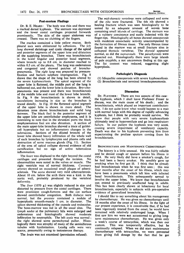
shrunk. (Natu-al size.)FIG. 11.-The wall of a small bronchus from theposterior basal segment. It showed bronchiectas:smacroscopically. Microscopically the wall is heavilyinfil rated by lymphocytes, plasma ce'ls, and sonmeneutrophils, and the vescels are engorged with blood.
(x212.)
Dec. 26, 1959
on 17 January 2022 by guest. Protected by copyright.
http://ww
w.bm
j.com/
Br M
ed J: first published as 10.1136/bmj.2.5164.1468 on 26 D
ecember 1959. D
ownloaded from
1472 DEC. 26, 1959 BRONCHIECTASIS WITH OSTEOPOROSIS
Post-mortem FindingsDr. B. E. HEARD: The body was thin and there was
a marked dorsal kyphosis. The sternum was depressedand the lower costal cartilages projected forwardsprominently. The skin of the upper abdomen wascreased. There was no clubbing of the fingers.The trachea contained a little yellow mucus. Both
pleural sacs were obliterated by adhesions. The leftlung showed shrinkage and cystic change of the apicaland posterior segments of the upper lobe and dilatationof the supplying bronchus. There was bronchiectasisin the lower lingular and posterior basal segments,where bronchi up to 0.8 cm. in diameter reached towithin 0.5 cm. of the pleura. The lung was oedematousand congested and showed patches of broncho-pneumonia. The right lung was prepared by pressure-fixation and barium sulphate impregnation. Fig. 8shows that the shape of the lung has been altered bythe severe kyphoscoliosis. The apical segment of theupper lobe is flattened, the middle part of the lung isballooned out, and the lower lobe is shrunken. Bro.cho-pneumonia was present and there was bronchiectasisof the middle lobe and most of the lower lobe bronchi.In Fig. 9 the bronchiectasis is shown in detail,sacculations increasing in size as the bronchus istraced distally. In Fig. 10 the flattened apical segmentof the upper lobe is shown in more detail. Theshrunken zone shows bronchiectasis and solid lungbetween. The black foci in the posterior segment ofthe upper lobe are centrilobular emphysema, and it isinteresting to note that in the shrunken parts the blackemphysematous foci are also shrunken. A section ofthe main upper-lobe bronchus showed a trace of goblet-cell hyperplasia but no inflammatory changes in thesubmucosa. Sections of the dilated bronchi of thelower lobe showed heavy infiltration of the whole wallof each bronchus by lymphocytes and plasma cells anddestruction of some mucous glands (Fig. 11). Sectionof the area of apical collapse showed evidence of oldcarnification but no sign of active tuberculousinflammation.The heart was displaced to the right beyond the costal
cartilages and presented through the incision. Noabnormalities were noted in the valves or muscle. Theright ventricle was of normal thickness. Coronaryarteries showed an occasional small plaque of athero-sclerosis. The aorta showed very mild atherosclerosis.About 10 cm. below the arch there was a kink in theaortic wall, probably produced by the vertebraldeformity.The liver (1070 g.) was slightly reduced in size and
distorted by pressure from the costal cartilages. Therewere prominent cough-furrows and the cut surfaceshowed centrilobular congestion. Both kidneys werenormal, but the prostate contained a nodule ofhyperplastic smooth-muscle I cm. in diameter. Thespleen showed thickening of the capsule and trabeculae.The bone-marrow was red in all the usual sites. Thelymph nodes at the pulmonary hila were enlarged andoedematous and histologically showed moderateinfiltration by neutrophils. The left testis was normal;the right showed some parenchymal pallor. Histo-logically there was atrophy of many of the seminiferoustubules with hyalinization. Leydig cells were veryscarce, presumably owing to testosterone therapy.The brain was not examined (permission refused).
The mid-thoracic vertebrae were collapsed and someof the ribs were fractured. The 6th rib showed ahealing fracture which was seen histologically to beunited by an adequate anmount of woven bonecontaining small islainds of cartilage. The sternum was
of a rubbery consistence and easily indented with thefinger-tips. Histologically all bones showed osteoporosiswith moderate osteoclastic activity unaccompanied bythe formation of fibrous tissue. The only fibrous tissuefound in the marrow was at small fracture sites incollapsed thoracic vertebrae. The thyroid appearednormal, as did the suprarenials. The parathyroids weredissected out. Histologically there were large islandsof pale oxyphils, a not uncommon finding at this age.The fat content was reduced, suggesting slighthyperplasia.
Pathologist's Diagnosis(1) Idiopathic osteoporosis with severe kyphoscoliosis.(2) Bronchiectasis and terminal bronchopneumonia.
DiscussionDr. FLETCHER: There are two aspects of this case:
the kyphosis, which I shall lcave Professor Fraser todiscuss, was the main cause of his death; and thebronchiectasis, which played an important contributoryrole. I do not quite know what would happen to a man
with normal lungs with this degree of rapidly advancingkyphosis, but I think he probably would survive. Weknow that people with very severe kyphoscoliosisultimately tend to hypoventilate and go into respiratorvfailure. In our patient the kyphosis was very acute.causing severe impairment of ventilatory capacity.Death was due to his kyphosis preventing him fromexpectorating the profuse sputum coming from hisbronchiectasis.
BRONCHIECTASIS AND MAINTENANCE CHEMOTHERAPYThe history is a little unusual. He was fairly reliable
and he denied cough or sputum before his illness in1954. He very likely did have a smoker's cough, forhe had been a heavy smoker. He sensibly gave up
smoking when he first got ill. I think that he alreadyhad bronchiectasis when he was first seen; this was
four months after the onset of his illness, which mavhave been a pneumonia which left him with infectedbasal bronchiectasis. This subsequently spread toinvolve the upper lobes. We know that bronchiectasiscan extend to previously unaffected lung in adults.This has been clearly shown at lobectomy for localbronchiectasis, especially in subjects with pre-operativeevidence of generalized bronchitis.
I should like to say something about his managementby chemotherapy. He was given no chemotherapy until18 months after the onset of his illness. In the light ofour present experience, it is conceivable that, if he hadbeen vigorously treated from the onset, he would haveremained with relatively undamaged lungs. When Ifirst saw him we were not accustomed to giving long-term maintenance chemotherapy. He was given onlya week's course of tetracycline, which was then our
custom. This was temporarily effective, but hecontinually relapsed. When we did start maintenancechemotherapy with tetracycline, we were preventedfrom giving full dosage by the fact that he got
BRmSHMEDICAL JOURNAL
on 17 January 2022 by guest. Protected by copyright.
http://ww
w.bm
j.com/
Br M
ed J: first published as 10.1136/bmj.2.5164.1468 on 26 D
ecember 1959. D
ownloaded from
DEC. 26, 1959 BRONCHIECTASIS WITH OSTEOPOROSIS BRMISI 1473MEDICAL JoURNAL
diarrhoea. This, of course, is an annoying side-effectof tetracycline, which we encounter in something like10-15% of our cases. It usually can be controlled bya simple opium mixture and very often does not persist.Later on we were forced to take the risk of maintenanceon chloramphenicol, which was much more effective,but the bronchial infection relapsed as soon as it wasdiscontinued.
Professor J. MCMICHAEL: Two questions. When hecame in last no clinical cyanosis was noted. Did wehave an arterial sample?
Dr. FLETCHER: No. He died within a day.Professor M1CMICHAEL: Was he never grossly
cyanotic?Dr. FLETCHER: There are occasional out-patient
notes of slight cyanosis, but he was never grosslycyanotic.
Professor MCMICHAEL: What was the cardiac state?Dr. FLETCHER: It seems to have been normal. There
was no evidence of right hypertrophy. 1 do not thinkhis ankle oedema was cardiac.
PROGRESSIVE OSTEOPOROSISProfessor T. RUSSELL FRASER: Well, this is an instance
of osteoporosis, unfortunately a condition which is notvery well understood nor very easy to treat. But Isuspect that his disorder must have been more severethan is usual in men, and perhaps more difficult to treatbecause of his associated cough. It is at least not at allcommon to see a progressive kyphosis advancing asrapidly as that. How much the cough contributed toit is hard to say. From the fact that he had osteoporoticbones in the radiological meaning of the term we caninfer that the man must already have lost over 50% ofhis bone mass. lf there is, in addition, the mechanicalproblem of coughing, I suppose progression may almostbe inevitable.The normal treatment would include a spinal brace
to place the support of his shoulders on to his iliaccrest; but doubtless he could not manage with hisbrace, since it must have increased his dyspnoea very
Ca,* * @ @ @* @ @ - -
* @ * - **---*
* * - * -*-*--****-* - * - -*---** - * * -* - * - ** * * * -
* - - - **----.....* * * - @
* - - - -* - - - -
* - - - -* - ....* * - - *
* * - * - .* - * - *
* - - . . .* - - - -
* - * - * ** - * - -* * - * -* * * * -......
* - - - -* @ - - @* @ @ @ @* - - - e* e - - -* - e - -
* v v v v* v v v -
* - - - -* - - * *
* - - - ** - * - **----* - - * -* - - - ** * * - -
* - - - ** * * * e* - - * -* * - - *
* - - - ** * - * -
* @ @ * ** - - * *
* * - - -* * - - *
* * - * ** * * * * ** - - * -*-----
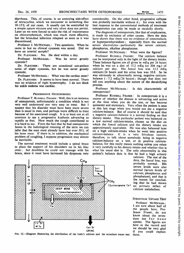
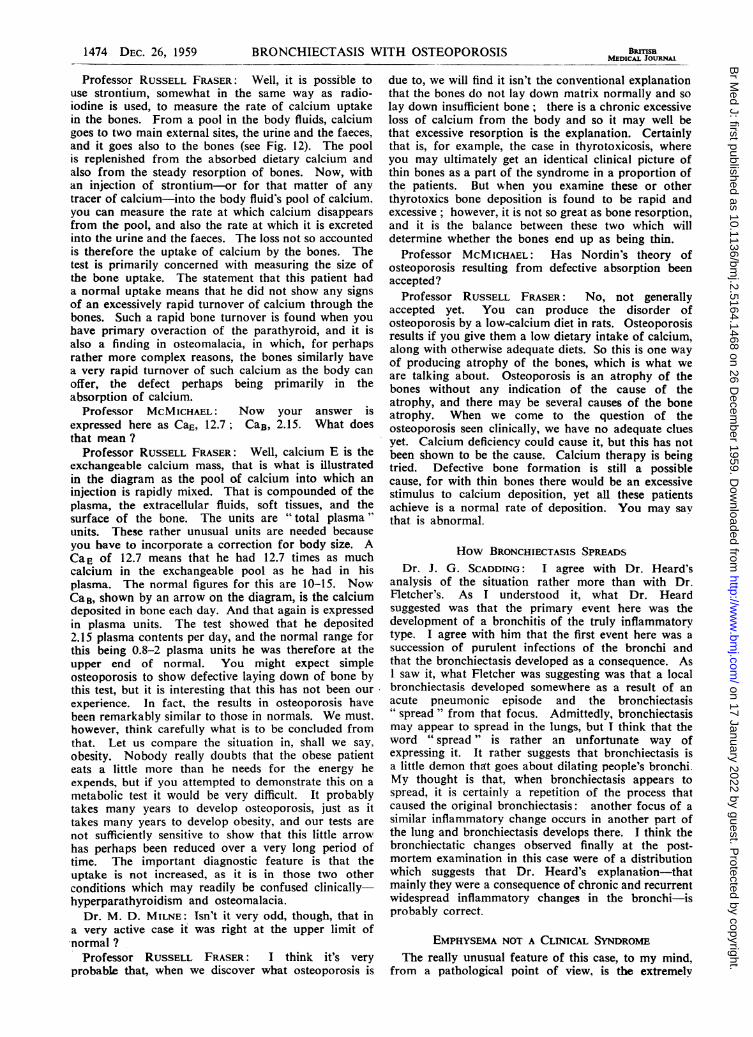
URIFFIG. 12.-Diagram illustrating the distribution of the body's calciu
considerably. On the other hand, progressive collapsewas probably inevitable without it; for even with thebest response to the conventional methods of treatmentosteoporotics can only be made to recalcify slowly.The diagnosis of osteoporosis, like that of emphysema,
is made by exclusion of other causes. Here the datahave shown that there was no evidence of osteomalaciaor hyperparathyroidism; especially from the normalserum electrolytes-particularly the serum calcium,phosphorus, alkaline phosphatase.
Professor MCMICHAEL: What were the figures?Professor RUSSELL FRASER: The calcium balance
can be interpreted only in the light of the dietary intake.These balance figures are all given in mEq per 24 hourswhen he was on a diet of 15.2 mEq (or 304 mg.) ofcalcium per day. His faecal loss was 12.2 mEqcalcium/24 hours and his urine loss was 10.8. So hewas obviously in abnormally strong, negative calcium-balance (-7.2 mEq/24 hours); though that does nottell you anything about the nature of the decalcifyingprocess.
Professor MCMICHAEL: Is this characteristic ofosteoporosis?
Professor RUSSELL FRASER: In osteoporosis it is amatter of whether the disease is advancing and activeat the time when you do the test, or has becomequiescent and stationary. Very often the patient is seenat this last stage when you would not see a negativecalcium-balance. But of course it should be said thata negative calcium-balance is a normal finding on thatdietary intake. This particular patient was balanced ona low normal calcium-intake, chosen as the diet onwhich the faecal calcium and the dietary intake areapproximately equivalent. He was also balanced lateron a high calcium-intake when he went into positivecalcium-balance It is a very frivolous custom,therefore, to talk about somebody being in negativecalcium-balance on a test, or in positive calcium-balance, for this really means nothing unless you relateit very carefully to his dietary intake and whether that iswhat hlis usual diet is. The only abnormality in thispatient's balance data is that he had a high urinary
calcium. The rest of thedata, the faecal loss, 'wasprobably normal. Hisserum levels were alsoSosut normal (serum levels of
Tissue calcium, phosphorus. andphosphatase), and that is
...... ~~~~the reason for conclud-....... CQE ing that he had shown
Exchangeable Ca) no primary defect ofcalcium metabolism.
STRONTIUM UPTAKE TEST::::::.0.0.0 Professor MCMICHAEL:
, Iam sure about half of:::::::.oooo0 the people here, Pro-:::::: 0. ..fessor Fraser, do not':::::: know about the stron-I----- -----, tium test for bone
uptake. The figures arehere in the record and
Sr we should be very gladJE if you could explainim and the strontium tracer test. them.
I-
0
on 17 January 2022 by guest. Protected by copyright.
http://ww
w.bm
j.com/
Br M
ed J: first published as 10.1136/bmj.2.5164.1468 on 26 D
ecember 1959. D
ownloaded from
BRONCHIECTASIS WITH OSTEOPOROSIS
Professor RUSSELL FRASER: Well, it is possible touse strontium, somewhat in the same way as radio-iodine is used, to measure the rate of calcium uptakein the bones. From a pool in the body fluids, calciumgoes to two main external sites, the urine and the faeces.and it goes also to the bones (see Fig. 12). The poolis replenished from the absorbed dietary calcium andalso from the steady resorption of bones. Now, withan injection of strontium-or for that matter of anytracer of calcium-into the body fluid's pool of calcium,you can measure the rate at which calcium disappearsfrom the pool, and also the rate at which it is excretedinto the urine and the faeces. The loss not so accountedis therefore the uptake of calcium by the bones. Thetest is primarily concerned with measuring the size ofthe bone uptake. The statement that this patient hada normal uptake means that he did not show any signsof an excessively rapid turnover of calcium through thebones. Such a rapid bone turnover is found when youhave primary overaction of the parathyroid, and it isalso a finding in osteomalacia, in which, for perhapsrather more complex reasons, the bones similarly havea very rapid turnover of such calcium as the body can
offer, the defect perhaps being primarily in theabsorption of calcium.
Professor MCMICHAEL: Now your answer isexpressed here as CaE, 12.7; CaB, 2.15. What doesthat mean ?
Professor RUSSELL FRASER: Well, calcium E is theexchangeable calcium mass, that is what is illustratedin the diagram as the pool of calcium into which aninjection is rapidly mixed. That is compounded of theplasma, the extracellular fluids, soft tissues, and thesurface of the bone. The units are "total plasma"units. These rather unusual units are needed becauseyou have to incorporate a correction for body size. ACaE of 12.7 means that he had 12.7 times as muchcalcium in the exchangeable pool as he had in hisplasma. The normal figures for this are 10-15. NowCa B, shown by an arrow on the diagram, is the calciumdeposited in bone each day. And that again is expressedin plasma units. The test showed that he deposited2.15 plasma contents per day, and the normal range forthis being 0.8-2 plasma units he was therefore at theupper end of normal. You might expect simpleosteoporosis to show defective laying down of bone bythis test, but it is interesting that this has not been ourexperience. In fact, the results in osteoporosis havebeen remarkably similar to those in normals. We must,however, think carefully what is to be concluded fromthat. Let us compare the situation in, shall we say,obesity. Nobody really doubts that the obese patienteats a little more than he needs for the energy heexpends, but if you attempted to demonstrate this on a
metabolic test it would be very difficult. It probablytakes many years to develop osteoporosis, just as ittakes many years to develop obesity, and our tests are
not sufficiently sensitive to show that this little arrow
has perhaps been reduced over a very long period oftime. The important diagnostic feature is that theuptake is not increased, as it is in those two otherconditions which may readily be confused clinically-hyperparathyroidism and osteomalacia.
Dr. M. D. MILNE: Isn't it very odd, though, that ina very active case it was right at the upper limit ofnormal ?Professor RUSSELL FRASER: I think it's very
probable that, when we discover what osteoporosis is
due to, we will find it isn't the conventional explanationthat the bones do not lay down matrix normally and so
lay down insufficient bone; there is a chronic excessiveloss of calcium from the body and so it may well bethat excessive resorption is the explanation. Certainlythat is, for example, the case in thyrotoxicosis, whereyou may ultimately get an identical clinical picture ofthin bones as a part of the syndrome in a proportion ofthe patients. But when you examine these or otherthyrotoxics bone deposition is found to be rapid andexcessive; however, it is not so great as bone resorption,and it is the balance between these two which willdetermine whether the bones end up as being thin.
Professor MCMICHAEL: Has Nordin's theory ofosteoporosis resulting from defective absorption beenaccepted?
Professor RUSSELL FRASER: No, not generallyaccepted yet. You can produce the disorder ofosteoporosis by a low-calcium diet in rats. Osteoporosisresults if you give them a low dietary intake of calcium,along with otherwise adequate diets. So this is one way
of producing atrophy of the bones, which is what we
are talking about. Osteoporosis is an atrophy of thebones without any indication of the cause of theatrophy, and there may be several causes of the boneatrophy. When we come to the question of theosteoporosis seen clinically, we have no adequate cluesyet. Calcium deficiency could cause it, but this has notbeen shown to be the cause. Calcium therapy is beingtried. Defective bone formation is still a possiblecause, for with thin bones there would be an excessivestimulus to calcium deposition, yet all these patientsachieve is a normal rate of deposition. You may savthat is abnormal.
How BRONCHIECTASIS SPREADSDr. J. G. SCADDING: I agree with Dr. Heard's
analysis of the situation rather more than with Dr.Fletcher's. As I understood it, what Dr. Heardsuggested was that the primary event here was thedevelopment of a bronchitis of the truly inflammatorvtype. I agree with him that the first event here was a
succession of purulent infections of the bronchi andthat the bronchiectasis developed as a consequence. AsI saw it, what Fletcher was suggesting was that a localbronchiectasis developed somewhere as a result of an
acute pneumonic episode and the bronchiectasis" spread" from that focus. Admittedly, bronchiectasismay appear to spread in the lungs, but I think that theword "spread" is rather an unfortunate way ofexpressing it. It rather suggests that bronchiectasis isa little demon that goes about dilating people's bronchi.My thought is that, when bronchiectasis appears tospread, it is certainly a repetition of the process thatcaused the original bronchiectasis: another focus of a
similar inflammatory change occurs in another part ofthe lung and bronchiectasis develops there. I think thebronchiectatic changes observed finally at the post-mortem examination in this case were of a distributionwhich suggests that Dr. Heard's explanation-thatmainly they were a consequence of chronic and recurrentwidespread inflammatory changes in the bronchi-isprobably correct.
EMPHYSEMA NOT A CLINICAL SYNDROMEThe really unusual feature of this case, to my mind,
from a pathological point of view, is the extremelv
BRrmsMEDICAL JOURNAL
1474 DEC. 26, 1959
on 17 January 2022 by guest. Protected by copyright.
http://ww
w.bm
j.com/
Br M
ed J: first published as 10.1136/bmj.2.5164.1468 on 26 D
ecember 1959. D
ownloaded from
DEC. 26, 1959 BRONCHIECTASIS WITH OSTEOPOROSIS BRrIlSH 1475MEDICAL JOURNAL
small amount of emphysema that was in evidence at theend of the process. I would have expected, in a casewhere all these changes had been going on for so manyyears, a good deal more emphysema. I was very gladto hear pin-pointed the difficulties over the diagnosisof emphysema. We are trying to establish the idea thatthe only tenable definition of emphysema is a morbidanatomical one, so that the only person who can sayfor certain whether emphysema is present is the morbidanatomist. When, as clinicians, we are bold enough tosay, " I think this patient has emphysema," what weshould mean is that we think that the morbid anatomistwill find certain morbid anatomical changes. We maybe wrong or we may be right, but that is what we shouldmean. Unfortunately the word " emphysema " hasbecome one of those witchwords which misleadseverybody, because it ias become equated with a clinicalsyndrome without any real justification. Indeed, Ithink this happens all over the world; certainly ithappens in the United States. Recently I received, froma centre in the United States for the journal which I edit,a paper in which a case report recorded that the patienthad had "recurrent attacks of emphysema." Thismakes clear that the word emphysema is used by somepeople there in the same sense in which most of us inthis country refer to recurrent or chronic bronchitis.[ don't think we'll overcome this confusion until we canget it established that ultimately emphysema is a morbidanatomical concept.What we urgently need is a convenient word to
describe the syndrome of chronic respiratory diseaseleading up to eventual respiratory insufficiency, whichis so common in this country, without necessarilyimplying any untenable hypothesis. The position inrespiratory disease at present, with the current use ofthe word emphysema, is rather similar to that whichwould exist in cardiology if you could not refer to" cardiac failure " without implying that there wasvalvular disease of the heart. We want functional termsin respiratory disease that would be as commonly usedand understood by everybody as " congestive cardiacfailure" is in cardiology. Having introduced that terminto our respiratory work, we could then go on to add,to complete a diagnosis, the morbid anatomical changesamong which might be emphysema.A VOICE: What about " respiratory failure"?Dr. SCADDING: Well, that would be a nice term to
use, but unfortunately it's not quite precise enough." Respiratory failure" might imply failure of therespiratory centre as well as of the lungs. " Pulmonaryinsufficiency" is the nearest I can get to it, implyinga local insufficiency in the lungs.Dr. FLETCHER: I should just like to say that I do not
look on bronchiectasis as a demon. I entirely agreewith Dr. Scadding that its spread is due to extension ofinfection with destructive results, but I do not think thiscase was one of bronchitis in the ordinary sense, becausewe do not see this process in most of our bronchitics.There was something different about this bronchitis.Did Dr. Heard say that he did not find hyperplasia ofmucus-secreting glands in the bronchi? In my mindbronchitis is characterized primarily by goblet-cell andmucous gland hypersecretion. In this case there was noreal history of chronic bronchitis preceding the firstillness. There was a rapidly progressive bronchiectasisstarting, I believe, with an acute infection. Later onbronchiectasis developed elsewhere, so that some
secondary consequence bronchiectasis. This may havebeen a peculiar kind of bronchitis, but it was not theordinary kind in which there is persistent mucoidsputum with recurrent episodes of infection. This manhad a persistently purulent sputum and radiologicalevidence of bronchiectasis at first in the lower lobes andsubsequently in the upper lobes.
EFFECT OF THE KYPHOSISDr. R. E. STEINER: The appearance of lesions in
the upper lobes coincided with the kyphosis.Dr. FLETCHER: Yes, they appeared in January, 1957.
when the P.-A. radiograph shows increased kyphosis.but this wasn't noticed clinically till about four monthslater. Do you suggest that he had a collapse of hisupper lobe and got a secondary infection in it, whichmay have led to the bronchiectasis?
Dr. STEINER: Yes, the mechanical compression wasthe first thing, at least in the case of the upper lobes.You are not dealing with a primary infection of thebronchi, on which you base your explanation of thebronchiectasis.
Dr. P. C. ELMES: I wonder which was causing thelung disease, the vertebral collapse or the bronchialinfection? The disease process may have been primarilya bronchial infection, perhaps associated with someother pulmonary disease. He was coughing up about200 ml. of purulent sputum per day, containing 10% ofprotein, for many years, and his disease was associatedwith anorexia. Thus protein deficiency could haveoccurred and given rise to bone absorption withvertebral collapse and progressive symptoms. This inturn made it impossible for us to clear his lungs ofinfection. Two processes were going on at the sametime and you never could say which came on first, orwhich was the more important; they added to eachother all the time. I wonder whether the final pictureof minimal emphysema was due to the fact that herewere lungs which were getting progressively scarred butwere not being asked to occupy the normal space. Hadthey been asked to occupy the normal 5 or 6 litres ofthoracic volume, then there would have been very muchmore likelihood of emphysema because the lungs wouldhave been stretched to fill that large volume, whereasbecause of the collapse they were not being stretchedat all.
Professor MCMICHAEL: Isn't it rather unusual, Dr.Heard, to see completely collapsed segments of the lungin these deformed hunchbacks? My observation ofthe ordinary kyphoscoliotic is that the lungs are smallbut otherwise normal.
Dr. HEARD: Complete collapse is unusual, but some
shrinkage is not uncommon with deformities of thechest wall or with pleural thickening. The defect isnot always one of compression. We have had a few cases
of severe kyphoscoliosis recently. One of them was a
man of 53 who developed kyphoscoliosis early inchildhood. He had very small lower lobes to bothlungs. The defect was not so much due to compressionby the deformity, but to the fact that his lungs appearednever to have developed to the normal size. Incidentally.he had no bronchiectasis and no emphysema was
demonstrable though the lungs were prepared bypressure-fixation and barium sulphate impregnation.*As regards the bronchial histology in the present case,
processs was spreading in his lungs which had as a 0 Heard, B. E., Thorax, 1958, 13, 136 ; ibid., 1959, 14, 58.
on 17 January 2022 by guest. Protected by copyright.
http://ww
w.bm
j.com/
Br M
ed J: first published as 10.1136/bmj.2.5164.1468 on 26 D
ecember 1959. D
ownloaded from
1476 DEc. 26, 1959 BRONCHIECTASIS WITH OSTEOPOROSIS
the small bronchus showing marked goblet-cell hyper-plasia was of the sort described by Reid.*We are grateful to Dr. J. P. Sbillingford and Dr. B. E.
Heard for assistance in preparing this report, and to thephotographic department of the Postgraduate MedicalSchool for the illustrations.
EXTRACTS AND HOWLERS TAKENFROM EXAMINATION SCRIPTS
BY
M. R. SHERIDAN, M.R.C.S., L.R.C.P.General Practitioner, Wood Green, London
The following are authentic extracts from examinationpapers collected through the years by the senior sistertutor of a well-known hospital.
AnatomyWax glands in the ear protect the organ from foreigners.The pupil is nothing more than a whole in the eNe. In
some people it is big, in others small, but unless you havebeen peculiarly born it is there in some size or another.The stomach is covered with a serious membrane.The stomach is supplied by an involuntary nerve called
the pneumatic.The aorta is a long tub whose contents are very precious
to the body.The exe is the organ of site, the sight of which is in a
holler in the temple.At the back of the eye one may see a retinue.The heart is the only organ in the body made of heart
muscle.The heart is a muscular organ acting like a human pump,
which pumps the heart around the body at regular intervals.The heart is an organ the shape of a heart. It is not
quite heart shaped because there are tubes protruding fromit, 4 from top left, an umbrella handle from bottom left,a " Y " shaped connection from bottom right and 2 tubesfrom top right.The brain will soon become a weekend one.At the back of the throat are 2 long pillars of faeces.The mouth is situated in the lower part of the face and
is known as a buckled cavity.
Personal and Communal HealthThe best room for eye trouble is one which is well alight.The shapkeeper should thorouehly rap all cakes before
selling them to make sure all disease is eliminated.Food should not be picked up by hand but with tongues.Clean overalls or trousers should be worn to polish the
fruit on.These are some of the types that help in hygiene of the
country: (I) Road sweepers. (2) Dustmen. (3) Nurses inthe district and so on. All these add up to the answerprevention of spread.
If blue bottles come through the larder window troubleis around.Cooks should be trained to wash their hands after a visit
to the toilet it is through wiping them on food that diseasesare spread.Overcrowding is not necessary. People when they get
married their parents say they can live with them until theyget a house built. Some people think, well as long as wecan live with them it is all right. BUT IT IS NOT. After
*Reid, L. M., Lancet, 1954, 1. 275.
they have been married a few months they start havingchildren, these grow up and this goes on. Overcrowdingthen occurs air is not allowed to enter, with too many inthe bed diseases are caught. Conditions like this can belikened to the many bundreds of people who were put ina black bole in Calcutta and burried alive.
Rest is essential if habits are to be good ones, if you donot rest then habits will be bad ones, so proper rest andgood cleaning of the body will produce a nurse who canbe relied upon to perform any old job the sister may haveon her list.Our Tutor has stated that in her opinion all infection in
hospitals is caused by dirty nurses, therefore personalhygiene and cleanliness is next to godliness, and is the firstthing to remember if you want to prevent wounds frombecoming pussy.The important thing in the maintenance of health is
personal cleanliness. This commeaces when the person isgetting up in the morning, and it continues until the personis ready for getting up again.You always find a person who does not practice personal
cleanliness is not nice to be near.Everything about personal cleanliness should be kept very
clean, making sure that the habits are not forgotten.The body needs a daily inspection to make sure every
part is in proper working order.The teeth should be brushed with a brush and toothpaste.
The best metlhod is to have 2 brushes and keep one of themin a septic solution.
Clothes should be well washed and ironed for hygienicconditions and also for decorating purposes.
In bad light people may screw up their eyes and thedamage they do may never be undone.
Housewives when baking should wear the appropriatehead gear.
Flies may land on composed heaps then pass through acrack in a house. settle on persons within and cause troubleamongst them. Few escape this vital pest.
Flies spread disease - you can say that again."Food should be kept covered in shops because of the
diseased types who buy it.If overcrowding continues water vapour increases and
people will shout for air, this when heard denotes air hunger.We seek them here, we seek them there, we seek those
horrid flies everywhere.Flies are abundant wherever rubbish, manure, filth and
hospital food are found.Flies may be shot down whilst in flight by the aid of
D.D.T. bombs.If open shoes are worn on duty the toe nails should be
kept clean and short as they are on view to all the patients.Curt shoes and swede shoes are generally baned by the
Matron.A nurse has no choice in the footwear she wears, the
Matron chooses ber shoes for her.The ear is the organ of earing.
NursingTreatment of ShockThe best treatment is to rape the patient in old blankets
making sure she is not overheated.The woman should have blankets under and over her
but onlookers must be kept outside.Coronary ThrombosisThe severity of the attack depends upon the sight of
the clot.The patient must adhere closely to the bed rest.
Common ColdThe common cold is caused by an organism called droplet
bacillus.The patient will suffer from a steaming nose.
on 17 January 2022 by guest. Protected by copyright.
http://ww
w.bm
j.com/
Br M
ed J: first published as 10.1136/bmj.2.5164.1468 on 26 D
ecember 1959. D
ownloaded from