Embed Size (px)
Citation preview
Journal of Clinical and Diagnostic Research. 2014 Jan, Vol-8(1): 203-205 203203
DOI: 10.7860/JCDR/2014/4475.3904 Review Article
An Imaging Review of Intra-ocular Calcifications
IntRoductIonWe all can enjoy the beauties of the world so long as our eyes are away from dreaded calcifications – their processes and determinants. But once calcification sets in any part of the eye; it is bound to make its presence felt.
The three main ways in which the intra-ocular calcifications manifest in our eyes are: Cataract, AH and Calcifications in tumours related to eye.
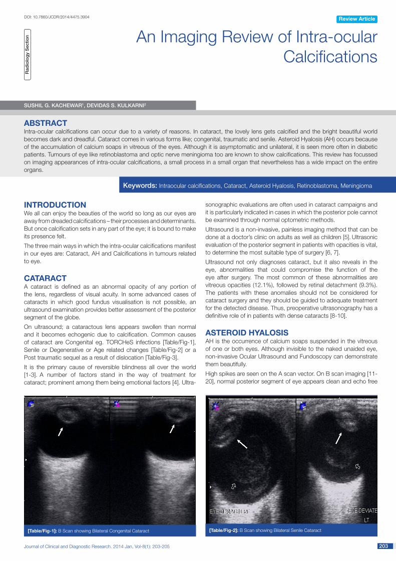
cAtARActA cataract is defined as an abnormal opacity of any portion of the lens, regardless of visual acuity. In some advanced cases of cataracts in which good fundus visualisation is not possible, an ultrasound examination provides better assessment of the posterior segment of the globe.
On ultrasound; a cataractous lens appears swollen than normal and it becomes echogenic due to calcification. Common causes of cataract are Congenital eg. TORCHeS infections [Table/Fig-1], Senile or Degenerative or Age related changes [Table/Fig-2] or a Post traumatic sequel as a result of dislocation [Table/Fig-3].
It is the primary cause of reversible blindness all over the world [1-3]. A number of factors stand in the way of treatment for cataract; prominent among them being emotional factors [4]. Ultra-
Rad
iolo
gy
Sec
tion
SuShil G. KachewaR1, DeviDaS S. KulKaRni2
Keywords: Intraocular calcifications, Cataract, Asteroid Hyalosis, Retinoblastoma, Meningioma
[table/Fig-1]: B Scan showing Bilateral Congenital Cataract [table/Fig-2]: B Scan showing Bilateral Senile Cataract
sonographic evaluations are often used in cataract campaigns and it is particularly indicated in cases in which the posterior pole cannot be examined through normal optometric methods.
Ultrasound is a non-invasive, painless imaging method that can be done at a doctor’s clinic on adults as well as children [5]. Ultrasonic evaluation of the posterior segment in patients with opacities is vital, to determine the most suitable type of surgery [6, 7].
Ultrasound not only diagnoses cataract, but it also reveals in the eye, abnormalities that could compromise the function of the eye after surgery. The most common of these abnormalities are vitreous opacities (12.1%), followed by retinal detachment (9.3%). The patients with these anomalies should not be considered for cataract surgery and they should be guided to adequate treatment for the detected disease. Thus, preoperative ultrasonography has a definitive role of in patients with dense cataracts [8-10].
AsteRoId HyAlosIs AH is the occurrence of calcium soaps suspended in the vitreous of one or both eyes. Although invisible to the naked unaided eye, non-invasive Ocular Ultrasound and Fundoscopy can demonstrate them beautifully.
High spikes are seen on the A scan vector. On B scan imaging [11-20], normal posterior segment of eye appears clean and echo free
ABstRActIntra-ocular calcifications can occur due to a variety of reasons. In cataract, the lovely lens gets calcified and the bright beautiful world becomes dark and dreadful. Cataract comes in various forms like; congenital, traumatic and senile. Asteroid Hyalosis (AH) occurs because of the accumulation of calcium soaps in vitreous of the eyes. Although it is asymptomatic and unilateral, it is seen more often in diabetic patients. Tumours of eye like retinoblastoma and optic nerve meningioma too are known to show calcifications. This review has focussed on imaging appearances of intra-ocular calcifications, a small process in a small organ that nevertheless has a wide impact on the entire organs.
Sushil G. Kachewar and Devidas S. Kulkarni, An Imaging Review of Intra-ocular Calcifications www.jcdr.net
Journal of Clinical and Diagnostic Research. 2014 Jan, Vol-8(1): 203-205204204
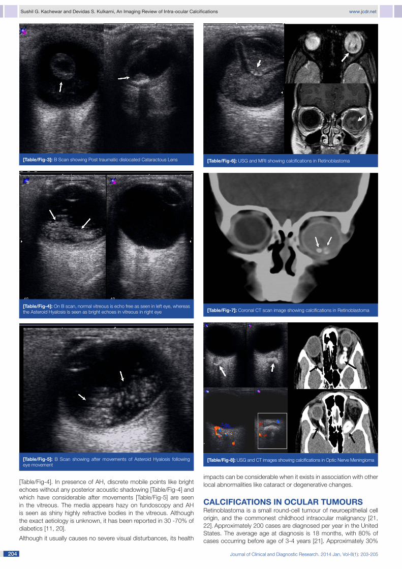
[table/Fig-3]: B Scan showing Post traumatic dislocated Cataractous Lens
[table/Fig-4]: On B scan, normal vitreous is echo free as seen in left eye, whereas the Asteroid Hyalosis is seen as bright echoes in vitreous in right eye
[table/Fig-5]: B Scan showing after movements of Asteroid Hyalosis following eye movement
[table/Fig-6]: USG and MRI showing calcifications in Retinoblastoma
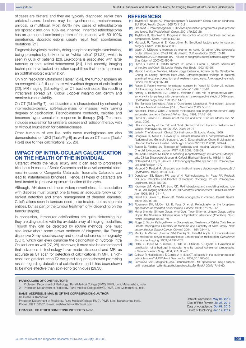
[table/Fig-7]: Coronal CT scan image showing calcifications in Retinoblastoma
[table/Fig-8]: USG and CT images showing calcifications in Optic Nerve Meningioma
[Table/Fig-4]. In presence of AH, discrete mobile points like bright echoes without any posterior acoustic shadowing [Table/Fig-4] and which have considerable after movements [Table/Fig-5] are seen in the vitreous. The media appears hazy on fundoscopy and AH is seen as shiny highly refractive bodies in the vitreous. Although the exact aetiology is unknown, it has been reported in 30 -70% of diabetics [11, 20].
Although it usually causes no severe visual disturbances, its health
impacts can be considerable when it exists in association with other local abnormalities like cataract or degenerative changes.
cAlcIFIcAtIons In oculAR tumouRsRetinoblastoma is a small round-cell tumour of neuroepithelial cell origin, and the commonest childhood intraocular malignancy [21, 22]. Approximately 200 cases are diagnosed per year in the United
States. The average age at diagnosis is 18 months, with 80% of
cases occurring before age of 3-4 years [21]. Approximately 30%
Journal of Clinical and Diagnostic Research. 2014 Jan, Vol-8(1): 203-205 205205
www.jcdr.net Sushil G. Kachewar and Devidas S. Kulkarni, An Imaging Review of Intra-ocular Calcifications
of cases are bilateral and they are typically diagnosed earlier than unilateral cases. Lesions may be synchronous, metachronous, unifocal, or multifocal. Most (90%) new cases of retinoblastoma are sporadic and only 10% are inherited. Inherited retinoblastoma has an autosomal dominant pattern of inheritance, with 80-100% penetrance. Sporadic lesions usually result from spontaneous mutations [21].
Diagnosis is typically made by doing an ophthalmologic examination, being prompted by leukocoria or “white reflex” [21,23], which is seen in 60% of patients [23]. Leukocoria is associated with large tumours or total retinal detachment [21]. Until recently, imaging techniques have lacked resolution capabilities that could approach an ophthalmologic examination.
On high resolution ultrasound [Table/Fig-6], the tumour appears as an echogenic soft-tissue mass with various degrees of calcification [22]. MR imaging [Table/Fig-6] or CT best delineates the resulting intracranial spread [21]. Colour Doppler imaging can identify and monitor tumour viability.
On CT [Table/Fig-7], retinoblastoma is characterised by enhancing intermediate-density soft-tissue mass or masses, with varying degrees of calcification. Retinoblastoma contracts, calcifies, and becomes hypo vascular in response to therapy [24]. Treatment includes enucleation for unilateral disease and radiation therapy with or without enucleation for bilateral disease.
Other tumours of eye like optic nerve meningiomas are also demonstrated on USG [Table/Fig-8] as well as on CT scans [Table/Fig-8] due to their calcifications [25, 26].
ImpAct oF IntRA-oculAR cAlcIFIcAtIon on tHe HeAltH oF tHe IndIvIduAlCataract affects the visual acuity and it can lead to progressive blindness in cases of Senile Cataracts and to developmental blind-ness in cases of Congenital Cataracts. Traumatic Cataracts can lead to instantaneous blindness. Hence, all types of cataracts are best treated to preserve optimum function of the eyes.
Although, AH does not impair vision; nevertheless, its association with diabetes must prompt one to keep an adequate follow up for earliest detection and timely management of dreaded diabetes. Calcifications seen in tumours need to be treated, not as separate entities, but as part of the tumour treatment only, depending on the tumour staging.
In conclusion, intraocular calcifications are quite distressing but they are diagnosable with the available array of imaging modalities. Though they can be detected by routine methods, one must also know about some newer methods of diagnosis, like Energy dispersive X-ray spectroscopy and optical coherence tomography (OCT), which can even diagnose the calcification of hydrogel Intra Ocular Lens as well [27, 28]. Moreover, it must also be remembered that advances in technology have made ultrasound and MRI as accurate as CT scan for detection of calcifications. In MRI, a high-resolution gradient-echo T2-weighted sequence showed promising results regarding detection of calcifications and it has been shown to be more effective than spin-echo techniques [29,30].
ReFeRences [1] Thylefors B, Négrel AD, Pararajasegaram R, Dadzie KY. Global data on blindness.
Bull World Health Organ. 1995;73:115-21. [2] Resnikoff S, Pararajasegaram R. Blindness prevention programmes: past, present
and future. Bull World Health Organ. 2001; 79:222-26. [3] Thylefors B, Resnikoff S. Progress in the control of world blindness and future
perspectives. Santé. 1998;8:140-43. [4] Marback R, Temporini E, Kara Júnior N. Emotional factors prior to cataract
surgery. Clinics. 2007;62:433-38. [5] Walsh A. Métodos e técnicas de exame. In: Abreu G, editor. Ultra-sonografia
ocular: atlas e texto. 3rd ed. Rio de Janeiro: Cultura Médica. 2002; 13-18. [6] Nassaralla Jr JJ, Nassaralla BA. The role of ecography before catarct surgery. Rev
Bras Oftalmol. 2003;62:480-84. [7] Byrne SF, Green RL. Orbital Tumors. In: Byme SF, Green RL, editors. Ultrasound
of the Eye and Orbit. St. Louis: Mosby Year Book; 1992; 53-58. [8] Marcio Henrique Mendes, Alberto Jorge Betinjane, Adhele de Sá Cavalcante,
Cheng Te Cheng, Newton Kara-José. Ultrasonographic findings in patients examined in cataract detection and treatment campaigns: A retrospective study. Clinics. 2009;64(7):637-40.
[9] Rosen E. Patient workup for cataract surgery. In: Yanoff M, Duker JS, editors. Ophthalmology. London: Mosby International; 1999; 181-84.
[10] Anteby II, Blumenthal EZ, Zamir E, Waindin P. The role of preoperative ultra-sonography for patients with dense cataract: a retrospective study of 509 cases. Ophthalmic Surg Laser. 1998;29:114-18.
[11] The Sankara Nethralaya Atlas of Ophthalmic Ultrasound. First edition. Jaypee Brothers Medical Publishers (P) Ltd, New Delhi. 2006; 56-57.
[12] Allison KL, Price J, Odin LJ. Asteroid hyalosis and axial length measurement using automated biometry. Cataract Refract Surg. 1991; 17:181-86.
[13] Byrne SF, Green RL. Ultrasound of the eye and orbit. 2 nd ed. Mosby, Inc. St. Louis. 2002.
[14] Ultrasonography of the EYE and Orbit. Second Edition. Lippincot Williams and Wilkins. Pennsylvania. 19106 USA. 2006; 76-77.
[15] Jaffe N. The Vitreous in Clinical Ophthalmology. St. Louis: Mosby, 1969.[16] Cosgrove D, Meire H, Dewbury K. Clinical Ultrasound a comprehensive text.
Abdominal and General Ultrasound. Vol. 2. Second edition. Churchill Livingstone. Harcourt Publishers Limited. Edinburgh: London W1P OLP. 2001; 673-74.
[17] Sutton D, Fielding JA. Textbook of Radiology and Imaging. Volume 2. Elsevier. Churchill Livingstone. London W1T 4LP. 2008;1558-59.
[18] McLeod D, Restori M. Rapid B-scanning in ophthalmology. In: Barnett E, Morley P, eds. Clinical Diagnostic Ultrasound. Oxford: Blackwell Scientific, 1985;111-120.
[19] Coleman DJ, Lizzy FL, Jack RL. Ultrasonography of the eye and orbit. Philadelphia: Lea and Febiger, 1977.
[20] McLeod D, Restori M. Ultrasonic examination in severe diabetic eye disease. Br J Ophthalmol. 1979; 63: 533-538.
[21] Donaldson SS, Egbert PR, Lee W-H. Retinoblastoma. In: Pizzo PA, Poplack DG, eds. Principles and Practice of Pediatric Oncology, 2nd ed. Philadelphia: Lippincott, 1993; 683-96.
[22] Kaufman LM, Mafee MF, Song CD. Retinoblastoma and simulating lesions: role of CT, MR imaging and use of Gd-DTPA contrast enhancement. Radiol Clin North Am. 1998; 36:1101 -17.
[23] Remit FG, Slovis TL, Baker JD. Orbital sonography in children. Pediatr Radiol. 1996; 26:245 -58.
[24] Abramson DH, McCormick B, Fass D, et al. Retinoblastoma: the long-term appearance of irradiated ocular lesions. Cancer. 1991; 67:2753 -55.
[25] Muna Bhende, Shriram Gopal, Anuj Gogi, Tarun Sharma, Lingam Gopal, Lekha Gopal: The Shankara Netralaya Atlas of Ophthalmic ultrasound (1st edition). Optic Nerve Disorders. 9: 261-78.
[26] Roger E. Turbin, Kathryn Pokorny. Diagnosis and Treatment of Orbital Optic Nerve Sheath Meningioma University of Medicine and Dentistry of New Jersey, New Jersey Medical School Cancer Control. 2004; 11(5): 334-41.
[27] Macky TA, Werner L, Soliman MM, Pandey SK, Izak AM, Apple DJ. Opacification of two hydrophilic acrylic intraocular lenses 3 months after implantation. Ophthalmic Surg Laser Imaging. 2003;34:197–202.
[28] Hatou S, Inoue M, Kurosaka D, Hida YR, Shinoda K, Oguchi Y. Evaluation of calcification of a hydrogel intraocular lens by optical coherence tomography. J Cataract Refract Surg. 2004;30:1590–92.
[29] Galluzzi P, Hadjistilianou T, Cerase A et al. Is CT still useful in the study protocol of retinoblastoma? AJNR Am J Neuroradiol. 2009;30:1760–65.
[30] Lemke AJ, Kazi I, Mergner U, et al. Retinoblastoma – MR appearance using a surface coil in comparison with histopathological results. Eur Radiol. 2007;17:49–60.
PaRTiculaRS OF cOnTRiBuTORS:1. Professor, Department of Radiology, IRural Medical College (RMC), PIMS, Loni, Maharashtra, India.2. Professor, Department of Radiology, Rural Medical College (RMC), PIMS, Lon, Maharashtra, India.
naMe, aDDReSS, e-Mail iD OF The cORReSPOnDinG auThOR:Dr. Sushil G. Kachewar, Professor, Department of Radiology, Rural Medical College (RMC), PIMS, Loni, Maharashtra, India.Phone: 9921160357: E-mail: [email protected]
Financial OR OTheR cOMPeTinG inTeReSTS: None.
Date of Submission: May 05, 2013 Date of Peer Review: Jul 27, 2013 Date of Acceptance: Oct 01, 2013
Date of Publishing: Jan 12, 2014