Embed Size (px)
Citation preview
LEMOINE AN1D MAcDoNALD:
in April, 1921, and Dr. Hans Barkan corroborated these findings inSeptember. Seven or eight weeks later a tumor nearlya centimeterin size was found successively by Dr. J. B. Ellis, Dr. W. H. Wil-der, and myself.DR. J. W. CHARLES (closing): From the fact that the gentlemen
have not said anything about deep pains in retinal detachment, Itake it that my query is answered and that it is an unusual thingto have deep pains which are non-glaucomatous in tumor of the eye.The first of these cases gave negative sinus findings until an acute
infection occurred, while the second gave positive nasal findingsto account for the original right neuralgia. The nose seemed nor-mal when the pain began to appear on the side of the detachment.Both eyes were hypermetropic.DR. EMORY HILL (closing): Just a word in regard to the question
of whether something should be done to release the fluid behindthe retina. It seems to me logical to do it, but practically it doesnot seem to be necessary. The removal of the fluid ought to bringabout a more prompt reattachment, and, presumably, the morequickly the reattachment occurs the better will be the visual result.My case would not prove this to be true by any means, and I doubtif I should do a scleral puncture in another such case. On the con-trary, it is rather a simple thing and I see no objection to it.
OBSERVATIONS ON PHACOANAPHYLACTICENDOPHTHALMITIS*
ALBERT N. LEMOINE, M.D.Boston, Mass.
ALEXANDER E. MACDONALD, M.B. (TOR.)Boston, Mass.(By invitation)
A report on 168 patients tested intradermally for hyper-sensitiveness to lens protein, including 14 patients givingpositive reactions, 8 of which developed intra-ocular inflam-mation, and 2 patients successfully desensitized and oper-ated upon for immature cataract by the ordinary method.
* From the Massachusetts Charitable Eye and Ear Infirmary, Boston.
202
Observations on Phacoanaphylactic Endophthalmitis 203
At the recent International Congress of Ophthalmologyheld in Washington, Verhoeff and Lemoinel showed thatabout 8 per cent. of individuals are hypersensitive to lensprotein, as demonstrated by the intradermal test for proteinhypersensitivity. It was further shown that individuals whoare hypersensitive to pigs' lens protein are also hypersensitiveto the protein of their own lenses, and that where there is arupture of the lens capsule, traumatic or operative, in suchindividuals, there results an intra-ocular inflammation whichVerhoeff and Lemoine have termed Phacoanaphylactic End-ophthalmitis.*
Further, they showed that eyes affected with phacoana-phylactic endophthalmitis have a characteristic histopatho-logic picture which apparently they have reproduced inanimals sensitized to lens protein.
In a more recent paper the same authors2 showed that anindividual hypersensitive to lens protein could be desensitizedand successfully operated upon, for immature cataract, bydiscission and linear extraction.
In the work of Verhoeff and Lemoine, pig, ox, and humanlens proteins were used, and both dermal and intradermaltests were employed. In the present work pigs' lens proteinand intradermal tests were used exclusively. The methodsof preparing the lens protein and of making the tests wereessentially the same as those employed by Verhoeff and Le-moine in their later work and were as follows:
Pigs' eyes are obtained as soon as possible, certainly notlater than four hours after the animal is slaughtered. Theanterior part of each eye is sterilized by flaming in a Bunsenburner; or, better still, by heating a small piece of metal untilred hot and singeing the cornea and the surrounding tissues
* In a discussion on cataract extraction before the Ophthalmic Section of theAmerican Medical Association in 1921 Dr. Verhoeff first called attention tothe probability that intra-ocular reaction following cataract extraction was, insome cases, due to hypersensitiveness to lens protein. His remarks on thissubject were omitted from the published transactions.
LEMOINE AND MACDONALD:
for a distance of about one cm. beyond the limbus. A cornealincision, similar to a cataract incision, is made, followed bya capsulotomy. The lens is then squeezed out of the eye intoa sterile mortar. The latter is wrapped in two layers ofsterile cloth and the lenses allowed to dry in a warm place.The process of desiccation requires twenty-four to seventy-two hours, depending upon atmospheric conditions. Onewith good laboratory facilities may use an air-tight calciumchlorid chamber. The lenses are then pulverized to a veryfine powder with a sterile pestle. The powder is put up inamounts of 0.03 gram in sterile ampules. These can be madeby drawing out Y8 inch test-tubes. After the weighed quan-tity of lens powder has been added, the ampule is sealed byheat. When a solution is desired (as a matter of fact, onlyabout one-half of the protein goes into solution, the otherhalf remains in the form of a suspension), the top of the tubeis broken, and 1 c.c. of fresh* sterile normal saline solution isadded. This makes a 3.3 per cent. solution when computedin terms of powdered lens protein, or a 10 per cent. solutionin terms of fresh lens protein.
In making the intradermal test, 0.04 c.c. of the above (1 :10)solution is injected between the layers of the skin with aLuer tuberculin syringe. For the control, normal saline solu-tion is injected in the same way and amounts, at least 8 cm.away from the area chosen for the test. Following are thesymbols employed in recording the reactions:++++ = a reaction with an area of erythema over 6 cm.in diameter, with an area of elevation at point of injection1 to 2 mm.
+++ = a reaction with an area of erythema from 4 to 6 cm.in diameter, with some elevation at the point of injection.
++ =a reaction with an area of erythema from 3 to 4 cm.in diameter, and a moderate elevation at the point of in-jection.* The normal saline solution should be fresh, as on standing it becomes
slightly acid and irritating to the skin.
204

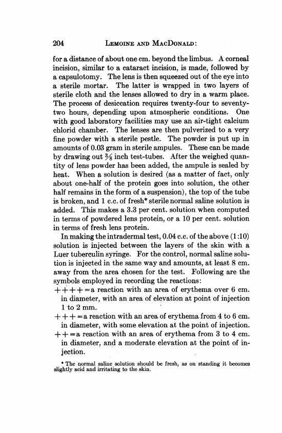
Fig. 1.-Showing wound in cornea, with cone-shaped mass ex-tending from the posterior surface of the lens to the center of thecornea, with purulent infiltration of the cataractous lens-substance.
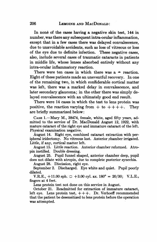
Fig. 2.-Showing extreme swelling of the optic disc.
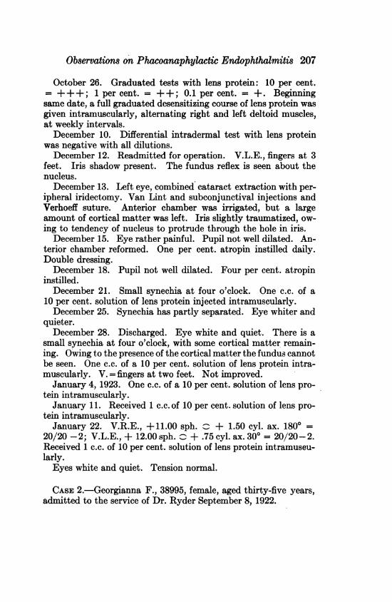
Fig. 3.-Showing infiltration of the cataractouslens with pus; endothelial cells and eosinophilessurrounding the hyaline balls of cataractous lens.

Observations on Phacoanaphylactic Endophthalmitis 205
+ =a reaction with an area of erythema from 2 to 3 cm. indiameter, with a slight elevation at the point of injection.=a reaction with an area of erythema less than 2 cm. indiameter, with very little elevation at point of injection,but definitely more marked than in the control.=when the control and test areas are practically the same.In a great many cases there is a reaction within a half
hour, which in some cases disappears within an hour. Thelatter cases are considered negative. Only reactions whichpersist for at least twenty-four hours are considered positive.Occasionally the reactidn is delayed twelve to twenty-fourhours. These cases are also considered positive.
Before desensitizing a patient one must first ascertain thedegree of hypersensitiveness. This is accomplished by mak-ing a differential intradermal test-i. e., by making the testsin three or more dilutions-as follows: 1:10, 1:100, 1:1000,etc., solutions of lens protein, computed as of fresh lens. Thefirst desensitizing injection should consist of 0.2 c.c. of thesolution giving a : reaction.The subsequent injections are given at intervals of seven
days, which interval is the same as that used by Dr. Walker3in his work with various other proteins, the dose beingdoubled each time until 1 c.c. of the full strength solution isinjected. At this time the patient should give a negativereaction to the full strength solution and may now beoperated upon.As shown by Verhoeff and Lemoine,2 it is best to continue
the injections with the maximum desensitizing dose at weeklyintervals after the operation, until the cortical matter is prac-tically all absorbed, as the desensitization is apparently notpermanent.The purpose of this paper is to report the results of the
intradermal tests with lens protein in 168 unselected cases ofinjury to the lens, traumatic or operative, with the end re-sults in the operated eyes.
LEMOINE AND MACDONALD:
In none of the cases having a negative skin test, 144 innumber, was there any subsequent intra-ocular inflammation,except that in a few cases there was delayed convalescence,due to unavoidable accidents, such as loss of vitreous or lossof the eye due to definite infection. These negative cases,also, include several cases of traumatic cataracts in patientsin middle life, whose lenses absorbed entirely without anyintra-ocular inflammatory reaction.
There were ten cases in which there was a = reaction.Eight of these patients made an uneventful recovery. In oneof the remaining two, in which congiderable cortical matterwas left, there was a marked delay in convalescence, andlater secondary glaucoma; in the other there was simply de-layed convalescence with an ultimately good end result.There were 14 cases in which the test to lens protein was
positive, the reaction varying from + to ++++. Theyare briefly summarized below:CASE 1.-Mary M., 38474, female, white, aged fifty years, ad-
mitted to the service of Dr. MacDonald August 12, 1922, withmature cataract of the right eye and immature cataract of the left.Physical examination negative.August 14. Right eye, combined cataract extraction with per-
ipheral iridectomy. No vitreous lost. Anterior chamber irrigated.Little, if any, cortical matter left.August 15. Little reaction. Anterior chamber reformed. Atro-
pin instilled. Double dressing.August 25. Pupil funnel shaped, anterior chamber deep, pupil
does not dilate with atropin, due to complete posterior synechia.August 28. Discission, right eye.September 2. Discharged. Eye white and quiet. Pupil poorly
dilated.V.R.E., +11.00 sph. +3.00 cyl. ax. 1800 = 20/30; V.L.E.,
fingers at 4 feet.Lens protein test not done on this service in August.October 21. Readmitted for extraction of immature cataract,
left eye. Lens protein test, ++ +. Dr. Verhoeff recommendedthat the patient be desensitized to lens protein before the operationwas attempted.
206
Observations on Phacoanaphylactic Endophthalmitis 207
October 26. Graduated tests with lens protein: 10 per cent.= +++; 1 per cent. = ++; 0.1 per cent. = +. Beginningsame date, a full graduated desensitizing course of lens protein wasgiven intramuscularly, alternating right and left deltoid muscles,at weekly intervals.December 10. Differential intradermal test with lens protein
was negative with all dilutions.December 12. Readmitted for operation. V.L.E., fingers at 3
feet. Iris shadow present. The fundus reflex is seen about thenucleus.December 13. Left eye, combined cataract extraction with per-
ipheral iridectomy. Van Lint and subconjunctival injections andVerhoeff suture. Anterior chamber was irrigated, but a largeamount of cortical matter was left. Iris slightly traumatized, ow-ing to tendency of nucleus to protrude through the hole in iris.December 15. Eye rather painful. Pupil not well dilated. An-
terior chamber reformed. One per cent. atropin instilled daily.Double dressing.December 18. Pupil not well dilated. Four per cent. atropin
instilled.December 21. Small synechia at four o'clock. One c.c. of a
10 per cent. solution of lens protein injected intramuscularly.December 25. Synechia has partly separated. Eye whiter and
quieter.December 28. Discharged. Eye white and quiet. There is a
small synechia at four o'clock, with some cortical matter remain-ing. Owing to the presence of the cortical matter the fundus cannotbe seen. One c.c. of a 10 per cent. solution of lens protein intra-muscularly. V. =fingers at two feet. Not improved.
January 4, 1923. One c.c. of a 10 per cent. solution of lens pro-tein intramuscularly.January 11. Received 1 c.c. of 10 per cent. solution of lens pro-
tein intramuscularly.January 22. V.R.E., +11.00 sph. + 1.50 cyl. ax. 1800 =
20/20'-2; V.L.E., + 12.00 sph. ^ + .75 cyl. ax. 300 = 20/20-2.Received 1 c.c. of 10 per cent. solution of lens protein intramuseu-larly.Eyes white and quiet. Tension normal.
CASE 2.-Georgianna F., 38995, female, aged thirty-five years,admitted to the service of Dr. Ryder September 8, 1922.
208 LEMOINE AND MACDONALD:
Previous History: March 2, 1921, the patient had a combinedcataract extraction for a nearly mature cataract of the right eye,and was discharged fifteen days later with a congested eye in whichconsiderable cortical matter remained. The out-patient depart-ment records show that on April 9th an iritis was present in theright eye.
April 12. Uveitis, right eye.June 14. An atropin dermatitis had developed from a 1 per cent.
solution of atropin t.i.d. The eye was still inflamed and painful.July 7. Posterior synechie and irregular pupil.September 8, 1921. Discission right eye, as a result of which the
eye was still inflamed on October 21.Physical examination was negative. Lens protein test, ++ +.
V.R.E. with correction was 15/200; L.E. = fingers at two feet, notimproved.
Local Examination: Drawn pupil, right eye. Immature cata-ract, left eye. Lateral nystagmus.September 9, 1922. Attempted intracapsular cataract extrac-
tion, left eye, during which the lens capsule ruptured, and the oper-ation was completed by the ordinary method, leaving much corticalmatter.
September 15. Patient gradually developing congestion, pain,photophobia, and lacrimation. In spite of treatment with atropin,dionin, and hot fomentation, the condition became worse, and adescemetitis developed. Pupil will not dilate. Patient has anatropin irritation and hyoscin hydrobromid 1/3 of 1 per cent. wassubstituted. Paracentesis performed.
October 16, 1922. No relief following paracentesis.October 22. Descemetitis, congestion, pain, and photophobia
are all more marked. Dr. Ryder advised desensitization to lensprotein. The graduated intradermal tests with lens protein werepositive as follows: 0.1 per cent. = +; 1 per cent. = ++ ; 10 percent. = +++; one hour after the tests were made.
October 23. Twenty-four hours after tests were made they werepositive as follows: 0.1 per cent. = ++; 1 per cent. = ++ +;10 per cent. = ++From October 23d to December 20th a full course of desensitizing
intramuscular injections of lens protein was given. Twenty-fourhours after each injection the patient had an exacerbation of pain,photophobia, and lacrimation, followed on the third or fourth dayby amelioration of symptoms. At the completion of the course the
Observations on Phacoanaphylactic Endophthalmitis 209
graduated intradermal tests were still all positive and further at-tempts to desensitize the patient were abandoned. Dischargedon January 2, 1923., Eye congested, descemetitis marked, andpupil drawn up; cortical matter still present. Pain persisted,also photophobia and lacrimation. V.L.E. = light projection,faulty.
CASE 3.-John G., 39549, male, aged forty-seven years, ad-mitted to the service of Dr. Derby on October 5, 1922, complain-ing of loss of vision, right eye.
Past History: Had scarlet fever at age of twelve years. At theage of nine years had an injury to left eye that never caused himany marked trouble, except occasional irritation and pain. Theleft eye was enucleated at the age of thirty-five on account of re-currmg irritation and pain.
Present Illness: On December 14, 1921, he was struck in theright eye by a piece of wood, which imnmediately destroyed thevision. An iritis developed which lasted six weeks. On March 31,1922, the patient was referred to Dr. Derby for extraction of theresulting traumatic cataract. An iritis followed the extractionwhich lasted three weeks.
Local Examination: Right eye, lids and lacrimal apparatus ap-parently normal. The cornea had a scar at 5.30 o'clock, extendingfrom 2 mm. from the limbus toward the center for a distance of 2.5mm. There was an operative scar in the limbus above. The an-terior chamber was shallow and the iris was incarcerated in thetraumatic corneal scar. The pupil was drawn up and occluded bya membrane extending to the limbus above and filling the coloboma,which was slit-like. No fundus reflex was seen. Physical examina-tion negative. V.R.E. = good light projection. L.E., anoph-thalmos. Lens protein test = + +.
October 6. Right eye, iridotomy and separation of anteriorsynechiae, by Dr. Derby. Dressed with double bandage and atro-pin ointment.
October 9. Little reaction. Good anterior chamber. Atropininstilled daily.
October 12. Eye quieting nicely.October 16. Discharged. Eye white and quiet. Good anterior
chamber. Has vitreous opacities, apparently from old hemorrhage.On discharge, V. with + 12.00 sph. = 20/200. Patient not seensince discharge.
14

LEMOINE AND MACDONALD:
CASE 4.-Charles K., 38113, male, white, aged twenty-sevenyears, admitted to the service of Dr. Thompson, July 24, 1922,with traumatic cataract of the left eye. V.L.E. = good light pro-jection. Physical examination, negative. Lens protein test, 10per cent. solution = ++; 1 per cent. solution = +.
History: Had steel intra-ocular foreign body May 2, 1922. Thesteel was removed by the anterior route, May 3d, with the giantand small magnets. Discharged, June 9th, with congted eye andpoorly dilated pupil. The lens slowly became cataractous and theeye developed signs of intra-ocular inflammation, and graduallybecame more painful.
Local examination showed conjunctival and deep circumcornealcongestion. Good anterior chamber. Iriswasmuddy, discolored, andbound down to the lens by four large synechie. Tension normal.
July 25. Left eye, linear extraction with separation of the syne-chi,e. Considerable cortical matter remained.
Following operation the eye remained painful and congested, inwhich condition he was discharged to the out-patient departienton August 5th, with good light projection. The patient reportedregularly at the out-patient department until December 19th,when, owing to the fact that the left eye was still congested, lightprojection faulty, tension minus, and still very painful, Dr. Ryderadvised enucleation. The patient returned to the clinic in Febru-ary, 1923; the eye was congested and soft, and the light projectionstill faulty. An intradermal test with uveal pigment-was negativeand enucleation was postponed.
CASE 5.-Margaret C., 37856, female, white, aged sixty-sevenyears, admitted to the service of Dr. Thompson July 11th, withnearly mature cataract of the right eye and immature cataract ofthe left. V.R.E. = shadows, light projection good; V.L.E., withcorrection, 20/100. The lens protein test, +++.
Physical examination negative, except that the teeth are in badcondition.
July 12. Combined cataract extraction, right eye.July 18. Pupil small, considerable cortical matter remains.July 20. Right eye began to be painful. The pupil was not well
dilated. Conjunctiva congested.July 25. Patient sensitive to one per cent. atropin and hyoscin
hydrobromid, one-third of one per cent., substituted. Eye verypainful. Cortical matter absorbing slowly.
210
Observations on Phacoanaphylactic Endophthalmitis 211
August 8. Eye quieting slowly, but still irritable and painful inspite of hyoscin, dionin, and hot fomentations.
September 1. Eye has quieted very slowly; now nearly white.The pupil is drawn up and there are posterior synechiw of iris withremnants of cortical matter and capsule that partly occlude pupil-lary opening. Vision, with correction, = 20/100. Tension, normal.CASE 6.-Annie W., 37224, female, white, aged sixty years, ad-
mitted to the service of Dr. Lancaster June 7, 1922, with nearlymature cataract of the right eye. V.R.E. = fingers at one foot.
Physical examination negative.Lens protein test = +++.June 8. Combined cataract extraction, right eye. Very little
cortical matter left.June 15. Good anterior chamber. Pupil well dilated. Very
little cortical matter present. There is a posterior synechia of theiris at four o'clock.June 24. Patient discharged. Eye congested. Vision = fingers
at one foot, not improved.June 26. Out-patient department. Eye still congested with
slight increase in tension.August 30. Vision, with correction, = 20/200.CASE 7.-Mary N., 38500, female, white, aged sixty-five years,
admitted to the service of Dr. Derby August 14, 1922, with scle-rosed lens, right eye. Left pupil blocked from former prolapse ofiris, after cataract operation. Lens protein test = +. The patientwas a diabetic but is now sugar free, and blood sugar within normalrange. Heart and lung negative.August 22. Combined cataract extraction, right eye. Practic-
ally no cortical matter left.August 26. Some striped keratitis.September 2. Discharged with pupil well dilated. Some cap-
sule in the pupillary opening. Moderate congestion. Vision, withcorrection, = fingers at six feet.CASE 8.-James S., 39203, male, white, aged sixty-eight years,
admitted to the service of Dr. Derby September 18, 1922, withimmature cataract of the right eye and nearly mature cataract ofthe left. Lens protein test = +. V.R.E., - 1.00 sph. = 20/100;L.E. = fingers at one foot. Light projection good.
Physical examination at the Massachusetts General Hospitalshowed arteriosclerosis, cardiosclerosis, and chronic nephritis.
LEMOINE AND MACDONALD:
September 19. Combined cataract extraction, left eye. Littlecortical matter left.September 21. Patient developed acute dementia and sustained
a severe fall that resulted in a fracture of left humerus.September 27. Transferred for surgical treatment to the Mas-
sachusetts General Hospital. No cortical matter in pupillary open-ing. Eye in good condition. Tension normal. Vision, with ap-proximate corrections, = 20/200 +.
CASE 9.-Herbert M., 39683, male, admitted October 13, 1922,as a private patient (Dr. Verhoeff). Hypermature cataract, lefteye. Physical examination, negative. Lens protein test = +. Hewas operated upon October 14, intracapsular extraction withoutiridectomy; no loss of vitreous; uneventful recovery.
October 30. Vision, with correction, = 20/15.CASE 10.-Jeanette H., 39854, female, white, aged seventy-four
years, admitted to the service of Dr. MacDonald October 25, 1922,with mature cataract of the left eye and incipient cataract of theright. Lens protein test =
Physical examination showed chronic cardiovascular disease(mitral stenosis, aortic insufficiency, and arteriosclerosis) andchronic right otitis media. On October 25th, a combined cataractextraction with peripheral iridectomy was performed on the lefteye. Practically no cortical matter remained. She was dischargedin ten days, after an uneventful recovery. Vision= 20/100. Therewas a small amount of lens capsule in the pupillary opening.CASE 11.-Peter M., 39874, male, white, aged seventy-two years,
admitted to the service of Dr. Verhoeff October 23, 1922, withmature cataract of the left eye. Physical examination negative.Lens protein test +. Was operated upon October 26th by theintracapsular method. The lens capsule ruptured as the lens wasbeing delivered, but practically no cortical matter was left. Hewas discharged in ten days after an uneventful recovery, except astriped keratitis. Vision = 20/20. Eye white bnd quiet. Pupilwell dilated.
CASE 12.-Sarah V., 39244, female, white, aged fifty years, ad-mitted on September 21, 1922, to the service of Dr. Thompson,with a mature cataract of the left eye. Aphakia, right eye, follow-ing intracapsular extraction.
Physical examination negative. Lens protein test = +. Sep-
212

Observations on Phacoanaphylactic Endophthalmitis 213
tember 22d, combined cataract extraction of the left eye withoutcomplications. No cortical matter left. Anterior chamber not ir-rigated. The patient was discharged in eleven days with the eyeslightly congested. The nasal pillar of the iris was bound down tothe corneal wound. V.R.E., with correction, = 20/20. L.E., withcorrection, = 20/30.CASE 13.-John O., 39455, male, white, aged seventy-seven years,
admitted to the service of Dr. Derby, September 2, 1922, withnearly mature cataract of the right eye. Lens protein test =September 3. Combined cataract extraction of the right eye
with Van Lint injection of the orbicularis oculi. Considerable cor-tical matter was left, which appeared to be absorbing well, althoughthe pupil did not dilate. The patient was discharged in ten dayswith the eye slightly congested and some cortical matter remaining.V.R.E., with correction, 20/200.
Patient was seen twelve days later in the out-patient depart-ment; eye was congested.
Fifty-seven days later the eye was still painful and congested.CASE 14.-Calvin R., 39459, male, white, aged eighty-two
years, admitted to the service of Dr. Derby on October 2, 1922,with early hypermature cataract, right eye. Immature cataractof the left eye.Examination negative, except for aneurysm of aorta. Lens pro-
tein, intradermal test = +. The right eye was operated uponOctober 3d by the combined method, leaving practically no cor-tical matter. He was discharged in eleven days with the eye whiteand quiet. Some capsule remained in the pupillary opening and atdischarge the vision = 5/200, not improved.
In addition to the foregoing cases there were two othercases with positive tests to lens protein, which are not in-cluded above, since.one (case 15) was, in a sense, selected, thediagnosis having been made by Dr. Verhoeff on the micro-scopic examination, and subsequently confirmed by the in-tradermal tests. In the other (case 16)" the test was doneafter the above series was completed.CASE 15.-Arthur M., 38900, Italian, aged six years, admitted to
the Massachusetts Charitable Eye and Ear Infirmary, on the ser-vice of Dr. MacDonald, on July 23, 1922. Past and family history

24LEMOINE AND MACDONALD:
negative. One and one-half hours before admission, while reachingto a shelf for an open knife, it fell and struck him in the right eye,inflicting a perforating wound of the eyeball.
Examination on admission: Right eye, the lids and conjunctivawere normal. The cornea was clear, except for a perforating woundextending from the limbus at twelve o'clock to the limbus at sixo'clock. There was no anterior chamber and a large portion of theiris, with lens matter and vitreous, protruded through the upperpart of the wound. Under ether narcosis all the prolapsed iris, vit-reous, and lens were excised, and the edges of the iris were replaced.The apposition of the wound was good, so no conjunctival flap wasmade. Instilled 1 per cent. solution of atropin sulphate andbichlorid ointment and applied a double bandage.
July 26. Lens much swollen, which caused the corneal wound togape with a small prolapse of the iris at the top. The prolapsed iriswas excised and the upper part of the cornea, where the wound wasgaping, was covered with a conjunctival flap dissected from above.
July 29. The conjunctival flap was in position. The anteriorchamber was not reformed. Pupil irregular. The eye was treatedtwice daily with boracic acid solution, one-half of one per cent. solu-tion of atropin sulphate, and bichlorid of mercury ointment.
August 8. The eye was fairly white and quiet. The flap was inposition over the upper part of the wound. The anterior chamberwas reformed but shallow; the pupil was not well dilated.August 14. Condition of the right eye about the same, but the
pupil was not so well dilated. The left eye showed no signs of in-volvement.
September 2. Right eye same as above. Left eye, there wascongestion of the conjunctiva, also deep circumcorneal congestion.The pupil reacted more sluggishly to light than before. No defi-nite descemetitis. Enucleation of the right eye was refused by theparents, so patient was discharged against advice.September 6. Readmitted to the wards. Condition of the right
eye the same, except that it was painful. Under ether narcosis theeye was enucleated. Left eye showed definite signs of photophobiaand tenderness to palpation. Ciliary congestion marked. Descem-etitis lower portion of cornea. Pupil dilated. Several posteriorsynechia. Fundus not clearly seen. Atropin sulphate, a half percent., t.i.d.September 8. Left eye, there was a fair amount of conjunctival
and ciliary congestion with more, but not marked, photophobia.
214
Observations on Phacoanaphylactic Endophthalmitis 215
Eye was sensitive to pressure. Increased deposits on Descemet'smembrane. Pupil fairly well dilated. Fundus obscure and dischazy. Vision = fingers at three feet. Weight, 38½ pounds. So-dium salicylate, gr. x, t.i.d. Blood count, white blood-corpuscles,8,000; polymorphonuclear, 44 per cent.; lymphocytes, 52 per cent.;large mononuclears, 4 per cent.September 20. Intradermal test = ++ +.September 28. Much better. Eye white and quiet. Vision,
with correction, = 20/50.September 30. Further improvement. Details of fundus made
out for first time. Still many dust-like opacities in the vitreous.October 8. White and quiet. Discharged to out-patient depart-
ment, with atropin sulphate, one-quarter of one per cent., t.i.d.Sodium salicylate, gr. x, t.i.d.February 4, 1923. Complement fixation with uveal pigment an-
tigen, by Dr. Alan Woods,4 was negative.February 14. The intradermal test with one-tenth of one per
cent. solution of uveal pigment was immediately positive, but neg-ative after twenty-four and forty-eight hours. At this time thesympathetic uveitis was not very active.
Pathologic report of the right eye, No. 4602. Fixation in 10 percent. formalin.There is a healed perforating wound, extending entirely across
the center of the cornea. The corneal epithelium is everywhereintact. The stroma is edematous, and the cornea is thickened,4 mm. The corneal corpuscles are somewhat increased. There isa cellular infiltration at the periphery of the cornea, extending 3mm. anterior to the canal of Schlemm, consisting of lymphocytes,eosinophiles, and a few polymorphonuclear leukocytes. A round-cell infiltration also occurs for 2 mm. on either side of the cornealwound, and here the thickness of the cornea is increased to 2 mm.Descemet's membrane, with its endothelial lining, is everywhereintact, except at the site of the wound. The posterior edges of thecorneal wound are separated 1.5 mm., and the gap is filled with amass of granulation tissue that extends backward through the lensand projects slightly into the vitreous. The central part of themass is markedly infiltrated with pus cells and eosinophiles sur-rounding numerous globules of lens matter, and shows here andthere areas of epithelioid cells and a few giant cells. The pupillarymargins of the iris are adherent to the edges of the wound and themass of tissue extending from it.
LEMOINE AND MACDONALD:
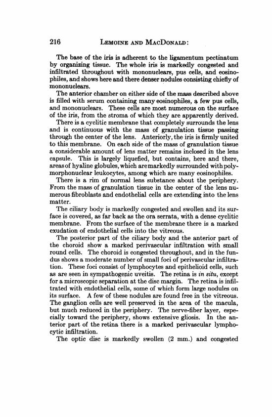
The base of the iris is adherent to the ligamentum pectinatumby organizing tissue. The whole iris is markedly congested andinfiltrated throughout with mononuclears, pus cells, and eosino-philes, and shows here and there denser nodules consisting chiefly ofmononuclears.The anterior chamber on either side of the mass described above
is filled with serum containing many eosinophiles, a few pus cells,and mononuclears. These cells are most numerous on the surfaceof the iris, from the stroma of which they are apparently derived.There is a cyclitic membrane that completely surrounds the lens
and is continuous with the mass of granulation tissue passingthrough the center of the lens. Anteriorly, the iris is firmly unitedto this membrane. On each side of the mass of granulation tissuea considerable amount of lens matter remains inclosed in the lenscapsule. This is largely liquefied, but contains, here and there,areas of hyaline globules, which aremarkedly surrounded with poly-morphonuclear leukocytes, among which are many eosinophiles.There is a rim of normal lens substance about the periphery.
From the mass of granulation tissue in the center of the lens nu-merous fibroblasts and endothelial cells are extending into the lensmatter.The ciliary body is markedly congested and swollen and its sur-
face is covered, as far back as the ora serrata, with a dense cycliticmembrane. From the surface of the membrane there is a markedexudation of endothelial cells into the vitreous.The posterior part of the ciliary body and the anterior part of
the choroid show a marked perivascular infiltration with smallround cells. The choroid is congested throughout, and in the fun-dus shows a moderate number of small foci of perivascular infiltra-tion. These foci consist of lymphocytes and epithelioid cells, suchas are seen in sympathogepic uveitis. The retina is in situ, exceptfor a microscopic separation at the disc margin. The retina is infil-trated with endothelial cells, some of which form large nodules onits surface. A few of these nodules are found free in the vitreous.The ganglion cells are well preserved in the area of the macula,but much reduced in the periphery. The nerve-fiber layer, espe-cially toward the periphery, shows extensive gliosis. In the an-terior part of the retina there is a marked perivascular lympho-cytic infiltration.The optic disc is markedly swollen (2 mm.) and congested
216
Observations on Phacoanaphylactic Endophthalmitis 217
throughout and shows a slight round-cell infiltration. The surfaceshows some glial proliferation.
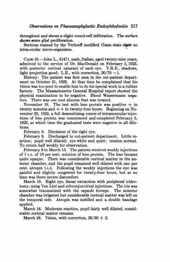
Sections stained by the Verhoeff modified Gram stain sl'v nointra-ocular micro-organisms. T;CASE 16.-John L., 41471, male, Italian, aged twenty-nine years,
admitted to the service of Dr. MacDonald on February 5, 1923,with posterior cortical cataract of each eye. V.R.E., shadows,light projection good; L.E., with correction, 20/70 -1.
History: The patient was first seen in the out-patient depart-ment on October 21, 1922. At that time he complained that hisvision was too poor to enable him to do his special work in a rubberfactory. The Massachusetts General Hospital report showed thephysical examination to be negative. Blood Wassermann nega-tive. There was one root abscess that was treated.November 19. The test with lens protein was positive + in
twenty minutes and ++ in twenty-four hours. Beginning on No-vember 25, 1922, a full desensitizing course of intramuscular injec-tions of lens protein was commenced and completed February 5,1923, at which time the graduated tests were negative in all dilu-tions.February 6. Discission of the right eye.February 8. Discharged to out-patient department. Little re-
action; pupil well dilated; eye white and quiet; tension, normal.To return half weekly for observation.February 8 to March 15. The patient received weekly injections
of 1 c.c. of 10 per cent. solution of lens protein. The lens becamequite opaque. There was considerable cortical matter in the an-terior chamber, and the pupil remained well dilated with one percent. atropin t.i.d. Following the weekly injections the eye waspainful and slightly congested for twenty-four hours, but at notime was there severe discomfort.March 16. Right eye, linear extraction with peripheral iridec-
tomy, using Van Lint and subconjunctival injections. The iris wassomewhat traumatized with the capsule forceps. The anteriorchamber was irrigated but considerable cortical matter was left onthe temporal side. Atropin was instilled and a double bandageapplied.March 18. Moderate reaction, pupil fairly well dilated, consid-
erable cortical matter remains.March 24. Vision, with correction, 20/30 + 2.
LEMOINE AND MACDONALD:
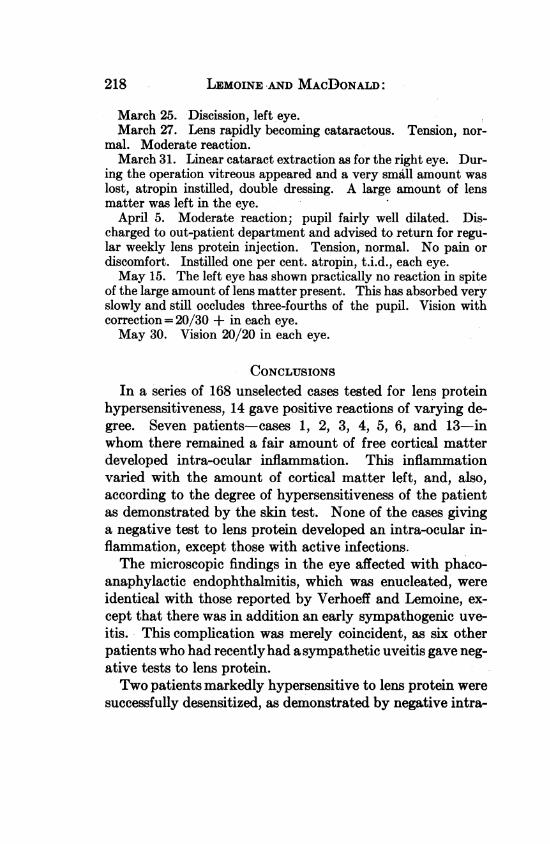
March 25. Discission, left eye.March 27. Lens rapidly becoming cataractous. Tension, nor-
mal. Moderate reaction.March 31. Linear cataract extraction as for the right eye. Dur-
ing the operation vitreous appeared and a very small amount waslost, atropin instilled, double dressing. A large amount of lensmatter was left in the eye.
April 5. Moderate reaction; pupil fairly well dilated. Dis-charged to out-patient department and advised to return for regu-lar weekly lens protein injection. Tension, normal. No pain ordiscomfort. Instilled one per cent. atropin, t.i.d., each eye.May 15. The left eye has shown practically no reaction in spite
of the large amount of lens matter present. This has absorbed veryslowly and still occludes three-fourths of the pupil. Vision withcorrection = 20/30 + in each eye.May 30. Vision 20/20 in each eye.
CONCLUSIONSIn a series of 168 unselected cases tested for lens protein
hypersensitiveness, 14 gave positive reactions of varying de-gree. Seven patients-cases 1, 2, 3, 4, 5, 6, and 13-inwhom there remained a fair amount of free cortical matterdeveloped intra-ocular inflammation. This inflammationvaried with the amount of cortical matter left, and, also,according to the degree of hypersensitiveness of the patientas demonstrated by the skin test. None of the cases givinga negative test to lens protein developed an intra-ocular in-flammation, except those with active infections.The microscopic findings in the eye affected with phaco-
anaphylactic endophthalmitis, which was enucleated, wereidentical with those reported by Verhoeff and Lemoine, ex-cept that there was in addition an early sympathogenic uve-itis. This complication was merely coincident, as six otherpatients who had recentlyhad asympathetic uveitis gave neg-ative tests to lens protein.Two patients markedly hypersensitive to lens protein were
successfully desensitized, as demonstrated by negative intra-
218
Observatiom on Phacoanaphylactic Endophthalmitis 219
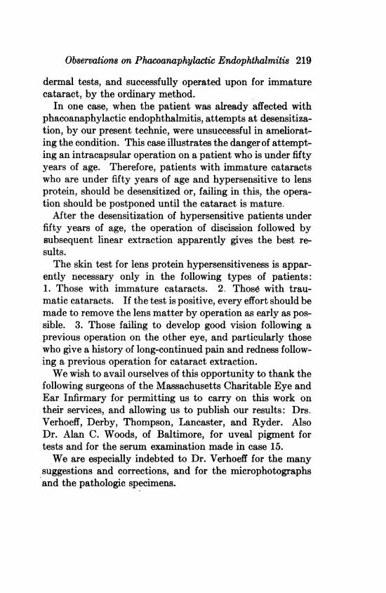
dermal tests, and successfully operated upon for immaturecataract, by the ordinary method.
In one case, when the patient was already affected withphacoanaphylactic endophthalmitis, attempts at desensitiza-tion, by our present technic, were unsuccessful in ameliorat-ing the condition. This case illustrates the danger of attempt-ing an intracapsular operation on a patient who is under fiftyyears of age. Therefore, patients with immature cataractswho are under fifty years of age and hypersensitive to lensprotein, should be desensitized or, failing in this, the opera-tion should be postponed until the cataract is mature.
After the desensitization of hypersensitive patients underfifty years of age, the operation of discission followed bysubsequent linear extraction apparently gives the best re-sults.The skin test for lens protein hypersensitiveness is appar-
ently necessary only in the following types of patients:1. Those with immature cataracts. 2. Those with trau-matic cataracts. If the test is positive, every effort should bemade to remove the lens matter by operation as early as pos-sible. 3. Those failing to develop good vision following aprevious operation on the other eye, and particularly thosewho give a history of long-continued pain and redness follow-ing a previous operation for cataract extraction.We wish to avail ourselves of this opportunity to thank the
following surgeons of the Massachusetts Charitable Eye andEar Infirmary for permitting us to carry on this work ontheir services, and allowing us to publish our results: Drs.Verhoeff, Derby, Thompson, Lancaster, and Ryder. AlsoDr. Alan C. Woods, of Baltimore, for uveal pigment fortests and for the serum examination made in case 15.We are especially indebted to Dr. Verhoeff for the many
suggestions and corrections, and for the microphotographsand the pathologic specimens.
LEMOINE AND MACDONALD:
REFERENCES1. Verhoeff and Lemoine: Endophthalmitis Phacoanaphylactica. Transac-
tions, An International Congress of Ophthalmology, Washington, 1922.2. Verhoeff and Lemoine: Hypersensitiveness to Lens Protein-Patient De-
sensitized and Successfuly Operated on for Immature Cataract, Amer.Jour. Ophth., voL v, No. 9, 1922.
3. Walker, I. C.: Personal communication regarding desensitizing of patientsin his routine protein therapy work. Aso,Study XIV: The Treatmentof Patients with Bronchial Asthma with Subcutaneous Injections ofProtein to which they are Sensitive, Jour. Med. Research, 1917, vol.xxxvi, p. 51.
4. Woods, Alan C.: Personal communication.
DISCUSSIONDR. W. B. LANCASTER, Boston, Mass.: I want to commend to
the members of the Society the use of this test. It is simple to per-form. We do it regularly on all the cases at the Infirmary and it isa useful guide. Bear in mind, however, that a vast majority ofcases are not sensitive-less than one in ten. So if you find a fewcases negative do not be surprised.
It is particularly useful in traumatic cases. If you have somecortical matter left in the anterior chamber of an eye that is in-jured, you can ordinarily treat the eye with expectant methods;but if you find the patient is sensitive to lens protein, you shouldimmediately evacuate as much as possible of the lens matter beforeserious inflammation is started.DR. GEORGE S. DERBY, Boston, Mass.: The lens protein test has
been used in all the cataract cases on my service at the Massa-chusetts Eye and Ear Infirmary during the last eighteen months,and also on most of my private cases. I am glad to testify to theextremely careful and conscientious work of Drs. Lemoine andMacDonald. I think they deserve a great deal of credit for pursu-ing an investigation of this sort during a busy and exacting service.As to the value of the test, I do not as yet feel able to form any
definite personal opinion. One or two cases have been suggestive.If the results of Verhoeff and the writers are confirmed, it is hardlynecessary to point out the great value of the test.DR. ARNOLD KNAPP, New York, May I ask Dr. Lemoine to tell
us if this anaphylactic iniflammation has definite clinical signs?Also where the material for the test may be obtained?DR. W. H. WILDER, Chicago: I think we should be very care-
ful before we come to any positive conclusions on a subject as
220

Observations on Phacoanaphylactic Endophthalmitis 221
complex as this. From experiments of Hektoen and others it hasbeen shown that the lens is not species specific at all, and, further-more, it seems to be true that certain tissues of the eye that havealready been sensitized to certain proteins or globulins or someother complex chemical substances, may react to whatever kind ofprotein may be injected into the circulation. It seems to me,therefore, before we can determine conclusively that a given eye isreacting because of some lens protein left in the anterior chamber,we must know in each individual case what the conditions are inthe tissues of that eye, particularly the uveal tissues. It has beenan observation of clinicians for many years that we frequently, butnot always, get pronounced reaction when cortical substance is leftin the chamber after cataract extraction. We should welcome,therefore, any safe procedure by which we could regulate this. Butwe cannot be positive, unless it is possible to make controls, that itis the lens protein that is affecting the eye. It may be that in agiven eye there is a condition which renders it sensitive to lens pro-tein, but it may be sensitive also to other proteins, such as peptoneor even bacterial proteins, that may gain entrance to the circula-tion. So I say, let us not be too hasty in forming our conclusions,but in all cases have something in the nature of a control.One other thought in regard to results after cataract operations:
There is a great disposition on the part of individuals who reportseries of cataract cases to judge of the value of any operative pro-cedure by the end results in terms of vision, and that is an errorinto which the protagonists of the intracapsular operation fre-quently fall. It would be much more accurate and enlightening indescribing the end results to give information as to the severity andduration of the reaction, the clearness and shape of the pupil, theamount of astigmatism, etc., rather than the degree of vision alone.A case that shows 20/200 vision might have been more of a
success in an operative way than one that showed 20/50, or even20/20. We all know that the degree of vision will depend as muchupon the transparency of the vitreous and the condition of thechoroid and retina around the posterior pole as it will on the clear-ness of the pupil. What we want to know is whether a certainmethod of treatment or operation gave the eye a better chance thansome other method, and we should not attempt to justify anyprocedure merely by end results obtained in terms of vision.
DR. W. H. WILMER, Washington, D. C.: It would seem that
LEMOINE AND MACDONALID:
there are definite lessons to be leamed from this very valuablepaper. All of us who have had a moderately large operative experi-ence, over a number of years, have considered cortex left in theeye as a foreign body. We have felt (for years before these scientificadvances were made) that it behooved us to get rid of this danger.For this reason we have believed that in certain types of cases theintracapsular operation is the ideal procedure-ither the operationas performed by Dr. Knapp or some other similar operation. If nosuch operative procedure is followed, it behooves us to remove thecortex either by a spoon, by expression, or by some other means. Ipersonally believe that the anterior syringe is a valuable instru-ment. One can syringe the anterior chamber thoroughly, after thecapsule is incised, until all cortex is removed, and we should not besatisfied until this removal is complete.
I believe further that the anterior syringe has another value. Itpushes back the little tags of capsule, forces the iris away from thecornea, and prevents adhesion of the cut iris to the corneal wound.With proper precautions it is not dangerous and should not leadto infection. I happened to look over my own operative cases fora number of years, and I find that since 1915 I have had no infec-tion of the wound in any operative procedure in spite of havingused the anterior syringe frequently, so one can safely say that itis not dangerous to use the syringe provided the proper antisepticprecautions are taken. Its use does not increase the traumatismand it does enable one to expel the cortex from the anterior cham-ber, so that I am certain that the anterior syringe still has itsplace in ophthalmic surgery today.
DR. ALBERT LEMOINE (closing): In reply to Dr. Knapp's ques-tions, the signs of intra-ocular inflammation are similar to those ofiridocycitis, a contracted pupil, congested iris, and the corticalmatter assumes a milky, granular appearance. It is whiter andmore granular than' ordinary cortical matter. Very frequentlythere is descemetitis associated with it, which may be very severe.There is also pain, and the lacrimation is usually very marked incomparison to the inflammation present. Case 2 was of this char-acter; the descemetitis covered the whole cornea and it persistedfor several weeks.
Lens protein is not on the market. The only place it can beobtained is at the Eye and Ear Infirmary. Miss Taylor, in Dr.Verhoeff's laboratory, is preparing some for the doctors who ask for
222

ZENTMAYER: Monocular Diplopia
it. I also have been preparing what I need for my personal use andhave supplied some of the oculists with small quantities.As to Dr. Wilder's remarks, we had 144 control cases. All of
these cases had cortical matter left in the eye. All gave negativetests to protein,and none had intra-ocular inflammation except-ing- where there was an active infection.
MONOCULAR DIPLOPIA, WITH ESPECIAL REFER-ENCE TO THAT ASSOCIATED WITH CEREBRAL
LESIONS
WILLIAM ZENTMAYER, M.D.Philadelphia, Pa.
The conditions in which monocular diplopia is met havebeen stated by de Schweinitz to be-(1) anomalies of refrac-tion, particularly astigmatism; (2) opacities in the corneaand lens; (3) irregular cramp of ciliary muscle; (4) hysteriaor allied functional nervous disturbances-for example, neu-rasthenia and cerebral asthenia; (5) organic disease of thebrain or its membranes; (6) simulation. To these should beadded (7) dislocated lens and (8) detachment of the retina.
In an excellent article on "Monocular Diplopia; Its Rela-tion to Hysteria," Charles makes a somewhat different classi-fication: (1) That form which proceeds from physical causes.(2) That resulting from the activity of a pseudo-fovea simul-taneous with that of the anatomic fovea. (3) A form asso-ciated with cerebral lesions. (4) A form not accounted for byany other hypothesis than that there is a cortical dissociation,which is the basis upon which hysteria is explained by themajority of modern neurologists.
Cases falling under several of these heads are well knownand of no particular interest. There are, however, cases ofexceptional causation, little known, which are deserving of abrief mention.
223