Embed Size (px)
DESCRIPTION
Methods of measuring intra ocular pressure. What is intra ocular pressure ?. Definition Intra ocular pressure refers to the pressure exerted by the intra ocular contents on the coats of the eyeball. - PowerPoint PPT Presentation
Citation preview
Methods of measuring intra ocular pressure
What is intra ocular pressure ?
Definition Intra ocular pressure refers to the pressure
exerted by the intra ocular contents on the coats of the eyeball.
It is maintained by the equilibrium between the aqueous humour formation, its outflow and the episcleral venous pressure
Normal IOP – 10.2mmhg to 20.6mmhg
Types of Tonometer
Schiotz Tonometer Applanation Tonometer- Goldman Tonopen Pulse air Tonometer Non contact Tonometer
Schiotz Tonometer
Devised in the year 1905 Schiotz tonometer is the instrument which is used
to measure the intra ocular pressure ( IOP )
Requirements (Schiotz tonometer)
Schiotz tonometer with 5.5gm ,7.5gm and 10gm weight
Measurement chart (Friedan Wald chart) 4% Xylocaine as local anaesthetic Wiper jar (Sterile) Kidney type tray Antibiotic drops (Chloramphenical ) 2% propanolol &Tissue paper
Method Explain the procedure to the patient and make
him comfortable. He should be lying down with head on the pillow.
Check the instrument by placing over the model. Now the indicator ‘0’in the scale
Wash and dry hands. Instill one drops of lignocaine 4%eyedrops in the
Eye (use pouch technique) – Explain the burning sensation & other points
Allow 2to5minutes for the local drops to act.
Ask the patient to look straight up Separate the eyelids, putting no pressure on the eye.
Fingers should rest on the orbital margin. Hold the tonometer so that footplate rests on the
center of the cornea. Look at the scale of the Tonometer to find out the
degree of indentation. If the weight is not enough to indent the cornea , use a heavier weight
Use the chart to calculate the intraocular pressure for that particular weight, and that degree of indentation
Record the intraocular pressure in the patients note’s
Sterilizing the instruments Before replacing the tonometer, dismantle it and
clean the parts. Remove the weights, unscrew the plunger and
clean them using swabs dipped in alcohol Heat the footplate over the blue flame of the spirit
lamp for two minutes. Now the footplate is sterilized
Keep the tonometer over the sterile wipers/pad so that it cools down to room temperature
Check the instruments by placing over the model. Now the indicator should show ‘0’in the scale
Tonopen
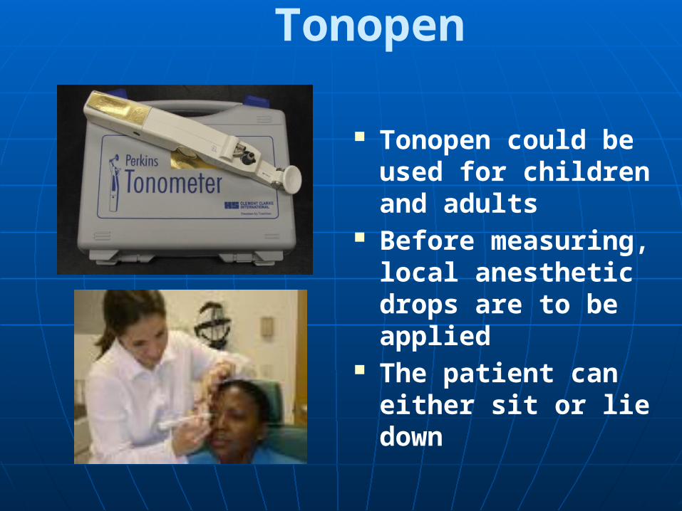
Tonopen could be used for children and adults
Before measuring, local anesthetic drops are to be applied
The patient can either sit or lie down
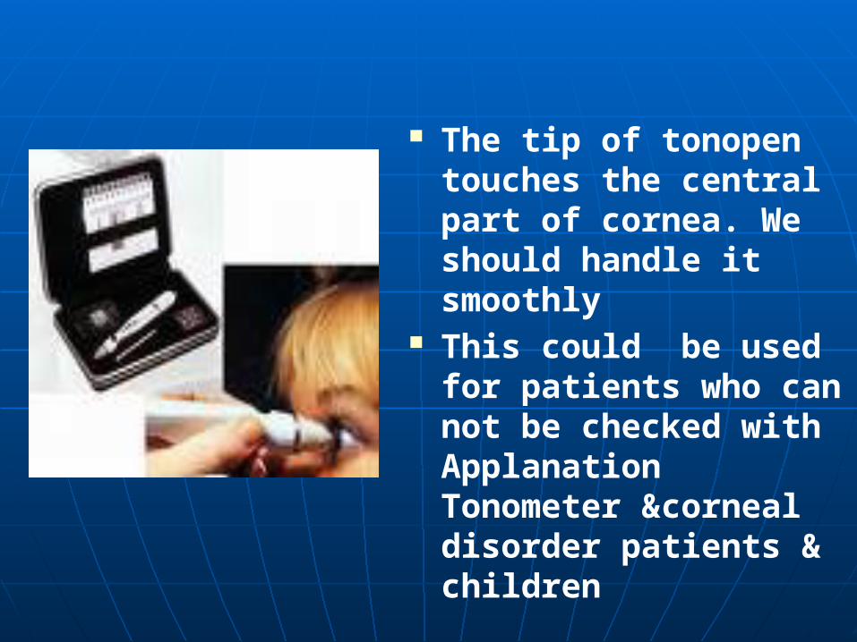
The tip of tonopen touches the central part of cornea. We should handle it smoothly
This could be used for patients who can not be checked with Applanation Tonometer &corneal disorder patients & children
Pulse Air Tonometer
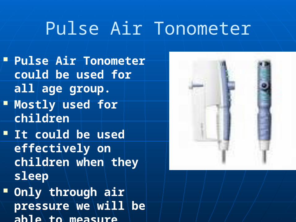
Pulse Air Tonometer could be used for all age group.
Mostly used for children It could be used effectively
on children when they sleep Only through air pressure
we will be able to measure
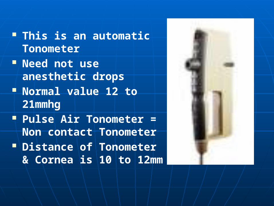
This is an automatic Tonometer Need not use anesthetic drops Normal value 12 to 21mmhg Pulse Air Tonometer = Non
contact Tonometer Distance of Tonometer &
Cornea is 10 to 12mm
Non contact Tonometer
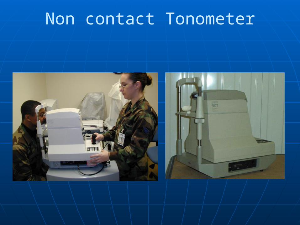
This is like the slit lamp Patients is to be focused to see the red-light
inside the Tonometer Picture of the eye could be seen on the screen A white dot will be seen in the cornea The MLOP should focus the white dot and click
it At the time of focusing, if you see the
word-’Forward’ ,you should adjust the joystick in front side.
At the time of focusing, if you see the word-’Too close’ ,you should adjust the joystick in back side.
Automatically the reading will be shown in the screen itself
This will be useful for injury and redness and post operative patients
No need to use local drops.
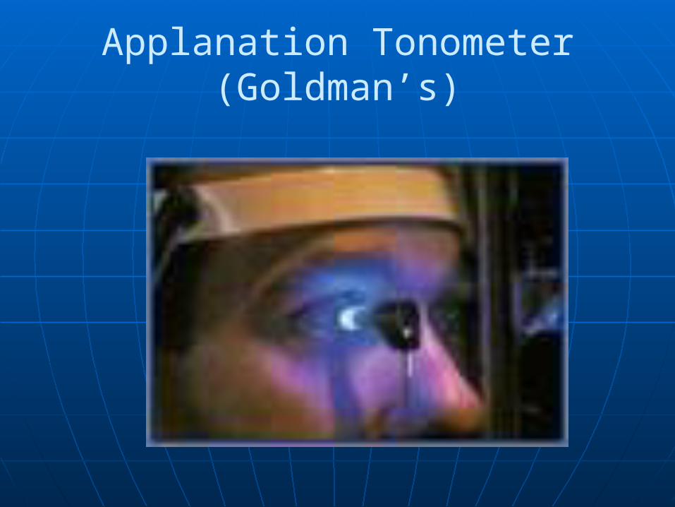
Goldman’s applanation Tonometer is capable of measuring intraocular pressure accurately to 0.5mm
It is more accurate than Schiotz Tonometer This is used with slit lamp microscope It measures the area of the circle made flat by the
instrument on the flat and spherical part of the cornea It has prism with external area of the Tonometer A measuring drum attached to the slit lamp is inserted
into the handle over the chamber

Procedure
Explain the method to the patient and keep the patient in front of the slit lamp
Apply local anesthetic to the eye and add a drop of flourescein with the help of a flourescein strip
Make the patients rest his chin in its place Use blue filter to focus the conjunctiva, open the
slit beam, which is angled at 45-60° to the chamber
Make the patient open her eyes and keep that straight .There should be no friction between the margin of the lid and the instrument lest it causes the patient to blink
When the patient is ready, arrange the measuring drum. Move it along with slit lamp control lever so that the Tonometer is near the centre of the cornea
Two bright yellow and green semicircles will be visible through the eyepiece of the microscope. Focus them with the help of the control lever so that the vertical lines separating the two parts is equidistant from top and bottom
Adjust the measuring drum towards upper valves. THE TWO SEMICIRCLES SUPERIMPOSE. The reading at which the two semicircles superimpose is noted.
Multiply the reading by 10. The intraocular pressure is read in mm of Hg
Note As the tonometer touches the cornea, it may cause
infection. So clean the applanating tip and sterilize it with an antiseptic