Embed Size (px)
Citation preview
RESEARCH PAPER
The mode of interaction between Vitis and Plasmoparaviticola Berk. & Curt. Ex de Bary depends on the hostspeciesG. Jurges1, H.-H. Kassemeyer2, M. Durrenberger3, M. Duggelin3 & P. Nick1
1 Institute of Botany 1, University of Karlsruhe, Karlsruhe, Germany
2 State Institute for Viticulture and Oenology, Freiburg, Germany
3 Centre of Microscopy, Biozentrum ⁄ Pharmazentrum, University of Basel, Basel, Switzerland
INTRODUCTION
The grapevine pathogen Plasmopara viticola originallyinfected wild species of Vitis in North America, butinvaded Europe in the 1870s, where it infected the previ-ously unexposed host cultivars of Vitis vinifera and soondeveloped into a major problem in vineyards throughoutthe continent. Since then, it has caused extensive losses inyield due to infection of leaves, inflorescences and clus-ters. Especially at high humidity, it can spread rapidlyover large areas within a very short period of time(Muller & Sleumer 1934). The first pathogen inoculum inspring derives from overwintering sexual oospores (Vercesiet al. 1999). It is, however, the rapid sequence of asexualpropagation by sporangia under optimal conditions, suchas high humidity and warm temperatures, that causes
severe epidemics and renders P. viticola a serious threatto viticulture (Muller & Sleumer 1934).
The lemon-shaped sporangia are coenocytic and con-tain four to eight nuclei (Riemann et al. 2002). Uponcontact with water, they release several flagellate zoosp-ores that swarm within the water film on the lower sur-face of the leaf. On susceptible hosts, the zoospores aretargeted to the stomata, where they shed their flagella,attach and encyst (Kiefer et al. 2002). Subsequently, theyform a germ tube that reaches into the substomatal cav-ity, where it dilates into a substomatal vesicle. From thissubstomatal vesicle, a primary hypha emerges, and devel-ops a mycelium that spreads within the leaf tissue,extending mainly into the intercellular spaces of thespongy parenchyma and forming haustoria that penetrateinto the cell wall of the host (Unger et al. 2007).
Keywords
Coevolution; guard cells; host–pathogen
interaction; resistance.
Correspondence
P. Nick, Botanisches Institut 1, Universitat
Karlsruhe, Kaiserstr. 2, 76128 Karlsruhe,
Germany.
E-mail: [email protected]
Editor
N. van Dam
Received: 18 August 2008; Accepted: 13
November 2008
doi:10.1111/j.1438-8677.2008.00182.x
ABSTRACT
In order to obtain insight into host responses to grapevine downy mildew(Plasmopara viticola), we compared pathogen development on a panel ofVitis species from North America, Asia and Europe. Leaf discs from differ-ent host species were inoculated in parallel, and the colonisation of themesophyll was visualised by aniline blue staining and quantified withrespect to infection incidence and mycelial growth. In parallel, the morphol-ogy of guard cells was screened for the presence of an internal cuticular rimafter staining with acridine orange and using low-temperature scanningelectron microscopy. We observed three response patterns: (i) inhibition ofpathogen development early after attachment of zoospores; (ii) successfulcolonisation of the mesophyll by the pathogen; and (iii) aberrant develop-ment, where the pathogen does not attach to guard cells, but produceshyphae on the leaf surface without formation of viable sporangiophores.Inhibition is observed in the North American and Siberian species, success-ful colonisation prevails in the European hosts, and surface hyphae arefound on non-Siberian Asiatic species. We propose that the interactionbetween host and pathogen is under control of specific signals that havebeen subject to evolutionary diversification.
Plant Biology ISSN 1435-8603
886 Plant Biology 11 (2009) 886–898 ª 2009 German Botanical Society and The Royal Botanical Society of the Netherlands
A comparison of early development between a host-freesystem (Riemann et al. 2002) and a biotic system basedon leaf discs (Kiefer et al. 2002) revealed host factors pro-mote and control early stages of the infection cycle, espe-cially the hatching of P. viticola zoospores from thesporangia, and the morphogenesis of the germ tube. Inaddition, it was demonstrated that the zoospores trackthe stomata actively and are guided by host factors thatare released from open stomata (Kiefer et al. 2002). Thenature of these host factors in the Vitis–P. viticola patho-system remains to be elucidated.
Historical evidence strongly suggests that P. viticolaoriginated in North America, where it is thought to haveoriginally co-evolved with native Vitis species. In Asia,P. viticola is not known, but related species such asPlasmopara cissii and Plasmopara amurensis (Grunzel1959; Dick 2002) have presumably coevolved with theVitaceae species native to their range of distribution. InEurope, Vitis evolved in the absence of Plasmopara speciesthat can infect grapevines. Thus, the host factors thatregulate pathogen development are expected to be subjectto evolutionary change. We therefore investigated andcompared guard cell morphology and the initial stages ofattempted infection (in leaf discs) by P. viticola in differ-ent Vitis species from North America, Asia and Europe.
MATERIALS AND METHODS
Plant material
The plant material used in this study originated from sev-eral sources, including the Institute of Viticulture of theState of Baden-Wurttemberg, the USDA National ClonalGermplasm Repository, University of California-Davis(USA), the Vitis collection at the Julius-Kuhn Institute,Geilweiler Hof, Siebeldingen (Germany) and the Botanical
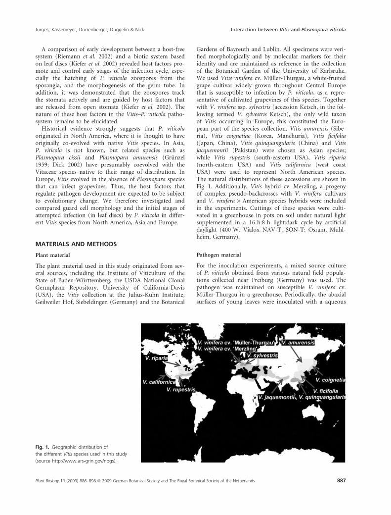
Gardens of Bayreuth and Lublin. All specimens were veri-fied morphologically and by molecular markers for theiridentity and are maintained as reference in the collectionof the Botanical Garden of the University of Karlsruhe.We used Vitis vinifera cv. Muller-Thurgau, a white-fruitedgrape cultivar widely grown throughout Central Europethat is susceptible to infection by P. viticola, as a repre-sentative of cultivated grapevines of this species. Togetherwith V. vinifera ssp. sylvestris (accession Ketsch, in the fol-lowing termed V. sylvestris Ketsch), the only wild taxonof Vitis occurring in Europe, this constituted the Euro-pean part of the species collection. Vitis amurensis (Sibe-ria), Vitis coignetiae (Korea, Manchuria), Vitis ficifolia(Japan, China), Vitis quinquangularis (China) and Vitisjacquemontii (Pakistan) were chosen as Asian species;while Vitis rupestris (south-eastern USA), Vitis riparia(north-eastern USA) and Vitis californica (west coastUSA) were used to represent North American species.The natural distributions of these accessions are shown inFig. 1. Additionally, Vitis hybrid cv. Merzling, a progenyof complex pseudo-backcrosses with V. vinifera cultivarsand V. vinifera · American species hybrids were includedin the experiments. Cuttings of these species were culti-vated in a greenhouse in pots on soil under natural lightsupplemented in a 16 h:8 h light:dark cycle by artificialdaylight (400 W, Vialox NAV-T, SON-T; Osram, Muhl-heim, Germany).
Pathogen material
For the inoculation experiments, a mixed source cultureof P. viticola obtained from various natural field popula-tions collected near Freiburg (Germany) was used. Thepathogen was maintained on susceptible V. vinifera cv.Muller-Thurgau in a greenhouse. Periodically, the abaxialsurfaces of young leaves were inoculated with a aqueous
Fig. 1. Geographic distribution of
the different Vitis species used in this study
(source http://www.ars-grin.gov/npgs).
Jurges, Kassemeyer, Durrenberger, Duggelin & Nick Interaction between Vitis and Plasmopara viticola
Plant Biology 11 (2009) 886–898 ª 2009 German Botanical Society and The Royal Botanical Society of the Netherlands 887
suspension containing approximately 2 · 104 sporangiaml)1 and kept overnight under high relative humidity(RH > 96%) at 24 �C. After incubation for 5–6 daysunder ambient greenhouse conditions, the plants wereagain maintained overnight under moist conditions toinduce sporulation. Formed sporangia were harvested andused for the next inoculation series. The harvested spo-rangia were frozen in dry ice and stored at )20 �C untiluse for the next inoculation series (Riemann et al. 2002).The sporangia maintained full viability for up to6 months. To verify the genetic identity of the sourceculture used in this study, genomic DNA was extractedfrom sporulating specimens, and the NL and LR regionsof the large subunit of the 25–28S RNA was used asmolecular marker to compare other samples of P. viticolacollected at different sites in Europe and North America(S. Schroder, W. Wilcox & P. Nick, unpublished data).The genetic identity of mycelia formed on the infectedleaves was verified at regular intervals by comparing theNL and LR sequences with those of the source culture.
Inoculation procedure
The sixth unfolded leaf (counted from the apex) wasexcised, surface-sterilized with 70% ethanol, rinsed withdistilled water and carefully dried with household tissue.Leaf discs 14 mm in diameter were excised from fullyexpanded leaves such that they did not contain majorveins and were placed top-down on water agar (0.8%w ⁄ v). Then, a portion of frozen sporangia was resus-pended in water and adjusted to 8 · 104 sporangiaÆml)1
using a hemacytometer (Fuchs-Rosenthal, Thoma, Frei-burg, Germany), and 100 ll of this suspension werespread with a micropipette onto the leaf disc for inocula-tion. After inoculation, the leaf discs were incubated at25 �C under continuous white light (5000 LxÆm)2) for72 h, when infections were analysed. To test for potentialcontamination of the leaf discs by other fungi, other leafdiscs were incubated and similarly examined. To excludethat the inoculum was contaminated by other fungi, onlysporangia derived from defined primary inocula wereused. In one set of experiments, the effect of an inoculumobtained from infected grape plantlets that had been axe-nically raised and then infected was compared to that ofan inoculum obtained as described above.
Staining of infection structures with aniline blue
To visualise the formation of hyphae on inoculated leafdiscs, we used the protocol of Kiefer et al. (2002). In thisprotocol, leaf discs (diameter 10 mm) are depigmentedby 10 min autoclaving at 121 �C and 120 kPa in 1 m
KOH prior to staining with aniline blue (Sigma-Aldrich,Deisenhofen, Germany). Depending on the species, resid-ual chlorophyll autofluorescence was sometimes observed,but this neither interfered with the quantification proto-col (see below) nor did it impair the visibility of the ani-line blue staining. After depigmentation, the leaf discs
were incubated for 5 min with 0.05% w ⁄ v aniline blue in0.067 m K2HPO4 (pH 9.8), washed twice for 5 min withdistilled water, and then directly viewed under an epifluo-rescence microscope (Axioskop, Zeiss, Gottingen, Ger-many) using the GFP-specific filter set 13 (excitation470 ± 20 nm, beam splitter 495 nm, and emissionthrough a band pass filter, 505–530 nm). From each leafdisc, five images were recorded in the centre and at fourequidistant points (distance to the centre: 3.00 mm) inthe four main directions, using a 20· Neofluar objective(Zeiss, Gottingen, Germany). Since the view field was3.00 mm in diameter, these images covered approximately80% of the disc area, but did not overlap. In sampleswith surface mycelium, the images were recorded on twofocal planes, either on the surface or about 100 lmwithin the tissue to assess penetration through the sto-mata. However, at a given infection site, there was eithermycelium on the surface of the leaf without colonisationof the mesophyll or colonisation of the mesophyll withoutany mycelium on the surface.
Assessment of host tissue colonisation
To separate background residual chlorophyll from theaniline blue signal, the images were processed usingPhotoshop software (Adobe Systems Inc., USA). Theimages (in the format Red–Blue–Green, RGB) were splitinto the individual channels, and the blue channel wassubjected to the autocontrast command (producing pixelintensity distributions that were comparable between dif-ferent images), and the red and green channels werereduced to 0%. The result was transformed into a greyscale and subjected to the auto-grey value command,ensuring that any aniline blue signal, irrespective of itsoriginal intensity, was scored with the same pixel inten-sity. The processed images were analysed using the ScionImage software (Scion Corporation, Frederick, MD,USA). The individual intercostal fields were selected usingthe freehand tool and analysed with respect to area andintegrated density of P. viticola mycelium. As a measureof the frequency of infection events, the area of infectedintercostal fields (i.e. the area delineated by leaf veins)was determined as a percentage of the total area of theintercostal fields within the viewed image. Because of thelow magnification, the geometry of the leaf surfaceremained visible as reference even when focused into themesophyll layers. To assess the colonisation intensity of agiven infection, the total projected area of mycelia withinthe mesophyll was determined. This was possible by mea-suring the integrated density within a given intercostalfield, since, due to the image processing, all pixels thatdid not originate from hyphae were black and thereforedid not contribute to the integrated density, whereas allpixels originating from hyphae contributed a pixel inten-sity of 255. The value obtained for the integrated densitywas thus proportional to the total cross-area of myceliawithin the analysed area. These values can therefore beused as (relative) measures of colonisation intensity. To
Interaction between Vitis and Plasmopara viticola Jurges, Kassemeyer, Durrenberger, Duggelin & Nick
888 Plant Biology 11 (2009) 886–898 ª 2009 German Botanical Society and The Royal Botanical Society of the Netherlands
compare surface growth of the mycelium, the total cross-area of mycelia on the leaf surface was measured directlyby selection of the (white) mycelia using the ‘magic stick’tool and quantifying the selected area in pixels. Since sur-face growth and colonisation of the mesophyll weremutually exclusive within a given intercostal field, thequantification of cross-area could be unequivocally attrib-uted to the colonisation mode.
Semi-thin sections and staining of host tissue with acridineorange
Small specimens of leaf tissue were fixed in 7 gÆl)1 para-formaldehyde (prepared freshly in 10 mm PIPES pH 7.0)for 1 h under vacuum. The specimens were then dehy-drated through an ethanol series (10%, 30% and 100%)and then infiltrated with xylol and embedded with paraf-fin (Roth, Karlsruhe, Germany) at 68 �C. Semithin sec-tions of 10 lm thickness were cut with a conventionalmicrotome, attached to chrome gelatine, and dried for5 h. The sections were stained for 5 min with a 10 gÆl)1
aqueous solution of acridine orange adjusted to pH 7.After washing with distilled water, the specimens weredried, covered with a drop of Entellan (Merck, Darms-tadt, Germany) and a coverslip, and retained for lateranalysis. The results were analysed under an epifluores-cence microscope (Axioskop, Zeiss, Gottingen, Germany)with filter set 1 (excitation 365 ± 12 nm, beam splitter395 nm, emission through a long-pass filter at 397 nm).
Low-temperature scanning electron microscopy
The structure of stomata was examined using low-tem-perature scanning electron microscopy (LT-SEM) asdescribed by Guggenheim et al. (1991). Fresh leaf pieceswith an area of 0.8–1.0 cm2 were excised and mountedon a specimen holder (Balzers AG; Balzers, Lichtenstein)using a low-temperature mounting medium. After cryo-fixation in liquid nitrogen, samples were transferredunder a nitrogen atmosphere into a Balzers cryoprepara-tion unit SCU 020 attached to a JEOL JSM 6300 scanningelectron microscope (SEM). Ice crystals on the surface ofthe specimen were allowed to sublimate from the surfaceby raising the temperature to )80 �C for about 10 min.The specimens were sputter-coated with gold (20 nm) inan argon atmosphere (Muller et al. 1991) and transferredinto the SEM under high-vacuum conditions. The sam-ples were observed at a stage temperature of )165 �C,using an acceleration voltage between 5 and 25 kV.
Sample sizes and statistical treatment
During this study, one to three genotypes were used peraccession. The experiments presented were repeated overthree consecutive vegetation periods. For analysis of guardcell morphology, each accession was investigated in fiveto seven independent experimental series from three dif-ferent vegetation periods. In each series, 20 to 50 individ-
ual leaf discs from five to 10 plants were employed. Fortyto 95 individual guard cells were viewed for the presenceof the inner cuticular rim. For analysis of host tissue col-onisation, 7924 images (from 70 to 119 leaf discs origi-nating from 10 to 15 plants per accession) were analysed.The average values plotted in Fig. 7 are pooled from fiveto seven independent infection series from three subse-quent years and between five to 10 different pathogeninocula. The variables (i) infected area (as a percentage oftotal area), (ii) colonisation intensity (for a given infec-tion event as a relative unit), and (iii) surface growth (asa relative unit) were analysed for the contributions ofhost versus inoculum using the non-parametrical Krus-kal–Wallis test. Throughout this study, the same pathogenmaterial was used originating from a source culture thathad been originally collected from different vineyardsfrom the region around Freiburg and was then main-tained in the greenhouse as described above.
RESULTS
Guard cell morphology
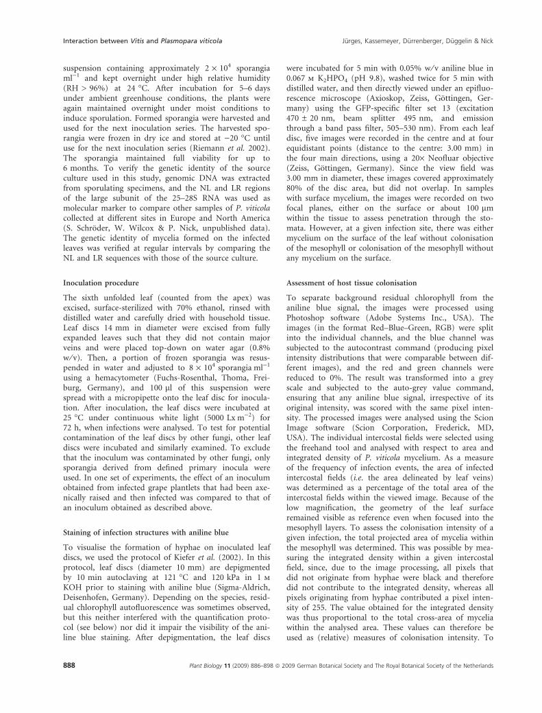
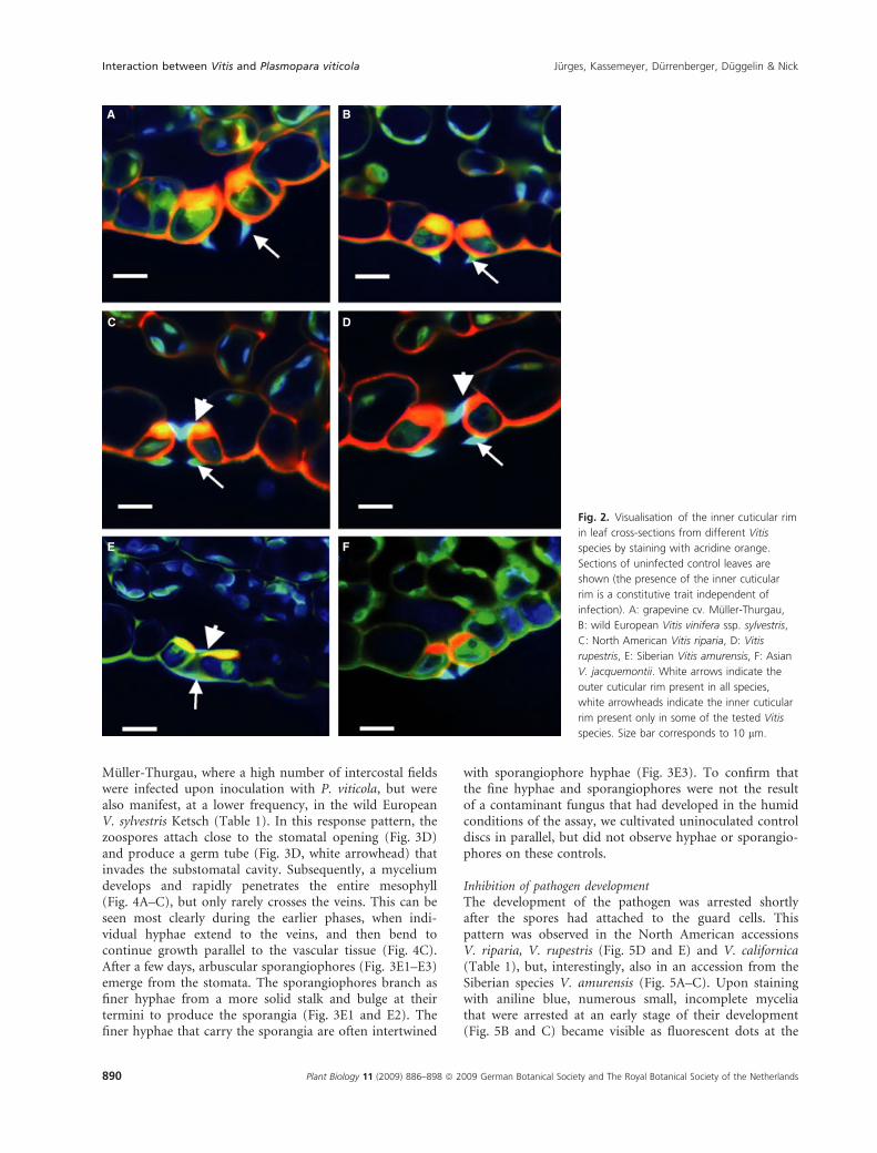
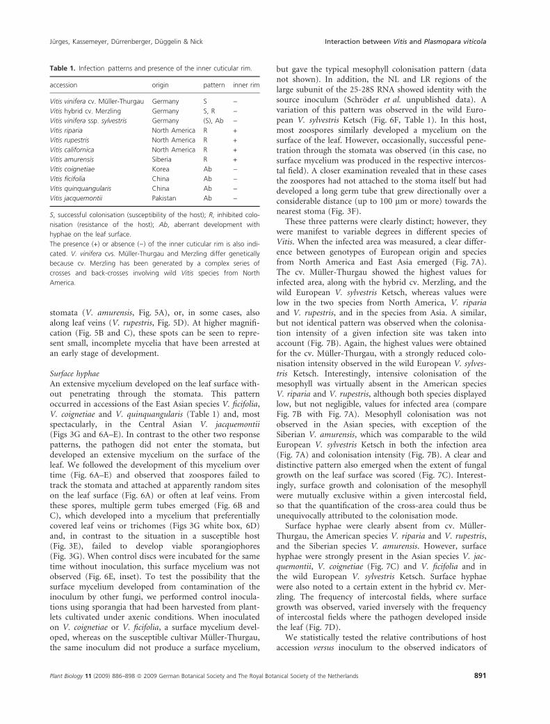
An inner cuticular rim at the neck region of the substom-atal cavity was present in many of the wild Vitis species,but was absent in V. vinifera cv. Muller-Thurgau and thehybrid cv. Merzling. This inner cuticular rim could bevisualised in transverse leaf sections, after staining withacridine orange, as a blue-green protrusion on the innerside of the stoma (Fig. 2, white arrowheads), similar tothe outer cuticular rim present in all tested Vitis speciesand cultivars (Fig. 2, white arrows). The inner cuticularrim was absent in the grapevine cultivars Muller-Thurgau(Fig. 2A) and the wild European V. sylvestris Ketsch(Fig. 2B), but was present in the North American speciesV. riparia, V. rupestris (Fig. 2C and D) and V. californica(Table 1), as well as in the Siberian species V. amurensis(Fig. 2E). However, the inner cuticular rim was notobserved in the Asian species V. jacquemontii (Fig. 2F),nor in V. coignetiae, V. ficifolia and V. quinquangularis(Table 1).
Under favourable circumstances, when the section hitthe median of the stoma, the inner cuticular rim couldalso be observed with SEM. It was clearly absent fromV. vinifera cv. Muller-Thurgau (Fig. 3A), but visible inthe stomata of the North American species V. riparia(Fig. 3B) and V. rupestris (Fig. 3C).
Interaction between Plasmopara viticola and Vitis
In assessing the responses of different Vitis species topathogen attack, we were able to define three distinctresponse patterns described below.
Successful colonisationHere, the pathogen penetrates through the stomata andsuccessfully colonises the mesophyll until sporulation.This pattern was most frequent in V. vinifera cv.
Jurges, Kassemeyer, Durrenberger, Duggelin & Nick Interaction between Vitis and Plasmopara viticola
Plant Biology 11 (2009) 886–898 ª 2009 German Botanical Society and The Royal Botanical Society of the Netherlands 889
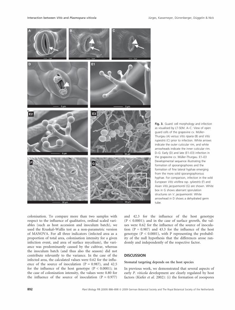
Muller-Thurgau, where a high number of intercostal fieldswere infected upon inoculation with P. viticola, but werealso manifest, at a lower frequency, in the wild EuropeanV. sylvestris Ketsch (Table 1). In this response pattern, thezoospores attach close to the stomatal opening (Fig. 3D)and produce a germ tube (Fig. 3D, white arrowhead) thatinvades the substomatal cavity. Subsequently, a myceliumdevelops and rapidly penetrates the entire mesophyll(Fig. 4A–C), but only rarely crosses the veins. This can beseen most clearly during the earlier phases, when indi-vidual hyphae extend to the veins, and then bend tocontinue growth parallel to the vascular tissue (Fig. 4C).After a few days, arbuscular sporangiophores (Fig. 3E1–E3)emerge from the stomata. The sporangiophores branch asfiner hyphae from a more solid stalk and bulge at theirtermini to produce the sporangia (Fig. 3E1 and E2). Thefiner hyphae that carry the sporangia are often intertwined
with sporangiophore hyphae (Fig. 3E3). To confirm thatthe fine hyphae and sporangiophores were not the resultof a contaminant fungus that had developed in the humidconditions of the assay, we cultivated uninoculated controldiscs in parallel, but did not observe hyphae or sporangio-phores on these controls.
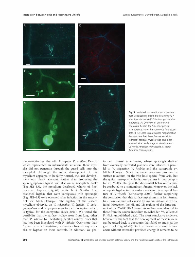
Inhibition of pathogen developmentThe development of the pathogen was arrested shortlyafter the spores had attached to the guard cells. Thispattern was observed in the North American accessionsV. riparia, V. rupestris (Fig. 5D and E) and V. californica(Table 1), but, interestingly, also in an accession from theSiberian species V. amurensis (Fig. 5A–C). Upon stainingwith aniline blue, numerous small, incomplete myceliathat were arrested at an early stage of their development(Fig. 5B and C) became visible as fluorescent dots at the
A B
DC
E F
Fig. 2. Visualisation of the inner cuticular rim
in leaf cross-sections from different Vitis
species by staining with acridine orange.
Sections of uninfected control leaves are
shown (the presence of the inner cuticular
rim is a constitutive trait independent of
infection). A: grapevine cv. Muller-Thurgau,
B: wild European Vitis vinifera ssp. sylvestris,
C: North American Vitis riparia, D: Vitis
rupestris, E: Siberian Vitis amurensis, F: Asian
V. jacquemontii. White arrows indicate the
outer cuticular rim present in all species,
white arrowheads indicate the inner cuticular
rim present only in some of the tested Vitis
species. Size bar corresponds to 10 lm.
Interaction between Vitis and Plasmopara viticola Jurges, Kassemeyer, Durrenberger, Duggelin & Nick
890 Plant Biology 11 (2009) 886–898 ª 2009 German Botanical Society and The Royal Botanical Society of the Netherlands
stomata (V. amurensis, Fig. 5A), or, in some cases, alsoalong leaf veins (V. rupestris, Fig. 5D). At higher magnifi-cation (Fig. 5B and C), these spots can be seen to repre-sent small, incomplete mycelia that have been arrested atan early stage of development.
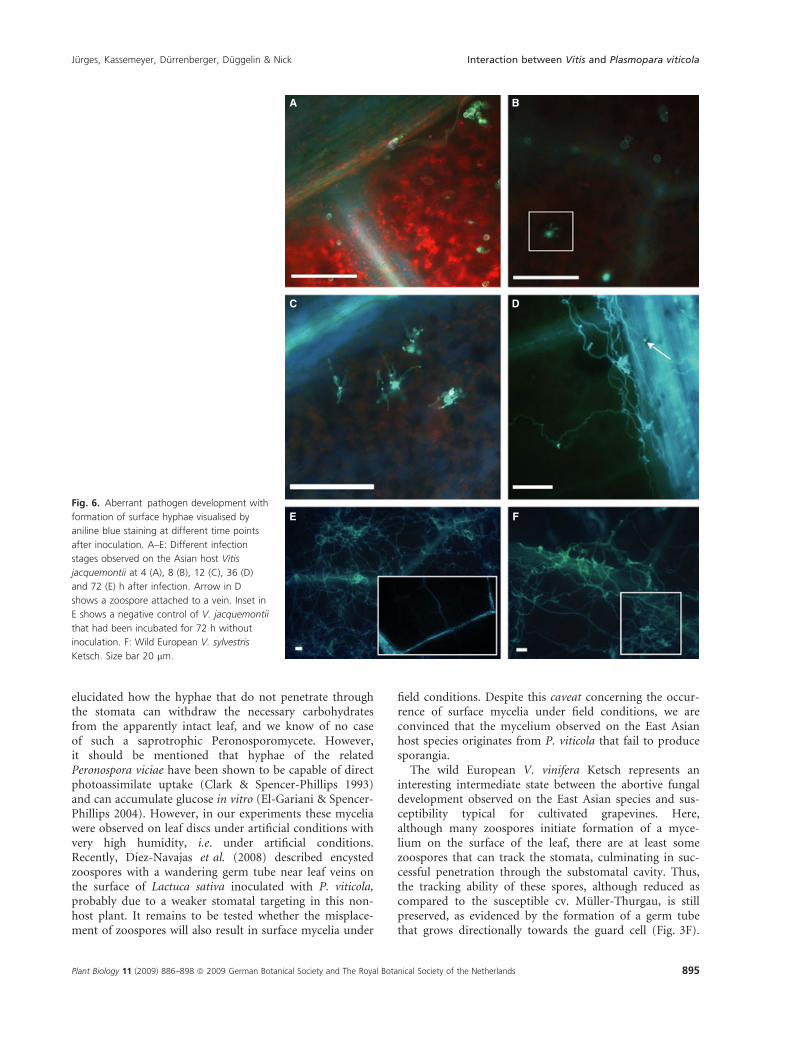
Surface hyphaeAn extensive mycelium developed on the leaf surface with-out penetrating through the stomata. This patternoccurred in accessions of the East Asian species V. ficifolia,V. coignetiae and V. quinquangularis (Table 1) and, mostspectacularly, in the Central Asian V. jacquemontii(Figs 3G and 6A–E). In contrast to the other two responsepatterns, the pathogen did not enter the stomata, butdeveloped an extensive mycelium on the surface of theleaf. We followed the development of this mycelium overtime (Fig. 6A–E) and observed that zoospores failed totrack the stomata and attached at apparently random siteson the leaf surface (Fig. 6A) or often at leaf veins. Fromthese spores, multiple germ tubes emerged (Fig. 6B andC), which developed into a mycelium that preferentiallycovered leaf veins or trichomes (Figs 3G white box, 6D)and, in contrast to the situation in a susceptible host(Fig. 3E), failed to develop viable sporangiophores(Fig. 3G). When control discs were incubated for the sametime without inoculation, this surface mycelium was notobserved (Fig. 6E, inset). To test the possibility that thesurface mycelium developed from contamination of theinoculum by other fungi, we performed control inocula-tions using sporangia that had been harvested from plant-lets cultivated under axenic conditions. When inoculatedon V. coignetiae or V. ficifolia, a surface mycelium devel-oped, whereas on the susceptible cultivar Muller-Thurgau,the same inoculum did not produce a surface mycelium,
but gave the typical mesophyll colonisation pattern (datanot shown). In addition, the NL and LR regions of thelarge subunit of the 25-28S RNA showed identity with thesource inoculum (Schroder et al. unpublished data). Avariation of this pattern was observed in the wild Euro-pean V. sylvestris Ketsch (Fig. 6F, Table 1). In this host,most zoospores similarly developed a mycelium on thesurface of the leaf. However, occasionally, successful pene-tration through the stomata was observed (in this case, nosurface mycelium was produced in the respective intercos-tal field). A closer examination revealed that in these casesthe zoospores had not attached to the stoma itself but haddeveloped a long germ tube that grew directionally over aconsiderable distance (up to 100 lm or more) towards thenearest stoma (Fig. 3F).
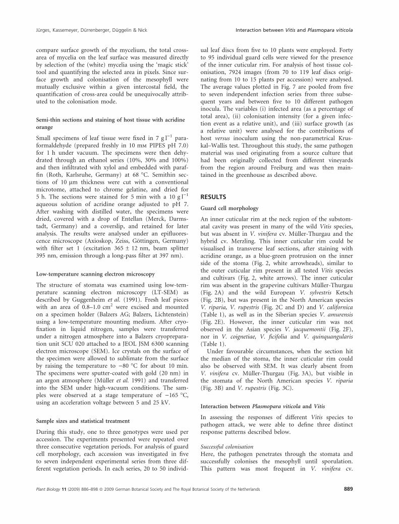
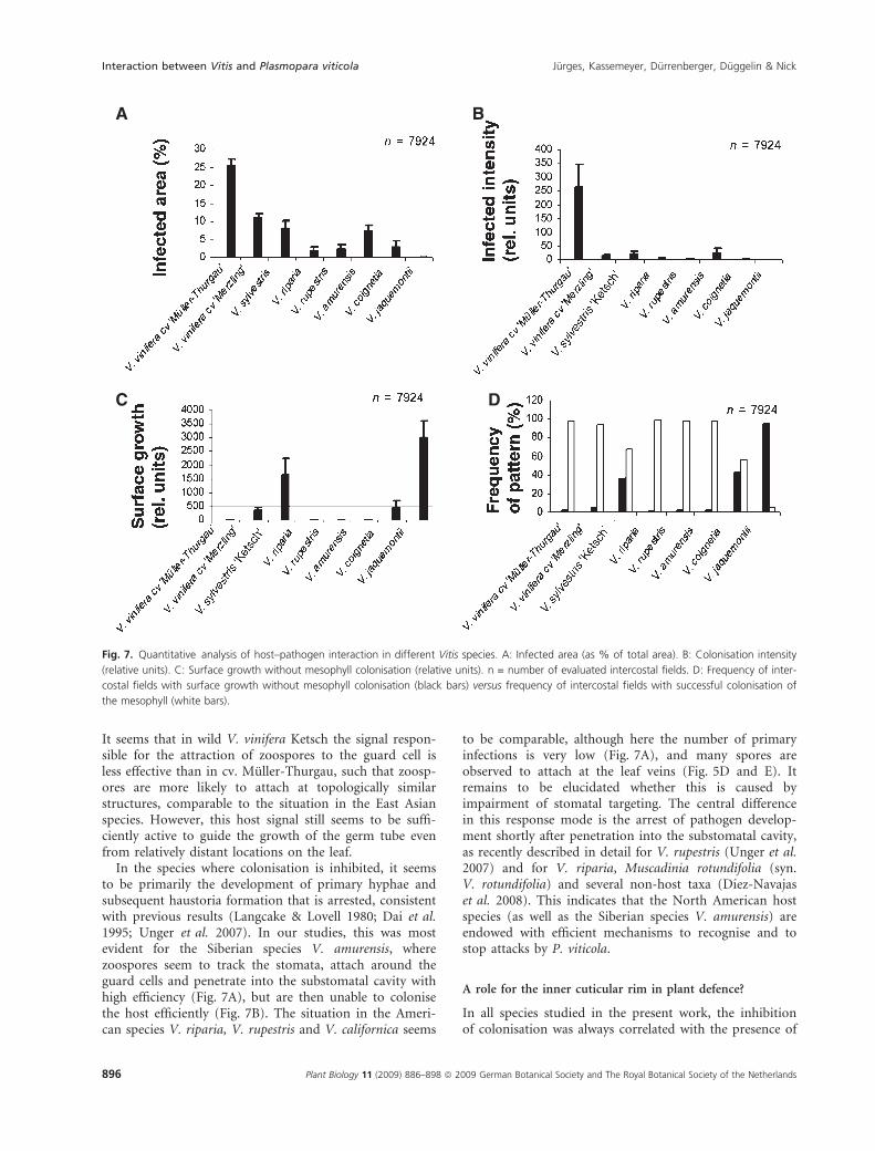
These three patterns were clearly distinct; however, theywere manifest to variable degrees in different species ofVitis. When the infected area was measured, a clear differ-ence between genotypes of European origin and speciesfrom North America and East Asia emerged (Fig. 7A).The cv. Muller-Thurgau showed the highest values forinfected area, along with the hybrid cv. Merzling, and thewild European V. sylvestris Ketsch, whereas values werelow in the two species from North America, V. ripariaand V. rupestris, and in the species from Asia. A similar,but not identical pattern was observed when the colonisa-tion intensity of a given infection site was taken intoaccount (Fig. 7B). Again, the highest values were obtainedfor the cv. Muller-Thurgau, with a strongly reduced colo-nisation intensity observed in the wild European V. sylves-tris Ketsch. Interestingly, intensive colonisation of themesophyll was virtually absent in the American speciesV. riparia and V. rupestris, although both species displayedlow, but not negligible, values for infected area (compareFig. 7B with Fig. 7A). Mesophyll colonisation was notobserved in the Asian species, with exception of theSiberian V. amurensis, which was comparable to the wildEuropean V. sylvestris Ketsch in both the infection area(Fig. 7A) and colonisation intensity (Fig. 7B). A clear anddistinctive pattern also emerged when the extent of fungalgrowth on the leaf surface was scored (Fig. 7C). Interest-ingly, surface growth and colonisation of the mesophyllwere mutually exclusive within a given intercostal field,so that the quantification of the cross-area could thus beunequivocally attributed to the colonisation mode.
Surface hyphae were clearly absent from cv. Muller-Thurgau, the American species V. riparia and V. rupestris,and the Siberian species V. amurensis. However, surfacehyphae were strongly present in the Asian species V. jac-quemontii, V. coignetiae (Fig. 7C) and V. ficifolia and inthe wild European V. sylvestris Ketsch. Surface hyphaewere also noted to a certain extent in the hybrid cv. Mer-zling. The frequency of intercostal fields, where surfacegrowth was observed, varied inversely with the frequencyof intercostal fields where the pathogen developed insidethe leaf (Fig. 7D).
We statistically tested the relative contributions of hostaccession versus inoculum to the observed indicators of
Table 1. Infection patterns and presence of the inner cuticular rim.
accession origin pattern inner rim
Vitis vinifera cv. Muller-Thurgau Germany S )Vitis hybrid cv. Merzling Germany S, R )Vitis vinifera ssp. sylvestris Germany (S), Ab )Vitis riparia North America R +
Vitis rupestris North America R +
Vitis californica North America R +
Vitis amurensis Siberia R +
Vitis coignetiae Korea Ab )Vitis ficifolia China Ab )Vitis quinquangularis China Ab )Vitis jacquemontii Pakistan Ab )
S, successful colonisation (susceptibility of the host); R, inhibited colo-
nisation (resistance of the host); Ab, aberrant development with
hyphae on the leaf surface.
The presence (+) or absence ()) of the inner cuticular rim is also indi-
cated. V. vinifera cvs. Muller-Thurgau and Merzling differ genetically
because cv. Merzling has been generated by a complex series of
crosses and back-crosses involving wild Vitis species from North
America.
Jurges, Kassemeyer, Durrenberger, Duggelin & Nick Interaction between Vitis and Plasmopara viticola
Plant Biology 11 (2009) 886–898 ª 2009 German Botanical Society and The Royal Botanical Society of the Netherlands 891
colonisation. To compare more than two samples withrespect to the influence of qualitative, ordinal scaled vari-ables (such as host accession and inoculum batch), weused the Kruskal–Wallis test as a non-parametric versionof MANOVA. For all three indicators (infected area as aproportion of total area, colonisation intensity for a giveninfection event, and area of surface mycelium), the vari-ance was predominantly caused by the cultivar, whereasthe inoculum batch (and thus also the season) did notcontribute relevantly to the variance. In the case of theinfected area, the calculated values were 0.62 for the influ-ence of the source of inoculation (P = 0.987), and 42.5for the influence of the host genotype (P < 0.0001); inthe case of colonization intensity, the values were 0.80 forthe influence of the source of inoculation (P = 0.977)
and 42.3 for the influence of the host genotype(P < 0.0001); and in the case of surface growth, the val-ues were 0.62 for the influence of the source of inocula-tion (P = 0.987) and 43.3 for the influence of the hostgenotype (P < 0.0001), with P representing the probabil-ity of the null hypothesis that the differences arose ran-domly and independently of the respective factor.
DISCUSSION
Stomatal targeting depends on the host species
In previous work, we demonstrated that several aspects ofearly P. viticola development are clearly regulated by hostfactors (Kiefer et al. 2002): (i) the formation of zoospores
A B C
E1D
E2 E3
GF
Fig. 3. Guard cell morphology and infection
as visualised by LT-SEM. A–C: View of open
guard cells of the grapevine cv. Muller-
Thurgau (A) versus Vitis riparia (B) and Vitis
rupestris (C) prior to infection. White arrows
indicate the outer cuticular rim, and white
arrowheads indicate the inner cuticular rim.
D–G: Early (D) and late (E1–E3) infection in
the grapevine cv. Muller-Thurgau. E1–E3
Developmental sequence illustrating the
formation of sporangiophores and the
formation of fine lateral hyphae emerging
from the more solid sporangiophorous
hyphae. For comparison, infection in the wild
European Vitis vinifera ssp. sylvestris (F) and
Asian Vitis jacquemontii (G) are shown. White
box in G shows aberrant sporulation
structures on V. jacquemontii. White
arrowhead in D shows a dehydrated germ
tube.
Interaction between Vitis and Plasmopara viticola Jurges, Kassemeyer, Durrenberger, Duggelin & Nick
892 Plant Biology 11 (2009) 886–898 ª 2009 German Botanical Society and The Royal Botanical Society of the Netherlands
within the mature sporangia was accelerated on the sur-face of the leaf of a compatible host genotype relative to ahost-free system (Kiefer et al. 2002); and (ii) the zoosp-ores track the stomata very efficiently within a few min-utes after their release from the sporangia, and are guidedby unknown host factors. When stomatal closure wasinduced by administering abscisic acid through the peti-ole, the accumulation of zoospores at the stomata wasimpaired in a concentration-dependent manner, whichargues strongly for active taxis guided by a factor releasedfrom open stomata. The comparison of infection patternsbetween different Vitis host species indicates that, in addi-tion to other mechanisms such as a hypersensitiveresponse, production or incorporation of toxic com-
pounds, callose formation and cell wall reinforcement,these host signals are targets of evolutionary change(Busam et al. 1997; Kortekamp et al. 1997; Gindro et al.2003; Hamiduzzaman et al. 2005).
In the infection pattern where hyphae are formed onthe surface of the leaf, stomatal targeting was stronglyimpaired. Here, the zoospores attached either to protrud-ing structures, such as leaf veins or trichomes, or some-where on the leaf surface (Figs 3G, 6 and 7C).Interestingly, this ‘tracking failure’ did not result inimmediate developmental arrest, but was followed byextensive formation of surface hyphae, which, in somecases, e.g. on V. jacquemontiii, (Figs 6A–E and 7C), pro-duced a mycelium covering large areas of the leaf. With
A
B C
Fig. 4. Successful colonisation on a
susceptible host (grapevine cv. Muller-
Thurgau) visualised by aniline blue staining
72 h after inoculation. A: Overview of an
infected intercostal field. B: Close-up of
penetrated hyphae. C: Barrier response of an
extending hyphae at the leaf vein.
Jurges, Kassemeyer, Durrenberger, Duggelin & Nick Interaction between Vitis and Plasmopara viticola
Plant Biology 11 (2009) 886–898 ª 2009 German Botanical Society and The Royal Botanical Society of the Netherlands 893
the exception of the wild European V. vinifera Ketsch,which represented an intermediate situation, these myc-elia did not penetrate through the guard cells into themesophyll. Although the initial development of thismycelium appeared to be fairly normal, the later develop-ment was clearly aberrant. Rather than producing thesporangiophores typical for infection of susceptible hosts(Fig. 3E1–E3), the mycelium developed whorls of fine,branched hyphae (Fig. 6F, white box). Similar fine,branched hyphae that were contiguous with sporangia(Fig. 3E1–E5) were observed after infection in the suscep-tible cv. Muller-Thurgau. The hyphae of the surfacemycelium observed on V. coignetiae, V. ficifolia, V. quin-quangularis and V. jacquemontii formed no septae, whichis typical for the oomycetes (Dick 2001). We tested thepossibility that the surface hyphae arose from fungi otherthan P. viticola by incubating parallel control discs thathad not been inoculated with P. viticola. Over more than3 years of experimentation, we never observed any myc-elia or hyphae on these controls. In addition, we per-
formed control experiments, where sporangia derivedfrom axenically cultivated plantlets were infected in paral-lel to V. coignetiae, V. ficifolia and the susceptible cv.Muller-Thurgau. Since the same inoculum produced asurface mycelium on the two host species from Asia, butthe typical mesophyll colonisation pattern in the suscepti-ble cv. Muller-Thurgau, the differential behaviour cannotbe attributed to a contaminant fungus. Moreover, the lackof septate hyphae in this surface mycelium is a typical fea-ture of P. viticola (Kortekamp 2005), further supportingthe conclusion that this surface mycelium is in fact formedby P. viticola and not caused by contamination with truefungi. Moreover, the NL and LR regions of the large sub-unit of the 25–28S RNA from this surface were identical tothose from the source inoculum (S. Schroder, W. Wilcox &P. Nick, unpublished data). The most conclusive evidence,however, is the fact that the development of these myceliacan be traced back to zoospores that failed to attach at theguard cell (Fig. 6A–E). Such extensive expansion cannotoccur without externally-provided energy. It remains to be
A D
E
B
C
Fig. 5. Inhibited colonisation on a resistant
host visualised by aniline blue staining 72 h
after inoculation. A–C: Siberian species Vitis
amurensis. A: Overview of an infected
intercostal field in the Siberian species
V. amurensis. Note the numerous fluorescent
dots. B, C: Close-ups at higher magnification
demonstrate that these fluorescent dots
represent residual mycelia that have been
arrested at an early stage of development.
D: North American Vitis riparia. E: North
American Vitis rupestris.
Interaction between Vitis and Plasmopara viticola Jurges, Kassemeyer, Durrenberger, Duggelin & Nick
894 Plant Biology 11 (2009) 886–898 ª 2009 German Botanical Society and The Royal Botanical Society of the Netherlands
elucidated how the hyphae that do not penetrate throughthe stomata can withdraw the necessary carbohydratesfrom the apparently intact leaf, and we know of no caseof such a saprotrophic Peronosporomycete. However,it should be mentioned that hyphae of the relatedPeronospora viciae have been shown to be capable of directphotoassimilate uptake (Clark & Spencer-Phillips 1993)and can accumulate glucose in vitro (El-Gariani & Spencer-Phillips 2004). However, in our experiments these myceliawere observed on leaf discs under artificial conditions withvery high humidity, i.e. under artificial conditions.Recently, Dıez-Navajas et al. (2008) described encystedzoospores with a wandering germ tube near leaf veins onthe surface of Lactuca sativa inoculated with P. viticola,probably due to a weaker stomatal targeting in this non-host plant. It remains to be tested whether the misplace-ment of zoospores will also result in surface mycelia under
field conditions. Despite this caveat concerning the occur-rence of surface mycelia under field conditions, we areconvinced that the mycelium observed on the East Asianhost species originates from P. viticola that fail to producesporangia.
The wild European V. vinifera Ketsch represents aninteresting intermediate state between the abortive fungaldevelopment observed on the East Asian species and sus-ceptibility typical for cultivated grapevines. Here,although many zoospores initiate formation of a myce-lium on the surface of the leaf, there are at least somezoospores that can track the stomata, culminating in suc-cessful penetration through the substomatal cavity. Thus,the tracking ability of these spores, although reduced ascompared to the susceptible cv. Muller-Thurgau, is stillpreserved, as evidenced by the formation of a germ tubethat grows directionally towards the guard cell (Fig. 3F).
A B
DC
E FFig. 6. Aberrant pathogen development with
formation of surface hyphae visualised by
aniline blue staining at different time points
after inoculation. A–E: Different infection
stages observed on the Asian host Vitis
jacquemontii at 4 (A), 8 (B), 12 (C), 36 (D)
and 72 (E) h after infection. Arrow in D
shows a zoospore attached to a vein. Inset in
E shows a negative control of V. jacquemontii
that had been incubated for 72 h without
inoculation. F: Wild European V. sylvestris
Ketsch. Size bar 20 lm.
Jurges, Kassemeyer, Durrenberger, Duggelin & Nick Interaction between Vitis and Plasmopara viticola
Plant Biology 11 (2009) 886–898 ª 2009 German Botanical Society and The Royal Botanical Society of the Netherlands 895
It seems that in wild V. vinifera Ketsch the signal respon-sible for the attraction of zoospores to the guard cell isless effective than in cv. Muller-Thurgau, such that zoosp-ores are more likely to attach at topologically similarstructures, comparable to the situation in the East Asianspecies. However, this host signal still seems to be suffi-ciently active to guide the growth of the germ tube evenfrom relatively distant locations on the leaf.
In the species where colonisation is inhibited, it seemsto be primarily the development of primary hyphae andsubsequent haustoria formation that is arrested, consistentwith previous results (Langcake & Lovell 1980; Dai et al.1995; Unger et al. 2007). In our studies, this was mostevident for the Siberian species V. amurensis, wherezoospores seem to track the stomata, attach around theguard cells and penetrate into the substomatal cavity withhigh efficiency (Fig. 7A), but are then unable to colonisethe host efficiently (Fig. 7B). The situation in the Ameri-can species V. riparia, V. rupestris and V. californica seems
to be comparable, although here the number of primaryinfections is very low (Fig. 7A), and many spores areobserved to attach at the leaf veins (Fig. 5D and E). Itremains to be elucidated whether this is caused byimpairment of stomatal targeting. The central differencein this response mode is the arrest of pathogen develop-ment shortly after penetration into the substomatal cavity,as recently described in detail for V. rupestris (Unger et al.2007) and for V. riparia, Muscadinia rotundifolia (syn.V. rotundifolia) and several non-host taxa (Dıez-Navajaset al. 2008). This indicates that the North American hostspecies (as well as the Siberian species V. amurensis) areendowed with efficient mechanisms to recognise and tostop attacks by P. viticola.
A role for the inner cuticular rim in plant defence?
In all species studied in the present work, the inhibitionof colonisation was always correlated with the presence of
A
C D
B
Fig. 7. Quantitative analysis of host–pathogen interaction in different Vitis species. A: Infected area (as % of total area). B: Colonisation intensity
(relative units). C: Surface growth without mesophyll colonisation (relative units). n = number of evaluated intercostal fields. D: Frequency of inter-
costal fields with surface growth without mesophyll colonisation (black bars) versus frequency of intercostal fields with successful colonisation of
the mesophyll (white bars).
Interaction between Vitis and Plasmopara viticola Jurges, Kassemeyer, Durrenberger, Duggelin & Nick
896 Plant Biology 11 (2009) 886–898 ª 2009 German Botanical Society and The Royal Botanical Society of the Netherlands

an inner cuticular rim at the transition between the sub-stomatal cavity and the stoma sensu strictu (Figs 2 and 3Table 1). This inner cuticular rim was absent in the EastAsian species, where P. viticola formed mycelia on the leafsurface, thereby refuting the hypothesis that this innercuticular rim somehow impairs zoospore tracking byinhibiting the release of the guiding signal through thestomata. On the other hand, the fact that all species thatare able to arrest the initiated colonisation of the meso-phyll are endowed with this inner cuticula rim (Table 1)suggests that this relationship is of functional significance.It also seems unlikely that this protrusion represents axeromorphic adaptation, because the majority of species inwhich it is observed originate from relatively humid habi-tats such as riverbanks (http://www.ars-grin.gov/). Thus,although the inner cuticular rim is probably not involvedin the suppression of zoospore tracking, it can be inter-preted in the context of plant defence. In the NorthAmerican species V. rupestris, V. riparia and V. californicaand in the Siberian species V. amurensis, where this innercuticular rim is present, zoospores can attach at the sto-mata but subsequent development is suppressed at anearly stage (Fig. 5). The American species presumablyhave undergone coevolution with P. viticola. The innercuticular rim might therefore be interpreted as a pre-formed resistance mechanism evolved as a consequence ofthe arms race between the host plant and the pathogen. Aplausible interpretation of the occurrence of this pre-formed barrier in V. amurensis may be the presence of aclosely related Peronosporomycete that also penetrates thehost tissue via the stomata. In fact, Grunzel (1959) men-tioned P. amurensis as a pathogen of East Asian Vitis spe-cies. The European taxa (cv Muller-Thurgau belonging tothe taxon V. vinifera ssp. sativa as well as the ancestralwild European species V. vinifera ssp. sylvestris) that aresusceptible do not possess this inner cuticular rim. Wetherefore hypothesise that the inner cuticular rim mightrepresent an adaptation to impair or at least delay growthof the germ tube through the stomata in a way similar tothe callosic plugs that have been shown to contribute todefence against fungal penetration in Vitis (Kortekampet al. 1997; Gindro et al. 2003; Hamiduzzaman et al.2005), and in other systems (Nishimura et al. 2003). Itshould be considered, however, that (1) the data pre-sented in this study have been obtained with leaf discs,the situation in the field might differ, and (2) in severalcases only one accession per host species was employedand, especially for the evolutionary advanced situation inNorth America, considerable intraspecies variation is tobe expected. However, we will soon publish a moreextensive infection study involving accessions from allknown wild Vitis species, along with data on molecularphylogeny of these species that confirm the biogeographi-cal context of the infection patterns reported in the pres-ent work.
The hypothesis that the inner cuticular rim represents apreformed defence response could be tested experimen-tally when the time courses in the expression of defence
genes are compared to those of host colonisation in pairsof closely related species that differ in the presence of thisguard cell structure. If the inner cuticular rim contributesto defence, the colonisation would proceed more slowlyin relation to the induction of defence genes.
ACKNOWLEDGEMENTS
The competent and highly motivated support of JoachimDaumann (Botanical Garden, Karlsruhe University) in thecultivation and propagation of the different Vitis speciesis gratefully ackowledged. We also gratefully acknowledgethe support of the USDA National Clonal GermplasmRepository, University of California-Davis (USA) whokindly provided some wild Vitis species.
REFERENCES
Busam G., Junghans K.T., Kneusel R.E., Kassemeyer H.H.,
Matern U. (1997) Characterization and expression of
Coffeoyl-coenzyme A 3-O-methyltransferase proposed for
the induction for the induced resistance response of Vitis
vinifera L. Plant Physiology, 115, 1039–1048.
Clark J.S.C., Spencer-Phillips P.T.N. (1993) Accumulation of
photoassimilate by Peronospora viciae (Berk.) Casp. and
leaves of Pisum sativum L.: Evidence for nutrient uptake via
intercellular hyphae. New Phytologist, 124, 107–119.
Dai G.H., Andary C., Mondolot-Cosson L., Boubals D. (1995)
Histochemical studies on the interaction between three spe-
cies of grapevine, Vitis vinifera, V. rupestris and V. rotundifo-
lia and the downy mildew fungus Plasmopara viticola.
Physiological Molecular Plant Pathology, 46, 177–188.
Dick M.W. (2001) The Peronosporomycetes. In: McLaughlin
D.J., McLaughlin E.G., Lemke P.A. (Eds), The Mycota VII
Part A. Systematics and Evolution. Springer, Berlin Heidel-
berg: 39–73.
Dick M.W. (2002) Towards an understanding of the evolution
of the downy mildews. In: Spencer-Phillips P.T.N., Gisi U.,
Lebeda A. (Eds), Advances in Downy Mildew Research, Vol.
1. Kluwer, Dordrecht: 1–59.
Dıez-Navajas A.M., Wiedemann-Merdinoglu S., Greif C., Mer-
dinoglu D. (2008) Nonhost versus host resistance to grape-
vine downy mildew, Plasmopara viticola, studied at the
tissue level. Phytopathology, 98, 776–780.
El-Gariani A., Spencer-Phillips P.T.N. (2004) Benzothidiazole-
induced resistance to Plasmopara halstedii (Farl.) Berl et de
Toni in sunflower. In Spencer-Phillips P.T.N., Jeger P.
(Eds), Advances in Downy Mildew Research, Vol. 2. Kluwer,
Dordrecht: 265–273.
Gindro K., Pezet R., Viret O. (2003) Histological studies of the
response of two Vitis vinifera cultivars (resistant and suscep-
tible) to Plasmopara viticola infections. Plant Physiology &
Biochemistry, 41, 846–853.
Grunzel H. (1959) Zur biologischen Differenzierung des Fals-
chen Mehltaus der Weinrebe (Peronospora viticola deBary).
Zentralblatt Bakteriologie II, 112, 454–472.
Jurges, Kassemeyer, Durrenberger, Duggelin & Nick Interaction between Vitis and Plasmopara viticola
Plant Biology 11 (2009) 886–898 ª 2009 German Botanical Society and The Royal Botanical Society of the Netherlands 897
Guggenheim R., Duggelin M., Mathys D., Grabski C. (1991)
Low-temperature SEM for the detection of fungicide activ-
ity. Journal of Microscopy, 161, 337–342.
Hamiduzzaman M.M., Jakab G., Barnavon L., Neuhaus J.M.,
Mauch-Mani B. (2005) b-Aminobutyric acid-induced resis-
tance against downy mildew in grapevine acts through the
potentiation of callose formation and jasmonic acid signal-
ling. Molecular Plant Microbe Interaction, 18, 819–829.
Kiefer B., Riemann M., Buche C., Kassemeyer H.H., Nick P.
(2002) The host guides morphogenesis and stomatal target-
ing in the grapevine pathogen Plasmopara viticola. Planta,
215, 387–393.
Kortekamp A. (2005) Growth, occurrence and development of
septa in Plasmopara viticola and other members of the Pero-
nosporaceae using light and epifluorescence microscopy.
Mycology Research, 109, 640–648.
Kortekamp A., Wind R., Zyprian E. (1997) The role of callose
deposits during infection to downy mildew-tolerant and two
susceptible Vitis cultivars. Vitis, 36, 103–104.
Langcake P., Lovell P.A. (1980) Light and electron microscopi-
cal studies on the infection of Vitis ssp. by Plasmopara viti-
cola, the downy mildew pathogen. Vitis, 19, 321–337.
Muller K., Sleumer H. (1934) Biologische Untersuchungen
uber die Peronosporakrankheit des Weinstocks mit beson-
derer Berucksichtigung ihrer Bekampfung nach Inkubations-
methode. Zeitschrift fur Wissenschaftliche Landwirtschaft, 79,
509–576.
Muller T., Guggenheim R., Duggelin M., Scheidegger C.
(1991) Freeze fracturing for conventional and field emission
low-temperature scanning electron microscopy: the cryo-
scanning unit SCU. Journal of Microscopy, 161, 73–83.
Nishimura M.T., Stein M., Hou B.-H., Vogel J.P., Edwards H.,
Somerville S.C. (2003) Loss of a callose synthase results in
salicylic acid-dependent disease resistance. Science, 301, 969–
972.
Riemann M., Buche C., Kassemeyer H.H., Nick P. (2002)
Microtubules and actin microfilaments guide the establish-
ment of cell polarity during early development of the wine
pathogen Plasmopara viticola. Protoplasma, 219, 13–22.
Unger S., Buche C., Boso S., Kassemeyer H.H. (2007) The
course of the colonization of two different Vitis geno-
types by Plasmopara viticola indicates compatible and
incompatible host–pathogen interactions. Phytopathology, 97,
781–786.
Vercesi A., Tornaghi R., Burruano S.S., Faoro F. (1999) A
cytological and ultrastructural study on the maturation and
germination of oospores of Plasmopara viticola from over-
wintering vine leaves. Mycological Research, 103, 193–202.
Interaction between Vitis and Plasmopara viticola Jurges, Kassemeyer, Durrenberger, Duggelin & Nick
898 Plant Biology 11 (2009) 886–898 ª 2009 German Botanical Society and The Royal Botanical Society of the Netherlands


![Pland propagation Protocol Vitis Riparia[1]](https://img.pdfslide.us/doc/110x75/618f63d2660b103f1b602956/pland-propagation-protocol-vitis-riparia1.jpg)

