Embed Size (px)
Citation preview

Red Blood Cells & Anemias
Andrew D. Leavitt, MDDepartments of Laboratory Medicine & Medicine
March 26, 2012
RBC: ~5 x 10⁶/l; Generate ~ 8,000,000,000/hour

Outline – topics we will cover

Erythropoiesis
Pro-Erythroblast
BasophilicErythroblast
PolychromaticErythroblast
OrthochromaticErythroblast Reticulocyte RBC
CHROMATIN STRUCTURE
NUCLEAR SIZE & COLOR
5 stages in the marrow

Red Blood Cell (RBC)
Delivers O2 from your lungs to all your tissuesTakes CO2 from your tissues to your lungs
Also – binds NO (a vasodilator)
Their size: 8 [capillaries have ~3 M diameter]
Their life-span: 120 days
Mature RBCs have no nucleus
Too few RBCs = anemia

What tells your body to make RBCs?
EPO:Is not stored, but expressed in response to the kidney sensing oxygen in the blood
It -1. Increases # of E-committed progenitors2. Increase GATA1 and FOG expression3. Enhances anti-apoptotic gene expression4. Increases transferrin receptor expression

Hemoglobin
Tetramer of two heterodimers (&)
Hemoglobin (Hgb) in the RBC carries O2, which is poorly soluble in water, to tissues & CO2 from tissues to lungs.
~640,000,000 Hemoglobin molecules/RBC
Must coordinate: 1.heme and globin synthesis 2. (Chromosome 11; 141 aa) & (Chromnosome 16; 146) chains
Colors of a bruise are globin breakdown products
HemeIron in a Porphyrin ring
Glu to Val
Linus Pauling1949 – molecular diagnosis
2012 – no therapy

Hemoglobin oxygen dissociation curve
ArterialO2 tension
Mean VenousO2 tension
HbF
; CO2

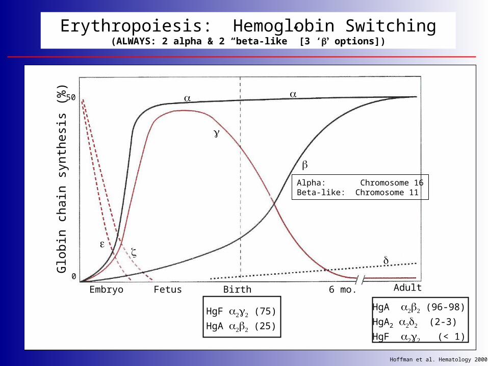
Erythropoiesis: Hemoglobin Switching(ALWAYS: 2 alpha & 2 “beta-like” [3 ‘’ options])
Embryo Fetus Birth Adult6 mo.
Glo
bin
chain
synth
esi
s (%
)
0
50
HgF (75)
HgA (25)
HgA (96-98)
HgA2 (2-3)
HgF (< 1)
Hoffman et al. Hematology 2000
Alpha: Chromosome 16Beta-like: Chromosome 11

*
*
*mutations lead to hereditary spherocytosis
Schematic of red cell membrane depicting proteins crucial for normal membrane mechanical strength

Normal Values for Hematology Tests - UCSF

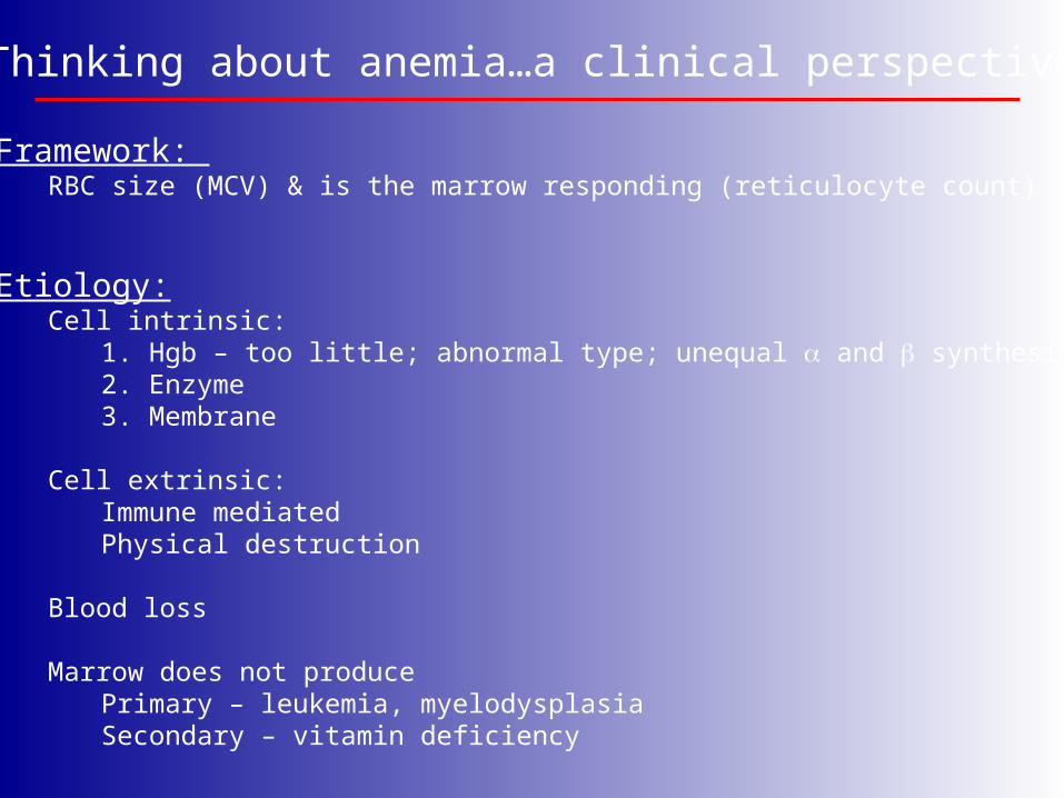
Thinking about anemia…a clinical perspective
Framework: RBC size (MCV) & is the marrow responding (reticulocyte count)
Etiology:Cell intrinsic:
1. Hgb – too little; abnormal type; unequal and synthesis2. Enzyme3. Membrane
Cell extrinsic:Immune mediatedPhysical destruction
Blood loss
Marrow does not producePrimary – leukemia, myelodysplasiaSecondary – vitamin deficiency

Reticulocyte Stain – detects the ribonucleoprotein in the young RBC
Reticulocytes

#1.1 Too little Hgb: Iron Deficiency Anemia
Small cells – microcytic (MCV < 80 fL)
Young women – OK
Older people – Worry about gastrointestinal blood loss
Iron deficiency anemia, peripheral blood (40x)normal peripheral blood (40x)
Lead poisoning can look like Fe deficiency – Lead blocks heme synthesis

Iron Deficiency Anemia
peripheral blood, 100X
HypochromicRBCs
Elliptocytes
Iron stian of bone marrow – none found
~ 500 million people worldwide

Daily absorption:Duodenum ~ 1 mg
Daily loss: urine, feces, skin, hair ~ 1 mg
Transferrin
Plasma (4 mg)
Circulating RBCs (1.7 – 2.4 gm)
Menstrual loss/hemorrhage
Macrophage(0.5 – 1.5 gm)
Ineffective erythropoiesis
Bone MarrowErythroblasts( ~ 150 mg)
The Iron Circuit – mostly a game of recycling
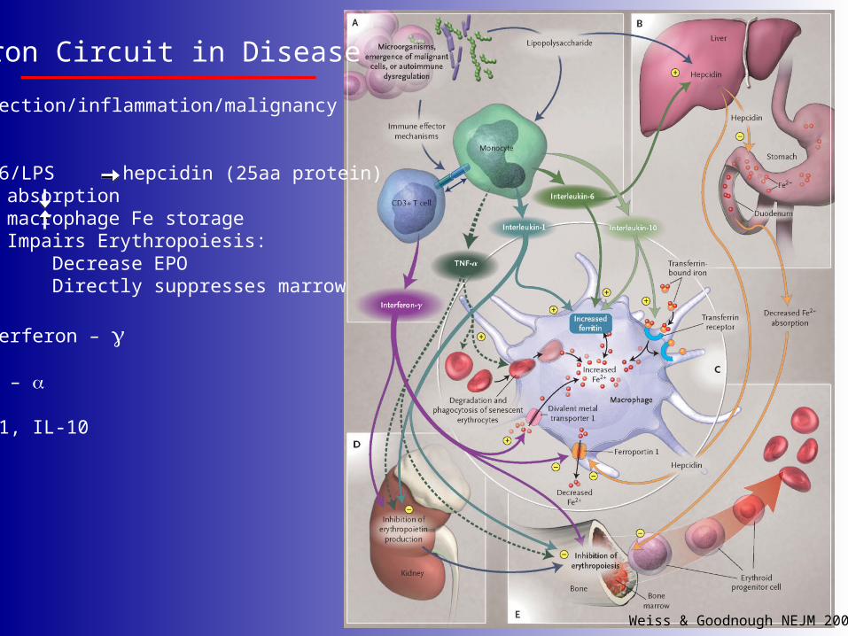
Weiss & Goodnough NEJM 2005
Iron Circuit in Disease
Infection/inflammation/malignancy
IL-6/LPS hepcidin (25aa protein)absorptionmacrophage Fe storageImpairs Erythropoiesis: Decrease EPO Directly suppresses marrow
Interferon –
TNF –
IL-1, IL-10

Storage iron(blue)
Iron deficienyc anemia - iron stain
Anemia of inflammation – iron stain
Iron stain of bone marrow aspirates

#1.2 Abnormal Hgb: Sickle cell disease
Multi-system disease
Cardiovascular/strokesKidneysSkinLungsImmune system
Treatment:
Supportive careTransfusionsDemethylating agentsStem cell transplants

#1.3 Abnormal / Globin ratio: Thalassemia
Small cells – microcytic (MCV < 80 fL)
Imbalance between and chains
Name based on which chain is deficient [ or thal]
Make lots of RBCs, just unstable and lyse, so increased retics
Alpha Thal:silent carrier trait or
Hgb H Hydrops fetalis
Beta Thal:MinorIntermediaMajor
Erythropoiesis: Hemoglobin Switching(ALWAYS: 2 alpha & 2 “beta-like” [3 ‘’ options])
Embryo Fetus Birth Adult6 mo.
Glo
bin
chain
synth
esi
s (%
)
0
50
HgF (75)
HgA (25)
HgA (96-98)
HgA2 (2-3)
HgF (< 1)
Hoffman et al. Hematology 2000
Alpha: Chromosome 16Beta-like: Chromosome 11
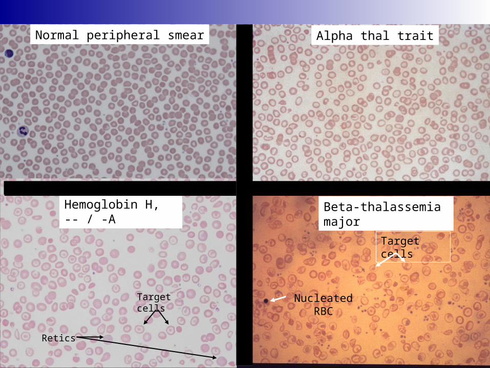
Beta-thalassemia major
Nucleated RBC
Target cells
Alpha thal traitNormal peripheral smear
Hemoglobin H, -- / -A
Target cells
Retics
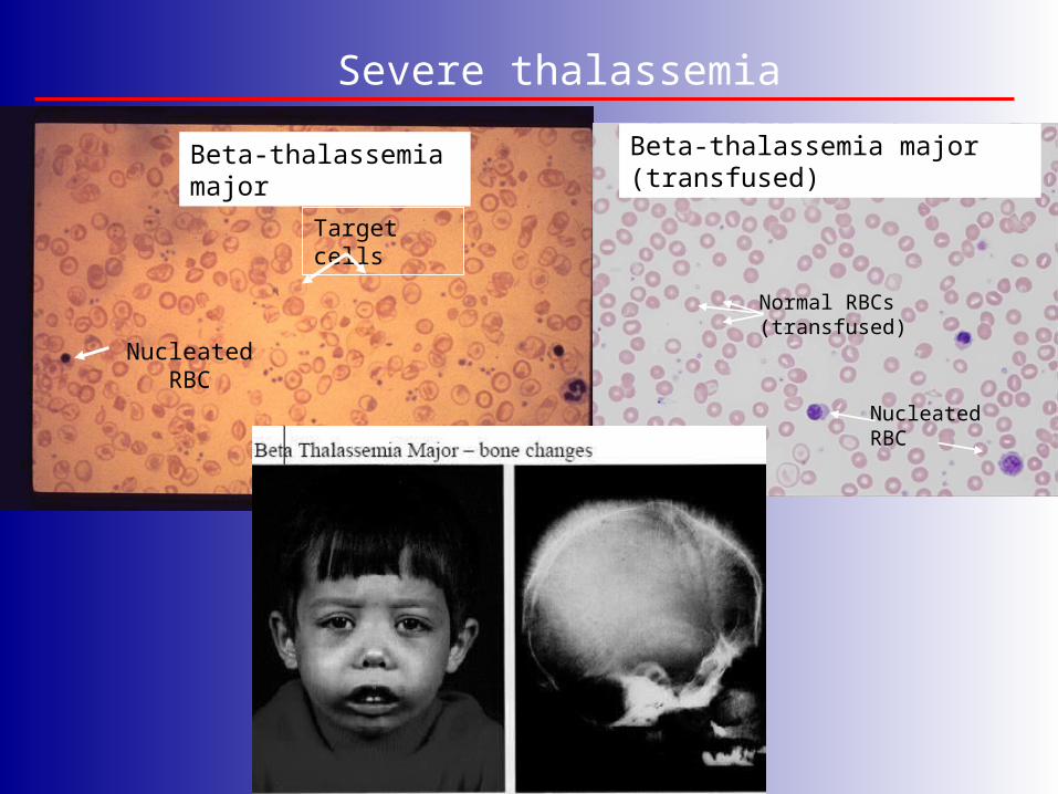
Severe thalassemia
Beta-thalassemia major
Nucleated RBC
Target cells
Beta-thalassemia major (transfused)
NucleatedRBC
Normal RBCs(transfused)

Bite cells
#2 Enzyme: G-6-PD deficiency (Heinz body hemolytic anemia)
Arrows show Heinz bodies
Sulfa drugs, fava beans, antimalarials..…
Reduced glutathione (GSH)is critical for RBCs to counter
oxidative stress.

*
*
*mutations lead to hereditary spherocytosis
#3: Membrane: red cell membrane depicting proteins crucial for normal membrane mechanical strength

Hereditary spherocytosis (HS)
spherocytes
Reticulocytes
Thinking about anemia…a clinical perspective
Framework: RBC size (MCV) & is the marrow responding (reticulocyte count)
Etiology:Cell intrinsic:
1. Hgb – too little; abnormal type; unequal and synthesis2. Enzyme3. Membrane
Cell extrinsic:Immune mediatedPhysical destruction
Blood loss
Marrow does not producePrimary – leukemia, myelodysplasiaSecondary – vitamin deficiency

Autoimmune hemolytic anemia
spherocytes
Reticulocytes

Testing for anti-RBC antibodies: attached or in circulation
Hemolytic disease of the newborn

RBC destruction - extrinsic
Hemolytic uremic syndrome (HUS)
Retics
schistocytes(RBC fragments)
Thrombotic thrombocytopenic purpura (TTP)schistocytes
(RBC fragments)
Erythroid Hyperplasia, TTP, bone marrow aspirate (100x)

Megaloblastic anemia (MCV > 100)
B12 deficiencyFolate deficiency
Affects DNA synthesis

Megaloblastic anemia (MCV > 100)
Anemia yet hypercellular marrow – ineffective erythropoiesis

Narla & Ebert 2010
Ribosomopathies and hematopoietic disorders
Our findings indicate that the erythroid lineage has a low threshold for the induction of p53, providing a basis for the failure of erythro- poiesis in the 5q- syndrome, DBA, and perhaps other bone marrow failure syn- dromes.

Thanks for your attention!

Take home message……
Blood is good…everyone should have some!
Share it…become a blood donor if you can
UCSF Blood donor center:
Millberry Union next to Subway sandwich shop
The best donor center nurses in town

![An Overview of the Anemias[1].ppt - School of Medicine · AO i fth A iAn Overview of the Anemias Iron Deficiency MegaloblasticIron Deficiency, Megaloblastic, ... {Malabsorption: Pernicious](https://img.pdfslide.us/doc/110x75/5ae1da537f8b9a5d648bed5f/an-overview-of-the-anemias1ppt-school-of-i-fth-a-ian-overview-of-the-anemias.jpg)