Embed Size (px)
Citation preview
HAL Id: hal-01608403https://hal.archives-ouvertes.fr/hal-01608403
Submitted on 26 May 2020
HAL is a multi-disciplinary open accessarchive for the deposit and dissemination of sci-entific research documents, whether they are pub-lished or not. The documents may come fromteaching and research institutions in France orabroad, or from public or private research centers.
L’archive ouverte pluridisciplinaire HAL, estdestinée au dépôt et à la diffusion de documentsscientifiques de niveau recherche, publiés ou non,émanant des établissements d’enseignement et derecherche français ou étrangers, des laboratoirespublics ou privés.
Copyright
Recognition of the Magnaporthe oryzae effectorAVR-Pia by the decoy domain of the rice NLR immune
receptor RGA5Diana Ortiz, Karine de Guillen, Stella Cesari, Veronique Chalvon-Poulier,
Jérôme Gracy, André Padilla, Thomas Kroj
To cite this version:Diana Ortiz, Karine de Guillen, Stella Cesari, Veronique Chalvon-Poulier, Jérôme Gracy, et al.. Recog-nition of the Magnaporthe oryzae effector AVR-Pia by the decoy domain of the rice NLR immunereceptor RGA5. The Plant cell, American Society of Plant Biologists (ASPB), 2017, 29 (1), pp.156-168.�10.1105/tpc.16.00435�. �hal-01608403�
Recognition of the Magnaporthe oryzae EffectorAVR-Pia by the Decoy Domain of the Rice NLR ImmuneReceptor RGA5 OPEN
Diana Ortiz,a,1 Karine de Guillen,b,1 Stella Cesari,a,c Véronique Chalvon,a Jérome Gracy,b André Padilla,b
and Thomas Kroja,2
a INRA, BGPI, Biology and Genetics of Plant-Pathogen Interactions, Campus International de Baillarguet, 34398 Montpellier, FrancebCNRS UMR 5048, INSERM U1054, Centre de Biochimie Structurale, Université Montpellier, 34090 Montpellier, FrancecCSIRO Agriculture Flagship, Canberra ACT 2601, Australia
ORCID IDs: 0000-0002-9778-0327 (J.G.); 0000-0001-9702-4612 (A.P.); 0000-0002-3752-1788 (T.K.)
Nucleotide binding domain and leucine-rich repeat proteins (NLRs) are important receptors in plant immunity that allowrecognition of pathogen effectors. The rice (Oryza sativa) NLR RGA5 recognizes the Magnaporthe oryzae effector AVR-Piathrough direct interaction. Here, we gained detailed insights into the molecular and structural bases of AVR-Pia-RGA5interaction and the role of the RATX1 decoy domain of RGA5. NMR titration combined with in vitro and in vivo protein-proteininteraction analyses identified the AVR-Pia interaction surface that binds to the RATX1 domain. Structure-informed AVR-Piamutants showed that, although AVR-Pia associates with additional sites in RGA5, binding to the RATX1 domain is necessaryfor pathogen recognition but can be of moderate affinity. Therefore, RGA5-mediated resistance is highly resilient to mutationsin the effector. We propose a model that explains such robust effector recognition as a consequence, and an advantage, ofthe combination of integrated decoy domains with additional independent effector-NLR interactions.
INTRODUCTION
Plant disease resistance largely relies on inducible immuneresponses that are triggered upon receptor-mediated recognitionof pathogen molecules and that often involve a localized pro-grammed cell death called the hypersensitive response (HR).Particularly important are NLRs, cytoplasmic nucleotide bindingoligomerization domain-like receptors with a multidomain archi-tecture composed of a C-terminal leucine-rich repeat domainand a central nucleotide binding domain (Takken and Goverse,2012; Jacob et al., 2013; Qi and Innes, 2013). Most NLRs carry inaddition an N-terminal coiled-coil or Toll/interleukin-1 receptordomain that have both been reported to mediate NLR homo-complex formation and to be crucial for the activation of down-stream signaling (Bernoux et al., 2011; Maekawa et al., 2011).Plant NLR proteins specifically recognize pathogen-derived ef-fectors that act inside plant cells (Cui et al., 2015).
Traditionally, both effector recognition and activation of re-sistance signaling are thought to be mediated by single plantNLRs, but recent studies revealed an increasing number of caseswhere different NLRs cooperate in pathogen recognition andresistance (Eitas and Dangl, 2010; Césari et al., 2014a). Fre-quently, the genes coding for these pairedNLRsoccur in a paired,inverted tandem arrangement in the genome. In the rare cases
investigated in more detail, the NLR pairs seem to act as hetero-complexes where only one of the paired NLRs acts directly in ef-fector recognition while the other is crucial for the activation ofdownstreamsignaling (Williams et al., 2014; Césari et al., 2014b). Inother cases, helperNLRs that actdownstreamof severalNLRswithdifferent recognition specificities were shown to be required forresistance and pathogen detection (Gabriëls et al., 2007; Bonardiet al., 2011; Wu et al., 2016).Some plant NLRs recognize effectors in an indirect manner.
They detect either a modification of the effector’s host targetprotein called a “guardee” or modifications of a host protein thatmimics the effector target and is called a “decoy” (van der Hoornand Kamoun, 2008). Effectors can also be recognized in a directmanner by binding, either alone, or in complex with a cofactorthat may be a guardee or a decoy, to the NLRs (Takken andGoverse, 2012; Collier and Moffett, 2009). In these cases, theleucine-rich repeat domain plays a crucial role in recognitionspecificity and has frequently been shown to mediate direct ef-fectorbinding (Elliset al., 2007;Krasilevaetal., 2010; Jiaetal., 2000).Alternatively, direct effector recognition can be mediated by non-canonicaldomains integrated intoNLRsat low frequencies (Kanzakiet al., 2012; Sarris et al., 2015; Maqbool et al., 2015; Le Roux et al.,2015; Césari et al., 2013). Recent work led to the hypothesis thatthese highly diverse integrated domains are mimics of effectortargets and can therefore be considered as integrated decoy do-mains (Le Roux et al., 2015; Sarris et al., 2015; Césari et al., 2014a).However, the molecular mechanisms of effector recognition byintegrated domains and the advantages of thismode of recognitionremain largely unknown.Rice blast, caused by the fungus Magnaporthe oryzae, is a
highly destructive crop disease and a serious threat for foodsecurity (Pennisi, 2010; Dean et al., 2012; Skamnioti and Gurr,
1 These authors contributed equally to this work.2 Address correspondence to [email protected] author responsible for distribution of materials integral to the findingspresented in this article in accordance with the policy described in theInstructions for Authors (www.plantcell.org) is: Thomas Kroj ([email protected]).OPENArticles can be viewed without a subscription.www.plantcell.org/cgi/doi/10.1105/tpc.16.00435
The Plant Cell, Vol. 29: 156–168, January 2017, www.plantcell.org ã 2017 American Society of Plant Biologists. All rights reserved.
2009). NLR-mediated pathogen recognition is the majormechanism in rice blast resistance. Among 25 different blastresistance genes cloned over the last 20 years, 24 code for NLRs(Liu et al., 2014). Blast resistance is frequently conferred bypaired NLRs with clustered tandem organization in the genome.Amongthese, theNLRpairRGA4/RGA5encodedby thePi-CO39/Pia resistance locus has been developed as a model for mo-lecular understanding of paired NLRs (Okuyama et al., 2011;Césari et al., 2013, 2014b). In this pair, RGA4 acts as a consti-tutively active disease resistance and cell death inducer that isrepressed by RGA5 in the absence of pathogen (Césari et al.,2014b). In addition to its repressor function, RGA5 acts asa receptor for theM. oryzae effectors AVR1-CO39 and AVR-Pia.Direct binding of RGA5 to these effectors results in derepressionof RGA4 and activation of resistance signaling. Effector bindinginvolves the unconventional C-terminal related to ATX1 (RATX1)domain of RGA5, which is similar to a heavy metal-associated(HMA) domain protein fromSaccharomyces cerevisiae that acts asa cytoplasmic copper chaperone (Césari et al., 2013). The RATX1domain of RGA5 is dispensable for RGA4 repression and seemsexclusivelydedicatedtoeffectorbinding (Césarietal.,2014b).Sincethe rice RATX1/HMA protein Pi21 is a blast susceptibility factorrequired for full disease development (Fukuoka et al., 2009), it hasbeen hypothesized that AVR1-CO39 and AVR-Pia target RATX1/HMAproteins for diseasedevelopment and that theRATX1domainis an integrated decoy domain (Césari et al., 2014b, 2013).
An HMA domain 53% identical to the RGA5 RATX1 domain isalsopresent in another rice (Oryza sativa) NLR, Pik-1,which actstogether with the NLR Pik-2 in the specific recognition of theM. oryzae effector AVR-Pik. Like in RGA5, this domain acts bydirectly binding the effector and is crucial for its recognition(Kanzaki et al., 2012). However, contrary to the C-terminal RATX1domain of RGA5, theHMAdomain of Pik-1 is located between thecoiled-coil and nucleotide binding domains, indicating in-dependent integration of the same domains in the two unrelatedNLRs (Césari et al., 2013). Recently, the determination of thecrystal structure of the AVR-PikD/Pikp-1 HMA domain complexallowed the precise identification of the AVR-PikD surface me-diating binding to the Pikp-1 HMA domain (Maqbool et al., 2015).
Although AVR-Pik, AVR-Pia, and AVR1-CO39 do not sharesequence similarities, they share a highly similar three-dimensionalstructure characterizedbya sixb-sandwich foldalsopresent in twoother effectors: AvrPiz-t from M. oryzae and ToxB from the wheatpathogenic fungus Pyrenophora tritici repentis (Zhang et al., 2013;deGuillen et al., 2015;Maqbool et al., 2015; Nyarko et al., 2014). Thecorresponding, structurally related Magnaporthe Avr and ToxB ef-fectors were termed MAX effectors. MAX effectors are present inother sometimes only distantly related phytopathogenic fungi andthe MAX effector family underwent strong expansion in M. oryzaewhere it accounts for roughly 10% of the effectors (de Guillen et al.,2015).
In this study,we investigated themolecular andstructural basesof AVR-Pia recognition by RGA5 with a focus on the role of theRATX1 domain in effector binding and recognition. We show thatAVR-Pia interacts with the RGA5RATX1 domain through a precisesurface thatsharessomesimilarity,butalso importantdifferences,with the HMA binding surface of AVR-Pik.We demonstrate thatbinding to the RATX1 domain is required for effector recognition
but that strong reduction in binding strength is tolerated. We alsoprovide evidence that the RATX1 domain is not required for as-sociation of AVR-Pia with RGA5 and that it associates with ad-ditional sites in the NLR, which could explain the high tolerance ofrecognition to reduced AVR-Pia-RATX1 binding strength. Basedon our results, we propose a model illustrating the advantages ofeffector recognition by integrated decoy domains as well as ad-ditional simultaneouslyoccurring interactionswithNLRreceptors.
RESULTS
The F24S and T46N Substitutions in the NonrecognizedAVR-Pia-H3 Allele Affect Surface Properties butNot Structure
We previously described the naturally occurring AVR-Piaallele AVR-Pia-H3 that carries two nonsynonymous poly-morphisms leading to the F24S and T46N substitutions(Césari et al., 2013). M. oryzae isolates carrying the AVR-Pia-H3 allele are virulent on rice varieties carrying the Piaresistance locus and AVR-Pia-H3 does not interact in yeasttwo-hybrid (Y2H) assays with the C-terminal part of the riceNLR immune receptor RGA5 containing the RATX1 domain(RGA5C-ter). The NMR structure of AVR-Pia showed that boththe F24 and T46 residues are surface exposed and suggestedthat the corresponding substitutions affect only AVR-Piasurface properties without major structural rearrangements(de Guillen et al., 2015).To test this hypothesis, the structures of AVR-Pia-H3 and the
single mutants AVR-PiaF24S or AVR-PiaT46N were analyzed byNMR spectroscopy. We performed sequential assignmentsusing 15N-labeled AVR-Pia samples, and the 13Ca and 13Cbassignments were performed using 13C-1H 2D experiments witha 13C-natural abundance sample in D2O (Supplemental Meth-ods). When compared with AVR-Pia wild type, 1H-15N chemicalshifts differed more in AVR-Pia-H3 than in AVR-PiaF24S orAVR-PiaT46N single mutants (Figure 1A). The NMR structure ofAVR-Pia-H3 proved to be very similar to the structure of AVR-Pia(PDB code 5JHJ) (Figure 1B; Supplemental Table 1 andSupplemental Figure 1). The backbone RMSD for superpositionof the AVR-Pia andAVR-Pia-H3 structures is 1.53 Å and drops to0.93 Å when the b1-b2 loop is excluded and the superpositionstarts at residue R23. Like the AVR-Pia wild-type protein, AVR-Pia-H3 shows the MAX-effector topology characterized by sixantiparallelb-strands (Figure 1C). The 1H-15Nchemical shift datafor AVR-PiaF24S and AVR-PiaT46N indicate that both singlemutants probably also keep the MAX-effector fold (Figure 1A).We also compared 15N relaxation data between AVR-Pia andAVR-Pia-H3, by aModel-Free analysis. The order parameter (S2)ranges from0 for a flexible residue to 1 for a rigid one and reflectstheamplitudeof the fast internalmotionof theHN-Nbondvectorsin the picoseconds-to-nanosecond time range. Our analysis in-dicated that both AVR-Piawt and AVR-Pia-H3 have rigid structureswith average S2 values of 0.8 and similar S2 profiles, indicatingsimilar protein dynamics (Supplemental Figure 2).The 3D structure of AVR-Pia-H3 therefore supports the
conclusion that the F24S and T46N substitutions do not result in
Recognition of the Effector AVR-Pia 157
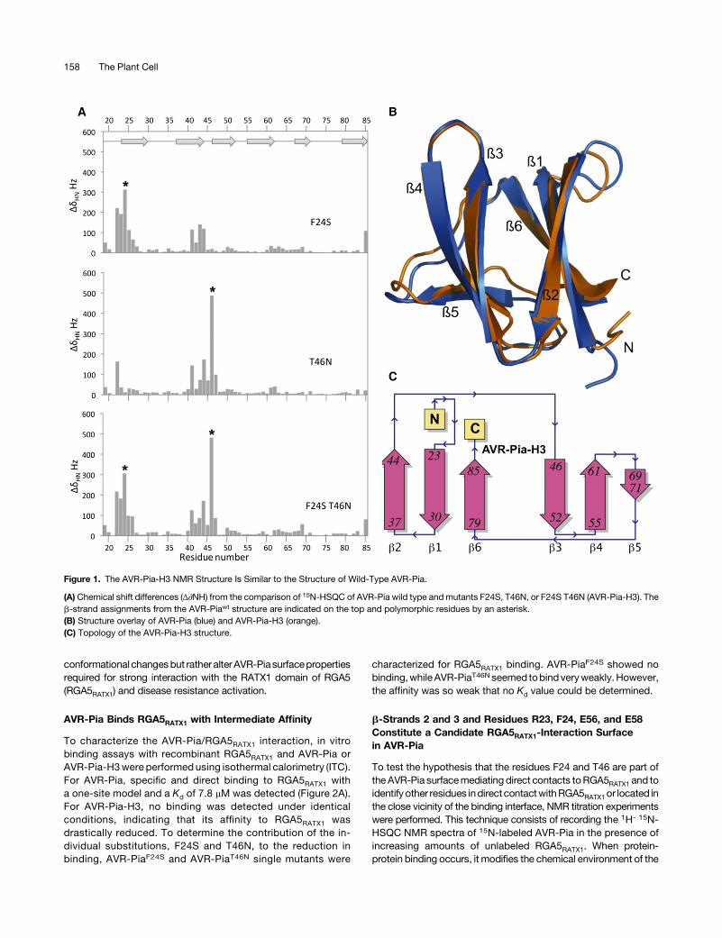
conformational changesbut rather alter AVR-Piasurfacepropertiesrequired for strong interaction with the RATX1 domain of RGA5(RGA5RATX1) and disease resistance activation.
AVR-Pia Binds RGA5RATX1 with Intermediate Affinity
To characterize the AVR-Pia/RGA5RATX1 interaction, in vitrobinding assays with recombinant RGA5RATX1 and AVR-Pia orAVR-Pia-H3were performed using isothermal calorimetry (ITC).For AVR-Pia, specific and direct binding to RGA5RATX1 witha one-site model and a Kd of 7.8 mM was detected (Figure 2A).For AVR-Pia-H3, no binding was detected under identicalconditions, indicating that its affinity to RGA5RATX1 wasdrastically reduced. To determine the contribution of the in-dividual substitutions, F24S and T46N, to the reduction inbinding, AVR-PiaF24S and AVR-PiaT46N single mutants were
characterized for RGA5RATX1 binding. AVR-PiaF24S showed nobinding,whileAVR-PiaT46Nseemed tobindveryweakly.However,the affinity was so weak that no Kd value could be determined.
b-Strands 2 and 3 and Residues R23, F24, E56, and E58Constitute a Candidate RGA5RATX1-Interaction Surfacein AVR-Pia
To test the hypothesis that the residues F24 and T46 are part ofthe AVR-Pia surfacemediatingdirect contacts toRGA5RATX1 and toidentify other residues indirect contactwithRGA5RATX1or located inthe close vicinity of the binding interface, NMR titration experimentswere performed. This technique consists of recording the 1H- 15N-HSQC NMR spectra of 15N-labeled AVR-Pia in the presence ofincreasing amounts of unlabeled RGA5RATX1. When protein-protein binding occurs, it modifies the chemical environment of the
Figure 1. The AVR-Pia-H3 NMR Structure Is Similar to the Structure of Wild-Type AVR-Pia.
(A) Chemical shift differences (Δ∂NH) from the comparison of 15N-HSQC of AVR-Pia wild type and mutants F24S, T46N, or F24S T46N (AVR-Pia-H3). Theb-strand assignments from the AVR-Piawt structure are indicated on the top and polymorphic residues by an asterisk.(B) Structure overlay of AVR-Pia (blue) and AVR-Pia-H3 (orange).(C) Topology of the AVR-Pia-H3 structure.
158 The Plant Cell
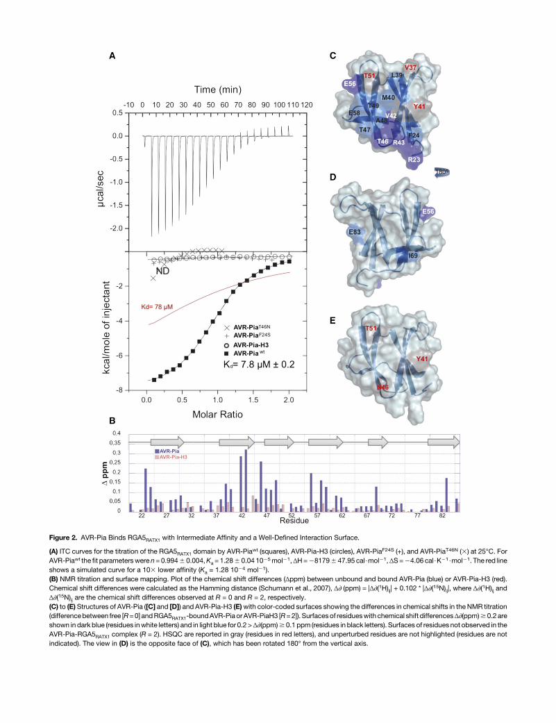
Figure 2. AVR-Pia Binds RGA5RATX1 with Intermediate Affinity and a Well-Defined Interaction Surface.
(A) ITC curves for the titration of the RGA5RATX1 domain by AVR-Piawt (squares), AVR-Pia-H3 (circles), AVR-PiaF24S (+), and AVR-PiaT46N (3) at 25°C. ForAVR-Piawt the fit parameterswere n= 0.9946 0.004,Ka = 1.286 0.04 1025mol21,ΔH=281796 47.95 cal$mol21,ΔS=24.06 cal$K21$mol21. The red lineshows a simulated curve for a 103 lower affinity (Ka = 1.28 1024 mol21).(B) NMR titration and surface mapping. Plot of the chemical shift differences (Δppm) between unbound and bound AVR-Pia (blue) or AVR-Pia-H3 (red).Chemical shift differences were calculated as the Hamming distance (Schumann et al., 2007), Δ∂ (ppm) = |Δ∂(1H)ij| + 0.102 * |Δ∂(15N)ij|, where Δ∂(1H)ij andΔ∂(15N)ij are the chemical shift differences observed at R = 0 and R = 2, respectively.(C) to (E) Structures of AVR-Pia ([C] and [D]) and AVR-Pia-H3 (E)with color-coded surfaces showing the differences in chemical shifts in the NMR titration(differencebetween free [R=0] andRGA5RATX1-boundAVR-Pia or AVR-PiaH3 [R=2]). Surfacesof residueswith chemical shift differencesΔ∂(ppm)$0.2areshown in dark blue (residues inwhite letters) and in light blue for 0.2 >Δ∂(ppm)$ 0.1 ppm (residues in black letters). Surfaces of residues not observed in theAVR-Pia-RGA5RATX1 complex (R = 2). HSQC are reported in gray (residues in red letters), and unperturbed residues are not highlighted (residues are notindicated). The view in (D) is the opposite face of (C), which has been rotated 180° from the vertical axis.
aminoacids locatedon thebindingsurface. This results ina changeof the chemical shift in NMRexperiments. Depending on the rate ofcomplex formation and dissociation, expressed by the exchangerate constant kex, and the chemical shift difference Δv between theunboundandboundstates (Δv=differencebetween the resonancefrequenciesof theexchangingsites), different exchange regimesoccur. NMR titration showed that the AVR-Pia-RGA5RATX1
complex was in slow exchange with kex << Δv since separateresonances appeared for individual species (bound and unboundstates) (SupplementalFigure3A).Residueswith importantchemicalshift changes between free AVR-Pia (R = 0) and AVR-Pia bound toRGA5RATX1 (molar ratio R = 2) were almost exclusively surfaceexposed and located in a region formed essentially by b-strands2 and 3 and including residuesR23and F24 fromb-strand 1 aswellasE56andE58 fromb-strand4 (Figures2Band2C).Nopeakswereobserved for residues Y27, V37, Y41, I44, and T51 in the complex.This candidate interaction surface largely overlaps with an ex-tended, solvent-exposed patch of hydrophobic/aromatic residuesformedbyF24,V26,andY28 inb1,V37,L38,andY41 inb2,andY85in b6. The residues on the other side of the AVR-Pia structure werenotshifted in theNMRtitrationandthereforeseemnot tobe involvedin the interaction with RGA5RATX1 (Figure 2D). Two exceptions wereE83,which probably senses aperturbation of the residueY41 that isclose in space, and the I69 residue, which may be involved in localconformational rearrangement of the short b5 strand.
RGA5RATX1 titration experiments were also performed with15N-labeledAVR-Pia-H3,which showsnobinding inY2H (Césariet al., 2013) and ITC (Figure 2A) analysis. Spectral perturbationswere strongly reduced and only few and limited changes ofchemical shiftsoccurredwhen titratingAVR-Pia-H3withRGA5RATX1(Figures 2B and 2E; Supplemental Figure 3B). Signals for the R23,S24,V42,R43,andE83residueswerestillobservedat theendof thetitration,while theyweremostly lostatamolar ratioof0.5 in thecaseof AVR-Pia (Supplemental Figure 3). Similarly, signals for E58, V59,and T47 were much less perturbed. Nevertheless, the peaks forY41, N46, and T51 were also perturbed, indicating a weak residualinteraction between RGA5RATX1 and AVR-Pia-H3 (Figures 2B and2E; Supplemental Figure 3).
In summary, NMR titration identified a candidate interactionsurface formed by b-strands 2 and 3 and including, in addition,residues R23, F24, E56, and E58 (Figure 2B). This surfaceoverlaps extensivelywith an extended hydrophobic patch on theAVR-Pia surface that contains F24 and has T46 on its border andthat may be crucial for RGA5RATX1 binding.
Y2H Experiments with Structure-Informed AVR-Pia MutantsConfirm an Important Role of the Candidate InteractionSurface in RGA5C-ter Binding
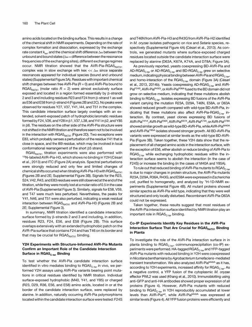
To test whether the AVR-Pia candidate interaction surfaceidentified in vitro mediates binding to RGA5C-ter in vivo, we per-formed Y2H assays using AVR-Pia variants bearing point muta-tions in critical residues identified by NMR titration. Individualsurface-exposed hydrophobic (M40, Y41, and Y85) or charged(R23, D29, R36, E56, and E58) amino acids, located in or at theborder of the candidate interaction surface, were replaced byalanine. In addition, naturally occurring AVR-Pia polymorphismslocatedwithin thecandidate interactionsurfacewere tested: F24S
andT46N fromAVR-Pia-H3andR43GfromAVR-Pia-H2 identifiedin M. oryzae isolates pathogenic on rice and Setaria species, re-spectively (Supplemental Figure 4A) (Césari et al., 2013). As con-trols, we generated mutants where surface-exposed chargedresidues located outside the candidate interaction surface werereplaced by alanine (D63A, K67A, K74A, and D78A; Figure 3A).As previously reported, yeasts coexpressing BD-AVR-Pia and
AD-RGA5C-ter or AD-RGA5C-ter and BD-RGA5C-ter grew on selectivemedium, indicatingphysicalbindingbetweenAVR-PiaandRGA5C-ter,and homo-interaction of the RGA5C-ter domain (Figure 3A) (Césariet al., 2013, 2014b). Yeasts coexpressing AD-RGA5C-ter and AVR-PiaF24S,AVR-PiaR43G,orAVR-PiaR36A fused to theBDdomaindidnotgrow on selective medium, indicating that these mutations abolishbinding to RGA5C-ter. Isolates expressing BD fusions of the AVR-Piavariant carrying the mutation R23A, D29A, T46N, E58A, or D63Ashowed reduced growth compared with wild-type BD-AVR-Pia, in-dicating that these mutations also affect AVR-Pia-RGA5C-ter in-teraction. By contrast, yeast clones expressing BD fusions ofAVR-PiaY41A,AVR-PiaE56A,AVR-PiaK67A,AVR-PiaK74A,orAVR-PiaD78A
showedsimilargrowthaswild-typeBD-AVR-Pia,whileAVR-PiaM40A
and AVR-PiaY85A isolates showed stronger growth. All BD-AVR-Piavariants were expressed at similar levels as the wild-type BD-AVR-Pia (Figure 3B). Taken together, these Y2H data show that the re-placement of all charged amino acids in the interaction surface, withthe exception of E56, either abolish or reduce binding of AVR-Pia toRGA5C-ter while exchanging hydrophobic residues within the in-teraction surface seems to abolish the interaction (in the case ofF24S) or increase the binding (in the cases of M40A and Y85A).To rule out that reducedbinding ofAVR-Piamutants toRGA5C-ter
is due to major changes in protein structure, the AVR-Pia mutantsR23A,D29A,R36A,R43G,andE58Awereexpressed inEscherichiacoli, purified to homogeneity, and analyzed by 1H-1D-NMR ex-periments (Supplemental Figure 4B). All mutant proteins showedsimilar spectra as AVR-Pia wild type, indicating that they were wellstructured and only locally disturbed. Recombinant AVR-Pia-D63Acould not be expressed.Taken together, these results suggest that most residues of
the AVR-Pia interaction surface identified byNMR titration play animportant role in RGA5C-ter binding.
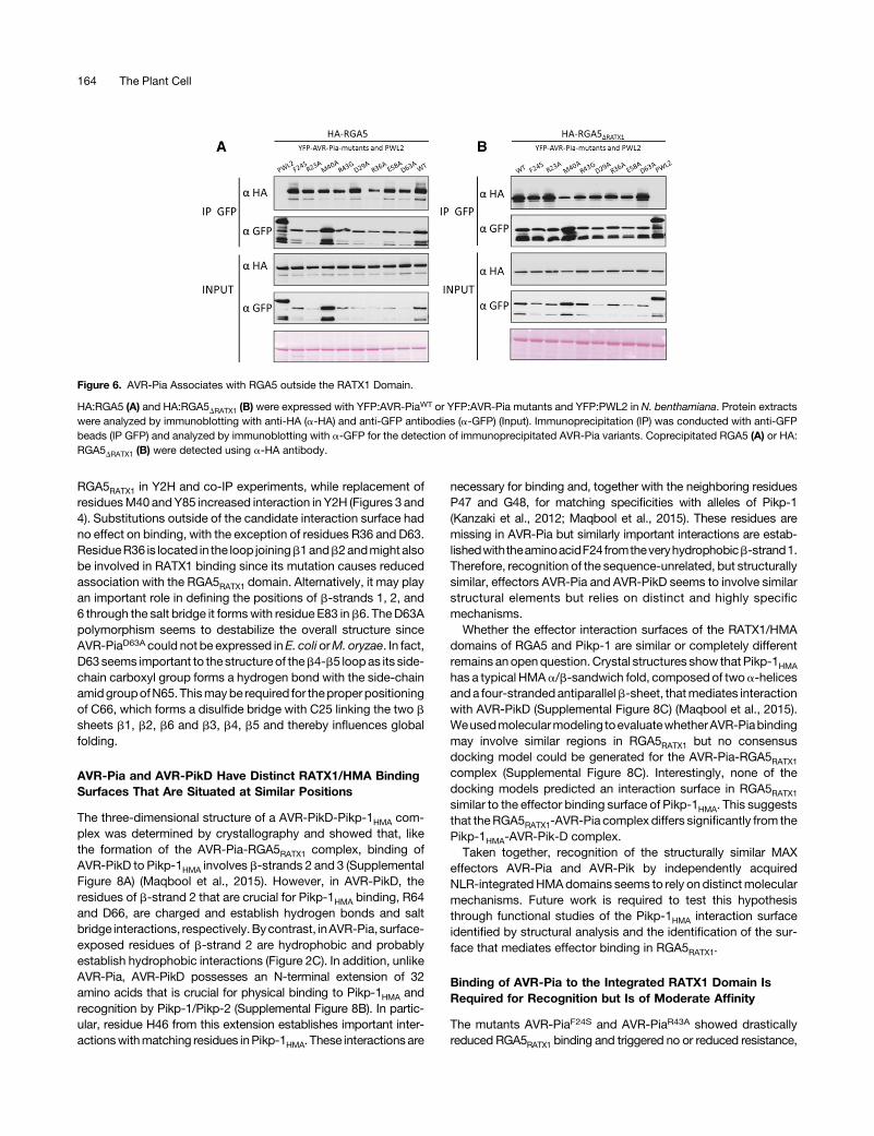
Co-IP Experiments Identify Key Residues in the AVR-PiaInteraction Surface That Are Crucial for RGA5RATX1 Bindingin Planta
To investigate the role of the AVR-Pia interaction surface in inplanta binding to RGA5C-ter, coimmunoprecipitation (co-IP) ex-perimentswereperformed.HA-taggedRGA5C-ter andYFP-taggedAVR-Pia mutants with reduced binding in Y2Hwere coexpressedinNicotianabenthamianabyAgrobacterium tumefaciens-mediatedtransient transformation. We also analyzed AVR-PiaM40A as it has,according to Y2H experiments, increased affinity for RGA5C-ter. Asa negative control, a YFP fusion of the cytoplasmic M. oryzaeeffector PWL2 was used (Khang et al., 2010). Immunoblotting usinganti-GFP and anti-HA antibodies showed proper expression of allproteins (Figure 4). However, AVR-Pia mutants with reducedbinding to RGA5C-ter in Y2H reproducibly accumulated at lowerlevels than AVR-Piawt, while AVR-PiaM40A was expressed atsimilar levels (Figure 4). All YFP fusionproteinswere efficiently and
160 The Plant Cell
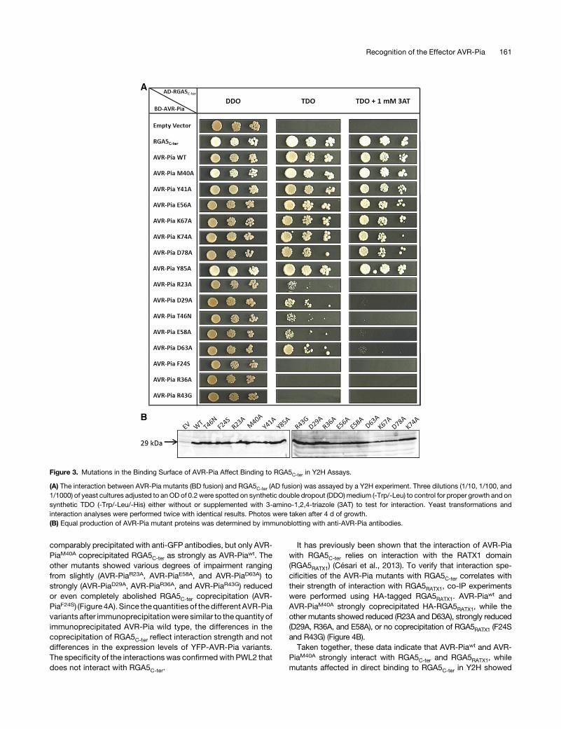
comparably precipitated with anti-GFP antibodies, but only AVR-PiaM40A coprecipitated RGA5C-ter as strongly as AVR-Piawt. Theother mutants showed various degrees of impairment rangingfrom slightly (AVR-PiaR23A, AVR-PiaE58A, and AVR-PiaD63A) tostrongly (AVR-PiaD29A, AVR-PiaR36A, and AVR-PiaR43G) reducedor even completely abolished RGA5C-ter coprecipitation (AVR-PiaF24S) (Figure 4A). Since thequantities of the different AVR-Piavariants after immunoprecipitationwere similar to the quantity ofimmunoprecipitated AVR-Pia wild type, the differences in thecoprecipitation of RGA5C-ter reflect interaction strength and notdifferences in the expression levels of YFP-AVR-Pia variants.The specificity of the interactions was confirmed with PWL2 thatdoes not interact with RGA5C-ter.
It has previously been shown that the interaction of AVR-Piawith RGA5C-ter relies on interaction with the RATX1 domain(RGA5RATX1) (Césari et al., 2013). To verify that interaction spe-cificities of the AVR-Pia mutants with RGA5C-ter correlates withtheir strength of interaction with RGA5RATX1, co-IP experimentswere performed using HA-tagged RGA5RATX1. AVR-Piawt andAVR-PiaM40A strongly coprecipitated HA-RGA5RATX1, while theother mutants showed reduced (R23A and D63A), strongly reduced(D29A, R36A, and E58A), or no coprecipitation of RGA5RATX1 (F24Sand R43G) (Figure 4B).Taken together, these data indicate that AVR-Piawt and AVR-
PiaM40A strongly interact with RGA5C-ter and RGA5RATX1, whilemutants affected in direct binding to RGA5C-ter in Y2H showed
Figure 3. Mutations in the Binding Surface of AVR-Pia Affect Binding to RGA5C-ter in Y2H Assays.
(A) The interaction between AVR-Pia mutants (BD fusion) and RGA5C-ter (AD fusion) was assayed by a Y2H experiment. Three dilutions (1/10, 1/100, and1/1000) of yeast cultures adjusted to anOD of 0.2 were spotted on synthetic double dropout (DDO)medium (-Trp/-Leu) to control for proper growth and onsynthetic TDO (-Trp/-Leu/-His) either without or supplemented with 3-amino-1,2,4-triazole (3AT) to test for interaction. Yeast transformations andinteraction analyses were performed twice with identical results. Photos were taken after 4 d of growth.(B) Equal production of AVR-Pia mutant proteins was determined by immunoblotting with anti-AVR-Pia antibodies.
Recognition of the Effector AVR-Pia 161
reduced association with RGA5C-ter and RGA5RATX1 in planta.Complete absenceof associationwithRGA5RATX1 for AVR-Pia
F24S
andAVR-PiaR43G,both inplantaand inY2H, indicatesacrucial roleof these residues in the binding interface and suggests that theyare pivotal for AVR-Pia recognition.
Direct Binding to the RATX1 Domain Is Required forAVR-Pia Recognition
To determine the role of the RATX1 binding surface of AVR-Pia inspecific recognition by the RGA4/RGA5 pair, AVR-Pia mutantswere coexpressed in N. benthamiana with RGA4/RGA5 and celldeath activation was monitored. Since tagged versions of AVR-Pia proved inactive in this assay, untaggedAVR-Piamutantswereused. AVR-Pia mutants with wild-type binding to RGA5RATX1 in-duced cell death, indicating that they are recognized by RGA5/RGA4 (Supplemental Figures 5A and 5B). Weakly or nonbindingmutants lostcelldeath inducingactivitybutwerealso lessabundantthan AVR-Piawt or recognized AVR-Pia mutants (SupplementalFigures 5A to 5C). They could be detected only after enrichmentby immunoprecipitation and showed in most cases only very lowabundance (Supplemental Figure 5D). Therefore, no clear con-clusions can be drawn for these mutants since lack of recognitionmay not only be due to reduced binding strength but also to lowprotein abundance or a combination of both effects. Differences inthe protein level of AVR-Piamutantswere previously observedwithYFP-taggedvariantsexpressed inN.benthamiana (Figure4)butnotupon expression in E. coli or yeast (Figure 3B). Therefore, differ-ences in the accumulation of AVR-Pia variants seem not related toan intrinsic destabilization of these proteins but rather to result fromreduced stability in N. benthamiana.
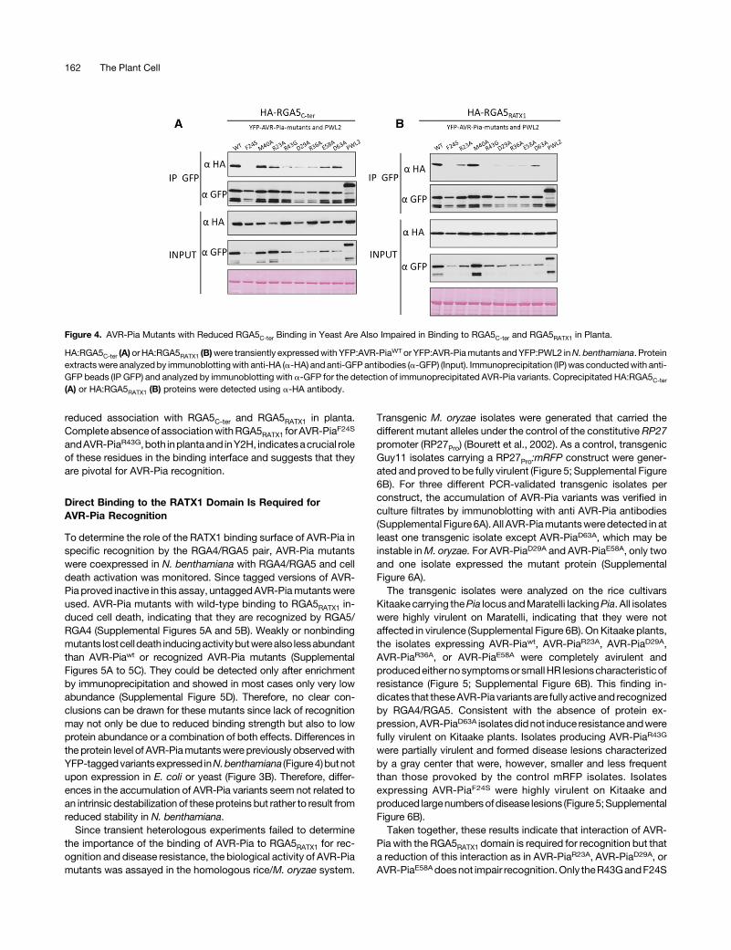
Since transient heterologous experiments failed to determinethe importance of the binding of AVR-Pia to RGA5RATX1 for rec-ognition and disease resistance, the biological activity of AVR-Piamutants was assayed in the homologous rice/M. oryzae system.
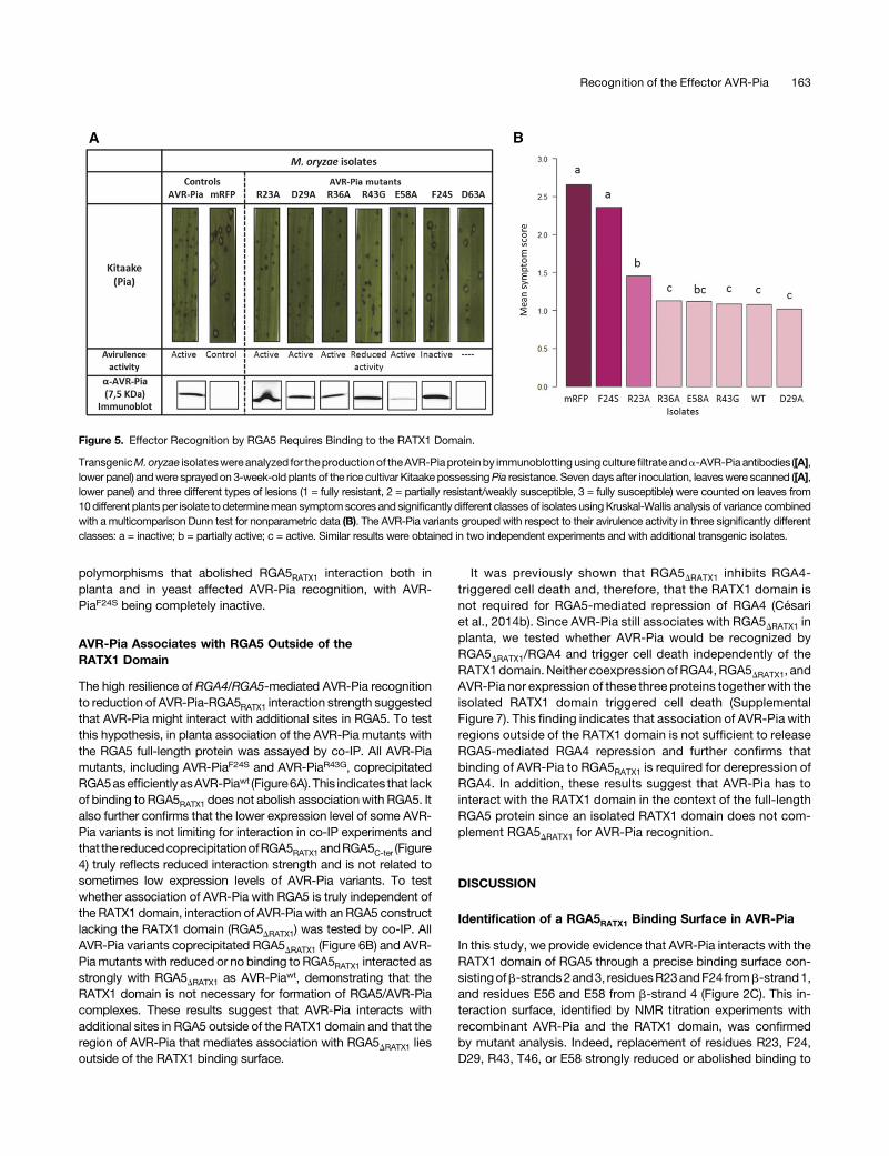
Transgenic M. oryzae isolates were generated that carried thedifferent mutant alleles under the control of the constitutive RP27promoter (RP27Pro) (Bourett et al., 2002). As a control, transgenicGuy11 isolates carrying a RP27Pro:mRFP construct were gener-ated and proved to be fully virulent (Figure 5; Supplemental Figure6B). For three different PCR-validated transgenic isolates perconstruct, the accumulation of AVR-Pia variants was verified inculture filtrates by immunoblotting with anti AVR-Pia antibodies(Supplemental Figure6A). All AVR-Piamutantsweredetected in atleast one transgenic isolate except AVR-PiaD63A, which may beinstable inM. oryzae. For AVR-PiaD29A and AVR-PiaE58A, only twoand one isolate expressed the mutant protein (SupplementalFigure 6A).The transgenic isolates were analyzed on the rice cultivars
Kitaake carrying thePia locus andMaratelli lackingPia. All isolateswere highly virulent on Maratelli, indicating that they were notaffected in virulence (Supplemental Figure 6B). On Kitaake plants,the isolates expressing AVR-Piawt, AVR-PiaR23A, AVR-PiaD29A,AVR-PiaR36A, or AVR-PiaE58A were completely avirulent andproducedeithernosymptomsorsmallHR lesionscharacteristicofresistance (Figure 5; Supplemental Figure 6B). This finding in-dicates that theseAVR-Pia variants are fully active and recognizedby RGA4/RGA5. Consistent with the absence of protein ex-pression,AVR-PiaD63A isolatesdidnot induce resistanceandwerefully virulent on Kitaake plants. Isolates producing AVR-PiaR43G
were partially virulent and formed disease lesions characterizedby a gray center that were, however, smaller and less frequentthan those provoked by the control mRFP isolates. Isolatesexpressing AVR-PiaF24S were highly virulent on Kitaake andproduced largenumbersofdisease lesions (Figure5;SupplementalFigure 6B).Taken together, these results indicate that interaction of AVR-
Pia with the RGA5RATX1 domain is required for recognition but thata reduction of this interaction as in AVR-PiaR23A, AVR-PiaD29A, orAVR-PiaE58Adoesnot impair recognition.Only theR43GandF24S
Figure 4. AVR-Pia Mutants with Reduced RGA5C-ter Binding in Yeast Are Also Impaired in Binding to RGA5C-ter and RGA5RATX1 in Planta.
HA:RGA5C-ter (A) orHA:RGA5RATX1 (B)were transiently expressedwith YFP:AVR-PiaWT or YFP:AVR-Piamutants andYFP:PWL2 inN. benthamiana. Proteinextractswere analyzedby immunoblottingwith anti-HA (a-HA) and anti-GFPantibodies (a-GFP) (Input). Immunoprecipitation (IP) was conductedwith anti-GFP beads (IP GFP) and analyzed by immunoblotting with a-GFP for the detection of immunoprecipitated AVR-Pia variants. Coprecipitated HA:RGA5C-ter
(A) or HA:RGA5RATX1 (B) proteins were detected using a-HA antibody.
162 The Plant Cell
polymorphisms that abolished RGA5RATX1 interaction both inplanta and in yeast affected AVR-Pia recognition, with AVR-PiaF24S being completely inactive.
AVR-Pia Associates with RGA5 Outside of theRATX1 Domain
The high resilience of RGA4/RGA5-mediated AVR-Pia recognitionto reduction of AVR-Pia-RGA5RATX1 interaction strength suggestedthat AVR-Pia might interact with additional sites in RGA5. To testthis hypothesis, in planta association of the AVR-Pia mutants withthe RGA5 full-length protein was assayed by co-IP. All AVR-Piamutants, including AVR-PiaF24S and AVR-PiaR43G, coprecipitatedRGA5asefficientlyasAVR-Piawt (Figure6A). This indicates that lackof binding to RGA5RATX1 does not abolish associationwith RGA5. Italso further confirms that the lower expression level of some AVR-Pia variants is not limiting for interaction in co-IP experiments andthat the reducedcoprecipitationofRGA5RATX1andRGA5C-ter (Figure4) truly reflects reduced interaction strength and is not related tosometimes low expression levels of AVR-Pia variants. To testwhether association of AVR-Pia with RGA5 is truly independent ofthe RATX1 domain, interaction of AVR-Pia with an RGA5 constructlacking the RATX1 domain (RGA5DRATX1) was tested by co-IP. AllAVR-Pia variants coprecipitated RGA5DRATX1 (Figure 6B) and AVR-Piamutants with reduced or no binding to RGA5RATX1 interacted asstrongly with RGA5DRATX1 as AVR-Piawt, demonstrating that theRATX1 domain is not necessary for formation of RGA5/AVR-Piacomplexes. These results suggest that AVR-Pia interacts withadditional sites in RGA5 outside of the RATX1 domain and that theregion of AVR-Pia that mediates association with RGA5DRATX1 liesoutside of the RATX1 binding surface.
It was previously shown that RGA5DRATX1 inhibits RGA4-triggered cell death and, therefore, that the RATX1 domain isnot required for RGA5-mediated repression of RGA4 (Césariet al., 2014b). Since AVR-Pia still associates with RGA5DRATX1 inplanta, we tested whether AVR-Pia would be recognized byRGA5DRATX1/RGA4 and trigger cell death independently of theRATX1domain. Neither coexpression ofRGA4,RGA5DRATX1, andAVR-Pia nor expression of these three proteins together with theisolated RATX1 domain triggered cell death (SupplementalFigure 7). This finding indicates that association of AVR-Pia withregions outside of the RATX1 domain is not sufficient to releaseRGA5-mediated RGA4 repression and further confirms thatbinding of AVR-Pia to RGA5RATX1 is required for derepression ofRGA4. In addition, these results suggest that AVR-Pia has tointeract with the RATX1 domain in the context of the full-lengthRGA5 protein since an isolated RATX1 domain does not com-plement RGA5DRATX1 for AVR-Pia recognition.
DISCUSSION
Identification of a RGA5RATX1 Binding Surface in AVR-Pia
In this study, we provide evidence that AVR-Pia interacts with theRATX1 domain of RGA5 through a precise binding surface con-sistingofb-strands2and3, residuesR23andF24 fromb-strand1,and residues E56 and E58 from b-strand 4 (Figure 2C). This in-teraction surface, identified by NMR titration experiments withrecombinant AVR-Pia and the RATX1 domain, was confirmedby mutant analysis. Indeed, replacement of residues R23, F24,D29, R43, T46, or E58 strongly reduced or abolished binding to
Figure 5. Effector Recognition by RGA5 Requires Binding to the RATX1 Domain.
TransgenicM.oryzae isolateswereanalyzed for theproductionof theAVR-Piaproteinby immunoblottingusingculturefiltrate anda-AVR-Pia antibodies([A],lower panel) andwere sprayed on 3-week-old plants of the rice cultivar Kitaake possessingPia resistance. Seven days after inoculation, leaveswere scanned ([A],lower panel) and three different types of lesions (1 = fully resistant, 2 = partially resistant/weakly susceptible, 3 = fully susceptible) were counted on leaves from10different plants per isolate to determinemean symptomscores and significantly different classes of isolates usingKruskal-Wallis analysis of variance combinedwith a multicomparison Dunn test for nonparametric data (B). The AVR-Pia variants grouped with respect to their avirulence activity in three significantly differentclasses: a = inactive; b = partially active; c = active. Similar results were obtained in two independent experiments and with additional transgenic isolates.
Recognition of the Effector AVR-Pia 163
RGA5RATX1 in Y2H and co-IP experiments, while replacement ofresiduesM40 andY85 increased interaction in Y2H (Figures 3 and4). Substitutions outside of the candidate interaction surface hadno effect on binding, with the exception of residues R36 and D63.ResidueR36 is located in the loop joiningb1andb2andmight alsobe involved in RATX1 binding since its mutation causes reducedassociation with the RGA5RATX1 domain. Alternatively, it may playan important role in defining the positions of b-strands 1, 2, and6 through the salt bridge it formswith residue E83 inb6. TheD63Apolymorphism seems to destabilize the overall structure sinceAVR-PiaD63A could not be expressed inE. coli orM. oryzae. In fact,D63seems important to the structure of theb4-b5 loopas its side-chain carboxyl group forms a hydrogen bond with the side-chainamidgroupofN65. Thismaybe required for theproper positioningof C66, which forms a disulfide bridge with C25 linking the two b
sheets b1, b2, b6 and b3, b4, b5 and thereby influences globalfolding.
AVR-Pia and AVR-PikD Have Distinct RATX1/HMA BindingSurfaces That Are Situated at Similar Positions
The three-dimensional structure of a AVR-PikD-Pikp-1HMA com-plex was determined by crystallography and showed that, likethe formation of the AVR-Pia-RGA5RATX1 complex, binding ofAVR-PikD to Pikp-1HMA involves b-strands 2 and 3 (SupplementalFigure 8A) (Maqbool et al., 2015). However, in AVR-PikD, theresidues of b-strand 2 that are crucial for Pikp-1HMA binding, R64and D66, are charged and establish hydrogen bonds and saltbridge interactions, respectively.Bycontrast, inAVR-Pia, surface-exposed residues of b-strand 2 are hydrophobic and probablyestablish hydrophobic interactions (Figure 2C). In addition, unlikeAVR-Pia, AVR-PikD possesses an N-terminal extension of 32amino acids that is crucial for physical binding to Pikp-1HMA andrecognition by Pikp-1/Pikp-2 (Supplemental Figure 8B). In partic-ular, residue H46 from this extension establishes important inter-actionswithmatching residues inPikp-1HMA. These interactionsare
necessary for binding and, together with the neighboring residuesP47 and G48, for matching specificities with alleles of Pikp-1(Kanzaki et al., 2012; Maqbool et al., 2015). These residues aremissing in AVR-Pia but similarly important interactions are estab-lishedwiththeaminoacidF24fromtheveryhydrophobicb-strand1.Therefore, recognition of the sequence-unrelated, but structurallysimilar, effectors AVR-Pia and AVR-PikD seems to involve similarstructural elements but relies on distinct and highly specificmechanisms.Whether the effector interaction surfaces of the RATX1/HMA
domains of RGA5 and Pikp-1 are similar or completely differentremains anopenquestion. Crystal structures show that Pikp-1HMA
has a typical HMAa/b-sandwich fold, composed of twoa-helicesanda four-strandedantiparallelb-sheet, thatmediates interactionwith AVR-PikD (Supplemental Figure 8C) (Maqbool et al., 2015).Weusedmolecularmodeling toevaluatewhetherAVR-Piabindingmay involve similar regions in RGA5RATX1 but no consensusdocking model could be generated for the AVR-Pia-RGA5RATX1
complex (Supplemental Figure 8C). Interestingly, none of thedocking models predicted an interaction surface in RGA5RATX1
similar to the effector binding surface of Pikp-1HMA. This suggeststhat theRGA5RATX1-AVR-Pia complex differs significantly from thePikp-1HMA-AVR-Pik-D complex.Taken together, recognition of the structurally similar MAX
effectors AVR-Pia and AVR-Pik by independently acquiredNLR-integratedHMAdomains seems to rely on distinctmolecularmechanisms. Future work is required to test this hypothesisthrough functional studies of the Pikp-1HMA interaction surfaceidentified by structural analysis and the identification of the sur-face that mediates effector binding in RGA5RATX1.
Binding of AVR-Pia to the Integrated RATX1 Domain IsRequired for Recognition but Is of Moderate Affinity
The mutants AVR-PiaF24S and AVR-PiaR43A showed drasticallyreduced RGA5RATX1 binding and triggered no or reduced resistance,
Figure 6. AVR-Pia Associates with RGA5 outside the RATX1 Domain.
HA:RGA5 (A) and HA:RGA5DRATX1 (B) were expressed with YFP:AVR-PiaWT or YFP:AVR-Pia mutants and YFP:PWL2 in N. benthamiana. Protein extractswere analyzed by immunoblotting with anti-HA (a-HA) and anti-GFP antibodies (a-GFP) (Input). Immunoprecipitation (IP) was conducted with anti-GFPbeads (IP GFP) and analyzed by immunoblotting with a-GFP for the detection of immunoprecipitated AVR-Pia variants. Coprecipitated RGA5 (A) or HA:RGA5DRATX1 (B) were detected using a-HA antibody.
164 The Plant Cell
respectively, indicating that the AVR-Pia-RGA5RATX1 interaction isrequired for RGA4/RGA5-mediated recognition. The presence ofthese polymorphisms in naturally occurring AVR-Pia alleles (Ribotet al., 2013) suggests that in rice isolatesofM.oryzae,AVR-Pia isundergoing selection for mutations in the RATX1-interactionsurface and escape from RGA4/RGA5-mediated recognition.These results therefore provide further support for a crucial roleof nonconventional, integrated decoy domains in effectorrecognition and NLR specificity.
However,wealso foundhigh resilience ofAVR-Pia recognition toa reduction in RGA5RATX1 binding strength since theweakly bindingAVR-Pia mutants AVR-PiaR23A, AVR-PiaE58A, AVR-PiaD29A, andAVR-PiaR36A were still able to trigger resistance. Similar ob-servations were made regarding AVR-PikD, the only other ex-ample where the affinity of an effector to the integrated decoydomain of its NLR receptor has been determined (Maqbool et al.,2015). Indeed, AVR-PikDA67D and AVR-PikDP47A G48D mutantsshowed drastically reduced binding to Pikp-1HMA but were nev-ertheless perfectly well recognized by Pik-1/Pik-2.
A possible explanation for this tolerance to a reduction in theaffinity between effectors and integrated decoys could be thateffectors interact with multiple independent sites in NLRreceptors. Indeed, our study suggests that, besides the RATX1domain, AVR-Pia interactswith other, not yet defined, regions inRGA5. In the simplest case, this interaction relies on directphysical binding, but since it was solely detected by co-IPexperiments, the possibility that the binding is indirect andinvolves additional cofactors cannot be excluded. This in-teraction seemsmediatedbyotherAVR-Pia surfaces than thoseinvolved inRGA5RATX1 binding sincemutantswith reducedbindingto RGA5RATX1 are not affected in interaction with RGA5DRATX1. As theRATX1 domain is covalently linked to the rest of the RGA5 receptor,AVR-Pia binding to these other sites has the potential to increase theoverall effector binding affinity to RGA5 despite the low affinitybinding to RGA5RATX1 (Kd = 7 mM). In this context, further mutation-induced reductionofAVR-Pia affinity toward theRATX1domainmaynot have a dramatic effect unless it completely abolishes AVR-Pia/RGA5RATX1 interaction. This situation highlights an advantage of the
integration of the decoy domain into the NLR receptor over a situ-ation where the decoy is a separate molecule and has to bind tothe effector before subsequent binding to the NLR receptor. In thelatter case, low affinity of the effector-decoy interaction would leadto drastically reduced receptor occupancy and render the corre-spondingresistancemorevulnerable toeffectormutationsaffectingdecoy binding.
Interaction of Effectors with Multiple Independent Sites Isa Hallmark of NLR Receptor Activation
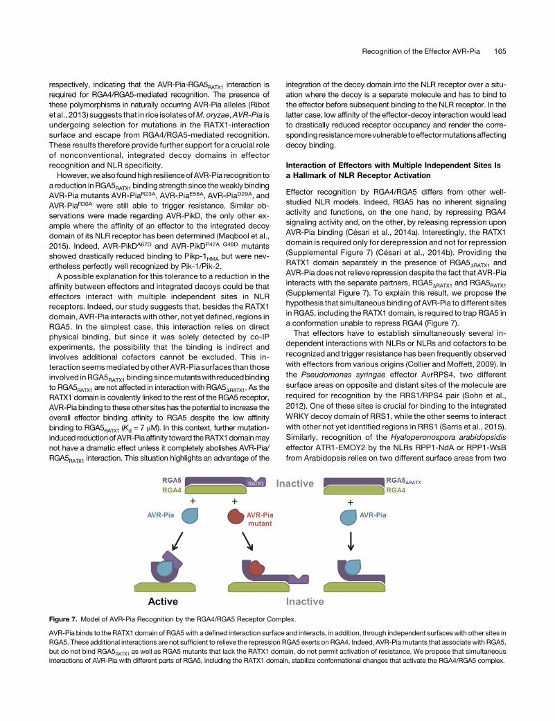
Effector recognition by RGA4/RGA5 differs from other well-studied NLR models. Indeed, RGA5 has no inherent signalingactivity and functions, on the one hand, by repressing RGA4signaling activity and, on the other, by releasing repression uponAVR-Pia binding (Césari et al., 2014a). Interestingly, the RATX1domain is required only for derepression and not for repression(Supplemental Figure 7) (Césari et al., 2014b). Providing theRATX1 domain separately in the presence of RGA5DRATX1 andAVR-Pia does not relieve repression despite the fact that AVR-Piainteracts with the separate partners, RGA5DRATX1 and RGA5RATX1
(Supplemental Figure 7). To explain this result, we propose thehypothesis that simultaneous binding of AVR-Pia to different sitesin RGA5, including the RATX1 domain, is required to trap RGA5 ina conformation unable to repress RGA4 (Figure 7).That effectors have to establish simultaneously several in-
dependent interactions with NLRs or NLRs and cofactors to berecognized and trigger resistance has been frequently observedwith effectors from various origins (Collier and Moffett, 2009). Inthe Pseudomonas syringae effector AvrRPS4, two differentsurface areas on opposite and distant sites of the molecule arerequired for recognition by the RRS1/RPS4 pair (Sohn et al.,2012). One of these sites is crucial for binding to the integratedWRKY decoy domain of RRS1, while the other seems to interactwith other not yet identified regions in RRS1 (Sarris et al., 2015).Similarly, recognition of the Hyaloperonospora arabidopsidiseffector ATR1-EMOY2 by the NLRs RPP1-NdA or RPP1-WsBfrom Arabidopsis relies on two different surface areas from two
Figure 7. Model of AVR-Pia Recognition by the RGA4/RGA5 Receptor Complex.
AVR-Pia binds to the RATX1 domain of RGA5with a defined interaction surface and interacts, in addition, through independent surfaces with other sites inRGA5. These additional interactions are not sufficient to relieve the repression RGA5 exerts on RGA4. Indeed, AVR-Piamutants that associate with RGA5,but do not bind RGA5RATX1 as well as RGA5 mutants that lack the RATX1 domain, do not permit activation of resistance. We propose that simultaneousinteractions of AVR-Pia with different parts of RGA5, including the RATX1 domain, stabilize conformational changes that activate the RGA4/RGA5 complex.
Recognition of the Effector AVR-Pia 165
different domains and on opposite sides of the molecule, sug-gesting simultaneous interactionwith independent binding sides inRPP1-NdA and RPP1WsB (Chou et al., 2011; Steinbrenner et al.,2015). Also in NLRs that recognize effector-cofactor complexes,simultaneous binding of these complexes to different parts of theNLR, generally involving the N terminus and the leucine-rich repeathave been frequently described (Collier and Moffett, 2009).Therefore, we propose the hypothesis that effectors or effector-cofactor complexes forcing or trappingNLRs in an activated statebysimultaneouslybinding tomultiplebindingsitesand inducingorstabilizing by this major conformational changes is a widespreadmechanism in NLR activation and particularly in NLRs with in-tegrated domains. Future structural and functional analysiswill benecessary to test this model and elucidate in more detail howactivation occurs at the molecular level.
METHODS
Growth Conditions of Plants and Fungi and Infection Assays
Nicotianabenthamianaplantsweregrowninagrowthchamberat22°Cunderfluorescent light (Radium;fluorescent lampSpectraluxPlusNL-T858W/865/G13) with a 16-h light period. Rice plants (Oryza sativa) were grown as de-scribed (Faivre-Rampant et al., 2008). TransgenicMagnaporthe oryzaeGUY11 strains were grown at 25°C during 5 d on rice flour agar for spore pro-duction (Berruyer et al., 2003) and in Tanaka complete culture medium(Villalba et al., 2008) agitated at 60 rpm and 25°C during 5 d for liquid culture.
For the analysis of interaction phenotypes, a suspension of M. oryzaeconidiospores inwaterwith 0.1%of gelatin and adjusted to 53104 sporesmL21 was sprayed on the leaves of 3-week-old rice plants (Berruyer et al.,2003).Symptomswereanalyzed7dafter inoculationon theyoungest leavethatwas fully expandedat the timeof inoculation. For quantitative analysis,lesions were classified and counted: resistant lesions, visible as smallbrown spots (type 1); weakly susceptible/partially resistant lesions char-acterized by a pronounced brown border and a small gray center (type 2);fully susceptible lesions characterized by a large gray center (type 3).
Constructs
Plasmids were generated by Gateway cloning (Thermo Fisher), restriction/ligation, site-directed mutagenesis using the QuickChange Lightning kit(Agilent), or gap-repair cloning in yeast (Bruno et al., 2004). Gateway entryclones were generated using the pDONR207 plasmid (Thermo Fisher).Gateway destination vectors were modified pBIN19 plasmids for ex-pression of tagged proteins in N. benthamiana (Césari et al. 2013) ormodified pGAD-T7 or pGBK-T7 plasmids (Clontech) for yeast two-hybridexperiments (Bernoux et al., 2011). For protein expression, the pET-15bvector (Merck-Millipore) was used. For M. oryzae transformation, con-structs were based on the pDL02 plasmid (Bruno et al., 2004). For detailson PCR and mutagenesis primers and generation of plasmids, refer toSupplemental Tables 2 and 3.
NMR Spectroscopy and Structure Determination
Spectrawereacquiredona700MHzAvanceBruker spectrometer equippedwith triple-resonance (1H, 15N, 13C) z-gradient cryoprobeat 305K. All spectraare referenced to the internal reference DSS for the 1H dimension and in-directly referenced for the 15N and 13C dimensions (Wishart et al., 1995).
Spectra were processed using Topspin (version 3.2) and analyzed usingstrip-plots with Cindy in house software and CCPN (Vranken et al., 2005)(analysis v 2.3). The 1H, 15N, and 13C assignments were derived by analogyfrom the assignments of AVR-Pia wild type without the need to prepare
a 13C-labeled sample and the details are given inSupplementalMethods.Briefly,oneproteinpreparationof15N-labeledAVR-Pia-H3 inwaterwasusedto record 3D 15N-1Hexperiments for backbone assignments and2DNOESYand 2D TOCSY for side-chain assignments. To solve ambiguous assign-ments and to obtain 13C chemical shift data, the sample was lyophilized anddissolved in D2O, and 13C-1H HSQC/TOCSY experiments were recorded.Distance restraints obtained from the 3D 15N-NOESY-HSQC and 2D-NO-ESY spectra, F/C dihedral angle constraints from TALOS+ (Shen et al.,2009), and H-bonds were used to generate structures by CYANA (Güntert,2004), CNS (Brunger, 2007), and the refinement in water of RECOORD(Nederveenet al., 2005) (Supplemental Table 1 andSupplementalMethods).
NMR Titration
For the assignments, protein samples (1mM) in 20mMpotassium-sodiumphosphate, pH 5.4, and 150 mM NaCl were used. For the titrations of15N-labeled AVR-Pia proteins, different samples with constant concen-trations of AVR-Pia wild type or H3 (50 mM) and various concentrations ofunlabeled RATX1 (ratios 2:1, 1:1, 0.5:1, 0.25:1, and 0:1 for the reference)were prepared. HSQC spectra were recorded at 305K on a Bruker Avance700MHzspectrometer.Chemical shift differencesweremeasured fromtheHSQC spectra of AVR-Pia or AVR-H3 alone and the AVR-RATX1 complexat R = 2. They are reported as Hamming distance weighted by the mag-netogyric ratios (Schumann et al., 2007).
Co-IP and Y2H Interaction Assays
Protein-protein interaction analyses by coimmunoprecipitation were per-formed with protein extracts fromN. benthamiana leaf discs harvested 2 dafter Agrobacterium infiltration (Césari et al., 2013). For the interaction ofAVR-Pia variants with RGA5C-ter and RGA5RATX1, five leaf disks per samplewere homogenized in extraction buffer (25 mM Tris-HCl, pH 7.5, 150 mMNaCl, 1 mM EDTA, 10 mM DTT, 1 mM PMSF, and 0.1% IGEPAL CA-630[Nonidet P-40]), supplemented with complete protease inhibitor cocktail(Roche) and polyvinylpolypyrrolidone (PVPP; 0.5%). After two cen-trifugations (30 min, 15,000g), 5 mL magnetic GFP-trap_M beads (Chro-motek) per samplewashed two timeswithproteinextractionbuffer (withoutPVPP) were added to 500 mL protein extract and incubated with gentlerotation for 2 h at 4°C. Beads were separated andwashed three times with600 mL protein extraction buffer (without PVPP).
For the interaction of AVR-Pia variants and RGA5 or RGA5DRATX1,a modified protein extraction buffer was used (50 mM Tris-HCl, pH 7.5,150mMNaCl, 1mMEDTA, 10mMDTT,1mMPMSF,1.0%IGEPALCA-630[Nonidet P-40], 0.5% sodium deoxycholate, and 0.1% SDS, supplementedwith a complete protease inhibitor cocktail [Roche] and 0.5% PVPP). Co-IPwasperformedwith 8mL agaroseGFP_trap_A suspension (Chromotek) andfour washes with the modified protein extraction buffer.
Bound proteins were eluted by boiling for 10 min at 70°C in 50 mLNuPAGE sample buffer, separated by polyacrylamide gel electrophoresisusing NuPAGE 4 to 12% gels (Invitrogen), transferred to nitrocellulosemembrane(Millipore),andanalyzedbyimmunoblotting.For immunodetectionofproteins, ratanti-HA-horseradishperoxidase(clone3F10;Sigma-Aldrich)ormouse anti-GFP (clones 7.1 and 13.1, Sigma-Aldrich) and goat anti-mouse-horseradish peroxidase (Sigma-Aldrich) were used in combination with theImmobilon western kit (Millipore).
Binding domain (BD) fusions of AVR-Pia variants in pGBKT7-53 and ac-tivation domain (AD) fusions of RGA5C-ter in pGADT7were transformed in goldand Y187 yeast strain, respectively. Interactions assays were performed ac-cording to theMatchmakerGold yeast two-hybrid systemprotocol (Clontech).
Transient Protein Expression and HR Assays in N. benthamiana
For agroinfiltration inN. benthamiana, pBIN19 binary vectors containing eitherAVR-Pia,PWL2, orRGA5variantswere transformed intoAgrobacteriumstrain
166 The Plant Cell
GV3101byelectroporation. IndividualcloneswereselectedandgrowninLuria-Bertani liquid medium containing 50 mg mL21 rifampicin, 15 mg mL21 gen-tamycin, and 50 mg mL21 kanamycin at 28°C for 24 h before agroinfiltration.Coinoculationmixtures adjusted to an OD600 of 1.0 were infiltrated in 4-week-oldN. benthamiana plants. The infiltrated plants were incubated for 48 or 96 hin growth chambers under controlled conditions for coimmunoprecipitations orcell death assays, respectively. Three days after infiltration,N. benthamianaleaves were scanned using a Typhoon FLA9000 fluorescence scanner (GEHealthcare)with excitation at 635nmanda long-pass red filter (LPR-665nm)to evaluate the HR response as a lack of red chlorophyll fluorescence.
Accession Numbers
Sequence data from this article correspond to those previously published(Césari, et al., 2013) and can be found in the GenBank/EMBL databasesunder the followingaccessionnumbers:AVR-Pia (AB498873),AVR-Pia-H3(KC777366), PWL2 (U26313), RGA4 (AB604622), Sasanishiki RGA5-A(AB604627), and Sasanishiki RGA5-B (KC777365). The Protein Data Bankaccession number for the AVR-Pia_H3 structure is 5JHJ.
Supplemental Data
Supplemental Figure 1. Solution structure of AVR-Pia-H3.
Supplemental Figure 2. Comparison of NMR relaxation of AVR-Piaand AVR-Pia-H3.
Supplemental Figure 3. HSQC spectra of AVR-Pia and AVR-Pia-H3recorded upon titration with RGA5RATX1.
Supplemental Figure 4. AVR-Pia mutants affected in RGA5RATX1
binding are well structured.
Supplemental Figure 5. AVR-Pia mutants not affected in RGA5RATX1
binding trigger HR in N. benthamiana.
Supplemental Figure 6. Characterization of transgenic M. oryzaeisolates carrying AVR-Pia mutant constructs.
Supplemental Figure 7. RGA5DRATX1 represses RGA4-mediated celldeath but does not recognize AVR-Pia.
Supplemental Figure 8. Comparison of the AVR-Pia and AVR-PikDstructures and their complexes with RATX1/HMA domains.
Supplemental Table 1. Statistics for 20 NMR structures of AVR-Pia-H3.
Supplemental Table 2. Primers.
Supplemental Table 3. Plasmids.
Supplemental Methods. Supplemental experimental procedures andmethods.
ACKNOWLEDGMENTS
This work was supported by the French Infrastructure for IntegratedStructural Biology (ANR-10-INSB-05-0) and the ANR project Immunere-ceptor (ANR-15-CE20-0007). D.O. was supported by a PhD grant from theMinistry of Research of Colombia (Colciencias). This work benefited frominteractions promoted by COST Action FA 1208 (https://www.cost-sustain.org). We thank Christian Roumestand for fruitful discussions.
AUTHOR CONTRIBUTIONS
T.K. and A.P. acquired funding and conceived, supervised, and designedthe research. D.O., J.G., K.G, V.C., and A.P. conducted the investigation.D.O., A.P., andT.K.wrote theoriginal draft. D.O., A.P., S.C., andT.K.wrote,
reviewed, and edited the article. D.O. and S.C. provided resources andgenerated vectors. D.O., S.C., K.G., V.C., andA.P. performed the research.A.P. andK.G. performedNMRanalyses. D.O. andV.C. produced transgenicM.oryzae strains. D.O. performed protein-protein interaction analyses.D.O.,J.G., K.G., A.P., and T.K. analyzeddata. D.O., T.K., andA.P. wrote the article
Received June 9, 2016; revised November 9, 2016; accepted January 12,2017; published January 13, 2017.
REFERENCES
Bernoux, M., Ve, T., Williams, S., Warren, C., Hatters, D., Valkov,E., Zhang, X., Ellis, J.G., Kobe, B., and Dodds, P.N. (2011).Structural and functional analysis of a plant resistance protein TIRdomain reveals interfaces for self-association, signaling, and auto-regulation. Cell Host Microbe 9: 200–211.
Berruyer, R., Adreit, H., Milazzo, J., Gaillard, S., Berger, A., Dioh, W.,Lebrun, M.-H.H., and Tharreau, D. (2003). Identification and fine map-ping of Pi33, the rice resistance gene corresponding to theMagnaporthegrisea avirulence gene ACE1. Theor. Appl. Genet. 107: 1139–1147.
Bonardi, V., Tang, S., Stallmann, A., Roberts, M., Cherkis, K., andDangl, J.L. (2011). Expanded functions for a family of plant intracellularimmune receptors beyond specific recognition of pathogen effectors.Proc. Natl. Acad. Sci. USA 108: 16463–16468.
Bourett, T.M., Sweigard, J.A., Czymmek, K.J., Carroll, A., andHoward, R.J. (2002). Reef coral fluorescent proteins for visualizingfungal pathogens. Fungal Genet. Biol. 37: 211–220.
Brunger, A.T. (2007). Version 1.2 of the crystallography and NMRsystem. Nat. Protoc. 2: 2728–2733.
Bruno, K.S., Tenjo, F., Li, L., Hamer, J.E., Xu, J., and Al, B.E.T.(2004). Cellular localization and role of kinase activity of PMK1 inMagnaporthe grisea. 3: 1525–1532.
Césari, S., et al. (2013). The rice resistance protein pair RGA4/RGA5recognizes the Magnaporthe oryzae effectors AVR-Pia and AVR1-CO39 by direct binding. Plant Cell 25: 1463–1481.
Césari, S., Bernoux, M., Moncuquet, P., Kroj, T., and Dodds,P.N. (2014a). A novel conserved mechanism for plant NLR proteinpairs: the “integrated decoy” hypothesis. Front. Plant Sci. 5: 606.
Césari, S., Kanzaki, H., Fujiwara, T., Bernoux, M., Chalvon, V.,Kawano, Y., Shimamoto, K., Dodds, P., Terauchi, R., and Kroj, T.(2014b). The NB-LRR proteins RGA4 and RGA5 interact functionallyand physically to confer disease resistance. EMBO J. 33: 1941–1959.
Chou, S., Krasileva, K.V., Holton, J.M., Steinbrenner, A.D., Alber,T., and Staskawicz, B.J. (2011). Hyaloperonospora arabidopsidisATR1 effector is a repeat protein with distributed recognition sur-faces. Proc. Natl. Acad. Sci. USA 108: 13323–13328.
Collier, S.M., and Moffett, P. (2009). NB-LRRs work a “bait andswitch” on pathogens. Trends Plant Sci. 14: 521–529.
Cui, H., Tsuda, K., and Parker, J.E. (2015). Effector-triggered im-munity: from pathogen perception to robust defense. Annu. Rev.Plant Biol. 66: 487–511.
Dean, R., Van Kan, J.A., Pretorius, Z.A., Hammond-Kosack, K.E.,Di Pietro, A., Spanu, P.D., Rudd, J.J., Dickman, M., Kahmann, R.,Ellis, J., and Foster, G.D. (2012). The top 10 fungal pathogens inmolecular plant pathology. Mol. Plant Pathol. 13: 414–430.
de Guillen, K., Ortiz-Vallejo, D., Gracy, J., Fournier, E., Kroj, T., andPadilla, A. (2015). Structure analysis uncovers a highly diverse butstructurally conserved effector family in phytopathogenic fungi.PLoS Pathog. 11: e1005228.
Eitas, T.K., and Dangl, J.L. (2010). NB-LRR proteins: pairs, pieces,perception, partners, and pathways. Curr. Opin. Plant Biol. 13: 472–477.
Recognition of the Effector AVR-Pia 167
Ellis, J.G., Dodds, P.N., and Lawrence, G.J. (2007). Flax rust re-sistance gene specificity is based on direct resistance-avirulenceprotein interactions. Annu. Rev. Phytopathol. 45: 289–306.
Faivre-Rampant, O., Thomas, J., Allègre, M., Morel, J.-B., Tharreau, D.,Nottéghem, J.-L., Lebrun, M.-H., Schaffrath, U., and Piffanelli, P.(2008). Characterization of the model system rice-Magnaporthe for thestudy of nonhost resistance in cereals. New Phytol. 180: 899–910.
Fukuoka, S., Saka, N., Koga, H., Ono, K., Shimizu, T., Ebana, K.,Hayashi, N., Takahashi, A., Hirochika, H., Okuno, K., and Yano, M.(2009). Loss of function of a proline-containing protein confers durabledisease resistance in rice. Science 325: 998–1001.
Gabriëls, S.H.E.J., Vossen, J.H., Ekengren, S.K., van Ooijen, G.,Abd-El-Haliem, A.M., van den Berg, G.C.M., Rainey, D.Y.,Martin, G.B., Takken, F.L.W., de Wit, P.J.G.M., and Joosten,M.H. (2007). An NB-LRR protein required for HR signalling mediatedby both extra- and intracellular resistance proteins. Plant J. 50: 14–28.
Güntert, P. (2004). Automated NMR structure calculation with CYANA.Methods Mol. Biol. 278: 353–378.
Jacob, F., Vernaldi, S., and Maekawa, T. (2013). Evolution andconservation of plant NLR functions. Front. Immunol. 4: 297.
Jia, Y., McAdams, S.A., Bryan, G.T., Hershey, H.P., and Valent, B.(2000). Direct interaction of resistance gene and avirulence geneproducts confers rice blast resistance. EMBO J. 19: 4004–4014.
Kanzaki, H., Yoshida, K., Saitoh, H., Fujisaki, K., Hirabuchi, A.,Alaux, L., Fournier, E., Tharreau, D., and Terauchi, R. (2012).Arms race co-evolution of Magnaporthe oryzae AVR-Pik and rice Pikgenes driven by their physical interactions. Plant J. 72: 894–907.
Khang, C.H., Berruyer, R., Giraldo, M.C., Kankanala, P., Park,S.-Y., Czymmek, K., Kang, S., and Valent, B. (2010). Translocationof Magnaporthe oryzae effectors into rice cells and their subsequentcell-to-cell movement. Plant Cell 22: 1388–1403.
Krasileva, K.V., Dahlbeck, D., and Staskawicz, B.J. (2010). Activationof an Arabidopsis resistance protein is specified by the in planta as-sociation of its leucine-rich repeat domain with the cognate oomyceteeffector. Plant Cell 22: 2444–2458.
Liu, W., Liu, J., Triplett, L., Leach, J.E., and Wang, G.-L. (2014).Novel insights into rice innate immunity against bacterial and fungalpathogens. Annu. Rev. Phytopathol. 52: 213–241.
Maekawa, T., et al. (2011). Coiled-coil domain-dependent homo-dimerization of intracellular barley immune receptors definesa minimal functional module for triggering cell death. Cell HostMicrobe 9: 187–199.
Maqbool, A., Saitoh, H., Franceschetti, M., Stevenson, C., Uemura,A., Kanzaki, H., Kamoun, S., Terauchi, R., and Banfield, M.(2015). Structural basis of pathogen recognition by an integratedHMA domain in a plant NLR immune receptor. Elife 4: 1–24.
Nyarko, A., Singarapu, K.K., Figueroa, M., Manning, V.A.,Pandelova, I., Wolpert, T.J., Ciuffetti, L.M., and Barbar, E.(2014). Solution NMR structures of Pyrenophora tritici-repentisToxB and its inactive homolog reveal potential determinants of toxinactivity. J. Biol. Chem. 289: 25946–25956.
Okuyama, Y., et al. (2011). A multifaceted genomics approach allowsthe isolation of the rice Pia-blast resistance gene consisting of twoadjacent NBS-LRR protein genes. Plant J. 66: 467–479.
Pennisi, E. (2010). Armed and dangerous. Science 327: 804–805.Qi, D., and Innes, R.W. (2013). Recent advances in plant NLR structure,
function, localization, and signaling. Front. Immunol. 4: 348.
Ribot, C., Césari, S., Abidi, I., Chalvon, V., Bournaud, C., Vallet, J.,Lebrun, M.-H., Morel, J.-B., and Kroj, T. (2013). The Magnaportheoryzae effector AVR1-CO39 is translocated into rice cells in-dependently of a fungal-derived machinery. Plant J. 74: 1–12.
Le Roux, C., et al. (2015). A receptor pair with an integrated decoyconverts pathogen disabling of transcription factors to immunity.Cell 161: 1074–1088.
Nederveen, A.J., et al. (2005). RECOORD: A recalculated coordinatedatabase of 5001 proteins from the PDB using restraints from theBioMagResBank. Proteins 59: 662–672.
Sarris, P.F.F., et al. (2015). A plant immune receptor detects pathogeneffectors that target WRKY transcription factors. Cell 161: 1089–1100.
Schumann, F.H., Riepl, H., Maurer, T., Gronwald, W., Neidig, K.P.,and Kalbitzer, H.R. (2007). Combined chemical shift changes andamino acid specific chemical shift mapping of protein-protein in-teractions. J. Biomol. NMR 39: 275–289.
Shen, Y., Delaglio, F., Cornilescu, G., and Bax, A. (2009). TALOS+:a hybrid method for predicting protein backbone torsion anglesfrom NMR chemical shifts. J. Biomol. NMR 44: 213–223.
Skamnioti, P., and Gurr, S.J. (2009). Against the grain: safeguardingrice from rice blast disease. Trends Biotechnol. 27: 141–150.
Sohn, K.H., Hughes, R.K., Piquerez, S.J., Jones, J.D.G., andBanfield, M.J. (2012). Distinct regions of the Pseudomonas syringaecoiled-coil effector AvrRps4 are required for activation of immunity.Proc. Natl. Acad. Sci. USA 109: 16371–16376.
Steinbrenner, A.D., Goritschnig, S., and Staskawicz, B.J. (2015).Recognition and activation domains contribute to allele-specificresponses of an Arabidopsis NLR receptor to an oomycete effectorprotein. PLoS Pathog. 11: e1004665.
Takken, F.L.W., and Goverse, A. (2012). How to build a pathogendetector: structural basis of NB-LRR function. Curr. Opin. PlantBiol. 15: 375–384.
van der Hoorn, R.A.L., and Kamoun, S. (2008). From Guard to De-coy: a new model for perception of plant pathogen effectors. PlantCell 20: 2009–2017.
Villalba, F., Collemare, J., Landraud, P., Lambou, K., Brozek, V.,Cirer, B., Morin, D., Bruel, C., Beffa, R., and Lebrun, M.-H. (2008).Improved gene targeting in Magnaporthe grisea by inactivation ofMgKU80 required for non-homologous end joining. Fungal Genet.Biol. 45: 68–75.
Vranken, W.F., Boucher, W., Stevens, T.J., Fogh, R.H., Pajon, A.,Llinas, M., Ulrich, E.L., Markley, J.L., Ionides, J., and Laue, E.D.(2005). The CCPN data model for NMR spectroscopy: developmentof a software pipeline. Proteins 59: 687–696.
Williams, S.J., et al. (2014). Structural basis for assembly and function ofa heterodimeric plant immune receptor. Science 344: 299–303.
Wishart, D.S., Bigam, C.G., Yao, J., Abildgaard, F., Dyson, H.J.,Oldfield, E., Markley, J.L., and Sykes, B.D. (1995). 1H, 13C and15N chemical shift referencing in biomolecular NMR. J. Biomol.NMR 6: 135–140.
Wu, C.-H., Belhaj, K., Bozkurt, T.O., Birk, M.S., and Kamoun, S.(2016). Helper NLR proteins NRC2a/b and NRC3 but not NRC1 arerequired for Pto-mediated cell death and resistance in Nicotianabenthamiana. New Phytol. 209: 1344–1352.
Zhang, Z.-M., Zhang, X., Zhou, Z.-R., Hu, H.-Y., Liu, M., Zhou, B.,and Zhou, J. (2013). Solution structure of the Magnaporthe oryzaeavirulence protein AvrPiz-t. J. Biomol. NMR 55: 219–223.
168 The Plant Cell
DOI 10.1105/tpc.16.00435; originally published online January 13, 2017; 2017;29;156-168Plant Cell
Thomas KrojDiana Ortiz, Karine de Guillen, Stella Cesari, Véronique Chalvon, Jérome Gracy, André Padilla and
Immune Receptor RGA5 Effector AVR-Pia by the Decoy Domain of the Rice NLRMagnaporthe oryzaeRecognition of the
This information is current as of March 27, 2017
Supplemental Data http://www.plantcell.org/content/suppl/2017/02/06/tpc.16.00435.DC2.html http://www.plantcell.org/content/suppl/2017/01/13/tpc.16.00435.DC1.html
References http://www.plantcell.org/content/29/1/156.full.html#ref-list-1
This article cites 44 articles, 12 of which can be accessed free at:
Permissions https://www.copyright.com/ccc/openurl.do?sid=pd_hw1532298X&issn=1532298X&WT.mc_id=pd_hw1532298X
eTOCs http://www.plantcell.org/cgi/alerts/ctmain
Sign up for eTOCs at:
CiteTrack Alerts http://www.plantcell.org/cgi/alerts/ctmain
Sign up for CiteTrack Alerts at:
Subscription Information http://www.aspb.org/publications/subscriptions.cfm
is available at:Plant Physiology and The Plant CellSubscription Information for
ADVANCING THE SCIENCE OF PLANT BIOLOGY © American Society of Plant Biologists


![circRNAs Are Involved in the Rice-Magnaporthe oryzae … · circRNAs Are Involved in the Rice-Magnaporthe oryzae Interaction1[OPEN] Jing Fan,a,2 Weili Quan,b,2 Guo-Bang Li,a,2 Xiao-Hong](https://img.pdfslide.us/doc/110x75/609760baf9f98337ca16efe8/circrnas-are-involved-in-the-rice-magnaporthe-oryzae-circrnas-are-involved-in-the.jpg)