Embed Size (px)
Citation preview
Conidial morphogenesis and septin-mediated plant infection require Smo1, a 1
Ras GTPase-activating protein in Magnaporthe oryzae 2
3
Michael J. Kershaw*, Magdalena Basiewicz*, 2, Darren M. Soanes*, Xia Yan*, 1, 4
Lauren S. Ryder*, 1, Michael Csukai†, Miriam Oses-Ruiz*, Barbara Valent‡ and 5
Nicholas J. Talbot*1, 3 6
7
*School of Biosciences, University of Exeter, Exeter EX4 4QD, UK. 8
†Biological Sciences, Syngenta, Jeallott’s Hill International Research Centre, 9
Bracknell, RG42 6EY, UK. 10
‡Department of Plant Pathology, Kansas State University, Manhattan, Kansas 11
66506, USA 12
13 14 15 16 17 18 19 20 21 22 KEYWORDS : Magnaporthe oryzae, Pyricularia oryzae, Rice blast, Smo, Ras-Gap, Bulked 23 segregant analysis 24 25
1Present Address : The Sainsbury Laboratory, Norwich Research Park, Norwich NR4 26
7UH, UK. 27
2Present Address : Biotechnology and Biological Sciences Research Council, Polaris 28
House, North Star Avenue, SN2 1UH 29
3Corresponding Author : The Sainsbury Laboratory, Norwich Research Park, 30
Norwich NR4 7UH, UK. E-mail : [email protected] 31
Genetics: Early Online, published on November 16, 2018 as 10.1534/genetics.118.301490
Copyright 2018.
2
Abstract 32
The pathogenic life cycle of the rice blast fungus Magnaporthe oryzae involves a 33
series of morphogenetic changes, essential for its ability to cause disease. The smo 34
mutation was identified more than twenty-five years ago and affects the shape and 35
development of diverse cell types in M. oryzae, including conidia, appressoria and 36
asci. All attempts to clone the SMO1 gene by map-based cloning or 37
complementation, have failed over many years. Here, we report the identification of 38
SMO1 by a combination of bulk segregant analysis and comparative genome 39
analysis. SMO1 encodes a GTPase-activating protein (GAP), which regulates Ras 40
signalling during infection-related development. Targeted deletion of SMO1 results in 41
abnormal, non-adherent conidia, impaired in their production of spore tip mucilage. 42
Smo1 mutants also develop smaller appressoria, with a severely reduced capacity to 43
infect rice plants. SMO1 is necessary for organisation of microtubules and for septin-44
dependent remodelling of the F-actin cytoskeleton at the appressorium pore. Smo1 45
physically interacts with components of the Ras2 signaling complex, and a range of 46
other signalling and cytoskeletal components, including the four core septins. SMO1 47
is therefore necessary for regulation of RAS activation required for conidial 48
morphogenesis and septin-mediated plant infection. 49
50
Introduction 51
Magnaporthe oryzae (synonym of Pyricularia oryzae) is an ascomycete fungus 52
responsible for rice blast disease (Zhang et al. 2016), a devastating plant disease 53
resulting in severe losses to the global rice harvest each year. The need for 54
increased rice production to feed the rapidly expanding human population, together 55

3
with the increasing energy costs of both fungicides and fertilizers, means there is an 56
urgent need to develop durable rice blast control strategies to be deployed as part of 57
an environmentally sustainable plan for increasing global rice production (Wilson and 58
Talbot, 2009; Yan and Talbot, 2016). 59
The rice blast fungus initiates plant infection when a three-celled spore, or conidium, 60
lands and germinates on a leaf surface. Conidia are able to adhere to the 61
hydrophobic leaf surface by means of spore tip mucilage, which is released from a 62
compartment at the tip of the apical cell of the spore. Apical conidial attachment, 63
together with the pyriform-shape of the spore, are hydrodynamically favourable for 64
resisting water flow and maintaining attachment to the leaf as the spore germinates 65
(Hamer et al. 1988). Typically, a single polarised germ tube emerges from the spore 66
and after 4 to 6 hours, the tip of the germ tube swells, and then differentiates into a 67
specialised infection cell called an appressorium (Ryder and Talbot, 2015; Talbot, 68
2003). In the appressorium, a discrete melanin cell wall layer is essential for 69
generation of high internal turgor pressure, by facilitating accumulation of glycerol to 70
very high concentrations (deJong et al. 1997). Penetration of the host cuticle results 71
from the application of turgor as mechanical force, leading to protrusion of a rigid 72
penetration peg to rupture the leaf cuticle. Re-polarisation of the appressorium 73
requires septin-mediated F-actin reorganisation at the base of the appressorium 74
(Dagdas et al. 2012). The fungus invades host cells, colonizing tissue rapidly, which 75
leads to formation of disease lesions from which the fungus produces large numbers 76
of spores allowing rapid spread of the disease to neighbouring plants (Ou, 1985). 77
The M. oryzae SMO1 locus was first defined from multiple mutants identified 78
spontaneously, or through genetic screens that took place more than 25 years ago 79
(Hamer et al. 1989b). One screen aimed to identify factors contributing to 80
4
appressorium development and another involved isolation of mutants that were 81
unable to adhere to hydrophobic surfaces, such as Teflon (poly-tetrafluoro-ethylene). 82
All mutants formed aberrantly shaped spores, with no visible axis of symmetry. Wild-83
type conidia in M. oryzae, by contrast, are bilaterally symmetrical and pyriform (tear-84
drop) shaped. These spore morphology mutants were named Smo and tetrad 85
analysis showed that the phenotype was due to a single gene mutation that defined 86
a new locus, SMO1, involved in cell shape determination. Smo1 mutants also 87
developed misshapen asci and affected appressorium morphogenesis (Hamer et al. 88
1989b). The original smo1 mutants were identified in a weeping lovegrass 89
(Eragrostis curvula)-infecting M. oryzae strain 4091-5-8 (Hamer et al. 1989b), but 90
smo1 mutants were later isolated and characterised in a rice pathogen of M. oryzae 91
and showed a virulence defect when inoculated on susceptible rice cultivars (Hamer 92
and Givan, 1990). The SMO1 locus was mapped based on the segregation of a 93
dispersed repeated DNA sequence, called MGR586 (Hamer et al. 1989a), and 94
shown to be located between two closely linked MGR sequences (Romao and 95
Hamer, 1992). An exhaustive series of map-based cloning experiments and 96
complementation analysis, however, failed to clone SMO1, so that its identity has 97
remained unknown for the last 25 years. 98
Here, we report the identification of SMO1 using comparative genome analysis and 99
bulked segregant analysis (Michelmore et al. 1991) of pooled DNA samples from the 100
progeny of a genetic cross of M. oryzae segregating for the Smo1 mutant phenotype. 101
Complementation of the original Smo1 mutants, followed by targeted gene deletion 102
confirmed the identity of SMO1, which encodes a GTPase-activating protein, most 103
similar to GapA in Aspergillus nidulans. We show that SMO1 is necessary for 104
determination of conidial shape and the ability of spores to attach to hydrophobic 105
5
substrates. Importantly, SMO1 is also necessary for septin-mediated F-actin 106
remodelling at the appressorium pore and therefore plays a critical role in plant 107
infection by the rice blast fungus. 108
109
Materials and Methods 110
Fungal strains, growth conditions, and DNA analysis. 111
Magnaporthe oryzae strains used in this study were the rice pathogens Guy11 112
(Leung et al. 1988) and a ∆ku-70 mutant impaired in non-homologous DNA-end 113
joining (Kershaw and Talbot, 2009), the weeping lovegrass pathogen 4091-5-8, and 114
12 smo1 mutants (see Table S1), either spontaneous mutants selected as non-115
adherent, appressorium development, or spore shape mutants, or mutants 116
generated by UV mutagenesis, from the original study (Hamer et al. 1989b). These 117
strains had been stored as desiccated, frozen filter paper stocks for the 25 years 118
preceding this study. Filter papers from these stocks were placed on complete 119
medium agar (Talbot et al., 1993b) and the fungus grown for 12 days before 120
preparation of liquid cultures from which mycelium was recovered for DNA 121
extraction. Growth, maintenance of M. oryzae, media composition, nucleic acid 122
extraction, and transformation were all as described previously (Talbot et al. 1993b). 123
Gel electrophoresis, restriction enzyme digestion (routinely purchased from Promega 124
UK Ltd Southampton, UK), gel blots, DNA manipulation and sequencing were 125
performed using standard procedures (Sambrook et al. 1989). 126
Sequencing and Single Nucleotide Polymorphism analysis 127
6
Genomic DNA was extracted from 12 smo1 mutant strains and sequenced using a 128
HiSeq 2500 (Illumina, Inc.), generating 100 base paired-end reads. Reads were 129
filtered using the fastq-mcf program from the ea-utils package 130
(http://code.google.com/p/ea-utils/). Filtered reads were mapped against the M. 131
oryzae (strain 70-15) reference genome version 8 (Dean et al. 2005), 132
(https://fungi.ensembl.org/Magnaporthe_oryzae/Info/Annotation/) using Burrows-133
Wheeler Aligner (BWA; (Li and Durbin, 2009)) and coverage details are provided in 134
Table S1. Bespoke Perl scripts were used to calculate mean aligned coverage of 135
reads against the reference genome, to discover SNPs (based on minimum read 136
depth of 10 and minimum base identity of 95%) and to identify genes in which SNPs 137
occurred. Table S1 shows details of raw DNA sequence information generated from 138
each mutant strain. Integrative Genome Viewer (IGV; (Robinson et al. 2011) was 139
used to manually inspect read alignments for evidence of mutations in each strain. 140
Sequence data for each mutant was submitted to the European Nucleotide Archive 141
database (http://www.ebi.ac.uk/ena/data/view/PRJEB27449), and accession 142
numbers are listed in Table S1. 143
Bulked segregant genome analysis 144
Bulked segregant analysis (BSA) (Michelmore et al. 1991) was performed on a 145
segregating ascospore population. Genetic crosses were performed, as described 146
previously (Valent et al. 1991). Briefly, the two rice pathogenic strains 4395-4-4 147
(smo1 alb1 Mat1-2) and wild type strain TH3 (Mat1-1) were inoculated together on 148
oatmeal agar and grown aseptically at 24oC for 7 days, and then at 20oC, until flask-149
shaped perithecia were visible at the mycelial junction. Perithecia were transferred to 150
4% distilled water agar, separated from all conidia, and broken open to reveal asci. 151
Mature asci were removed with a glass needle, and ascospores dissected from 152
7
them. Ascospores were transferred individually to a 48-well plate containing 153
complete medium and incubated for 4 to 5 days (Talbot et al. 1996). At this time, 154
monoconidial re-isolations were made from each well by picking individual conidia 155
using a mounted glass needle and removing them to individual complete medium 156
agar plates for growth at 24oC for 12 days. Progeny were screened by microscopy 157
and genomic DNA extracted from mycelial cultures of progeny using the CTAB 158
method, described previously (Talbot et al. 1993a). DNA samples were bulked into 159
two samples; wild type progeny and mutant progeny, and sequenced using HiSeq. 160
Sequenced reads were aligned against the M. oryzae reference strain (70-15) 161
assembly and examined for occurrence of SNPs segregating with the smo1 162
mutation, as described above. 163
Generation of targeted deletion mutants and strains expressing GFP fusions. 164
Targeted gene replacement was carried out using a split-marker strategy (Catlett et 165
al. 2003). Vectors were constructed using a hygromycin B resistance selectable 166
marker, hph (Sweigard et al. 1997). To amplify split hph templates, the primers used 167
were M13F with HY and M13R with YG, as described previously (Kershaw and 168
Talbot, 2009). Sequence data for the SMO1 candidate gene, MGG_03846, was 169
retrieved from M. oryzae genome database 170
(https://fungi.ensembl.org/Magnaporthe_oryzae/) and used to design specific primer 171
pairs (5’-Smo50.1/3’-SmoM13f/ and 5’-Smo30.1/3’-SmoM13r) to amplify regions 172
flanking the open reading frame of MGG_03846 (Table S2). M. oryzae strain Guy-11 173
was transformed with the deletion cassettes (2 μg of DNA of each flank) and 174
transformants selected in the presence of hygromycin B (200 μg ml-1). Two 175
independent deletion mutants were obtained, as assessed by Southern blot analysis. 176
A translational C-terminal MGG_03846 GFP fusion construct was generated by in-177
8
fusion cloning based on in vitro homologous recombination (Takara Clontech, San 178
Germain-en-Laye, France). The primers 5’-Smop and 3’-SmoGFP were used to 179
amplify a 4.5 kb fragment which included 1.9 kb of the MGG_03846 promoter region 180
and 2.6 kb of the MGG_03846 open reading frame minus the stop codon. A 1.4 kb 181
GFP fragment with trpC terminator, was amplified using primers 5’-smoGFP and 3’-182
TrpC, as listed in Table S2. Amplicons were cloned into pCB1532 (Sweigard et al. 183
1997), linearized with BamHI and HindIII, which carries the ILV1 cassette conferring 184
resistance to sulfonylurea. Homologous recombination results in assembly of 185
fragments (5.9 kb) in the correct orientation to generate a gene fusion construct of 186
11.2kb. The construct was transformed into the wild type strain Guy11. For 187
complementation of a smo1 mutant, the SMO1-GFP fusion cassette was 188
transformed into the smo1-3 deletion mutant. A full length 5.6 kb fragment of the 189
SMO1 gene from Guy11 was amplified with primers 5’-Smop and 3’-smo30.1 (Table 190
S2) and cloned into pCB1532 (Sweigard, 1997) and this construct was transformed 191
into the smo1 mutant CP751 (Hamer et al. 1989b). Transformants were selected in 192
the presence of sulfonylurea (50 μg ml-1). For localisation of fluorescent fusion 193
proteins in the smo1 mutant, SEP3-GFP (Dagdas et al. 2012), Gelsolin-GFP 194
(Ryder et al. 2013) Lifeact-GFP (Berepiki et al. 2010), GFP-ATG8 (Kershaw and 195
Talbot, 2009) H1-RFP (tdTomato) (Saunders et al. 2010) and -tubulin:sGFP 196
(Saunders et al. 2010) constructs were transformed into smo1 and transformants 197
selected on either sulfonylurea (50 μg ml-1) or bialophos (50 μg ml-1). 198
Appressorium development, penetration assays and rice infections. 199
Magnaporthe oryzae conidia were obtained by harvesting suspensions in water from 200
the surface of 12-day-old plate cultures prepared on CM agar. Infection-related 201
9
development was assessed by incubating conidia on hydrophobic glass coverslips 202
and allowing appressoria to form, before visualisation by epifluorescence or laser 203
confocal microscopy. To visualize spore tip mucilage, FITC conjugated fluorescein 204
isothiocyanate-conjugated concanavalin (FITC-ConA) was added at 1μg ml-1 to 205
harvested conidia and incubated at 24C for 20 min before examination. Rice leaf 206
sheath (Oryza sativa) inoculations were performed, as described previously, using 207
the susceptible rice cultivar CO-39 (Kankanala et al. 2007). Appressorium-mediated 208
penetration of onion (Allium cepa) epidermal strips was assayed, as described 209
previously (Balhadere et al. 1999), and assessed by recording the frequency of 210
hyphal penetration from an appressorium. An incipient cytorrhysis assay was carried 211
out by allowing appressoria to form in water on borosilicate cover slips for 24 h after 212
which the water was replaced with a range of aqueous glycerol ranging from 0.25M 213
to 2.5M and after 30 min, the frequency of cytorrhysis determined (de Jong et al. 214
1997). Plant infection assays were performed by spraying seedlings of rice cultivar 215
CO-39 with a suspension of 105 conidia ml-1, as previously described (Talbot et al. 216
1993b). Occurrence of blast symptoms was recorded 5 days after inoculation and 217
experiments performed three times. 218
Protein-protein interaction studies 219
A yeast two-hybrid screen was performed to determine physical interactions of Smo1 220
and to investigate its function as a Ras-GAP, using the Matchmaker GAL4 Two-221
Hybrid system 3 (Takara Clontech, San Germain-en-Laye, France). SMO1, RAS2, 222
and RAS1 cDNA were cloned into the bait vector pGBKT7 using primer 223
combinations, 5’-smoGB/ 3’-smoGB, 5’-ras2GB/3’ras2GB and 5’-ras1GB/ras1GB. 224
SMO1, RAS2, and GEF1 cDNA were cloned into the prey vector PGADT7 using 225
10
primer combinations 5’-smoGA/ 3’-smoGA, 5’-ras2GA/3’ras2GA and 5’-226
gef1GA/gef1GA. Cloning was performed using in-fusion cloning (Takara Clontech, 227
San Germain-en-Laye, France). Sequencing was performed to ensure constructs 228
were in-frame (MWG operon). Yeast two-hybrid analysis was then carried out using 229
the Matchmaker GAL4 Two-Hybrid system 3 (Takara Clontech, San Germain-en-230
Laye, France) according to the manufacturer’s instructions (Wilson et al. 2010). For 231
in vivo co-immunoprecipitation studies, total protein was extracted from lyophilized 232
M. oryzae mycelium of strains expressing Smo1:GFP and ToxA:GFP (control) after 233
growth in liquid CM for 48 h. Protein extracts were co-immunoprecipitated using the 234
GFP-Trap protocol, according to the manufacturer’s protocol (ChromoTek). Protein 235
extracts were prepared for LC-MS/MS and separated by SDS/PAGE. Gels were cut 236
into slices and LC-MS/MS analysis was performed at the University of Bristol 237
Proteomics Facility. 238
Light and Epifluorescence Microscopy. 239
Epifluorescence microscopy was used to visualize localisation of fluorescent fusion 240
proteins expressing eGFP or RFP using an IX81 motorized inverted microscope 241
(Olympus Microscopy UK, Southend-on-Sea, UK) equipped with a UPlanSApo 242
100X/1.40 Oil objective (Olympus). Excitation of fluorescently-labelled proteins was 243
carried out using a VS-LMS4 Laser-Merge-System with solid-state lasers (488 nm/50 244
mW). Laser intensity was controlled by a VS-AOTF100 System and coupled into the 245
light path using a VS-20 Laser-Lens-System (Visitron Systems GmbH, Puchheim, 246
Germany). Images were captured using a Charged-Coupled Device camera 247
(Photometric CoolSNAP HQ2, Roper Scientific). All parts of the system were under 248
the control of the software package MetaMorph (Molecular Devices, Downingtown, 249
USA). High resolution imaging of M. oryzae Tub2-GFP-expressing transformants 250
11
was performed using a Leica SP8 laser confocal microscope, with an argon laser 251
line (488 nm) to excite GFP for imaging. 252
Data availability 253
The authors affirm that all data necessary for confirming the conclusions of the article are 254
present within the article, figures, and tables. Strains and plasmids are available from the 255
corresponding author upon request. Genome sequence data are available at the 256
European Nucleotide Archive (ENA); the accession numbers are listed in Table S1. 257
Supplemental material is available at Figshare 258
259
Results 260
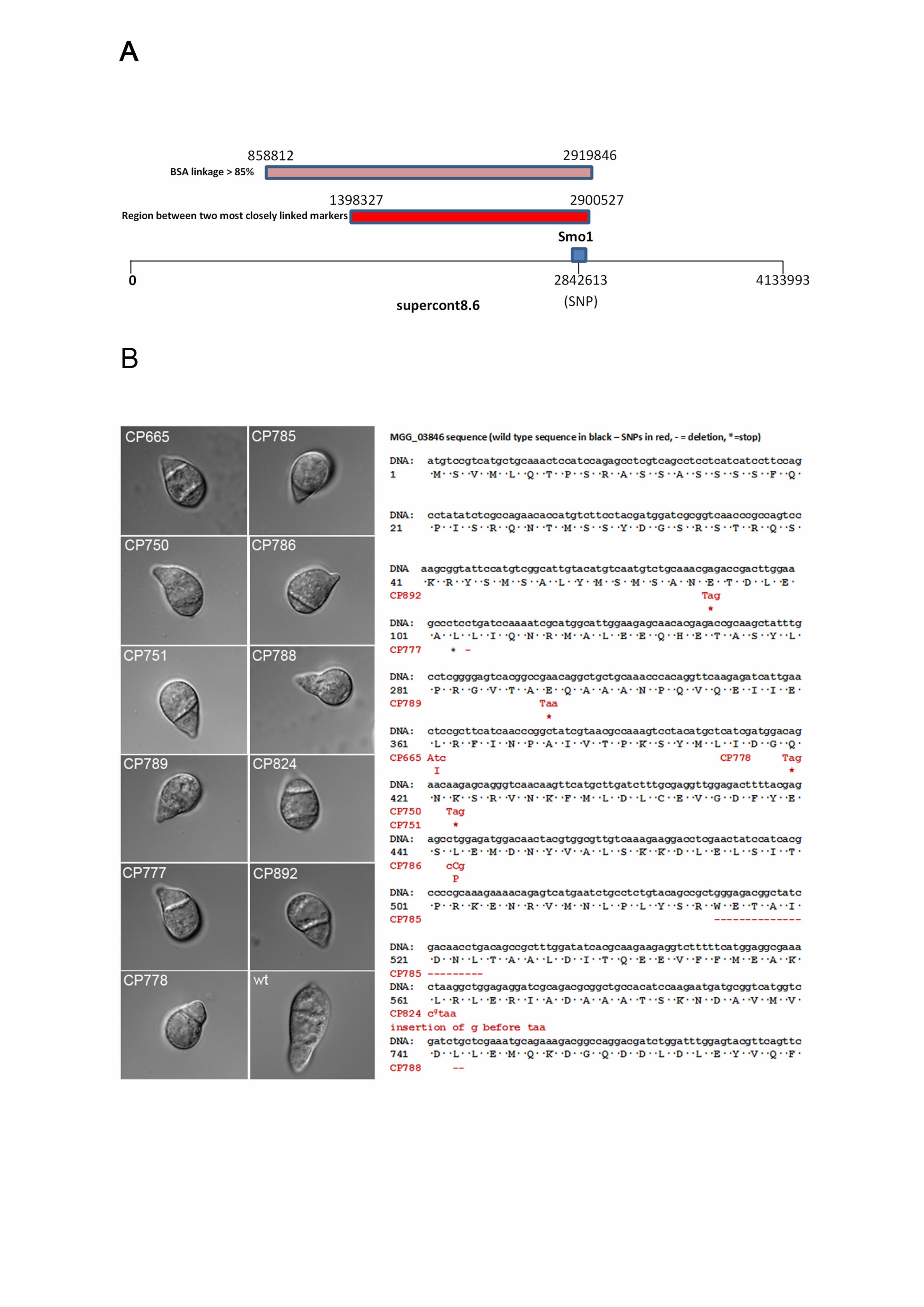
Identification of the SMO1 locus 261
To identify the SMO1 locus, we carried out bulked segregant analysis (Michelmore et 262
al. 1991) using whole genome sequencing to identify single nucleotide 263
polymorphisms. M. oryzae rice pathogenic strain 4395-4-4 (smo1 alb1 Mat1-2) was 264
crossed with a wild type rice pathogenic strain TH3 (Mat1-1) and a total of 50 265
ascospores collected. Ascospore progeny were phenotypically characterised based 266
on spore shape, as smo1 mutants have one, or two-celled spherical, or misshapen 267
conidia, compared to the three-celled pyriform wild-type conidia. Progeny were 268
therefore selected according to the Smo1 phenotype, DNA extracted from each 269
individual and then bulked. Whole genome sequencing of bulked DNA samples 270
identified a region of 2,061,034 bases on supercontig 8.6, which was defined by 271
SNPs showing greater than 85% linkage to SMO1 using bulk segregant analysis 272
(BSA) (Figure 1A). This conformed to the region originally defined by MGR586-273
12
based genetic mapping spanning the SMO1 locus (Romao and Hamer, 1992). 274
Consistent with this, two single copy RFLP probes previously shown to be closely 275
linked to the SMO1 locus (Hamer and Givan, 1990; Romao and Hamer, 1992): 276
JH4.28 and JH5.00, were sequenced and mapped to the M. oryzae genome. They 277
were both found on supercontig 8.6, separated by a distance of 1,502,200 bases. 278
The identified SMO1 gene lies within this region, as identified by BSA (Figure 1A). 279
We then analysed an allelic series of original smo1 mutants, which were either 280
selected by UV mutagenesis as non-adherent mutants, appressorium development 281
mutants, or spontaneous Smo1 mutants (Hamer et al. 1989b), as described in Table 282
S1, and confirmed their phenotype by spore morphology, as shown in Figure 1B. 283
Genomic DNA was extracted from each strain and whole genome sequencing 284
carried out. We also carried out genome sequence analysis of the parental 4091-5-8 285
strain, from which the Smo1 mutants were originally selected (Hamer et al. 1989b). 286
All Smo1 mutants shared 202,020 SNPs which distinguished 4091-5-8 from the M. 287
oryzae genome reference strain 70-15. Subtracting these shared SNPs from the 288
derived SNP datasets for each strain, identified SNPs unique to each mutant. Six of 289
the ten mutant smo1 strains possessed a SNP in gene MGG_03846. Manual 290
inspection of reads aligned to MGG_03846 identified mutations in five other mutant 291
strains (Figure 1B). Only CP790 did not contain a mutation in MGG_03846, and our 292
analysis suggests that this strain does not exhibit the Smo1 phenotype and therefore 293
may have reverted since the original study (Hamer et al. 1989b). With the exception 294
of strains CP750 and CP751, which had identical SNPs, the mutations were different 295
in each strain, consistent with the allelic variability reported originally (Hamer et al. 296
1989b). Five of the strains had a SNP which introduces a stop codon into the open 297
reading frame (nonsense mutation), four had either an insertion or deletion that 298
13
produces a frameshift mutation and two possessed a base pair substitution that 299
resulted in a change in an amino acid residue (Figure 1B). The mutation in CP786, 300
for instance, changes a leucine to proline, which is likely to produce a distinct 301
alteration in secondary structure, given that proline acts as a structural disruptor in 302
the middle of regular secondary structure elements, such as alpha helices and beta 303
sheets (Williamson, 1994). 304
Cloning and characterisation of the SMO1 gene of M. oryzae 305
The SMO1 candidate gene, MGG_03846, is 2643 bp in length with four introns of 81, 306
83, 78 and 65 bp, respectively, and encodes a putative 780 aa protein. Bioinformatic 307
analysis predicted that MGG_03846 encodes a Ras GTPase activating protein 308
(RasGAP), and the predicted gene product possesses two domains, a GTPase 309
activator domain for Ras-like GTPase from amino acids 193-401 and a RAS GAP C-310
terminal motif from amino acids 580-699. There are four putative GapA encoding 311
genes in M. oryzae and phylogenetic analysis (Figure S1) revealed potential SMO1 312
orthologs of GapA from Aspergillus nidulans (Harispe et al. 2008) and Gap1 from 313
Schizosaccharomyces pombe (Imai et al. 1991). Phylogenetic analysis of other 314
putative GAPs in M. oryzae suggests that MGG_03700 is a homolog of the 315
Saccharomyces cerevisiae IQG1, which controls actin-ring formation and cytokinesis 316
(Epp and Chant, 1997). MGG_08105 is a homolog of the S. cerevisiae BUD2, which 317
plays a role in spindle position checkpoint and bud site selection (Park et al. 1993), 318
whilst MGG_11425 is a homolog of the S. cerevisiae RasGAPs IRA1 and IRA2, 319
which are negative regulators of Ras-cAMP signaling pathway required for reducing 320
cAMP levels under nutrient limiting conditions (Tanaka et al. 1989; Tanaka et al. 321
1990) (Figure S1). 322
14
To determine whether the candidate RAS-GAP-encoding gene is SMO1 we 323
cloned a full length copy of MGG_03846 under control of its native promoter and 324
transformed this into smo1 mutant, CP750 (Hamer et al. 1989b). Re-introduction of 325
SMO1 fully complemented the smo1 spore shape phenotype, and spores in the 326
complemented strain germinated to produce short germ tubes and normal 327
appressoria (Figure S2). 328
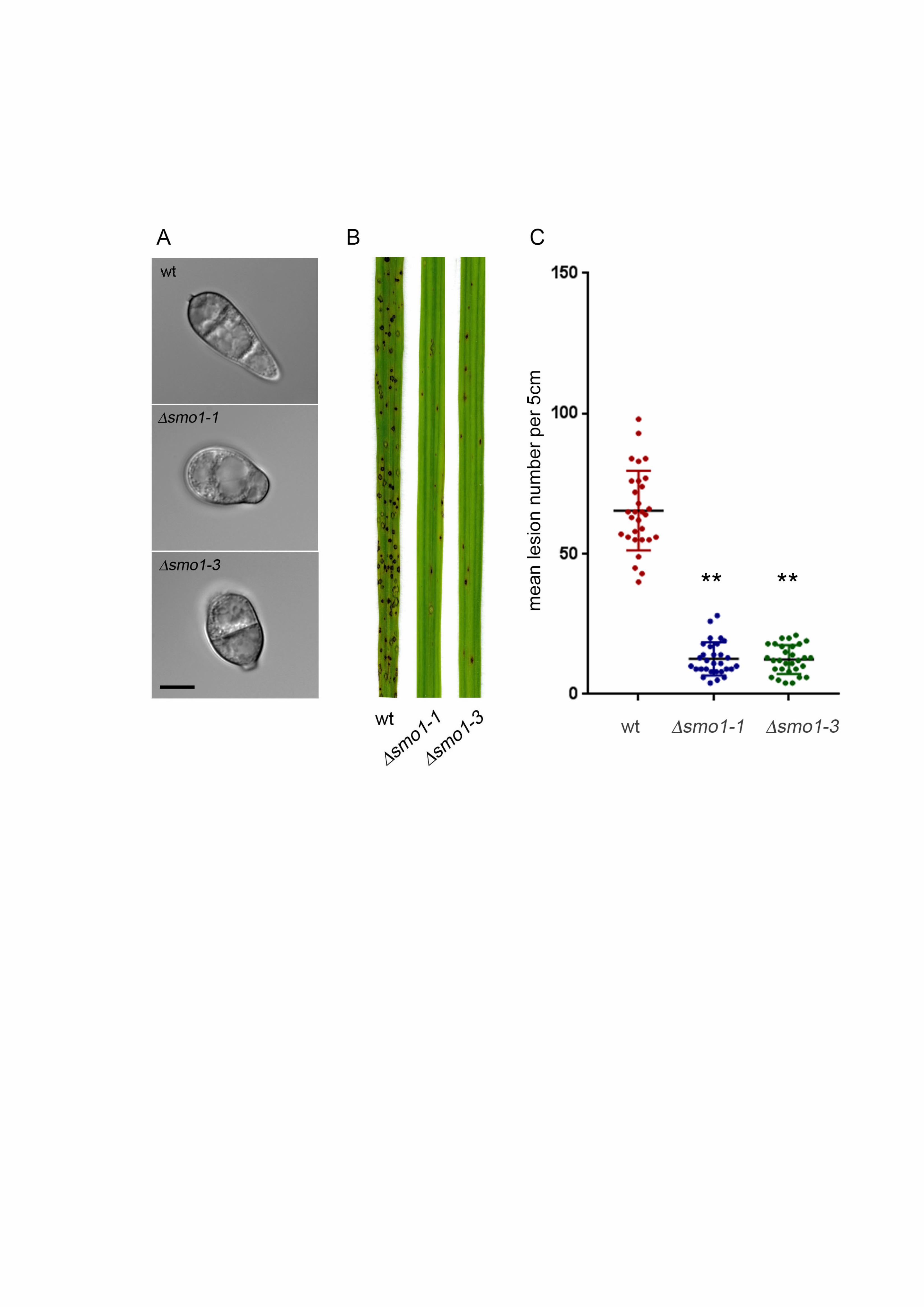
Targeted deletion of SMO1 leads to cell shape defects 329
We next carried out targeted deletion to generate a smo1 mutant in the wild type, 330
rice pathogenic strain of M. oryzae, Guy11, and two independent deletion strains 331
were selected and confirmed by Southern blot analysis for further analysis. The 332
morphology of smo1 mutants was similar to the original smo1 mutants, with 333
mycelial colonies more compact, white and fluffy, compared to those of the isogenic 334
wild type strain Guy-11. Conidia were more rounded and predominantly unicellular, 335
or two-celled (Figure 2A). By contrast, wild type spores are typically 22-25 m in 336
length, and 8.75 and 5.5 m in diameter, whereas smo1 conidia are typically 12.5-337
15 m in length, and 8.5-10 m in diameter. Conidia of smo1 mutants germinate 338
normally, but produce longer germ tubes than the wild type, typically 87 m in 339
smo1 compared to 22 m in Guy11 (Figure 3A). Appressorium development was 340
delayed and appressoria were typically misshapen and slightly smaller, 8.64 ± 0.56 341
m in diameter in smo1, compared to 9.55 ± 0.56 m in Guy11 (p < 0.01), as 342
shown in Figure 3. 343
As a consequence of the abnormal spore morphology and delay in 344
appressorium formation in smo1 mutants, we decided to investigate the pattern of 345
15
nuclear division during appressorium development. In M. oryzae, a single round of 346
mitosis occurs prior to appressorium development, followed by conidial cell death 347
and degradation of nuclei in each conidial cell (Veneault-Fourrey et al. 2006; 348
Saunders et al. 2010). We introduced an H1-RFP fusion into the smo1 mutant to 349
visualize nuclear dynamics by live-cell imaging (Figure S3A). Nuclear division in 350
smo1 takes place within 4-6 hours post inoculation (hpi), as observed in Guy11 351
and, in addition, one daughter nucleus migrates to the developing appressorium and 352
the other nucleus returned to the conidium, in the same way as the wild type (Figure 353
S3A). Nuclear material was, however, often observed in the longer germ tube, as 354
well as the conidium. We used Calcofluor-white to examine septation events in the 355
germ tube and observed that one septum normally forms in the germ tube, often 356
near to the conidium (Figure S3B). After 16 h, nuclei in smo1 conidia start to 357
degrade and by 24 h the spore had collapsed, as was observed in Guy11 (Figure 358
S3A). We conclude that smo1 mutants show defects in spore shape and 359
organisation and exhibit extended germ tube growth associated with a delay in 360
appressorium development. 361
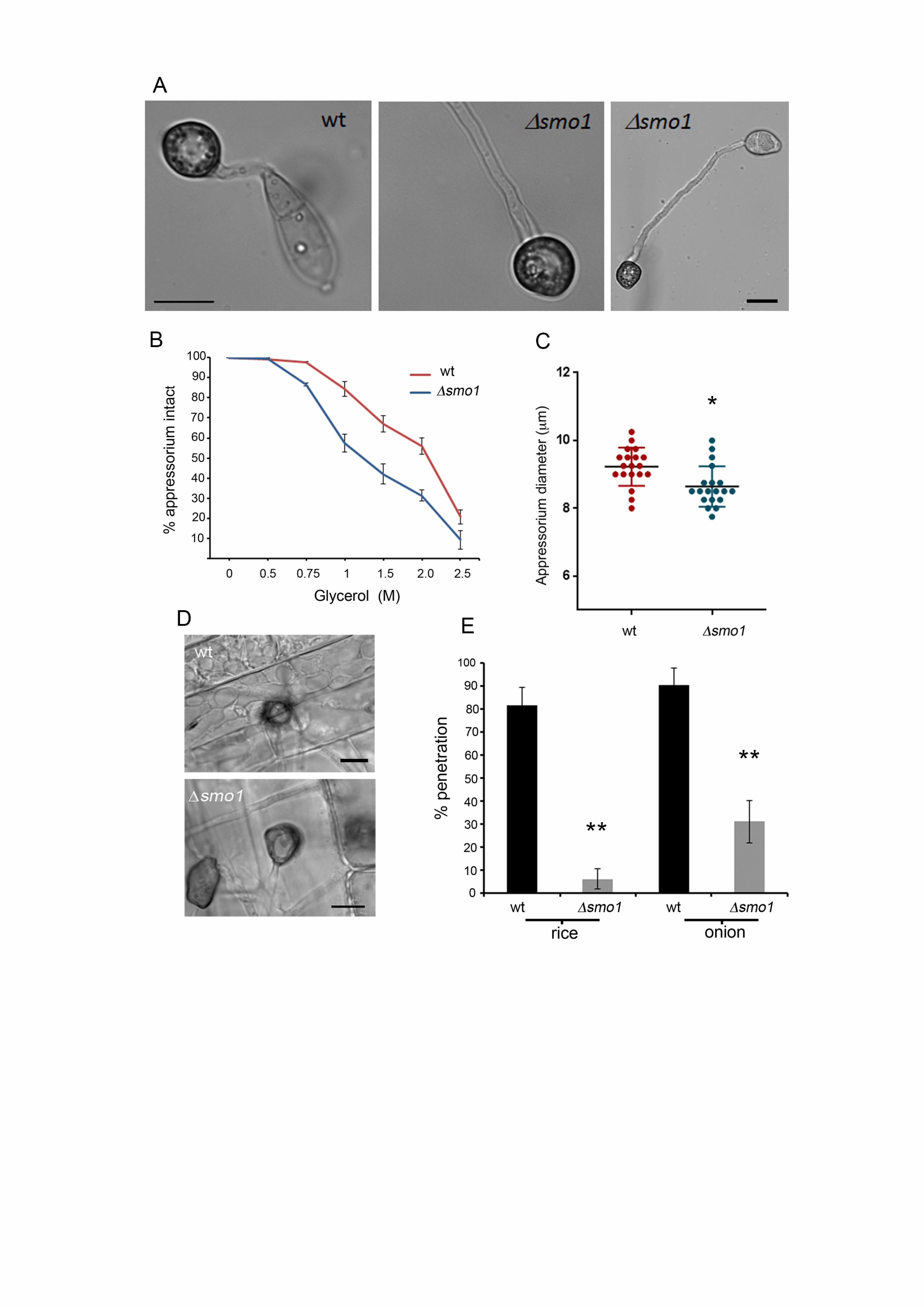
The SMO1 gene encodes a virulence factor in M. oryzae 362
To determine the role of SMO1 in fungal pathogenicity, we inoculated the susceptible 363
rice cultivar CO-39 with spore suspensions of two smo1 mutant strains and Guy11. 364
The smo1 mutants generated significantly reduced numbers of disease lesions, 365
13.44 ± 1.34 per 5 cm leaf, compared to 65 ± 6.94 lesions per 5 cm leaf in Guy11 366
(P<0.0001) (Figure 2B & C). To determine whether the reduced ability of smo1 367
mutants to cause disease lesions was due to reduced appressorium turgor, we 368
incubated appressoria of smo1 mutants in a series of glycerol solutions of 369
16
increasing molarity and measured the frequency of incipient cytorrhysis (cell 370
collapse) (Howard et al. 1991). A concentration of 1.25 M glycerol caused the 371
collapse of 50% of appressoria in the smo1 mutant whereas in the wild type 2.25 M 372
glycerol was required for 50% (P < 0.0001) (Figure 3C). This reduction in turgor is 373
consistent with an appressorium penetration defect. We therefore applied the smo1 374
mutant to excised rice leaf sheath to determine the frequency of appressorium-375
mediated penetration. We observed that Guy11 had a frequency of successful 376
penetration events of 81.66% ± 7.59 penetration, compared to 6.3 ± 4.37 in smo1 377
(P<0.001), as shown in Figure 3D & E. We also tested whether smo1 mutants were 378
able to penetrate onion epidermis and found slightly increased penetration compared 379
to that of smo1 mutants on rice leaf sheaths, but still significantly reduced 380
compared to the wild type (90.33. ± 7.59 (wt) compared to 31 ± 9.23 in smo1; P < 381
0.001), as shown in Figure 3E. We conclude that smo1 mutants are reduced in 382
their ability to cause rice blast disease because of impairment in appressorium 383
function, including a reduction in turgor and the frequency of penetration peg 384
development. 385
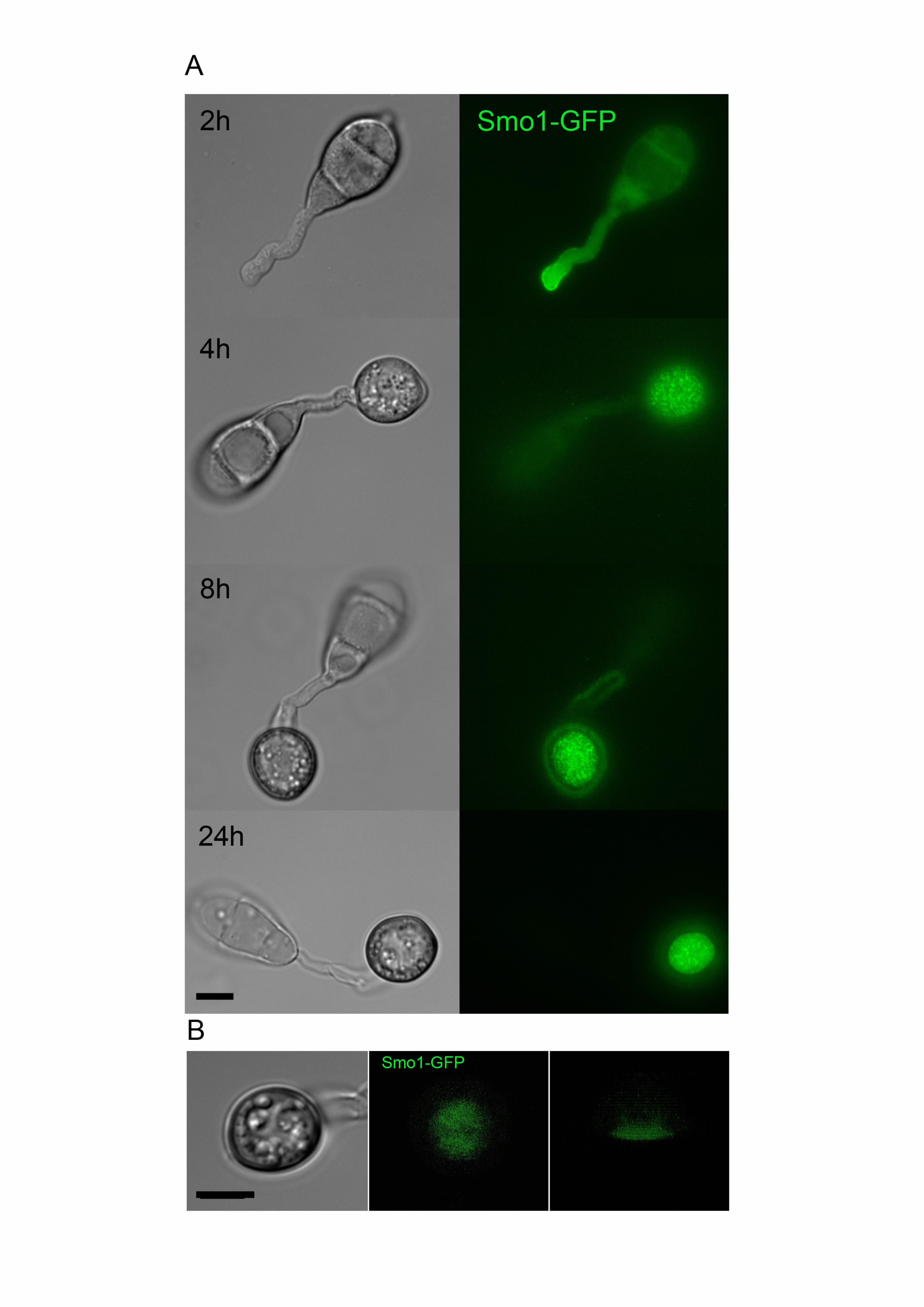
Smo1 localises to the appressorium pore during plant infection. 386
To determine the subcellular localisation of the Smo1 protein and its temporal 387
dynamics during infection-related development, we generated a SMO1-GFP fusion 388
which was introduced into Guy11 and a smo1 mutant. Expression of SMO1-GFP in 389
the smo1 mutant strain was sufficient to restore wild type spore and appressorium 390
morphologies and the ability to infect rice and cause disease (Figure S2). Analysis of 391
the cellular localisation showed that Smo1 localises to the tip of germ tubes during 392
germination. As the appressorium forms, Smo1 localised initially as small puncta 393
17
throughout the appressorium (Figure 4A). However, after 24 h when maximal turgor 394
is established in the appressorium, Smo1 localisation became more condensed and 395
by using 3-D reconstruction of a mature appressorium, Smo1-GFP was observed to 396
localise predominantly to the base of the appressorium around the appressorium 397
pore (Figure 4B, Supplemental Movie 1). Smo1 distribution is therefore associated 398
with regions of polarised growth, such as the germ tube tip. In the appressorium, 399
Smo1 localises to the point at which anisotropic growth is re-established for 400
penetration peg development and plant infection. 401
smo1 mutants are defective in spore tip mucilage generation and surface 402
attachment. 403
The tight adhesion of conidia to the rice leaf surface is critical for rice blast disease 404
and involves release of spore tip mucilage (STM) from the tip of the conidium 405
(Hamer et al. 1988), prior to germination. We used FITC-labelled concanavalin-A 406
(ConA-FITC) to compare STM released from spores of Guy-11 and the smo1 407
mutant. We evaluated levels of STM secretion between 1 h and 24 h post-408
inoculation. The adhesive is released from the conidial apex initially and then germ 409
tube tip (Hamer et al., 1988). This revealed a clear reduction in STM secretion in 410
smo1 mutants compared to Guy-11. During the early stages of conidial attachment 411
and germination, STM in the smo1 mutant was noticeably reduced compared to 412
Guy-11 (Figure 5A & 5B). We also observed Guy-11 and smo1 mutants after 24 h 413
and observed a similar reduction in a ConA-positive mucilage layer around the 414
mature appressorium in a smo1 mutant (Figure 5C). To examine conidial adhesion, 415
we then counted the number of conidia that could be removed from the surface of 416
hydrophobic coverslips by washing 30 min after inoculation. In Guy11, 67 ± 6.9% of 417

18
conidia remained attached to PTFE Teflon surfaces after washing, whereas in 418
smo1 mutants 48.4 ± 9.97% (P < 0.01) remained attached (Figure 5D). We 419
conclude that STM secretion is impaired in smo1 mutants which show reduced 420
adhesion to hydrophobic surfaces. 421
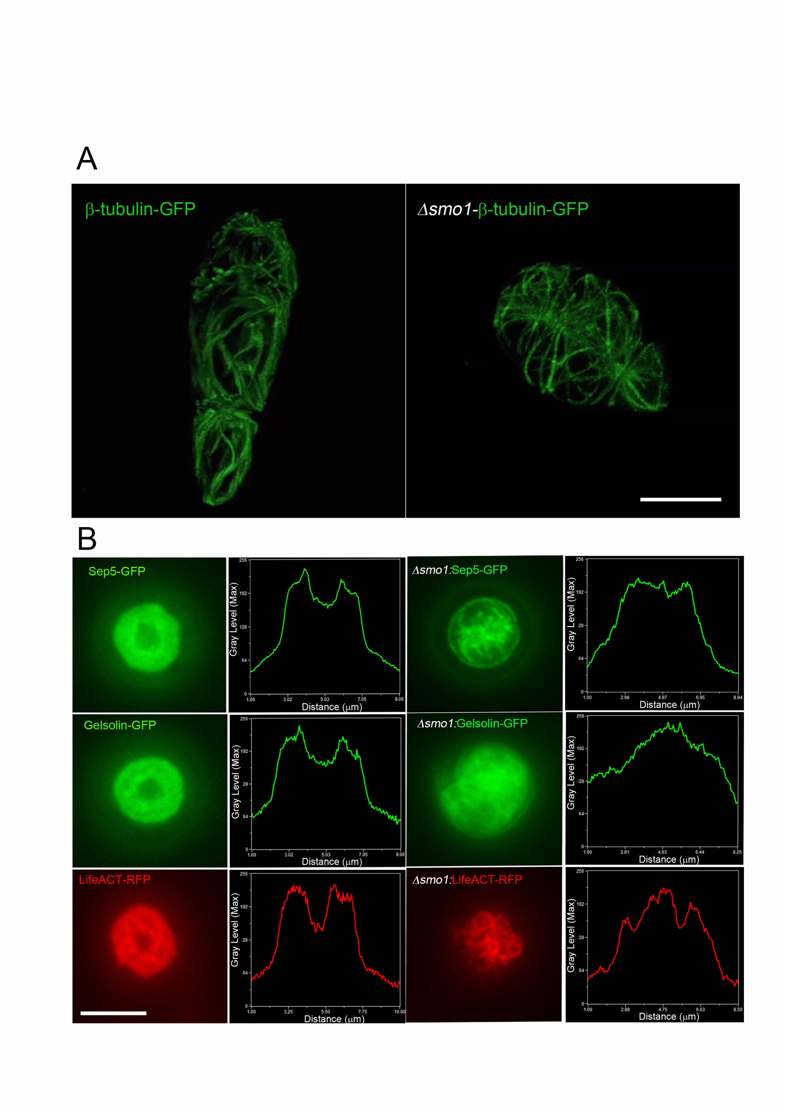
smo1 mutants are impaired in septin-mediated F-actin reorganisation at the 422
appressorium pore 423
The conidial shape phenotype of smo1 mutants suggested an effect on the 424
distribution and organisation of cytoskeletal components. We therefore visualized the 425
distribution of microtubules based on expression of the -tubulin Tub2-GFP fusion 426
protein. Conidia of the wild type Guy11 showed a network of long microtubules 427
defining each of the three-cells within the spore (Figure 6A and Supplemental Movie 428
2). By contrast microtubules observed in spores of a smo1 mutant showed an 429
abnormal distribution, consistent with the spherical shape of spores (Figure 6A and 430
Supplemental Movie 3). A key requirement for appressorium function in M. oryzae is 431
the recruitment and organisation of a septin-dependent toroidal F-actin network at 432
the appressorium pore. Septins provide cortical rigidity to the infection cell at the 433
point of penetration and act as a diffusion barrier for organisation of polarity 434
determinants required for penetration hypha development (Dagdas et al. 2012). We 435
decided to investigate organisation of the septin Sep5-GFP (Dagdas et al. 2012) in 436
the smo1 mutant. In Guy11, a septin ring was visible surrounding the appressorium 437
pore, but this was mis-localised in the smo1 mutant (Figure 6B). We therefore 438
observed the F-actin cytoskeleton, by expressing actin-binding protein gene fusions 439
LifeAct-RFP and Gelsolin-GFP (Berepiki et al. 2010; Ryder et al. 2013) in the smo1 440
mutants and observing appressoria at 24 hpi. In Guy-11, Lifeact-GFP and Gelsolin-441
19
GFP fluorescence revealed the toroidal F-actin network at the appressorium pore 442
which marks the point at which the penetration peg emerges (Figure 6B). By 443
contrast, the smo1 mutant showed dispersed and non-specific localisation of 444
LifeAct-RFP and Gelsolin-GFP (Figure 6B). We conclude that the septin-mediated F-445
actin dynamics necessary for host penetration are regulated by signalling pathways 446
acting downstream of Smo1. 447
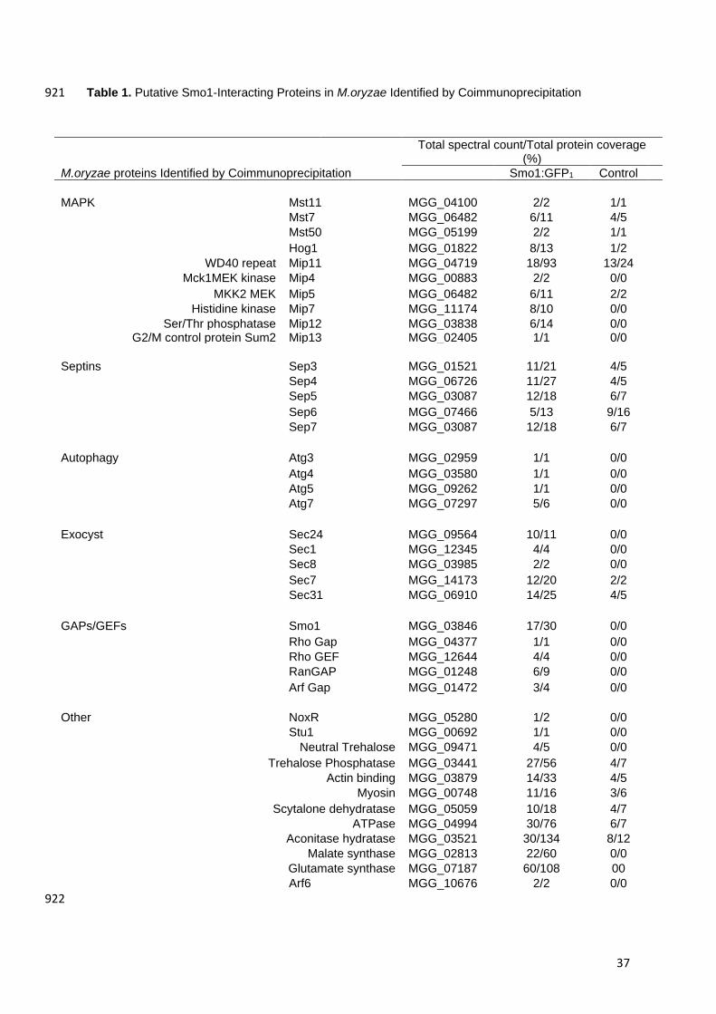
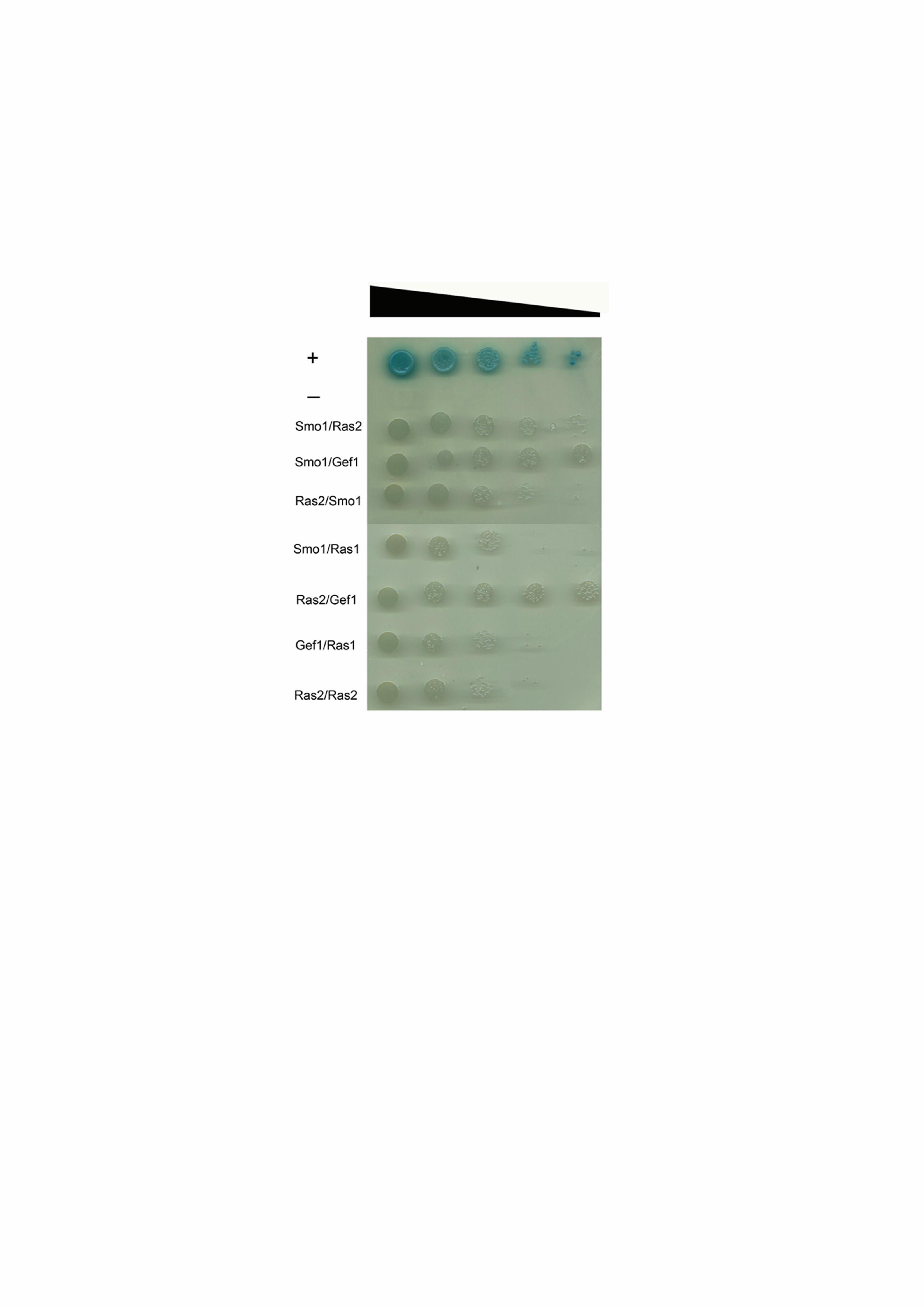
Protein-protein interaction studies to identify Smo1-interacting partners 448
The identification of Smo1 as a putative Ras-Gap protein prompted us to identify its 449
potential interacting partners. Two independent lines of investigation were followed. 450
First of all, yeast two hybrid analysis was carried out between Smo1 and confirmed 451
Ras signalling components from M. oryzae. Initially, control experiments were 452
performed in which the pGAD-Smo1 (prey–Smo1), pGAD-Ras2 (prey–Ras2), 453
pGAD-Gef1(prey–Smo1), and also pGBK-Ras(bait–Ras2), pGBK-Smo 1 (bait–454
Smo1) and pGBK-Ras1 (bait–Ras1) were independently transformed into the yeast 455
two hybrid Gold strain before plating onto SD/-Leu and SD/-Trp media, respectively. 456
The lack of growth on these media demonstrates that none of the vectors are 457
capable of auto-activating reporter genes. Simultaneous co-transformation of the 458
pGBK-Ras2 (bait–Ras2) and pGAD-Smo1 (prey–Smo1) vectors into the Y2H Gold 459
strain resulted in activation of all four reporter genes and growth on high stringency 460
medium (-His/-Ade/-Leu/-Trp/+X-α-Gal) (Figure 7). Co-transformation also activated 461
MEL1 expression in which the enzyme α–galactosidase is secreted into the medium, 462
resulting in hydrolysis of X-α-Gal in the medium and turning the yeast colony blue. 463
Growth on such high stringency media supports the hypothesis that Smo1 and Ras2 464
can physically interact. Putative interactions were also observed between Smo1 and 465
20
Gef1, and between Ras2 and Gef1. Weaker interactions were also observed 466
between Ras1 and both Gef1 and Smo1. When considered together, the 467
interactions are consistent with Smo1 acting as a GTPase-activating protein on 468
Ras2. We cannot preclude that Smo1 also plays a regulatory role in Ras1 signalling, 469
but it shows much higher affinity to Ras2. 470
Secondly, we carried out co-immunoprecipitation of Smo1-GFP from hyphae of M. 471
oryzae and identified interacting proteins by mass spectrometry. This revealed 472
putative interactions with the MAP kinase signalling pathway components, previously 473
implicated in appressorium development, such as Mst11, Mst7 and Mst50, which all 474
operate upstream of the Pmk1 MAP kinase, as well as the WD40 repeat protein 475
Mip11, as shown in Table1. Moreover, Smo1 appears to interact with the four core 476
septins and with components of the exocyst complex, which are known to be 477
associated with appressorium pore function (Dagdas et al. 2012; Gupta et al. 2015), 478
as well as autophagy components that are also necessary for appressorium function 479
(Table 1). (Kershaw and Talbot, 2009). 480
481
482
483
Discussion 484
In this report, we have provided evidence that SMO1 encodes a Ras GTPase-485
activating protein that plays important functions in cell shape determination and 486
infection-related development in the rice blast fungus. SMO1 is critical for rice blast 487
disease and plays a significant role in conidium and appressorium shape 488
21
determination and attachment to the leaf surface, in addition to an important 489
regulatory function in the re-polarisation apparatus that operates within the 490
appressorium. Smo1 is essential for septin recruitment and organisation at the 491
appressorium pore, which in turn is necessary for F-actin re-organisation and 492
penetration peg development (Dagdas et al. 2012). Smo1 physically interacts with 493
Ras2, suggesting a model in which Ras signalling is required for appressorium re-494
polarisation, as shown in Figure 8. 495
Smo1 mutants were so frequently identified during the early days of rice blast 496
molecular genetic analysis that Hamer and colleagues (1989b) suggested that the 497
SMO locus might be highly mutable. For example, isolation of spontaneous mutants 498
with melanin pigment defects and benomyl-resistance identified double mutants that 499
were also Smo1 (CP665 and CP892). Separate genetic screens to identify mutants 500
with a defect in appressorium development or a search for STM mutants with 501
reduced capacity to attach to hydrophobic surfaces also mainly identified Smo1 502
mutants (Hamer et al. 1989b). However, in spite of the rapid genetic mapping of the 503
SMO1 locus (Hamer and Givan, 1990), the gene proved to be extremely difficult to 504
clone and after around six years of effort, SMO1 cloning was finally abandoned. 505
Recent advances in genome sequencing, the availability of numerous independent 506
Smo1- mutants, and the ability to carry out genetic crosses readily in M. oryzae, have 507
now enabled us to identify SMO1 and to understand the genetic events leading to 508
frequent loss of gene function. Surprisingly, mutational events leading to inactivation 509
of SMO1 were all SNPs or small insertions/deletions in the coding sequence. This 510
result contrasts with frequent deletion of the highly mutable BUF1 melanin 511
biosynthesis gene, which presumably occurs by transposon-mediated recombination 512
(Chumley and Valent, 1991; Farman, 2002). The SMO1 gene does not reside in a 513
22
particularly transposon-rich region of the genome. Therefore, mechanisms for 514
frequent isolation of Smo mutants and the original difficulties in cloning SMO1 515
remain to be explained. It is possible that the number of transposable elements and 516
repeated DNA sequences normally distributed across the M. oryzae genome, was 517
sufficient to prevent map-based cloning efforts from being effective in the early 518
1990’s, given the absence of extensive DNA sequence information, and large insert 519
genomic libraries (such as Bacterial Artifical Chromosome Libraries) at that time. 520
SMO1 encodes a GTPase activating protein (GapA) involved in regulation of 521
Ras proteins, and is one of four found in the M. oryzae genome. Ras-GA proteins 522
work in conjunction with Ras-GEFs to regulate the activity of Ras proteins in 523
response to external stimuli, affecting downstream signalling pathways necessary for 524
regulation of morphological transitions necessary for growth in Eukaryotes (Boguski 525
and McCormick, 1993). A Ras-GAP protein MadC, from the fungus Phycomyces 526
blakesleeanus has, for example, been shown to form part of the photosensory 527
pathway for light-dependent fruiting body formation (Polaino et al 2017). 528
We have shown by yeast 2 hybrid analysis that Smo1 interacts with both 529
Ras2 and Gef1, providing further evidence that Smo1 functions as a Ras-GAP and 530
that it can form a complex with the corresponding guanine nucleotide exchange 531
factor. Gap proteins are classified based on sequence homology within their Gap 532
domains, with each domain specific for a class of G-proteins (Donovan et al. 2002). 533
The different arrangements of domains, and in many cases the inclusion of distinct 534
additional domains in the RasGAP family, suggest these proteins are subject to a 535
diverse range of cellular interactions (Donovan et al. 2002). In M. oryzae, four 536
putative RasGAPs can be classified into four different clusters according to their 537
sequences and domain structures. Smo1 possess two domains, a GTPase activation 538
23
domain for Ras-like GTPase and a RAS GAP C-terminal motif. These domains are 539
characteristic of proteins belonging to the Ras-specific GAPs. The sequence placed 540
Smo1 in a cluster with the Aspergillus nidulans GapA (Harispe et al. 2008) and Gap1 541
from Schizosaccharomyces pombe (Imai et al. 1991). In A. nidulans, GapA mutants 542
exhibit abnormal conidiophores, delayed polarity maintenance characterised by 543
apical swelling, and sub-apical hyphal branching (Harispe et al. 2008). In addition, F-544
actin distribution is lost in gapA cells, suggesting a role for GapA in F-actin 545
cytoskeleton organisation, required for hyphal growth (Harispe et al. 2008). Mutation 546
of Gap1 in S. pombe results in hypersensitivity to mating factor pheromone and the 547
inability to perform efficient mating, which are identical phenotypes to those caused 548
by activated ras1 mutations (Imai et al. 1991). The defect in polar growth suggests 549
that GAP1 is involved in polarity maintenance, working antagonistically with Ste6 in 550
the regulation of Ras-GTPase in S. pombe. Involvement of a Ras-GAP in fungal 551
morphogenesis was first reported for the basidiomycete white rot fungus, 552
Schizophyllum commune in which Gap1 deletion was shown to affect sexual 553
development with mutants unable to form gills on fruiting bodies and producing no 554
basidiospores. In addition, growth phenotypes suggested involvement in the 555
maintenance of polarity (Schubert et al. 2006). The Smo1 mutant phenotypes 556
observed are therefore consistent with those of GAP genes identified in other fungi, 557
with effects on cell shape determination, polar/non-polar growth transitions, and 558
regulation of the F-actin cytoskeleton. 559
Three other putative RasGAPs predicted in M. oryzae, MGG_11425.6 and 560
MGG_08105.6 and MGG_03700.6, have not yet been characterised. MGG_03700 is 561
a homolog of the Saccharomyces cerevisiae Iqg1, an essential gene shown by 562
depletion and over-expression analysis to be required for cytokinesis and actin-ring 563
24
formation (Epp and Chant, 1997). Iqg1 possesses a calponin-homology (CH) domain 564
and IQ repeats in addition to the RAS GAP C-terminal motif (Epp and Chant, 1997). 565
MGG_08105 is a homolog of the S. cerevisiae GAP BUD2 which stimulates 566
hydrolysis of the Ras2 homolog BUD1. Mutants defective in BUD2 display random 567
budding but no obvious growth defect (Park et al. 1993). MGG_11425 is a homolog 568
of the S. cerevisiae RasGAPs IRA1 and IRA2, which are negative regulators of Ras-569
cAMP signaling pathway required for reducing cAMP levels under nutrient limiting 570
conditions (Tanaka et al. 1989; Tanaka et al. 1990). 571
Ras proteins are low molecular weight monomeric G-proteins which localise to the 572
plasma membrane (Wennerberg et al. 2005) and switch between the active GTP-573
bound and inactive GDP-bound status, competitively regulated by GEFs and GAPs 574
(Boguski and McCormick, 1993). Ras proteins have intrinsic GTPase and GDP/GTP 575
exchange activity, but GAP and GEF proteins work to ensure a more tightly 576
regulated process. In S. cerevisiae, the two Ras proteins, Ras1 and Ras2 are both 577
essential for growth and both function to activate adenylate cyclase (Tamanoi, 2011). 578
The RAS/cAMP/PKA pathway in S. cerevisae regulates a variety of processes, 579
including cell cycle progression and life span (Tamanoi, 2011). M. oryzae also has 580
two Ras-encoding genes, MoRAS1 and MoRAS2, and both have been 581
characterised. In the ras1 deletion mutant no distinct phenotypes were observed 582
other than a slight reduction in conidiation (Zhou et al. 2014). RAS2, however, is 583
thought to be an essential gene and has only been characterised by generation and 584
expression of a Ras2 dominant active allele. MoRAS2G18V transformants formed 585
morphologically abnormal appressoria on both hydrophilic and hydrophobic surfaces, 586
suggesting that dominant active RAS2 can bypass surface attachment requirements 587
for appressorium formation (Zhou et al. 2014). MoRAS2G18V showed increased Pmk1 588
25
phosphorylation and elevated cAMP levels in aerial hyphae. cAMP-PKA signalling 589
has been shown to be important for initial surface recognition and appressorium 590
generation and for generation of turgor pressure necessary for infection. The cAMP-591
dependent PKA mutant, cpkA, produces long germ tubes and small non-functional 592
appressoria (Xu et al. 1997), whilst the mitogen–activated protein (MAP) kinase 593
Pmk1 is essential for appressorium formation and invasive growth (Xu and Hamer, 594
1996). In both pmk1 and cpka mutants, expression of MoRAS2G18V had no effect 595
on appressorium morphogenesis, suggesting that Ras2 functions upstream of both 596
cAMP and Pmk1 signalling pathways (Zhou et al. 2014). Deletion of several 597
upstream components of the Pmk1 pathway, including MST50, MST11 and MST7 598
result in defects in appressorium development and plant infection (Park et al. 2006; 599
Zhao et al. 2005; Zhou et al. 2014). Mst50 functions as an adapter protein of the 600
Mst7-Mst11-Pmk1 cascade involved in activating Pmk1 in M. oryzae (Zhao et al. 601
2005). Both Mst50 and Mst11 have been shown to interact with MoRas1 and 602
MoRas2, by yeast two-hybrid assays (Park et al. 2006) and deletion of the Ras-603
association (RA) domain of Mst11 blocked Pmk1 activation and appressorium 604
formation (Qi et al. 2015), supporting a role for Ras signalling in activation of the 605
Pmk1 pathway. It has been shown recently that the transmembrane mucins, Msb2 606
and Cbp1, function together to recognise extracellular signals through Ras2 (Wang 607
et al. 2015). Pmk1 phosphorylation was reduced in a Momsb2 mutant but blocked in 608
a Momsb2 cbp1 double mutant, which was non-pathogenic (Wang et al. 2015). 609
Affinity purification was used to identify a series of Mst50-interacting proteins (MIPs) 610
as well as upstream kinases of the Mps1 pathway, and also the histidine kinase Hik1 611
(Li et al. 2017). These interactions suggest a role for Mst50 in three different 612
signalling pathways. However, domain deletion experiments showed that the Mst50 613
26
Ras-association domain was not important for response to oxidative stress (Li et al. 614
2017). M.oryzae Ras2 is therefore essential for cellular viability, and a key mediator 615
between both the Pmk1 MAPK and cAMP signalling cascades (Qi et al. 2015; Zhou 616
et al. 2014). Activation and deactivation of Ras2 regulates developmental 617
switches/pathways necessary for growth and pathogenicity of the fungus (Zhou et al. 618
2014). 619
When considered together, our results suggest that Smo1 acts as a negative 620
regulator of Ras2 and this is why smo1 mutants display such severe developmental 621
defects, including mishappen spores and appressoria, long germ tubes and a failure 622
in penetration peg development (see model in Figure 8). These phenotypes point to 623
a defect in the maintenance of polarity that is required for morphological transitions 624
in the fungus. These developmental effects are a consequence of the disruptions to 625
both septin and F-actin dynamics in smo1 mutants, which are essential for plant 626
infection. SMO1-dependent regulation is therefore required for the morphological 627
transitions, and cell shape generation processes that are associated with asexual 628
reproduction and plant infection by the blast fungus. 629
630
631
Acknowledgments 632
This work was funded by a Biological Sciences and Biotechnology Research Council 633
(BBSRC) Industrial Partnership Award with Syngenta (BB/) and a European 634
Research Council Advanced Investigator Award to NJT under the European Union's 635
Seventh Framework Programme (FP7/2007-2013) / ERC grant agreement n° 636

27
294702 GENBLAST. We acknowledge George Littlejohn (Plymouth University) for 637
help with microscopy. This is contribution number 18-374-J from the Kansas 638
Agricultural Experiment Station. This paper is dedicated to John Hamer and the 639
brave students and postdocs in his research group who tried so hard to clone SMO1 640
in the early 1990’s; including Kathy Dobinson, Scott Givan, Steve Harris, Michelle 641
Momany, Yankyo Salch, and Verel Shull. The corresponding author (NJT) shares 642
your pain, having spent much of 1991 also trying in vain. The authors all salute you. 643
644
28
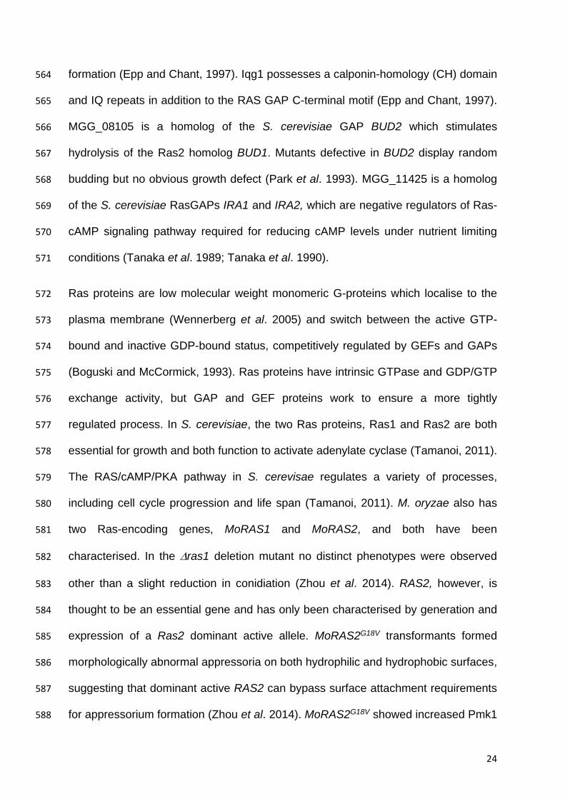
Figure 1. Identification of the SMO1 locus in Magnaporthe oryzae. (A) The identified 645
SMO1 gene lies on supercontig 8.6 (Chromosome 6: 2,838,047-2,844,822, ensembl 646
database). Single copy RFLP probes JH4.28 and JH5.00, which have previously 647
been shown to be closely linked to the SMO1 locus (Hamer and Givan, 1990; 648
Romao and Hamer, 1992), were sequenced and mapped to the M. oryzae genome, 649
and also found on supercontig 8.6, separated by a distance of 1,502,200 bases. A 650
region in a similar position on supercontig 8.6 was defined by SNPs that showed a 651
greater than 85% linkage to SMO1 using bulk segregant analysis (BSA). (B) 652
Micrographs showing spore morphology of smo1 strains used in this study compared 653
to wild type strain Guy11 (Bars = 10 m). Smo1 mutants were originally obtained 654
spontaneously or after UV mutagenesis (Hamer et al. 1989b). Nucleotide and amino 655
acid sequences of MGG_03846 indicating position of SNPs and subsequent 656
mutation in each of the smo1 mutants. Wild type sequence in black, SNPs in red, * = 657
stop codon. 658
659
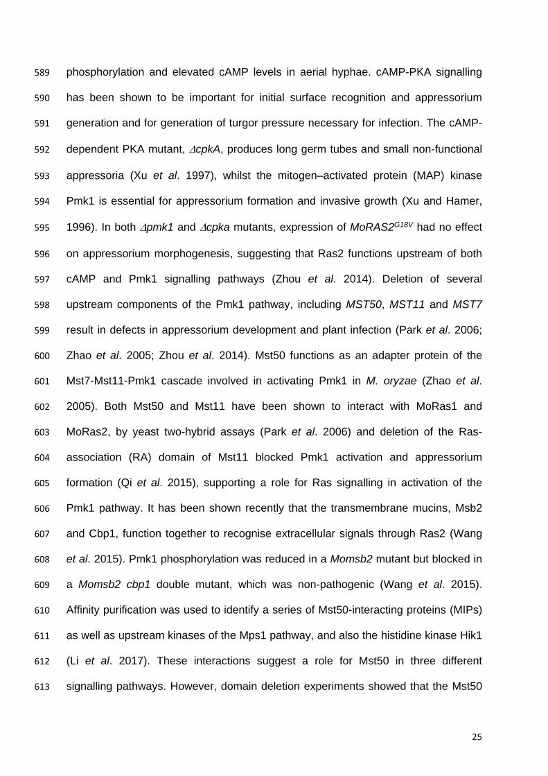
Figure 2. Deletion of MGG_03846 resulted in smo1 spore phenotype and mutants 660
were reduced in plant infection. (A) Micrographs showing spores of two MGG_03846 661
deletion strains (smo1) as compared to wild type strain Guy11 (Bar = 5 m). (B) 662
Photographs of leaves from infected plants. Seedlings of rice cultivar CO-39 were 663
inoculated with conidial suspensions (5 x 10⁴ ml-1). Seedlings were incubated for 5 664
days to allow symptom development. (C) Box plot of mean lesion density per 5 cm 665
leaf after infection with MGG_03846 deletion mutants compared to wild type. Error 666
bar equals standard error of the mean. * P < 0.0001 (unpaired Students’s t test (n = 667
3 experiments of 40 leaves). 668
29
669
Figure 3. The smo1 mutant is able to elaborate an appressorium, which is 670
impaired in function. (A) Micrograph showing smo1 mutant with extended germ 671
tube compared to wild type strain (Bar = 5 m). (B) Incipient cytorrhysis assays 672
measuring intracellular glycerol were carried out by allowing appressoria to form in 673
water on borosilicate cover slips for 24 h, after which the water was replaced with a 674
range of aqueous glycerol solutions ranging from 0.25 M to 2.5 M. The rate of cell 675
collapse was determined after 30 min. A concentration of 1.25 M glycerol caused the 676
collapse of 50% of appressoria in the smo1 mutant whereas 2.25 M glycerol was 677
required for 50% collapse in the wild type. P < 0.0001 unpaired Student’s t test, n = 678
100). (C) Box plot showing reduced appressorium diameter of smo1 mutant 679
compared to wild type, Guy11. *P < 0.01 unpaired Student’s t test, n = 50. (D) 680
Micrograph comparing penetration of rice leaf sheath by Guy11 and the smo1 681
mutant. Inoculations were performed as described previously using the susceptible 682
rice line CO-39. (E) Bar chart showing percentage penetration of Guy11 and smo1 683
mutant on leaf sheath and onion epidermis assessed by recording the frequency of 684
hyphal penetration from an appressorium **P< 0.001 unpaired Student’s t test, n = 685
24. 686
687
Figure 4. Live cell imaging of M. oryzae wild type strain expressing Smo1-GFP (A) 688
Cellular localisation of Smo1-GFP at the tip of the germ tube and as punctate 689
structures in both the developing appressoria (B) Distribution of Smo1-GFP in 690
mature appressorium visualized by laser confocal miscoscopy. The right hand panel 691
shows confocal image of transverse view of appressorium with Smo1-GFP present 692
30
predominantly at the appressorium-substrate interface (see also Supplemental 693
Movie 1). Spores were harvested and inoculated onto hydrophobic cover slips and 694
visualized by epifluorescence or laser confocal microscopy. Bar = 10 m. 695
696
Figure 5. Live cell imaging of M. oryzae wildtype and smo1 mutant showing 697
release and levels of spore tip mucilage (STM) by addition of ConA FITC to 698
germinating M. oryzae conidia of (A) Guy11 and (B) smo1 mutant. Spores were 699
harvested and inoculated onto hydrophobic cover slips before addition of ConA FITC 700
(100 g ml-1). Bar = 10 m. (C) ConA FITC staining of mucilage in mature 701
appressoria of Guy11 and smo1 mutant after 24 h. Bar = 5 m. (D) Box plot 702
showing percentage of germinated conidia of Guy11 and smo1 remaining attached 703
to cover slips after washing. Conidia were harvested and counted (1 x 10⁵ ml-1) and 704
allowed to attach to hydrophobic cover slips 30 minutes before washing with water. 705
*P< 0.01 unpaired Student’s t test, n = 100. 706
Figure 6. smo1 mutants are unable to undergo septin-mediated F-actin re-707
modelling in the appressorium. (A) Expression of -tubulin-GFP in conidia of M. 708
oryzae Guy11 (left hand panel) and smo1 mutant (right hand panel). Microtubules 709
showed aberrant distribution and organisation, consistent with spore-shape defect in 710
smo1 mutant (B) Live cell imaging of septin-dependent F-actin network in 711
appressoria of M. oryzae Guy11 and smo1 mutant at 24 hpi, visualized by laser-712
assisted epifluorescence microscopy. Localisation of Sep5-GFP, Gelsolin-GFP, and 713
LifeAct-RFP at the appressorium pore with corresponding line-scan graphs to show 714
31
distribution of fluorescence signal in a transverse section. Organisation of 715
appressorium pore components requires Smo1. Bar = 5 m. 716
Figure 7. Yeast Two-hybrid analysis. Yeast two-hybrid screens were performed to 717
determine putative physical interactions of Smo1 and to confirm its function as a 718
Ras-GAP. SMO1, RAS2, and RAS1 cDNAs were cloned into the bait vector 719
pGBKT7, SMO1, RAS2 and GEF1 cDNA were cloned into the prey vector PGADT7. 720
Simultaneous co-transformation of the pGBK-Ras2 (bait–Ras2) and pGAD-Smo1 721
(prey–Smo1) vectors into the Y2H Gold strain results in the activation of all four 722
reporter genes and growth on high stringency media (-His/-Ade/-Leu/-Trp/+X-α-Gal). 723
Smo1/Gef1 showed the highest stringency interaction. (+ = positive control, - = 724
negative empty prey vector control) 725
726
Figure 8. Model for the potential action of Smo1 in Ras2 signalling and its regulation 727
of septin-dependent appressorium re-polarisation and plant infection. 728
729
Table 1. Putative Smo1-Interacting Proteins in M.oryzae Identified by 730
Coimmunoprecipitation 731
732
Table S1 Smo1 mutants used in this study and characteristics of the genome 733
sequences generated 734
735
Table S2 Primers used in this study 736
32
737
Figure S1. Multiple sequence alignment using Clustal X (Larkin et al. 2007) showing 738
GTPase activating proteins from M. oryzae in Maximum likelihood phylogenetic tree 739
created using PhyML. Bootstrap support values of 70 or greater are indicated on 740
tree. 741
742
Figure S2. Complementation of the smo1 mutant restores wild type phenotypes. 743
(A) Micrographs showing spore morphology of Guy11 and smo1 mutant 744
complemented with the Smo1-GFP fusion construct, and the smo1- strain CP750 745
complemented with wild type SMO1 allele. Bar = 10 m (B) Photographs of leaves 746
from infected seedlings of rice cultivar CO-39 inoculated with conidial suspensions (1 747
x 105 ml-1) of wild type Guy11, smo1 and smo1 mutant complemented with SMO1-748
GFP. 749
750
Figure S3. Cellular localisation of H1-RFP by live cell imaging in (A) a wild type 751
Guy11 background and (B) the smo1 mutant background, visualized by 752
epifluorescence microscopy over a time course of infection related development. Bar 753
= 10 m. (C) Calcofluor-white staining of conidia, germ tubes and appressoria of the 754
wild type strain Guy11 and smo1 mutant at 24 hpi. Bar = 10 m. 755
756
References 757
33
Balhadere, P.V., Foster, A.J., and Talbot, N.J. (1999). Identification of pathogenicity 758
mutants of the rice blast fungus Magnaporthe grisea by insertional 759
mutagenesis. Mol Plant Microbe Interact. 12:129-142. 760
Berepiki, A., Lichius, A., Shoji, J.Y., Tilsner, J., and Read, N.D. (2010). F-actin 761
dynamics in Neurospora crassa. Eukaryot Cell 9:547-557. 762
Boguski, M.S., and McCormick, F. (1993). Proteins regulating Ras and its relatives. 763
Nature 366:643-654. 764
Catlett, N.L., Lee, B.-N., Yoder, O.C., and Turgeon, B.G. (2003). Split-Marker 765
Recombination for Efficient Targeted Deletion of Fungal Genes. Fungal 766
Genet. Newsl. 50:9-11. 767
Chumley, F.G., and Valent, B. (1991). Strategies for Characterizing and Cloning 768
Host Specificity Genes in Magnaporthe grisea, the Rice Blast Fungus. 769
Molecular Strategies of Pathogens and Host Plants:131-138. 770
Dagdas, Y.F., Yoshino, K., Dagdas, G., Ryder, L.S., Bielska, E., et al. (2012). Septin-771
mediated plant cell invasion by the rice blast fungus, Magnaporthe oryzae. 772
Science 336:1590-1595. 773
Dean, R.A., Brown, D., Donofrio, N., and Oh, Y. (2004). Functional genome analysis 774
of Magnaporthe grisea. Phytopathology 94:S122-S122. 775
Dean, R.A., Talbot, N.J., Ebbole, D.J., Farman, M.L., Mitchell, T.K., et al. (2005). The 776
genome sequence of the rice blast fungus Magnaporthe grisea. Nature 777
434:980-986. 778
deJong, J.C., MCCormack, B.J., Smirnoff, N., and Talbot, N.J. (1997). Glycerol 779
generates turgor in rice blast. Nature 389:244-245. 780
Donovan, S., Shannon, K.M., and Bollag, G. (2002). GTPase activating proteins: 781
critical regulators of intracellular signaling. Biochimica et biophysica acta 782
1602:23-45. 783
Epp, J.A., and Chant, J. (1997). An IQGAP-related protein controls actin-ring 784
formation and cytokinesis in yeast. Current Biology 7:921-929. 785
Farman, M.L. (2002). Meiotic deletion at the BUF1 locus of the fungus Magnaporthe 786
grisea is controlled by interaction with the homologous chromosome. Genetics 787
160:137-148. 788
Hamer, J.E., Farrall, L., Orbach, M.J., Valent, B., and Chumley, F.G. (1989a). Host 789
species-specific conservation of a family of repeated DNA sequences in the 790
genome of a fungal plant pathogen. Proc Natl Acad Sci U S A 86:9981-9985. 791
Hamer, J.E., and Givan, S. (1990). Genetic mapping with dispersed repeated 792
sequences in the rice blast fungus: mapping the SMO locus. Mol Gen Genet 793
223:487-495. 794
Hamer, J.E., Howard, R.J., Chumley, F.G., and Valent, B. (1988). A mechanism for 795
surface attachment in spores of a plant pathogenic fungus. Science 239:288-796
290. 797
Hamer, J.E., Valent, B., and Chumley, F.G. (1989b). Mutations at the smo genetic 798
locus affect the shape of diverse cell types in the rice blast fungus. Genetics 799
122:351-361. 800
Harispe, L., Portela, C., Scazzocchio, C., Penalva, M.A., and Gorfinkiel, L. (2008). 801
Ras GTPase-activating protein regulation of actin cytoskeleton and hyphal 802
polarity in Aspergillus nidulans. Eukaryot Cell 7:141-153. 803
Howard, R.J., Ferrari, M.A., Roach, D.H., and Money, N.P. (1991). Penetration of 804
hard substrates by a fungus employing enormous turgor pressures. Proc Natl 805
Acad Sci U S A 88:11281-11284. 806
34
Imai, Y., Miyake, S., Hughes, D.A., and Yamamoto, M. (1991). Identification of a 807
GTPase-activating protein homolog in Schizosaccharomyces pombe. 808
Molecular and Cellular Biology 11:3088-3094. 809
Kankanala, P., Czymmek, K., and Valent, B. (2007). Roles for rice membrane 810
dynamics and plasmodesmata during biotrophic invasion by the blast fungus. 811
Plant Cell 19:706-724. 812
Kershaw, M.J., and Talbot, N.J. (2009). Genome-wide functional analysis reveals 813
that infection-associated fungal autophagy is necessary for rice blast disease. 814
P Natl Acad Sci USA 106:15967-15972. 815
Larkin, M.A., Blackshields, G., Brown, N.P., Chenna, R., McGettigan, P.A., et al. 816
(2007). Clustal W and Clustal X version 2.0. Bioinformatics 23: 2947–2948. 817
Leung, H., Borromeo, E.S., Borromeo, M.A., and Notteghem, J.L. (1988). Genetic 818
Analysis of Virulence in the Rice Blast Fungus Magnaporthe grisea 78:1227 - 819
1233 820
Li, G., Zhang, X., Tian, H., Choi, Y.E., Andy Tao, et al. (2017). MST50 Is Involved in 821
Multiple MAP Kinase Signaling Pathways in Magnaporthe oryzae. Environ 822
Microbiol. 823
Li, H., and Durbin, R. (2009). Fast and accurate short read alignment with Burrows-824
Wheeler transform. Bioinformatics 25:1754-1760. 825
Michelmore, R.W., Paran, I., and Kesseli, R.V. (1991). Identification of markers 826
linked to disease-resistance genes by bulked segregant analysis: a rapid 827
method to detect markers in specific genomic regions by using segregating 828
populations. Proc Natl Acad Sci U S A 88:9828-9832. 829
Ou, S., H. (1985). Rice Diseases: CABI, Wallingford, United Kingdom 830
Park, G., Xue, C., Zhao, X., Kim, Y., Orbach, M., et al. (2006). Multiple upstream 831
signals converge on the adaptor protein Mst50 in Magnaporthe grisea. The 832
Plant Cell 18:2822-2835. 833
Park, H.O., Chant, J., and Herskowitz, I. (1993). BUD2 encodes a GTPase-activating 834
protein for Bud1/Rsr1 necessary for proper bud-site selection in yeast. Nature 835
365:269-274. 836
Polaino, S., Villalobos-Escobedo, J.M., Shaya, V.P.S., Miralles-Durán, A., 837
Chaudhary, S., et al. (2017). A Ras GTPase associted protein is involved in 838
phototropic and circadian photobiology responses in fungi. Sci Reports 7: 839
44790. 840
Qi, L., Kim, Y., Jiang, C., Li, Y., Peng, Y., et al. (2015). Activation of Mst11 and 841
Feedback Inhibition of Germ Tube Growth in Magnaporthe oryzae. Molecular 842
plant-microbe interactions : MPMI 28:881-891. 843
Robinson, J.T., Thorvaldsdottir, H., Winckler, W., Guttman, M., Lander, E.S., et al. 844
(2011). Integrative genomics viewer. Nat Biotechnol 29:24-26. 845
Romao, J., and Hamer, J.E. (1992). Genetic organization of a repeated DNA 846
sequence family in the rice blast fungus. Proc Natl Acad Sci U S A 89:5316-847
5320. 848
Ryder, L.S., Dagdas, Y.F., Mentlak, T.A., Kershaw, M.J., Thornton, C.R., et al. 849
(2013). NADPH oxidases regulate septin-mediated cytoskeletal remodeling 850
during plant infection by the rice blast fungus. Proc Natl Acad Sci U S A 851
110:3179-3184. 852
Ryder, L.S., and Talbot, N.J. (2015). Regulation of appressorium development in 853
pathogenic fungi. Curr Opin Plant Biol 26:8-13. 854
35
Sambrook, J., Fritsch, E.F., Maniatis, T., and (1989). Molecular cloning : a laboratory 855
manual: New York : Cold Spring Harbor Laboratory Press. 856
Saunders, D.G.O., Aves, S.J., and Talbot, N.J. (2010). Cell Cycle-Mediated 857
Regulation of Plant Infection by the Rice Blast Fungus. Plant Cell 22:497-507. 858
Schubert, D., Raudaskoski, M., Knabe, N., and Kothe, E. (2006). Ras GTPase-859
activating protein gap1 of the homobasidiomycete Schizophyllum commune 860
regulates hyphal growth orientation and sexual development. Eukaryot Cell 861
5:683-695. 862
Sweigard, J.C., F. Carroll, A. Farrall, L and Valent B (1997). A series of vectors for 863
fungal transformation. Fungal Genet. Newsl. 44:52-53. 864
Talbot, N.J. (2003). On the trail of a cereal killer: Exploring the biology of 865
Magnaporthe grisea. Annual Review of Microbiology 57:177-202. 866
Talbot, N.J., Ebbole, D.J., and Hamer, J.E. (1993a). Identification and 867
Characterization of Mpg1, a Gene Involved in Pathogenicity from the Rice 868
Blast Fungus Magnaporthe grisea. Plant Cell 5:1575-1590. 869
Talbot, N.J., Ebbole, D.J., and Hamer, J.E. (1993b). Identification and 870
characterization of MPG1, a gene involved in pathogenicity from the rice blast 871
fungus Magnaporthe grisea. Plant Cell 5:1575-1590. 872
Talbot, N.J., Kershaw, M.J., Wakley, G.E., deVries, O.M.H., Wessels, J.G.H., et al. 873
(1996). MPG1 encodes a fungal hydrophobin involved in surface interactions 874
during infection-related development of Magnaporthe grisea. Plant Cell 8:985-875
999. 876
Tamanoi, F. (2011). Ras signaling in yeast. Genes & cancer 2:210-215. 877
Tanaka, K., Matsumoto, K., and Toh, E.A. (1989). IRA1, an inhibitory regulator of the 878
RAS-cyclic AMP pathway in Saccharomyces cerevisiae. Mol Cell Biol 9:757-879
768. 880
Tanaka, K., Nakafuku, M., Tamanoi, F., Kaziro, Y., Matsumoto, K., et al. (1990). 881
IRA2, a second gene of Saccharomyces cerevisiae that encodes a protein 882
with a domain homologous to mammalian ras GTPase-activating protein. Mol 883
Cell Biol 10:4303-4313. 884
Valent, B., Farrall, L., and Chumley, F.G. (1991). Magnaporthe grisea genes for 885
pathogenicity and virulence identified through a series of backcrosses. 886
Genetics 127:87-101. 887
Wang, G., Li, G., Zhang, S., Jiang, C., Qin, J., et al. (2015). Activation of the 888
signalling mucin MoMsb2 and its functional relationship with Cbp1 in 889
Magnaporthe oryzae. Environ Microbiol 17:2969-2981. 890
Wennerberg, K., Rossman, K.L., and Der, C.J. (2005). The Ras superfamily at a 891
glance. Journal of Cell Science 118:843-846. 892
Williamson, M.P. (1994). The structure and function of proline-rich regions in 893
proteins. The Biochemical Journal 297 ( Pt 2):249-260. 894
Wilson, R.A., Gibson, R.P., Quispe, C.F., Littlechild, J.A., and Talbot, N.J. (2010). An 895
NADPH-dependent genetic switch regulates plant infection by the rice blast 896
fungus. Proc Natl Acad Sci USA 107:21902-21907. 897
Wilson, R.A., and Talbot, N.J. (2009). Under pressure: investigating the biology of 898
plant infection by Magnaporthe oryzae. Nature Reviews Microbiology 7:185-899
195. 900
Xu, J.R., and Hamer, J.E. (1996). MAP kinase and cAMP signaling regulate infection 901
structure formation and pathogenic growth in the rice blast fungus 902
Magnaporthe grisea. Genes & Development 10:2696-2706. 903
36
Xu, J.R., Urban, M., Sweigard, J.A., and Hamer, J.E. (1997). The CPKA gene of 904
Magnaporthe grisea is essential for appressorial penetration. Mol Plant 905
Microbe Interact. 10:187-194. 906
Yan, X., and Talbot, N.J. (2016). Investigating the cell biology of plant infection by 907
the rice blast fungus Magnaporthe oryzae. Curr Opin Microbiol 34:147-153. 908
Zhang, N., Luo, J., Rossman, A.Y., Aoki, T., Chuma, I., et al. (2016). Generic names 909
in Magnaporthales. IMA fungus 7:155-159. 910
Zhao, X., Kim, Y., Park, G., and Xu, J.R. (2005). A mitogen-activated protein kinase 911
cascade regulating infection-related morphogenesis in Magnaporthe grisea. 912
Plant Cell 17:1317-1329. 913
Zhou, X., Zhao, X., Xue, C., Dai, Y., and Xu, J.R. (2014). Bypassing both surface 914
attachment and surface recognition requirements for appressorium formation 915
by overactive ras signaling in Magnaporthe oryzae. Molecular plant-microbe 916
interactions : Molec. Plant Microbe Interact. 27:996-1004. 917
918
919
920
37
Table 1. Putative Smo1-Interacting Proteins in M.oryzae Identified by Coimmunoprecipitation 921
922
Total spectral count/Total protein coverage (%)
M.oryzae proteins Identified by Coimmunoprecipitation Smo1:GFP1 Control
MAPK Mst11 MGG_04100 2/2 1/1
Mst7 MGG_06482 6/11 4/5
Mst50 MGG_05199 2/2 1/1
Hog1 MGG_01822 8/13 1/2
WD40 repeat Mip11 MGG_04719 18/93 13/24
Mck1MEK kinase Mip4 MGG_00883 2/2 0/0
MKK2 MEK Mip5 MGG_06482 6/11 2/2
Histidine kinase Mip7 MGG_11174 8/10 0/0
Ser/Thr phosphatase Mip12 MGG_03838 6/14 0/0 G2/M control protein Sum2 Mip13 MGG_02405 1/1 0/0
Septins Sep3 MGG_01521 11/21 4/5
Sep4 MGG_06726 11/27 4/5
Sep5 MGG_03087 12/18 6/7
Sep6 MGG_07466 5/13 9/16
Sep7 MGG_03087 12/18 6/7
Autophagy Atg3 MGG_02959 1/1 0/0
Atg4 MGG_03580 1/1 0/0
Atg5 MGG_09262 1/1 0/0
Atg7 MGG_07297 5/6 0/0
Exocyst Sec24 MGG_09564 10/11 0/0
Sec1 MGG_12345 4/4 0/0
Sec8 MGG_03985 2/2 0/0
Sec7 MGG_14173 12/20 2/2
Sec31 MGG_06910 14/25 4/5
GAPs/GEFs Smo1 MGG_03846 17/30 0/0
Rho Gap MGG_04377 1/1 0/0
Rho GEF MGG_12644 4/4 0/0
RanGAP MGG_01248 6/9 0/0
Arf Gap MGG_01472 3/4 0/0
Other NoxR MGG_05280 1/2 0/0
Stu1 MGG_00692 1/1 0/0
Neutral Trehalose MGG_09471 4/5 0/0
Trehalose Phosphatase MGG_03441 27/56 4/7
Actin binding MGG_03879 14/33 4/5
Myosin MGG_00748 11/16 3/6
Scytalone dehydratase MGG_05059 10/18 4/7
ATPase MGG_04994 30/76 6/7
Aconitase hydratase MGG_03521 30/134 8/12
Malate synthase MGG_02813 22/60 0/0
Glutamate synthase MGG_07187 60/108 00
Arf6 MGG_10676 2/2 0/0

38
1 Co-immuno precipitation of protein extracts from mycelium expressing Smo1-GFP or ToxA-GFP (control) using anti-GFP antibodies 923
924


![circRNAs Are Involved in the Rice-Magnaporthe oryzae … · circRNAs Are Involved in the Rice-Magnaporthe oryzae Interaction1[OPEN] Jing Fan,a,2 Weili Quan,b,2 Guo-Bang Li,a,2 Xiao-Hong](https://img.pdfslide.us/doc/110x75/609760baf9f98337ca16efe8/circrnas-are-involved-in-the-rice-magnaporthe-oryzae-circrnas-are-involved-in-the.jpg)