Embed Size (px)
Citation preview
MoCDC14 is important for septation during conidiation andappressorium formation in Magnaporthe oryzae
CHAOHUI L I 1 , †, SHUL IN CAO 1 , †, CHENGKANG ZHANG 2 , YONGHUI ZHANG 1 , Q IANG ZHANG 1 ,J IN -RONG XU 1 , 2 AND CHENFANG WANG 1 , *1NWAFU-PU Joint Research Center, State Key Laboratory of Crop Stress Biology for Arid Areas, College of Plant Protection, Northwest A&F University, Yangling,
Shaanxi, 712100, China2Department of Botany and Plant Pathology, Purdue University, West Lafayette, IN 47907, USA
SUMMARY
As a typical foliar pathogen, appressorium formation and pene-
tration are critical steps in the infection cycle of Magnaporthe
oryzae. Because appressorium formation and penetration are
closely co-regulated with the cell cycle, and Cdc14 phosphatases
have an antagonistic relationship with cyclin-dependent kinases
(CDKs) on proteins related to mitotic exit and cytokinesis, in this
study, we functionally characterized the MoCDC14 gene in
M. oryzae. The Mocdc14 deletion mutant showed significantly
reduced growth rate and conidiation. It was also defective in sep-
tum formation and nuclear distribution. Septation was irregular
in Mocdc14 hyphae and hyphal compartments became multi-
nucleate. Mutant conidia often showed incomplete septa or
lacked any septum. During appressorium formation, the septum
delimiting appressoria from the rest of the germ tubes was often
formed far away from the neck of the appressoria or not formed
at all. Unlike the wild-type, some mutant appressoria had more
than one nucleus at 24 h. In addition to appressoria, melaniza-
tion occurred on parts of the germ tubes and conidia, depending
on the irregular position of the appressorium-delimiting septum.
The Mocdc14 mutant was also defective in glycogen degradation
during appressorium formation and appressorial penetration of
intact plant cells. Similar defects in septum formation, melaniza-
tion and penetration were observed with appressorium-like struc-
tures formed at hyphal tips in the Mocdc14 mutant. Often a long
fragment of mutant hyphae was melanized, together with the
apical appressorium-like structures. These results indicate that
MoCDC14 plays a critical role in septation, nuclear distribution
and pathogenesis in M. oryzae, and correct septum formation
during conidiogenesis and appressorium formation requires the
MoCdc14 phosphatase.
Keywords: cell cycle, conidiation, cytokinesis, plant infection,
Pyricularia oryzae, rice blast.
INTRODUCTION
Magnaporthe oryzae (synonym: Pyricularia oryzae) is the causal
agent of rice blast, one of the most important diseases threaten-
ing rice production worldwide, and a model system for the study
of fungal–plant interactions (Dean et al., 2012; Ebbole, 2007; Xu
et al., 2007). Although it also produces single-celled microconidia
(Kato et al., 1994; Zhang et al., 2014), pyriform macroconidia
(commonly referred to as conidia) are the primary inoculum and
main source of disease dissemination (Ebbole, 2007; Wilson and
Talbot, 2009). Young conidia are single-celled and contain one
nucleus migrating from the conidiophore. After one round of mito-
sis and formation of a septum, developing conidia become two-
celled. The tip compartment will then undergo one more round of
mitosis and cytokinesis to form mature, three-celled conidia with
one nucleus in each compartment (Howard and Valent, 1996; Liu
et al., 2010). Various mutants with conidiogenesis defects have
been identified in M. oryzae, and a number of them, such as the
con1, con7, com1, cdc15 and chs1 mutants, are defective in plant
infection (Goh et al., 2011; Kong et al., 2012; Shi and Leung,
1995; Yang et al., 2010).
Infection by M. oryzae begins with the attachment and germi-
nation of conidia on plant surfaces (Wilson and Talbot, 2009).
Germ tubes emerging from conidia undergo one round of mitosis
and develop dome-shaped appressoria at the tip (Os�es-Ruiz et al.,
2016). In M. oryzae, cytokinesis is spatially uncoupled from mito-
sis during appressorium formation (Saunders et al., 2010b). A spe-
cial septum is formed at the neck of a developing appressorium
after one daughter nucleus migrates into appressorium (Os�es-Ruiz
et al., 2016). When appressoria mature, conidial compartments
and germ tubes undergo autophagic cell death (Liu et al., 2007,
2012; Veneault-Fourrey et al., 2006). Carbon storage in conidia is
mobilized and degraded in appressoria to generate enormous tur-
gor pressure, which is used by M. oryzae to physically penetrate
the cuticle and underlying plant cells (Ebbole, 2007; Saunders
et al., 2010a; Wilson and Talbot, 2009). Appressorium-like struc-
tures can also be formed at the hyphal tips, which also involves
the formation of a special septum at the neck for delimitation
(Kong et al., 2013). Both appressoria formed by germ tubes and
appressorium-like structures formed at hyphal tips are heavily*Correspondence: Email: [email protected]†These authors contributed equally to this work.
VC 201 6 BS P P A N D JOHN W I LEY & S ONS LT D 1
MOLECULAR PLANT PATHOLOGY DOI : 10.1111/mpp.12523
bs_bs_banner
melanized, which is necessary for the generation of high intracel-
lular turgor pressure and plant penetration (Howard and Valent,
1996; Martin-Urdiroz et al., 2016; Wilson and Talbot, 2009). The
septum that separates appressoria or appressorium-like structures
from the rest of the germ tubes or hyphae must be complete and
have no septal pore when intracellular turgor builds up. Indeed,
melanization occurs at this special septum, together with appres-
soria and appressorium-like structures, but not in the rest of the
germ tubes or hyphae.
In eukaryotic organisms, reversible phosphorylation of proteins
by protein kinases and phosphatases is known to regulate various
growth and developmental processes. As a model for the study of
fungal–plant interactions, M. oryzae has been extensively studied
with regard to the molecular mechanisms regulating plant infec-
tion processes. A number of protein kinases involved in well-
conserved signal transduction pathways have been shown to reg-
ulate surface recognition, appressorium formation, penetration
and invasive hyphae in this important pathogen (Li et al., 2012;
Zhao et al., 2007). Appressorium morphogenesis is also tightly
regulated by the cell cycle in M. oryzae (Saunders et al., 2010a,b;
Veneault-Fourrey et al., 2006). Premitotic DNA replication and
one round of mitosis are essential for appressorium formation.
Hydroxyl urea treatment before germination or expression of a
temperature-sensitive NimA kinase blocks appressorium develop-
ment (Saunders et al., 2010a). Expression of a temperature-
sensitive allele of the SEP1 kinase gene that is involved in the
determination of the position and frequency of cell division results
in increased septation and nuclear division in the germ tubes and
in defects in appressorium formation and plant infection (Saunders
et al., 2010b). Protein kinases involved in cell cycle progression
also probably play pivotal roles in the differentiation and growth
of bulbous invasive hyphae inside plant cells in M. oryzae (Goh
et al., 2011; Perez-Martin et al., 2006; Saunders et al., 2010a).
Similar observations of distinct cell cycle regulation during infec-
tious growth have been observed in Ustilago maydis and Fusarium
graminearum (Liu et al., 2015; Perez-Martin et al., 2006; Sgarlata
and Perez-Martin, 2005).
Cdc14 belongs to a family of highly conserved dual-specificity
phosphatases that are present in fungi and animals, but absent in
plants (Kerk et al., 2008; Mocciaro and Schiebel, 2010). Cdc14
enzymes play a direct role in the promotion of cytokinesis by act-
ing on components of the contractile actomyosin ring and cell sep-
aration machineries (Meitinger et al., 2012). In the budding yeast
Saccharomyces cerevisiae, CDC14 is an essential gene that regu-
lates mitotic exit, cytokinesis and septation (Bardin and Amon,
2001; Meitinger et al., 2012). Dephosphorylation of Iqg1 by
Cdc14 at its cyclin-dependent kinase (CDK) phosphorylation sites
promotes the formation of the actomyosin ring that is necessary
for cytokinesis (Miller et al., 2015). Endoplasmic reticulum (ER)
export of the Chs2 chitin synthase also requires its
dephosphorylation by Cdc14 at the CDK phosphorylation sites to
ensure septum formation following mitosis (Chin et al., 2012). In
the fission yeast Schizosaccharomyces pombe, the Cdc14 ortho-
logue Clp1 functions together with the septation initiation net-
work (SIN) to coordinate cytokinesis with nuclear division, but is
not essential for mitosis and growth (Trautmann et al., 2001).
Clp1 reverses Cdk1-mediated phosphorylation of the mitotic
inducer Cdc25 and promotes its degradation by the anaphase-
promoting complex at the end of mitosis (Esteban et al., 2004;
Wolfe and Gould, 2004). In the filamentous fungi Aspergillus nidu-
lans, F. graminearum and Beauveria bassiana, the CDC14 ortho-
logues are also dispensable for viability, but important for asexual
development (Li et al., 2015; Son and Osmani, 2009; Wang et al.,
2013). In the human pathogen Candida albicans, deletion of
CaCDC14 has no effect on growth rate, but results in defects in
cell separation, mitotic exit and morphogenesis (Clemente-Blanco
et al., 2006).
The M. oryzae genome has one distinct orthologue of CDC14,
but its functions during appressorium morphogenesis and invasive
growth have not been characterized. Because of the relationship
between appressorium morphogenesis and cell cycle regulation,
and the function of Cdc14 phosphatases as major antagonists of
CDKs (Mocciaro and Schiebel, 2010; Queralt and Uhlmann, 2008),
in this study, we characterized the MoCDC14 phosphatase gene
in M. oryzae. The Mocdc14 mutant showed significantly reduced
growth rate and conidiation. It was also defective in septum for-
mation and nuclear distribution during vegetative growth, conidio-
genesis and appressorium formation. Mutant conidia often
showed incomplete septa or lacked any septum, and produced
appressoria with abnormal appressorium-delimiting septation and
melanization. In addition, the Mocdc14 mutant was defective in
appressorial penetration and infection through wounds. Similar
defects in septation, melanization and penetration were observed
with appressorium-like structures formed by Mocdc14 at hyphal
tips. Taken together, our results show that MoCDC14 plays a criti-
cal role in septation, nuclear distribution and pathogenesis in M.
oryzae, and correct septum formation during conidiogenesis and
appressorium formation requires the MoCdc14 phosphatase.
RESULTS
MoCDC14 is important for normal vegetative growth
and nuclear distribution in hyphae
The MoCDC14 gene (MGG_04637) is orthologous to CDC14 of S.
cerevisiae and clp1 of Sc. pombe, and has the typical HCKAGLGR
catalytic sequence. To determine its function in M. oryzae, we
generated the Mocdc14 deletion mutant by the split-marker
approach (Catlett et al., 2003). Putative Mocdc14 deletion
mutants were identified by polymerase chain reaction (PCR). Four
Mocdc14 mutants, M1, M49, M51 and M69 (Table 1), with the
2 C. LI et al .
MOLECULAR PLANT PATHOLOGY (2017) VC 2016 BSPP AND JOHN WILEY & SONS LTD
same phenotypes (described below) were confirmed by Southern
blot analysis (Fig. S1, see Supporting Information).
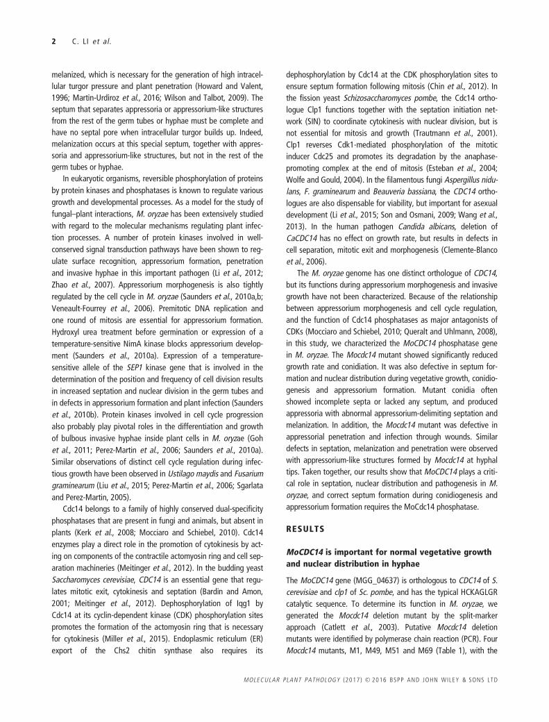
The Mocdc14 mutant showed a reduced growth rate of
approximately 75% (Table 2) and formed colonies with limited
aerial hyphae on oatmeal agar (OTA) plates (Fig. 1A), indicating
the importance of MoCDC14 during vegetative growth. To deter-
mine whether deletion of MoCDC14 affects cytokinesis, we trans-
formed the H1-GFP fusion construct (Luo et al., 2014) into Guy11
and the Mocdc14 mutant M1. In Guy11, septation occurred regu-
larly in hyphae and each hyphal compartment had one nucleus
(Fig. 1B). In the Mocdc14 mutant, hyphae had fewer and unevenly
distributed septa, and individual hyphal compartments often con-
tained multiple nuclei (Fig. 1B). These results suggest that the
Mocdc14 mutant is defective in mitotic exit and nuclei continue to
divide in the absence of cytokinesis in vegetative hyphae.
The Mocdc14 mutant is defective in conidiogenesis
The Mocdc14 mutant also showed significantly reduced conidia-
tion. The average number of conidia produced by the mutant on
each OTA plate was (2.2 6 1.1) 3 103, which was reduced by
more than 100-fold in comparison with the wild-type (Table 2).
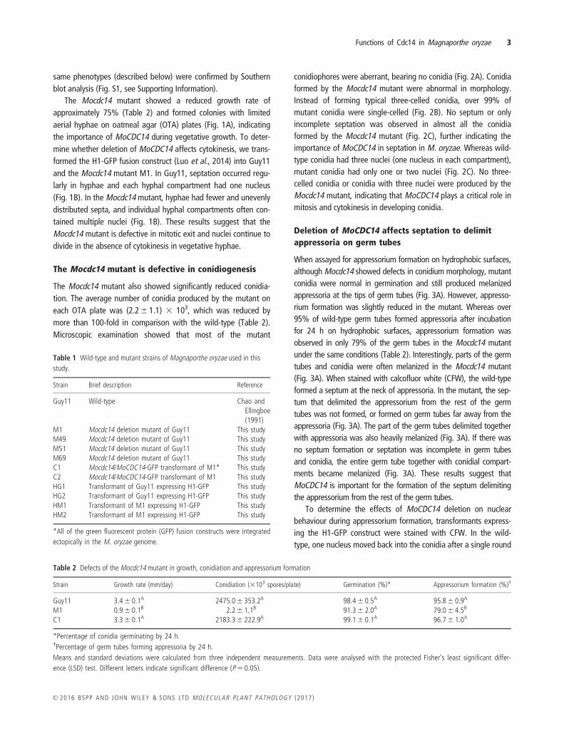
Microscopic examination showed that most of the mutant
conidiophores were aberrant, bearing no conidia (Fig. 2A). Conidia
formed by the Mocdc14 mutant were abnormal in morphology.
Instead of forming typical three-celled conidia, over 99% of
mutant conidia were single-celled (Fig. 2B). No septum or only
incomplete septation was observed in almost all the conidia
formed by the Mocdc14 mutant (Fig. 2C), further indicating the
importance of MoCDC14 in septation in M. oryzae. Whereas wild-
type conidia had three nuclei (one nucleus in each compartment),
mutant conidia had only one or two nuclei (Fig. 2C). No three-
celled conidia or conidia with three nuclei were produced by the
Mocdc14 mutant, indicating that MoCDC14 plays a critical role in
mitosis and cytokinesis in developing conidia.
Deletion of MoCDC14 affects septation to delimit
appressoria on germ tubes
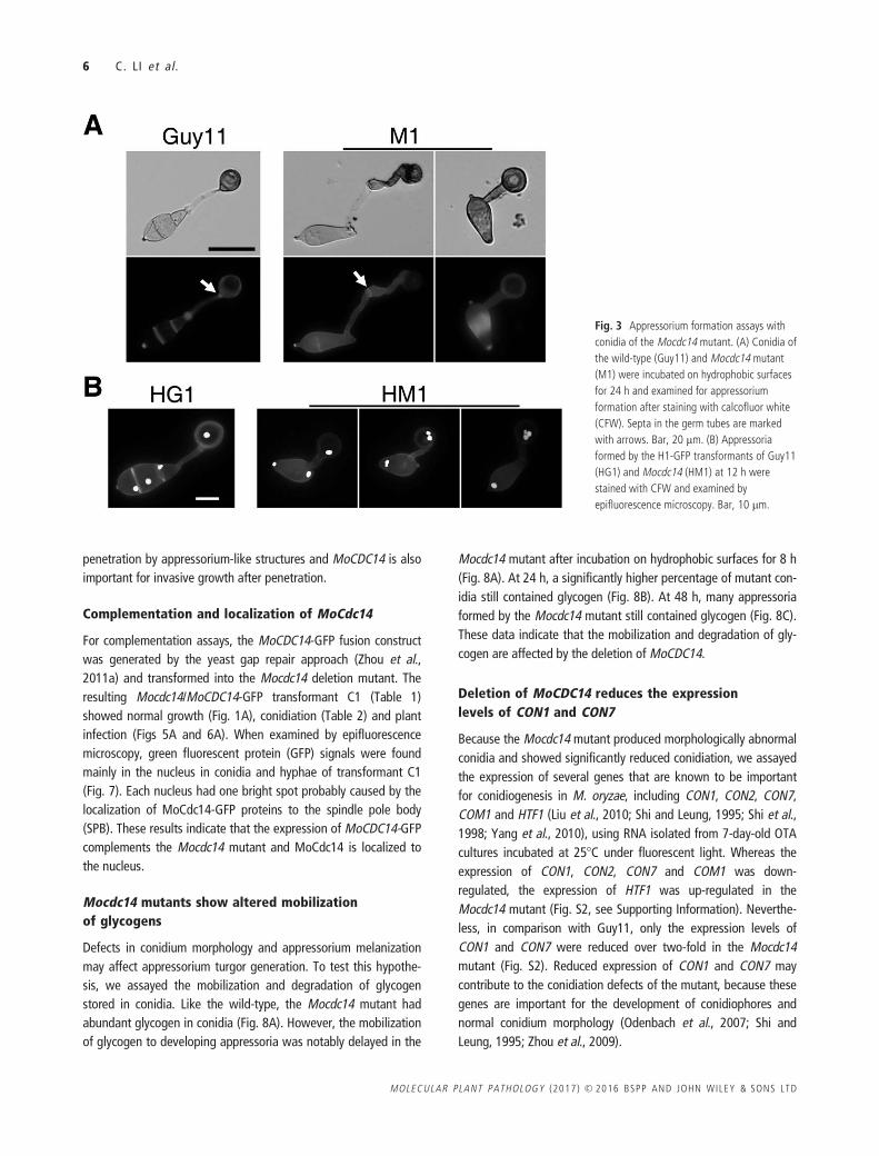
When assayed for appressorium formation on hydrophobic surfaces,
although Mocdc14 showed defects in conidium morphology, mutant
conidia were normal in germination and still produced melanized
appressoria at the tips of germ tubes (Fig. 3A). However, appresso-
rium formation was slightly reduced in the mutant. Whereas over
95% of wild-type germ tubes formed appressoria after incubation
for 24 h on hydrophobic surfaces, appressorium formation was
observed in only 79% of the germ tubes in the Mocdc14 mutant
under the same conditions (Table 2). Interestingly, parts of the germ
tubes and conidia were often melanized in the Mocdc14 mutant
(Fig. 3A). When stained with calcofluor white (CFW), the wild-type
formed a septum at the neck of appressoria. In the mutant, the sep-
tum that delimited the appressorium from the rest of the germ
tubes was not formed, or formed on germ tubes far away from the
appressoria (Fig. 3A). The part of the germ tubes delimited together
with appressoria was also heavily melanized (Fig. 3A). If there was
no septum formation or septation was incomplete in germ tubes
and conidia, the entire germ tube together with conidial compart-
ments became melanized (Fig. 3A). These results suggest that
MoCDC14 is important for the formation of the septum delimiting
the appressorium from the rest of the germ tubes.
To determine the effects of MoCDC14 deletion on nuclear
behaviour during appressorium formation, transformants express-
ing the H1-GFP construct were stained with CFW. In the wild-
type, one nucleus moved back into the conidia after a single round
Table 1 Wild-type and mutant strains of Magnaporthe oryzae used in this
study.
Strain Brief description Reference
Guy11 Wild-type Chao andEllingboe(1991)
M1 Mocdc14 deletion mutant of Guy11 This studyM49 Mocdc14 deletion mutant of Guy11 This studyM51 Mocdc14 deletion mutant of Guy11 This studyM69 Mocdc14 deletion mutant of Guy11 This studyC1 Mocdc14/MoCDC14-GFP transformant of M1* This studyC2 Mocdc14/MoCDC14-GFP transformant of M1 This studyHG1 Transformant of Guy11 expressing H1-GFP This studyHG2 Transformant of Guy11 expressing H1-GFP This studyHM1 Transformant of M1 expressing H1-GFP This studyHM2 Transformant of M1 expressing H1-GFP This study
*All of the green fluorescent protein (GFP) fusion constructs were integrated
ectopically in the M. oryzae genome.
Table 2 Defects of the Mocdc14 mutant in growth, conidiation and appressorium formation
Strain Growth rate (mm/day) Conidiation (3103 spores/plate) Germination (%)* Appressorium formation (%)†
Guy11 3.4 6 0.1A 2475.0 6 353.2A 98.4 6 0.5A 95.8 6 0.9A
M1 0.9 6 0.1B 2.2 6 1.1B 91.3 6 2.0A 79.0 6 4.5B
C1 3.3 6 0.1A 2183.3 6 222.9A 99.1 6 0.1A 96.7 6 1.0A
*Percentage of conidia germinating by 24 h.†Percentage of germ tubes forming appressoria by 24 h.
Means and standard deviations were calculated from three independent measurements. Data were analysed with the protected Fisher’s least significant differ-
ence (LSD) test. Different letters indicate significant difference (P 5 0.05).
Functions of Cdc14 in Magnaporthe oryzae 3
VC 2016 BSPP AND JOHN WILEY & SONS LTD MOLECULAR PLANT PATHOLOGY (2017)
of mitosis had occurred in the germ tube, and the other moved
into developing appressoria (Saunders et al., 2010b). Each wild-
type appressorium contained a single nucleus (Fig. 3B). However,
approximately 10.4 6 3.0% of the appressoria formed by the
Mocdc14 mutant had two or more nuclei (Fig. 3B). Therefore,
deletion of MoCDC14 affects mitotic division and cytokinesis (sep-
tation) during appressorium formation in M. oryzae.
MoCDC14 is also important for the delimitation of
appressoria at hyphal tips
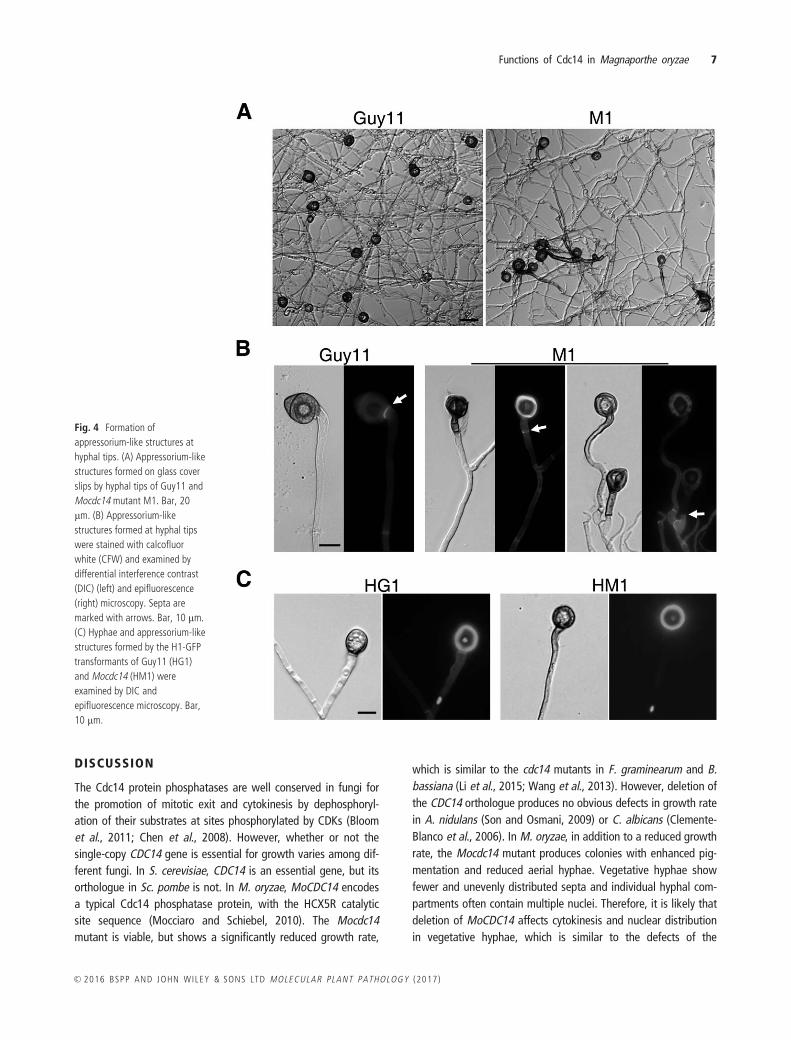
In addition to the formation of appressoria at the tips of germ
tubes, M. oryzae also forms appressorium-like structures at hyphal
tips (Kong et al., 2013). On artificial hydrophobic surfaces, hyphal
tips of the Mocdc14 mutant still developed appressorium-like
structures. However, fragments of hyphae were often delimited
and melanized together with appressorium-like structures in the
Mocdc14 mutant (Fig. 4A). Unlike the wild-type, septation in the
mutant occurred far away from the neck of appressorium-like
structures along the hyphae. Therefore, similar to its septation
defects during appressorium formation on germ tubes, the
Mocdc14 mutant was defective in the formation of the septum to
delimit appressorium-like structures at the hyphal tips (Fig. 4B).
MoCDC14 must also be important for the formation of the septum
delimiting the appressorium-like structures on hyphae.
We also examined the number of nuclei in appressorium-like
structures formed at the hyphal tips of transformants expressing
the H1-GFP construct. Like the wild-type, the Mocdc14 mutant
had a single nucleus in appressorium-like structures (Fig. 4C).
MoCDC14 is important for plant infection
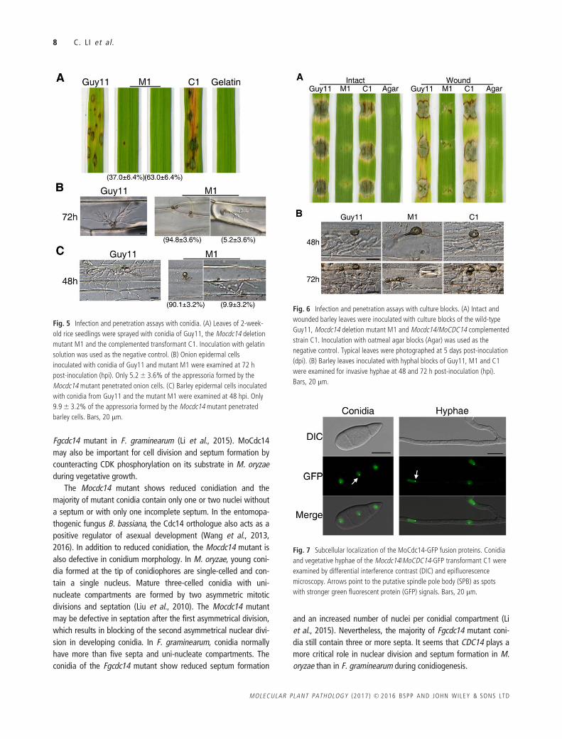
To determine the effect of MoCDC14 deletion on virulence, 2-
week-old seedlings of rice cultivar CO-39 were used for spray infec-
tion assays. At 7 days post-inoculation (dpi), numerous typical blast
lesions were observed on leaves inoculated with Guy11 (Fig. 5A).
Under the same conditions, 63.0 6 6.4% of the leaves sprayed
with the Mocdc14 mutant had no typical blast lesions. Although
some leaves had rare small black spots, blast lesions with extensive
necrotic zones were never observed on leaves inoculated with the
Mocdc14 mutant (Fig. 5A), indicating that MoCDC14 plays a critical
role in plant infection and lesion development.
MoCDC14 is important for appressorium penetration
and invasive growth
In penetration assays with onion epidermal cells, whereas
67.3 6 4.2% of Guy11 appressoria penetrated and formed inva-
sive hyphae by 72 hpi, only 5.2 6 3.6% of the appressoria formed
by the Mocdc14 mutant penetrated onion epidermal cells
(Fig. 5B). The majority of Mocdc14 appressoria appeared to be
melanized, together with parts of or entire germ tubes on onion
epidermis, and failed to penetrate underlying plant cells. Further-
more, in comparison with the wild-type, invasive hyphae formed
by the Mocdc14 mutant in rare onion epidermal cells penetrated
by mutant appressoria were narrower and less bulbous (Fig. 5B).
Similar results were obtained in penetration assays with barley
epidermal cells. Whereas 74.6 6 5.1% of Guy11 appressoria
penetrated and formed invasive hyphae by 48 hpi, only
9.9 6 3.2% of the appressoria formed by the Mocdc14 mutant
Fig. 1 Growth and nuclear distribution
defects of the Mocdc14 mutant. (A) Ten-day-
old oatmeal cultures of the wild-type (Guy11),
Mocdc14 mutant (M1) and Mocdc14/
MoCDC14 complemented transformant (C1).
(B) Hyphae of transformants of Guy11 and
Mocdc14 mutant expressing the H1-GFP
construct were stained with calcofluor white
(CFW) and examined by epifluorescence
microscopy. Bar, 10 lm.
4 C. LI et al .
MOLECULAR PLANT PATHOLOGY (2017) VC 2016 BSPP AND JOHN WILEY & SONS LTD
penetrated barley epidermal cells (Fig. 5C). The Mocdc14 mutant
also showed delayed spread to neighbouring cells. By 48 hpi,
wild-type invasive hyphae had spread from the penetrated cells to
neighbouring barley epidermal cells. Under the same conditions,
invasive hyphae of the mutant were limited to the penetrated cell
(Fig. 5C). These results indicate that the Mocdc14 mutant is defec-
tive in penetration and infectious growth after penetration.
MoCDC14 is required for appressorium-like
structure-mediated penetration and infection
We also conducted infection assays with culture blocks (Liu et al.,
2010) to determine the effect of MoCDC14 deletion on penetra-
tion by appressorium-like structures formed at hyphal tips. On
intact barley leaves inoculated with the Mocdc14 mutant,
only limited necrosis was observed directly beneath culture blocks
(Fig. 6A). On wounded leaves, the Mocdc14 mutant also caused
only limited blast lesions outside the wounding sites (Fig. 6A).
Under the same conditions, Guy11 caused extensive necrosis on
both intact and wounded leaves surrounding the inoculation sites
(Fig. 6A).
In barley epidermal cell penetration assays with culture blocks,
whereas the wild-type penetrated into host cells through
appressorium-like structures and developed invasive hyphae by 48
hpi (Fig. 6B), the majority of the appressorium-like structures
formed by Mocdc14 failed to penetrate under the same condi-
tions. Even at 72 hpi, only less than 2% of the appressorium-like
structures formed by the Mocdc14 mutant were able to penetrate
and showed limited invasive growth in the penetrated cells. Under
the same conditions, Guy11 showed extensive invasive growth in
the initial penetrated and neighbouring cells (Fig. 6B). These
results indicate that the Mocdc14 mutant is defective in
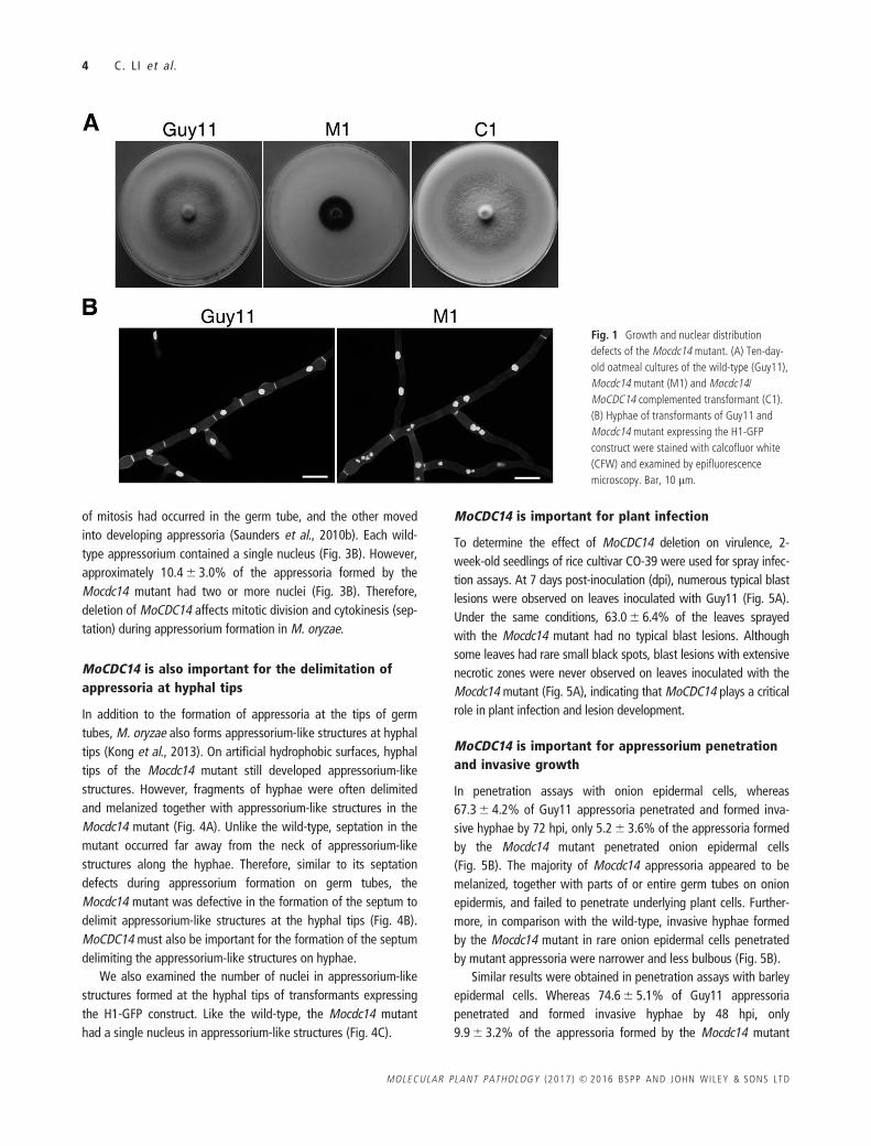
Fig. 2 Defects of the Mocdc14 mutant in conidiogenesis. (A) Ten-day-old oatmeal cultures of the wild-type (Guy11) and Mocdc14 mutant (M1) were examined for
conidia and conidiophores under a dissection microscope. Bar, 50 lm. (B) Conidia of Guy11 and Mocdc14 mutant were examined by differential interference contrast
(DIC) microscopy. Bar, 10 lm. (C) Conidia of transformants of Guy11 (HG1) and Mocdc14 mutant (HM1) expressing the H1-GFP construct were stained with
calcofluor white (CFW) and examined by epifluorescence microscopy. Bar, 10 lm.
Functions of Cdc14 in Magnaporthe oryzae 5
VC 2016 BSPP AND JOHN WILEY & SONS LTD MOLECULAR PLANT PATHOLOGY (2017)
penetration by appressorium-like structures and MoCDC14 is also
important for invasive growth after penetration.
Complementation and localization of MoCdc14
For complementation assays, the MoCDC14-GFP fusion construct
was generated by the yeast gap repair approach (Zhou et al.,
2011a) and transformed into the Mocdc14 deletion mutant. The
resulting Mocdc14/MoCDC14-GFP transformant C1 (Table 1)
showed normal growth (Fig. 1A), conidiation (Table 2) and plant
infection (Figs 5A and 6A). When examined by epifluorescence
microscopy, green fluorescent protein (GFP) signals were found
mainly in the nucleus in conidia and hyphae of transformant C1
(Fig. 7). Each nucleus had one bright spot probably caused by the
localization of MoCdc14-GFP proteins to the spindle pole body
(SPB). These results indicate that the expression of MoCDC14-GFP
complements the Mocdc14 mutant and MoCdc14 is localized to
the nucleus.
Mocdc14 mutants show altered mobilization
of glycogens
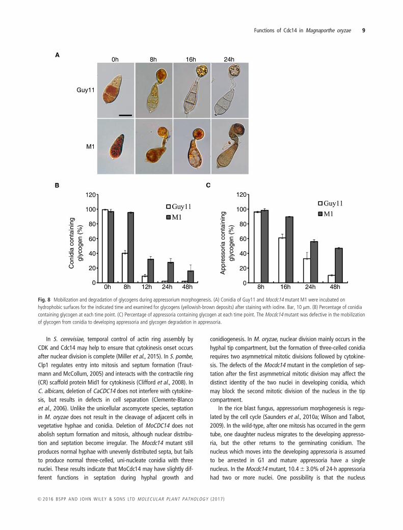
Defects in conidium morphology and appressorium melanization
may affect appressorium turgor generation. To test this hypothe-
sis, we assayed the mobilization and degradation of glycogen
stored in conidia. Like the wild-type, the Mocdc14 mutant had
abundant glycogen in conidia (Fig. 8A). However, the mobilization
of glycogen to developing appressoria was notably delayed in the
Mocdc14 mutant after incubation on hydrophobic surfaces for 8 h
(Fig. 8A). At 24 h, a significantly higher percentage of mutant con-
idia still contained glycogen (Fig. 8B). At 48 h, many appressoria
formed by the Mocdc14 mutant still contained glycogen (Fig. 8C).
These data indicate that the mobilization and degradation of gly-
cogen are affected by the deletion of MoCDC14.
Deletion of MoCDC14 reduces the expression
levels of CON1 and CON7
Because the Mocdc14 mutant produced morphologically abnormal
conidia and showed significantly reduced conidiation, we assayed
the expression of several genes that are known to be important
for conidiogenesis in M. oryzae, including CON1, CON2, CON7,
COM1 and HTF1 (Liu et al., 2010; Shi and Leung, 1995; Shi et al.,
1998; Yang et al., 2010), using RNA isolated from 7-day-old OTA
cultures incubated at 258C under fluorescent light. Whereas the
expression of CON1, CON2, CON7 and COM1 was down-
regulated, the expression of HTF1 was up-regulated in the
Mocdc14 mutant (Fig. S2, see Supporting Information). Neverthe-
less, in comparison with Guy11, only the expression levels of
CON1 and CON7 were reduced over two-fold in the Mocdc14
mutant (Fig. S2). Reduced expression of CON1 and CON7 may
contribute to the conidiation defects of the mutant, because these
genes are important for the development of conidiophores and
normal conidium morphology (Odenbach et al., 2007; Shi and
Leung, 1995; Zhou et al., 2009).
Fig. 3 Appressorium formation assays with
conidia of the Mocdc14 mutant. (A) Conidia of
the wild-type (Guy11) and Mocdc14 mutant
(M1) were incubated on hydrophobic surfaces
for 24 h and examined for appressorium
formation after staining with calcofluor white
(CFW). Septa in the germ tubes are marked
with arrows. Bar, 20 lm. (B) Appressoria
formed by the H1-GFP transformants of Guy11
(HG1) and Mocdc14 (HM1) at 12 h were
stained with CFW and examined by
epifluorescence microscopy. Bar, 10 lm.
6 C. LI et al .
MOLECULAR PLANT PATHOLOGY (2017) VC 2016 BSPP AND JOHN WILEY & SONS LTD
DISCUSSION
The Cdc14 protein phosphatases are well conserved in fungi for
the promotion of mitotic exit and cytokinesis by dephosphoryl-
ation of their substrates at sites phosphorylated by CDKs (Bloom
et al., 2011; Chen et al., 2008). However, whether or not the
single-copy CDC14 gene is essential for growth varies among dif-
ferent fungi. In S. cerevisiae, CDC14 is an essential gene, but its
orthologue in Sc. pombe is not. In M. oryzae, MoCDC14 encodes
a typical Cdc14 phosphatase protein, with the HCX5R catalytic
site sequence (Mocciaro and Schiebel, 2010). The Mocdc14
mutant is viable, but shows a significantly reduced growth rate,
which is similar to the cdc14 mutants in F. graminearum and B.
bassiana (Li et al., 2015; Wang et al., 2013). However, deletion of
the CDC14 orthologue produces no obvious defects in growth rate
in A. nidulans (Son and Osmani, 2009) or C. albicans (Clemente-
Blanco et al., 2006). In M. oryzae, in addition to a reduced growth
rate, the Mocdc14 mutant produces colonies with enhanced pig-
mentation and reduced aerial hyphae. Vegetative hyphae show
fewer and unevenly distributed septa and individual hyphal com-
partments often contain multiple nuclei. Therefore, it is likely that
deletion of MoCDC14 affects cytokinesis and nuclear distribution
in vegetative hyphae, which is similar to the defects of the
Fig. 4 Formation of
appressorium-like structures at
hyphal tips. (A) Appressorium-like
structures formed on glass cover
slips by hyphal tips of Guy11 and
Mocdc14 mutant M1. Bar, 20
lm. (B) Appressorium-like
structures formed at hyphal tips
were stained with calcofluor
white (CFW) and examined by
differential interference contrast
(DIC) (left) and epifluorescence
(right) microscopy. Septa are
marked with arrows. Bar, 10 lm.
(C) Hyphae and appressorium-like
structures formed by the H1-GFP
transformants of Guy11 (HG1)
and Mocdc14 (HM1) were
examined by DIC and
epifluorescence microscopy. Bar,
10 lm.
Functions of Cdc14 in Magnaporthe oryzae 7
VC 2016 BSPP AND JOHN WILEY & SONS LTD MOLECULAR PLANT PATHOLOGY (2017)
Fgcdc14 mutant in F. graminearum (Li et al., 2015). MoCdc14
may also be important for cell division and septum formation by
counteracting CDK phosphorylation on its substrate in M. oryzae
during vegetative growth.
The Mocdc14 mutant shows reduced conidiation and the
majority of mutant conidia contain only one or two nuclei without
a septum or with only one incomplete septum. In the entomopa-
thogenic fungus B. bassiana, the Cdc14 orthologue also acts as a
positive regulator of asexual development (Wang et al., 2013,
2016). In addition to reduced conidiation, the Mocdc14 mutant is
also defective in conidium morphology. In M. oryzae, young coni-
dia formed at the tip of conidiophores are single-celled and con-
tain a single nucleus. Mature three-celled conidia with uni-
nucleate compartments are formed by two asymmetric mitotic
divisions and septation (Liu et al., 2010). The Mocdc14 mutant
may be defective in septation after the first asymmetrical division,
which results in blocking of the second asymmetrical nuclear divi-
sion in developing conidia. In F. graminearum, conidia normally
have more than five septa and uni-nucleate compartments. The
conidia of the Fgcdc14 mutant show reduced septum formation
and an increased number of nuclei per conidial compartment (Li
et al., 2015). Nevertheless, the majority of Fgcdc14 mutant coni-
dia still contain three or more septa. It seems that CDC14 plays a
more critical role in nuclear division and septum formation in M.
oryzae than in F. graminearum during conidiogenesis.
Fig. 5 Infection and penetration assays with conidia. (A) Leaves of 2-week-
old rice seedlings were sprayed with conidia of Guy11, the Mocdc14 deletion
mutant M1 and the complemented transformant C1. Inoculation with gelatin
solution was used as the negative control. (B) Onion epidermal cells
inoculated with conidia of Guy11 and mutant M1 were examined at 72 h
post-inoculation (hpi). Only 5.2 6 3.6% of the appressoria formed by the
Mocdc14 mutant penetrated onion cells. (C) Barley epidermal cells inoculated
with conidia from Guy11 and the mutant M1 were examined at 48 hpi. Only
9.9 6 3.2% of the appressoria formed by the Mocdc14 mutant penetrated
barley cells. Bars, 20 lm.
Fig. 6 Infection and penetration assays with culture blocks. (A) Intact and
wounded barley leaves were inoculated with culture blocks of the wild-type
Guy11, Mocdc14 deletion mutant M1 and Mocdc14/MoCDC14 complemented
strain C1. Inoculation with oatmeal agar blocks (Agar) was used as the
negative control. Typical leaves were photographed at 5 days post-inoculation
(dpi). (B) Barley leaves inoculated with hyphal blocks of Guy11, M1 and C1
were examined for invasive hyphae at 48 and 72 h post-inoculation (hpi).
Bars, 20 lm.
Fig. 7 Subcellular localization of the MoCdc14-GFP fusion proteins. Conidia
and vegetative hyphae of the Mocdc14/MoCDC14-GFP transformant C1 were
examined by differential interference contrast (DIC) and epifluorescence
microscopy. Arrows point to the putative spindle pole body (SPB) as spots
with stronger green fluorescent protein (GFP) signals. Bars, 20 lm.
8 C. LI et al .
MOLECULAR PLANT PATHOLOGY (2017) VC 2016 BSPP AND JOHN WILEY & SONS LTD
In S. cerevisiae, temporal control of actin ring assembly by
CDK and Cdc14 may help to ensure that cytokinesis onset occurs
after nuclear division is complete (Miller et al., 2015). In S. pombe,
Clp1 regulates entry into mitosis and septum formation (Traut-
mann and McCollum, 2005) and interacts with the contractile ring
(CR) scaffold protein Mid1 for cytokinesis (Clifford et al., 2008). In
C. albicans, deletion of CaCDC14 does not interfere with cytokine-
sis, but results in defects in cell separation (Clemente-Blanco
et al., 2006). Unlike the unicellular ascomycete species, septation
in M. oryzae does not result in the cleavage of adjacent cells in
vegetative hyphae and conidia. Deletion of MoCDC14 does not
abolish septum formation and mitosis, although nuclear distribu-
tion and septation become irregular. The Mocdc14 mutant still
produces normal hyphae with unevenly distributed septa, but fails
to produce normal three-celled, uni-nucleate conidia with three
nuclei. These results indicate that MoCdc14 may have slightly dif-
ferent functions in septation during hyphal growth and
conidiogenesis. In M. oryzae, nuclear division mainly occurs in the
hyphal tip compartment, but the formation of three-celled conidia
requires two asymmetrical mitotic divisions followed by cytokine-
sis. The defects of the Mocdc14 mutant in the completion of sep-
tation after the first asymmetrical mitotic division may affect the
distinct identity of the two nuclei in developing conidia, which
may block the second mitotic division of the nucleus in the tip
compartment.
In the rice blast fungus, appressorium morphogenesis is regu-
lated by the cell cycle (Saunders et al., 2010a; Wilson and Talbot,
2009). In the wild-type, after one mitosis has occurred in the germ
tube, one daughter nucleus migrates to the developing appresso-
ria, but the other returns to the germinating conidium. The
nucleus which moves into the developing appressoria is assumed
to be arrested in G1 and mature appressoria have a single
nucleus. In the Mocdc14 mutant, 10.4 6 3.0% of 24-h appressoria
had two or more nuclei. One possibility is that the nucleus
Fig. 8 Mobilization and degradation of glycogens during appressorium morphogenesis. (A) Conidia of Guy11 and Mocdc14 mutant M1 were incubated on
hydrophobic surfaces for the indicated time and examined for glycogens (yellowish-brown deposits) after staining with iodine. Bar, 10 lm. (B) Percentage of conidia
containing glycogen at each time point. (C) Percentage of appressoria containing glycogen at each time point. The Mocdc14 mutant was defective in the mobilization
of glycogen from conidia to developing appressoria and glycogen degradation in appressoria.
Functions of Cdc14 in Magnaporthe oryzae 9
VC 2016 BSPP AND JOHN WILEY & SONS LTD MOLECULAR PLANT PATHOLOGY (2017)
entering the developing appressorium may continue to divide
without cytokinesis as a result of defects in mitotic exit associated
with MoCDC14 deletion. Because the Mocdc14 mutant is defec-
tive in the formation of the septum that delimits the appressoria
from the rest of the germ tubes, it is also possible that more than
one nucleus migrates into the developing appressoria. Further-
more, when mature appressoria are melanized, conidia and germ
tubes undergo autophagic cell death in the wild-type (Veneault-
Fourrey et al., 2006). However, conidial compartments often still
contain nuclei after appressoria have been melanized in the
Mocdc14 mutant when appressorium-delimiting septa are not
properly formed. Complete septation of appressoria from the rest
of the germ tubes by the appressorium-delimiting septum may be
a prerequisite for the triggering of autophagic cell death.
In M. oryzae, appressorium melanization is important for
appressorium turgor generation (deJong et al., 1997). The
Mocdc14 mutant still forms melanized appressoria on germ tubes,
but parts of the germ tubes and sometimes conidia are also
melanized. Similar defects in melanization have been observed in
the Mocdc14 mutant when assayed for the formation of
appressorium-like structures at hyphal tips. Fragments of hyphae
are often melanized together with appressorium-like structures,
depending on the position of the septum that delimits the
appressorium-like structures. These results indicate that the for-
mation of these special septum-delimiting appressoria or
appressorium-like structures from the rest of the germ tubes or
hyphae plays a critical role in defining the boundary of melanin
deposition. Enzymes involved in melanin biosynthesis may be only
expressed or active after the completion of the appressorium-
delimiting septum. In M. oryzae, deletion of the MoAND1 gene,
an orthologue of A. nidulans ApsA, results in septation defects in
hyphae, but has no effect on the formation of appressoria and the
appressorium-delimiting septum (Jeon et al., 2014). An earlier
study in M. oryzae with hydroxyl urea treatment and NimA muta-
tions has also shown that the differentiation of appressoria
requires a cytokinetic event that is distinct from cell divisions
within hyphae (Saunders et al., 2010a). Our studies show that
MoCdc14 is involved in deciding the occurrence and position of
this special septum during appressorium formation.
In infection assays, the Mocdc14 mutant is almost non-
pathogenic. The defects of the Mocdc14 mutant in plant infection
can be related directly to its defects in growth and appressorium
morphogenesis. In addition, the Mocdc14 mutant is defective in
glycogen mobilization and degradation, which is important for
appressorium turgor generation and penetration. However,
mutants with deletion of MoCDC14 are also defective in infection
through wounding, suggesting a critical role of MoCdc14 during
invasive growth. In rare plant cells penetrated by the Mocdc14
mutant, only limited growth of invasive hyphae is seen and the
mutant fails to spread into neighbouring cells. Rare invasive
hyphae formed by the mutant inside plant cells are less branching
than those of the wild-type. In M. oryzae, bulbous invasive hyphae
are considered to show pseudohyphal-like growth in plant cells
(Kankanala et al., 2007; Yi and Valent, 2013). It is possible that
MoCdc14 is important for constriction or septum formation in
invasive hyphae with pseudohyphal growth.
EXPERIMENTAL PROCEDURES
Strains and culture conditions
The M. oryzae wild-type strain Guy11 and mutants used in this study
(Table 1) were cultured on OTA or complete medium (CM) plates at 258C
as described previously (Xu and Hamer, 1996; Zhou et al., 2011b). For fun-
gal transformation, protoplast preparation and polyethylene glycol (PEG)-
mediated transformation of M. oryzae were performed as described previ-
ously (Park et al., 2006). Hygromycin B (Calbiochem, La Jolla, CA, USA)
and geneticin (MP Biochemicals, Santa Ana, CA, USA) were added to final
concentrations of 300 and 500 lg/mL, respectively, for transformant selec-
tion. For DNA isolation, vegetative hyphae were harvested from 2-day-old
liquid CM cultures (Zhao et al., 2005). Measurements of growth rate and
conidiation were performed as described previously (Li et al., 2004; Park
et al., 2004).
Generation of the Mocdc14 deletion mutants
To delete the MoCDC14 gene, the double-joint PCR method (Yu et al.,
2004) was used to generate the CDC14 gene replacement construct (Fig.
S1A). The 1080-bp upstream and 992-bp downstream flanking sequences
of MoCDC14 were amplified with the primer pairs C1F/C2R and C3F/C4R
(Table S1, see Supporting Information), respectively, and ligated with the
hph cassette amplified with primers HYG/F and HYG/R from pCB1003
(Carroll et al., 1994). The products of double-joint PCR were amplified
with primers CCF and CCR (Table S1) and transformed into protoplasts of
Guy11. Hygromycin-resistant transformants were screened by PCR and
putative gene replacement mutants were confirmed by Southern blot
analysis.
Generation of the MoCDC14-GFP fusion construct
To generate the MoCDC14-GFP fusion construct, the entire MoCDC14
gene, including its promoter region, was amplified with primers 14GFP-F
and 14GFP-R (Table S1) and cloned into XhoI-digested pFL2 (Zhou et al.,
2011b) by the yeast gap repair approach (Zhou et al., 2011a). The
MoCDC14-GFP fusion construct recovered from yeast Trp1 transformants
was confirmed by sequencing analysis and transformed into the Mocdc14
deletion mutant M1 (Table 1). Geneticin-resistant transformants express-
ing the MoCDC14-GFP construct were verified by PCR and examined for
GFP signals.
Appressorium formation, penetration and plant
infection assays
Conidia were harvested from 10-day-old OTA cultures and resuspended to
a concentration of 5 3 104 conidia/mL (Zhou et al., 2012). Appressorium
formation by germ tubes on artificial surfaces was assayed as described
10 C. LI et al .
MOLECULAR PLANT PATHOLOGY (2017) VC 2016 BSPP AND JOHN WILEY & SONS LTD
previously (Wang et al., 2015; Zhou et al., 2011b). Appressorial penetra-
tion and invasive hyphal development were assayed with barley and onion
epidermal cells (Chi et al., 2009; Kong et al., 2013). For spray infection
assays, conidia were adjusted to 1 3 105 conidia/mL in 0.25% gelatin
and used for inoculation of 14-day-old seedlings of rice cultivar CO-39
(Kong et al., 2013).
Assays for the formation and penetration of
appressorium-like structures
Assays for appressorium-like structures at hyphal tips were performed as
described previously (Liu et al., 2010). For infection assays with culture
blocks, the second leaves of 8-day-old seedlings of barley cultivar Golden
Promise were inoculated with 1–2-mm2 blocks of 10-day-old OTA cultures
as described previously (Liu et al., 2010). Penetration, invasive growth
and lesion development were examined as described previously (Liu et al.,
2010; Yang et al., 2010).
Cell wall and glycogen staining
The cell wall was stained with 10 mg/mL CFW (Sigma-Aldrich, St. Louis,
MO, USA), as described previously (Zhou et al., 2011b), to visualize septa
in hyphae and during appressorium formation. Conidia incubated at room
temperature on hydrophobic surfaces were stained for glycogens with
60 mg of KI and 10 mg of I2 (Thines et al., 2000; Zhang et al., 2014). Gly-
cogen mobilization and degradation during appressorium formation were
examined with an Olympus BX51 epifluorescence microscope (Olympus
Corporation, Tokyo, Japan).
Quantitative reverse transcription-polymerase chain
reaction (qRT-PCR) analysis
Hyphae harvested from 7-day-old OTA cultures of the wild-type strain
Guy11 and Mocdc14 mutant M1 were used for RNA isolation with TRIzol
reagent (Invitrogen, Carlsbad, CA, USA). After treatment with the DNA-
free kit (Promega, Madison, WI, USA), purified RNA samples were used
for cDNA synthesis with the Fermentas first cDNA synthesis kit (Hanover,
MD, USA). The resulting first cDNA was used for qRT-PCR assays as
described previously (Ding et al., 2010) with the primers listed in Table
S1. Data from three biological replicates were used to estimate the rela-
tive expression levels of CON1, CON2, CON7, COM1 and HTF1 with the
2–DDCt method (Livak and Schmittgen, 2001). The M. oryzae actin gene
MGG_03982.6 was used as the endogenous control for normalization.
ACKNOWLEDGEMENTS
We thank Xuli Gao for assistance with appressorium formation and pene-
tration assays. We also thank Drs Huiquan Liu, Cong Jiang and Guanghui
Wang for fruitful discussions. This work was supported by grants from
the United States Department of Agriculture-National Institute of Food
and Agriculture (USDA-NIFA) (2012-67013-19381), Specialized Research
Cultivation Fund for Excellent Young Scholars of Northwest Agricultural
and Forestry University (NWSUAF) and Nature Science Foundation of
China (No. 31271989; No. 31201464).
REFERENCES
Bardin, A.J. and Amon, A. (2001) Men and sin: what’s the difference? Nat. Rev.
Mol. Cell Biol. 2, 815–826.
Bloom, J., Cristea, I.M., Procko, A.L., Lubkov, V., Chait, B.T., Snyder, M. and
Cross, F.R. (2011) Global analysis of Cdc14 phosphatase reveals diverse roles in
mitotic processes. J. Biol. Chem. 286, 5434–5445.
Carroll, A., Sweigard, J. and Valent, B. (1994) Improved vectors for selecting
resistance to hygromycin. Fungal Genet. Newsl. 41, 135–143.
Catlett, N., Lee, B.N., Yoder, O. and Turgeon, B.G. (2003) Split-marker recombi-
nation for efficient targeted deletion of fungal genes. Fungal Genet. Newsl. 9–11.
Chao, C.-C.T. and Ellingboe, A.H. (1991) Selection for mating competence in Mag-
naporthe grisea pathogenic to rice. Canadian Journal of Botany 69, 2130–2134.
Chen, C.T., Feoktistova, A., Chen, J.S., Shim, Y.S., Clifford, D.M., Gould, K.L.
and McCollum, D. (2008) The SIN kinase Sid2 regulates cytoplasmic retention of
the S. pombe Cdc14-like phosphatase Clp1. Curr. Biol. 18, 1594–1599.
Chi, M.H., Park, S.Y., Kim, S. and Lee, Y.H. (2009) A novel pathogenicity gene is
required in the rice blast fungus to suppress the basal defenses of the host. PLoS
Pathog. 5, e1000401.
Chin, C.F., Bennett, A.M., Ma, W.K., Hall, M.C. and Yeong, F.M. (2012) Depend-
ence of Chs2 ER export on dephosphorylation by cytoplasmic Cdc14 ensures that
septum formation follows mitosis. Mol. Biol. Cell. 23, 45–58.
Clemente-Blanco, A., Gonz�alez-Novo, A., Mach�ın, F., Caballero-Lima, D.,
Arag�on, L., S�anchez, M., de Aldana, C.R.V., Jim�enez, J. and Correa-Bordes,
J. (2006) The Cdc14p phosphatase affects late cell-cycle events and morphogenesis
in Candida albicans. J. Cell Sci. 119, 1130–1143.
Clifford, D.M., Wolfe, B.A., Roberts-Galbraith, R.H., McDonald, W.H., Yates,
J.R., 3rd. and Gould, K.L. (2008) The Clp1/Cdc14 phosphatase contributes to the
robustness of cytokinesis by association with anillin-related Mid1. J. Cell Biol. 181,
79–88.
Dean, R., Van Kan, J.A., Pretorius, Z.A., Hammond-Kosack, K.E., Di Pietro, A.,
Spanu, P.D., Rudd, J.J., Dickman, M., Kahmann, R., Ellis, J. and Foster, G.D.
(2012) The top 10 fungal pathogens in molecular plant pathology. Mol. Plant
Pathol. 13, 414–430.
deJong, J.C., MCCormack, B.J., Smirnoff, N. and Talbot, N.J. (1997) Glycerol
generates turgor in rice blast. Nature, 389, 244–245.
Ding, S.L., Liu, W., Iliuk, A., Ribot, C., Vallet, J., Tao, A., Wang, Y., Lebrun,
M.H. and Xu, J.R. (2010) The Tig1 histone deacetylase complex regulates infec-
tious growth in the rice blast fungus Magnaporthe oryzae. Plant Cell, 22,
2495–2508.
Ebbole, D.J. (2007) Magnaporthe as a model for understanding host–pathogen
interactions. Annu. Rev. Phytopathol. 45, 437–456.
Esteban, V., Blanco, M., Cueille, N., Simanis, V., Moreno, S. and Bueno, A.
(2004) A role for the Cdc14-family phosphatase Flp1p at the end of the cell cycle in
controlling the rapid degradation of the mitotic inducer Cdc25p in fission yeast. J.
Cell Sci. 117, 2461–2468.
Goh, J., Kim, K.S., Park, J., Jeon, J., Park, S.Y. and Lee, Y.H. (2011) The cell cycle
gene MoCDC15 regulates hyphal growth, asexual development and plant infection
in the rice blast pathogen Magnaporthe oryzae. Fungal Genet. Biol. 48, 784–792.
Howard, R.J. and Valent, B. (1996) Breaking and entering: host penetration by
the fungal rice blast pathogen Magnaporthe grisea. Annu. Rev. Microbiol. 50,
491–512.
Jeon, J., Rho, H., Kim, S., Kim, K.S. and Lee, Y.H. (2014) Role of MoAND1-medi-
ated nuclear positioning in morphogenesis and pathogenicity in the rice blast fun-
gus, Magnaporthe oryzae. Fungal Genet. Biol. 69, 43–51.
Kankanala, P., Czymmek, K. and Valent, B. (2007) Roles for rice membrane
dynamics and plasmodesmata during biotrophic invasion by the blast fungus. Plant
Cell, 19, 706–724.
Kato, H., Mayama, R., Sekine, R. and Urashima, A. (1994) Microconidium forma-
tion in Magnaporthe grisea. Annu. Phytopathol. Soc. Jpn. 60, 175–185.
Kerk, D., Templeton, G. and Moorhead, G.B. (2008) Evolutionary radiation pat-
tern of novel protein phosphatases revealed by analysis of protein data from the
completely sequenced genomes of humans, green algae, and higher plants. Plant
Physiol. 146, 351–367.
Kong, L.A., Yang, J., Li, G.T., Qi, L.L., Zhang, Y.J., Wang, C.F., Zhao, W.S., Xu,
J.R. and Peng, Y.L. (2012) Different chitin synthase genes are required for various
developmental and plant infection processes in the rice blast fungus Magnaporthe
oryzae. PLoS Pathog. 8, e1002526.
Kong, L.A., Li, G.T., Liu, Y., Liu, M.G., Zhang, S.J., Yang, J., Zhou, X.Y., Peng,
Y.L. and Xu, J.R. (2013) Differences between appressoria formed by germ tubes
Functions of Cdc14 in Magnaporthe oryzae 11
VC 2016 BSPP AND JOHN WILEY & SONS LTD MOLECULAR PLANT PATHOLOGY (2017)
and appressorium-like structures developed by hyphal tips in Magnaporthe oryzae.
Fungal Genet. Biol. 56, 33–41.
Li, C.H., Melesse, M., Zhang, S.J., Hao, C.F., Wang, C.F., Zhang, H.C., Hall, M.C.
and Xu, J.R. (2015) FgCDC14 regulates cytokinesis, morphogenesis, and pathoge-
nesis in Fusarium graminearum. Mol. Microbiol. 98, 770–786.
Li, G.T., Zhou, X.Y. and Xu, J.R. (2012) Genetic control of infection-related develop-
ment in Magnaporthe oryzae. Curr. Opin. Microbiol. 15, 678–684.
Li, L., Xue, C.Y., Bruno, K., Nishimura, M. and Xu, J.R. (2004) Two PAK kinase
genes, CHM1 and MST20, have distinct functions in Magnaporthe grisea. Mol.
Plant–Microbe Interact. 17, 547–556.
Liu, H.Q., Zhang, S.J., Ma, J.W., Dai, Y.F., Li, C.H., Lyu, X., Wang, C.F. and Xu,
J.R. (2015) Two Cdc2 kinase genes with distinct functions in vegetative and infec-
tious hyphae in Fusarium graminearum. PLoS Pathog. 11, e1004913.
Liu, W.D., Xie, S.Y., Zhao, X.H., Chen, X., Zheng, W.H., Lu, G.D., Xu, J.R. and
Wang, Z.H. (2010) A homeobox gene is essential for conidiogenesis of the rice
blast fungus Magnaporthe oryzae. Mol. Plant–Microbe Interact. 23, 366–375.
Liu, X.H., Lu, J.P. and Lin, F.C. (2007) Autophagy during conidiation, conidial germi-
nation and turgor generation in Magnaporthe grisea. Autophagy, 3, 472–473.
Liu, X.H., Gao, H.M., Xu, F., Lu, J.P., Devenish, R.J. and Lin, F.C. (2012) Autoph-
agy vitalizes the pathogenicity of pathogenic fungi. Autophagy, 8, 1415–1425.
Livak, K.J. and Schmittgen, T.D. (2001) Analysis of relative gene expression data
using real-time quantitative PCR and the 2(–Delta Delta C(T)) method. Methods,
25, 402–408.
Luo, Y.P., Zhang, H.C., Qi, L.L., Zhang, S.J., Zhou, X.Y., Zhang, Y.M. and Xu, J.R.
(2014) FgKin1 kinase localizes to the septal pore and plays a role in hyphal growth,
ascospore germination, pathogenesis, and localization of Tub1 beta-tubulins in
Fusarium graminearum. New Phytol. 204, 943–954.
Martin-Urdiroz, M., Oses-Ruiz, M., Ryder, L.S. and Talbot, N.J. (2016) Investigat-
ing the biology of plant infection by the rice blast fungus Magnaporthe oryzae.
Fungal Genet. Biol. 90, 61–68.
Meitinger, F., Palani, S. and Pereira, G. (2012) The power of MEN in cytokinesis.
Cell Cycle, 11, 219–228.
Miller, D.P., Hall, H., Chaparian, R., Mara, M., Mueller, A., Hall, M.C. and
Shannon, K.B. (2015) Dephosphorylation of Iqg1 by Cdc14 regulates cytokinesis
in budding yeast. Mol. Biol. Cell, 26, 2913–2926.
Mocciaro, A. and Schiebel, E. (2010) Cdc14: a highly conserved family of phospha-
tases with non-conserved functions? J. Cell Sci. 123, 2867–2876.
Odenbach, D., Breth, B., Thines, E., Weber, R.W., Anke, H. and Foster, A.J.
(2007) The transcription factor Con7p is a central regulator of infection-related
morphogenesis in the rice blast fungus Magnaporthe grisea. Mol. Microbiol. 64,
293–307.
Os�es-Ruiz, M., Sakulkoo, W. and Talbot, N.J. (2016) Septation and cytokinesis in
pathogenic fungi. In: Growth, Differentiation and Sexuality, Wendland, J. (ed.) pp.
67–79. Berlin: Springer.
Park, G., Bruno, K.S., Staiger, C.J., Talbot, N.J. and Xu, J.R. (2004) Independent
genetic mechanisms mediate turgor generation and penetration peg formation dur-
ing plant infection in the rice blast fungus. Mol. Microbiol. 53, 1695–1707.
Park, G., Xue, C., Zhao, X., Kim, Y., Orbach, M. and Xu, J.R. (2006) Multiple
upstream signals converge on the adaptor protein Mst50 in Magnaporthe grisea.
Plant Cell, 18, 2822–2835.
Perez-Martin, J., Castillo-Lluva, S., Sgarlata, C., Flor-Parra, I., Mielnichuk, N.,
Torreblanca, J. and Carbo, N. (2006) Pathocycles: Ustilago maydis as a model to
study the relationships between cell cycle and virulence in pathogenic fungi. Mol.
Genet. Genomics, 276, 211–229.
Queralt, E. and Uhlmann, F. (2008) Cdk-counteracting phosphatases unlock mitotic
exit. Curr. Opin. Cell Biol. 20, 661–668.
Saunders, D.G., Aves, S.J. and Talbot, N.J. (2010a) Cell cycle-mediated regulation
of plant infection by the rice blast fungus. Plant Cell, 22, 497–507.
Saunders, D.G., Dagdas, Y.F. and Talbot, N.J. (2010b) Spatial uncoupling of mito-
sis and cytokinesis during appressorium-mediated plant infection by the rice blast
fungus Magnaporthe oryzae. Plant Cell, 22, 2417–2428.
Sgarlata, C. and Perez-Martin, J. (2005) Inhibitory phosphorylation of a mitotic
cyclin-dependent kinase regulates the morphogenesis, cell size and virulence of
the smut fungus Ustilago maydis. J. Cell Sci. 118, 3607–3622.
Shi, Z.X. and Leung, H. (1995) Genetic analysis of sporulation in Magnaporthe
grisea by chemical and insertional mutagenesis. Mol. Plant–Microbe Interact. 8,
949–959.
Shi, Z.X., Christian, D. and Leung, H. (1998) Interactions between spore morpho-
genetic mutations affect cell types, sporulation, and pathogenesis in Magnaporthe
grisea. Mol. Plant–Microbe Interact. 11, 199–207.
Son, S. and Osmani, S.A. (2009) Analysis of all protein phosphatase genes in Asper-
gillus nidulans identifies a new mitotic regulator, Fcp1. Eukaryot. Cell, 8, 573–585.
Thines, E., Weber, R.W. and Talbot, N.J. (2000) MAP kinase and protein kinase A-
dependent mobilization of triacylglycerol and glycogen during appressorium turgor
generation by Magnaporthe grisea. Plant Cell, 12, 1703–1718.
Trautmann, S. and McCollum, D. (2005) Distinct nuclear and cytoplasmic functions
of the S. pombe Cdc14-like phosphatase Clp1p/Flp1p and a role for nuclear shut-
tling in its regulation. Curr. Biol. 15, 1384–1389.
Trautmann, S., Wolfe, B.A., Jorgensen, P., Tyers, M., Gould, K.L. and McCollum, D.
(2001) Fission yeast Clp1p phosphatase regulates G2/M transition and coordination of
cytokinesis with cell cycle progression. Curr. Biol. 11, 931–940.
Veneault-Fourrey, C., Barooah, M., Egan, M., Wakley, G. and Talbot, N.J.
(2006) Autophagic fungal cell death is necessary for infection by the rice blast fun-
gus. Science, 312, 580–583.
Wang, G.H., Li, G.T., Zhang, S.J., Jiang, C., Qin, J. and Xu, J.R. (2015) Activation
of the signalling mucin MoMsb2 and its functional relationship with Cbp1 in Mag-
naporthe oryzae. Environ. Microbiol. 17, 2969–2981.
Wang, J., Liu, J., Hu, Y., Ying, S.H. and Feng, M.G. (2013) Cytokinesis-required
Cdc14 is a signaling hub of asexual development and multi-stress tolerance in
Beauveria bassiana. Sci. Rep. 3, 3086.
Wang, Z.K., Wang, J., Liu, J., Ying, S.H., Peng, X.J. and Feng, M.G. (2016) Pro-
teomic and phosphoproteomic insights into a signaling hub role for Cdc14 in asex-
ual development and multiple stress responses in Beauveria bassiana. PLoS One,
11, e0153007.
Wilson, R.A. and Talbot, N.J. (2009) Under pressure: investigating the biology of
plant infection by Magnaporthe oryzae. Nat. Rev. Microbiol. 7, 185–195.
Wolfe, B.A. and Gould, K.L. (2004) Fission yeast Clp1p phosphatase affects G2/M
transition and mitotic exit through Cdc25p inactivation. EMBO J. 23, 919–929.
Xu, J.R. and Hamer, J.E. (1996) MAP kinase and cAMP signaling regulate infection
structure formation and pathogenic growth in the rice blast fungus Magnaporthe
grisea. Genes Dev. 10, 2696–2706.
Xu, J.R., Zhao, X.H. and Dean, R.A. (2007) From genes to genomes: a new para-
digm for studying fungal pathogenesis in Magnaporthe oryzae. Adv. Genet. 57,
175–218.
Yang, J., Zhao, X.Y., Sun, J., Kang, Z.S., Ding, S.L., Xu, J.R. and Peng, Y.L.
(2010) A novel protein Com1 is required for normal conidium morphology and full
virulence in Magnaporthe oryzae. Mol. Plant–Microbe Interact. 23, 112–123.
Yi, M. and Valent, B. (2013) Communication between filamentous pathogens and
plants at the biotrophic interface. Annu. Rev. Phytopathol. 51, 587–611.
Yu, J.H., Hamari, Z., Han, K.H., Seo, J.A., Reyes-Dominguez, Y. and
Scazzocchio, C. (2004) Double-joint PCR: a PCR-based molecular tool for gene
manipulations in filamentous fungi. Fungal Genet. Biol. 41, 973–981.
Zhang, H.L., Wu, Z.S., Wang, C.F., Li, Y. and Xu, J.R. (2014) Germination and
infectivity of microconidia in the rice blast fungus Magnaporthe oryzae. Nat Com-
mun. 5, 4518.
Zhao, X.H., Kim, Y., Park, G. and Xu, J.R. (2005) A mitogen-activated protein
kinase cascade regulating infection-related morphogenesis in Magnaporthe grisea.
Plant Cell, 17, 1317–1329.
Zhao, X.H., Mehrabi, R. and Xu, J.R. (2007) Mitogen-activated protein kinase path-
ways and fungal pathogenesis. Eukaryot. Cell, 6, 1701–1714.
Zhou, X.Y., Li, G.T. and Xu, J.R. (2011a) Efficient approaches for generating GFP
fusion and epitope-tagging constructs in filamentous fungi. Methods Mol. Biol.
722, 199–212.
Zhou, X.Y., Liu, W.D., Wang, C.F., Xu, Q.J., Wang, Y., Ding, S.L. and Xu, J.R.
(2011b) A MADS-box transcription factor MoMcm1 is required for male fertility,
microconidium production and virulence in Magnaporthe oryzae. Mol. Microbiol.
80, 33–53.
Zhou, X.Y., Zhang, H.F., Li, G.T., Shaw, B. and Xu, J.R. (2012) The cyclase-
associated protein Cap1 is important for proper regulation of infection-related mor-
phogenesis in Magnaporthe oryzae. PLoS Pathog. 8, e1002911.
Zhou, Z.Z., Li, G.H., Lin, C.H. and He, C.Z. (2009) Conidiophore stalk-less1 encodes a
putative zinc-finger protein involved in the early stage of conidiation and mycelial infec-
tion in Magnaporthe oryzae. Mol Plant–Microbe Interact. 22, 402–410.
SUPPORTING INFORMATION
Additional Supporting Information may be found in the online
version of this article at the publisher’s website:
12 C. LI et al .
MOLECULAR PLANT PATHOLOGY (2017) VC 2016 BSPP AND JOHN WILEY & SONS LTD
Fig. S1 The MoCDC14 gene replacement construct and dele-
tion mutants. (A) The MoCDC14 locus and gene replacement
construct. The MoCDC14 and hph genes are marked with white
and black arrows, respectively. H, HindIII. (B) Southern blot
analysis with the wild-type (Guy11) and Mocdc14 transform-
ants (M1, M49, M51 and M69). All the DNA samples were
digested with HindIII. The blots were hybridized with probe A
(left) amplified with primers C5F and C6R and probe B (right)
amplified with H852 and H850.
Fig. S2 Assays of the expression levels of CON1, CON2, CON7,
COM1 and HTF1 by quantitative reverse transcription-polymerase
chain reaction (qRT-PCR). For each gene, the relative expression
level in Guy11 was set to unity. The mean and standard deviation
were calculated using data from three independent replicates.
Table S1 Polymerase chain reaction (PCR) primers used in this
study.
Functions of Cdc14 in Magnaporthe oryzae 13
VC 2016 BSPP AND JOHN WILEY & SONS LTD MOLECULAR PLANT PATHOLOGY (2017)