Embed Size (px)
Citation preview
Effector-Mediated Suppression of Chitin-TriggeredImmunity by Magnaporthe oryzae Is Necessaryfor Rice Blast Disease C W
Thomas A. Mentlak,a Anja Kombrink,b Tomonori Shinya,c Lauren S. Ryder,a Ippei Otomo,c Hiromasa Saitoh,d
Ryohei Terauchi,d Yoko Nishizawa,e Naoto Shibuya,c Bart P.H.J. Thomma,b and Nicholas J. Talbota,1
a School of Biosciences, University of Exeter, Geoffrey Pope Building, Exeter EX4 4QD, United Kingdomb Laboratory of Phytopathology, Wageningen University, 6708 PB Wageningen, The Netherlandsc Department of Life Sciences, Faculty of Agriculture, Meiji University, Kawasaki, Kanagawa 214-8571, Japand Iwate Rice Biotechnology Center, Kitakami, Iwate 024-0003, Japane Division of Plant Sciences, National Institute of Agrobiological Sciences, Tsukuba, Ibaraki 305-8634, Japan
Plants use pattern recognition receptors to defend themselves from microbial pathogens. These receptors recognize
pathogen-associated molecular patterns (PAMPs) and activate signaling pathways that lead to immunity. In rice (Oryza
sativa), the chitin elicitor binding protein (CEBiP) recognizes chitin oligosaccharides released from the cell walls of fungal
pathogens. Here, we show that the rice blast fungus Magnaporthe oryzae overcomes this first line of plant defense by
secreting an effector protein, Secreted LysM Protein1 (Slp1), during invasion of new rice cells. We demonstrate that Slp1
accumulates at the interface between the fungal cell wall and the rice plasma membrane, can bind to chitin, and is able to
suppress chitin-induced plant immune responses, including generation of reactive oxygen species and plant defense gene
expression. Furthermore, we show that Slp1 competes with CEBiP for binding of chitin oligosaccharides. Slp1 is required by
M. oryzae for full virulence and exerts a significant effect on tissue invasion and disease lesion expansion. By contrast, gene
silencing of CEBiP in rice allows M. oryzae to cause rice blast disease in the absence of Slp1. We propose that Slp1
sequesters chitin oligosaccharides to prevent PAMP-triggered immunity in rice, thereby facilitating rapid spread of the
fungus within host tissue.
INTRODUCTION
The filamentous fungus Magnaporthe oryzae is one of the
most devastating plant pathogens, causing blast disease in a
significant number of agronomically important crops, including
rice (Oryza sativa), barley (Hordeum vulgare), and finger millet
(Eleusine coracana) (Wilson and Talbot, 2009). To cause disease,
infection structures called appressoria are required for penetra-
tion of the host plant (Talbot, 2003;Wilson and Talbot 2009). After
penetration of the host surface, the fungal penetration peg
differentiates to form a thin filamentous primary hypha and the
fungus grows without causing disease symptoms. At this time,
an intimate relationship between the host and pathogen is
established, in which the host plasmamembrane is not breached,
but instead appears to become invaginated, thereby sealing the
invading fungus in a host-derived plasma membrane, known as
the extrainvasive hyphal membrane (EIHM) (Kankanala et al.,
2007). Filamentous hyphae grow briefly within host cells before
differentiating into bulbous secondary pseudohyphae, which
propagate rapidly within the host cell (Kankanala et al., 2007).
The fungus then moves into neighboring plant cells at pit field
sites, potentially using plasmodesmata to traverse between rice
cells (Kankanala et al., 2007). Rice blast disease symptoms only
become visible following a prolonged biotrophic phase in which
the fungus spreads extensively within rice tissue, suggesting that
M. oryzae can evade host recognition and proliferate in living
plant cells by active suppression of plant immunity.
During the early stages of infection, M. oryzae is believed to
secrete effector proteins to suppress host defenses (Mosquera
et al., 2009; Khang et al., 2010), although the precise function of
rice blast effectors has not yet been determined. The best
characterized M. oryzae effector, Avr-Pita, was first identified
because it is recognized in rice cultivars carrying the Pi-ta
resistance gene. The intracellular Pi-ta resistance gene product
and Avr-Pita have been shown to interact directly (Jia et al.,
2000), suggesting that Avr-Pita is secreted by the fungus and
delivered across the host plasma membrane into rice cells. Avr-
Pita is predicted to encode a metalloprotease, but its role in
fungal virulence and the targets of its putative proteolytic activity
have not yet been determined (Jia et al., 2000). Recent studies
have confirmed that Avr-Pita is delivered into the cytoplasm of
rice cells and have also led to the discovery of an infection
structure, known as the biotrophic interfacial complex (BIC),
1 Address correspondence to [email protected] author responsible for distribution of materials integral to thefindings presented in this article in accordance with the policy describedin the Instructions for Authors (www.plantcell.org) is: Nicholas J. Talbot([email protected]).CSome figures in this article are displayed in color online but in blackand white in the print edition.WOnline version contains Web-only data.www.plantcell.org/cgi/doi/10.1105/tpc.111.092957
The Plant Cell, Vol. 24: 322–335, January 2012, www.plantcell.org ã 2012 American Society of Plant Biologists. All rights reserved.
which forms as a subapical bulbous structure at the periphery of
invasive pseudohyphal cells (Mosquera et al., 2009; Khang et al.,
2010). During biotrophic intracellular growth, this structure ac-
cumulates effector proteins by an unknown mechanism, and it
has been proposed that BICs may be used to mediate the
delivery of rice blast effectors into the host cytoplasm (Khang
et al., 2010).
In this study, we set out to identify novel effectors secreted by
the rice blast fungus. We were particularly interested in deter-
mining whether M. oryzae deploys effectors in the apoplast,
the space between the fungal cell wall and the host plasma
membrane. Secretion of apoplastic effectors is a common strat-
egy of many extracellular fungal pathogens, but it is not clear
whether intracellular colonizing fungi, such asM. oryzae, require
extracellular effectors during tissue invasion (Mosquera et al.,
2009; Jia et al., 2000).
The intercellular fungal pathogen Cladosporium fulvum, which
causes leaf-mold disease of tomato (Solanum lycopersicum),
colonizes the spaces between tomato spongy mesophyll cells
and secretes several apoplastic effectors during colonization of
tomato leaves (Thomma et al., 2005; van Esse et al., 2008). Many
of these effectors also have Avr functions and are perceived by
cognate Cf receptor gene products residing in the host plasma
membrane (Wang et al., 2010). Effectors ofC. fulvum are thought
to be entirely apoplastic, which reflects the nature of pathogenic
colonization. Interestingly, an effector known as Ecp6 was re-
cently identified from C. fulvum that is secreted during infection
(Bolton et al., 2008). Ecp6 contains LysM domains that have
previously been implicated in carbohydrate binding and has
been shown to bind chitin (de Jonge et al., 2010). Ecp6 may
therefore suppress host recognition of chitin and pathogen-
associated molecular pattern (PAMP)–triggered immunity
through the scavenging of PAMP molecules (de Jonge et al.,
2010). Although experiments have suggested a virulence func-
tion for this effector (Bolton et al., 2008), the cognate chitin
elicitor receptor in tomato with which Ecp6 competes has yet to
be identified.
In this report, we show thatM. oryzae secretes a novel effector,
which we have named Slp1, for Secreted LysM Protein 1.
Intriguingly, although M. oryzae colonizes rice intracellularly,
Slp1 shows strong similarity to C. fulvum Ecp6 and contains two
LysM domains. Using live-cell imaging of rice tissue, we show
that Slp1 specifically accumulates at the plant-fungal interface
during the early stages of rice blast infections and that its delivery
to this interface is vital for its biological function. We also
demonstrate that Slp1 specifically binds chitin and is able to
suppress chitin-triggered immunity in rice suspension cells,
including the generation of reactive oxygen species (ROS).
Slp1 competes for chitin binding with the rice pattern recognition
receptor (PRR) chitin elicitor binding protein (CEBiP), which is
required for chitin-triggered immunity in rice, acting in cooper-
ation with the LysM receptor-like kinase Os-CERK1 (Shimizu
et al., 2010). Finally, we show that Slp1 is important for rice blast
disease and necessary for disease lesion expansion. When con-
sidered together, our results provide evidence that although the
rice blast fungus invades and occupies plant cells, it must deploy
an apoplastic effector to suppress PAMP-triggered immunity to
facilitate its growth within rice tissue.
RESULTS
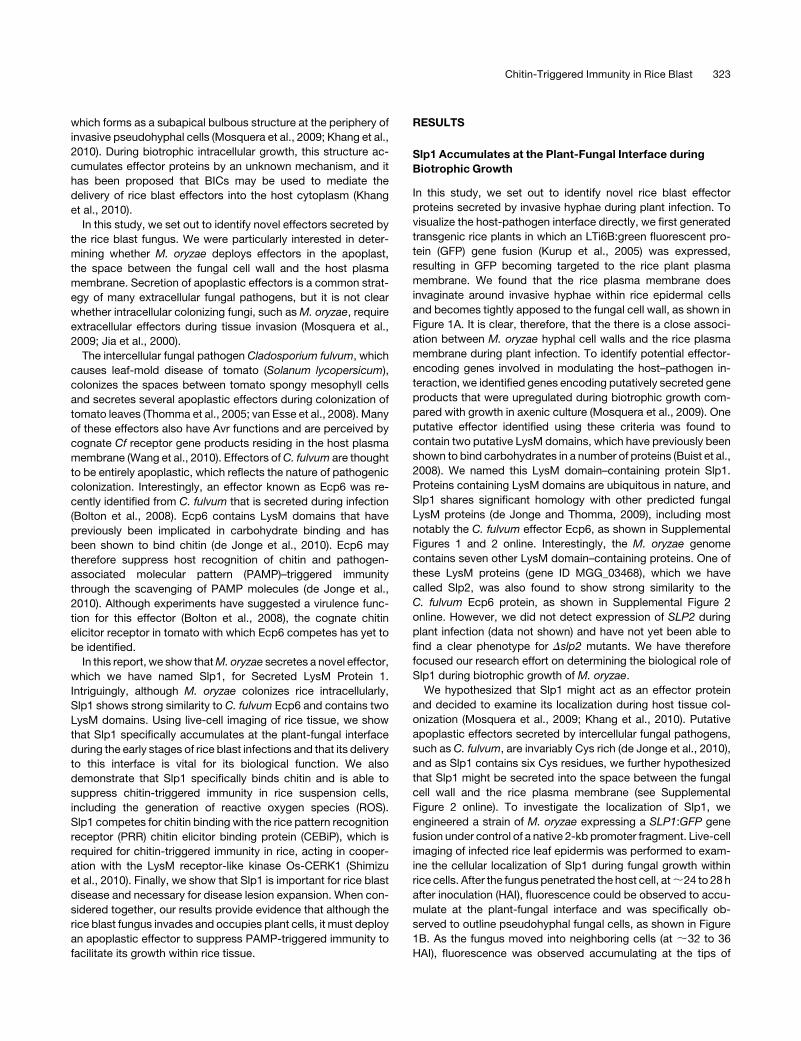
Slp1 Accumulates at the Plant-Fungal Interface during
Biotrophic Growth
In this study, we set out to identify novel rice blast effector
proteins secreted by invasive hyphae during plant infection. To
visualize the host-pathogen interface directly, we first generated
transgenic rice plants in which an LTi6B:green fluorescent pro-
tein (GFP) gene fusion (Kurup et al., 2005) was expressed,
resulting in GFP becoming targeted to the rice plant plasma
membrane. We found that the rice plasma membrane does
invaginate around invasive hyphae within rice epidermal cells
and becomes tightly apposed to the fungal cell wall, as shown in
Figure 1A. It is clear, therefore, that the there is a close associ-
ation between M. oryzae hyphal cell walls and the rice plasma
membrane during plant infection. To identify potential effector-
encoding genes involved in modulating the host–pathogen in-
teraction, we identified genes encoding putatively secreted gene
products that were upregulated during biotrophic growth com-
pared with growth in axenic culture (Mosquera et al., 2009). One
putative effector identified using these criteria was found to
contain two putative LysM domains, which have previously been
shown to bind carbohydrates in a number of proteins (Buist et al.,
2008). We named this LysM domain–containing protein Slp1.
Proteins containing LysM domains are ubiquitous in nature, and
Slp1 shares significant homology with other predicted fungal
LysM proteins (de Jonge and Thomma, 2009), including most
notably the C. fulvum effector Ecp6, as shown in Supplemental
Figures 1 and 2 online. Interestingly, the M. oryzae genome
contains seven other LysM domain–containing proteins. One of
these LysM proteins (gene ID MGG_03468), which we have
called Slp2, was also found to show strong similarity to the
C. fulvum Ecp6 protein, as shown in Supplemental Figure 2
online. However, we did not detect expression of SLP2 during
plant infection (data not shown) and have not yet been able to
find a clear phenotype for Dslp2 mutants. We have therefore
focused our research effort on determining the biological role of
Slp1 during biotrophic growth of M. oryzae.
We hypothesized that Slp1 might act as an effector protein
and decided to examine its localization during host tissue col-
onization (Mosquera et al., 2009; Khang et al., 2010). Putative
apoplastic effectors secreted by intercellular fungal pathogens,
such as C. fulvum, are invariably Cys rich (de Jonge et al., 2010),
and as Slp1 contains six Cys residues, we further hypothesized
that Slp1 might be secreted into the space between the fungal
cell wall and the rice plasma membrane (see Supplemental
Figure 2 online). To investigate the localization of Slp1, we
engineered a strain of M. oryzae expressing a SLP1:GFP gene
fusion under control of a native 2-kb promoter fragment. Live-cell
imaging of infected rice leaf epidermis was performed to exam-
ine the cellular localization of Slp1 during fungal growth within
rice cells. After the fungus penetrated the host cell, at;24 to 28 h
after inoculation (HAI), fluorescence could be observed to accu-
mulate at the plant-fungal interface and was specifically ob-
served to outline pseudohyphal fungal cells, as shown in Figure
1B. As the fungus moved into neighboring cells (at ;32 to 36
HAI), fluorescence was observed accumulating at the tips of
Chitin-Triggered Immunity in Rice Blast 323
invasive hyphae that were invading new host cells. At this time,
fluorescence ceased to accumulate at the host-pathogen inter-
face within the initially infected host cell (Figure 1C). At no stage
was fluorescence observed within host cells nor could fluores-
cence be observed in other fungal structures, including conidia,
germ tubes, or appressoria, as shown in Supplemental Figure 3
online. From these observations, we conclude that Slp1 is
specifically expressed when the fungus is growing intracellularly
within its host, a feature associated with putative rice blast
effector proteins (Mosquera et al., 2009; Khang et al., 2010).
Next, we wanted to examine whether the localization pattern of
Slp1 differs from that of previously described rice blast effectors,
which accumulate at BIC structures (Mosquera et al., 2009;
Khang et al., 2010).We therefore engineered a strain ofM. oryzae
that simultaneously expressed a SLP1:GFP gene fusion and a
Pathogenicity on Weeping Lovegrass2 (PWL2):monomeric red
fluorescent protein (mRFP) gene fusion. Pwl2 is a previously
characterized BIC-localized effector, known to be delivered into
the cytoplasm of rice cells during plant infection by M. oryzae
(Khang et al., 2010). We undertook live-cell imaging of infected
rice epidermis, as shown in Figure 1D and Supplemental Movie
1 online. Interestingly, at >80% of infection sites observed
between 24 and 32 HAI, colocalization between SLP1:GFP and
PWL2:mRFP could not be observed (n > 100). Taken together,
we conclude that Slp1 accumulates between the invaginated
host plasma membrane and the fungal cell wall during initial
invasion of rice cells and is therefore distinct from previously
identified BIC-localized effectors.
Having established that Slp1 was not a BIC-localized effector
protein, we were interested in trying to colocalize Slp1 with other
rice blast effectors that appear to accumulate in the apoplast.
One potential effector, presumed to be apoplastic in localization,
is Bas4 (Mosquera et al., 2009; Khang et al., 2010). We therefore
engineered an M. oryzae strain that simultaneously expresses
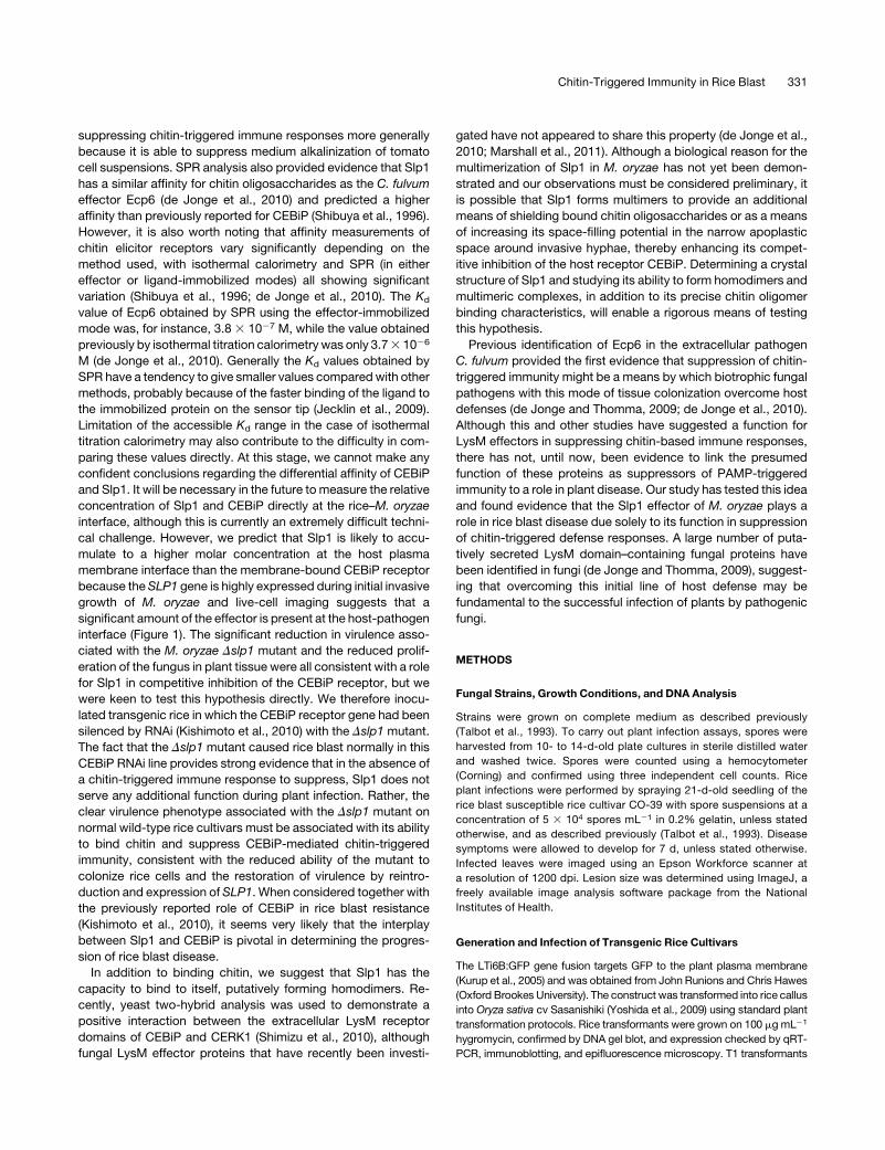
Figure 1. Slp1 Accumulates at the Plant-Fungal Interface during
Biotrophic Growth.
(A) Laser confocal micrograph of M. oryzae invasive hyphae-colonizing
epidermal leaf cells of a transgenic line of rice expressing LTi6B:GFP.
The rice cell plasma membrane becomes invaginated around the grow-
ing fungal hyphae.
(B) Cellular localization of Slp1:GFP in M. oryzae during biotrophic
growth on epidermal rice cells at 24 HAI. Fluorescence was initially
observed accumulating at the tips of invasive hyphae at the plant-fungal
interface and was later found to surround invasive hyphae.
(C) At 36 HAI, Slp1:GFP fluorescence could be observed accumulating at
the tips of filamentous hyphae-invading adjacent cells. At this time,
fluorescence was no longer observed in initially infected host cells.
(D) Lack of colocalization between SLP1:GFP and PWL2:mRFP.
A Guy11 M. oryzae transformant expressing both the SLP1:GFP and
PWL2:mRFP constructs was used to visualize the cellular localization
of Slp1 and the BIC-localized effector Pwl2 in planta. At 24 HAI, the
Slp1-Gfp signal surrounded invasive hyphae, whereas Pwl2 accumulates
at the BIC (white arrow).
(E) Partial colocalization of M. oryzae Slp1:GFP and Bas4:mRFP fusion
proteins in the apoplastic space surrounding fungal invasive hyphae.
White arrow indicates site of colocalization.
(F) Cellular localization of Slp27-162:GFP at 24 HAI on leaf sheath tissue.
Slp27-162:GFP aggregates can be seen localizing within the cytoplasm of
fungal invasive hyphae. White asterisk indicates the site of appressorium
formation at the leaf surface.
Bars = 10 mm.
324 The Plant Cell
SLP1:GFP and BAS4:mRFP. At 24 HAI, there did appear to be
some colocalization between Slp1 and Bas4, as shown in Figure
1E. Although the two proteins appeared to colocalize, there
were, however, significant areas where Slp1:GFP accumulated,
but Bas4:mRFP did not. In view of our observation that Slp1
accumulates at the fungal-plant interface, we next investigated
how the protein is delivered to the apoplast during biotrophic
growth. SLP1 encodes a small secreted protein of 162 amino
acids, with a predicted N-terminal signal peptide of 27 amino
acids in length (based on SignalP 3.0 analysis). To test the
significance of this secretion sequence, we engineered an
M. oryzae strain in which the coding region of the first 27 amino
acids of SLP1 was removed. A new start codon was introduced
and the resulting coding region fused to GFP. Expression of the
SLP127-162:GFP construct was driven by the native 2.0-kb SLP1
promoter fragment. Removal of the signal peptide prevented
Slp1:GFP from reaching the tips of invasively growing hyphae,
and Slp1 was no longer observed accumulating in the apoplastic
space (Figure 1F). The resultant intracellular Slp127-162:GFP
instead appeared to accumulate as aggregates in the fungal
cytoplasm. Cellular mislocalization of SLP127-162:GFP is consis-
tent with the hypothesis that Slp1 is an apoplastic effector, the
secretion of which is dependent on a peptide sequence within
the initial 27 amino acids.
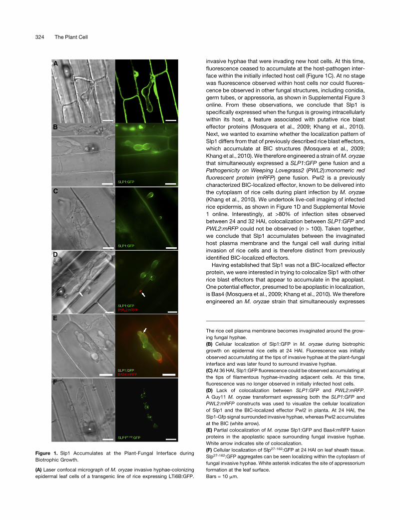
Slp1 Is a Virulence Determinant inM. oryzae
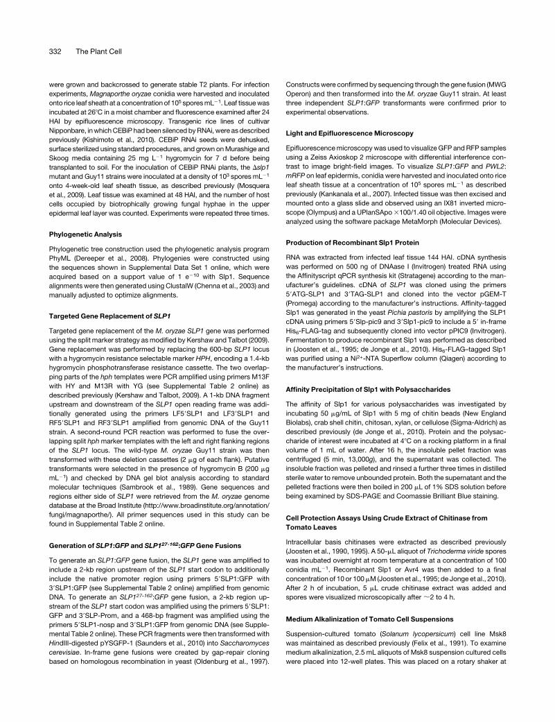
To test the contribution of Slp1 to rice blast disease, a targeted
gene deletion of SLP1 was performed in M. oryzae (see Supple-
mental Figure 4 online). Fungal spores of the resulting Dslp1
mutant and the isogenic wild-type Guy11 strain were harvested,
adjusted to uniform concentrations, and applied to 21-d-old
seedlings of the blast-susceptible rice cultivar CO-39 (Figure 2).
Deletion of SLP1 significantly reduced the ability of M. oryzae to
cause disease, and the symptoms of plants inoculated with
Dslp1 spores were highly reduced when compared with plants
infected with the wild-type Guy11 strain (Figure 2A). To quantify
the reduction in virulence, both lesion density and lesion size
were analyzed using image analysis software (ImageJ). The
mean lesion size generated by the Dslp1mutant was found to be
significantly smaller than that of the Guy11 wild type (t test, P <
0.01) (Figure 2B). The mean lesion size for Guy11 was calculated
to be 1.15 mm2 (6SE 0.049, n > 100 lesions), while the mean
lesion size of the Dslp1 mutant was calculated to be 0.31 mm2
(6SE 0.025, n > 100). Additionally, the mean lesion density per
unit area of the Dslp1mutant (11.16 5.7, n = 49) was found to be
significantly lower than that of the wild type (40.76 10.8, n = 28;
t test, P < 0.01) (Figure 2C). Complementation analysis using the
SLP1:GFP construct was performed, and reintroduction of the
SLP1 gene was found to restore virulence to M. oryzae (see
Supplemental Figure 5 online). Deletion of the Slp1 signal peptide
prevented complementation of the Dslp1mutant phenotype (see
Supplemental Figure 6 online).
We also evaluated whether Dslp1 mutants were impaired in
their ability to form functional infection structures or whether the
virulence phenotype was simply a consequence of a reduction in
fitness. We harvested spores of the Dslp1 mutant and wild-type
strains and compared their ability to form appressoria on an
inductive glass surface (Figure 2D). After 24 h, Dslp1 mutants
were capable of forming mature appressoria in a manner iden-
tical to that of the wild-type M. oryzae strain. Vegetative growth
rates and behavior in axenic culture were also identical to Guy11.
From these observations, we conclude that the virulence phe-
notype of theDslp1mutant is associated with a reduced ability of
the Dslp1 mutant to proliferate within host tissues, rather than a
reduced capacity to make successful penetration structures. To
test this idea, we examined and compared host tissues infected
with a Dslp1 mutant compared with the isogenic Guy11. We
initially counted the number of cells occupied by the fungus at 48
HAI and found that the number of host cells occupied by a Dslp1
mutant was significantly lower than the wild-type Guy11 strain
(n = 15, two-tailed t test, P = 0.014) (Figure 2E). At 48 HAI, the
mean number of host cells occupied by Dslp1 was found to be
4.29 cells (SD6 2.5), while themean number of cells occupied by
Guy11 was 7.33 (SD6 3.6). At 48 HAI, the Dslp1mutant had only
just started to colonize neighboring cells, while Guy11 had
become well established at 48 HAI, with bulbous hyphae fully
ramified in host tissues (Figure 2F). We conclude that Slp1 is
necessary for efficient rice tissue invasion by M. oryzae to bring
about rice blast disease.
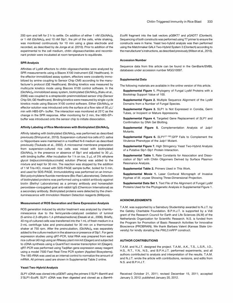
M. oryzae Slp1 Is a Chitin Binding Protein
To define the biological function of Slp1, we cloned and overex-
pressed a SLP1 cDNA in Pichia pastoris (de Jonge et al., 2010).
Recombinant Slp1 protein was isolated and purified. As Slp1
contains two putative LysM domains, we were initially interested
to see whether the protein was capable of binding to specific
polysaccharides. After incubating purified Slp1 protein with
insoluble cell wall polysaccharides, we observed that Slp1
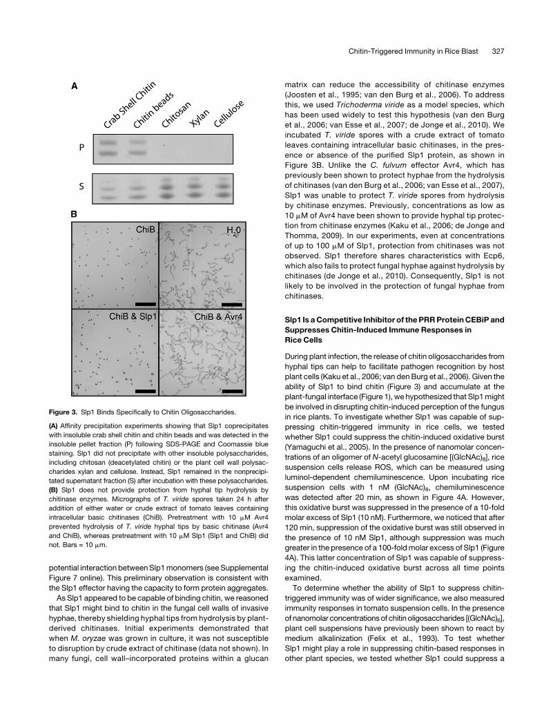
specifically coprecipitated with insoluble crab shell chitin and
chitin beads and was detected in the insoluble pellet fraction
following affinity precipitation (Figure 3). Slp1 did not, however,
precipitate with any other tested cell wall polysaccharides,
including chitosan (deacetylated chitin) and the plant cell wall
polysaccharides cellulose and xylan, as evidenced by Slp1
remaining in the supernatant fraction after affinity precipitation
(Figure 3A). Interestingly, not only did Slp1 appear to bind
specifically to chitin and not to other polysaccharides, several
bands were evident in both the pellet and supernatant fractions,
suggesting that Slp1 is likely to be glycosylated or potentially
forms oligomers. To ensure that these higher molecular weight
protein bands were not contaminants from the protein over-
expression system, mass spectrometry was performed on the
gel fragments after in-gel trypsin digestion. Slp1 was detected in
all of these experiments, confirming that the higher molecular
weight protein bands were not due to expression artifacts and
suggesting that Slp1 is likely to show abnormal electrophoretic
mobility due to being a glycoprotein as reported for other LysM
proteins (Kaku et al., 2006) or potentially to form multimers. We
were interested in determining whether Slp1 had the capacity to
form multimers based on a protein–protein interaction. We
therefore performed high-stringency yeast two-hybrid analysis
in which an SLP1 cDNA was simultaneously cloned into bait and
prey vectors of the Matchmaker GAL4 two-hybrid system
(Clontech). Using this system, we were able to detect a strong
Chitin-Triggered Immunity in Rice Blast 325
Figure 2. SLP1 Is a Virulence Determinant in M. oryzae Required for Rice Tissue Invasion.
(A) Conidial suspensions of equal concentration (53 10�4 spores mL�1) fromM. oryzaeGuy11 (wild type [WT]) or Dslp1mutants were used to inoculate
21-d-old seedlings of the blast susceptible rice cultivar CO-39. Disease symptoms were reduced on plants inoculated with Dslp1 mutants.
(B) Bar chart of mean lesion size of plants inoculated with Guy11 and the Dslp1mutant. Mean lesion size was significantly reduced in plants inoculated
with the Dslp1 mutant compared with the isogenic wild type (t test, P < 0.01). Error bars denote 6 1 SE.
(C) Bar chart of mean lesion density of seedlings infected with Guy11 strain and the Dslp1 mutant per unit area. Mean lesion density was significantly
reduced in Dslp1 mutant infections (t test, P < 0.01). Error bars denote 1 SD. Double asterisks in (B) and (C) denote P < 0.01 from two-tailed t test.
(D) Null Dslp1 mutants produce normal conidia and form appressoria in a time-dependent manner comparable to that of the wild-type Guy11 strain.
Conidia of both Guy11 and Dslp1 mutant strains were harvested and set to a concentration of 5 3 10�4 spores mL�1. Spores were inoculated onto
hydrophobic glass cover slips and incubated in a moist chamber at 268C and examined by light microscopy. The morphology of conidia and
appressoria was not altered in Dslp1 mutants. Bars = 10 mm.
(E) Bar chart showing the number of rice host cells occupied after 48 HAI with the Dslp1 mutant compared with Guy11. After 48 h, the number of host
cells occupied by the fungus was recorded (n = 15 infection sites). At this time point, the number of host cells occupied by the Dslp1mutant was found to
be significantly lower than that of the wild-type Guy11 strain (two-tailed t test, P = 0.014). Asterisk denotes P < 0.05.
(F) Typical infection sites of rice leaf sheath inoculated with Dslp1 and Guy11, showing greater fungal proliferation and tissue invasion by the wild-type
strain. Images were recorded 48 HAI. Asterisk marks the first infected host cell. Bar = 30 mm.
[See online article for color version of this figure.]
326 The Plant Cell
potential interaction betweenSlp1monomers (seeSupplemental
Figure 7 online). This preliminary observation is consistent with
the Slp1 effector having the capacity to form protein aggregates.
As Slp1 appeared to be capable of binding chitin, we reasoned
that Slp1 might bind to chitin in the fungal cell walls of invasive
hyphae, thereby shielding hyphal tips from hydrolysis by plant-
derived chitinases. Initial experiments demonstrated that
when M. oryzae was grown in culture, it was not susceptible
to disruption by crude extract of chitinase (data not shown). In
many fungi, cell wall–incorporated proteins within a glucan
matrix can reduce the accessibility of chitinase enzymes
(Joosten et al., 1995; van den Burg et al., 2006). To address
this, we used Trichoderma viride as a model species, which
has been used widely to test this hypothesis (van den Burg
et al., 2006; van Esse et al., 2007; de Jonge et al., 2010). We
incubated T. viride spores with a crude extract of tomato
leaves containing intracellular basic chitinases, in the pres-
ence or absence of the purified Slp1 protein, as shown in
Figure 3B. Unlike the C. fulvum effector Avr4, which has
previously been shown to protect hyphae from the hydrolysis
of chitinases (van den Burg et al., 2006; van Esse et al., 2007),
Slp1 was unable to protect T. viride spores from hydrolysis
by chitinase enzymes. Previously, concentrations as low as
10 mM of Avr4 have been shown to provide hyphal tip protec-
tion from chitinase enzymes (Kaku et al., 2006; de Jonge and
Thomma, 2009). In our experiments, even at concentrations
of up to 100 mM of Slp1, protection from chitinases was not
observed. Slp1 therefore shares characteristics with Ecp6,
which also fails to protect fungal hyphae against hydrolysis by
chitinases (de Jonge et al., 2010). Consequently, Slp1 is not
likely to be involved in the protection of fungal hyphae from
chitinases.
Slp1 Is aCompetitive Inhibitor of thePRRProteinCEBiP and
Suppresses Chitin-Induced Immune Responses in
Rice Cells
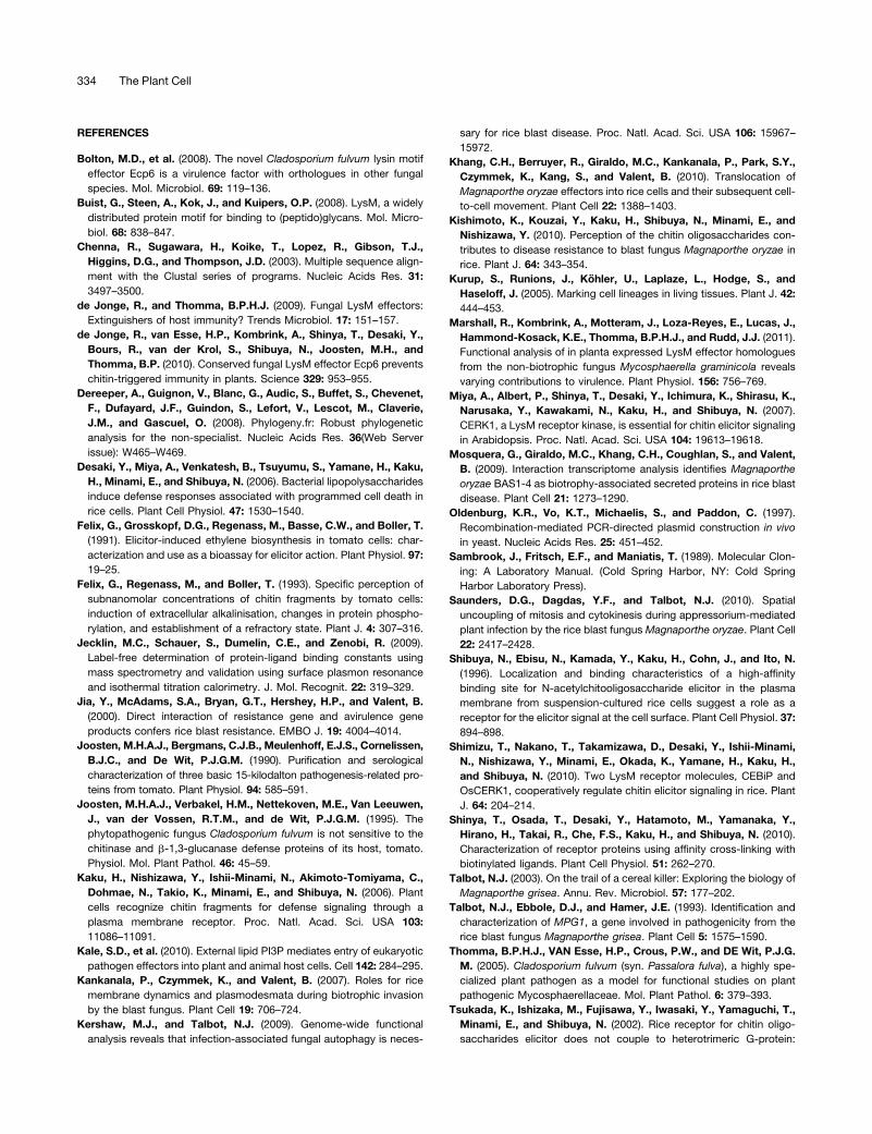
During plant infection, the release of chitin oligosaccharides from
hyphal tips can help to facilitate pathogen recognition by host
plant cells (Kaku et al., 2006; van den Burg et al., 2006). Given the
ability of Slp1 to bind chitin (Figure 3) and accumulate at the
plant-fungal interface (Figure 1), we hypothesized that Slp1might
be involved in disrupting chitin-induced perception of the fungus
in rice plants. To investigate whether Slp1 was capable of sup-
pressing chitin-triggered immunity in rice cells, we tested
whether Slp1 could suppress the chitin-induced oxidative burst
(Yamaguchi et al., 2005). In the presence of nanomolar concen-
trations of an oligomer of N-acetyl glucosamine [(GlcNAc)8], rice
suspension cells release ROS, which can be measured using
luminol-dependent chemiluminescence. Upon incubating rice
suspension cells with 1 nM (GlcNAc)8, chemiluminescence
was detected after 20 min, as shown in Figure 4A. However,
this oxidative burst was suppressed in the presence of a 10-fold
molar excess of Slp1 (10 nM). Furthermore, we noticed that after
120 min, suppression of the oxidative burst was still observed in
the presence of 10 nM Slp1, although suppression was much
greater in the presence of a 100-foldmolar excess of Slp1 (Figure
4A). This latter concentration of Slp1 was capable of suppress-
ing the chitin-induced oxidative burst across all time points
examined.
To determine whether the ability of Slp1 to suppress chitin-
triggered immunity was of wider significance, we also measured
immunity responses in tomato suspension cells. In the presence
of nanomolar concentrationsof chitin oligosaccharides [(GlcNAc)6],
plant cell suspensions have previously been shown to react by
medium alkalinization (Felix et al., 1993). To test whether
Slp1 might play a role in suppressing chitin-based responses in
other plant species, we tested whether Slp1 could suppress a
Figure 3. Slp1 Binds Specifically to Chitin Oligosaccharides.
(A) Affinity precipitation experiments showing that Slp1 coprecipitates
with insoluble crab shell chitin and chitin beads and was detected in the
insoluble pellet fraction (P) following SDS-PAGE and Coomassie blue
staining. Slp1 did not precipitate with other insoluble polysaccharides,
including chitosan (deacetylated chitin) or the plant cell wall polysac-
charides xylan and cellulose. Instead, Slp1 remained in the nonprecipi-
tated supernatant fraction (S) after incubation with these polysaccharides.
(B) Slp1 does not provide protection from hyphal tip hydrolysis by
chitinase enzymes. Micrographs of T. viride spores taken 24 h after
addition of either water or crude extract of tomato leaves containing
intracellular basic chitinases (ChiB). Pretreatment with 10 mM Avr4
prevented hydrolysis of T. viride hyphal tips by basic chitinase (Avr4
and ChiB), whereas pretreatment with 10 mM Slp1 (Slp1 and ChiB) did
not. Bars = 10 mm.
Chitin-Triggered Immunity in Rice Blast 327
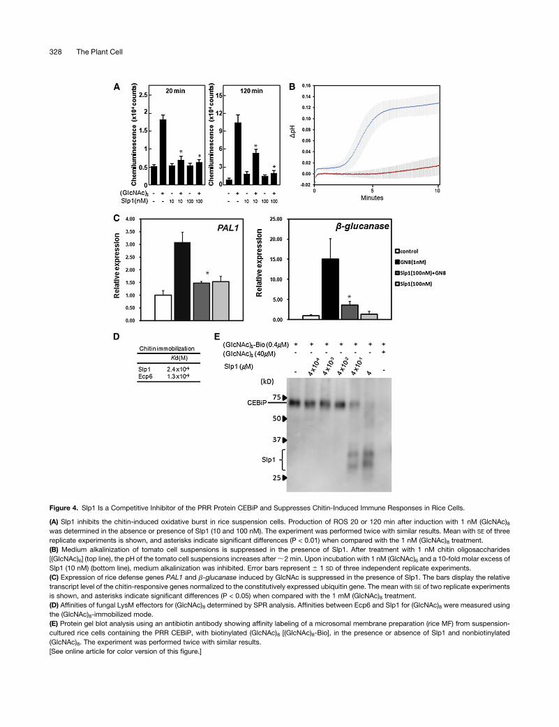
Figure 4. Slp1 Is a Competitive Inhibitor of the PRR Protein CEBiP and Suppresses Chitin-Induced Immune Responses in Rice Cells.
(A) Slp1 inhibits the chitin-induced oxidative burst in rice suspension cells. Production of ROS 20 or 120 min after induction with 1 nM (GlcNAc)8was determined in the absence or presence of Slp1 (10 and 100 nM). The experiment was performed twice with similar results. Mean with SE of three
replicate experiments is shown, and asterisks indicate significant differences (P < 0.01) when compared with the 1 nM (GlcNAc)8 treatment.
(B) Medium alkalinization of tomato cell suspensions is suppressed in the presence of Slp1. After treatment with 1 nM chitin oligosaccharides
[(GlcNAc)6] (top line), the pH of the tomato cell suspensions increases after;2 min. Upon incubation with 1 nM (GlcNAc)6 and a 10-fold molar excess of
Slp1 (10 nM) (bottom line), medium alkalinization was inhibited. Error bars represent 6 1 SD of three independent replicate experiments.
(C) Expression of rice defense genes PAL1 and b-glucanase induced by GlcNAc is suppressed in the presence of Slp1. The bars display the relative
transcript level of the chitin-responsive genes normalized to the constitutively expressed ubiquitin gene. The mean with SE of two replicate experiments
is shown, and asterisks indicate significant differences (P < 0.05) when compared with the 1 mM (GlcNAc)8 treatment.
(D) Affinities of fungal LysM effectors for (GlcNAc)8 determined by SPR analysis. Affinities between Ecp6 and Slp1 for (GlcNAc)8 were measured using
the (GlcNAc)8-immobilized mode.
(E) Protein gel blot analysis using an antibiotin antibody showing affinity labeling of a microsomal membrane preparation (rice MF) from suspension-
cultured rice cells containing the PRR CEBiP, with biotinylated (GlcNAc)8 [(GlcNAc)8-Bio], in the presence or absence of Slp1 and nonbiotinylated
(GlcNAc)8. The experiment was performed twice with similar results.
[See online article for color version of this figure.]
328 The Plant Cell
chitin-induced pH shift in tomato cell suspensions. We observed
that in the presence of a 10-fold molar excess of Slp1 (10 nM),
medium alkalinization of tomato suspensions cells was inhibited
(Figure 4B). We therefore conclude that Slp1 is capable of
suppressing chitin-induced immune responses in plant cells.
Chitin-triggered immunity is known to result in induction of
pathogenesis-related genes, and we therefore sought to deter-
mine the effect of the Slp1 effector on induction of rice defense
gene expression. We therefore performed quantitative RT-PCR
(qRT-PCR) and examined changes in expression of the rice Phe
ammonia lyase gene, PAL1, and the b-glucanase–encoding
gene, rBG. In the presence of 1 nM (GlcNAc)8, expression of
both PAL1 and b-glucanase increased significantly (Figure 4C).
However, the increase in gene expression was suppressedwhen
a 100-fold molar excess of Slp1 was also included, consistent
with the role of Slp1 in preventing chitin-triggered immunity
responses in rice.
In rice, the pattern recognition receptor LysM protein CEBiP
resides at the rice plasma membrane and is able to bind to chitin
oligosaccharides (Shibuya et al., 1996; Kaku et al., 2006). We
hypothesized that Slp1 might therefore function to compete with
the CEBiP recognition receptor residing at the invaginated rice
cell membrane. CEBiP is a LysM domain–containing protein and
interacts with the LysM receptor-like kinase protein CERK1 to
bring about plant defense responses (Shimizu. et al., 2010).
CeBiP has been shown to contribute to rice blast disease
resistance (Kishimoto et al., 2010). We therefore performed a
competition assay inwhich amicrosomalmembrane preparation
containing the receptor protein CEBiP was isolated from rice
suspension cells. When this membrane fraction was incubated
with 0.4 mM biotinylated N-acetylchito-octaose (GlcNAc)8, la-
beling ofCEBiP occurred (Figure 4E).When an equimolar amount
of Slp1 (0.4 mM) was added, a significant portion of biotinylated
(GlcNAc)8 bound to the effector, suggesting that Slp1 is capable
of competing with CEBiP for chitin binding in this assay. When a
10-fold molar excess of Slp1 (4 mM) was added, binding of
biotinylated (GlcNAc)8 to the membrane fraction containing
CEBiP was almost entirely blocked and resulted in the almost
exclusive labeling of Slp1 (Figure 4E).
We also determined the affinity kinetics of Slp1 for chitin
oligosaccharides using surface plasmon resonance (SPR) tech-
nology. Using SPR, we tested for binding of Slp1 and Ecp6 to
the ligand (chitin oligosaccharides [(GlcNAc)8]). Using the ligand-
immobilized method, in which chitin oligosaccharides are
immobilized to the sensor chip, we were able to calculate
dissociation constants (Kd values) for both Slp1 and Ecp6 (Figure
4D). We estimated that the affinity for chitin oligosaccharides
was similar for both Slp1 and Ecp6, with Kd values of 2.43 1029
M and 1.3 3 1029 M, respectively. Previously, the Kd value of
CEBiP for chitin oligosaccharides was calculated as 2.9 3 1028
M (Shibuya et al., 1996). Full rate constant values, including
the Kd and Kon values, for the association of Slp1 for chitin
oligosaccharides can be found in Supplemental Table 1 online.
These results suggest that Slp1 and Ecp6 both show a high
affinity for chitin oligosaccharides, which is consistent with
the ability of Slp1 to act as competitive inhibitor of CEBiP.
When all of these results are considered together, we conclude
that the M. oryzae Slp1 protein competes directly with chitin
receptor proteins in rice and is able to suppress chitin-induced
immunity.
Targeted Gene Silencing of CEBiP in Rice Restores the
Ability of Dslp1Mutants ofM. oryzae to Cause Rice
Blast Disease
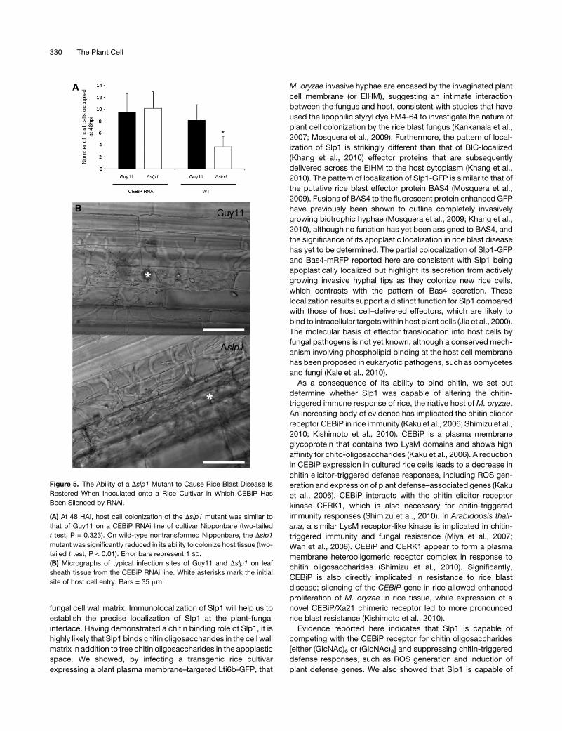
Wewere interested in establishing whether the ability of the Slp1
effector to act as a competitive inhibitor of CEBiP, thereby
suppressing PAMP-triggered immunity, was the reason why
M. oryzae Dslp1 mutants showed a significant reduction in their
ability to cause rice blast disease. We therefore obtained trans-
genic rice lines of cultivar Nipponbare, in which the CEBiP-
encoding gene had been silenced using RNA interference (RNAi;
Kishimoto et al., 2010). These rice lines have previously been
shown to lack chitin-triggered immune responses and to exhibit
increased susceptibility to rice blast disease (Kishimoto et al.,
2010). We inoculated the CEBiP RNAi plants, and corresponding
wild-type Nipponbare rice lines, with theM. oryzae Dslp1mutant
and Guy11 strain. Strikingly, we observed that the Dslp1 mutant
was as virulent as Guy11 when inoculated onto CEBiP RNAi
plants (Figure 5). On CEBiP RNAi plants, the mean number of
host cells occupied by the fungus at 48 HAI by the Guy11 and
Dslp1 strain was 9.4 (SD 6 3.21) and 10.2 (SD 6 2.83), respec-
tively. Furthermore, on CEBiP RNAi plants, no significant differ-
ence in host tissue colonization was observed between Guy11
and the Dslp1mutant (two-tailed t test, n = 34 infection sites, P =
0.322). By contrast, when nonsilenced Nipponbare rice lines
were inoculated, the mean number of host cells occupied
by Guy11 and the Dslp1 mutant was 8.1 (SD 6 2.63) and 3.7
(SD 6 1.78), respectively. The mean number of host cells colo-
nized by the Dslp1 mutant was significantly lower than the wild-
type Guy11 (two-tailed t test, P < 0.01) (Figure 5). We also found
that spray inoculation of CEBiP-RNAi seedlings with the Dslp1
mutant led to restoration of the number of disease lesions (data
not shown). We conclude that it is the ability of Slp1 to act as a
competitive inhibitor of CEBiP that is its principal function during
rice blast disease and that this role is highly significant in
determining the outcome of the host–pathogen interaction.
DISCUSSION
In this study, we set out to investigate the mechanisms used by
the rice blast fungus to colonize living rice tissue. We focused on
whether effector proteins secreted by M. oryzae during biotro-
phic growth could be involved in perturbing the way that rice
plants initially detect the invading fungus by means of PAMP
molecules, such as chitin oligosaccharides. Our results provide
evidence that M. oryzae deploys an effector, Slp1, to suppress
chitin-induced host defense responses in rice tissue and that this
is significant in the development of rice blast disease. By contrast
with previously described rice blast effectors (Jia et al., 2000;
Khang et al., 2010), Slp1 accumulates in the apoplastic space at
the plant-fungal interface and, in particular, is associated with
colonization of new rice cells by the fungus during invasive
growth. At this stage, however, we cannot exclude that Slp1 is
secreted and subsequently becomes incorporated into the
Chitin-Triggered Immunity in Rice Blast 329
fungal cell wall matrix. Immunolocalization of Slp1 will help us to
establish the precise localization of Slp1 at the plant-fungal
interface. Having demonstrated a chitin binding role of Slp1, it is
highly likely that Slp1 binds chitin oligosaccharides in the cell wall
matrix in addition to free chitin oligosaccharides in the apoplastic
space. We showed, by infecting a transgenic rice cultivar
expressing a plant plasma membrane–targeted Lti6b-GFP, that
M. oryzae invasive hyphae are encased by the invaginated plant
cell membrane (or EIHM), suggesting an intimate interaction
between the fungus and host, consistent with studies that have
used the lipophilic styryl dye FM4-64 to investigate the nature of
plant cell colonization by the rice blast fungus (Kankanala et al.,
2007; Mosquera et al., 2009). Furthermore, the pattern of local-
ization of Slp1 is strikingly different than that of BIC-localized
(Khang et al., 2010) effector proteins that are subsequently
delivered across the EIHM to the host cytoplasm (Khang et al.,
2010). The pattern of localization of Slp1-GFP is similar to that of
the putative rice blast effector protein BAS4 (Mosquera et al.,
2009). Fusions of BAS4 to the fluorescent protein enhanced GFP
have previously been shown to outline completely invasively
growing biotrophic hyphae (Mosquera et al., 2009; Khang et al.,
2010), although no function has yet been assigned to BAS4, and
the significance of its apoplastic localization in rice blast disease
has yet to be determined. The partial colocalization of Slp1-GFP
and Bas4-mRFP reported here are consistent with Slp1 being
apoplastically localized but highlight its secretion from actively
growing invasive hyphal tips as they colonize new rice cells,
which contrasts with the pattern of Bas4 secretion. These
localization results support a distinct function for Slp1 compared
with those of host cell–delivered effectors, which are likely to
bind to intracellular targetswithin host plant cells (Jia et al., 2000).
The molecular basis of effector translocation into host cells by
fungal pathogens is not yet known, although a conserved mech-
anism involving phospholipid binding at the host cell membrane
has been proposed in eukaryotic pathogens, such as oomycetes
and fungi (Kale et al., 2010).
As a consequence of its ability to bind chitin, we set out
determine whether Slp1 was capable of altering the chitin-
triggered immune response of rice, the native host of M. oryzae.
An increasing body of evidence has implicated the chitin elicitor
receptor CEBiP in rice immunity (Kaku et al., 2006; Shimizu et al.,
2010; Kishimoto et al., 2010). CEBiP is a plasma membrane
glycoprotein that contains two LysM domains and shows high
affinity for chito-oligosaccharides (Kaku et al., 2006). A reduction
in CEBiP expression in cultured rice cells leads to a decrease in
chitin elicitor-triggered defense responses, including ROS gen-
eration and expression of plant defense–associated genes (Kaku
et al., 2006). CEBiP interacts with the chitin elicitor receptor
kinase CERK1, which is also necessary for chitin-triggered
immunity responses (Shimizu et al., 2010). In Arabidopsis thali-
ana, a similar LysM receptor-like kinase is implicated in chitin-
triggered immunity and fungal resistance (Miya et al., 2007;
Wan et al., 2008). CEBiP and CERK1 appear to form a plasma
membrane heterooligomeric receptor complex in response to
chitin oligosaccharides (Shimizu et al., 2010). Significantly,
CEBiP is also directly implicated in resistance to rice blast
disease; silencing of the CEBiP gene in rice allowed enhanced
proliferation of M. oryzae in rice tissue, while expression of a
novel CEBiP/Xa21 chimeric receptor led to more pronounced
rice blast resistance (Kishimoto et al., 2010).
Evidence reported here indicates that Slp1 is capable of
competing with the CEBiP receptor for chitin oligosaccharides
[either (GlcNAc)6 or (GlcNAc)8] and suppressing chitin-triggered
defense responses, such as ROS generation and induction of
plant defense genes. We also showed that Slp1 is capable of
Figure 5. The Ability of a Dslp1 Mutant to Cause Rice Blast Disease Is
Restored When Inoculated onto a Rice Cultivar in Which CEBiP Has
Been Silenced by RNAi.
(A) At 48 HAI, host cell colonization of the Dslp1 mutant was similar to
that of Guy11 on a CEBiP RNAi line of cultivar Nipponbare (two-tailed
t test, P = 0.323). On wild-type nontransformed Nipponbare, the Dslp1
mutant was significantly reduced in its ability to colonize host tissue (two-
tailed t test, P < 0.01). Error bars represent 1 SD.
(B) Micrographs of typical infection sites of Guy11 and Dslp1 on leaf
sheath tissue from the CEBiP RNAi line. White asterisks mark the initial
site of host cell entry. Bars = 35 mm.
330 The Plant Cell
suppressing chitin-triggered immune responses more generally
because it is able to suppress medium alkalinization of tomato
cell suspensions. SPR analysis also provided evidence that Slp1
has a similar affinity for chitin oligosaccharides as the C. fulvum
effector Ecp6 (de Jonge et al., 2010) and predicted a higher
affinity than previously reported for CEBiP (Shibuya et al., 1996).
However, it is also worth noting that affinity measurements of
chitin elicitor receptors vary significantly depending on the
method used, with isothermal calorimetry and SPR (in either
effector or ligand-immobilized modes) all showing significant
variation (Shibuya et al., 1996; de Jonge et al., 2010). The Kd
value of Ecp6 obtained by SPR using the effector-immobilized
mode was, for instance, 3.8 3 1027 M, while the value obtained
previously by isothermal titration calorimetry was only 3.73 1026
M (de Jonge et al., 2010). Generally the Kd values obtained by
SPR have a tendency to give smaller values compared with other
methods, probably because of the faster binding of the ligand to
the immobilized protein on the sensor tip (Jecklin et al., 2009).
Limitation of the accessible Kd range in the case of isothermal
titration calorimetry may also contribute to the difficulty in com-
paring these values directly. At this stage, we cannot make any
confident conclusions regarding the differential affinity of CEBiP
and Slp1. It will be necessary in the future to measure the relative
concentration of Slp1 and CEBiP directly at the rice–M. oryzae
interface, although this is currently an extremely difficult techni-
cal challenge. However, we predict that Slp1 is likely to accu-
mulate to a higher molar concentration at the host plasma
membrane interface than the membrane-bound CEBiP receptor
because theSLP1 gene is highly expressed during initial invasive
growth of M. oryzae and live-cell imaging suggests that a
significant amount of the effector is present at the host-pathogen
interface (Figure 1). The significant reduction in virulence asso-
ciated with the M. oryzae Dslp1 mutant and the reduced prolif-
eration of the fungus in plant tissue were all consistent with a role
for Slp1 in competitive inhibition of the CEBiP receptor, but we
were keen to test this hypothesis directly. We therefore inocu-
lated transgenic rice in which the CEBiP receptor gene had been
silenced by RNAi (Kishimoto et al., 2010) with the Dslp1 mutant.
The fact that the Dslp1 mutant caused rice blast normally in this
CEBiP RNAi line provides strong evidence that in the absence of
a chitin-triggered immune response to suppress, Slp1 does not
serve any additional function during plant infection. Rather, the
clear virulence phenotype associated with the Dslp1 mutant on
normal wild-type rice cultivars must be associated with its ability
to bind chitin and suppress CEBiP-mediated chitin-triggered
immunity, consistent with the reduced ability of the mutant to
colonize rice cells and the restoration of virulence by reintro-
duction and expression of SLP1. When considered together with
the previously reported role of CEBiP in rice blast resistance
(Kishimoto et al., 2010), it seems very likely that the interplay
between Slp1 and CEBiP is pivotal in determining the progres-
sion of rice blast disease.
In addition to binding chitin, we suggest that Slp1 has the
capacity to bind to itself, putatively forming homodimers. Re-
cently, yeast two-hybrid analysis was used to demonstrate a
positive interaction between the extracellular LysM receptor
domains of CEBiP and CERK1 (Shimizu et al., 2010), although
fungal LysM effector proteins that have recently been investi-
gated have not appeared to share this property (de Jonge et al.,
2010; Marshall et al., 2011). Although a biological reason for the
multimerization of Slp1 in M. oryzae has not yet been demon-
strated and our observations must be considered preliminary, it
is possible that Slp1 forms multimers to provide an additional
means of shielding bound chitin oligosaccharides or as a means
of increasing its space-filling potential in the narrow apoplastic
space around invasive hyphae, thereby enhancing its compet-
itive inhibition of the host receptor CEBiP. Determining a crystal
structure of Slp1 and studying its ability to form homodimers and
multimeric complexes, in addition to its precise chitin oligomer
binding characteristics, will enable a rigorous means of testing
this hypothesis.
Previous identification of Ecp6 in the extracellular pathogen
C. fulvum provided the first evidence that suppression of chitin-
triggered immunity might be a means by which biotrophic fungal
pathogens with this mode of tissue colonization overcome host
defenses (de Jonge and Thomma, 2009; de Jonge et al., 2010).
Although this and other studies have suggested a function for
LysM effectors in suppressing chitin-based immune responses,
there has not, until now, been evidence to link the presumed
function of these proteins as suppressors of PAMP-triggered
immunity to a role in plant disease. Our study has tested this idea
and found evidence that the Slp1 effector of M. oryzae plays a
role in rice blast disease due solely to its function in suppression
of chitin-triggered defense responses. A large number of puta-
tively secreted LysM domain–containing fungal proteins have
been identified in fungi (de Jonge and Thomma, 2009), suggest-
ing that overcoming this initial line of host defense may be
fundamental to the successful infection of plants by pathogenic
fungi.
METHODS
Fungal Strains, Growth Conditions, and DNA Analysis
Strains were grown on complete medium as described previously
(Talbot et al., 1993). To carry out plant infection assays, spores were
harvested from 10- to 14-d-old plate cultures in sterile distilled water
and washed twice. Spores were counted using a hemocytometer
(Corning) and confirmed using three independent cell counts. Rice
plant infections were performed by spraying 21-d-old seedling of the
rice blast susceptible rice cultivar CO-39 with spore suspensions at a
concentration of 5 3 104 spores mL21 in 0.2% gelatin, unless stated
otherwise, and as described previously (Talbot et al., 1993). Disease
symptoms were allowed to develop for 7 d, unless stated otherwise.
Infected leaves were imaged using an Epson Workforce scanner at
a resolution of 1200 dpi. Lesion size was determined using ImageJ, a
freely available image analysis software package from the National
Institutes of Health.
Generation and Infection of Transgenic Rice Cultivars
The LTi6B:GFP gene fusion targets GFP to the plant plasma membrane
(Kurup et al., 2005) and was obtained from John Runions and Chris Hawes
(Oxford Brookes University). The construct was transformed into rice callus
into Oryza sativa cv Sasanishiki (Yoshida et al., 2009) using standard plant
transformation protocols. Rice transformants were grown on 100 mg mL21
hygromycin, confirmed by DNA gel blot, and expression checked by qRT-
PCR, immunoblotting, and epifluorescence microscopy. T1 transformants
Chitin-Triggered Immunity in Rice Blast 331
were grown and backcrossed to generate stable T2 plants. For infection
experiments, Magnaporthe oryzae conidia were harvested and inoculated
onto rice leaf sheath at a concentration of 105 sporesmL21. Leaf tissuewas
incubated at 268C in a moist chamber and fluorescence examined after 24
HAI by epifluorescence microscopy. Transgenic rice lines of cultivar
Nipponbare, inwhichCEBiP hadbeen silenced byRNAi,were asdescribed
previously (Kishimoto et al., 2010). CEBiP RNAi seeds were dehusked,
surface sterilized using standard procedures, and grown onMurashige and
Skoog media containing 25 mg L21 hygromycin for 7 d before being
transplanted to soil. For the inoculation of CEBiP RNAi plants, the Dslp1
mutant and Guy11 strains were inoculated at a density of 103 spores mL21
onto 4-week-old leaf sheath tissue, as described previously (Mosquera
et al., 2009). Leaf tissue was examined at 48 HAI, and the number of host
cells occupied by biotrophically growing fungal hyphae in the upper
epidermal leaf layer was counted. Experiments were repeated three times.
Phylogenetic Analysis
Phylogenetic tree construction used the phylogenetic analysis program
PhyML (Dereeper et al., 2008). Phylogenies were constructed using
the sequences shown in Supplemental Data Set 1 online, which were
acquired based on a support value of 1 e210 with Slp1. Sequence
alignments were then generated using ClustalW (Chenna et al., 2003) and
manually adjusted to optimize alignments.
Targeted Gene Replacement of SLP1
Targeted gene replacement of the M. oryzae SLP1 gene was performed
using the split marker strategy asmodified by Kershaw and Talbot (2009).
Gene replacement was performed by replacing the 600-bp SLP1 locus
with a hygromycin resistance selectable marker HPH, encoding a 1.4-kb
hygromycin phosphotransferase resistance cassette. The two overlap-
ping parts of the hph templates were PCR amplified using primers M13F
with HY and M13R with YG (see Supplemental Table 2 online) as
described previously (Kershaw and Talbot, 2009). A 1-kb DNA fragment
upstream and downstream of the SLP1 open reading frame was addi-
tionally generated using the primers LF59SLP1 and LF39SLP1 and
RF59SLP1 and RF39SLP1 amplified from genomic DNA of the Guy11
strain. A second-round PCR reaction was performed to fuse the over-
lapping split hphmarker templates with the left and right flanking regions
of the SLP1 locus. The wild-type M. oryzae Guy11 strain was then
transformed with these deletion cassettes (2 mg of each flank). Putative
transformants were selected in the presence of hygromycin B (200 mg
mL21) and checked by DNA gel blot analysis according to standard
molecular techniques (Sambrook et al., 1989). Gene sequences and
regions either side of SLP1 were retrieved from the M. oryzae genome
database at the Broad Institute (http://www.broadinstitute.org/annotation/
fungi/magnaporthe/). All primer sequences used in this study can be
found in Supplemental Table 2 online.
Generation of SLP1:GFP and SLP127-162:GFP Gene Fusions
To generate an SLP1:GFP gene fusion, the SLP1 gene was amplified to
include a 2-kb region upstream of the SLP1 start codon to additionally
include the native promoter region using primers 59SLP1:GFP with
39SLP1:GFP (see Supplemental Table 2 online) amplified from genomic
DNA. To generate an SLP127-162:GFP gene fusion, a 2-kb region up-
stream of the SLP1 start codon was amplified using the primers 59SLP1:
GFP and 39SLP-Prom, and a 468-bp fragment was amplified using the
primers 59SLP1-nosp and 39SLP1:GFP from genomic DNA (see Supple-
mental Table 2 online). These PCR fragments were then transformed with
HindIII-digested pYSGFP-1 (Saunders et al., 2010) into Saccharomyces
cerevisiae. In-frame gene fusions were created by gap-repair cloning
based on homologous recombination in yeast (Oldenburg et al., 1997).
Constructs were confirmed by sequencing through the gene fusion (MWG
Operon) and then transformed into the M. oryzae Guy11 strain. At least
three independent SLP1:GFP transformants were confirmed prior to
experimental observations.
Light and Epifluorescence Microscopy
Epifluorescencemicroscopywas used to visualize GFP andRFP samples
using a Zeiss Axioskop 2 microscope with differential interference con-
trast to image bright-field images. To visualize SLP1:GFP and PWL2:
mRFP on leaf epidermis, conidia were harvested and inoculated onto rice
leaf sheath tissue at a concentration of 105 spores mL21 as described
previously (Kankanala et al., 2007). Infected tissue was then excised and
mounted onto a glass slide and observed using an IX81 inverted micro-
scope (Olympus) and a UPlanSApo3100/1.40 oil objective. Images were
analyzed using the software package MetaMorph (Molecular Devices).
Production of Recombinant Slp1 Protein
RNA was extracted from infected leaf tissue 144 HAI. cDNA synthesis
was performed on 500 ng of DNAase I (Invitrogen) treated RNA using
the Affinityscript qPCR synthesis kit (Stratagene) according to the man-
ufacturer’s guidelines. cDNA of SLP1 was cloned using the primers
59ATG-SLP1 and 39TAG-SLP1 and cloned into the vector pGEM-T
(Promega) according to the manufacturer’s instructions. Affinity-tagged
Slp1 was generated in the yeast Pichia pastoris by amplifying the SLP1
cDNA using primers 59Slp-pic9 and 39Slp1-pic9 to include a 59 in-frame
His6-FLAG-tag and subsequently cloned into vector pPIC9 (Invitrogen).
Fermentation to produce recombinant Slp1 was performed as described
in (Joosten et al., 1995; de Jonge et al., 2010). His6-FLAG–tagged Slp1
was purified using a Ni2+-NTA Superflow column (Qiagen) according to
the manufacturer’s instructions.
Affinity Precipitation of Slp1 with Polysaccharides
The affinity of Slp1 for various polysaccharides was investigated by
incubating 50 mg/mL of Slp1 with 5 mg of chitin beads (New England
Biolabs), crab shell chitin, chitosan, xylan, or cellulose (Sigma-Aldrich) as
described previously (de Jonge et al., 2010). Protein and the polysac-
charide of interest were incubated at 48C on a rocking platform in a final
volume of 1 mL of water. After 16 h, the insoluble pellet fraction was
centrifuged (5 min, 13,000g), and the supernatant was collected. The
insoluble fraction was pelleted and rinsed a further three times in distilled
sterile water to remove unbounded protein. Both the supernatant and the
pelleted fractions were then boiled in 200 mL of 1% SDS solution before
being examined by SDS-PAGE and Coomassie Brilliant Blue staining.
Cell Protection Assays Using Crude Extract of Chitinase from
Tomato Leaves
Intracellular basis chitinases were extracted as described previously
(Joosten et al., 1990, 1995). A 50-mL aliquot of Trichoderma viride spores
was incubated overnight at room temperature at a concentration of 100
conidia mL21. Recombinant Slp1 or Avr4 was then added to a final
concentration of 10 or 100mM (Joosten et al., 1995; de Jonge et al., 2010).
After 2 h of incubation, 5 mL crude chitinase extract was added and
spores were visualized microscopically after;2 to 4 h.
Medium Alkalinization of Tomato Cell Suspensions
Suspension-cultured tomato (Solanum lycopersicum) cell line Msk8
was maintained as described previously (Felix et al., 1991). To examine
medium alkalinization, 2.5 mL aliquots of Msk8 suspension cultured cells
were placed into 12-well plates. This was placed on a rotary shaker at
332 The Plant Cell
200 rpm and left for 2 h to settle. On addition of either 1 nM (GlcNAc)6or 1 nM (GlcNAc)6 and 10 nM Slp1, the pH of the cells, while shaking,
was monitored continuously for 10 min using a glass electrode and
recorded, as described by de Jonge et al. (2010). Prior to addition of the
experimental to the cell medium, chitin oligosaccharides and recombi-
nant protein were incubated at room temperature to equilibrate.
SPR Analysis
Affinities of LysM effectors to chitin oligosaccharides were analyzed by
SPR measurements using a Biaore X100 instrument (GE Healthcare). In
the effector-immobilized assay system, effectors were covalently immo-
bilized by amine coupling to Sensor Chip CM5 according to the manu-
facturer’s protocol (GE Healthcare). Binding kinetics was measured by
multicycle kinetics mode using Biacore X100 control software. In the
(GlcNAc)8-immobilized assay system, biotinylated (GlcNAc)8 (Kaku et al.,
2006) was coupled to a streptavidin preimmobilized sensor chip (Sensor
Chip SA; GEHealthcare). Binding kinetics weremeasured by single-cycle
kinetics mode using Biacore X100 control software. Either (GlcNAc)8 or
effector solution was introduced onto the surface at a flow rate of 30 mL/
min with HBS-EP+ buffer. The interaction was monitored at 258C as the
change in the SPR response. After monitoring for 2 min, the HBS-EP+
buffer was introduced onto the sensor chip to initiate dissociation.
Affinity Labeling of Rice Membranes with Biotinylated (GlcNAc)8
Affinity labeling with biotinylated (GlcNAc)8 was performed as described
previously (Shinya et al., 2010). Suspension-cultured rice cells ofO. sativa
cv Nipponbare were maintained in a modified N-6 medium as described
previously (Tsukada et al., 2002). A microsomal membrane preparation
from suspension-cultured rice cells was mixed with biotinylated
(GlcNAc)8 in the presence or absence of Slp1 and adjusted to 30 mL
with binding buffer. After incubation for 1 h on ice, 3 mL of 3% ethylene
glycol bis[succinimidylsuccinate] solution (Pierce) was added to the
mixture and kept for 30 min. The reaction was stopped by the addition
of 1 M Tris-HCl, mixed with SDS-PAGE sample buffer, boiled for 5 min,
and used for SDS-PAGE. Immunoblotting was performed on an Immun-
Blot polyvinylidene fluoride membrane (Bio-Rad Laboratories). Detection
of biotinylated proteins was performed using a rabbit antibody against
biotin (Bethyl Laboratories) as a primary antibody and horseradish
peroxidase–conjugated goat anti-rabbit IgG (Chemicon International) as
a secondary antibody. Biotinylated proteins were detected by the chem-
iluminescence with Immobilon Western Detection reagents (Millipore).
Measurement of ROS Generation and Gene Expression Analysis
ROS generation induced by elicitor treatment was analyzed by chemilu-
minescence due to the ferricyanide-catalyzed oxidation of luminol
(5-amino-2,3-dihydro-1,4-phthalazinedione) (Desaki et al., 2006). Briefly,
40 mg of cultured cells was transferred into the 1 mL of fresh medium in a
2-mL centrifuge tube and preincubated for 30 min on a thermomixer
shaker at 750 rpm. After the preincubation, (GlcNAc)8 was separately
added to the culturemedium in the absence or presence of Slp1. For gene
expression studies using qRT-PCR, total RNA was prepared from each
rice cultivar (40mg) using anRNeasy plantmini kit (Qiagen) and subjected
to cDNA synthesis using a QuantiTect reverse transcription kit (Qiagen).
qRT-PCR was performed using TaqMan gene expression assay reagent
using a model 7500 Fast Real-Time PCR system (Applied Biosystems).
The 18S rRNA was used as an internal control to normalize the amount of
mRNA. All primers used are shown in Supplemental Table 2 online.
Yeast Two-Hybrid Analysis
SLP1 cDNA was cloned into pGEMT using the primers 59SLP1-BamHI and
39SLP1-EcoRI. SLP1 cDNA was then digested and cloned as a BamHI-
EcoRI fragment into the bait vectors pGBKT7 and pGADT7 (Clontech).
Sequencing of both constructswasperformed using T7primer to ensure the
constructs were in frame. Yeast two-hybrid analysis was then performed
using the Matchmaker GAL4 Two-Hybrid System 3 (Clontech) according to
themanufacturer’s instructions, asdescribedpreviously (Wilsonet al., 2010).
Accession Number
Sequence data from this article can be found in the GenBank/EMBL
databases under accession number MGG10097.
Supplemental Data
The following materials are available in the online version of this article.
Supplemental Figure 1. Phylogeny of Fungal LysM Proteins with a
Bootstrap Support Value of 100.
Supplemental Figure 2. Multiple Sequence Alignment of the LysM
Domains from a Number of Fungal Species.
Supplemental Figure 3. SLP1 Is Not Expressed in Conidia, Germ
Tubes, or Incipient or Mature Appressoria.
Supplemental Figure 4. Targeted Gene Replacement of SLP1 and
Confirmation by DNA Gel Blotting.
Supplemental Figure 5. Complementation Analysis of Dslp1
Mutants.
Supplemental Figure 6. SLP127-162:GFP Fails to Complement the
Virulence Phenotype of the Dslp1 Mutant.
Supplemental Figure 7. High Stringency Yeast Two-Hybrid Analysis
of a Putative Slp1-Slp1 Protein Interaction.
Supplemental Table 1. Rate Constants for Association and Disso-
ciation of Slp1 with Chitin Oligomers Derived by Surface Plasmon
Resonance Analysis.
Supplemental Table 2. Primers Used in This Work.
Supplemental Movie 1. Laser Confocal Micrograph of Invasive
Hyphae of M. oryzae Showing Three-Dimensional Projection.
Supplemental Data Set 1. Text File of the Alignment of Fungal LysM
Proteins Used for the Phylogenetic Analysis in Supplemental Figure 1.
ACKNOWLEDGMENTS
T.A.M. was supported by a Sainsbury Studentship awarded to N.J.T. by
the Gatsby Charitable Foundation. B.P.H.J.T. is supported by a Vidi
grant of the Research Council for Earth and Life Sciences (ALW) of the
Netherlands Organization for Scientific Research. N.S. is funded from
the Program for Promotion of Basic Research Activities for Innovative
Bioscience (PROBRAIN). We thank Barbara Valent (Kansas State Uni-
versity) for kindly donating the PWL2:mRFP construct.
AUTHOR CONTRIBUTIONS
T.A.M. and N.J.T. designed the project. T.A.M., A.K., T.S., L.S.R., I.O.,
H.S., R.T., Y.N., N.S., and B.P.H.J.T. performed experiments, and all
authors contributed to analysis and interpretation of the results. T.A.M.
and N.J.T. wrote the article with contributions, revisions, and edits from
N.S. and B.P.H.J.T.
Received October 21, 2011; revised December 15, 2011; accepted
January 3, 2012; published January 20, 2012.
Chitin-Triggered Immunity in Rice Blast 333
REFERENCES
Bolton, M.D., et al. (2008). The novel Cladosporium fulvum lysin motif
effector Ecp6 is a virulence factor with orthologues in other fungal
species. Mol. Microbiol. 69: 119–136.
Buist, G., Steen, A., Kok, J., and Kuipers, O.P. (2008). LysM, a widely
distributed protein motif for binding to (peptido)glycans. Mol. Micro-
biol. 68: 838–847.
Chenna, R., Sugawara, H., Koike, T., Lopez, R., Gibson, T.J.,
Higgins, D.G., and Thompson, J.D. (2003). Multiple sequence align-
ment with the Clustal series of programs. Nucleic Acids Res. 31:
3497–3500.
de Jonge, R., and Thomma, B.P.H.J. (2009). Fungal LysM effectors:
Extinguishers of host immunity? Trends Microbiol. 17: 151–157.
de Jonge, R., van Esse, H.P., Kombrink, A., Shinya, T., Desaki, Y.,
Bours, R., van der Krol, S., Shibuya, N., Joosten, M.H., and
Thomma, B.P. (2010). Conserved fungal LysM effector Ecp6 prevents
chitin-triggered immunity in plants. Science 329: 953–955.
Dereeper, A., Guignon, V., Blanc, G., Audic, S., Buffet, S., Chevenet,
F., Dufayard, J.F., Guindon, S., Lefort, V., Lescot, M., Claverie,
J.M., and Gascuel, O. (2008). Phylogeny.fr: Robust phylogenetic
analysis for the non-specialist. Nucleic Acids Res. 36(Web Server
issue): W465–W469.
Desaki, Y., Miya, A., Venkatesh, B., Tsuyumu, S., Yamane, H., Kaku,
H., Minami, E., and Shibuya, N. (2006). Bacterial lipopolysaccharides
induce defense responses associated with programmed cell death in
rice cells. Plant Cell Physiol. 47: 1530–1540.
Felix, G., Grosskopf, D.G., Regenass, M., Basse, C.W., and Boller, T.
(1991). Elicitor-induced ethylene biosynthesis in tomato cells: char-
acterization and use as a bioassay for elicitor action. Plant Physiol. 97:
19–25.
Felix, G., Regenass, M., and Boller, T. (1993). Specific perception of
subnanomolar concentrations of chitin fragments by tomato cells:
induction of extracellular alkalinisation, changes in protein phospho-
rylation, and establishment of a refractory state. Plant J. 4: 307–316.
Jecklin, M.C., Schauer, S., Dumelin, C.E., and Zenobi, R. (2009).
Label-free determination of protein-ligand binding constants using
mass spectrometry and validation using surface plasmon resonance
and isothermal titration calorimetry. J. Mol. Recognit. 22: 319–329.
Jia, Y., McAdams, S.A., Bryan, G.T., Hershey, H.P., and Valent, B.
(2000). Direct interaction of resistance gene and avirulence gene
products confers rice blast resistance. EMBO J. 19: 4004–4014.
Joosten, M.H.A.J., Bergmans, C.J.B., Meulenhoff, E.J.S., Cornelissen,
B.J.C., and De Wit, P.J.G.M. (1990). Purification and serological
characterization of three basic 15-kilodalton pathogenesis-related pro-
teins from tomato. Plant Physiol. 94: 585–591.
Joosten, M.H.A.J., Verbakel, H.M., Nettekoven, M.E., Van Leeuwen,
J., van der Vossen, R.T.M., and de Wit, P.J.G.M. (1995). The
phytopathogenic fungus Cladosporium fulvum is not sensitive to the
chitinase and b-1,3-glucanase defense proteins of its host, tomato.
Physiol. Mol. Plant Pathol. 46: 45–59.
Kaku, H., Nishizawa, Y., Ishii-Minami, N., Akimoto-Tomiyama, C.,
Dohmae, N., Takio, K., Minami, E., and Shibuya, N. (2006). Plant
cells recognize chitin fragments for defense signaling through a
plasma membrane receptor. Proc. Natl. Acad. Sci. USA 103:
11086–11091.
Kale, S.D., et al. (2010). External lipid PI3P mediates entry of eukaryotic
pathogen effectors into plant and animal host cells. Cell 142: 284–295.
Kankanala, P., Czymmek, K., and Valent, B. (2007). Roles for rice
membrane dynamics and plasmodesmata during biotrophic invasion
by the blast fungus. Plant Cell 19: 706–724.
Kershaw, M.J., and Talbot, N.J. (2009). Genome-wide functional
analysis reveals that infection-associated fungal autophagy is neces-
sary for rice blast disease. Proc. Natl. Acad. Sci. USA 106: 15967–
15972.
Khang, C.H., Berruyer, R., Giraldo, M.C., Kankanala, P., Park, S.Y.,
Czymmek, K., Kang, S., and Valent, B. (2010). Translocation of
Magnaporthe oryzae effectors into rice cells and their subsequent cell-
to-cell movement. Plant Cell 22: 1388–1403.
Kishimoto, K., Kouzai, Y., Kaku, H., Shibuya, N., Minami, E., and
Nishizawa, Y. (2010). Perception of the chitin oligosaccharides con-
tributes to disease resistance to blast fungus Magnaporthe oryzae in
rice. Plant J. 64: 343–354.
Kurup, S., Runions, J., Kohler, U., Laplaze, L., Hodge, S., and
Haseloff, J. (2005). Marking cell lineages in living tissues. Plant J. 42:
444–453.
Marshall, R., Kombrink, A., Motteram, J., Loza-Reyes, E., Lucas, J.,
Hammond-Kosack, K.E., Thomma, B.P.H.J., and Rudd, J.J. (2011).
Functional analysis of in planta expressed LysM effector homologues
from the non-biotrophic fungus Mycosphaerella graminicola reveals
varying contributions to virulence. Plant Physiol. 156: 756–769.
Miya, A., Albert, P., Shinya, T., Desaki, Y., Ichimura, K., Shirasu, K.,
Narusaka, Y., Kawakami, N., Kaku, H., and Shibuya, N. (2007).
CERK1, a LysM receptor kinase, is essential for chitin elicitor signaling
in Arabidopsis. Proc. Natl. Acad. Sci. USA 104: 19613–19618.
Mosquera, G., Giraldo, M.C., Khang, C.H., Coughlan, S., and Valent,
B. (2009). Interaction transcriptome analysis identifies Magnaporthe
oryzae BAS1-4 as biotrophy-associated secreted proteins in rice blast
disease. Plant Cell 21: 1273–1290.
Oldenburg, K.R., Vo, K.T., Michaelis, S., and Paddon, C. (1997).
Recombination-mediated PCR-directed plasmid construction in vivo
in yeast. Nucleic Acids Res. 25: 451–452.
Sambrook, J., Fritsch, E.F., and Maniatis, T. (1989). Molecular Clon-
ing: A Laboratory Manual. (Cold Spring Harbor, NY: Cold Spring
Harbor Laboratory Press).
Saunders, D.G., Dagdas, Y.F., and Talbot, N.J. (2010). Spatial
uncoupling of mitosis and cytokinesis during appressorium-mediated
plant infection by the rice blast fungus Magnaporthe oryzae. Plant Cell
22: 2417–2428.
Shibuya, N., Ebisu, N., Kamada, Y., Kaku, H., Cohn, J., and Ito, N.
(1996). Localization and binding characteristics of a high-affinity
binding site for N-acetylchitooligosaccharide elicitor in the plasma
membrane from suspension-cultured rice cells suggest a role as a
receptor for the elicitor signal at the cell surface. Plant Cell Physiol. 37:
894–898.
Shimizu, T., Nakano, T., Takamizawa, D., Desaki, Y., Ishii-Minami,
N., Nishizawa, Y., Minami, E., Okada, K., Yamane, H., Kaku, H.,
and Shibuya, N. (2010). Two LysM receptor molecules, CEBiP and
OsCERK1, cooperatively regulate chitin elicitor signaling in rice. Plant
J. 64: 204–214.
Shinya, T., Osada, T., Desaki, Y., Hatamoto, M., Yamanaka, Y.,
Hirano, H., Takai, R., Che, F.S., Kaku, H., and Shibuya, N. (2010).
Characterization of receptor proteins using affinity cross-linking with
biotinylated ligands. Plant Cell Physiol. 51: 262–270.
Talbot, N.J. (2003). On the trail of a cereal killer: Exploring the biology of
Magnaporthe grisea. Annu. Rev. Microbiol. 57: 177–202.
Talbot, N.J., Ebbole, D.J., and Hamer, J.E. (1993). Identification and
characterization of MPG1, a gene involved in pathogenicity from the
rice blast fungus Magnaporthe grisea. Plant Cell 5: 1575–1590.
Thomma, B.P.H.J., VAN Esse, H.P., Crous, P.W., and DE Wit, P.J.G.
M. (2005). Cladosporium fulvum (syn. Passalora fulva), a highly spe-
cialized plant pathogen as a model for functional studies on plant
pathogenic Mycosphaerellaceae. Mol. Plant Pathol. 6: 379–393.
Tsukada, K., Ishizaka, M., Fujisawa, Y., Iwasaki, Y., Yamaguchi, T.,
Minami, E., and Shibuya, N. (2002). Rice receptor for chitin oligo-
saccharides elicitor does not couple to heterotrimeric G-protein:
334 The Plant Cell
elicitor responses of suspension cultured rice cells from Daikoku
dwarf (d1) mutants lacking a functional G-protein a-subunit. Physiol.
Plant. 116: 373–382.
van den Burg, H.A., Harrison, S.J., Joosten, M.H.A.J., Vervoort, J.,
and de Wit, P.J.G.M. (2006). Cladosporium fulvum Avr4 protects
fungal cell walls against hydrolysis by plant chitinases accumulating
during infection. Mol. Plant Microbe Interact. 19: 1420–1430.
van Esse, H.P., Bolton, M.D., Stergiopoulos, I., de Wit, P.J.G.M., and
Thomma, B.P.H.J. (2007). The chitin-binding Cladosporium fulvum
effector protein Avr4 is a virulence factor. Mol. Plant Microbe Interact.
20: 1092–1101.
van Esse, H.P., Van’t Klooster, J.W., Bolton, M.D., Yadeta, K.A., van
Baarlen, P., Boeren, S., Vervoort, J., de Wit, P.J.G.M., and
Thomma, B.P.H.J. (2008). The Cladosporium fulvum virulence protein
Avr2 inhibits host proteases required for basal defense. Plant Cell 20:
1948–1963.
Wan, J., Zhang, X.-C., Neece, D., Ramonell, K.M., Clough, S., Kim, S.
Y., Stacey, M.G., and Stacey, G. (2008). A LysM receptor-like kinase
plays a critical role in chitin signaling and fungal resistance in
Arabidopsis. Plant Cell 20: 471–481.
Wang, G., Fiers, M., Ellendorff, U., Wang, Z., de Wit, P.J.G.M.,
Angenent, G.C., and Thomma, B.P.H.J. (2010). The diverse roles of
extracellular leucine-rich repeat-containing receptor-like proteins in
plants. Crit. Rev. Plant Sci. 29: 285–299.
Wilson, R.A., Gibson, R.P., Quispe, C.F., Littlechild, J.A., and Talbot,
N.J. (2010). An NADPH-dependent genetic switch regulates plant
infection by the rice blast fungus. Proc. Natl. Acad. Sci. USA 107:
21902–21907.
Wilson, R.A., and Talbot, N.J. (2009). Under pressure: Investigating the
biology of plant infection by Magnaporthe oryzae. Nat. Rev. Microbiol.
7: 185–195.
Yamaguchi, T., Minami, E., Ueki, J., and Shibuya, N. (2005). Elicitor-
induced activation of phospholipases plays an important role for the
induction of defense responses in suspension-cultured rice cells.
Plant Cell Physiol. 46: 579–587.
Yoshida, K., Saitoh, H., Fujisawa, S., Kanzaki, H., Matsumura, H.,
Yoshida, K., Tosa, Y., Chuma, I., Takano, Y., Win, J., Kamoun, S.,
and Terauchi, R. (2009). Association genetics reveals three novel
avirulence genes from the rice blast fungal pathogen Magnaporthe
oryzae. Plant Cell 21: 1573–1591.
Chitin-Triggered Immunity in Rice Blast 335
DOI 10.1105/tpc.111.092957; originally published online January 20, 2012; 2012;24;322-335Plant Cell
Saitoh, Ryohei Terauchi, Yoko Nishizawa, Naoto Shibuya, Bart P.H.J. Thomma and Nicholas J. TalbotThomas A. Mentlak, Anja Kombrink, Tomonori Shinya, Lauren S. Ryder, Ippei Otomo, Hiromasa
Necessary for Rice Blast Disease IsMagnaporthe oryzaeEffector-Mediated Suppression of Chitin-Triggered Immunity by
This information is current as of June 11, 2018
Supplemental Data /content/suppl/2012/01/13/tpc.111.092957.DC1.html
References /content/24/1/322.full.html#ref-list-1
This article cites 41 articles, 17 of which can be accessed free at:
Permissions https://www.copyright.com/ccc/openurl.do?sid=pd_hw1532298X&issn=1532298X&WT.mc_id=pd_hw1532298X
eTOCs http://www.plantcell.org/cgi/alerts/ctmain
Sign up for eTOCs at:
CiteTrack Alerts http://www.plantcell.org/cgi/alerts/ctmain
Sign up for CiteTrack Alerts at:
Subscription Information http://www.aspb.org/publications/subscriptions.cfm
is available at:Plant Physiology and The Plant CellSubscription Information for
ADVANCING THE SCIENCE OF PLANT BIOLOGY © American Society of Plant Biologists