Embed Size (px)
Citation preview
Doug Reed" 2
William D. Robertson' Jack Rootman3
Gordon Douglas3
Received February 12, 1985; accepted after revision July 2, 1985.
Presented in part at the annual meeting of the Western Society of Neuroradiology, Santa Barbara, CA, October 1984.
1 Department of Radiology, University of British Columbia, and Vancouver General Hospital , Vancouver, British Columbia, Canada V5Z 1 M9.
> Present address: Department of Diagnostic Imaging , Yale University School of Medicine, 333 Cedar St., New Haven, CT 06510. Address reprint requests to D. Reed.
3 Department of Ophthalmology, University of British Columbia, and Vancouver General Hospital , Vancouver, British Columbia, Canada V5Z 1 M9.
AJNR 7:259-263, March/April 1986 0195-6108/86/0702-0259 © American Society of Neuroradiology
259
Plexiform Neurofibromatosis of the Orbit: CT Evaluation
Seven patients with orbital plexiform neurofibromas (PNFs) were evaluated by computed tomography (CT). These lesions are pathognomonic of von Recklinghausen disease and manifest a spectrum of radiologiC abnormalities ranging from thickening of the eyelid and periorbital soft tissues to enlargement of the bony orbit and extensive infiltration of the orbital soft tissues. In analyzing the CT features of orbital PNF, three findings that have not been previously reported were identified, Increased density of the intraconal fat present in four of seven patients was believed to represent PNF involvement of small intraconal nerves. Three patients demonstrated enhancing, irregularly nodular thickening of the optic nerve sheath complex thought to be caused by PNF of the posterior ciliary nerves surrounding the optic nerve, Three patients with buphthalmos and congenital glaucoma demonstrated thickening and enhancement of the uveal/scleral layer believed to represent PNF of these structures. Involvement of the choroid and sclera, posterior ciliary nerves, and small intraconal nerves has been recognized pathologically but has not been previously identified by CT.
Plexiform neurofibromas (PNFs) are diffusely infiltrating benign overgrowths of all of the elements of a peripheral nerve [1]. Some controversy exists in the pathology literature as to whether they represent a hamartomatous or neoplastic process. The face and the extremities are the locations most commonly involved by PNF, with many of the facial lesions originating in or eventually involving the orbit. These lesions are pathognomonic of neurofibromatosis (von Recklinghausen disease) even in the absence of other typical abnormalities [1-3] , and while they are a relatively uncommon manifestation of this disease, they are the most common orbital mass in patients with neurofibromatosis.
PNF of the orbit has a characteristic appearance on CT with findings including enlargement of the orbit and thickening of the eyelids, periorbital soft tissues, and extraocular muscles [4, 5]. In this retrospective study of seven patients with orbital PNF, three new CT findings were identified that have not been described in the radiologic literature.
Materials and Methods
The CT findings in seven patients with PNF involving the orbit were reviewed. Scans were obtained with either a General Electric 8800 or 9800 scanner using either 3- or 5-mm slice thicknesses. All patients had scans obtained in the axial plane, and six of the seven were also examined in the direct coronal projection. Noncontrast scans were obtained in each case, followed by repeat scans with intravenous contrast enhancement in six . Four of the seven patients had histologic confirmation of the diagnosis of orbital PNF. The other three were diagnosed clinically on the basis of classical presentation and findings of neurofibromatosis and PNF. The CT scans were reviewed with specific assessment made of the size and bony structures of the orbit, the soft tissues of the orbit, and the size and configuration of the globe.
260 REED ET AL. AJNR :7, March/April 1986
Results
Seven patients (six male and one female) were aged 1-49 years (mean age, 17.3 years). All lesions were unilateral. The CT findings are summarized in table 1. The spectrum of
TABLE 1: CT Findings in Plexiform Neurofibromatosis of the Orbit
Findings
Bony changes: Enlargement of orbit .................. . ... . Enlargement of superior orbital fissure . Enlargement of inferior orbital fissure .. Diminution of adjacent ethmoid sinus . Diminution of adjacent maxillary sinus Defect in greater wing of sphenoid Elevated lesser wing of sphenoid Ipsilateral enlargement of middle cranial fossa
Soft tissues: Thickened upper eyelid . . ......... . .. . Thickened lower eyelid . . . . . . . . . . . . . . Thickened periorbital soft tissue, medial . Thickened periorbital soft tissue, lateral ....... . Enlargement of rectus muscles ......... .. . . . . Increased density of orbital fat ........ . . Thickened nodular optic nerve outline Enlargement of cavernous sinus Plexiform neurofibromas in pterygopalatine fossa .
Globe: Buphthalmos . Phthisis bulbi . . ... ... . Proptosis ........ . ....... . . Enophthalmos ..... .. . ...... . . .. . . . . . . . . .
A B
No. of Cases (n = 7)
6 6 6 6 5 2 3 3
6 1 3 5 5 4 3 4 3
3 1 4 2
disease ranged from involvement of only the eyelid and periorbital soft tissues to widespread infiltration of all the soft tissues of the orbit. Figures 1 and 2 demonstrate the findings in the most severely affected patients. The most severe involvement tended to occur in the youngest patients, implying a more significant genetic penetrance.
Osseous Abnormalities
Bony abnormalities were identified frequently; enlargement of the orbit and of both the superior and inferior orbital fissures occurred in six patients. In the only patient without orbital enlargement, the PNF was confined to the eyelid and periorbital soft tissues. Decreased size of the ipsilateral ethmoid and maxillary sinuses was present in six and five patients, respectively. Varying manifestations of osseous dysplasia were noted in five patients, with defects in the greater wing of the sphenoid (two patients), elevation of the lesser wing of the sphenoid (three patients), and/or enlargement of the middle cranial fossa (three patients).
Soft-Tissue Abnormalities
All seven patients had thickening of the eyelids (six upper and one lower). Thickening and loss of fascial planes involving the medial and/or lateral periorbital soft tissues was present in five patients. In five patients, the PNF caused enlargement of the extraocular muscles. The superior oblique muscle was not clearly identified in every case; however, the superior, medial , inferior, and lateral recti were thickened in all five. Three patients revealed PNF involving the pterygopalatine
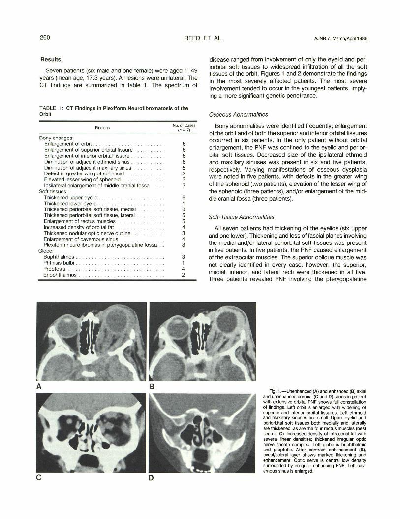
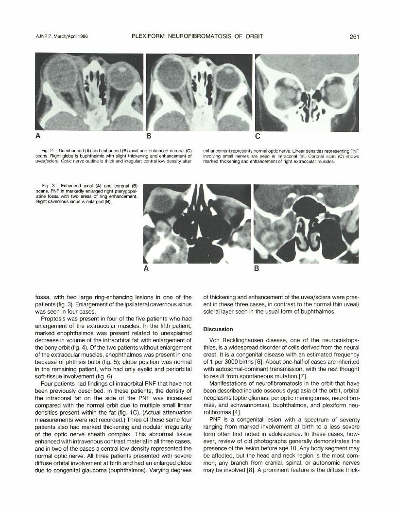
Fig. 1.-Unenhanced (A) and enhanced (8) axial and unenhanced coronal (C and 0) scans in patient with extensive orbital PNF shows full constellation of findings . Left orbit is enlarged with widening of superior and inferior orbital fissures . Left ethmoid and maxillary sinuses are small. Upper eyelid and periorbital soft tissues both medially and laterally are thickened, as are the four rectus muscles (best seen in C). Increased density of intraconal fat with several linear densities; thickened irregUlar optic nerve sheath complex. Left globe is buphthalmic and proptotic. After contrast enhancement (8), uveal/scleral layer shows marked thickening and enhancement. Optic nerve is central low density surrounded by irregular enhancing PNF. Left cavernous sinus is enlarged.
AJNR:7, March/April 1986 PLEXIFORM NEUROFIBROMATOSIS OF ORBIT 261
B
Fig. 2.-Unenhanced (A) and enhanced (8) axial and enhanced coronal (C) scans. Right globe is buphthalmic with slight thickening and enhancement of uvea/sclera. Optic nerve outline is thick and irregular; central low density after
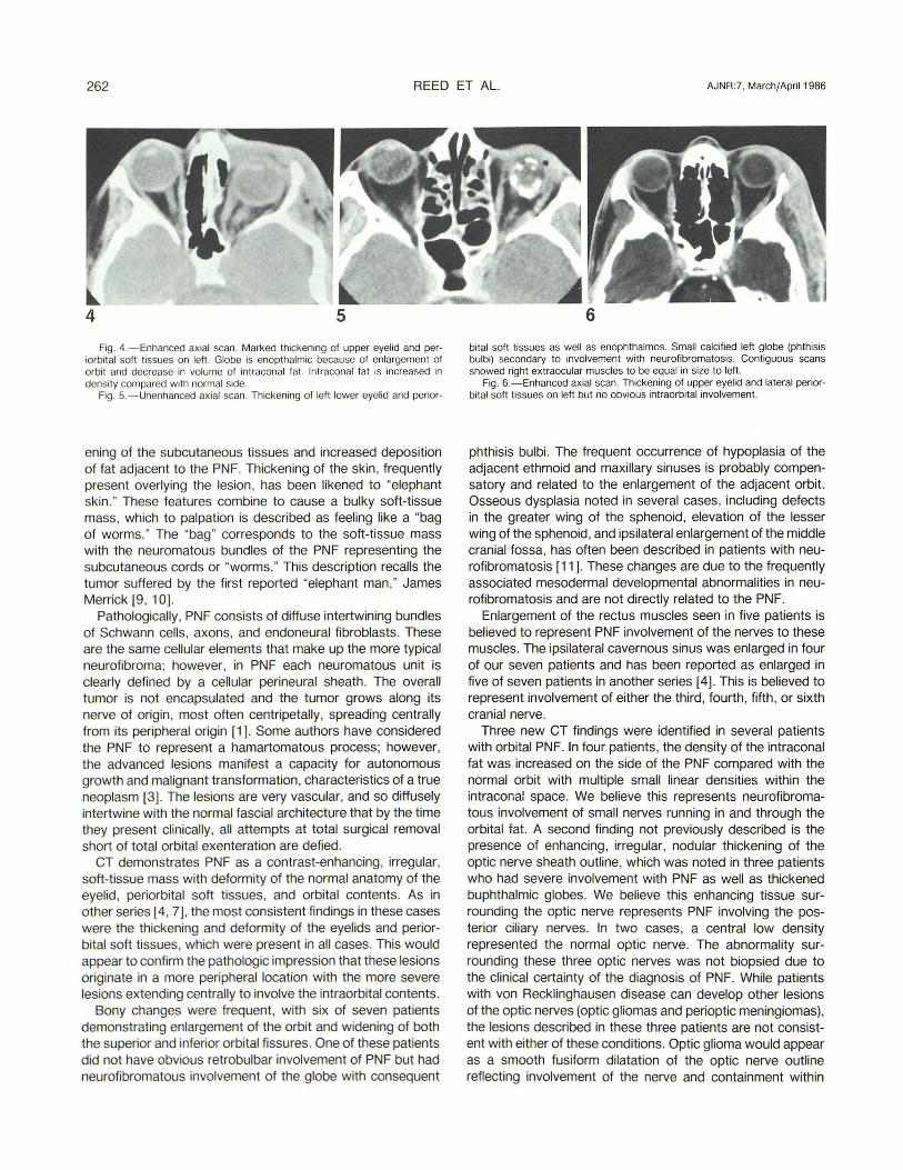
Fig. 3.-Enhanced axial (A) and coronal (8) scans. PNF in markedly enlarged right pterygopalatine fossa with two areas of ring enhancement. Right cavernous sinus is enlarged (8).
A
fossa, with two large ring-enhancing lesions in one of the patients (fig. 3). Enlargement of the ipsilateral cavernous sinus was seen in four cases.
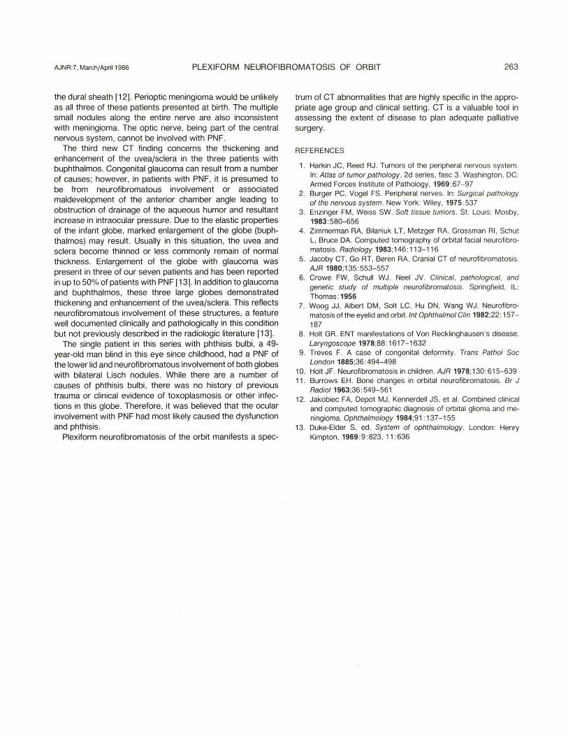
Proptosis was present in four of the five patients who had enlargement of the extraocular muscles. In the fifth patient, marked enophthalmos was present related to unexplained decrease in volume of the intraorbital fat with enlargement of the bony orbit (fig . 4). Of the two patients without enlargement of the extraocular muscles, enophthalmos was present in one because of phthisis bulbi (fig. 5); globe position was normal in the remaining patient, who had only eyelid and periorbital soft-tissue involvement (fig. 6).
Four patients had findings of intraorbital PNF that have not been previously described. In these patients, the density of the intraconal fat on the side of the PNF was increased compared with the normal orbit due to multiple small linear densities present within the fat (fig. 1 C). (Actual attenuation measurements were not recorded.) Three of these same four patients also had marked thickening and nodular irregularity of the optic nerve sheath complex . This abnormal tissue enhanced with intravenous contrast material in all three cases , and in two of the cases a central low density represented the normal optic nerve. All three patients presented with severe diffuse orbital involvement at birth and had an enlarged globe due to congenital glaucoma (buphthalmos). Varying degrees
c enhancement represents normal optic nerve. Linear densities representing PNF involving small nerves are seen in intraconal fat. Coronal scan (C) shows marked thickening and enhancement of right extraocular muscles.
B
of thickening and enhancement of the uvea/sclera were present in these three cases, in contrast to the normal thin uveal/ scleral layer seen in the usual form of buphthalmos.
Discussion
Von Recklinghausen disease, one of the neurocristopathies, is a widespread disorder of cells derived from the neural crest. It is a congenital disease with an estimated frequency of 1 per 3000 births [6]. About one-half of cases are inherited with autosomal-dominant transmission , with the rest thought to result from spontaneous mutation [7].
Manifestations of neurofibromatosis in the orbit that have been described include osseous dysplasia of the orbit, orbital neoplasms (optic gliomas, perioptic meningiomas, neurofibromas, and schwannomas), buphthalmos, and plexiform neurofibromas [4].
PNF is a congenital lesion with a spectrum of severity ranging from marked involvement at birth to a less severe form often first noted in adolescence. In these cases, however, review of old photographs generally demonstrates the presence of the lesion before age 10. Any body segment may be affected, but the head and neck region is the most common; any branch from cranial, spinal, or autonomic nerves may be involved [8] . A prominent feature is the diffuse thick-
262 REED ET AL. AJNR :7, March/April 1986
4 5
Fig. 4.- Enhanced axial scan. Marked thickening of upper eyelid and periorbital soft tissues on left . Globe is enopthalmic because of enlargement of orbit and decrease in volume of intraconal fat. Intraconal fat is increased in density compared with normal side.
Fig. 5.- Unenhanced ax ial scan. Thickening of left lower eyelid and perior-
ening of the subcutaneous tissues and increased deposition of fat adjacent to the PNF. Thickening of the skin, frequently present overlying the lesion, has been likened to "elephant skin." These features combine to cause a bulky soft-tissue mass, which to palpation is described as feeling like a "bag of worms." The "bag" corresponds to the soft-tissue mass with the neuromatous bundles of the PNF representing the subcutaneous cords or "worms." This description recalls the tumor suffered by the first reported "elephant man, " James Merrick [9, 10].
Pathologically, PNF consists of diffuse intertwining bundles of Schwann cells, axons, and endoneural fibroblasts. These are the same cellular elements that make up the more typical neurofibroma; however, in PNF each neuromatous unit is clearly defined by a cellular perineural sheath. The overall tumor is not encapsulated and the tumor grows along its nerve of origin, most often centripetally , spreading centrally from its peripheral origin [1] . Some authors have considered the PNF to represent a hamartomatous process; however, the advanced lesions manifest a capacity for autonomous growth and malignant transformation, characteristics of a true neoplasm [3] . The lesions are very vascular, and so diffusely intertwine with the normal fascial architecture that by the time they present clinically , all attempts at total surgical removal short of total orbital exenteration are defied.
CT demonstrates PNF as a contrast-enhancing, irregular, soft-tissue mass with deformity of the normal anatomy of the eyelid, periorbital soft tissues , and orbital contents. As in other series [4, 7] , the most consistent findings in these cases were the thickening and deformity of the eyelids and periorbital soft tissues , which were present in all cases. This would appear to confirm the pathologic impression that these lesions originate in a more peripheral location with the more severe lesions extending centrally to involve the intraorbital contents.
Bony changes were frequent , with six of seven patients demonstrating enlargement of the orbit and widening of both the superior and inferior orbital fissures . One of these patients did not have obvious retrobulbar involvement of PNF but had neurofibromatous involvement of the globe with consequent
bital soft tissues as well as enophthalmos. Small calcified left globe (phthisis bulbi) secondary to involvement with neurofibromatosis. Contiguous scans showed right extraocular muscles to be equal in size to left .
Fig. 6.-Enhanced axial scan. Thickening of upper eyelid and lateral periorbital soft tissues on left but no obvious intraorbital involvement.
phthisis bulbi. The frequent occurrence of hypoplasia of the adjacent ethmoid and maxillary sinuses is probably compensatory and related to the enlargement of the adjacent orbit. Osseous dysplasia noted in several cases, including defects in the greater wing of the sphenoid, elevation of the lesser wing of the sphenoid, and ipsilateral enlargement of the middle cranial fossa, has often been described in patients with neurofibromatosis [11]. These changes are due to the frequently associated mesodermal developmental abnormalities in neurofibromatosis and are not directly related to the PNF.
Enlargement of the rectus muscles seen in five patients is believed to represent PNF involvement of the nerves to these muscles. The ipsilateral cavernous sinus was enlarged in four of our seven patients and has been reported as enlarged in five of seven patients in another series [4] . This is believed to represent involvement of either the third , fourth, fifth , or sixth cranial nerve.
Three new CT findings were identified in several patients with orbital PNF. In four patients, the density of the intraconal fat was increased on the side of the PNF compared with the normal orbit with multiple small linear densities within the intracona: space. We believe this represents neurofibromatous involvement of small nerves running in and through the orbital fat. A second finding not previously described is the presence of enhancing, irregular, nodular thickening of the optic nerve sheath outline, which was noted in three patients who had severe involvement with PNF as well as thickened buphthalmic globes. We believe this enhancing tissue surrounding the optic nerve represents PNF involving the posterior ciliary nerves. In two cases, a central low density represented the normal optic nerve. The abnormality surrounding these three optic nerves was not biopsied due to the clinical certainty of the diagnosis of PNF. While patients with von Recklinghausen disease can develop other lesions of the optic nerves (optic gliomas and perioptic meningiomas), the lesions described in these three patients are not consistent with either of these conditions. Optic glioma would appear as a smooth fusiform dilatation of the optic nerve outline reflecting involvement of the nerve and containment within
AJNR :7, March/April 1986 PLEXIFORM NEUROFIBROMATOSIS OF ORBIT 263
the dural sheath [12] . Perioptic meningioma would be unlikely as all three of these patients presented at birth. The multiple small nodules along the entire nerve are also inconsistent with meningioma. The optic nerve, being part of the central nervous system, cannot be involved with PNF.
The third new CT finding concerns the thickening and enhancement of the uvea/sclera in the three patients with buphthalmos. Congenital glaucoma can result from a number of causes; however, in patients with PNF, it is presumed to be from neurofibromatous involvement or associated maldevelopment of the anterior chamber angle leading to obstruction of drainage of the aqueous humor and resultant increase in intraocular pressure. Due to the elastic properties of the infant globe, marked enlargement of the globe (buphthalmos) may result. Usually in this situation, the uvea and sclera become thinned or less commonly remain of normal thickness. Enlargement of the globe with glaucoma was present in three of our seven patients and has been reported in up to 50% of patients with PNF [13] . In addition to glaucoma and buphthalmos, these three large globes demonstrated thickening and enhancement of the uvea/sclera. This reflects neurofibromatous involvement of these structures, a feature well documented clinically and pathologically in this condition but not previously described in the radiologic literature [13].
The single patient in this series with phthisis bulbi, a 49-year-old man blind in this eye since childhood, had a PNF of the lower lid and neurofibromatous involvement of both globes with bilateral Lisch nodules. While there are a number of causes of phthisis bulbi, there was no history of previous trauma or clinical evidence of toxoplasmosis or other infections in this globe. Therefore, it was believed that the ocular involvement with PNF had most likely caused the dysfunction and phthisis.
Plexiform neurofibromatosis of the orbit manifests a spec-
trum of CT abnormalities that are highly specific in the appropriate age group and clinical setting. CT is a valuable tool in assessing the extent of disease to plan adequate palliative surgery.
REFERENCES
1. Harkin JC, Reed RJ . Tumors of the peripheral nervous system. In : Atlas of tumor pathology , 2d series, fasc 3. Washington, DC: Armed Forces Institute of Pathology, 1969 :67-97
2. Burger PC, Vogel FS. Peripheral nerves. In: Surgical pathology of the nervous system. New York: Wiley , 1975 :537
3. Enzinger FM, Weiss SW. Soft tissue tumors. St. Louis: Mosby, 1983 :580-656
4. Zimmerman RA, Bilaniuk LT, Metzger RA, Grossman RI , Schut L, Bruce OA. Computed tomography of orbital facial neurofibromatosis. Radiology 1983;146 : 113-116
5. Jacoby CT, Go RT, Beren RA. Cranial CT of neurofibromatosis. AJR 1980;135:553-557
6. Crowe FW, Schull WJ. Neel JV. Clinical, pathological, and genetic study of multiple neurofibromatosis. Springfield , IL: Thomas: 1956
7. Woog JJ, Albert OM, Solt LC, Hu ON , Wang WJ . Neurofibromatosis of the eyelid and orbit.lnt Ophthalmol Clin 1982;22 : 157-187
8. Holt GR. ENT manifestations of Von Recklinghausen's disease. Laryngoscope 1978;88 : 1617- 1632
9. Treves F. A case of congenital deformity. Trans Pathol Soc London 1885 ;36 :494-498
10. Holt JF. Neurofibromatosis in children. AJR 1978;130 : 615-639 11 . Burrows EH . Bone changes in orbital neurofibromatosis. Br J
Radio/1963;36:549-561 12. Jakobiec FA, Depot MJ, Kennerdell JS, et al. Combined clinical
and computed tomographic diagnosis of orbital glioma and meningioma. Ophthalmology 1984;91: 137-155
13. Duke-Elder S, ed. System of ophthalmology. London: Henry Kimpton , 1969 :9 :823, 11 :636


![· Web view[18F]-Fluorodeoxyglucose positron emission tomography in children with neurofibromatosis type 1 and plexiform neurofibromas: correlation with malignant transformation.J](https://img.pdfslide.us/doc/110x75/5b1c5e287f8b9a37258fdaa9/-web-view18f-fluorodeoxyglucose-positron-emission-tomography-in-children-with.jpg)
![Solitary Intraparotid Facial Nerve Plexiform Neurofibroma · peripheral nerve sheath tumor, which occurs in 2% - 5% of patients with plexiform neurofibroma [8]. Malignat peripheral](https://img.pdfslide.us/doc/110x75/5f7de695ec881b64331afe7f/solitary-intraparotid-facial-nerve-plexiform-neurofibroma-peripheral-nerve-sheath.jpg)