Embed Size (px)
Citation preview
EXPERIENCES WITH THE MANAGEMENT OF
PLEXIFORM NEUROFIBROMA
By Charles L. Minor, M.D., and C. Everett Koop, M.D.
Surgical Clinic, the Children’s Hospital of Philadelphia, and the Harrison Department of Surgical
Research, School of Medicine, University of Pennsylvania
Presented at the Annual Meeting of the American Academy of Pediatrics, October, 1958.
ADDRESS: (C.L.M.) Children’s Hospital of Philadelphia, 1740 Bainbridge Street, Philadelphia,
Pennsylvania.
482
PEDIATRICS, September i959
P LEXIFORM NEUROFIBROMA is a subject
which presents many very interesting
problems which have been discussed only
sporadically in the literature. The most
striking finding in the start of a search for
a concise presentation of the disease is the
difficulty in uncovering an apt description
of either gross or microscopic picture. We
have collected here a brief resume of the
clinical and pathologic problem with a re-
port of 11 cases treated at the Children’s
Hospital of Philadelphia.
Thomson in 1900’ wrote the most corn-
plete review of the problem and classified
plexiform neurofibroma as one of the most
interesting types of neurofibroma. It is but
one of the manifestations of diffuse neuro-
fibromatosis or von Recklinghausen’s dis-
ease, being given the descriptive name be-
cause of the characteristic gross appearance
of this benign tumor wherever it appears.
Typically, the growth is in the distnibu-
tion of one or more contiguous, usually
sensory, nerves or of a plexus of nerves, and
it often appears at birth or soon afterward.
The skin over superficial masses may be
normal, discolored brown, or thickened and
hairy as well as pigmented. The sensation
on palpation is unmistakable, being de-
scribed as feeling like a bag full of worms,
a tangle of spaghetti or grains of boiled
tapioca on a string. The disease may also
appear as a diffuse thickening of a single
nerve trunk without the nodular sensation.
The patient’s or parents’ attention is aroused
because of deformity and almost never be-
cause of pain or discomfort. The tumor may
appear as a sharply localized and relatively
small mass or as a diffuse and sometimes
quite large growth. Seldom is there any de-
tectable neunologic deficit. Most patients
who present with this problem have obvious
cafe-au-lait patches over the surface of the
body, these having been noted at birth or
shortly after and almost always before the
appearance of the plexiform tumor.
The distribution of the growths is quite
wide, although a majority of them first ap-
pear about the head and neck (41 of 58 in
the study by Thomson, 6 of 11 in the pres-
ent series). In one of its more favorite
sites, the peniorbital area, the growth may
cause marked deformity of the eye, either
pushing it out of position or covering it with
a grossly distorted eyelid. Nevertheless, the
optic nerve is not destroyed and careful
reconstructive surgery can restore normal
vision. It is important in this regard to know
of the propensity for this process to appear
in one location and then to crop up in an
entirely different area, probably a manifes-
tation of multiple foci of disease.
Major concern centers about this particu-
lan tumor because of its relentless progres-
sion along the distribution of a nerve, espe-
cially about the head and neck, following it
through the tiniest foramen and thence in-
vading the skull and leading to compres-
sion of adjacent cranial nerves or brain
substance. This occurs in similar fashion
in the spine passing through the vertebral
foramina to cause compression of the cord.
For this reason it is important to try to re-
move the mass completely as soon as possi-
ble in order to stall the process, evens
though we know it may involve some other
by guest on April 12, 2019www.aappublications.org/newsDownloaded from
AMERICAN ACADEMY OF PEDIATRICS - PROCEEDINGS 483
focus at a later date. Except for this phe-
nomenon, our approach would be to correct
only the deformity.
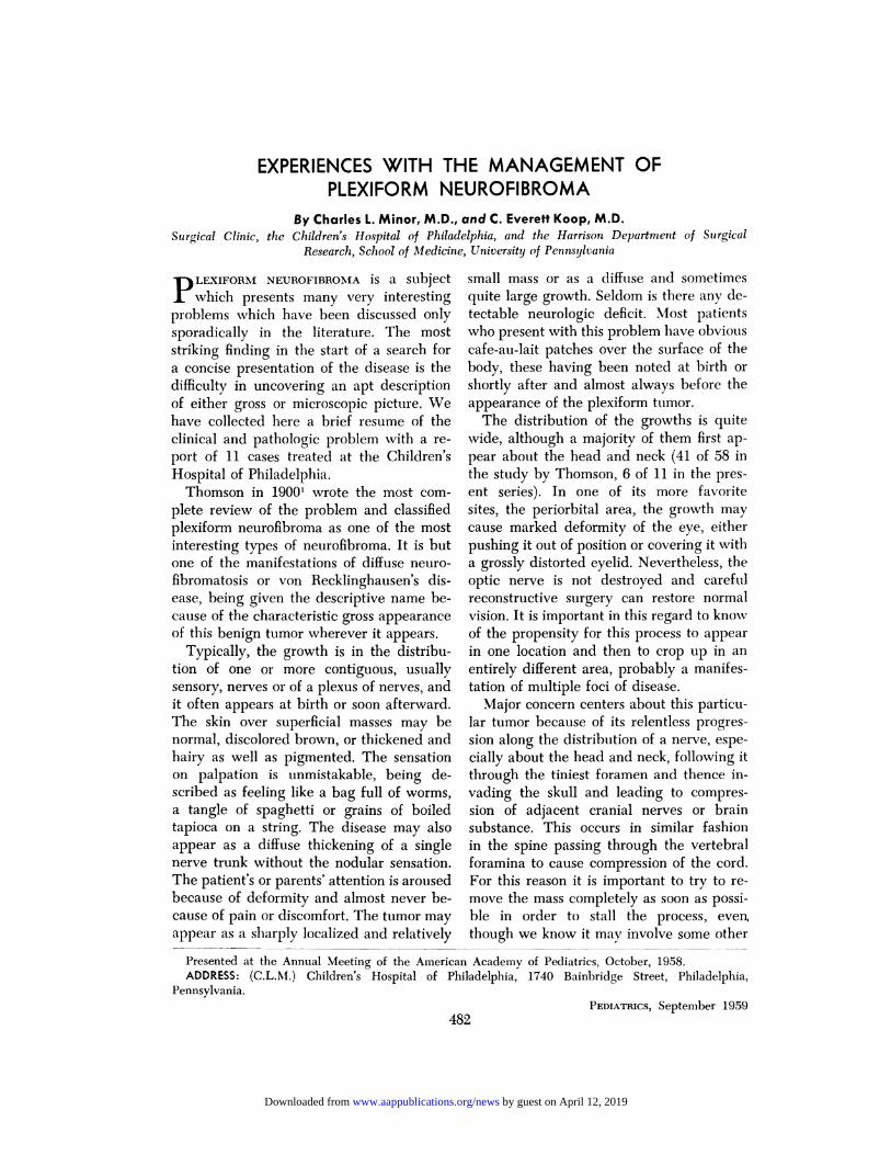
At operation the tumor appears very
much as it was described on palpation, with
masses or strands of smooth, shining nerves
which are easily separated from each other.
They vary in size from that of an adult
finger down to the most attenuated fiber
and are typically not cylindrical but pre-
sent nodular, fusiform or beaded swellings.
The tissue has a soft, gelatinous consistency
without elasticity so that it is very easily
torn and hence lost. It is important to fol-
low the tiniest tendrils out to the end in
order to eradicate the tumor.
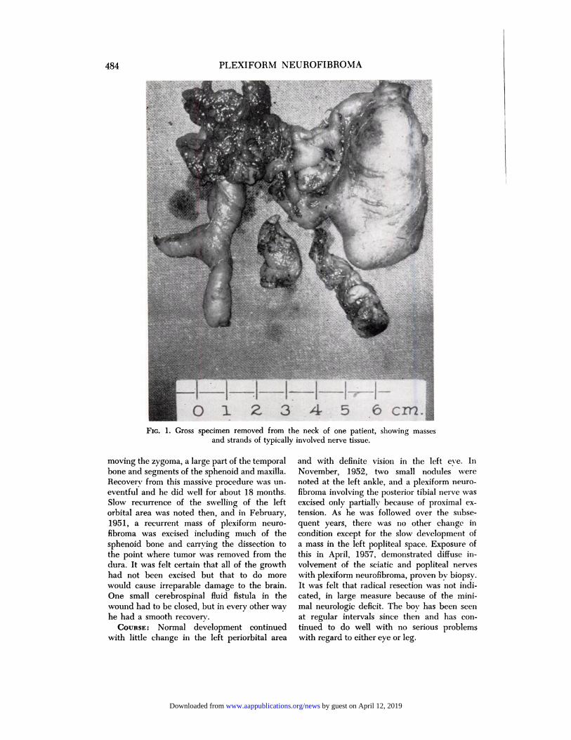
The histologic picture is similar to that
of generalized neurofibromatosis with over-
growth of endoneunium surrounding or sep-
arating individual nerve fibers. Whether
this is a proliferation only of connective
tissue elements or also of the sheath cells of
Schwann is unknown. The large amount of
edema causes a lattice-like appearance with
axis cylinders persisting with displacement
or dissociation but very rarely with degen-
eration or disappearance.
In the past 9% years we have seen 11 pa-
tients with various manifestations of this
problem. The youngest patient was 8
months old when first seen, the oldest, 9
years. There were five girls and six boys,
and one child was a Negro, all the rest be-
ing white. Each presented because of a
mass except one which was detected on
roentgenographic examination of the chest.
Three of the children had complained of
slight discomfort on palpation of the mass.
Six of the tumors appeared first about the
head and neck, two were on the thoracic
�vall, one was in the posterior mediastinum
with extension into the neck, one appeared
in the lumbar area near the midline and
one presented in the rectovaginal septum.
None of the children presented with any
abnormality of the neurologic examination,
and seven of them had typical cafe-au-lait
spots, often noted soon after birth and be-
fore the mass of tumor appeared.
Five patients had primary resection of the
entire presenting mass and have shown no
evidence of further disease in follow-up cx-
aminations up to 3 years later. The most
revealing material we have, however, is in
the study of the other six children. Two of
them had excision of what was believed to
be all of the tumor, one after two previous
operative procedures elsewhere. One boy
has slowly developed another focus of sim-
ilar appearance adjacent to the oniginal and
excision of this is planned for the near fu-
tune. The other had to have another excision
9 months after the operation performed by
us, and there has been no recurrence in 4
years since then. One 9-year-old girl, who
was found to have plexiform neunofibroma
arising in the rectovaginal septum with cx-
tension into the right adnexa, had biopsy
only and has been followed for 2 years dur-
ing which time she has done well except for
persistence of difficulty controlling the
rectal sphincter, which had been present
previously.
The other three present the most complex
situations. We shall give summaries of the
disease in two. The third has had excision
of three foci of tumor to the present: in
May, 1955, excision of a mass posterior to
the left ear with extension up to the carotid
foramen; in October, 1957, excision of the
left penoneal nerve, which was diffusely in-
volved from the ankle to the knee; and in
October, 1958, excision of another plexiform
mass in the right side of the neck.
Case �
CASE REPORTS
HISTORY AND PHYSICAL FINDINGS: CF., an
8-month-old white boy, was first seen in Jan-
uary, 1949, with a grossly swollen, discolored
left eyelid which had been present and had
slowly grown since birth causing some prop-
tosis of the eye. The only other abnormal find-
ing was scattered cafe-au-lait spots.
ROENTGENOGRAPHIC AND OPERATIVE Fn’�D-
INGS: Roentgenograms showed erosion of the
lower border of the left orbit and of the superior
portion of the maxilla and the sphenoid bone.
A biopsy showed this to be plexiform
fibroma, and wide excision was carried out re-
by guest on April 12, 2019www.aappublications.org/newsDownloaded from
484 PLEXIFORM NEUROFIBROMA
Fic. 1. Gross specimen removed from the neck of one patient, showing massesand strands of typically involved nerve tissue.
moving the zygoma, a large part of the temporal
bone and segments of the sphenoid and maxilla.Recovery from this massive procedure was un-
eventful and he did well for about 18 months.Slow recurrence of the swelling of the left
orbital area was noted then, and in February,
1951, a recurrent mass of plexiform neuno-fibroma was excised including much of thesphenoid bone and carrying the dissection tothe point where tumor was removed from theduna. It was felt certain that all of the growthhad not been excised but that to do morewould cause irreparable damage to the brain.
One small cerebrospinal fluid fistula in the
wound had to be closed, but in every other way
he had a smooth recovery.
COURSE : Normal development continuedwith little change in the left peniorbital area
and with definite vision in the left eve. In
November, 1952, two small nodules were
noted at the left ankle, and a plexiform neuro-
fibroma involving the posterior tibial nerve was
excised only partially because of proximal cx-tension. As he was followed over the subse-
quent years, there was no other change in
condition except for the slow development of
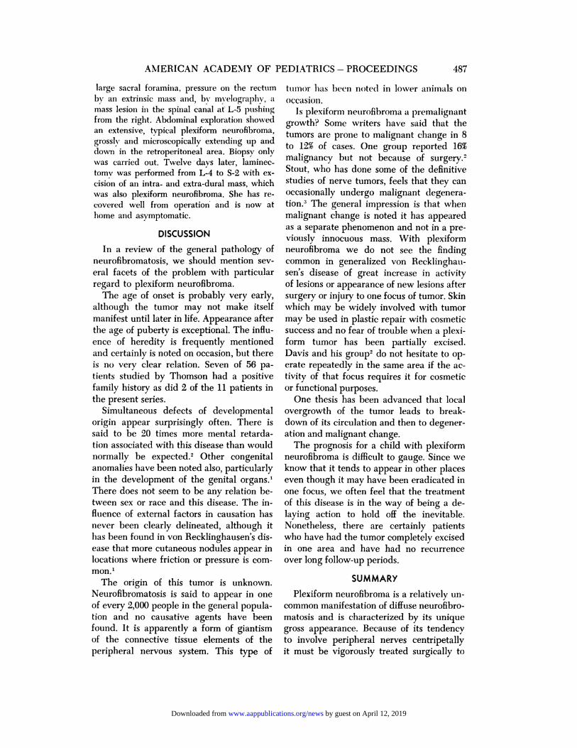
a mass in the left popliteal space. Exposure of
this in April, 1957, demonstrated diffuse in-
volvement of the sciatic and popliteal nerveswith plexiform neurofibroma, proven by biopsy.
It was felt that radical resection was not mdi-
cated, in large measure because of the mini-mal neurologic deficit. The boy has been seen
at regular intervals since then and has con-
tinued to do well with no serious problemswith regard to either eye or leg.
by guest on April 12, 2019www.aappublications.org/newsDownloaded from
FIG. 2. Nerve bundle, demonstrating marked involvement of one axis cylinder with normal or slightlyinvolved cylinders in close approximation to it.
FIG. 3. Specimen shown in Figure 2; a normal nerve fiber is in the center with
the periphery of the involved fiber to the left.
by guest on April 12, 2019www.aappublications.org/newsDownloaded from
486 PLEXIFORM NEUROFIBROMA
1�I(;. 5. Popliteal �icrvc ( Case 1 ), �xposc�l at op-
(ration in April, 1957, slu)wing tvpic(Ll. (liflus�
change ill the nervc.
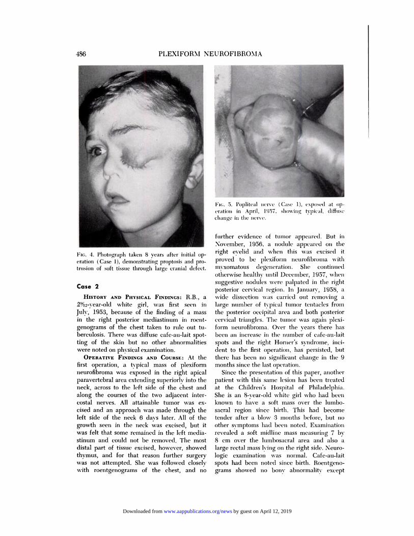
Flo. 4. Photograph taken 8 years after initial op-
eration ( Case 1), demonstrating proptosis and pro-
trusion of soft tissue through large cranial defect.
Case 2
HISTORY AND PHYSICAL FINDINGS: RB., a
2ih2�vear�old white girl, was first seen in
Jul�, 1953, because of the finding of a mass
in the right I)osterior mediastinurn in roent-
genograms of the chest taken to rule out tu-
berculosis. There was diffuse cafe-au-lait spot-
ting of the skin but no other abnormalities
were noted On physical examination.
OPERATIVE FINDINGS AND COURSE: At thefirst operation, a typical mass of plexiform
neurofibroma was exposed in the night apical
paravertebral area extending superiorly into the
neck, across to the left side of the chest and
along the courses of the two adjacent inter-
costal nerves. All attainable tumor was cx-
cised and an approach was made through the
left side of the neck 6 days later. All of thegrowth seen in the neck was excised, but it
was felt that some remained in the left media-
stinum and could not be removed. The mostdistal part of tissue excised, however, showedthymus, and for that reason further surgery
was not attempted. She was followed closely
with roentgenograms of the chest, and 110
further evidence of tumor appeared. But in
November, 1956, a no(lule a�)peared 011 the
right eyelid and when this was excised it
proved to be plexiforni neurofibroma with
mvxomatous degeneration. She continued
otherwise healthy until December, 1957, when
suggestive nodules were palpated in the right
I)osterior cervical region. In January, 1958, awide dissection was carried out removing a
large number of typical tumor tentacles fron�
the posterior occipital area and both posterior
cervical triangles. The tumor was again plexi-
form neurofibroma. Over the years there has
been ClII increase ill the Iluniber of cafe-au-lait
spots and the right Horller’s syndrome, mci-dent to the first operation, has persisted, but
there has been nO significant change in the 9
months since the last operation.
Since the �rese1�tati�11 of this paper, another
patient with this same lesion has been treated
at the Children’s Hospital of Philadelphia.
She is an 8-year-old white girl who had been
known to have a soft mass over the lumbo-
sacral region since birth. This had become
tender after a blow 3 months before, but no
other symptoms had been noted. Examination
revealed a soft midline mass measuring 7 by
8 cm over the lumbosacral area and also a
large rectal mass lying on the right side. Neuro-
logic examination was normal. Cafe-au-lait
spots had been noted since birth. Roentgeno-
grams showed no born abnormality except
by guest on April 12, 2019www.aappublications.org/newsDownloaded from
AMERICAN ACADEMY OF PEDIATRICS - PROCEEDINGS 487
large sacral foramina, pressure on the rectum
by an extrinsic mass and, b�’ mvelography, a
mass lesion ill the spinal canal at L-5 pushing
from the right. Abdominal exploration showed
an extensive, typical plexiform neurofibroma,
grossly and microscopically extending up and
down in the retnopenitoneal area. Biopsy only
was carried out. Twelve � days later, laminec-
tomy was performed from L-4 to S-2 with cx-
cision of an intra- and extra-dural mass, whichwas also plexiform neurofibroma. She has ne-
covered well from operation and is now at
home and asymptomatic.
DISCUSSION
In a review of the general pathology of
neurofibromatosis, we should mention 5ev-
eral facets of the problem with particular
regard to plexiform neurofibroma.
The age of onset is probably very early,
although the tumor may not make itself
manifest until later in life. Appearance after
the age of puberty is exceptional. The influ-
ence of heredity is frequently mentioned
and certainly is noted on occasion, but there
is no very clear relation. Seven of 56 pa-
tients studied by Thomson had a positive
family history as did 2 of the 11 patients in
the present series.
Simultaneous defects of developmental
origin appear surprisingly often. There is
said to be 20 times more mental retarda-
tion associated with this disease than would
normally be expected.2 Other congenital
anomalies have been noted also, particularly
in the development of the genital organs.’
There does not seem to be any relation be-
tween sex or race and this disease. The in-
fluence of external factors in causation has
never been cleanly delineated, although it
has been found in von Recklinghausen’s dis-
ease that more cutaneous nodules appear in
locations where friction on pressure is com-
mon.’
The origin of this tumor is unknown.
Neurofibromatosis is said to appear in one
of every 2,000 people in the general popula-
tion and no causative agents have been
found. It is apparently a form of giantism
of the connective tissue elements of the
peripheral nervous system. This type of
tumor has l)een noted in lower animals on
occasion.
Is plexiform neurofibroma a premalignant
growth? Some writers have said that the
tumors are prone to malignant change in 8
to 12% of cases. One group reported 16%
malignancy but not because of surgery.’
Stout, who has done some of the definitive
studies of nerve tumors, feels that they can
occasionally undergo malignant degenera-
tion.3 The general impression is that when
malignant change is noted it has appeared
as a separate phenomenon and not in a pre-
viously innocuous mass. With plexiform
neurofibroma we do not see the finding
common in generalized von Recklinghau-
sen’s disease of great increase in activity
of lesions or appearance of new lesions after
surgery or injury to one focus of tumor. Skin
which may be widely involved with tumor
may be used in plastic repair with cosmetic
success and no fear of trouble when a plexi-
form tumor has been partially excised.
Davis and his group’ do not hesitate to op-
crate repeatedly in the same area if the ac-
tivity of that focus requires it for cosmetic
or functional purposes.
One thesis has been advanced that local
overgrowth of the tumor leads to break-
down of its circulation and then to degener-
ation and malignant change.
The prognosis for a child with plexiform
neurofibroma is difficult to gauge. Since we
know that it tends to appear in other places
even though it may have been eradicated in
one focus, we often feel that the treatment
of this disease is in the way of being a de-
laying action to hold off the inevitable.
Nonetheless, there are certainly patients
who have had the tumor completely excised
in one area and have had no recurrence
over long follow-up periods.
SUMMARY
Plexiform neurofibroma is a relatively un-
common manifestation of diffuse neurofibro-
matosis and is characterized by its unique
gross appearance. Because of its tendency
to involve peripheral nerves centnipetally
it must be vigorously treated surgically to
by guest on April 12, 2019www.aappublications.org/newsDownloaded from
PLEXIFORM NEUROFIBROMA488
prevent damage to vital areas, notably the
brain and spinal cord, although the tumor
is basically benign.
We have presented our experience with
11 children who had this disease, demon-
strating that despite the propensity for the
growth to crop up in several areas it can be
eradicated locally, and the patient may have
no further trouble for a long time.
We believe that nesectable lesions should
be removed even though this may require
repeated operations over a period of several
years.
Acknowledgment
We wish to express our thanks to Dr.
Twining F. Campbell, Jr., who as a medicalstudent did the initial studies of these cases.Also to Dr. William C. Yakovac, Pathologist of
the Children’s Hospital, who helped in clarify-
ing a confusing picture and in reviewing the
pathologic sections for us.
REFERENCES
1. Thomson, A. : On Neunoma and Neuro-fibromatosis. Edinburgh, Turnbull andSpears, 1900.
2. Davis, W. B., Edgerton, M. T., and Hoff-meister, S. F. : Neurofibromatosis of thehead and neck. Plast. & Reconstruct.Surg., 14:186, 1954.
3. Stout, A. P. : Neunofibroma and neunil-
emoma Clin. Proc., 5: 1, 1946.
by guest on April 12, 2019www.aappublications.org/newsDownloaded from
1959;24;482Pediatrics Charles L. Minor and C. Everett Koop
NEUROFIBROMAEXPERIENCES WITH THE MANAGEMENT OF PLEXIFORM
ServicesUpdated Information &
http://pediatrics.aappublications.org/content/24/3/482including high resolution figures, can be found at:
Permissions & Licensing
http://www.aappublications.org/site/misc/Permissions.xhtmlentirety can be found online at: Information about reproducing this article in parts (figures, tables) or in its
Reprintshttp://www.aappublications.org/site/misc/reprints.xhtmlInformation about ordering reprints can be found online:
by guest on April 12, 2019www.aappublications.org/newsDownloaded from
1959;24;482Pediatrics Charles L. Minor and C. Everett Koop
NEUROFIBROMAEXPERIENCES WITH THE MANAGEMENT OF PLEXIFORM
http://pediatrics.aappublications.org/content/24/3/482the World Wide Web at:
The online version of this article, along with updated information and services, is located on
Copyright © 1959 by the American Academy of Pediatrics. All rights reserved. Print ISSN: 1073-0397. American Academy of Pediatrics, 141 Northwest Point Boulevard, Elk Grove Village, Illinois, 60007.been published continuously since 1948. Pediatrics is owned, published, and trademarked by the Pediatrics is the official journal of the American Academy of Pediatrics. A monthly publication, it has
by guest on April 12, 2019www.aappublications.org/newsDownloaded from
![Solitary Intraparotid Facial Nerve Plexiform Neurofibroma · peripheral nerve sheath tumor, which occurs in 2% - 5% of patients with plexiform neurofibroma [8]. Malignat peripheral](https://img.pdfslide.us/doc/110x75/5f7de695ec881b64331afe7f/solitary-intraparotid-facial-nerve-plexiform-neurofibroma-peripheral-nerve-sheath.jpg)