Embed Size (px)
Citation preview
PHENOTYPIC AND MOLECULAR CHARACTERIZATION OF METHICILLIN
RESISTANT STAPHYLOCOCCUS AUREUS FROM SURGICAL PATIENTS AND
NORMAL DOGS
CECILIA NJOROGE (BVM)
A THESIS SUBMITTED IN PARTIAL FULFILMENT OF THE REQUIREMENTS
FOR THE AWARD OF THE DEGREE OF MASTER OF VETERINARY SURGERY
(MVET SURGERY) OF THE UNIVERSITY OF NAIROBI
FACULTY OF VETERINARY MEDICINE
OCTOBER 2016
ii
DECLARATION
This is my original work and I declare that it has not been submitted for award of a degree in
any other University.
DR. CECILIA W. NJOROGE, BVM (UNIVERSITY OF NAIROBI)
SIGNATURE: DATE:
This thesis has been submitted for examination with our approval as University
Supervisors:
PROF. JOHN DEMESI MANDE (BVM, MSc, PhD)
SIGNATURE: DATE:
PROF. SIMON ERIC MITEMA (BVM, MSc, PhD)
SIGNATURE: DATE:
DR. JAFRED M.A. KITAA (BVM, MSc, PhD)
SIGNATURE: DATE:

iii
DEDICATION
To my parents, Dr. B.Ngaruiya and Mrs. Pauline Njoroge.

iv
ACKNOWLEDGEMENTS
I would like to express my sincerest gratitude to my supervisors Prof. J.D. Mande, Prof. S.E.
Mitema and Dr. J.M.A. Kitaa, their expertise; vast knowledge and guidance during all levels
of this project were invaluable. Special thanks to Prof. Mande, who gave me the foundation to
start my postgraduate degree by encouraging me to enrol for graduate study. His mentorship,
persistence and technical assistance during the course of my study truly made a difference in
my life. I also want to acknowledge Dr. Aboge for taking time out of his busy schedule to
help me navigate the world that is Molecular Biology. His vast expertise in this field and
willingness to guide me through it was remarkable and I am eternally grateful. I am grateful to
Prof. Mitema and NACOSTI (National Commission for Science, Technology and
Innovation), for the financial support towards the research project.
I would like to thank the staff at the Department of Public Health and Pharmacology and
Toxicology, especially Mr. Nduhiu Gitahi, Mr. Alfred Mainga and James Macharia for the
technical assistance, support and encouragement they offered me during the course of my
project. Appreciation also to Beatrice, Carol, Masinde and Lucy.
I acknowledge the staff at the University of Nairobi Small Animal Hospital and Andy’s
Veterinary clinic for facilitating access to clinical cases in particular, Dr. W. Mwangi, Dr. L.
Mathai and Dr. D. Muasya of the UoN Small Animal Clinic and Dr. A. Matole and Dr. A.
Gitari of the Andy’s Veterinary Clinic.
I would also like to express my deepest gratitude to my friends Maryanne Kagai, Robert
Ndung’u, Anne Olang’o, Paul Kimweli and David Ochieng’. Their support through what has
been a very challenging journey have truly made a lasting impact in my life and taught me
how to face challenges head on. My gratitude also goes to my postgraduate classmates Dr.
Hassan Mohammed and Dr. Jamleck Muriuki for their friendship, company, useful insights
and exchange of ideas throughout my study.
I would also like to thank my family for the support they provided me through my entire life
and in particular, I must acknowledge my parents for their unwavering support, guidance and
love. Their dedication to education and inspires me to keep aiming higher.
v
TABLE OF CONTENTS
DECLARATION ...................................................................................................................... ii
DEDICATION ......................................................................................................................... iii
ACKNOWLEDGEMENTS .................................................................................................... iv
TABLE OF CONTENTS ......................................................................................................... v
LIST OF TABLES ................................................................................................................... ix
LIST OF FIGURES .................................................................................................................. x
ABSTRACT ............................................................................................................................. xi
CHAPTER ONE ....................................................................................................................... 1
1.0. INTRODUCTION .......................................................................................................... 1
1.1. Justification .................................................................................................................................. 4
1.2. General objective ......................................................................................................................... 5
1.3. Specific objectives ....................................................................................................................... 5
CHAPTER TWO ...................................................................................................................... 6
2.0. LITERATURE REVIEW ............................................................................................. 6
2.1. Structure and function of the canine skin .................................................................................... 6
2.2. Concepts of surgical asepsis, surgical site infection and infection control in small animal
practice ......................................................................................................................................... 8
2.3. Methicillin resistant Staphylococcus aureus and methicillin resistant Staphylococcus
pseudintermedius in dogs............................................................................................................. 9
2.4. Mechanism of MRSA resistance ............................................................................................... 13
vi
2.5. Detection of methicillin resistance ............................................................................................ 15
2.6. Contamination, colonisation and infection ................................................................................ 19
CHAPTER THREE................................................................................................................ 23
3.0. MATERIAL AND METHODS .................................................................................. 23
3.1. Study site ................................................................................................................................... 23
3.2. Study design ............................................................................................................................... 23
3.3. Retrospective study: Survey of common bacterial isolates from wounds and otitis externa and
their respective antimicrobial susceptibility profiles. ................................................................ 23
3.3.1. Animal patient biodata ................................................................................................. 23
3.3.2. Bacterial profile ............................................................................................................. 24
3.3.3. Antimicrobial susceptibility testing (AST) ................................................................... 24
3.3.4. Wound characteristics ................................................................................................... 24
3.4. Data analysis .............................................................................................................................. 25
3.5. Prospective study: Prevalence of MRSA/MRSP in dogs .......................................................... 25
3.5.1. Study population ........................................................................................................... 25
3.5.2. Sample collection .......................................................................................................... 26
3.5.3. Bacteriological examination .......................................................................................... 26
3.5.4. Biochemical tests for confirmation ............................................................................... 27
3.5.6. Antimicrobial susceptibility testing ............................................................................... 27
3.6. Molecular identification and PCR detection of mecA ................................................................ 29
3.6.1. DNA extraction ............................................................................................................. 29
3.6.2. Validation of isolates ..................................................................................................... 29

vii
3.6.3. Identification of coagulase positive staphylococci ........................................................ 30
3.6.4. Detection of mecA ......................................................................................................... 30
3.7. Sequencing of resistant genes .................................................................................................... 30
3.8. Basic Local Alignment Sequence Tool (BLAST) analysis ....................................................... 33
3.9. Submission to NCBI GenBank .................................................................................................. 33
3.10. Ethical issues ............................................................................................................................. 33
CHAPTER FOUR .................................................................................................................. 34
4.0. RESULTS ..................................................................................................................... 34
4.1. Retrospective study: Survey of common bacterial isolates from wounds and otitis externa of
dogs and their antimicrobial susceptibility patterns ................................................................... 34
4.1.1. Animal Patient Biodata ................................................................................................. 34
4.1.2. Microbial isolates .......................................................................................................... 35
4.1.3. Antibiogram profile ....................................................................................................... 37
4.1.4. Wound characteristics ................................................................................................... 39
4.2. Prevalence of MRSA/MRSP from normal dogs and surgical patients ...................................... 41
4.2.1. Clinical history and animal biodata ............................................................................... 41
4.2.2. Prevalence of staphylococci. ........................................................................................ 41
4.2.3. Phenotypic characterisation of resistance ..................................................................... 41
4.2.4. Validation of isolates .................................................................................................... 41
4.2.5. Identification of coagulase positive staphylococci (COPS) ......................................... 42
4.2.6. MecA gene .................................................................................................................... 42
4.2.7. BLAST analysis ............................................................................................................ 42
viii
CHAPTER FIVE .................................................................................................................... 47
5.0. DISCUSSION, CONCLUSION AND RECOMMENDATIONS ............................ 47
5.1. Discussion .................................................................................................................................. 47
5.2. Conclusions ................................................................................................................................ 56
5.3. Recommendations ...................................................................................................................... 56
CHAPTER SIX ....................................................................................................................... 58
6.0. REFERENCES AND APPENDICES ........................................................................ 58
6.1. References .................................................................................................................................. 58
6.2. Appendices ................................................................................................................................ 77
Appendix 1: Microbial isolates from wounds and ear swabs of dogs from the UoN Small Animal
Clinic (2004-2013). .................................................................................................................... 77
Appendix 2: Antimicrobial susceptibility pattern of bacterial isolates (2004-2013) ..................... 84
Appendix 3: Sample collection form ................................................................................................ 91
Appendix 4: Tests for Differentiation of Staphylococcus spp. ......................................................... 92
Appendix 5: Oxacillin disk diffusion antimicrobial susceptibility testing zone diameter readings
for presumptive coagulase positive Staphylococcus isolates. .................................................... 93
Appendix 6: Nucleotide and amino acid sequences of the resistant genes ........................................ 94

ix
LIST OF TABLES
Table 1: Comparison of efficiency of methods used for susceptibility testing ....................... 16
Table 2: Primer pairs and sequences used in the PCR reactions for identification of the Genus
Staphylococcus, species identification of Staphylococcus aureus and detection of
mecA medicated resistance. ......................................................................................... 32
Table 3: Prevalence of bacterial isolates from clinical samples of wounds and ear swabs in
dogs ............................................................................................................................... 36
Table 4: Resistance (%) of six bacterial isolates from dogs to various antimicrobial agents
(n=262). ........................................................................................................................ 38
Table 5: Phenotypic multidrug resistance profiles displayed by the bacterial isolates from
dogs to various antimicrobial agents. ........................................................................... 38
Table 6: Number of dogs presented with injury to different regions of the body ................... 40
Table 7: Causes of wounds sampled for culture and sensitivity in dogs presented to the clinic.
.................................................................................................................................................. 40
Table 8: Resistant gene nucleotide homologues and their identities in expressed in
percentages ............................................................................................................................... 46
Table 9: Diversity of hosts and geographical distribution of resistant gene homologues ....... 46
x
LIST OF FIGURES
Figure 1: Skin Structure ............................................................................................................. 6
Figure 2: Agarose gel electrophoresis of PCR products of control PCR done for
identification of Staphylococcus targeting 16S rRNA gene. Lane 1: 100bp ladder
DNA marker, Lane 2: Negative Control; Lanes 3-11 Representative
Staphylococcus (PCR Product 416 bp); Lane 13: Staphylococcus aureus ATCC
25923 ...................................................................................................................... 44
Figure 3: Agarose gel electrophoresis of PCR products of mecA positive strains. Lane 1:
1KB ladder DNA marker, Lane 3-6 mecA positive isolates (Positive PCR product
286 bp), Lane 7 Positive control. ............................................................................ 45
xi
ABSTRACT
Staphylococcus spp. are globally recognized as colonisers of the skin and important causes of
infection in the skin of animals and humans. The increasing prevalence of antimicrobial
resistance, and in particular multi-drug resistant methicillin resistant Staphylococcus aureus
(MRSA) and the emergence of methicillin-resistant Staphylococcus pseudintermedius
(MRSP) in dogs has made treatment more challenging. The objectives of this study were to
determine bacterial ecology and their antimicrobial susceptibilities from wound and ear swabs
with emphasis on Staphylococcus aureus and to determine the prevalence of MRSA/MRSP in
normal dogs and surgical patients using phenotypic and genotypic assays. The study also
undertook Basic Local Alignment Search Tool (BLAST) analysis of sequenced polymerase
chain reaction (PCR) amplicons of the resistance determinant.
The study was divided into two parts, retrospective and prospective components. The
retrospective component of the study was designed to determine the bacterial ecology and
antimicrobial susceptibility from samples taken from surgical patients. Records were retrieved
from clinical laboratory of 291 bacteriological samples collected from 200 dogs submitted to
the University of Nairobi Small Animal Clinic over a 10 year period between January 2004
and December 2013. Information collected included the location from where the sample was
collected (wound or ear swab) as well as age, sex of the animal, microbial isolates and
antimicrobial susceptibility profile. In addition, for samples obtained from wounds, records
were further reviewed to determine the type, nature, location and causes of the wounds.
In the prospective component of the study, investigations were done on 191 samples obtained
from dogs presented at the University of Nairobi Small Animal Clinic and a community
veterinary clinic. Identification of coagulase positive Staphylococcus spp. (COPS) was
undertaken using mannitol salt agar as a selective medium and coagulase testing using
xii
reconstituted rabbit plasma. Final confirmation of COPS was done by PCR using primers
specific to the 16S rRNA gene of Staphylococcus Genus and primers specific to the nuc
(thermonuclease) gene of Staphylococcus aureus and Staphylococcus pseudintermedius spp.
Antimicrobial susceptibility testing (AMST) was performed using Kirby-Bauer disc diffusion
method according to Clinical and Laboratory Standards Institute (CLSI) guidelines.
Staphylococcus aureus ATCC 25923 was used as the reference organism. Oxacillin was used
as a surrogate for methicillin. For each isolate, susceptibility testing was done twice and the
mean zone diameter of inhibition calculated. The mean diameter was then compared to the
CLSI interpretive standard break points for Staphylococcus aureus and Staphylococcus
pseudintermedius for oxacillin and the number of resistant isolates noted.
DNA of the phenotypically resistant COPS isolates were extracted and thereafter specific
PCR assays were used to detect the resistance determinant among the resistant isolates. The
PCR amplicons were electrophoresed on 1.5 % agarose gel in Tris-acetate-EDTA buffer
supplemented with 0.5µg/ml of ethidium bromide and calibrated using 100 bp DNA ladder.
The gels were visually inspected by Ultra Violet (UV)-transilluminator. The amplicons
obtained were purified and sequenced using the ABI PRISM 3770 genetic analyzer. BLAST
analysis was done to confirm the identities of the sequenced amplicons, their location on
chromosomal DNA, the geographical distribution and diversity of hosts from which genes’
homologues had previously been isolated. The sequenced resistance gene was submitted to
the National Center for Biotechnology Information genetic sequence database (NCBI
GenBank) for assignment of accession numbers.
xiii
The retrospective study findings revealed that the most prevalent microbial isolates recovered
from dogs diagnosed with wounds, surgical site infections and otitis externa, were
Staphylococcus aureus 50 % (133/267) and Proteus spp. 14 % (38/267) respectively. Other
frequently recovered isolates included Pseudomonas spp. 10 % (28/267), other
Staphylococcus spp. 8.2 % (22/267), Streptococcus spp. 6.7 % (18/267) and E. coli 5.6 %
(15/267) respectively.
Resistance to antimicrobial drugs was observed in the majority of the isolates in the
retrospective study, with 97% (262/267) of the isolates demonstrating antimicrobial resistance
to at least one drug. Resistance to sulphonamides (96%), potentiated sulphonamides (89%),
ampicillin (68%), amoxicillin (62%) and tetracycline (56%) was relatively high for all
bacterial species examined.
Staphylococcus aureus isolates showed 95% resistance to sulfamethoxazole, 55% to
ampicillin, 52% to tetracycline and 52% to amoxicillin/clavulanic acid respectively.
Pseudomonas spp. showed the highest multidrug resistance with all (100%) isolates showing
resistance to amoxicillin, amoxicillin/clavulanic acid and sulfamethoxazole, the isolates also
showed high resistance to cotrimoxazole (93%), ampicillin (93%) and tetracyclines (80%)
respectively. Low resistance to gentamicin (9%), norfloxacin (24%) and chloramphenicol
(33%) was observed in all bacterial isolates.
The cause of 33% (18/54) wounds was not specified in the records. Common source of wound
swabs included, surgical site infections (SSI) 23.9% (11/46) followed by bite wounds 21.7%
(10/46), and traumatic injuries 15.2% (7/46). Majority of the wounds 67% (31/46) were
recorded on the limbs of affected animals with hindlimbs 32.6% (15/46) being more affected
than hindlimbs 28.3% (13/46).
xiv
Data from the prospective study revealed that presumptive Staphylococcus spp. were isolated
from 34% (65/191) of the samples. Coagulase positive Staphylococcus spp. (COPS)
accounted for 43% (28/65) of the Staphylococcus spp. isolated. Phenotypic resistance to
oxacillin was detected in 53.6% (15/28) of COPS. The PCR assay detected mecA gene as a
286 bp gene fragment amplicon in 2 of the 15 (7%) oxacillin resistant phenotypes. BLAST
analysis of the sequenced PCR products revealed that one of the resistance genes had 99 %
nucleotide identity to sequences in the NCBI GenBank database, while the other sample had a
95 - 97% identity. Further analysis of the resistant determinants by BLAST revealed that all
the resistant Staphylococcus strains were Staphylococcus aureus strains.
This study confirms Staphylococcus aureus as the most prevalent bacterial isolate from
wounds, surgical site infections and otitis externa. Proteus spp., Pseudomonas spp., other
Staphylococcus spp., Streptococcus spp. and Escherichia coli in descending order, were also
frequently isolated. Gentamicin, norfloxacin and chloramphenicol in that order were the most
effective antimicrobial agents in management of wounds, surgical site infections and otitis
externa in the retrospective study. The study reports the first case of MRSA strains in dogs in
Kenya which were associated with mobile genetic elements (SCCmec) and have the potential
to be transferred from dogs to humans. The MRSA resistant determinants observed are similar
to some human like isolates reported in several countries.
1
CHAPTER ONE
1.0. INTRODUCTION
Staphylococcal species are commensal bacteria and leading causes of community and
hospital-associated disease in humans and animals worldwide (Vengust et al., 2006). The
most clinically relevant staphylococci in veterinary medicine are the coagulase positive
Staphylococcus aureus and members of the Staphylococcus intermedius group, particularly
Staphylococcus pseudintermedius (Weese and Duijkeren, 2009).
Although S. aureus can colonize and infect companion animal species, the most common
commensal staphylococci of canines is Staphylococcus pseudintermedius (formerly S.
intermedius) with isolation rates of between 46- 92% in healthy dogs compared to 10% S.
aureus (Hanselman et al., 2009; Rubin and Chirino-trejo, 2011; Paul et al., 2011).
Staphylococcus pseudintermedius can be isolated from the nares, mouth, pharynx, forehead,
groin and anus of healthy dogs and cats. It is an opportunistic pathogen and a leading cause of
skin and ear infections, infections of other body tissues and cavities, and post-operative
wound infections in dogs and cats (Guardabassi et al., 2004; van Duijkeren et al., 2011).
Staphylococcal infections are frequently treated with antibiotics and, consequently, antibiotic
resistance and⁄or acquired resistance have developed (Normand et al., 2000). The increasing
prevalence of antimicrobial resistance has made staphylococcal infections become more
dangerous and costly to treat. Of considerable concern is Methicillin-resistant S. aureus
(MRSA) and emergence of Methicillin-resistant Staphylococcus pseudintermedius (MRSP) in
dogs and cats. These resistant strains of bacteria pose a new threat to animal health due to the
limitations in their management (EMA, 2011).
2
MRSA was first identified in the United Kingdom, and was then recognized as a nosocomial
pathogen worldwide (HA-MRSA) (Petinaki and Spiliopoulou, 2012). Subsequently, there
have been reports of MRSA infections occuring in people with no exposure to a healthcare
setting; these have been designated community acquired (CA-MRSA). There are differences
in epidemiology of HA-MRSA and CA-MRSA including resistance determinants, SCCmec
types and clonal complexes. HA-MRSA has also been found to be resistant to more
antimicrobials than CA-MRSA, and to be responsible for more invasive infections (Cohn and
Middleton, 2010). In animals, methicillin resistance was first documented in the early 1970’s
after isolation of MRSA from a dairy cow with mastitis. The emergence of MRSA in
livestock and in people in contact with livestock have introduced a new epidemiological
dimension to MRSA infections. These strains are designated LA-MRSA and are
phenotypically and genotypically distinct from the HA-MRSA and CA-MRSA genotypes
(Petinaki and Spiliopoulou, 2012). Although pet animals, especially dogs and cats may
become contaminated, colonized, or infected with S. aureus, including MRSA, these species
are not believed to be natural reservoir hosts for S. aureus. The MRSA strains found in
companion animals are frequently identical to human epidemic strains of MRSA, making it
more likely that MRSA originates from a person than a pet (Cohn and Middleton, 2010).
Majority of MRSA infections in dogs and cats appear to be in high-risk patients and are
acquired by direct contact with human carriers (Duquette and Nutall, 2004).
The MRSA isolates in dogs have been associated with clinical samples from surgical site
infections, wound infections (Baptiste et al., 2005; Vincze et al., 2014), catheter site
infections, urinary tract infections, pneumonia, and skin infections (Vengust et al., 2006).

3
These observations demonstrate the clinical importance and therapeutic challenge of MRSA
in the management of conditions of dogs and cats.
Methicillin-resistant S. pseudintermedius (MRSP) has recently emerged in small animals
worldwide and represents a major challenge for small animal practitioners due to its
characteristic multidrug resistance phenotype (Paul et al., 2011) and its characteristics of a
nosocomial pathogen (Frank and Loeffler, 2012). It has been isolated from various conditions
including wound infections, otitis externa and canine pyoderma (Beck et al., 2012). An
important aspect of MRSA and MRSP control is identification of potential sources of
exposure.
There are limited reports of MRSA in humans in Kenya; Maina et al. (2013) found MRSA
prevalence of 84.1% amongst Staphylococcus aureus isolated from patients with skin and soft
tissue conditions. A different study by Aiken et al. (2014) reported low carriage rate of
Staphylococcus aureus (85/950) in hospitalized patients, with only 7.0% of these isolates
being MRSA. There is limited data in literature on the prevalence as well as phenotypic and
molecular characteristics of microbial isolates from normal and surgical conditions in dogs in
Kenya. Mande and Kitaa (2005) found Staphylococcus aureus as the most common isolate
from ear swabs of dogs suffering from otitis externa and also reported multidrug resistance
among bacterial isolates. Available records in the UoN Small Animal Clinic laboratory
indicated frequent isolation of bacteria from dogs and cats. However, no systematic data or
meta-analysis was available describing the full extent of the phenotypic and molecular
characteristics of the different types of microbial isolates in dogs in Kenya.

4
This study was therefore designed with the aim of addressing the identified gap in the
knowledge and skills in order to improve the therapeutic and clinical management of dogs
undergoing surgical or medical procedures in Kenya.
1.1. Justification
Increasing isolation of methicillin resistant staphylococci in dogs has serious implications not
just on canines but also for in contact humans due to the potential of zoonotic transmission.
MRS are primarily transmitted via contact with contaminated objects/ environment, persons
or animals.
The relationship between many pets and their owners has dramatically changed. Most dogs no
longer live in kennels outside the home, people keep pets who live in the household almost as
family members and thus there is frequent contact between the pets and family members. The
intimate contact between pets (namely, cats and dogs) and their owners creates favourable
conditions for MRSA/MRSP transmission.
Preliminary review of facilities at the University of Nairobi’s Small Animal Clinic revealed
that surgical patients, clinical cases and healthy dogs share the same environment. These
facilities include:- common reception area, consultation rooms, corridors, surgical theatres)
and in some cases are housed in the same kennel. Although veterinary patients are often
attended to by the same clinicians, there was no documentary evidence of existence of
Standard Operating Procedures for decontamination of facilities and staff between cases.
Animals colonised with MRSA/MRSP may serve as sources of these pathogens in hospital
environments. Contamination of contact surfaces may be a risk factor for acquisition of these
resistant pathogens by surgical cases, with subsequent infection.

5
Bacteria of the genus Staphylococcus and in particular Staphylococcus aureus are the most
commonly isolated microbes from samples collected from cases at the small animal clinic.
These microbes are on average resistant to 3 or more antibiotics suggesting existence of
multidrug resistance phenotypes. These microbes pose a danger due to the possibility of
transfer of these resistant genes amongst other staphylococci and some clinically important
pathogens.
1.2. General objective
To determine the prevalence of Staphylococcus aureus and other microbial isolates in surgical
patients and normal dogs with emphasis on MRSA/MRSP at the University of Nairobi small
animal clinic, upper Kabete and a community veterinary clinic.
1.3. Specific objectives
The specific objectives of the study were;
(i) To determine retrospectively the prevalence of common bacterial flora in isolates from
wounds surgical site infections and otitis externa with emphasis on Staphylococcus
aureus.
(ii) To determine the antimicrobial suscpetibility profiles of the various bacterial isolates
to antibiotics used in the antimicrobial susceptibility tests.
(iii) To determine the prevalence of MRSA/MRSP in normal dogs and surgical patients
using phenotypic and genotypic tools.
(iv) To sequence resistant PCR amplicons and thereafter validate the sequences.

6
CHAPTER TWO
2.0. LITERATURE REVIEW
2.1. Structure and function of the canine skin
The skin (integument) is composed of two major layers; the outer stratified epithelium known
as epidermis and an underlying dermis (Figure 1). The integument provides a primary barrier
against infectious agents, thus serving as the body’s first line of defence against
microorganisms. The epidermis consists three principal layers; stratum basale, stratum
spinosum and stratum corneum (Pavletic, 2003).
Figure 1: Skin Structure
Adapted from http://lpi.oregonstate.edu/mic/micronutrients-health/skin-health
7
The skin is a primary source for harboring microorganisms that present as being a potential
cause of cross contamination (AST Standards, 2008). There is an intimate relationship
between this population of micro-organisms, particularly the bacterial component, and the
host. This relationship has a critical role in both protection and development of disease
(Weese, 2012). The principal types of skin flora include resident and transient flora. The
resident flora consist permanent inhabitants of the skin. Resident bacteria become established
on the skin where they multiply and are able to persist on a long-term basis (Kampf and
Kramer, 2004). Resident skin flora are mainly found under the superficial cells of stratum
corneum (Verwilghen et al. 2011).
Transient skin flora consist of micro-organisms including bacteria and fungi that are found
passively on the skin. They do not replicate on the skin, but they survive and take advantage
of changing conditions. If the conditions allow, they multiply and may cause clinical infection
(Saijonmaa- Koulumies and Lloyd, 1996; Mason et al., 1996). Transient flora only colonise
the superficial layers of intact skin. They are acquired by contact with other people, animals,
or contaminated environmental surfaces (Verwilghen et al., 2011). Upon wounding, there is
damage to the epidermis, local vasculature, possibly the dermis and underlying tissue,
depending on the extent (Daunton et al., 2012). Exposure of subcutaneous tissue provides a
favourable substratum for a wide variety of microbes to contaminate and colonise (Padhy et
al., 2014).
Isolation of low numbers of coagulase positive Staphylococci from the canine skin surface
indicates transient status. However, these organisms are readily isolated from the muco-
cutaneous junction. This suggests a resident status in mucosal surfaces which then act as
reservoir for transmission to the skin and hair through grooming (Mason et al., 1996).
8
2.2. Concepts of surgical asepsis, surgical site infection and infection control in small
animal practice
Surgical hand antisepsis aims to reduce the number of transient microorganisms as much as
possible as well as to depress resident microflora of the hands and forearms (Slatter, 2003).
However, traditional methods of hand antisepsis such as scrubbing, have been implicated as
one of the factors leading to skin damage. In one study, the hands of surgical staff were found
to have higher bacterial counts and more pathogenic organisms than hands of others.
Prolonged or repeated washing leads to damaged barrier function of the stratum corneum and
strips the skin of protective agents like amino acids and antimicrobial factors present in the
water–lipid layers of the superficial skin (Verwilghen et al., 2011).
The hands of healthcare workers are often contaminated with opportunistic pathogens.
Methicillin-resistant Staphylococcus aureus (MRSA) outbreaks in veterinary patients have
been associated with colonized surgeons and staff (McLean and Ness, 2008). This is due to
frequent and close contact with patients and the hospital environment. Healthcare workers
have been implicated as critical sources of hospital acquired infections (Wang et al., 2001;
Weber et al., 2002). Hand carriage of Staphylococcus aureus and other multi-drug resistant
(MDR) bacteria on the hands of medical professionals, including veterinary surgeons, makes
prevention of transmission of skin bacteria to the surgical wound particularly important.
Staphylococcus aureus survives on hands for at least 150 minutes and for an even longer time
on surfaces, with MRSA being isolated for upto seven months on inanimate surfaces (Neely
et al., 2000). However, compliance rates with hand hygiene practices have been reported to be
low, with an overall average of 40% (Kampf and Kramer, 2004). Lack of routine hand
washing after handling household pets has been found to be significantly associated with S.
9
pseudintermedius colonization in humans (Hanselmann et al., 2009). Contaminated surfaces,
including hands of healthcare workers may therefore act as sources of transient colonisation
for in-contact humans and animals.
The normal skin flora is a major cause of postoperative infections in animals. The most
commonly isolated genus from surgical site infections is Staphylococcus (Turk, 2013; Padhy
et al., 2014). Prevention of exposure to this flora is most important at the time of surgery and
is achieved through pre-operative preparation of the patients including clipping of hair and
scrubbing of the surgical site (Slatter, 1993). However, a patient’s skin cannot be completely
sterile and all surgical wounds become contaminated with bacteria; increasing the risk of
infection. For this reason, use of prophylactic antibiotics in surgical patients is common place
in veterinary clinics (Turk, 2013).
2.3. Methicillin resistant Staphylococcus aureus and methicillin resistant
Staphylococcus pseudintermedius in dogs
Methicillin resistant Staphylococcus aureus and methicillin resistant Staphylococcus
pseudintermedius have emerged as important pathogens in companion animals. MRSA is an
important pathogen that has been implicated as a leading cause of hospital acquired infections
in people (Singh et al., 2013). Methicillin belongs to a class of semi-synthetic β-lactamase
resistant penicillins introduced to treat infections caused by β-lactamase-producing
Staphylococcus strains. Within a year of its introduction to clinical use, the first reports of
methicillin (oxacillin) resistant Staphylococcus aureus emerged.
Historically, the vast majority of MRSA infections were nosocomial and were isolated from
patients associated with hospitals (Cohn and Middleton, 2010). Such strains were designated
10
hospital associated MRSA (HA-MRSA) clones. Subsequently, these resistant Staphylococcus
aureus organisms established themselves in hospitals and communities, spreading throughout
the world (Hafeez et al., 2004). MRSA infections due to HA-MRSA were associated with
serious illness and even death (Kuehnert et al., 2005). In recent years, there has been a shift in
the epidemiology of MRSA infections with an increase in the proportion of MRSA infections
occurring in humans with no exposure to healthcare settings. These MRSA infections have
been designated community- acquired (CA-MRSA) lineages and these can be carried for long
periods by healthy people (Harris et al., 2013; Cohn and Middleton, 2010). Methicillin
resistance in staphylococci in samples from animals has been documented since the early
1970s with the isolation of MRSA from a dairy cow. Indeed, MRSA have been isolated from
wound infections (Vincze et al., 2014), canine pyoderma (Beck et al., 2012), otitis externa,
bovine mastitis, equine wound infections (Vengust et al., 2006), porcine exudative
epidermitis and soft tissue infections of cats (Weese, 2010).
Methicillin-resistant Staphylococcus aureus infections in people occur in high-risk
environments such as intensive care units, or are associated with infections acquired during or
after orthopaedic surgery (Manian, 2003). The reservoir of infection is usually other colonised
or infected patients or hospital staff, and the organism is frequently transmitted via the
transiently colonised hands of healthcare workers (Baptiste et al., 2005; Leonard et al., 2006).
Dogs and cats are not considered resevoir hosts of Staphylococcus aureus. However, they
may become contaminated, colonized, or infected with S. aureus, including MRSA (Cohn and
Middleton, 2010).
In dogs, S. pseudintermedius is the predominant Staphylococcus spp. with reported isolation
frequencies between 20% and 90% from healthy canine skin and mucosal sites (Griffeth et
11
al., 2008; Hanselman et al., 2009; Rubin and Chirino-trejo, 2011). Staphylococcus
pseudintermedius can be isolated from the nares, mouth, anus, groin and forehead of healthy
dogs and cats as well as from dogs and cats with inflammatory skin disease (Abraham et al.,
2007; Griffeth et al., 2008). The perineum and the mouth are the most frequently colonized
body sites. The combination of the samples from the two body sites, allowed detection of
90% (75/82) of dog carriers in one study (Paul et al., 2012). Rubin and Chirino-Trejo (2011)
recommended screening of at least the pharynx and rectum, which together accounted for
99.3% of the carriers in their study. MRSP has been isolated from dermatologic conditions,
especially canine pyoderma, otitis externa and wound infections (Beck et al., 2012).
Simultaneous sampling of the pharynx, perineum, the corner of the mouth and wounds (if
present) is recommended for MRSP screening (Windahl et al., 2012). However, a negative
culture from a non-purulent wound should not be used as a criterion for a dog being MRSP
negative. In a study by Windahl et al. (2012), almost 20% of the wound samples were
negative, despite the bacteria being found in cultures from other sites that were sampled
simultaneously. Staphylococcus aureus is a common isolate from the skin and can persist in
the nares, and up to 60% of humans are thought to be carriers of S. aureus. Nasal carriage is
indicative of exposure and is associated with an increased risk of clinical infection in
hospitalized patients (Davis et al., 2004; Stevens et al., 2010). There has been an increase in
reports of MRSA infections in animals; MRSA has been reported in almost all domesticated
species, including dogs, cats, horses, cattle and sheep (Hartmann et al., 1997; Tomlin et al.,
1999; Goñi et al., 2004; Rich and Roberts, 2004).
Prevalence of MRSA is variable, with documented studies reporting prevalence ranging
between 0-4% in healthy animals (Loeffler et al., 2005; Abraham et al., 2007; Griffeth et al.,
12
2008). Various studies on MRSA colonization or infection among pets have shown that both
human-to-animal and animal-to-human transmission can occur, and that environmental
sources in veterinary clinics, veterinary staff and other hospitalized animals play a crucial role
(Petinaki and Spiliopoulou, 2012). Loefller et al. (2005) in their study isolated MRSA from
staff, dogs and environmental sites. Eighty two (82%) percent of the isolates were
indistinguishable from EMRSA-15, an epidemic strain dominant in UK hospitals. The high
prevalence of MRSA in people and pets in known infected households as well as the
identification of indistinguishable strains in humans and domestic animals suggested that
there was interspecies transmission of MRSA (Faires et al., 2009) though the direction of
transmission remains unclear. This demonstrates the zoonotic importance of MRSA/MRSP in
veterinary practice as well as pet owning households. Transmission of MRSA between
veterinary personnel and their patients is a concern in veterinary facilities (Baptiste et al.,
2005; Bergstrom et al., 2012) with both animal health and zoonotic implications. Leonard et
al. (2006) isolated MRSA from five dogs with wound discharges after surgical procedures at
a veterinary practice in Ireland. In the same study, MRSA with similar molecular and
phenotypic characteristics was isolated from the nares of one veterinary surgeon. While the
direction of transmission is not known, it suggests that veterinary hospitals and colonised staff
may play a role in the dissemination of MRSA and thus emphasises the zoonotic potential of
MRSA and the need for infection control in hospitals to prevent outbreaks of nosocomial
MRSA infections (van Duijkeren et al., 2011).
Risk factors include repeated courses of antibiotics, hospitalizations, intravenous
catheterization and surgery. Nienhoff et al. (2011) reported an association between
antimicrobial treatment and MRSP carriage. This finding was also reported by Bergstrom et
13
al. (2012). In their study, all dogs that tested positive for MRSP had been treated with
antimicrobials and although healthy dogs were included in their study, none of them were
MRSP positive, despite sharing a common environment with the sick dogs (Bergstrom et al.,
2012). Thus, antimicrobial treatment should be considered as one potential factor contributing
to MRSP isolation from patients.
2.4. Mechanism of MRSA resistance
Methicillin-resistance in MRSP is mediated by acquisition of mecA, which is carried on a
mobile genetic element identified as the Staphylococcal Cassette Chromosome mec
(SCCmec) encoding the penicillin binding protein 2a (PBP2a). β-lactam antibiotics bind to
PBP of S. pseudintermedius to prevent cell wall construction by the bacterium. The modified
PBP of MRSP has a low affinity for β -lactams and therefore cell wall construction is not
prevented by these antimicrobials (van Duijkeren et al., 2011). Thus, cell wall construction in
these MRS strains continues even in the presence of otherwise inhibitory concentrations of β -
lactam antibiotics (Paterson et al., 2014). According to the Clinical Laboratory Standards
Institute (CLSI) standards, MRS should be considered resistant to all β -lactams agents i.e
penicillins, β-lactam/ β-lactamase inhibitor combinations, both oral and parenteral cephems
including cephalosporins and carbepenems regardless of the results obtained from
susceptibility testing. This is since most cases of documented MRS infections have responded
poorly to therapy with β -lactam agents (CLSI, 2013).
Some MRSA strains encode a novel mecA homologue termed mecC originally designated as
mecALGA251. This homologue has 70% nucleotide identity with mecA and encodes a PBP that
is 63% identical at the amino acid level to the PBP2α encoded by mecA (Paterson et al.,
2014). The mecC wielding MRSA isolates have been isolated from bovine milk (Paterson et
14
al., 2014), domestic dog (Paterson et al., 2014) and from a cat suffering from chronic
conjunctivitis (Medhus et al., 2012). Prevalence of mecC from animal species is low, as
screening of bovine milk samples for MRSA yielded a prevalence of 2% in Britain (Paterson
et al., 2014). A study on samples from Dutch cattle did not isolate any mecC MRSA (van
Duijkeren et al., 2014). The mecC MRSA pose a potential diagnostic loophole since the
mecC gene is not detected by the PCR method established for the detection of mecA and
consequently mecC strains will be potentially misidentified as methicillin susceptible
Staphylococcus aureus (Paterson et al., 2014; Gomez et al., 2014).
There are two methicillin resistance phenotypes namely, homogenous and heterogenous.
While homogenous strains express a uniformly high level resistance, heterogenous strains
have a small proportion of a highly resistant subpopulation in a largely susceptible population.
Due to the selective pressure of antibiotics, the resistant minority predominates providing
clinical resistance (Niemeyer et al., 1996). Detection of resistance is made difficult by
additional genes, which are also found in susceptible isolates. These genes can affect the
expression of methicillin resistance in S. aureus, resulting in heterogenecity of strains (Brown
et al., 2005).
Additional genetic determinants frequently confer concurrent resistance to other clinically
relevant antibiotics (Bond and Loeffler, 2012). High resistance rates in MRS isolates have
been observed in isolates from different regions. In Germany, majority of the isolates were
resistant to fluoroquinolones, aminoglycosides and macrolides (Ruscher et al., 2009). In
North America, a study of 103 isolates found that aside from β-lactam resistance, 90 %
isolates were also resistant to ciprofloxacin, clindamycin, erythromycin, kanamycin,
streptomycin and trimethoprim; resistance to gentamicin and tetracycline was observed in
15
70 % and to chloramphenicol in 57 % (Perreten et al., 2010). The multidrug resistance profile
of MRSP in Europe and North America includes resistance to all oral antimicrobials routinely
used for the treatment of infections in pets, and the drugs to which they remain susceptible are
not authorized for use in animals (Perreten et al., 2010).
2.5. Detection of methicillin resistance
Various methods have been described for identification of MRS from clinical samples
(Chambers, 1997; Brown, 2001; Brown et al., 2005). These include genotypic and phenotypic
methods. Phenotypic tests rely on standard culture media together with conventional
laboratory tests and AST for the identification of MRSA. Conventional methods require
isolation of S. aureus first before antimicrobial susceptibility testing (Velasco et al., 2014).
Clinical Laboratory Standards Institute recommends the use of broth microdilution and disk
diffusion for detection of methicillin resistance in coagulase positive staphylococci.
Interpretive criteria of ≤21mm for disk diffusion and >4µg/mL for broth microdilution is
used for methicillin resistance (CLSI, 2013).
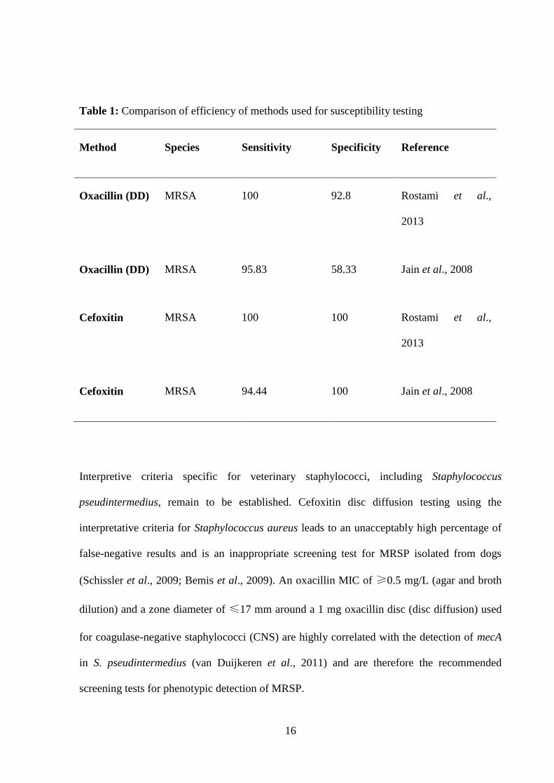
Most clinical laboratories use either oxacillin or cefoxitin as a surrogate for methicillin
(Loeffler et al., 2007; Bemis et al., 2009). However, studies have indicated that cefoxitin
testing is more superior and reliable than oxacillin (Table 1) for detection of MRSA strains
(Rostami et al., 2013). Oxacillin disk testing has been proven to be unreliable for MRSA
detection, since it suffers from lower specificity relative to cefoxitin (Chambers, 1997).
16
Table 1: Comparison of efficiency of methods used for susceptibility testing
Method Species Sensitivity Specificity Reference
Oxacillin (DD) MRSA 100 92.8 Rostami et al.,
2013
Oxacillin (DD) MRSA 95.83 58.33 Jain et al., 2008
Cefoxitin MRSA 100 100 Rostami et al.,
2013
Cefoxitin MRSA 94.44 100 Jain et al., 2008
Interpretive criteria specific for veterinary staphylococci, including Staphylococcus
pseudintermedius, remain to be established. Cefoxitin disc diffusion testing using the
interpretative criteria for Staphylococcus aureus leads to an unacceptably high percentage of
false-negative results and is an inappropriate screening test for MRSP isolated from dogs
(Schissler et al., 2009; Bemis et al., 2009). An oxacillin MIC of ≥0.5 mg/L (agar and broth
dilution) and a zone diameter of ≤17 mm around a 1 mg oxacillin disc (disc diffusion) used
for coagulase-negative staphylococci (CNS) are highly correlated with the detection of mecA
in S. pseudintermedius (van Duijkeren et al., 2011) and are therefore the recommended
screening tests for phenotypic detection of MRSP.
17
Phenotypic methods for AST are time consuming and laborius; in addition, several culture
conditions can also influence methicillin resistance such as the temperature, pH and
concentration of sodium chloride (NaCl) in the medium (Brown et al., 2005). These factors
impair the process of detection and may cause misidentification of some strains as methicillin
susceptible Staphylococcus aureus (MSSA) when in fact they are MRSA.
Genotypic methods are more accurate in detecting methicillin resistant staphylococci as
compared to conventional susceptibility methods and detection of the mecA gene by PCR is
considered the gold standard for identification of MRS (Schissler et al., 2009; Cohn et al.,
2010). PCR can produce results within 24 hours as compared to the conventional methods
which require at least 48 hours. This quick turnaround time ensures that MRS infections are
quickly diagnosed and appropriate therapy started (Sajith Khan et al., 2012). However, few
laboratories perform PCR for mecA in routine diagnostics, since it has greater technical
demands, uses expensive reagents and requires specialised laboratory equipment (Han et al.,
2007; Schissler et al., 2009).
Detection of the altered gene product of mecA, i.e. Penicillin Binding Protein (PBP2a), in
MRSA can also be used to diagnose MRSA (Hanselmann et al., 2006; Griffeth et al., 2008;
Julian et al., 2012). This test reliably differentiates between MRSA and MSSA. However,
PBP2a latex agglutination testing developed for MRSA can result in false-positive reactions
when applied to S. pseudintermedius isolates, and is therefore not recommended as the sole
test for confirmation of methicillin resistant Staphylococcus pseudintermedius (van Duijkeren
et al., 2011). In the study by Griffeth et al. (2008), it was found that the latex agglutination
test failed to identify 2 out of 13 MR isolates. Both the isolates were methicillin resistant
Staphylococcus pseudintermedius isolates. This finding could be due to the fact that the test

18
has not been validated for Staphylococcus pseudintermedius as it has for Staphylococcus
aureus.
Several chromogenic media have been approved for the detection of MRSA in pure cultures.
These media have been shown to reliably identify MRSA with sufficient sensitivity and
specificity for routine use (Han et al., 2007; Riedel et al., 2010). In the study by Han et al.
(2007), CHROMagar S. aureus (CSA) recovered 89.7 % and 94.9 % MRSA at 24 and 48
hours, respectively while CHROMagar MRSA (CSA-MRSA) recovered 87.2 % and 94.9 %
of the MRSA isolates at 24 and 48 hours. There was no significant difference between the two
agars in detection of MRSA. MRSA Select agar demonstrated a sensitivity and specificity of
99 % and 98 % respectively in detecting MRSA from blood cultures. However, the
specificity of the tests can be greatly improved by combining with either the tube coagulase
test or a commercial biochemical typing system to presumptively identify staphylococci.
Once presumptive MRSA are identified, molecular detection of mecA or latex agglutination
test for PB2a is recommended.
Selective media for detection of MRSP have not been identified. A recent study compared the
use of conventional MRSA selective media for isolation of MRSP. Five different screening
media were used in the study :- mannitol salt agar with oxacillin, CHROMagar MRSA,
chromID MRSA agar, oxacillin resistance screening agar base (ORSAB) and Brilliance
MRSA agar. The study found ORSAB and Brillance MRSA agar to be the most reliable in
detection and isolation of MRSP from clinical material (Horstmann et al., 2012).
19
2.6. Contamination, colonisation and infection
Colonization is the presence, growth and multiplication of MRS in one or more body sites
without observable clinical signs or immune reaction. Colonization by methicillin resistant
Staphylococci (MRS) of any species poses a risk for plasmid encoded transfer of
antimicrobial resistance determinants between staphylococci and other bacterial organisms.
Colonisation in humans has been associated with a four-fold risk of infection compared to
non- colonised patients (Safdar and Bradley, 2008). Colonization is incriminated as a risk
factor for S. pseudintermedius infection, since most dogs are infected with strains residing on
their body (Pinchbeck et al., 2006; Sasaki et al., 2007; Fazakerley et al., 2010). In a study on
dogs presented to a private dermatology clinic, Beck et al. (2012) demonstrated persistence of
MRSP after resolution of MRSP pyoderma. Of the dogs that initially had an MRSP
pyoderma, 26 of 42 (61.9 %) were colonized at one or more sites at follow-up, even though
the pyoderma had resolved.
Contamination on the other hand means that the bacteria can be easily washed off and often
only one culture is MRSP positive, while subsequent cultures are negative. Most studies done
on MRSA/MRSP are cross-sectional, making it difficult to determine if individuals with
MRSP positive cultures are merely contaminated or carriers. A longitudinal study carried out
by Paul et al. (2012) demonstrated that dogs were either persistent, transient or sporadic
carriers of S. pseudintermedius. Dogs positive for S. pseudintermedius at all sampling times
were classified as persistent carriers. Intermittent carriers were distinguished between
transient carriers that tested positive in at least three consecutive samples and sporadic carriers
that were positive at only one or two of the nine sampling times. Non-carriers were defined as
dogs testing negative at all sampling times (Paul et al., 2012).
20
Methicillin resistant Staphylococcus pseudintermedius has been reported as a contaminant in
cages for large dogs, the top surface of X-ray stand and the intensive care unit (Ishihara et al.,
2010). Another study found hospital clothing to have a high prevalence of methicillin
resistant Staphylococci (17.5 %); of these 3.5 % were MRSA and 14.0 % were MRSP (Singh
et al., 2013). In this study, technicians were 9.5 times more likely than students to have
clothing contaminated with MRSA. Julian et al. (2012) isolated MRS from 3/123 (2.4 %)
cellular phones (CPs) belonging to personnel in a veterinary teaching hospital; MRSP was
isolated from two (1.6 %) CPs, while MRSA was isolated from one (0.8 %) CP. Cellular
phones and hospital clothing may serve as formites for pathogenic bacteria with transmission
to patients or personnel through subsequent contamination of the hands.
Infections with methicillin resistant staphylococci in small animals, especially dogs, have
been reported. Baptiste et al. (2005) isolated MRSA from 3 dogs with clinical infections; joint
infection, pleuro-pneumonia and wound infection respectively. The dog with joint infection
also tested positive for nasal and faecal carriage of MRSA. Two months after the initial
isolation, a similar MRSA strain was associated with clinical disease in two other dogs. These
dogs had no history of contact with the other dogs, suggesting hospital acquired transmission
could also occur in veterinary centres. Beck et al. (2012) collected skin, nasal and rectal
swabs of dogs that were presented to a dermatology referral service with pyoderma and
healthy control dogs. Skin cultures yielded MRSP in 70 (40.5 %) dogs, methicillin-resistant
Staphylococcus aureus (MRSA) in three (1.7 %) and methicillin-resistant Staphylococcus
schleiferi ssp. coagulans (MRSScoag) in five (2.9 %).
Contact with other MRSP colonized dogs or humans might also serve as a source of
reinfection, as well as contaminated objects in the household (Windahl et al., 2012). Isolation

21
of MRSP remains uncommon in humans, screening of veterinarians and veterinary personnel
via nasal culture for MRSP carriage, revealed a carriage rate of 3.9–5.3 % (Ishihara et al.,
2010). Pet owners of animals with MRSP were screened and a nasal carriage rate of 4–13 %
was observed. The genetic identity of some isolates from owner–pet pairs supported
interspecies transmission (Frank and Loeffler, 2012). Carriage rate has been reported to be
higher in veterinarians attending to known MRSA/MRSP cases. Loeffler et al. (2010) in their
case-control study on colonisation rate in veterinarians and owners of small animals with
known MRSA infection reported carriage rates of 12.3 % and 7.5 % respectively. The rates in
the control group i.e animals with MSSA (methicillin Susceptible Staphylococcus aureus)
were significantly lower at 4.8 % and 0 % respectively for veterinarians and owners. The
findings of this study indicated an occupational risk for MRSA carriage in small animal
general practitioners, veterinary staff and owners of MRSA-infected pets.
Although methicillin resistant staphylococci are not necessarily more virulent than
methicillin-susceptible staphylococci, treatment options are often severely limited by multi-
drug resistance (Cain, 2013). MRS infections are more resistant to some treatments than
methicillin-sensitive Staphylococcus (MSS). There are concerns regarding the role of pets in
MRSA transmission with various authors reporting concurrent colonisation of humans and
their pets with indistinguishable MRSA strains. Many companion animals if not all, have
come into contact with humans and other animals of the same species, creating the potential
for transmission of organisms such as MRS (Vengust et al., 2006). Some reports have noted
that infection of human subjects with MRSA persisted until the pet and any other colonized or
infected cohabitants was treated with antimicrobials to which the bacteria were susceptible
(Manian 2003, van Duijkeren et al., 2004, Sing et al., 2008).
22
There are speculations that epidemic MRSA in humans drives the parallel epidemic in
companion animals. Despite the growing importance of these pathogens in veterinary
medicine, especially for surgical patients, no studies have been reported on the prevalence of
these pathogens in dogs in small animal practices in Kenya. In a preliminary study,
Staphylococcus species was identified as the most common isolate from wound swabs from
the University of Nairobi Small Animal Clinic. A high percentage of these isolates were
resistant to ampicillin and other B-lactam antibiotics such as amoxycillin and amoxycillin-
clavulanic (Njoroge et al., 2016). These preliminary findings led to a suspicion of the
existence of Methicillin resistant Staphylococcus spp.. in dogs in Kenya and prompted further
research to substantiate these claims.
23
CHAPTER THREE
3.0. MATERIAL AND METHODS
3.1. Study site
The study was undertaken at the University of Nairobi Small Animal Clinic, Upper Kabete.
This facility receives patients mostly from the suburbs of Nairobi region and its environs. It
also serves as a referral center for cases from other small animal clinics in Kenya. The Andy’s
community clinics whose patients are drawn from the Nairobi region and surrounding areas.
3.2. Study design
This study involved a retrospective and prospective component. The retrospective study
component involved review of microbial isolates and antibiogram data from the bacteriology
laboratory of samples submitted from surgical patients and dogs with otitis externa at the
University of Nairobi Small Animal Clinic. The prospective component was a cross-sectional
study that involved sampling of surgical patients and normal dogs presented at the Univeristy
of Nairobi Small Animal Clinic and at a Community veterinary clinic located in Nairobi
County.
3.3. Retrospective study: Survey of common bacterial isolates from wounds and otitis
externa and their respective antimicrobial susceptibility profiles.
3.3.1. Animal patient biodata
The bacteriology laboratory records of clinical samples submitted between January 2004 and
December 2013 were investigated. All the samples were from animals presented to the
University of Nairobi’s Small Animal Clinic during the study period. The records were
24
examined to retrieve data on culture samples of dogs and cats presented with otitis externa
and wounds. Animal biodata retrieved from these records included: date of submission, sex
and site where the sample was collected from (wound or ear swab).
3.3.2. Bacterial profile
For each clinical sample submitted, the number of microbial isolates and microorganisms
isolated from either wounds or ear swab were recorded. The total number of various bacterial
flora isolated were calculated and expressed as percentages. Bacteria of the Genus
Staphylococcus were recorded as Staphylococcus aureus or broadly classified as other
Staphylococcus spp. (for those that did not fit the characteristics of S. aureus in biochemical
tests).
3.3.3. Antimicrobial susceptibility testing (AST)
Routine disk diffusion procedures were employed in AST by the laboratory. The bacterial
isolates were tested against a panel of 8 antimicrobial agents namely, ampicillin (2µg),
gentamicin (10µg), cotrimoxazole (25µg), chloramphenicol (10µg), tetracycline (10µg),
potentiated amoxicillin (amoxycillin-clavulanic acid) (30µg), norfloxacin and
sulfamethoxazole (25µg). Various bacteria in the AST were scored by the laboratory as either
being susceptible or resistant to the respective antibiotic. If the zone of inhibition around the
disk was found to be ≤14mm, the organism was scored as being resistant to that drug.
3.3.4. Wound characteristics
Patient case records from which wound and abscess swabs were collected were retrieved for
further review. Information recorded for analysis included the cause and location (body
region) of the wound or abscess swab.
25
3.4. Data analysis
All data was entered into a spreadsheet (Microsost Excel 2010) and a pivot table generated.
The frequency of the various parameters (species, breed, sex) over the study period was
calculated and expressed as percentages. The total number of bacterial flora isolated was
calculated and expressed as percentages. Antimicrobial susceptibility was expressed as either
susceptible or resistant. Overall resistance for each antimicrobial agent was calculated.
Percentage resistance for each bacteria was calculated for each antimicrobial agent.
3.5. Prospective study: Prevalence of MRSA/MRSP in dogs
3.5.1. Study population
The following formula was used to calculate an appropriate sample size for the study
2
2 )1(96.1
d
ppn
Where (p) = Estimate of the expected proportion (15%)
(d) = Desired level of absolute precision (0.05)
An estimated MRSA prevalence of 15% (Bond and Loeffler, 2012) in the population was
used at 95% confidence interval. From the formula, we estimated our sample size to be 196
samples.
A total of 191 dogs were enrolled in this cross-sectional study, which entailed convenience
sampling at the UoN Small Animal Clinic and a Community Owned Clinic. Criteria for
inclusion entailed: - dogs of any age, sex, breed and obtaining written consent from owner or
attending veterinarian to collect samples; preference was given to dogs presented for surgery,
those with wounds and/or otitis externa. A brief questionnaire was filled by the owner or
26
attending veterinarian in order to obtain information on the patient including biodata like
breed, sex, age, presenting complaint, history of the condition (first time/recurrent) and prior
treatment administered (antibiotic use) in the past three months preceding the study.
3.5.2. Sample collection
Sampling was carried out between March 2014 and June 2015. Samples were collected from
four sites on the affected surgical patients and normal dogs, specifically, anterior nares, buccal
mucosa, perianal area, a wound swab if the patient presented with a wound and an ear swab in
patients presenting with otitis externa. A sterile cotton tipped swab moistened with sterile
normal saline was used to collect samples by swabbing the aforementioned sites. A separate
swab was used for each anatomic location and swabs from each dog were pooled in a bijou
bottle containing 3 ml of transport medium (Stuart’s medium) and transported to the
laboratory where they were stored in a refrigerator at 4oC awaiting processing.
3.5.3. Bacteriological examination
3.5.3.1. Recovery of isolates
Samples were removed from the refrigerator and kept at room temperature for 4 hours before
being cultured onto nutritive medium, tryptone soya broth supplemented with 6.5 % NaCl for
selective enrichment of Staphylococcus. After incubation at 37oC for 24 hrs, a loopful of broth
was taken and cultured to Mannitol Salt Agar (MSA), a selective medium and incubated at
35oC for 24- 48 hrs. Growth of yellow colonies on this medium and colour change of the
media to yellow was taken as positive fermentation of mannitol and presumptive
Staphylococcus aureus (Kateete et al., 2010). Pink colonies on mannitol salt agar were also
sub cultured and designated as presumptive Staphylococcus pseudintermedius.
27
The presumptive Staphylococcus aureus or Staphylococcus pseudintermedius colonies were
subcultured on 5 % sheep blood agar (SBA) and incubated at 37oC for 24 hours to isolate a
pure culture. Those SBA plates that did not show any growth after 24 hours were incubated
for a further 24 hours. Final identification of the presumptive coagulase positive
Staphylococcus spp. characteristic colonies was on basis of colonial morphology, gram stain
reaction, and positive catalase and coagulase tests. The presumed staphylococcus colonies
were subjected to a Gram stain and the slide examined under a light microscope to check for
gram reaction, size and shape of the colonies. Gram positive cocci that appeared as grapelike
clusters in pairs and singles were presumed to be Staphylococcus spp.
3.5.4. Biochemical tests for confirmation
3.5.4.1. Catalase test
A sterile loop was used to pick organisms from the plate and place them on a slide. A drop of
3 % Hydrogen peroxide was added to the slide and mixed with the organisms. Visualization
of bubbles was regarded as a positive reaction.
3.5.4.2. Tube coagulase test
This test was performed by transferring a single colony of inoculum to 1 ml of reconstituted
rabbit plasma. The two were mixed by gently rotating the tubes. The tubes were then
incubated at 37oC and evaluated after 24 hrs. Formation of a clot in the tube was taken as a
positive reaction. Presumptive coagulase positive Staphylococcus colonies were sub-cultured
on Tryptic soy agar, awaiting susceptibility testing.
3.5.6. Antimicrobial susceptibility testing
Antimicrobial susceptibility testing was performed according to the Kirby-Bauer disc
diffusion method. A sterile loop was used to pick organisms from the tryptone soy agar plate.
28
The organisms were added to a tube containing 4.5 ml of sterile physiological saline. The
mixture was vortexed to create a smooth suspension. The turbidity of the suspension was
adjusted to 0.5 McFarland standard. A sterile swab was dipped into the inoculum suspension.
The Mueller Hinton (MH) plate was then inoculated by streaking across the agar surface
ensuring that the entire plate was covered. The lid of the plate was left slightly open for 3-5
minutes for the agar surface to dry up.
Oxacillin was used as the surrogate antibiotic to methicillin (CLSI, 2008). Oxacillin (1 µg)
discs (HiMedia Laboratories Pvt. Ltd, Mumbai, India) were peeled from the cartridge using
forceps. The lid of the MH agar was lifted to allow placement of the discs on the agar surface.
Once the disc was placed, it was gently pressed with forceps to ensure total contact with the
agar surface. Plates were incubated at 35-37oC for 24 hrs. The zone diameters of complete
inhibition, including that of the disks, were measured to the nearest whole millimetre using a
ruler. To measure the zones of inhibition, the ruler was held on the back of an inverted petri
dish while holding it a few inches from a black non-reflecting background illuminated with
reflected light.
For each isolate, antimicrobial susceptibility testing was done in duplicate and the mean zone
diameter of inhibition calculated. The resistance zone diameter of ˂17mm around a 1 µg
oxacillin disc was used as an indicator for methicillin resistance as recommended by Bemis et
al. (2009), and approved by the Clinical and Laboratory Standards Institute (CLSI)
subcommittee on Veterinary Antimicrobial Susceptibility Testing (CLSI, 2013).
29
3.6. Molecular identification and PCR detection of mecA
Isolates found to be resistant were amplified by polymerase chain reaction (PCR).
Staphylococcus aureus ATCC 25923 served as the reference quality control strain. Primer
pairs, sequences and amplicon size of primers used in the PCR reactions are shown in Table
2.
3.6.1. DNA extraction
Extraction of DNA was performed as described by Diaz-Campos (2012). Two or three
colonies were obtained from 18 – 48 hours cultures inoculated on tryptic soy agar (4.1 %) and
suspended in 400 μl of sterile distilled water. The bacterial suspension was boiled at 95°C for
7 minutes and then centrifuged at 15,000 g for 1 min and the supernatant collected. The DNA
supernatant extracts were stored at -20°C until used as a template for the PCR reactions.
3.6.2. Validation of isolates
Amplification of 16S rRNA gene of all strains were performed at first to confirm that they
were Staphylococcus strains. This was performed in a protocol adapted from Kondo et al.
(2007). PCR reaction was done in a total volume of 20 μl containing 5 μl of DNA template
and 0.25 μl of primers Staph-F and Staph-R. Thermal cycling reactions consisted of initial
denaturation at 94°C for 10 min; followed by 35 cycles of denaturation at 94°C for 15 s,
annealing at 50°C for 15 s, extension at 72°C for 1 min; and a final elongation at 72°C for 5
min. Amplification products were analyzed by electrophoresis in a 1.5% agarose gel stained
with ethidium bromide. Gels were visualised under U.V light. Amplification of the 416bp
PCR product indicated the strain to belong to the genus Staphylococcus.
30
3.6.3. Identification of coagulase positive staphylococci
Primers for species identication were designed to amplify a portion of the nuc gene. The
procedure used was adapted from Asfour and Darwish (2014). The reaction was established in
25 µl reaction volume containing 10 µl of DNA as template. The amplification cycles were
carried out in a thermocycler. Reaction conditions were optimized to be 94°C for 5 min, as
initial denaturation, followed by 35 cycles of denaturation at 94°C for 30 seconds, annealing
at 55° C for 30 seconds and extension at 72°C for 60 seconds. A final extension step at 72°C
for 10 min was followed. DNA isolated from Staphylococcus aureus ATCC 25923 was used
as positive control. Amplification of 295 bp and 381 bp indicated the isolate to be
Staphylococcus aureus and Staphylococcus pseudintermedius respectively.
3.6.4. Detection of mecA
Detection of the mecA gene was performed as previously described by Kondo et al. (2007).
PCR reaction was performed in a final reaction volume of 25 µl containing 5 μl of DNA
template. Amplification was done in a MJ minicycler (MJ Research Inc., USA) under the
following conditions: initial denaturation at 94oC for 2 minutes, followed by 30 cycles of
94oC for 2 minutes, annealing temperature at 57oC for 1 minute, extension temperature at
72oC for 2 minutes, and a final extension step of 72oC for 2 minutes. A 1.5 % agarose gel was
used for electrophoresis after staining with ethidium bromide. Gels were visualized under
ultraviolet illumination. A 100 bp DNA ladder was run simultaneously as a DNA marker.
Amplification of the 286 bp band indicated the strains to harbour the mecA gene.
3.7. Sequencing of resistant genes
The PCR products obtained using gene-specific primers for resistance were purified and
submitted for sequencing. The PCR products were purified with QIAquick PCR Purification
31
Kit (Qiagen, USA). This was done to remove excess primers, salts and Taq polymerase which
interfere with the sequencing reaction. The purified products together with the forward and
reverse primers initially used for the PCR detection of resistance were submitted to
International Livestock Research Institute (ILRI), Segolip laboratory for sequencing which
was done using the ABI PRISM 3770 genetic analyser (Applied Biosystems, US).

32
Table 2: Primer pairs and sequences used in the PCR reactions for identification of the Genus
Staphylococcus, species identification of Staphylococcus aureus and detection of
mecA medicated resistance.
Key: F- Forward Primer R- Reverse Primer Sau- Staphylococcus aureus
Primer Name Sequence 5’- 3’ Gene Amplicon Size
MecA1 F- TGC TAT CCA CCC TCA AAC AGG
R- AAC GTT GTA ACC ACC CCA AGA
mecA 286bp
MecA2 F- AGA AAT GAC TGA ACG TCC GAT TT
R- CAC CTG TTT GAG GGT GGA TAG
mecA 887bp
Sau F- CGA AAG GGC AAT ACG CAA AG
R- GGA TGC TTT GTT TCA GGT GTA TC
Nuc 295bp
Staph F- GTA GGT GGC AAG CGTTAT CC
R- CGC ACA TCA GCG TCA G
16S rRNA 416bp
33
3.8. Basic Local Alignment Sequence Tool (BLAST) analysis
The BLASTn tool of the NCBI Genbank database (http:/blast.ncbi.nlm.nih.gov/Blast.cgi) was
used to analyze the sequenced DNAs. The nucleotides were first read using GeneRunner
software for further analysis. Analysis of the BLAST output was used to determine the
Staphylococcus spp. harbouring the assayed resistance genes, their geographical distribution
and hosts from which these homologues had been previously isolated. The homologues to the
sequences including their nucleotide and amino-acid identity were identified using the
BLASTn output.
3.9. Submission to NCBI GenBank
The sequenced resistance gene that was longer than 200 bp was submitted to the NCBI
GenBank database for validation and assignment of an accession number.
3.10. Ethical issues
Ethical approval for the study was granted by the University of Nairobi’s Faculty of
Veterinary Medicine’s Biosecurity, Animal Use and Ethics Committee (Ref No. 15/10).
Informed consent was sought from the owners prior to sample collection. Consent to collect
samples from the University Small Animal Clinic was requested and granted by the Chairman
of the Department of Clinical Studies. The dogs used were the sole responsibility of the
owner. Hospitalised dogs were housed in kennels located at the clinic.

34
CHAPTER FOUR
4.0. RESULTS
4.1. Retrospective study: Survey of common bacterial isolates from wounds and otitis
externa of dogs and their antimicrobial susceptibility patterns
During the period between January 2004 and December 2013, a total of 291 samples were
recorded from 191 individual dogs. The swab samples were obtained from wounds 27%
(n=80) and ear infections 73% (n=211) respectively. Of these samples, growth was observed
in 267 (92%) of the samples with 24 (8%) of the samples showing no growth after culture.
4.1.1. Animal Patient Biodata
The samples (n=291) were submitted from 200 dogs of which of which 145 were sampled
once, 34 sampled twice, 15 sampled thrice, 3 sampled four times, 2 sampled 5 times and one
animal sampled 12 times over the study period. Adult animals accounted for 89% (178/200)
compared to 6% (12/200) young animals. Males accounted for 68% (136/200) compared to
27% (53/200) females while the sex of 11 animals was not indicated. Of the 200 samples from
dogs, 119 (59.5 %) were German shepherd dogs, 29 (14.5 %) dogs were cross breeds, 8 (4%)
Japanese spitz, 7 (3.5%) rottweilers dogs. The rest were breeds with 4 or less dogs in each
breed.
35
4.1.2. Microbial isolates
The predominant isolates were Staphylococcus aureus 50% (133/267) and Proteus spp. 14%
(38/267). Other frequently isolated bacteria included Pseudomonas spp. 10% (28/267),
Staphylococcus spp. 8.2% (22/267), Streptococcus spp. 6.7% (18/267), E. coli 5.6% (15/267).
The frequency and source of isolation of the different spp. is represented in Table 3.
Staphylococcus aureus remained the most common isolate, regardless of the source of the
sample. Proteus spp. were more frequently isolated in ear swabs (16.5%), than from wounds
(3.7%). Pseudomonas spp. were also recorded as important pathogens in ear infections
(12.3%) but were found to be minor pathogens in wound infections with isolation rate of
2.5%. E. coli was a common cause of contamination in wounds (16.3%) but did not seem to
be an important cause of ear infections (0.95%).

36
Table 3: Prevalence of bacterial isolates from clinical samples of wounds and ear swabs in
dogs
Isolate Ear (Percent) Wound (Percent) Total Percent
Staphylococcus aureus 103 (48.8%) 30 (37.5%) 133 49.8
Proteus spp. 35 (16.5%) 3 (3.75%) 38 14.2
Pseudomonas spp. 26 (12.3%) 2 (2.5%) 28 10
Staphylococcus spp. 19 (9%) 3 (3.75%) 22 8.2
Streptococcus spp. 10 (4.7%) 8 (10%) 18 6.7
Escherichia coli 3 (1%) 12 (16.3%) 15 5.6
Corynebacterium spp. 3 (1.4%) 2 (2.5%) 5 1.9
Actinomyces pyogenes 1 (0.5%) 1 (1.25%) 2 0.75
Diphtheroids 1 (0.5%) - 1 0.4
Klebsiella spp. - 1 (1.25%) 1 0.4
Norcardia spp. - 1 (1.25%) 1 0.4
Pasteurella spp. - 1 (1.25%) 1 0.4
Total 201 66 267 100
37
4.1.3. Antibiogram profile
Resistance to antimicrobial drugs was observed in the majority of the isolates in the study,
with 97% (262/267) of the isolates demonstrating antimicrobial resistance to at least one drug.
4 isolates were not resistant to any drug and one of the isolates was a fungal, thus
antimicrobial susceptibility was not done. Resistance to sulphonamides (96%), potentiated
sulphonamides (89%), ampicillin (68%), amoxicillin (62%) and tetracycline (56%) was
relatively high for all bacterial species examined (Table 4).
Staphylococcus aureus isolates displayed high multidrug resistance to sulfamethoxazole
(95%), cotrimoxazole (87%), ampicillin (55%) and amoxicillin/clavulanic acid (51%).
Resistance to sulfamethoxazole was a common finding, with more than (90%) of the isolates
being resistant to this drug. Proteus spp. isolates were 100% resistant to amoxicillin and
sulfamethoxazole and showed high level resistance to ampicillin (94%), cotrimoxazole (97%)
and tetracyclines (69%). All Pseudomonas spp. isolates (100%) were resistant to
sulfamethoxazole and amoxycillin. High level resistances to ampicillin (93%),
amoxicillin/clavulanic acid (87%) tetracyclines (79%) and chloramphenicol (64%) were also
observed among the Pseudomonas spp. isolates (Table 4).
Low resistance to gentamicin (9%), norfloxacin (22%) was observed in all bacterial isolates.
The results of antimicrobial susceptibility testing are presented in Table 4. Multidrug
resistance was also observed with majority of the isolates displaying resistance to 2 or more
drugs (Table 5).
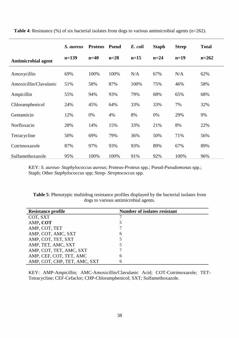
38
Table 4: Resistance (%) of six bacterial isolates from dogs to various antimicrobial agents (n=262).
KEY: S. aureus- Staphylococcus aureus; Proteus-Proteus spp.; Pseud-Pseudomonas spp.;
Staph; Other Staphylococcus spp; Strep- Streptococcus spp.
Table 5: Phenotypic multidrug resistance profiles displayed by the bacterial isolates from
dogs to various antimicrobial agents.
Resistance profile Number of isolates resistant
COT, SXT 7
AMP, COT 5
AMP, COT, TET 7
AMP, COT, AMC, SXT 6
AMP, COT, TET, SXT 5
AMP, TET, AMC, SXT 5
AMP, COT, TET, AMC, SXT 7
AMP, CEF, COT, TET, AMC 6
AMP, COT, CHP, TET, AMC, SXT 6
KEY: AMP-Ampicillin; AMC-Amoxicillin/Clavulanic Acid; COT-Cotrimoxazole; TET-
Tetracycline; CEF-Cefaclor; CHP-Chloramphenicol; SXT; Sulfamethoxazole.
Antimicrobial agent
S. aureus
n=139
Proteus
n=40
Pseud
n=28
E. coli
n=15
Staph
n=24
Strep
n=19
Total
n=262
Amoxycillin 69% 100% 100% N/A 67% N/A 62%
Amoxicillin/Clavulanic
acid
51% 58% 87% 100% 75% 46% 58%
Ampicillin 55% 94% 93% 79% 68% 65% 68%
Chloramphenicol 24% 45% 64% 33% 33% 7% 32%
Gentamicin 12% 0% 4% 8% 0% 29% 9%
Norfloxacin 28% 14% 15% 33% 21% 8% 22%
Tetracycline 50% 69% 79% 36% 50% 71% 56%
Cotrimoxazole 87% 97% 93% 93% 89% 67% 89%
Sulfamethoxazole 95% 100% 100% 91% 92% 100% 96%

39
4.1.4. Wound characteristics
Of the 80 samples collected from wounds in the retrospective study, only 58% (46/80) of
records were retrievable from the medical records. Wounds commonly involved the limbs of
the affected animals, with hindlimbs (32.6%) more affected than forelimbs (27.8%). The head
region was also frequently presented with wounds 8 out of 46 (17.3%), Table 6.
Surgical site infections were a more frequent source of wound swabs than other causes,
representing 23.9% of the sources. Bite wounds and traumatic wounds were also frequently
sampled for culture and susceptibility testing (Table 7). The cause of 18 wounds sampled
(33%) was not specified.
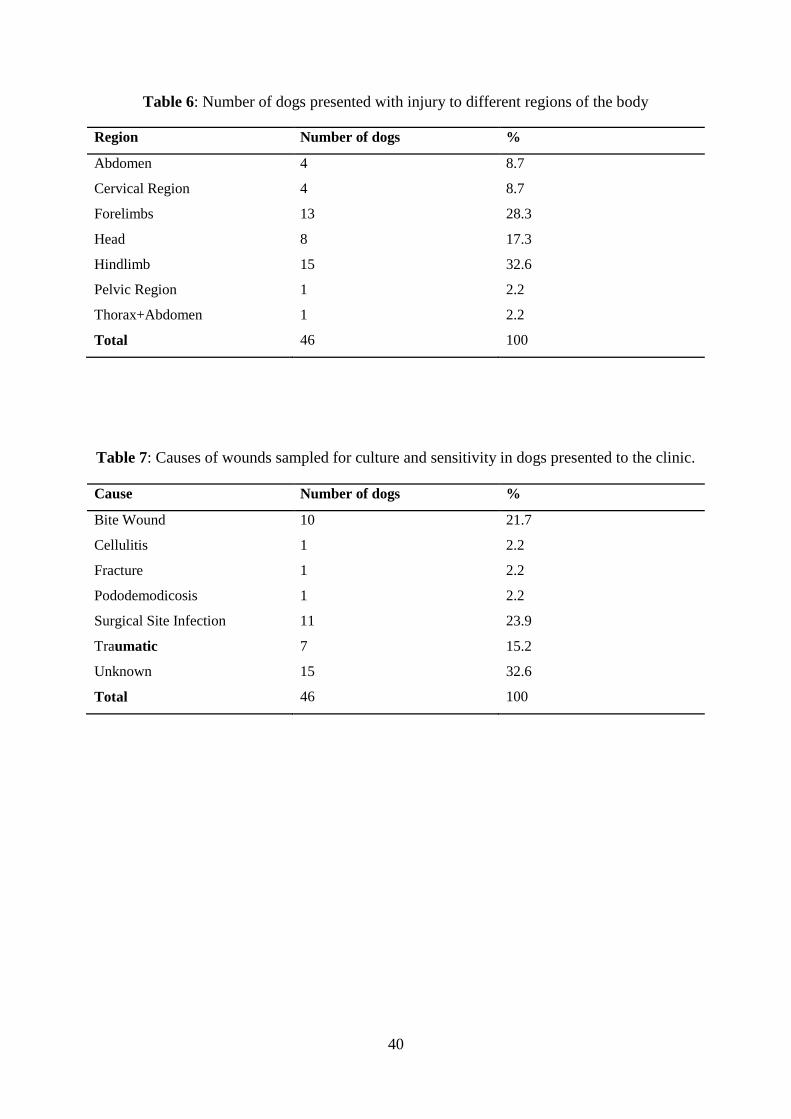
40
Table 6: Number of dogs presented with injury to different regions of the body
Region Number of dogs %
Abdomen 4 8.7
Cervical Region 4 8.7
Forelimbs 13 28.3
Head 8 17.3
Hindlimb 15 32.6
Pelvic Region 1 2.2
Thorax+Abdomen 1 2.2
Total 46 100
Table 7: Causes of wounds sampled for culture and sensitivity in dogs presented to the clinic.
Cause Number of dogs %
Bite Wound 10 21.7
Cellulitis 1 2.2
Fracture 1 2.2
Pododemodicosis 1 2.2
Surgical Site Infection 11 23.9
Traumatic 7 15.2
Unknown 15 32.6
Total 46 100
41
4.2. Prevalence of MRSA/MRSP from normal dogs and surgical patients
4.2.1. Clinical history and animal biodata
Samples from the Community veterinary clinic accounted for 103 (54%) of the samples,
while 88 samples (46%), were collected from the University of Nairobi Small Animal Clinic.
Seventy two (37.7%) of the 191 dogs sampled presented with wound(s) on their body. Males
were the predominant dogs sampled accounting for 56% (n=107) of the samples with 44 %
(n=84) being females. Majority of the animals (60 %) had received antimicrobial treatment in
the past three months prior to sampling.
4.2.2. Prevalence of staphylococci.
All the 191 samples successfully formed colonies in the enrichment media (Tryptone soya
broth). The selective media, MSA, detected 65 (34 %) presumptive staphylococci species, the
other samples yielded gram –ve bacteria which were not considered for further screening.
The Staphylococcus spp. were subjected to a tube coagulase test and only 28 (14.7%) of the
isolates tested positive and thus designated coagulase positive staphylococci.
4.2.3. Phenotypic characterisation of resistance
Antimicrobial susceptibility testing was done on the 28 coagulase positive isolates of which,
13 isolates (46.4%) were susceptible to oxacillin. Phenotypic resistance to oxacillin was
observed in 15 isolates (53.6%).
4.2.4. Validation of isolates
The control PCR was performed to exclude any false positive results. It was done using a
control primer pair targeting 416 bp fragment of 16S rRNA gene of genus Staphylococcus.
Eleven out of the 15 presumptive coagulase positive staphylococci, were confirmed to be
staphylococci (Figure 2)

42
4.2.5. Identification of coagulase positive staphylococci (COPS)
This PCR assay was done to differentiate the COPS by amplification of the 295 bp and 381bp
specific PCR product for Staphylococcus aureus and Staphylococcus pseudintermedius
respectively. Out of the 11 confirmed Staphylococcus species, this assay identified 7 (63.6%)
Staphylococcus aureus strains. No Staphylococcus pseudintermedius strains were detected in
this study.
4.2.6. MecA gene
Two mecA-positive MRSA strains were isolated from two dogs (Figure 3). One of the strains
was from the wound of a dog with a post-operative infection that resulted after inguinal
herniorrhaphy while the other was from a normal healthy puppy presented for vaccination.
4.2.7. BLAST analysis
4.2.7.1. Identification of DNA sequences
Analysis of the sequenced resistant determinants from the two samples revealed the genes
were harboured by Staphylococcus spp. strains. The nucleotide sequence of isolate 1 (Lab ID:
CS 100), was 97% identical to GenBank accession number AB547235.1, which is a
Staphylococcus sciuri mecA gene and 96% identical to GenBank accession number
KF058902.1 which is a Staphylococcus aureus mecA gene.
The nucleotide sequence of isolate 2 (Lab ID: CS 148) revealed 99% nucleotide identity to
sequences in the NCBI databases belonging to different Staphylococcus spp. This isolate was
99% identical to GenBank accession numbers KR187111.1, KP265312.1 and HE984157.2
which were mecA genes from Staphylococcus aureus, Staphylococcus epidermidis and
Staphylococcus pseudintermedius respectively (Table 8).
43
4.2.7.2. Geographical distribution and host diversity of the homologue genes
The homologues containing the mecA gene showed varied global distribution with isolates
from Brazil, Japan, China, Madagascar, Israel and Ireland. These strains were isolated from
diverse sources including human, dogs, rodents and primates and with different conditions
(Table 9).
4.2.7.3. Accesion numbers
The sequenced resistance gene submitted to the GenBank database was validated and
subsequently assigned the accession number KX689749.
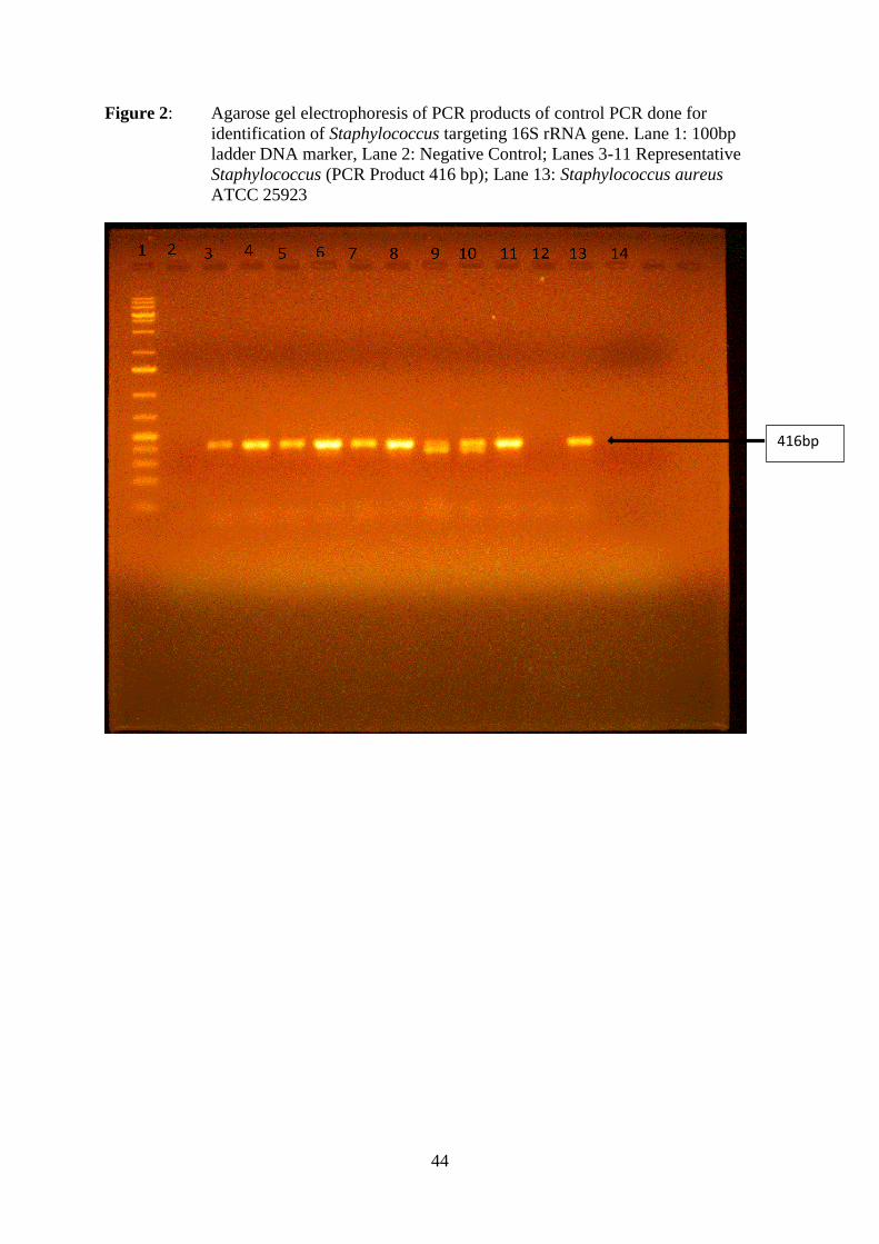
44
Figure 2: Agarose gel electrophoresis of PCR products of control PCR done for
identification of Staphylococcus targeting 16S rRNA gene. Lane 1: 100bp
ladder DNA marker, Lane 2: Negative Control; Lanes 3-11 Representative
Staphylococcus (PCR Product 416 bp); Lane 13: Staphylococcus aureus
ATCC 25923
416bp

45
Figure 3: Agarose gel electrophoresis of PCR products of mecA positive strains. Lane 1:
1KB ladder DNA marker, Lane 3-6 mecA positive isolates (Positive PCR
product 286 bp), Lane 7 Positive control.
286bp 200bp
400bp
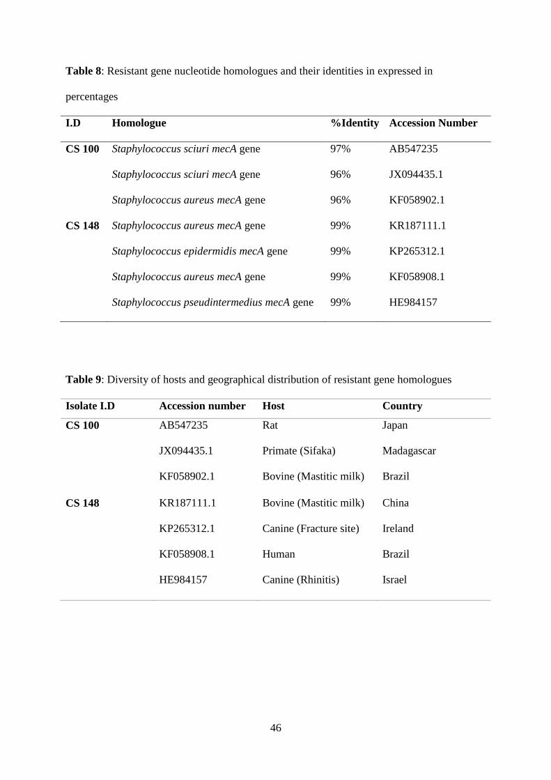
46
Table 8: Resistant gene nucleotide homologues and their identities in expressed in
percentages
I.D Homologue %Identity Accession Number
CS 100 Staphylococcus sciuri mecA gene 97% AB547235
Staphylococcus sciuri mecA gene 96% JX094435.1
Staphylococcus aureus mecA gene 96% KF058902.1
CS 148 Staphylococcus aureus mecA gene 99% KR187111.1
Staphylococcus epidermidis mecA gene 99% KP265312.1
Staphylococcus aureus mecA gene 99% KF058908.1
Staphylococcus pseudintermedius mecA gene 99% HE984157
Table 9: Diversity of hosts and geographical distribution of resistant gene homologues
Isolate I.D Accession number Host Country
CS 100 AB547235
JX094435.1
KF058902.1
Rat
Primate (Sifaka)
Bovine (Mastitic milk)
Japan
Madagascar
Brazil
CS 148 KR187111.1
KP265312.1
KF058908.1
HE984157
Bovine (Mastitic milk)
Canine (Fracture site)
Human
Canine (Rhinitis)
China
Ireland
Brazil
Israel
47
CHAPTER FIVE
5.0. DISCUSSION, CONCLUSION AND RECOMMENDATIONS
5.1. Discussion
In this study, Staphylococcus spp. were the most common isolates from samples submitted in
the laboratory. The findings of the retrospective study confirmed the etiological and clinical
importance of Staphylococcus organisms as colonisers of skin and important causes of
infection in the skin of animals. The high percentage of staphylococci (59.1%) was expected
since Staphylococcal species are present on or in clinically normal individuals as commensals
(Weese, 2010). However, they are opportunistic pathogens including S. pseudintermedius as
well as S. aureus as leading cause of surgical site infections in animals (Vengust et al., 2006;
Turk, 2015). The observation in this study that Staphylococcus aureus was the most prevalent
isolate from wounds is similar to reports from Bangladesh (Rahman et al., 2003) In contrast,
Vincze et al. (2014) recorded a low prevalence of Staphylococcus aureus from wounds with
isolation rates of 5.8% and 12.2% for dogs and cats respectively in Germany. Staphylococcus
aureus has been recognized as an important wound pathogen and a major cause of delayed
wound healing and infection. The prevalence of Staphylococcus aureus (37.5%) isolated from
wounds of dogs in Kenya has previously not been reported.
The high prevalence of Staphylococcus aureus in this study was surprising. Other authors
(Meyers et al., 2007; Urumova et al., 2012; Padhy et al., 2014) have reported Staphylococcus
intermedius to be the major isolate from wounds in dogs. While dogs and cats may become
colonised, contaminated and infected with Staphylococcus aureus, they are not considered
reservoir hosts of this organism (Cohn and Middleton, 2010). The predominant
Staphylococcus spp. in dogs has been reported to be Staphylococcus pseudintermedius

48
(Griffeth et al., 2008; Hanselmann et al., 2009). This finding may be due to the fact that at the
laboratory, all coagulase positive Staphylococci were designated as Staphylococcus aureus.
In the present study, E. coli, Streptococcus spp., other Staphylococcus spp. and Proteus spp.,
were other microorganisms isolated from the wound swabs. This finding is similar to the
study by Rahman et al. (2003) in Bangladesh, who isolated E. coli, Klebsiella spp. and
Proteus spp. in wound swabs. The results of the present study are also in agreement with
Urumova et al. (2012) who also found a high incidence of enterobactericiae in particlular
E.coli in wounds. In this study, the polymicrobial growth was demonstrated, with 24 % of the
swabs yielding more than one organism was consistent with other reports of similar nature
conducted elsewhere (Meyers, 2007; Padhy et al., 2014). Colonisation in wounds is mostly
polymicrobial involving different potentially pathogenic microorganisms (Bowler et al.,
2001). The number and diversity of microorganisms in any wound is influenced by several
factors among them are wound type, depth, location, and quality, the level of tissue perfusion,
and the antimicrobial efficacy as well as the host immune response.
In vitro antimicrobial agent susceptibility of the isolates showed a high frequency of resistant
strains, with 97% of the isolates showing resistance to at least one drug.. These observations
are the cause for concern as they are an indication of existence of multidrug resistant isolates
among dogs that might pose a clinical as well as therapeutical challenges.
In the retrospective study, 58% of bacteria isolated from ear swabs belonged to the Genus
Staphylococcus, this is comparable to other studies (Lilenbaum et al., 2000; Lyskova et al.,
2007; Petrov et al., 2013). Malayeri et al. (2010) reported a high prevalence of 73.8% of
Staphylococcus spp. in Iran. Other bacteria isolated in the present study included Proteus spp.
16.5%, Pseudomonas spp. 12.3% and Streptococcus spp. 4.7% were comparable to a previous
study by Mande and Kitaa (2005) where Staphylococcus aureus was found to be the most
49
prevalent isolate (51.2%), and Streptococcus spp. (14%), Pseudomonas spp. (14%) and
Proteus spp. (10%) also commonly isolated (Mande and Kitaa, 2005). This study
demonstrated an increase in staphylococcal isolation from otitis externa to 58% vs 51.2%
compared to a previous Kenyan study. This study also shows that Proteus spp. is increasingly
becoming an important pathogen with a prevalence of 16.5% up from 10% in the study by
Mande and Kitaa (2005).
Previous studies in dogs, reported the pathogens isolated from wounds to be most sensitive to
potentiated sulphonamides and amoxicillin/clavulanic acid preparations (Meyers et al., 2007;
Urumova et al., 2012). This observation is not in agreement with findings in this study where
comparatively higher resistance rates were observed to potentiated sulphonamides (89%) and
amoxycillin/clavulanic (58%). Interestingly, Pedersen et al. (2007) found no resistance to
amoxicillin/clavulanic acid in their study which involved bacterial isolates from clinical
submissions in Denmark. Earlier reports by Authier et al. (2006) suggested
amoxicillin/clavulanic acid to be an appropriate antimicrobial for treatment of skin infections
by Staphylococcus spp. However, based on the results of this study, the use of these
antimicrobials as the first line of treatment for empirical therapy might result in treatment
failure, if the observation made represnts the general population in Kenya. The findings of
this study demonstrated that gentamicin and norfloxacin were the most effective antimicrobial
agents against majority of the isolates. Gentamicin has been indicated for the treatment of
Staphylococcal infections (Lilenbaum et al., 2000). However, Authier et al. (2006) suggests
that its use should be limited to cases where initial treatment has failed.
In the present study, surgical site infections were found to be a frequent cause for wound
swabbing representing 24% of the wound swabs. Seventy five percent of surgical site
infections sampled resulted from fracture fixation using an implant. Turk et al. (2015)
reported the use of implants increases the risk for surgical site infections. Gallagher et al.

50
(2012) and Turk et al. (2015) further points out that, implants frequently become colonized
with bacteria and may also act as substrates for bacterial biofilm formation. Majority of the
wounds sampled were located on the extremeties with the hindlimbs being more affected than
the forelimbs. These results are comparable with the report by Shamir et al. (2002) where the
extremeties and the head were reported as the most frequent sites of bite wounds in dogs.
Similarly, Meyers et al. (2007) also observed majority of wounds to involve the cranial half
of the body, especially the head and thoracic limbs in dogs.
With respect to the susceptibility of coagulase positive Staphylococcus spp. to various
antimicrobials the present study found 97% of the Staphylococcus isolates to be resistant to at
least one drug.This finding is in agreement with a previous report which is in agreement with
a report by Lilenbaum et al. (2000) who reported Staphylococcus isolates to display a high
level of resistance in Brazil. They found 90.9 % of the isolates in their study to show
resistance to at least one drug. However, the findings of this study were in contrast with
thefindings reported by Junco and Barrasa (2002) who reported only 64.8% of COPS
displaying resistance.
In the present study, the least effective antimicrobials against Staphylococcus aureus were
sulphonamides (sulfamethoxazole), potentiated sulphonamides (cotrimoxazole), ampicillin
and tetracycline. The highest level of resistance noted was for potentiated sulphonamides with
a resistance rate of 95 %. Lilenbaum et al. (2000) also found majority of staphylococcal
isolates in Brazil resistant to this drug though at a lower rate 72.7 %. On the other hand, a
study in Denmark by Pedersen et al. (2007) described very low resistance of S.intermedius ear
isolates to this drug combination. The most effective agent against Staphylococci was
gentamicin, chloramphenicol and norfloxacin. Gentamicin susceptibility rate was 88% which
is similar to the one reported by Lilenbaum et al. (2000). Most of the isolates were also found
51
to be susceptible to amoxicillin (62%), suggesting that this drug can be used as a first line of
treatment prior to results of antimicrobial susceptibility testing.
Pseudomonas and Proteus isolates observed in this study displayed the highest resistance to
most antimicrobial agents. Pseudomonas spp. are mostly isolated in chronic cases of canine
otitis externa (Scott et al., 2001; Greene, 2006). This organism has been reputed for its high
level of resistance to most antimicrobials. The multidrug resistance was observed to be the
case in this study, with 92% of Pseudomonas spp. isolates showing resistance to 4 or more
drugs. Highest resistance was recorded to amoxicillin, and sulfamethoxazole, with all the
isolates tested against these drugs showing 100% resistance. These isolates also showed high
resistance to ampicillin (93%) and amoxicillin/clavulanic acid (87%). Significant resistance to
chloramphenicol (64%) and tetracycline (79%) was also observed in the Pseudomonas spp.
isolates in this study. Pedersen et al. (2007) in their study found that all the Pseudomonas spp.
isolates were resistant to ampicillin, amoxicillin/clavulanic acid and erythromicin. Malayeri et
al. (2010) also concurred with these observations with all Pseudomonas spp. isolates in their
study showing 100% resistance to ampicillin, amoxicillin/clavulanic, erythromicin, rifampin
and penicillin G. In another study, Hariharan et al. (2006) found that Pseudomonas isolates to
be highly resistant to chloramphenicol (99%) and doxycycline (98%). The least antimicrobial
resistance in this study was observed against gentamicin (4%) and norfloxacin (15%). This
observation is in agreement with Petersen et al. (2002) who reported most Pseudomonas
aeruginosa isolates to be 100% susceptible to the two drugs.
In the present study, Proteus spp. was the second most frequently isolated microorganism
after Staphylococcus aureus, accounting for 14.2% of all isolates; a finding that was similar to
reports by other researchers (Pedersen et al., 2007; Lyskova et al., 2007; Petrov et al., 2013 ).
The present study found all isolates to be resistant to amoxicillin and sulphonamides, but
susceptible to gentamicin. High resistance was observed against ampicillin (94%),
52
cotrimoxazole (97%) and moderate resistance to tetracyclines (69%), amoxicillin/clavulanic
(58%) and chloramphenicol (45%). Similar results have previously been reported by Petrov et
al. (2013), who found all isolates to be susceptible to gentamicin. In addition, they observed
resistance to tetracycline (81%) and chloramphenicol (74%) though at higher rates, and the
isolates in their study were resistant to ampicillin. In contrast to this study, Pedersen et al.
(2007), found all Proteus spp. isolates in their study to be resistant to tetracyclines and
majority of the isolates susceptible to gentamicin and ciprofloxacin, which is in agreement to
the findings in this study.
Prospective screening of dogs in this study showed a carriage rate of 34% (65/191) of
Staphylococcus spp. This may be due to the prolonged storage time of some samples (up to 8
months for a few samples). In other studies, samples were cultured within 12 hrs (Gingrich et
al., 2011) and 24 - 36 hours of collection (Bergstrom et al., 2012; Walther et al., 2012). The
recent use of antimicrobial agents in most of the study animals prior to sampling may have led
to suppression of the number of commensal bacteria, especially those resident on the skin.
Detection of the mecA gene by PCR revealed 2 out of the 15 (13%) phenotypically resistant
isolates to be genotypically resistant to methicillin (oxacillin). These two isolates contained
the mecA gene that encodes for resistance to β-lactam antibiotics. Ozturk et al. (2010)
reported similar results in their study where all 5 Staphylococcus spp. isolates that were
phenotypically resistant to oxacillin were mecA negative on PCR. This discrepancy between
phenotypic and genotypic resistance in the isolates has been reported by Schmidt et al., 2014
and Elhassan et al., 2015. This discrepancy could be due to existence of the so-called
borderline (low-level resistant) strains. These mecA negative strains are thought to result from
overproduction of β-lactamase (Chambers, 1997). Other mechanisms associated with
borderline resistance include acquisition of modified PBPs (Elhassan et al., 2015). The
53
existence of these borderline strains emphasise the need to screen mecA negative strains for
other resistance mechanisms.
Two genes are known to encode for methicillin resistance in Staphylococcus spp. namely
mecA and mecC. However in this study only mecA was investigated for genotypic
characterization of methicillin resistance, since reports of mecC positive MRSA isolates are
low with prevalence of 0-3% reported in European countries (Paterson et al., 2014). Previous
studies have also shown mecA to be the most common gene encoding for methicillin
resistance in Staphylococcus spp. (Weese, 2010; van Duijkeren et al., 2011).
No MRSPs were observed in this study, which is similar to reports by Garbacz et al. (2011),
their study involved 39 Staphylococcus pseudintermedius isolates from clinical submissions
and found all the isolates to be susceptible to oxacillin. Several studies by different authors
have also failed to isolate any MRSP isolates (Murphy et al., 2009; Rubin and Chirino-Trejo,
2011; Schmidt et al., 2014). In most of these studies, the investigators collected samples from
healthy animals. In the present study, samples were collected both from normal and clinically
sick animals, some of which had received antibiotic treatment prior to sampling. This study
found a prevalence rate of 7% (2/28) of MRSA among coagulase positive Staphylococcus
spp. and an overall prevalence of 1% (2/191). In a Swedish animal hospital, no MRSA was
isolated from surgical patients and healthy animals, although the prevalence of MRSA in the
environment was found to be 5.3% (Bergstrom et al., 2012). The low prevalence of MRSA in
this study is also similar to reports by Couto et al. (2011) who reported MRSA prevalence of
1% from 287 dogs and cats presented to a veterinary teaching hospital in Portugal, and is
consistent with findings of Quitoco et al. (2013). A higher prevalence of MRSA of 15.8% was
observed in a study on surgical site infections (Turk et al., 2015). A recent report by Aiken et
al. (2014) found a similar prevalence (7%) of MRSA among Staphylococcus aureus strains
isolated from patients admitted to a hospital in Kiambu County, Kenya. One of the MRSA
54
isolates in this study was recovered from a patient with a surgical site infection. The patient
had been hospitalised for a week prior to the infection and was under treatment with an
antimicrobial agent. This finding is similar to that by Middleton et al. (2005) whose sole
postoperative MRSA isolate was from a canine patient with an orthopaedic pin tract infection.
MRSA has emerged as an important pathogen in post-operative infections in previous studies
(Tomlin et al., 1999; Turk et al., 2015). Antimicrobial drug therapy, hospitalization and
surgery have been cited as factors predisposing to MRSA infection (Loeffler et al., 2005;
Magalhaes et al., 2010; Faires et al., 2010; Davis et al., 2013). Multidrug resistant
Staphylococci isolated from dogs with post-operative infections and wounds should raise
suspicion of MRSA infection and appropriate care taken in handling such patients.
Analysis of the sequenced resistant determinants showed that the resistant genes were
harboured by Staphylococcus spp. strains and that the resistant determinant is geographically
widespread across various regions of the globe having previously been isolated from countries
such as Ireland (McManus et al., 2015), China (Wu et al., 2015), Brazil (Melo et al., 2013),
Israel (Perreten et al., 2013). The strains have been isolated from different Staphylococcus
spp. isolated from bovine (milk), canine (orthopaedic implant and rhinitis) and human clinical
submissions. The mecA genes contained in SCCmec have been reported to be almost identical
regardless of the Staphylococcus species carrying it (Tsubakishita et al., 2010). This
observation alludes to the fact that the mecA gene is transferable from different
Staphylococcus spp. The mecA gene encodes for penicillin binding (PBP2) protein. PBP 2a,
is a low-affinity penicillin-binding protein (PBP) that mediates methicillin resistance in
Staphylococcus spp. (Weese, 2010). This modified PBP2 has a low affinity for β -lactams and
therefore cell wall construction is not prevented by these antimicrobials (van Duijkeren et al.,
2011).
55
Haphazard use of antimicrobials prior to testing can lead to selection of multidrug resistant
strains. This may have been the case for one of the Staphylococcus aureus strains isolated that
showed resistance to the complete panel of the 8 drugs tested against. This strain was isolated
from a dog suffering from recurrent otitis externa. Selective pressure exerted by previous
antimicrobial treatment in recurrent cases may lead to emergence of resistant strains
(Guardabassi et al., 2004). Occurrence of antimicrobial resistance in companion animals is of
significance to human health (Hawkey, 2008). The close contact between household pets and
humans offers favourable conditions for the transmission of bacteria by direct contact or
indirectly through contamination of the environment. Transmission of mobile resistance
determinants between companion animals and humans may also occur (Guardabassi et al.,
2004).
56
5.2. Conclusions
1. The present study confirms that the most prevalent microorganisms associated with
wounds and otitis externa were Staphylococcus aureus (50.5%), Proteus spp.
(14.04%), Pseudomonas spp. (9.82%), Other Staphylococcus spp. (8.42%),
Streptococcus spp. (7.67%) and E. coli (5.62%).
2. From the results of our study, gentamicin, an aminoglycoside, is the most effective
antimicrobial agent against all the isolates from wounds, surgical site infections and
otitis externa in dogs. Norfloxacin, a fluoroquinolone, is relatively effective against
Gram negative isolates (Proteus spp. and Pseudomonas spp.) and Streptococcus spp.
3. Pseudomonas spp. and E. coli from otitis externa and wounds respectively, are the
most challenging organisms to treat in dogs.
4. The study findings report the first two cases of MRSA isolated from a normal healthy
dog and a dog with a surgical site infection in Kenya. The study observed MRSA
prevalence of 7% among coagulase positive Staphylococcus spp.
5. The resistant determinant mecA in this study was similar to some MRSA strains from
human patients in other parts of the world and therefore demonstrates the zoonotic
importance of these resistant strains.
5.3. Recommendations
1. Judicial use of antimicrobial agents is recommended especially the newer antibiotics
to prevent development of resistance.
2. Development of antimicrobial treatment guidelines in companion animals or adoption
of existing ones for empiric therapy.

57
3. Establishment of veterinary practice infection control policies to prevent spread of
resistant isolates between patients and in- contact staff.
4. Further investigation is necessary to determine the extent of MRSA carriage by
companion animals and livestock.
5. Further research is necessary to evaluate the factors leading to acquisition of these
resistant isolates
58
CHAPTER SIX
6.0. REFERENCES AND APPENDICES
6.1. References
Abraham, J., Morris D., Griffeth, G., Shofer, F., Rankin, S. (2007). Surveillance of
healthy cats and cats with inflammatory skin disease for colonization of the skin by
methicillin-resistant coagulase-positive staphylococci and Staphylococcus schleiferi
ssp. schleiferi. Veterinary Dermatology 18: 252-259.
Aiken, A.M., Mutuku, I.M., Sabat, A.J., Akkerboom, V., Mwangi, J., Scott, J.A.G,
Morpeth, S.C., Friedrich, A.W. and Grundmann, H. (2014). Carriage of
Staphylococcus aureus in Thika Level 5 Hospital, Kenya: a cross-sectional study.
Antimicrobial resistance and infection control 3(1): 22.
Asfour, H.A.E. and Darwish, S.F. (2014). Evaluation of Phenotypic Methods versus
Molecular Methods for Differentiation of Coagulase Positive Staphylococci causing
Bovine Mastitis with a Special Reference to atypical Staphylococcus aureus.
International Journal of Current Microbiology and Applied Science 3(5): 543-558.
Association of Surgical Technologists (AST) standards. (2008). Standards of practice for
surgical attire, surgical scrub, hand hygiene and hand washing. Retrieved from:
http://www.ast.org/uploadedFiles/Main_Site/Content/About_Us/Standard_Surgical_A
ttire_Surgical_Scrub.pdf.
Authier, S., Paquette, D., Labrecque, O. and Messier, S. (2006). Comparison of
susceptibility to antimicrobials of bacterial isolates from companion animals in a
veterinary diagnostic laboratory in Canada between 2 time points 10 yrs apart.
Canadian Veterinary Journal 47: 774-778.
59
Baptiste, K., Williams, K., Williams, N., Wattret, A., Clegg, P., Dawson, S., Corkill, J.,
O’Neill, T. and Hart C. (2005). Methicillin-resistant staphylococci in companion
animals. Emerging Infectious Diseases 11: 1942–1944.
Beck, K. M., Waisglass, S. E., Dick, H. L. and Weese, J. S. (2012). Prevalence of
methicillin resistant Staphylococcus pseudintermedius (MRSP) from skin and carriage
sites of dogs after treatment of their methicillin-resistant or methicillin-sensitive
staphylococcal pyoderma. Veterinary Dermatology 23:369-75.
Bemis, D. A., Jones, R. D., Frank, L. A. and Kania, S. A. (2009). Evaluation of
susceptibility test breakpoints used to predict mecA-mediated resistance in
Staphylococcus pseudintermedius isolated from dogs. Journal of Veterinary
Diagnostic Investigation 21: 53-58.
Bergström, A., Gustafsson, C., Leander, M., Fredriksson, M., Grönlund, U., and
Trowald-Wigh, G. (2012). Occurrence of methicillin-resistant Staphylococci in
surgically treated dogs and the environment in a Swedish animal hospital. Journal of
Small Animal Practice 53(7): 404-410.
Bond, R. and Loeffler, A. (2012). What’s happened to Staphylococcus intermedius?
Taxonomic revision and emergence of multi-drug resistance. Journal of Small Animal
Practice 53: 147–154.
Bowler, P.G., Duerden, B.I. and Armstrong, D.G. (2001). Wound microbiology and
associated approaches to wound management. Clinical Microbiology Reviews 14:
244-269.
Brown, D.F. (2001). Detection of methicillin/oxacillin resistance in staphylococci. Journal of
Antimicrobial Chemotherapy 48(1): 65–70.
60
Brown, J., Edwards, I. and Hawkey, M. (2005). Guidelines for the laboratory diagnosis and
susceptibility testing of methicillin resistant Staphylococcus aureus (MRSA). Journal
of Antimicrobial Chemotherapy 56(6): 1000-1018.
Cain, C.L. (2013). Methicillin resistant Staphylococcal infections. Today’s Veterinary
Practice, 3 (3):26-32.
Chambers, H.F. (1997). Methicillin resistance in staphylococci: molecular and biochemical
basis and clinical implications. Clinical Microbiology Review 10: 781–791.
CLSI (Ed) (2013) Performance standards for antimicrobial disk and dilution susceptibility
tests for bacteria isolated from animals; Approved Standard, 4th edition. CLSI
document VET01-A. Wayne, PA: Clinical and Laboratory Standards Institute
CLSI (2008). Performance Standards for Antimicrobial Disk and Dilution Susceptibility
Test for Bacteria Isolated from Animals; Approved Standard, 3rd edition. CLSI
document M31-A3. Wayne, PA: Clinical and Laboratory Standards Institute.
Cohn, L.A. and Middleton, J.R. (2010). A Veterinary Perspective on Methicillin Resistant
Staphylococci. Journal of Veterinary Emergency and Critical Care 20: 31-45.
Couto, N., Pomba, C., Moodley, A. and Guardabassi, L. (2011). Prevalence of methicillin-
resistant staphylococci among dogs and cats at a veterinary teaching hospital in
Portugal. Veterinary Record 162: 72.
Daunton, C., Kothari, S., Smith, L. and Steele, D.A. (2012). A History Of Materials And
Practices For Wound Management. Wound Practice and Research 20(4): 174-186.
Davis, K.A., Stewart, J.J., Crouch, H.K., Florez, C.E. and Hospenthal, D.R. (2004).
Methicillin-resistant Staphylococcus aureus (MRSA) nares colonization at hospital
admission and its effect on subsequent MRSA infection. Clinical Infectious Disease
39:776–82.
61
Davis, M.F., Peterson, A.E., Julian, K.G., Greene, W.H., Price, L.B., Nelson, K.,
Whitener, C.J., Silbergeld, E.K. (2013). Household risk factors for colonization with
multidrug-resistant Staphylococcus aureus isolates
8.doi:10.1371/journal.pone.0054733
Diaz- Campos, D.V. (2012). Molecular epidemiology and genetic analysis of Staphylococcus
species in companion animal medicine (Doctorate dissertation). Retrieved from
https://etd.auburn.edu/bitstream/handle/10415/3066/Dissertation,%20D%20Diaz,%20
4%2030%202012.pdf?sequence=2
Duquette, R.A. and Nuttall, T.J. (2004). Methicillin-resistant Staphylococcus aureus in
dogs and cats: an emerging problem? Journal of Small Animal Practice 45: 591-597.
Elhassan, M.M., Hani, A.O., Hassan, A.H., Miskelyemen A.E., and Leila, M.A. (2015).
Absence of the mec A Gene in Methicillin Resistant Staphylococcus aureus Isolated
from Different Clinical Specimens in Shendi City , Sudan. BioMed Research
International. doi: 10.1155/2015/895860
European Medicines Agency (EMA). (2010). Reflection paper on Methicillin-resistant
Staphylococcus pseudintermedius.
Faires, M., Tater, K. and Weese, J.S. (2009). An investigation of methicillin-resistant
Staphylococcus aureus colonization in people and pets in the same household with an
infected person or infected pet. Journal of the American Veterinary Medical
Association 235(5): 540-543.
Faires, M.C., Traverse, M., Tater, K.C., Pearl, D.L., Weese, J.S. (2010). Methicillin-
resistant and -susceptible Staphylococcus aureus infections in dogs. Emerging Infectious
Diseases. 16:69–75. doi:10.3201/eid1601.081758
62
Fazakerley, J., Williams, N., Carter, S., McEwan, N. and Nuttall, T. (2010).
Heterogeneity of Staphylococcus pseudointermedius isolates from atopic and healthy
dogs. Veterinary Dermatology 21: 578–585.
Frank, L.A. and Loeffler, A. (2012). Methicillin resistant Staphylococcus
pseudointermedius; Clinical challenge and treatment options. Veterinary Dermatology
23: 283-291
Gallagher, A.D., and Mertens, W.D. (2012). Implant removal rate from infection after tibial
plateau leveling osteotomy in dogs. Veterinary Surgery 41: 705- 711.
Garbacz, K., Piechowicz, L., Żarnowska, S., Haras, K. and Dąbrowska-Szponar, M.
(2011). Heterogeneity of methicillin-sensitive Staphylococcus pseudintermedius
strains isolated from diseased dogs. Polish Journal of Veterinary Sciences 14(2): 283-
284.
Gingrich, E.N., Kurt, T., Hyatt, D.R., Lappin, M.R. and Ruch-Gallie, R. (2011).
Prevalence of methicillin-resistant staphylococci in northern Colorado shelter animals.
Journal of Veterinary Diagnostic Investigation 23: 947-950.
Gomez, P., Gonzalez-Barrio, D., Benito, D., Garcıa, J.T., Vinuela, J., Zarazaga, M.,
Ruiz-Fons, F. and Torres, C. (2014). Detection of methicillin-resistant
Staphylococcus aureus (MRSA) carrying the mecC gene in wild small mammals in
Spain. Journal of Antimicrobial Chemotherapy 69(8): 2061-2064.
Goni, P., Vergara, Y., Ruiz, J., Albizu, I., Vila, J. and Gomez-Lus, R. (2004). Antibiotic
resistance and epidemiological typing of Staphylococcus aureus strains from ovine
and rabbit mastitis. International Journal of Antimicrobial Agents 23(3): 268-272
Greene, C.E. (2006). Otitis Externa. In: Greene CE (ed) Infectious Diseases of the Dog and
Cat, 3rd edn. Saunders, Missouri: 815–823.
63
Griffeth, G.C., Morris, D.O., Abraham, J.L., Shofer, F.S. and Rankin, S.C. (2008).
Screening for skin carriage of methicillin-resistant coagulase-positive staphylococci
and Staphylococcus schleiferi in dogs with healthy and inflamed skin. Veterinary
Dermatology 19: 142–149.
Guardabassi, L., Loeber, M.E. and Jacobson, A. (2004). Transmission of multiple
antimicrobial-resistant Staphylococcus intermedius between dogs affected by deep
pyoderma and their owners. Veterinary Microbiology 98: 23-27.
Hafeez, R., Chughtai, A.S. and Aslam, M. (2004). Prevalence and antimicrobial
susceptibility of methicillin resistant S. aureus (MRSA). International Journal of
Pathology 2(1): 10–15.
Han, Z., Lautenbach, E., Fishman, N. and Nachamkin, I. (2007). Evaluation of mannitol
salt agar, CHROMagar Staph aureus and CHROMagar MRSA for detection of
meticillin-resistant Staphylococcus aureus from nasal swab specimens. Journal of
Medical Microbiology 56(1):43-46.
Hanselman, B.A., Kruth, S.A., Rousseau, J. and Weese, J.S. (2009). Coagulase positive
staphylococcal colonization of humans and their household pets. Canadian Veterinary
Journal 50: 954-958.
Hanselman, B.A., Kruth, S.A., Rousseau, J., Low, D.E., Willey, B.M. (2006). Methicillin
resistant Staphylococcus aureus colonization in veterinary personnel. Emerging
Infectious Diseases 12(12): 1933–1938.
Hariharan, H., Coles, M., Poole, D., Lund, L. and Page, R. (2006). Update on
antimicrobial susceptibilities of bacterial isolates from canine and feline otitis externa.
Canadian Veterinary Journal 47:253–255.
Harris, S.R., Cartwright, E.J., Török, M.E., Holden, M.T., Brown, N.M., Ogilvy Stuart,
A.L., Ellington, M.J., Quail, M.A., Bentley, S.D., Parkhill, J. and Peacock, S.J.
64
(2013). Whole-genome sequencing for analysis of an outbreak of methicillin resistant
Staphylococcus aureus: a descriptive study. Lancet Infectious Diseases 13: 130–136.
Hartmann, F.A., Trostle, S.S. and Klohnen, A.A. (1997). Isolation of methicillin-resistant
Staphylococcus aureus from a postoperative wound infection in a horse. Journal of the
American Veterinary Medical Association 211:590-592.
Hawkey, P.M. (2008). The growing burden of antimicrobial resistance. Journal of
Antimicrobial Chemotherapy 62 Suppl. 1: i1-i9.
Horstmann, C., Mueller, R.S., Straubinger, R.K. and Werckenthin, C. (2012). Detection
of methicillin resistant Staphylococcus pseudintermedius with commercially available
selective media. Letters in Applied Microbiology 54: 26-31.
Ishihara, K., Shimokubo, N., Sakagami, A., Ueno, H., Muramatsu, Y., Kadosawa, T.,
Yanagisawa, C., Hanaki, H., Nakajima, C., Suzuki, Y. and Tamura, Y. (2010).
Occurrence and molecular characteristics of methicillin-resistant Staphylococcus
aureus and methicillin-resistant Staphylococcus pseudintermedius in an academic
Veterinary Hospital. Applied Environmental Microbiology 76: 5165-5174.
Jain, A., Agarwal, A., Verma, R.K. (2008). Cefoxitin disc diffusion test for detection of
Methicillin resistant Staphylococci. Journal of Medical Microbiology 57: 957-961.
Julian, T., Singh, A., Rousseau, J. and Weese, J.S. (2012). Methicillin resistant
staphylococcal contamination of cellular phones of personell in a veterinary teaching
hospital. BMC Research Notes 5: 193-197.
Junco, T.M. T. and Barrasa Martin J.L. (2002). Identification and antimicrobial
susceptibility of coagulase positive Staphylococci isolated from healthy dogs and dogs
suffering from otitis externa. Journal of Veterinary Medicine B 49: 419–423.
65
Kampf, G. and Kramer, A. (2004). Epidemiologic background of hand hygiene and
evaluation of the most important agents for scrubs and rubs. Clinical Microbiology
Review 17:863–893.
Kateete, D.P., Kimani, C.N., Katabazi, F.A., Okeng, A., Okee, M.S., Nanteza, A., Joloba,
M.L. and Najjuka, F.L. (2010). Identification of Staphylococcus aureus: Dnase and
Mannitol salt agar improve the efficiency of the tube coagulase test. Annals of Clinical
Microbiology and Antimicrobials 9:23-30.
Kondo, Y., Ito, T., Ma, X. X., Watanabe, S., Kreiswirth, B. N., Etienne, J. and
Hiramatsu, K. (2007). Combination of Multiplex PCRs for Staphylococcal Cassette
Chromosome mec Type Assignment: Rapid Identification System formec, ccr, and
Major Differences in Junkyard Regions. Antimicrobial Agents and
Chemotherapy 51(1):264–274.
Kuehnert, M.J., Hill, H.A., Kupronis, B.A., Tokars, J.I., Solomon, S.L., and Jernigan,
D.B. (2005). Methicillin-resistant Staphylococcus aureus hospitalizations, United
States. Emerging Infectious Diseases 11:868–72.
Leonard, F., Abbott, Y., Rossney, A., Quinn, P., O’Mahony, R. and Markey, B. (2006).
Methicillin-resistant Staphylococcus aureus isolated from a veterinary surgeon and
five dogs in one practice. Veterinary Record 158: 155–159.
Lilenbaum, W., Veras, M., Blum, E. and Souza, G.N. (2000). Antimicrobial susceptibility
of Staphylococci isolated from otitis externa in dogs. Letters in Applied Microbiology
31:42–45.
Loeffler, A., Boag, A.K., Sung, J., Lindsay, J.A. and Guardabassi, L., Dalsgaard, A.,
Smith, H., Stevens, K. B. and Lloyd, D. H. (2005). Prevalence of methicillin-
resistant Staphylococcus aureus among staff and pets in a small animal referral
hospital in the UK. Journal of Antimicrobial Chemotherapy 56(4): 692–697.

66
Loeffler, A., Linek, M., Moodley, A., Guardabassi, L., Sung, J.M., Winkler, M., Weiss,
R. and Lloyd, D.H. (2007). First report of multiresistant, mecA-positive
Staphylococcus intermedius in Europe: 12 cases from a veterinary dermatology
referral clinic in Germany. Veterinary Dermatology 18:412-421.
Loeffler, A., Pfeiffer, D.U., Lloyd, D.H., Smith, H., Soares-Magalhaes, R. and Lindsay,
J.A. (2010). Methicillin resistant Staphylococcus aureus carriage in U.K veterinary
staff and owners of infected pets: new risk groups. Journal of Hospital Infections
74(3): 282-288.
Lyskova, P., Vydrzalova, M. and Mazurova, J. (2007). Identification and antimicrobial
susceptibility of bacteria and yeasts isolated from healthy dogs and dogs with otitis
externa. Journal of Veterinary Medicine A 54: 559-563.
Magalhaes, S.R.J., Loeffler, A., Lindsay, J., Rich, M., Roberts, L., Smith, H., Lloyd, D.
H. and Pfeiffer, D. U. (2010). Risk factors for methicillin-resistant Staphylococcus
aureus (MRSA) infection in dogs and cats: a case-control study. Veterinary Research
41:55.
Maina, E. K., Kiiyukia, C., Wamae, C. N., Waiyaki, P. G. and Kariuki, S. (2013).
Characterization of methicillin-resistant Staphylococcus aureus from skin and soft
tissue infections in patients in Nairobi, Kenya. International Journal of Infectious
Diseases 17(2): e115-119.
Malayeri, H.Z., Jamshidi, S. and Salehi, T.Z. (2010). Identification and antimicrobial
susceptibility patterns of bacteria causing otitis externa in dogs Veterinary Research
Communications 34:435–444.
Mande, J.D and Kitaa, J.M.A. (2005). Microbial profile and antimicrobial susceptibility of
isolates from dogs with otitis in Kenya. The Kenya Veterinarian 29: 40-44.
67
Manian, F.A. (2003). Asymptomatic nasal carriage of mupirocin-resistant, methicillin-
resistant Staphylococcus aureus (MRSA) in a pet dog associated with MRSA infection
in household contacts. Clinical Infection and Disease 36: e26–e28.
Mason, I.S., Mason, K.V. and Llov, D.H. (1996). A review of the biology of canine skin
with respect to the commensals Staphylococcus intermedius, Demodex canis and
Malassezia pachydermatis. Veterinary Dermatology 7: 119-132.
McLean, C.L and Ness, M.G. (2008). Methicillin-resistant Staphylococcus aureus in a
veterinary orthopaedic referral hospital: staff nasal colonisation and incidence of
clinical cases. Journal of Small Animal Practice 49(4):170–177
McManus, B.A., Coleman, D.C., Deasy, E.C., Brennan, G.I., O’ Connell, B., Monecke,
S., Ralf Ehricht, R., Leggett, B., Leonard, N. and Shore, A.C. (2015). Comparative
Genotypes, Staphylococcal Cassette Chromosome mec (SCCmec) Genes and
Antimicrobial Resistance amongst Staphylococcus epidermidis and Staphylococcus
haemolyticus Isolates from Infections in Humans and Companion Animals. PLoS
ONE 10(9): e0138079.
Medhus A., Slettemea J.S., Marstein L, Larssen K.W. and Sunde M. (2012). Methicillin-
resistant Staphylococcus aureus with the novel mecC gene variant isolated from a cat
suffering from chronic conjunctivitis. Journal of Antimicrobial Chemotherapy, 0: 1–2.
Melo, D.A., Coelho, I.S., Motta, C.C., Rojas, A.C., Dubenczuk, F.C., Coelho, S.M.O. and
Souza, M.M.S. (2013). Implications of the use of human parameters for the mecA
gene detection to predict beta-lactam resistance in bovine Staphylococcus spp. isolated
from mastitis. Staphylococcus aureus strain 28C penicillin-binding protein (mecA)
gen - Nucleotide - NCBI.htm (Unpublished).
68
Meyers, B., Schoeman, J.P., Goddard, A. and Picard, J. (2007). The bacteriology and
antimicrobial susceptibility of infected and non-infected dog bite wounds: fifty cases.
Veterinary Microbiology 127(3-4): 360–368.
Middleton, J., Fales, W., Luby, C., Oaks, J., Sanchez, S., Kinyon, J., Wu, C., Maddox,
C., Welsh, R. and Hartmann, F. (2005). Surveillance of Staphylococcus aureus in
veterinary teaching hospitals. Journal of Clinical Microbiology 43:2916-2919.
Murphy, C., Reid-Smith, R. J., Prescott, J. F., Bonnett, B. N., Poppe, C., Boerlin, P.,
Weese, J. S., Janecko, N. and Mcewen, S. A. (2009). Occurrence of antimicrobial
resistant bacteria in healthy dogs and cats presented to private veterinary hospitals in
southern Ontario: preliminary study. Canadian Veterinary Journal 50: 1047-53
Neely, A. N. and Maley, M. P. (2000). Survival of Enterococci and Staphylococci on
hospital fabrics and plastic. Journal of Clinical Microbiology 38: 724-726.
Nelson, L.L. (2011). Surgical site infections in small animal practice. The Veterinary Clinics
of North America Small Animal Practice 41: 1041-1056.
Niemeyer, D.M., Pucci, M.J., Thanassi, J.A., Sharma, V.K. and Archer, G.L. (1996).
Role of mecA transcriptional regulation in the phenotypic expression of methicillin
resistance in Staphylococcus aureus. Journal of Bacteriology 178 (18): 5464–5471.
Nienhoff, U., Kadlec, K., Chaberny, I.F., Verspohl, J., Gerlach, G.F., Kreienbrock, L.,
Schwarz, S., Simon, D. and Nolte, I. (2011). Methicillin-resistant Staphylococcus
pseudintermedius among dogs admitted to a small animal hospital. Veterinary
Microbiology 150:191-197.
Njoroge, C.W., Mande, J.D., Mitema, E.S. and Kitaa, J.M.A. (2016). Multidrug resistance
of common bacterial patogens from wounds and otitis externa in small animals during
a 10 year period in Kenya. International Journal of Veterinary Science 5(4): 262-267

69
Normand, E.H., Gibson, N.R., Reid, S.W., Carmichael, S. and Taylor, D.J. (2000).
Antimicrobial-resistance trends in bacterial isolates from companion animal
community practice in the UK. Preventative Veterinary Medicine 46: 267–278.
Öztürk, D., Avki, S., Türütoğlu, H., Yiğitarslan, K. and Sağnak, S. (2010). Methicillin
resistance among Coagulase-positive Staphylococci isolated from dogs with Otitis
Externa, Skin Wounds and Pyoderma. Kafkas Universitesi Veteriner Fakultesi Dergisi
16(4): 651-656.
Padhy, A., Mishra, R., Behera, S.S., Sahu, A.R. and Sahoo, S. (2014). Microbial profile of
canine persistent wound infections. Veterinary World 7(4): 244-247.
Paterson, G.K., Harrison, E.M. and Holmes, M.A. (2014). The emergence of mecC
methicillin-resistant Staphylococcus aureus. Trends in Microbiology 22: 42–47.
Paterson, G.K., Morgan, F.J.E., Harrison, E.M., Peacock, S.J., Parkhill, J., Zadoks, R.N.
and Holme, M.A. (2014). Prevalence and properties of mecC methicillin resistant
Staphylococcus aureus (MRSA) in bovine bulk tank milk in Great Britain. Journal of
Antimicrobial Chemotherapy 69: 598–602.
Paul, N.C., Bargman, S.C., Moodley, A., Nielsen, S.S. and Guardabassi, L. (2012).
Staphylococcus pseudintermedius colonization patterns and strain diversity in healthy
dogs: a cross-sectional and longitudinal study. Veterinary Microbiology 160: 420–427.
Paul, N.C., Moodley, A., Ghibaudo, G. and Guardabassi, L. (2011). Carriage of
Methicillin-Resistant Staphylococcus pseudointermedius in small animal
veterinarians; Indirect Evidence of Zoonotic transmission. Zoonoses and Public
Health 8: 533-539.

70
Pedersen, K., Jensen, H., Finster, K., Jensen, V.F. and Heuer, O.E. (2007). Occurrence of
antimicrobial resistance in bacteria from diagnostic samples from dogs. Journal of
Antimicrobial Chemotherapy 60:775 – 781.
Perreten, V., Kadlec K., Schwarz S., Grönlund Andersson U., Finn M., Greko C.,
Moodley A., Kania S.A., Frank L.A., Bemis D.A., Franco A., Iurescia M.,
Battisti A., Duim B., Wagenaar J.A., van Duijkeren E., Weese J.S., Fitzgerald
J.R., Rossano A. and Guardabassi L. (2010). Clonal spread of methicillin- resistant
Staphylococcus pseudintermedius in Europe and North America: an international
multicentre study. Journal of Antimicrobial Chemotherapy 65:1145 – 1154.
Perreten,V., Chanchaithong, P., Nuvee Prapasarakul, N., Rossano, A., Shlomo, E.,
Blum, S.E., Elad, D. and Schwendener S. (2013). Novel Pseudo-Staphylococcal
Cassette Chromosome mec Element (SCCmec57395) in Methicillin-Resistant
Staphylococcus pseudintermedius CC45. Antimicrobial Agents and Chemotherapy
57(11): 5509–5515.
Petersen, A. D., Walker, R. D., Bowman, M. M., Schott, H. C. and Rosser Jr., E. J.
(2002). Frequency of isolation and antimicrobial susceptibility patterns of
Staphylococcus intermedius and Pseudomonas aeruginosa isolates from canine skin
and ear samples over a 6-year period (1992– 1997). Journal of American Animal
Hospitals Association 38: 407–13
Petinaki, E. and Spiliopoulou, I. (2012). Methicillin-resistant Staphylococcus aureus among
companion and food-chain animals; Impact of human contact. Clinical Microbiology
and Infection 18: 626-634.
Petrov, V., Mihaylov, G., Tsachev, I., Zhelev G., Marutsov, P. and Koe, K. (2013). Otitis
externa in dogs: microbiology and antimicrobial susceptibility. Revue de Médecine
Vétérinaire 164 (1): 18-22.
71
Pinchbeck, L.R., Cole, L.K., Hillier, A., Kowalski, J.J., Rajala-Schultz, P.J., Bannerman,
T.L. and York, S. (2006). Genotypic relatedness of staphylococcal strains isolated
from pustules and carriage sites in dogs with superficial bacterial folliculitis. American
Journal of Veterinary Research 67: 1337–1346.
Quitoco, I.M., Raimundo, M.S., Silva-Carvalho, M.C., Souza, R.R., Beltrame, C.O.,
Oliveira, T.F., Araújo, R., del Peloso, P.F., Coelho, L.R. and Figueiredo, A.M.
(2013). First report in South America of companion animal colonization by the
USA1100 clone of community-acquired methicillin-resistant Staphylococcus aureus
(ST30) and by the European clone of methicillin-resistant Staphylococcus
pseudintermedius (ST71) 336. BMC Research Notes 6:336. doi: 10.1186/1756-0500-
6-336.
Rahman, M.M., Biswas, D.B., Islam, M.M. and Islam, M.A. (2003). Cultural sensitivity of
septic wounds in animals. Pakistan Journal of Biological Sciences 6: 741-744.
Rich, M. and Roberts, L. (2004). Methicillin-resistant Staphylococcus aureus isolates from
companion animals. Veterinary Record 159: 535-536.
Riedel, S., Dam, L., Stamper, P.D., Shah, S.A.R. and Carro, K.C. (2010). Evaluation of
Bio-Rad MRSASelect Agar for detection of methicillin-resistant Staphylococcus
aureus directly from blood cultures. Journal of Clinical Microbiology 48(6): 2285–
2288.
Rostami, S., Moosavian, M., Shoja, S., Torabipour, M. and Farshadzadeh, Z. (2013).
Comparison of mecA gene based PCR with CLSI cefoxitin and oxacillin disc diffusion
methods for detecting methicillin resistance in Staphylococcus aureus clinical isolates.
African Journal of Microbiology Research 7(21): 2438- 2441.
Rubin, J.E. and Chirino-Trejo, M. (2011). Prevalence, sites of colonization, and
antimicrobial resistance among Staphylococcus pseudintermedius isolated from

72
healthy dogs in Saskatoon, Canada. Journal of Veterinary Diagnostic Investigation 23:
351–354.
Ruscher, C., Lübke-Becker, A., Wleklinski, C.G., Soba, A., Wieler, L.H. and Walther, B.
(2009). Prevalence of methicillin-resistant Staphylococcus pseudintermedius isolated
from clinical samples of companion animals and equidaes. Veterinary Microbiology
136:197-201.
Safdar, N. and Bradley, E.A. (2008). The risk of infection after nasal colonization with
Staphylococcus aureus. American Journal of Medicine 121: 310–315.
Saijonmaa-Koulumies, L.E. and Lloyd, D.H. (1996). Colonization of the canine skin with
bacteria. Veterinary Dermatology, 7:153-162.
Sajith- Khan, A.K., Preetha, J.S., Lakshi, S.Y., Anandi, C. And Ramesh, R. (2012).
Detection of mecA genes of methicillin resistant Staphylococcus aureus by PCR.
International Journal of Health and Rehabilitation Science 1(2): 64-68.
Sasaki, T., Kikuchi, K., Tanaka, Y., Takahashi, N., Kamata, S. and Hiramatsu, K.
(2007). Methicillin-resistant Staphylococcus pseudintermedius in a veterinary teaching
hospital. Journal of Clinical Microbiology 45:1118–1125.
Schissler, J.R., Hillier, A., Daniels, J.B., Cole, L.K. and Gebreyes, W.A. (2009).
Evaluation of Clinical Laboratory Standards Institute interpretive criteria for
methicillin-resistant Staphylococcus pseudintermedius isolated from dogs. Journal of
Veterinary Diagnostic Investigation 21: 684-688.
Schmidt, V.M., Williams, N.J., Pinchbeck, G., Corless, C.E., Shaw, S., McEwan, N.,
Dawson, S. and Nuttall, T. (2014). Antimicrobial resistance and characterisation of
staphylococci isolated from healthy Labrador retrievers in the United Kingdom. BMC
Veterinary Research 10: 17.
73
Scott, D.W., Miller, W.H. and Griffin, C.E. (2001). Muller and Kirk's small animal
dermatology. 6th ed. W.B. Saunders; Philadelphia:pp 913.
Shamir, M.H., Leisner, S., Klement, E., Gonen, E. and Johnston, De. (2002). Dog bite
wounds in dogs and cats: a retrospective study of 196 cases. Journal of Veterinary
Science A 49: 107-112.
Sing, A., Tuschak, C. and Hörmansdorfer, S. (2008). Methicillin-resistant Staphylococcus
aureus in a family and its pet cat. New England Journal of Medicine 358:1200–1.
Singh, A., Walker, M., Rousseau, J., Monteith, G.J. and Weese, J.S. (2013). Methicillin-
resistant Staphylococcal contamination of clothing worn by personell in a veterinary
teaching hospital. Veterinary Surgery 42: 643-648.
Slatter, D. (2003). Textbook of small animal surgery, 3rd Edition. (ed. Slatter, D). W.B
Saunders Company, Philadelphia: pp 163-178.
Stevens, A.M., Hennesy, T., Bagett, H.C., Bruden, D., Parks, D. and Klejka, J. (2010).
Methicillin resistant Staphylococcus aureus carriage and risk factorsfor skin
infections, Southwestern Alaska, USA. Emerging Infectious Diseases 16: 797-803.
Pavletic, M.M. (2003). The Integument. In Slatter D.J. (Ed): Textbook of Small Animal
Surgery, 3rd edn. W.B. Saunders Company, Philadelphia.pp 250-259.
Petersen, A. D., Walker, R.D., Bowman, M. M., Schott, H. C., Rosser, E. J. JR.
(2002). Frequency of isolation and antimicrobial susceptibility patterns of
Staphylococcus intermedius and Pseudomonas aeruginosa isolates from canine skin
and ear samples over a 6-year period (1992-1997). Journal of American Animal
Hospitals Assocciation. 38: 407-413.
Perreten, V., Kadlec, K., Schwarz, S., Gronlund A.U., Finn, M., Greko, C., Moodley, A.,
Kania, S. A., Frank, L. A., Bemis, D. A., Franco, A., Iurescia, M., Battisti, A.,
Duim, B., Wagenaar, J. A., Van Duijkeren, E., Weese, J. S., Fitzgerald, J.R.,

74
Rossano, A. and Guardabassi, L. (2010). Clonal spread of methicillin-resistant
Staphylococcus pseudintermedius in Europe and North America: an international
multicentre study. Journal of Antimicrobial Chemotherapy. 65:1145-54.
Tomlin, J., Pead, M.J., Lloyd, D.H., Howell, S., Hartmann, F., Jackson, H.A. and Muir,
P. (1999). Methicillin-resistant Staphylococcus aureus infections in 11 dogs.
Veterinary Record 144: 60–64.
Tsubakishita, S., Kuwahara-Arai, K., Sasaki, T., and Hiramatsu, K. (2010). Origin and
Molecular Evolution of the Determinant of Methicillin Resistance in Staphylococci.
Antimicrobial Agents and Chemotherapy 54(10): 4352–4359.
Turk, R. (2013). Prospective evaluation of epidemiology and microbiology of surgical site
infections (Masters Thesis). Retrieved from
https://atrium.lib.uoguelph.ca/xmlui/bitstream/handle/10214/7431/Turk_Ryen_20130
Msc.pdf?sequence=1.
Turk, R., Singh, A. and Weese, J.S. (2015). Prospective Surgical Site Infection Surveillance
in Dogs. Veterinary Surgery 44(1): 2-8.
Urumova, V., Chaprazov, T.S., Lyutskanov, M. and Borisov, I. (2012). Microbiological
analyses of canine infected wounds. Revue Médecin Veterinaire 163(4): 201-205.
Van Duijkeren, E., Catry, B., Greko, C., Moreno, M.A., Pomba, M.C., Pyörälä, S.,
Ruzauskas, M., Sanders, P., Threlfall, E.J., Torren-Edo, J. and Törneke, K.
(2011). Review on methicillin-resistant Staphylococcus pseudintermedius. Journal of
Antimicrobial Chemotherapy 66 (12): 2705-2714.
Van Duijkeren, E., Jansen, M.D., Flemming, S.C., de Neeling, H., Wagenaar, J.A.,
Schoormans, A.H., van Nes, A. and Fluit, A.C. (2007). Methicillin-resistant
Staphylococcus aureus in pigs with exudative epidermitis. Emerging Infectious
Diseases 13:1408-1410.

75
Van Duijkeren, E., Box, A.T., Heck, M.E., Wannet, W.J. and Fluit, A.C. (2004).
Methicillin resistant staphylococci isolated from animals. Veterinary Microbiology
103:91–97.
Van Duijkeren, E., Hengeveld, P.D., Albers, M., Pluister, G., Jacobs, P. and Heres, L.
(2014). Prevalence of methicillin resistance Staphylococcus aureus carrying mecA or
mecC in dairy cattle. Veterinary Microbiology. 171(3-4): 364-367.
Velasco V., Sherwood J.S., Rojas-Garcı P.P. and Logue, C.M. (2014). Multiplex Real-
Time PCR for Detection of Staphylococcus aureus, mecA and Panton Valentine
Leukocidin (PVL) Genes from Selective Enrichments from Animals and Retail Meat.
PLoS One 9(5): e97617.
Vengust, M., Anderson, M.E.C., Rousseau, J. and Weese, J.S. (2006). Methicillin-resistant
staphylococcal colonization in clinically normal dogs and horses in the community.
Letters in Applied Microbiology 43:602-606.
Verwilghen, D. and Kampf, G. (2011). Presurgical Hand Antisepsis: Concepts and Current
Habits of Veterinary Surgeons. Veterinary Surgery 40:515–521.
Vincze, S., Stamm, I., Kopp, P. A., Hermes, J., Adlhoch, C., Semmler, T., Wieler, L. H.,
Lubke-Becker, A. and Walther, B. (2014). Alarming proportions of methicillin-
resistant Staphylococcus aureus (MRSA) in wound samples from companion animals,
Germany 2010-2012. PLoS One 9(1): e85656.
Walther, B., Hermes, J., Cuny, C., Wieler, L.H., Vincze, S., Abou E.Y., Stamm, I., Kopp,
P.A., Kohn, B., Witte, W., Jansen, A., Conraths, F.J., Semmler, T., Eckmanns, T.
and Lübke-Becker, A. (2012). Sharing more than friendship--nasal colonization with
coagulase-positive staphylococci (CPS) and co-habitation aspects of dogs and their
owners. PloS One 7(4): e35197.
Wang, J.T., Chang S.C., Ko, W.J., Chang, Y.Y., Chen, M.L., Pan, H.J. and Luh, K.T.
(2001). A hospital-acquired outbreak of methicillin-resistant Staphylococcus aureus
infection initiated by a surgeon carrier. Journal of Hospital Infection 47:104–109.
76
Weber, S., Herwaldt, L. A., Mcnutt, L.A., Rhomberg, P.,Vaudaux, P., Pfaller, M.A. and
Perl. T.M. (2002). An outbreak of Staphylococcus aureus in a pediatric cardiothoracic
surgery unit. Infection Control and Hospital Epidemiology 23:77–81.
Weese, J.S. and Duijkeren, V.E. (2009). Methicillin-resistant Staphylococcus aureus and
Staphylococcus pseudintermedius in veterinary medicine. Veterinary. Microbiology
140:418-429.
Weese, J.S. (2010). Methicillin resistant Staphylococcus aureus in animals. Institute for
Laboratory Animal Research Journal 51:233–244.
Weese, J. S. (2012). Staphylococcal control in the veterinary hospital. Veterinary
Dermatology 23: 292-298, e57-8.
Windahl, U., Reimega˚, R.D., Holst, B.S., Egenvall, A., Fernstro¨, M.L., Fre-Driksson,
M., Trowald-Wigh, G. and Andersson, U.G. (2012). Carriage of methicillin-
resistant Staphylococcus pseudintermedius in dogs—a longitudinal study. BMC
Veterinary Research 8: 34- 42.
Wu, Z., Li, F., Liu, D., Xue, H. and Zhao, X. (2015). Novel type XII of Staphylococcal
cassette chromosome mec harboring a new cassette chromosome recombinase, CcrC2
allele. Antimicrobial Agents and Chemotherapy 59(12):7597-7601.
http://lpi.oregonstate.edu/mic/micronutrients-health/skin-health.

77
6.2. Appendices
Appendix 1: Microbial isolates from wounds and ear swabs of dogs from the UoN
Small Animal Clinic (2004-2013).
No. Year Species Sex Age Breed Source IRT
1 2005 Canine M A GSD Abscess Actinomyces pyogenes
2 2006 Canine M A GSD Ear swab Actinomyces pyogenes
3 2009 Canine F Cross wound Coliforms
4 2007 Canine M A GSD Ear swab Corynebacterium
5 2007 Canine M A GSD Ear swab Corynebacterium
6 2007 Canine F A Cross Ear swab Corynebacterium
7 2012 Canine M A J.Spitz wound Corynebacterium
8 2013 Canine M A GSD wound Corynebacterium
9 2007 Canine M * GSD Ear swab Diphtheroids
10 2004 Canine F A J.Spitz wound E.coli
11 2004 Canine F A Cross wound E.coli
12 2007 Canine M A GSD Ear swab E.coli
13 2008 Canine M 7yrs+ Cross wound E.coli
14 2008 Canine M A Dachschund wound E.coli
15 2009 Canine F 6mnths J.Spitz wound E.coli
16 2009 Canine F 4mnt/yr J.Spitz wound E.coli
17 2009 Canine F A J.Spitz wound E.coli
18 2009 Canine F A J.Spitz wound E.coli
19 2009 Canine M A Cross wound E.coli
20 2009 Canine F 11mnths GSD wound E.coli
21 2010 Canine M A GSD Abscess E.coli
22 2013 Canine F A GSD wound E.coli
23 2010 Canine M * GSD Ear Swab E.coli
24 2010 Canine M A GSD wound E.coli
25 2006 Canine M A Terrier Ear swab Fungi
26 2009 Canine F A GSD wound Klebsiella
27 2007 Canine F * GSD Abscess Nocardia
28 2008 Canine M A Cross wound Pasteurella
29 2004 Canine M A Cross Ear Swab Proteus spp.
30 2004 Canine M A Cross Ear swab Proteus spp.
31 2004 Canine M A Cross Ear Swab Proteus spp.
32 2004 Canine F A * Ear Swab Proteus spp.
33 2005 Canine M A GSD Ear swab Proteus spp.
34 2005 Canine M A GSD Ear Swab Proteus spp.
35 2005 Canine M A GSD Ear Swab Proteus spp.
36 2005 Canine M A GSD Ear Swab Proteus spp.
37 2005 Canine M A GSD Ear swab Proteus spp.
38 2005 Canine * * * Ear swab Proteus spp.

78
39 2005 Canine M A GSD Ear swab Proteus spp.
40 2005 Canine M A Husky
Cross
Ear swab Proteus spp.
41 2005 Canine M A GSD Ear swab Proteus spp.
42 2005 Canine M A GSD Ear swab Proteus spp.
43 2006 Canine M A * Ear swab Proteus spp.
44 2007 Canine M A GSD Ear swab Proteus spp.
45 2007 Canine F A GSD Ear swab Proteus spp.
46 2007 Canine M A Jack Rusell Ear swab Proteus spp.
47 2007 Canine M A Jack Rusell Ear swab Proteus spp.
48 2007 Canine M A GSD Ear swab Proteus spp.
49 2007 Canine M A GSD Ear swab Proteus spp.
50 2007 Canine M A * Ear swab Proteus spp.
51 2007 Canine M A GSD Ear swab Proteus spp.
52 2008 Canine M A Cross Ear Swab Proteus spp.
53 2008 Canine F A GSD Ear Swab Proteus spp.
54 2009 Canine M Puppy GSD Ear Swab Proteus spp.
55 2009 Canine M A * wound Proteus spp.
56 2011 Canine M A Cross Ear Swab Proteus spp.
57 2011 Canine M A GSD Ear Swab Proteus spp.
58 2012 Canine F A GSD Ear Swab Proteus spp.
59 2012 Canine M A GSD Ear Swab Proteus spp.
60 2012 Canine M 3yrs Rottweiler wound Proteus spp.
61 2012 Canine M A GSD Ear Swab Proteus spp.
62 2013 Canine M A J.Spitz wound Proteus spp.
63 2013 Canine M A GSD Ear Swab Proteus spp.
64 2013 Canine M A Ridgeback Ear Swab Proteus spp.
65 2010 Canine M A GSD Ear Swab Proteus spp.
66 2010 Canine F A GSD Ear Swab Proteus spp.
67 2004 Canine F A Beagle Ear Swab Pseudomonas spp.
68 2004 Canine M A GSD Ear Swab Pseudomonas spp.
69 2005 Canine M A GSD Ear swab Pseudomonas spp.
70 2005 Canine M A GSD Ear swab Pseudomonas spp.
71 2005 Canine * A GSD Ear Swab Pseudomonas spp.
72 2005 Canine F Puppy Poodle Ear Swab Pseudomonas spp.
73 2005 Canine M Puppy Poodle Ear Swab Pseudomonas spp.
74 2006 Canine M A GSD Ear swab Pseudomonas spp.
75 2006 Canine * A * Ear swab Pseudomonas spp.
76 2006 Canine M A GSD X Ear swab Pseudomonas spp.
77 2006 Canine M A GSD Ear swab Pseudomonas spp.
78 2008 Canine F A GSD Ear Swab Pseudomonas spp.
79 2008 Canine F A Boerboel Ear Swab Pseudomonas spp.
80 2009 Canine M A Ridgeback Ear Swab Pseudomonas spp.
81 2009 Canine M A GSD Ear Swab Pseudomonas spp.
82 2009 Canine F A GSD Ear Swab Pseudomonas spp.
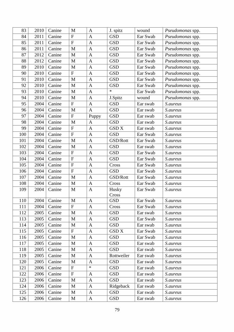
79
83 2010 Canine M A J. spitz wound Pseudomonas spp.
84 2011 Canine F A GSD Ear Swab Pseudomonas spp.
85 2011 Canine F A GSD Ear Swab Pseudomonas spp.
86 2011 Canine M A GSD Ear Swab Pseudomonas spp.
87 2012 Canine M A GSD Ear Swab Pseudomonas spp.
88 2012 Canine M A GSD Ear Swab Pseudomonas spp.
89 2010 Canine M A GSD Ear Swab Pseudomonas spp.
90 2010 Canine F A GSD Ear Swab Pseudomonas spp.
91 2010 Canine M A GSD Ear Swab Pseudomonas spp.
92 2010 Canine M A GSD Ear Swab Pseudomonas spp.
93 2010 Canine M A * Ear Swab Pseudomonas spp.
94 2010 Canine M A J.Spitz wound Pseudomonas spp.
95 2004 Canine F A GSD Ear swab S.aureus
96 2004 Canine M A GSD Ear swab S.aureus
97 2004 Canine F Puppy GSD Ear swab S.aureus
98 2004 Canine M A GSD Ear swab S.aureus
99 2004 Canine F A GSD X Ear swab S.aureus
100 2004 Canine F A GSD Ear Swab S.aureus
101 2004 Canine M A GSD/Rott Ear Swab S.aureus
102 2004 Canine M A GSD Ear swab S.aureus
103 2004 Canine F A GSD Ear Swab S.aureus
104 2004 Canine F A GSD Ear Swab S.aureus
105 2004 Canine F A Cross Ear Swab S.aureus
106 2004 Canine F A GSD Ear Swab S.aureus
107 2004 Canine M A GSD/Rott Ear Swab S.aureus
108 2004 Canine M A Cross Ear Swab S.aureus
109 2004 Canine M A Husky
Cross
Ear Swab S.aureus
110 2004 Canine M A GSD Ear Swab S.aureus
111 2004 Canine F A Cross Ear Swab S.aureus
112 2005 Canine M A GSD Ear swab S.aureus
113 2005 Canine M A GSD Ear Swab S.aureus
114 2005 Canine M A GSD Ear swab S.aureus
115 2005 Canine F A GSD X Ear Swab S.aureus
116 2005 Canine M A GSD Ear Swab S.aureus
117 2005 Canine M A GSD Ear swab S.aureus
118 2005 Canine M A GSD Ear swab S.aureus
119 2005 Canine M A Rottweiler Ear swab S.aureus
120 2005 Canine M A GSD Ear swab S.aureus
121 2006 Canine F * GSD Ear swab S.aureus
122 2006 Canine F A GSD Ear swab S.aureus
123 2006 Canine M A GSD Ear swab S.aureus
124 2006 Canine M A Ridgeback Ear swab S.aureus
125 2006 Canine M A GSD Ear swab S.aureus
126 2006 Canine M A GSD Ear swab S.aureus
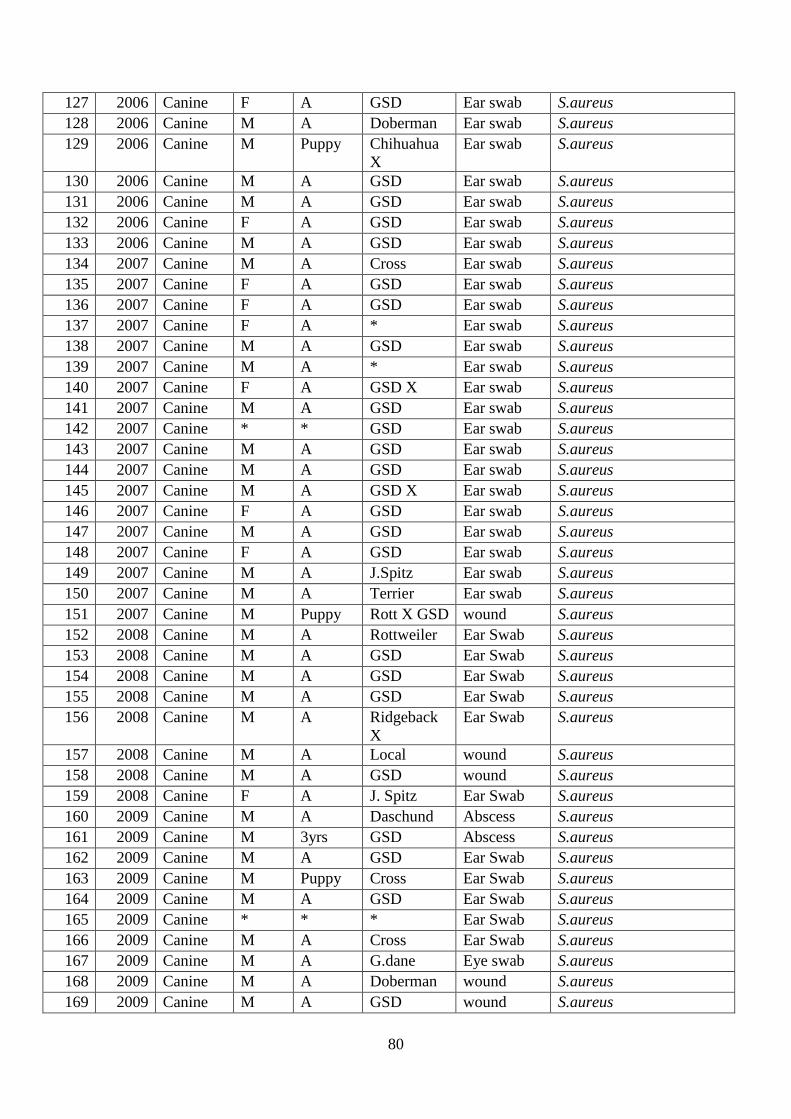
80
127 2006 Canine F A GSD Ear swab S.aureus
128 2006 Canine M A Doberman Ear swab S.aureus
129 2006 Canine M Puppy Chihuahua
X
Ear swab S.aureus
130 2006 Canine M A GSD Ear swab S.aureus
131 2006 Canine M A GSD Ear swab S.aureus
132 2006 Canine F A GSD Ear swab S.aureus
133 2006 Canine M A GSD Ear swab S.aureus
134 2007 Canine M A Cross Ear swab S.aureus
135 2007 Canine F A GSD Ear swab S.aureus
136 2007 Canine F A GSD Ear swab S.aureus
137 2007 Canine F A * Ear swab S.aureus
138 2007 Canine M A GSD Ear swab S.aureus
139 2007 Canine M A * Ear swab S.aureus
140 2007 Canine F A GSD X Ear swab S.aureus
141 2007 Canine M A GSD Ear swab S.aureus
142 2007 Canine * * GSD Ear swab S.aureus
143 2007 Canine M A GSD Ear swab S.aureus
144 2007 Canine M A GSD Ear swab S.aureus
145 2007 Canine M A GSD X Ear swab S.aureus
146 2007 Canine F A GSD Ear swab S.aureus
147 2007 Canine M A GSD Ear swab S.aureus
148 2007 Canine F A GSD Ear swab S.aureus
149 2007 Canine M A J.Spitz Ear swab S.aureus
150 2007 Canine M A Terrier Ear swab S.aureus
151 2007 Canine M Puppy Rott X GSD wound S.aureus
152 2008 Canine M A Rottweiler Ear Swab S.aureus
153 2008 Canine M A GSD Ear Swab S.aureus
154 2008 Canine M A GSD Ear Swab S.aureus
155 2008 Canine M A GSD Ear Swab S.aureus
156 2008 Canine M A Ridgeback
X
Ear Swab S.aureus
157 2008 Canine M A Local wound S.aureus
158 2008 Canine M A GSD wound S.aureus
159 2008 Canine F A J. Spitz Ear Swab S.aureus
160 2009 Canine M A Daschund Abscess S.aureus
161 2009 Canine M 3yrs GSD Abscess S.aureus
162 2009 Canine M A GSD Ear Swab S.aureus
163 2009 Canine M Puppy Cross Ear Swab S.aureus
164 2009 Canine M A GSD Ear Swab S.aureus
165 2009 Canine * * * Ear Swab S.aureus
166 2009 Canine M A Cross Ear Swab S.aureus
167 2009 Canine M A G.dane Eye swab S.aureus
168 2009 Canine M A Doberman wound S.aureus
169 2009 Canine M A GSD wound S.aureus

81
170 2009 Canine M A Cross wound S.aureus
171 2009 Canine M A Cross wound S.aureus
172 2009 Canine M A Doberman wound S.aureus
173 2009 Canine M 4yrs G.dane Abscess S.aureus
174 2009 Canine F A GSD Ear Swab S.aureus
175 2010 Canine * 8yrs * Abscess S.aureus
176 2010 Canine F A Cross wound S.aureus
177 2010 Canine M A GSD wound S.aureus
178 2011 Canine F A Terrier Abscess S.aureus
179 2011 Canine M A GSD Ear Swab S.aureus
180 2011 Canine F A GSD Ear Swab S.aureus
181 2011 Canine M A Ridgeback Ear Swab S.aureus
182 2011 Canine M A GSD Ear Swab S.aureus
183 2011 Canine M A GSD Ear Swab S.aureus
184 2011 Canine F A Cross Ear Swab S.aureus
185 2011 Canine M A GSD Ear Swab S.aureus
186 2011 Canine F A GSD Ear Swab S.aureus
187 2011 Canine M A GSD Ear Swab S.aureus
188 2011 Canine M A GSD Ear Swab S.aureus
189 2011 Canine M A GSD Ear Swab S.aureus
190 2011 Canine M 6mnths * wound S.aureus
191 2011 Canine M * Labrador wound S.aureus
192 2011 Canine F 2.5yrs Rottweiler wound S.aureus
193 2012 Canine F A GSD Ear Swab S.aureus
194 2012 Canine F A French
Bullmastiff
Ear Swab S.aureus
195 2012 Canine F A Cross Ear Swab S.aureus
196 2012 Canine M A GSD Ear Swab S.aureus
197 2012 Canine M A GSD Ear Swab S.aureus
198 2012 Canine F A GSD Ear Swab S.aureus
199 2012 Canine F A GSD Ear Swab S.aureus
200 2012 Canine M A GSD Ear Swab S.aureus
201 2012 Canine M A GSD Ear Swab S.aureus
202 2012 Canine M A Local Ear Swab S.aureus
203 2012 Canine M A GSD wound S.aureus
204 2012 Canine F A Rottweiler wound S.aureus
205 2012 Canine M A Cross wound S.aureus
206 2012 Canine M 1yr GSD wound S.aureus
207 2012 Canine M A GSD Ear Swab S.aureus
208 2012 Canine F A GSD wound S.aureus
209 2012 Canine M A GSD Ear Swab S.aureus
210 2013 Canine M A GSD Ear Swab S.aureus
211 2013 Canine F A Cross wound S.aureus
212 2013 Canine M A Rottweiler wound S.aureus
213 2013 Canine * * Rottweiler wound S.aureus
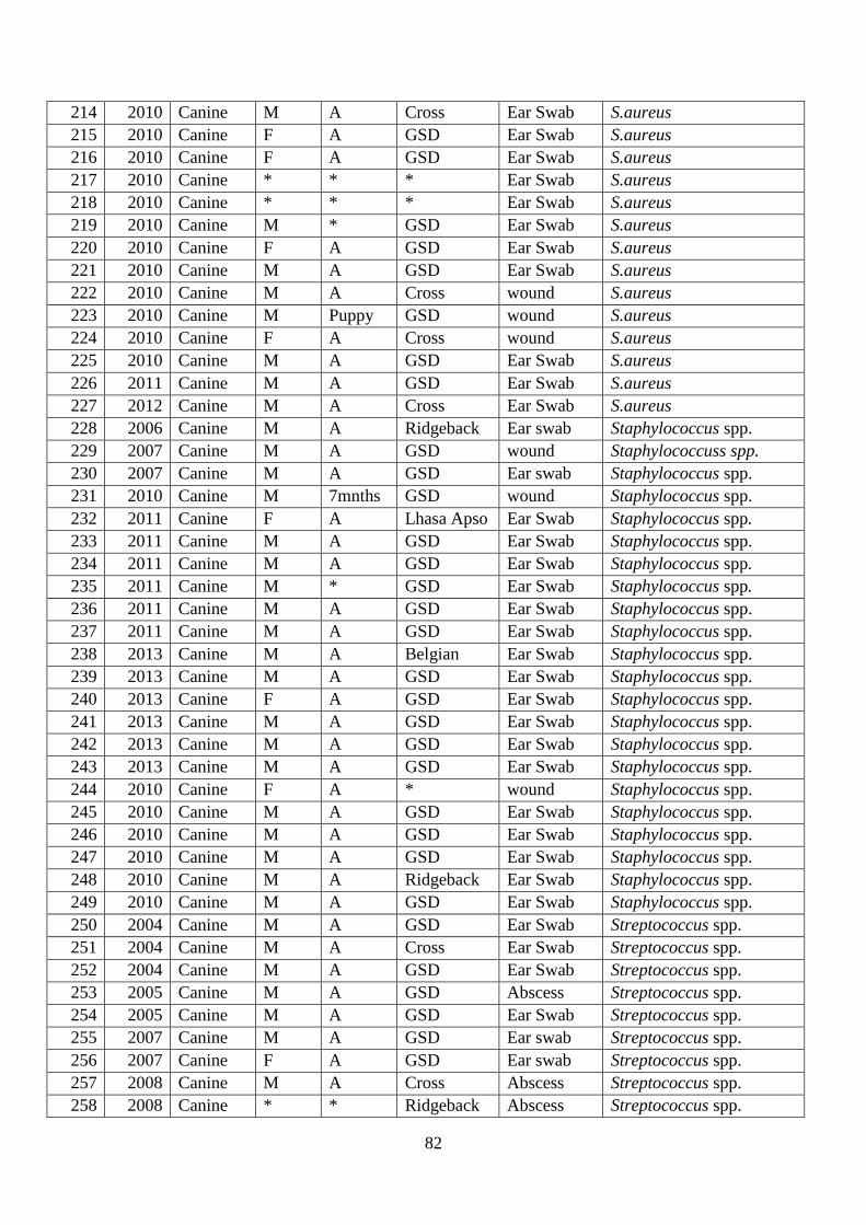
82
214 2010 Canine M A Cross Ear Swab S.aureus
215 2010 Canine F A GSD Ear Swab S.aureus
216 2010 Canine F A GSD Ear Swab S.aureus
217 2010 Canine * * * Ear Swab S.aureus
218 2010 Canine * * * Ear Swab S.aureus
219 2010 Canine M * GSD Ear Swab S.aureus
220 2010 Canine F A GSD Ear Swab S.aureus
221 2010 Canine M A GSD Ear Swab S.aureus
222 2010 Canine M A Cross wound S.aureus
223 2010 Canine M Puppy GSD wound S.aureus
224 2010 Canine F A Cross wound S.aureus
225 2010 Canine M A GSD Ear Swab S.aureus
226 2011 Canine M A GSD Ear Swab S.aureus
227 2012 Canine M A Cross Ear Swab S.aureus
228 2006 Canine M A Ridgeback Ear swab Staphylococcus spp.
229 2007 Canine M A GSD wound Staphylococcuss spp.
230 2007 Canine M A GSD Ear swab Staphylococcus spp.
231 2010 Canine M 7mnths GSD wound Staphylococcus spp.
232 2011 Canine F A Lhasa Apso Ear Swab Staphylococcus spp.
233 2011 Canine M A GSD Ear Swab Staphylococcus spp.
234 2011 Canine M A GSD Ear Swab Staphylococcus spp.
235 2011 Canine M * GSD Ear Swab Staphylococcus spp.
236 2011 Canine M A GSD Ear Swab Staphylococcus spp.
237 2011 Canine M A GSD Ear Swab Staphylococcus spp.
238 2013 Canine M A Belgian Ear Swab Staphylococcus spp.
239 2013 Canine M A GSD Ear Swab Staphylococcus spp.
240 2013 Canine F A GSD Ear Swab Staphylococcus spp.
241 2013 Canine M A GSD Ear Swab Staphylococcus spp.
242 2013 Canine M A GSD Ear Swab Staphylococcus spp.
243 2013 Canine M A GSD Ear Swab Staphylococcus spp.
244 2010 Canine F A * wound Staphylococcus spp.
245 2010 Canine M A GSD Ear Swab Staphylococcus spp.
246 2010 Canine M A GSD Ear Swab Staphylococcus spp.
247 2010 Canine M A GSD Ear Swab Staphylococcus spp.
248 2010 Canine M A Ridgeback Ear Swab Staphylococcus spp.
249 2010 Canine M A GSD Ear Swab Staphylococcus spp.
250 2004 Canine M A GSD Ear Swab Streptococcus spp.
251 2004 Canine M A Cross Ear Swab Streptococcus spp.
252 2004 Canine M A GSD Ear Swab Streptococcus spp.
253 2005 Canine M A GSD Abscess Streptococcus spp.
254 2005 Canine M A GSD Ear Swab Streptococcus spp.
255 2007 Canine M A GSD Ear swab Streptococcus spp.
256 2007 Canine F A GSD Ear swab Streptococcus spp.
257 2008 Canine M A Cross Abscess Streptococcus spp.
258 2008 Canine * * Ridgeback Abscess Streptococcus spp.

83
259 2009 Canine M A GSD Ear Swab Streptococcus spp.
260 2009 Canine M A * Ear Swab Streptococcus spp.
261 2010 Canine M A Cross wound Streptococcus spp.
262 2010 Canine F A Jack
Russell
wound Streptococcus spp.
263 2012 Canine F 2yrs G. dane Abscess Streptococcus spp.
264 2013 Canine M A GSD Ear Swab Streptococcus spp.
265 2010 Canine M A Rottweiler Abscess Streptococcus spp.
266 2010 Canine M A GSD Ear Swab Streptococcus spp.
267 2010 Canine M A Cross wound Streptococcus spp.
KEY: S. aureus: Staphylococcus aureus, Staphylococcus: Other Staphylococcus spp., E. coli:
Escherichia coli; AMST- Antimicrobial susceptibility testing. M: Male, F: Female, A: Adult,
GSD: German Shepherd *: Information missing from record.
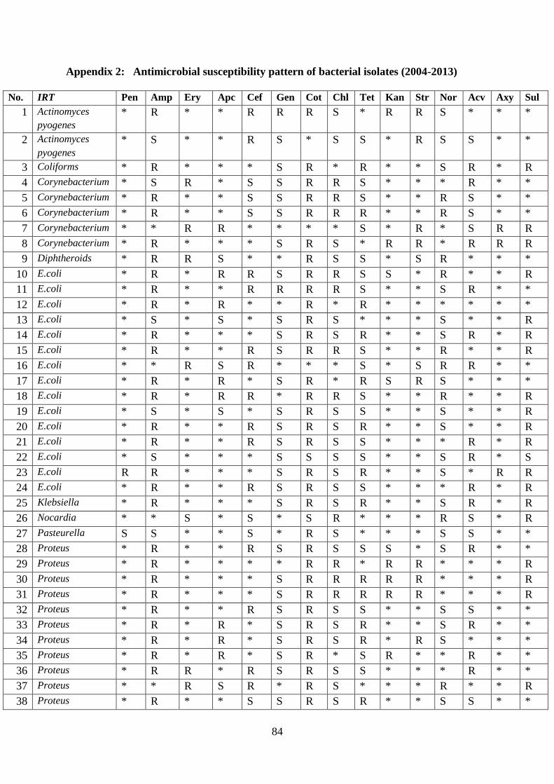
84
Appendix 2: Antimicrobial susceptibility pattern of bacterial isolates (2004-2013)
No. IRT Pen Amp Ery Apc Cef Gen Cot Chl Tet Kan Str Nor Acv Axy Sul
1 Actinomyces
pyogenes
* R * * R R R S * R R S * * *
2 Actinomyces
pyogenes
* S * * R S * S S * R S S * *
3 Coliforms * R * * * S R * R * * S R * R
4 Corynebacterium * S R * S S R R S * * * R * *
5 Corynebacterium * R * * S S R R S * * R S * *
6 Corynebacterium * R * * S S R R R * * R S * *
7 Corynebacterium * * R R * * * * S * R * S R R
8 Corynebacterium * R * * * S R S * R R * R R R
9 Diphtheroids * R R S * * R S S * S R * * *
10 E.coli * R * R R S R R S S * R * * R
11 E.coli * R * * R R R R S * * S R * *
12 E.coli * R * R * * R * R * * * * * *
13 E.coli * S * S * S R S * * * S * * R
14 E.coli * R * * * S R S R * * S R * R
15 E.coli * R * * R S R R S * * R * * R
16 E.coli * * R S R * * * S * S R R * *
17 E.coli * R * R * S R * R S R S * * *
18 E.coli * R * R R * R R S * * R * * R
19 E.coli * S * S * S R S S * * S * * R
20 E.coli * R * * R S R S R * * S * * R
21 E.coli * R * * R S R S S * * * R * R
22 E.coli * S * * * S S S S * * S R * S
23 E.coli R R * * * S R S R * * S * R R
24 E.coli * R * * R S R S S * * * R * R
25 Klebsiella * R * * * S R S R * * S R * R
26 Nocardia * * S * S * S R * * * R S * R
27 Pasteurella S S * * S * R S * * * S S * *
28 Proteus * R * * R S R S S S * S R * *
29 Proteus * R * * * * R R * R R * * * R
30 Proteus * R * * * S R R R R R * * * R
31 Proteus * R * * * S R R R R R * * * R
32 Proteus * R * * R S R S S * * S S * *
33 Proteus * R * R * S R S R * * S R * *
34 Proteus * R * R * S R S R * R S * * *
35 Proteus * R * R * S R * S R * * R * *
36 Proteus * R R * R S R S S * * * R * *
37 Proteus * * R S R * R S * * * R * * R
38 Proteus * R * * S S R S R * * S S * *
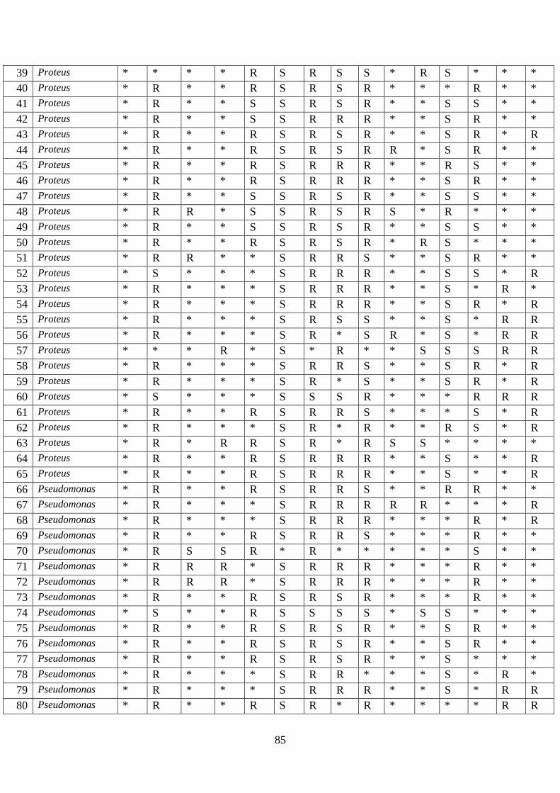
85
39 Proteus * * * * R S R S S * R S * * *
40 Proteus * R * * R S R S R * * * R * *
41 Proteus * R * * S S R S R * * S S * *
42 Proteus * R * * S S R R R * * S R * *
43 Proteus * R * * R S R S R * * S R * R
44 Proteus * R * * R S R S R R * S R * *
45 Proteus * R * * R S R R R * * R S * *
46 Proteus * R * * R S R R R * * S R * *
47 Proteus * R * * S S R S R * * S S * *
48 Proteus * R R * S S R S R S * R * * *
49 Proteus * R * * S S R S R * * S S * *
50 Proteus * R * * R S R S R * R S * * *
51 Proteus * R R * * S R R S * * S R * *
52 Proteus * S * * * S R R R * * S S * R
53 Proteus * R * * * S R R R * * S * R *
54 Proteus * R * * * S R R R * * S R * R
55 Proteus * R * * * S R S S * * S * R R
56 Proteus * R * * * S R * S R * S * R R
57 Proteus * * * R * S * R * * S S S R R
58 Proteus * R * * * S R R S * * S R * R
59 Proteus * R * * * S R * S * * S R * R
60 Proteus * S * * * S S S R * * * R R R
61 Proteus * R * * R S R R S * * * S * R
62 Proteus * R * * * S R * R * * R S * R
63 Proteus * R * R R S R * R S S * * * *
64 Proteus * R * * R S R R R * * S * * R
65 Proteus * R * * R S R R R * * S * * R
66 Pseudomonas * R * * R S R R S * * R R * *
67 Pseudomonas * R * * * S R R R R R * * * R
68 Pseudomonas * R * * * S R R R * * * R * R
69 Pseudomonas * R * * R S R R S * * * R * *
70 Pseudomonas * R S S R * R * * * * * S * *
71 Pseudomonas * R R R * S R R R * * * R * *
72 Pseudomonas * R R R * S R R R * * * R * *
73 Pseudomonas * R * * R S R S R * * * R * *
74 Pseudomonas * S * * R S S S S * S S * * *
75 Pseudomonas * R * * R S R S R * * S R * *
76 Pseudomonas * R * * R S R S R * * S R * *
77 Pseudomonas * R * * R S R S R * * S * * *
78 Pseudomonas * R * * * S R R * * * S * R *
79 Pseudomonas * R * * * S R R R * * S * R R
80 Pseudomonas * R * * R S R * R * * * * R R
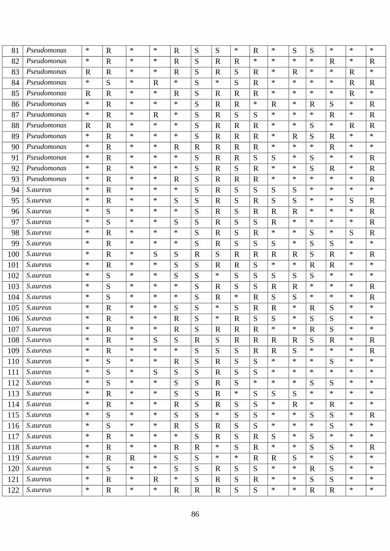
86
81 Pseudomonas * R * * R S S * R * S S * * *
82 Pseudomonas * R * * R S R R * * * * R * R
83 Pseudomonas R R * * R S R S R * R * * R *
84 Pseudomonas * S * R * S * S R * * * * R R
85 Pseudomonas R R * * R S R R R * * * * R *
86 Pseudomonas * R * * * S R R * R * R S * R
87 Pseudomonas * R * R * S R S S * * * R * R
88 Pseudomonas R R * * * S R R R * * S * R R
89 Pseudomonas * R * * * S R R R * R S R * *
90 Pseudomonas * R * * R R R R R * * * R * *
91 Pseudomonas * R * * * S R R S S * S * * R
92 Pseudomonas * R * * * S R S R * * S R * R
93 Pseudomonas * R * * R S R R R * * * * * R
94 S.aureus * R * * * S R S S S S * * * *
95 S.aureus * R * * S S R S R S S * * S R
96 S.aureus * S * * * S R S R R R * * * R
97 S.aureus * S * * S S R S S R * * * * R
98 S.aureus * R * * * S R S R * * S * S R
99 S.aureus * R * * * S R S S S * S S * *
100 S.aureus * R * S S R S R R R R S R * R
101 S.aureus * R * * S S R R S * * R R * *
102 S.aureus * S * * S S * S S S S S * * *
103 S.aureus * S * * * S R S S R R * * * R
104 S.aureus * S * * * S R * R S S * * * R
105 S.aureus * R * * S S * S R R * R S * *
106 S.aureus * R * * R S * R S S * S S * *
107 S.aureus * R * * R S R R R * * R S * *
108 S.aureus * R * S S R S R R R R S R * R
109 S.aureus * R * * * S S S R R S * * * R
110 S.aureus * S * * R S R S S * * * S * *
111 S.aureus * S * S S S R S S * * * * * *
112 S.aureus * S * * S S R S * * * S S * *
113 S.aureus * R * * S S R * S S S * * * *
114 S.aureus * R * * R S R S S * R * R * *
115 S.aureus * S * * S S * S S * * S S * R
116 S.aureus * S * * R S R S S * * * S * *
117 S.aureus * R * * * S R S R S * S * * *
118 S.aureus * R * * R R * S R * * S S * R
119 S.aureus * R R * S S * * R R S * S * *
120 S.aureus * S * * S S R S S * * R S * *
121 S.aureus * R * R * S R S R * * S S * *
122 S.aureus * R * * R R R S S * * R R * *

87
123 S.aureus * S * * S S R S S S * S * * *
124 S.aureus * R * * S S R S S * * S S * *
125 S.aureus * R * R S S R S R * * S * * *
126 S.aureus * R * * S S R S R * * S S * *
127 S.aureus * S * * S S S S S * * S S * *
128 S.aureus * R * * S S R R R * * R S * *
129 S.aureus * R * * R S R S R * * S S * *
130 S.aureus * R * * S R R S S * * S S * *
131 S.aureus * R * * R R R S R * S S * * *
132 S.aureus * S * * S S R S R * * R S * *
133 S.aureus * R * * S S S R R * * S S * *
134 S.aureus * S * * S S R S R * * S S * *
135 S.aureus * R * * S R R S R * * S R * *
136 S.aureus * R * * R S R R R * * S R * *
137 S.aureus * R * * S S R S S * * R R * *
138 S.aureus * R * * S S R S R * * S R * *
139 S.aureus * R * * S R R R R * * S R * *
140 S.aureus * R * * S S R * S R * S R * *
141 S.aureus * R * * S S R R S * * S R * *
142 S.aureus * S * S S S R S S * * S * * *
143 S.aureus * R * * R * R * R * * * R * *
144 S.aureus * S * S S S R S S * R * * * *
145 S.aureus * S * * S R R S S * * R S * *
146 S.aureus * R * * S S S S R * * S R * *
147 S.aureus * S * * S R R S R * * S S * *
148 S.aureus * R * * S S R S R * S R * * *
149 S.aureus * R * * * * R * * * * * * * *
150 S.aureus * R S * * S R S S * * R S * *
151 S.aureus * R R * * R R S R * * R R * *
152 S.aureus * S * * S S R R S * * S S * *
153 S.aureus R S * * S S R S * * * S S * *
154 S.aureus * R * * S S R S R * * S S * *
155 S.aureus * R * * * S R R R * * S R * *
156 S.aureus * R * * * S R S S * * S R * R
157 S.aureus S S * * S R R S * * * S R * *
158 S.aureus * R * * R S * S * S R * R * *
159 S.aureus * R * * * S R S R * * R R * S
160 S.aureus * R * * * R R R R * * S * * R
161 S.aureus * S * * * S R R S * * S * * R
162 S.aureus * R * * S S R R R * * * * S R
163 S.aureus * S * * S S R S R * * * * S R
164 S.aureus * S * R S S R R * R * * S * R
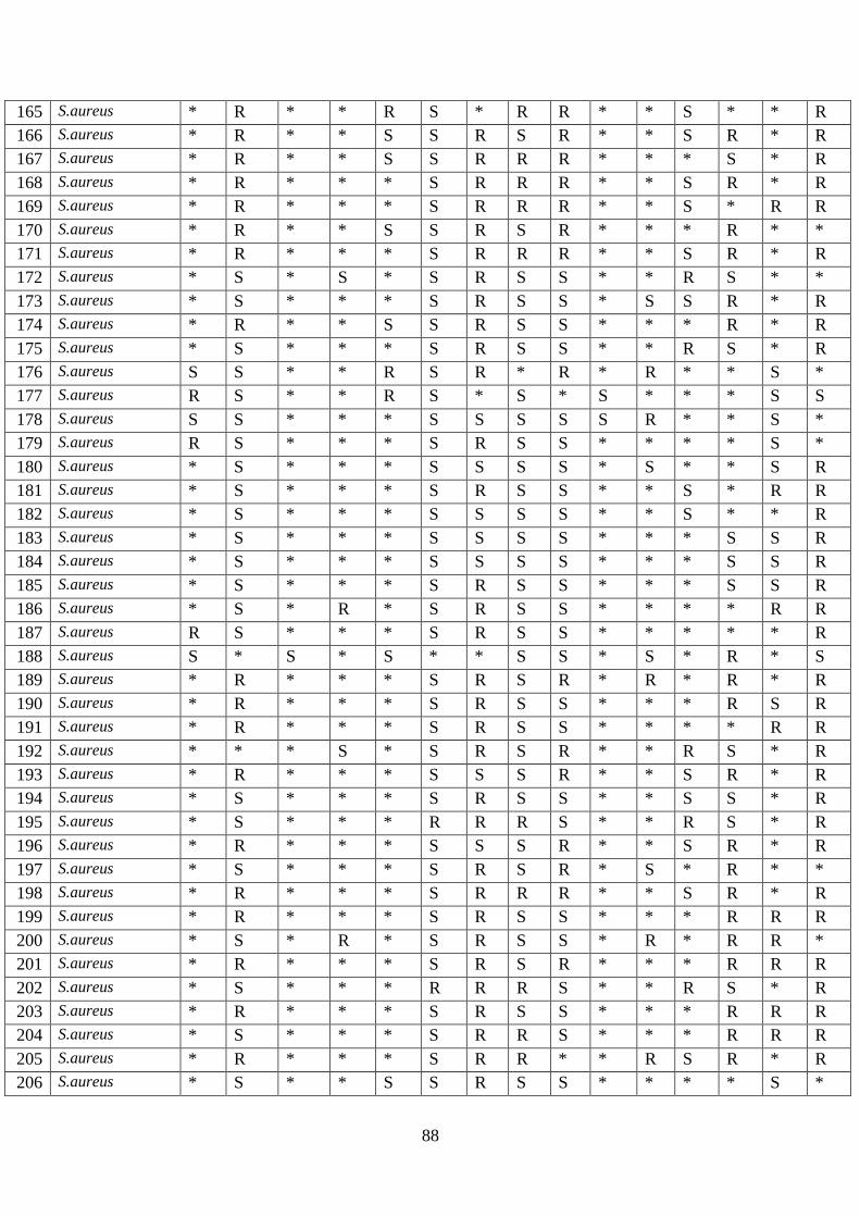
88
165 S.aureus * R * * R S * R R * * S * * R
166 S.aureus * R * * S S R S R * * S R * R
167 S.aureus * R * * S S R R R * * * S * R
168 S.aureus * R * * * S R R R * * S R * R
169 S.aureus * R * * * S R R R * * S * R R
170 S.aureus * R * * S S R S R * * * R * *
171 S.aureus * R * * * S R R R * * S R * R
172 S.aureus * S * S * S R S S * * R S * *
173 S.aureus * S * * * S R S S * S S R * R
174 S.aureus * R * * S S R S S * * * R * R
175 S.aureus * S * * * S R S S * * R S * R
176 S.aureus S S * * R S R * R * R * * S *
177 S.aureus R S * * R S * S * S * * * S S
178 S.aureus S S * * * S S S S S R * * S *
179 S.aureus R S * * * S R S S * * * * S *
180 S.aureus * S * * * S S S S * S * * S R
181 S.aureus * S * * * S R S S * * S * R R
182 S.aureus * S * * * S S S S * * S * * R
183 S.aureus * S * * * S S S S * * * S S R
184 S.aureus * S * * * S S S S * * * S S R
185 S.aureus * S * * * S R S S * * * S S R
186 S.aureus * S * R * S R S S * * * * R R
187 S.aureus R S * * * S R S S * * * * * R
188 S.aureus S * S * S * * S S * S * R * S
189 S.aureus * R * * * S R S R * R * R * R
190 S.aureus * R * * * S R S S * * * R S R
191 S.aureus * R * * * S R S S * * * * R R
192 S.aureus * * * S * S R S R * * R S * R
193 S.aureus * R * * * S S S R * * S R * R
194 S.aureus * S * * * S R S S * * S S * R
195 S.aureus * S * * * R R R S * * R S * R
196 S.aureus * R * * * S S S R * * S R * R
197 S.aureus * S * * * S R S R * S * R * *
198 S.aureus * R * * * S R R R * * S R * R
199 S.aureus * R * * * S R S S * * * R R R
200 S.aureus * S * R * S R S S * R * R R *
201 S.aureus * R * * * S R S R * * * R R R
202 S.aureus * S * * * R R R S * * R S * R
203 S.aureus * R * * * S R S S * * * R R R
204 S.aureus * S * * * S R R S * * * R R R
205 S.aureus * R * * * S R R * * R S R * R
206 S.aureus * S * * S S R S S * * * * S *

89
207 S.aureus * S * * * S R * S * * * S R *
208 S.aureus * R * * S S * R S R * R * * *
209 S.aureus * S * * S S S S * * * R S * R
210 S.aureus * S * * S S R S S * * S * * R
211 S.aureus * R * * * S S S R * R S R * *
212 S.aureus * S * R S S R S R * * * * * R
213 S.aureus * S * * R S R S R * R * R * *
214 S.aureus * S * * R S R S R * * * S * R
215 S.aureus * S * * * S R R S * * S R * R
216 S.aureus * R * * * S R S R * R S * S *
217 S.aureus R S * * * S R S R * * S * S *
218 S.aureus * R * * * S R S R * * S * * R
219 S.aureus * S * * * S R R S * * R S * R
220 S.aureus * S * * S S R * * * * S R * R
221 S.aureus * R * * S S R S R * * * R * R
222 S.aureus * S * * * S R S S * R * * R *
223 S.aureus * R * * * S R S R * * R S * R
224 Staphylococcus * R * S S S * * S S S S * * *
225 Staphylococcus * S * * S S R S R * * S S * *
226 Staphylococcus * R * * * S R R R * * S R * *
227 Staphylococcus * S * * * S R R S * * R S * R
228 Staphylococcus * R * * * S R S S * * S * R R
229 Staphylococcus R R * * * S R * R * * S * R *
230 Staphylococcus * S * R * S * S R * S * * R R
231 Staphylococcus * S * * * S R S R * R R * R *
232 Staphylococcus * R * * * S R R S * * R * R R
233 Staphylococcus * R * R * S R S S * * * R * R
234 Staphylococcus * S * * * S R R R * * S * S R
235 Staphylococcus * R * * S S * * R * * S * S R
236 Staphylococcus * S * * * S S * R * R S * * *
237 Staphylococcus * R * * S S R * S * R * R * *
238 Staphylococcus * R * * * S R S S * * * R * R
239 Staphylococcus * S * * * S R S S * * * * * R
240 Staphylococcus * R * * S S R S S * * * R * R
241 Staphylococcus * R * R * S R * R R * * * * R
242 Staphylococcus * R * * R S S S R * * * R * S
243 Staphylococcus * R * * * S R * R S R S * * *
244 Staphylococcus R R * * * S R R S * * S * S R
245 Staphylococcus R R * * * S R S S * R S * R *
246 Streptococcus * R * * * S R * R R R * * * R
247 Streptococcus * R * * S R R S S R * S S * *
248 Streptococcus * R * * S S * S R * * S R * R
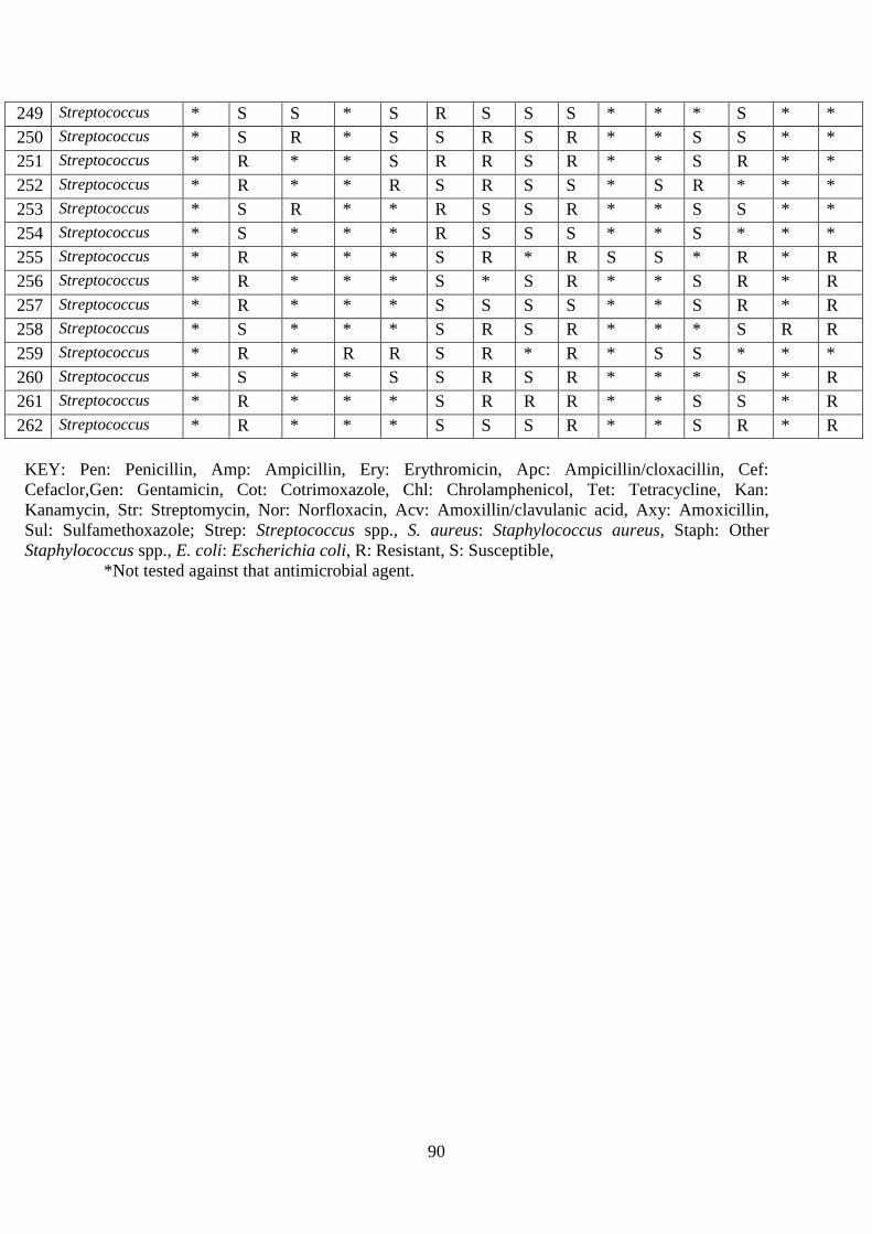
90
249 Streptococcus * S S * S R S S S * * * S * *
250 Streptococcus * S R * S S R S R * * S S * *
251 Streptococcus * R * * S R R S R * * S R * *
252 Streptococcus * R * * R S R S S * S R * * *
253 Streptococcus * S R * * R S S R * * S S * *
254 Streptococcus * S * * * R S S S * * S * * *
255 Streptococcus * R * * * S R * R S S * R * R
256 Streptococcus * R * * * S * S R * * S R * R
257 Streptococcus * R * * * S S S S * * S R * R
258 Streptococcus * S * * * S R S R * * * S R R
259 Streptococcus * R * R R S R * R * S S * * *
260 Streptococcus * S * * S S R S R * * * S * R
261 Streptococcus * R * * * S R R R * * S S * R
262 Streptococcus * R * * * S S S R * * S R * R
KEY: Pen: Penicillin, Amp: Ampicillin, Ery: Erythromicin, Apc: Ampicillin/cloxacillin, Cef:
Cefaclor,Gen: Gentamicin, Cot: Cotrimoxazole, Chl: Chrolamphenicol, Tet: Tetracycline, Kan:
Kanamycin, Str: Streptomycin, Nor: Norfloxacin, Acv: Amoxillin/clavulanic acid, Axy: Amoxicillin,
Sul: Sulfamethoxazole; Strep: Streptococcus spp., S. aureus: Staphylococcus aureus, Staph: Other
Staphylococcus spp., E. coli: Escherichia coli, R: Resistant, S: Susceptible,
*Not tested against that antimicrobial agent.

91
Appendix 3: Sample collection form
Sample Collection Form For dogs
Section A
Date: ………………………………………..
Name: ………………………………………
Breed: ………………………………………
Case No: …………………………………….
Age: …………………………………………
Sex: …………………………………………
Owner’s Name: ……………………………
Lab I.D:……………………………………..
Section B
Tick the Appropriate answer
1. Presenting Complaint
a) Wound
Yes No
b) If yes to (a) above, how long has
the wound been there
˂1wk
˃1wk
N/A
2. Has the dog been treated with
antimicrobials/antibiotics
Yes No
3. If yes to (2), when was the last
treatment administered
Past one week
Past one month
3months ago
Place of Residence…………………………
Section C: TO BE FILLED BY THE
ATTENDING VETERINARIAN
Reason for Visit to clinic
Vaccination
Routine Check-up
Clinical Case
Surgical Case
Physical Examination Findings
Diagnosis
Attending Clinician
Name………………………………………..
Signature……………………………………
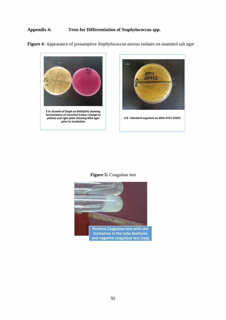
92
Appendix 4: Tests for Differentiation of Staphylococcus spp.
Figure 4: Appearance of presumptive Staphylococcus aureus isolates on mannitol salt agar
Figure 5: Coagulase test
6 A: Growth of Staph on MSA(left) showing fermentation of mannitol (colour change to yellow) and right plate showing MSA agar
prior to incubation6 B : Standard organism on MSA ATCC 25923
Positive Coagulase test with clot formation in the tube (bottom)
and negative coagulase test (top)
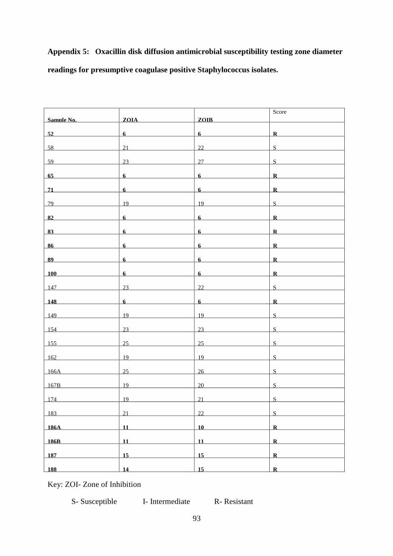
93
Appendix 5: Oxacillin disk diffusion antimicrobial susceptibility testing zone diameter
readings for presumptive coagulase positive Staphylococcus isolates.
Sample No. ZOIA ZOIB
Score
52 6 6 R
58 21 22 S
59 23 27 S
65 6 6 R
71 6 6 R
79 19 19 S
82 6 6 R
83 6 6 R
86 6 6 R
89 6 6 R
100 6 6 R
147 23 22 S
148 6 6 R
149 19 19 S
154 23 23 S
155 25 25 S
162 19 19 S
166A 25 26 S
167B 19 20 S
174 19 21 S
183 21 22 S
186A 11 10 R
186B 11 11 R
187 15 15 R
188 14 15 R
Key: ZOI- Zone of Inhibition
S- Susceptible I- Intermediate R- Resistant
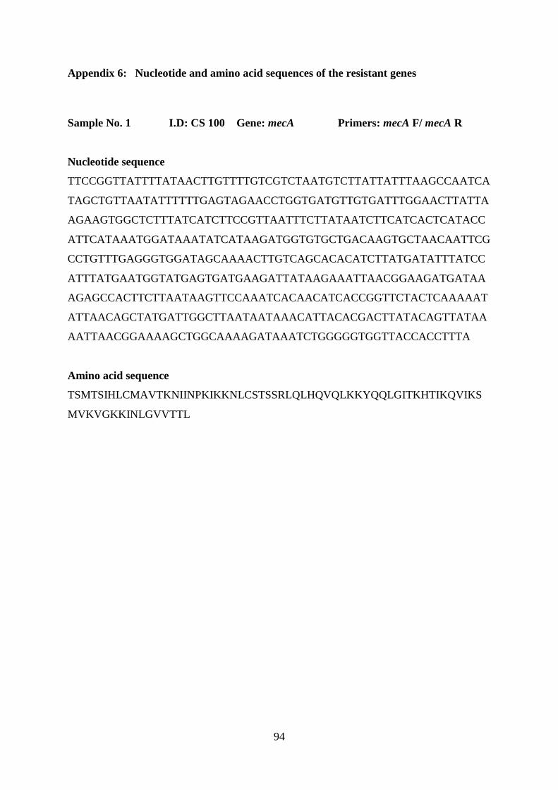
94
Appendix 6: Nucleotide and amino acid sequences of the resistant genes
Sample No. 1 I.D: CS 100 Gene: mecA Primers: mecA F/ mecA R
Nucleotide sequence
TTCCGGTTATTTTATAACTTGTTTTGTCGTCTAATGTCTTATTATTTAAGCCAATCA
TAGCTGTTAATATTTTTTGAGTAGAACCTGGTGATGTTGTGATTTGGAACTTATTA
AGAAGTGGCTCTTTATCATCTTCCGTTAATTTCTTATAATCTTCATCACTCATACC
ATTCATAAATGGATAAATATCATAAGATGGTGTGCTGACAAGTGCTAACAATTCG
CCTGTTTGAGGGTGGATAGCAAAACTTGTCAGCACACATCTTATGATATTTATCC
ATTTATGAATGGTATGAGTGATGAAGATTATAAGAAATTAACGGAAGATGATAA
AGAGCCACTTCTTAATAAGTTCCAAATCACAACATCACCGGTTCTACTCAAAAAT
ATTAACAGCTATGATTGGCTTAATAATAAACATTACACGACTTATACAGTTATAA
AATTAACGGAAAAGCTGGCAAAAGATAAATCTGGGGGTGGTTACCACCTTTA
Amino acid sequence
TSMTSIHLCMAVTKNIINPKIKKNLCSTSSRLQLHQVQLKKYQQLGITKHTIKQVIKS
MVKVGKKINLGVVTTL
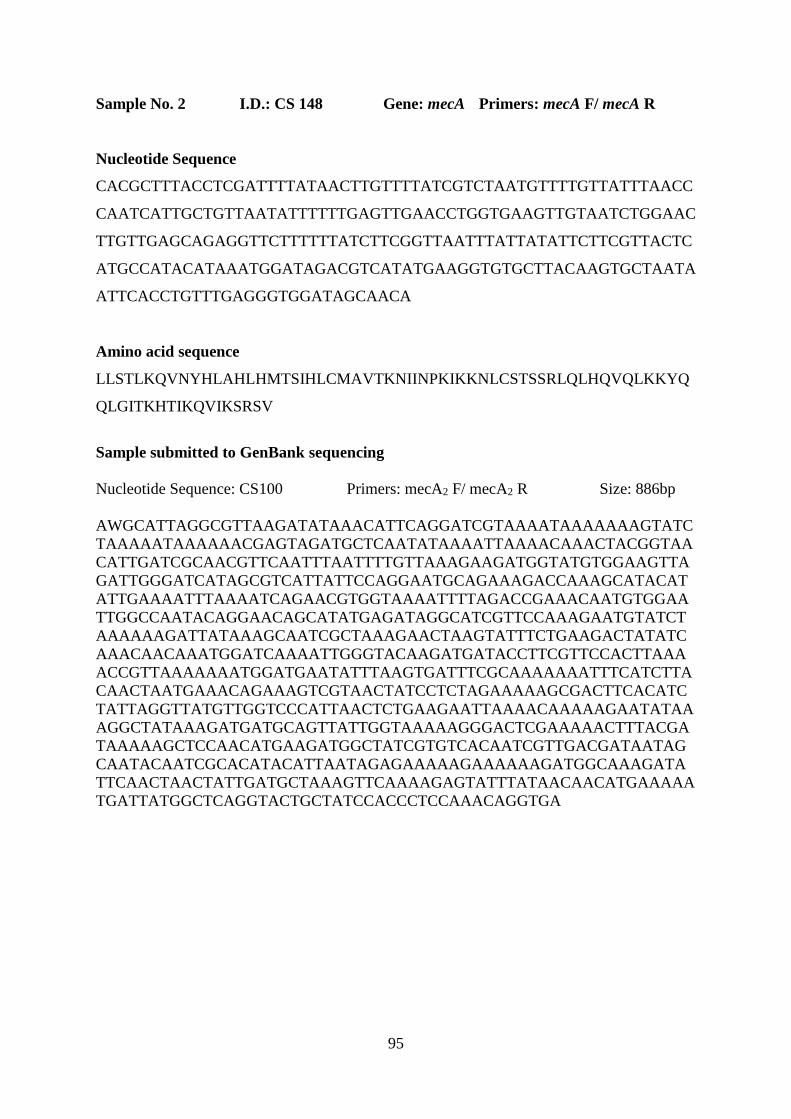
95
Sample No. 2 I.D.: CS 148 Gene: mecA Primers: mecA F/ mecA R
Nucleotide Sequence
CACGCTTTACCTCGATTTTATAACTTGTTTTATCGTCTAATGTTTTGTTATTTAACC
CAATCATTGCTGTTAATATTTTTTGAGTTGAACCTGGTGAAGTTGTAATCTGGAAC
TTGTTGAGCAGAGGTTCTTTTTTATCTTCGGTTAATTTATTATATTCTTCGTTACTC
ATGCCATACATAAATGGATAGACGTCATATGAAGGTGTGCTTACAAGTGCTAATA
ATTCACCTGTTTGAGGGTGGATAGCAACA
Amino acid sequence
LLSTLKQVNYHLAHLHMTSIHLCMAVTKNIINPKIKKNLCSTSSRLQLHQVQLKKYQ
QLGITKHTIKQVIKSRSV
Sample submitted to GenBank sequencing
Nucleotide Sequence: CS100 Primers: mecA2 F/ mecA2 R Size: 886bp
AWGCATTAGGCGTTAAGATATAAACATTCAGGATCGTAAAATAAAAAAAGTATC
TAAAAATAAAAAACGAGTAGATGCTCAATATAAAATTAAAACAAACTACGGTAA
CATTGATCGCAACGTTCAATTTAATTTTGTTAAAGAAGATGGTATGTGGAAGTTA
GATTGGGATCATAGCGTCATTATTCCAGGAATGCAGAAAGACCAAAGCATACAT
ATTGAAAATTTAAAATCAGAACGTGGTAAAATTTTAGACCGAAACAATGTGGAA
TTGGCCAATACAGGAACAGCATATGAGATAGGCATCGTTCCAAAGAATGTATCT
AAAAAAGATTATAAAGCAATCGCTAAAGAACTAAGTATTTCTGAAGACTATATC
AAACAACAAATGGATCAAAATTGGGTACAAGATGATACCTTCGTTCCACTTAAA
ACCGTTAAAAAAATGGATGAATATTTAAGTGATTTCGCAAAAAAATTTCATCTTA
CAACTAATGAAACAGAAAGTCGTAACTATCCTCTAGAAAAAGCGACTTCACATC
TATTAGGTTATGTTGGTCCCATTAACTCTGAAGAATTAAAACAAAAAGAATATAA
AGGCTATAAAGATGATGCAGTTATTGGTAAAAAGGGACTCGAAAAACTTTACGA
TAAAAAGCTCCAACATGAAGATGGCTATCGTGTCACAATCGTTGACGATAATAG
CAATACAATCGCACATACATTAATAGAGAAAAAGAAAAAAGATGGCAAAGATA
TTCAACTAACTATTGATGCTAAAGTTCAAAAGAGTATTTATAACAACATGAAAAA
TGATTATGGCTCAGGTACTGCTATCCACCCTCCAAACAGGTGA

96