Embed Size (px)
Citation preview
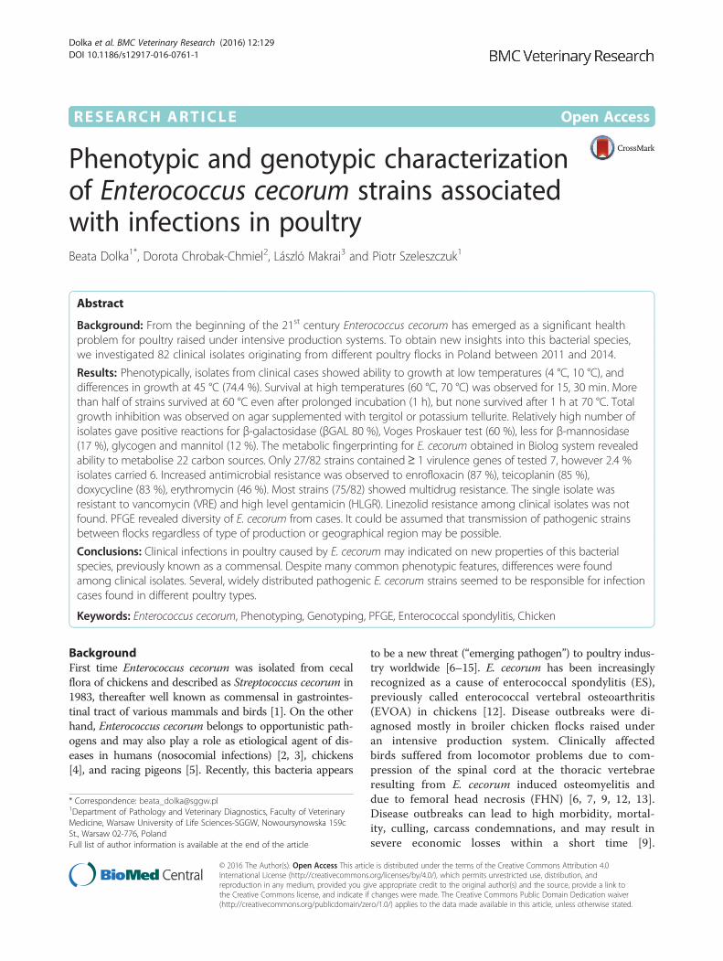
RESEARCH ARTICLE Open Access
Phenotypic and genotypic characterizationof Enterococcus cecorum strains associatedwith infections in poultryBeata Dolka1*, Dorota Chrobak-Chmiel2, László Makrai3 and Piotr Szeleszczuk1
Abstract
Background: From the beginning of the 21st century Enterococcus cecorum has emerged as a significant healthproblem for poultry raised under intensive production systems. To obtain new insights into this bacterial species,we investigated 82 clinical isolates originating from different poultry flocks in Poland between 2011 and 2014.
Results: Phenotypically, isolates from clinical cases showed ability to growth at low temperatures (4 °C, 10 °C), anddifferences in growth at 45 °C (74.4 %). Survival at high temperatures (60 °C, 70 °C) was observed for 15, 30 min. Morethan half of strains survived at 60 °C even after prolonged incubation (1 h), but none survived after 1 h at 70 °C. Totalgrowth inhibition was observed on agar supplemented with tergitol or potassium tellurite. Relatively high number ofisolates gave positive reactions for β-galactosidase (βGAL 80 %), Voges Proskauer test (60 %), less for β-mannosidase(17 %), glycogen and mannitol (12 %). The metabolic fingerprinting for E. cecorum obtained in Biolog system revealedability to metabolise 22 carbon sources. Only 27/82 strains contained≥ 1 virulence genes of tested 7, however 2.4 %isolates carried 6. Increased antimicrobial resistance was observed to enrofloxacin (87 %), teicoplanin (85 %),doxycycline (83 %), erythromycin (46 %). Most strains (75/82) showed multidrug resistance. The single isolate wasresistant to vancomycin (VRE) and high level gentamicin (HLGR). Linezolid resistance among clinical isolates was notfound. PFGE revealed diversity of E. cecorum from cases. It could be assumed that transmission of pathogenic strainsbetween flocks regardless of type of production or geographical region may be possible.
Conclusions: Clinical infections in poultry caused by E. cecorum may indicated on new properties of this bacterialspecies, previously known as a commensal. Despite many common phenotypic features, differences were foundamong clinical isolates. Several, widely distributed pathogenic E. cecorum strains seemed to be responsible for infectioncases found in different poultry types.
Keywords: Enterococcus cecorum, Phenotyping, Genotyping, PFGE, Enterococcal spondylitis, Chicken
BackgroundFirst time Enterococcus cecorum was isolated from cecalflora of chickens and described as Streptococcus cecorum in1983, thereafter well known as commensal in gastrointes-tinal tract of various mammals and birds [1]. On the otherhand, Enterococcus cecorum belongs to opportunistic path-ogens and may also play a role as etiological agent of dis-eases in humans (nosocomial infections) [2, 3], chickens[4], and racing pigeons [5]. Recently, this bacteria appears
to be a new threat (“emerging pathogen”) to poultry indus-try worldwide [6–15]. E. cecorum has been increasinglyrecognized as a cause of enterococcal spondylitis (ES),previously called enterococcal vertebral osteoarthritis(EVOA) in chickens [12]. Disease outbreaks were di-agnosed mostly in broiler chicken flocks raised underan intensive production system. Clinically affectedbirds suffered from locomotor problems due to com-pression of the spinal cord at the thoracic vertebraeresulting from E. cecorum induced osteomyelitis anddue to femoral head necrosis (FHN) [6, 7, 9, 12, 13].Disease outbreaks can lead to high morbidity, mortal-ity, culling, carcass condemnations, and may result insevere economic losses within a short time [9].
* Correspondence: [email protected] of Pathology and Veterinary Diagnostics, Faculty of VeterinaryMedicine, Warsaw University of Life Sciences-SGGW, Nowoursynowska 159cSt., Warsaw 02-776, PolandFull list of author information is available at the end of the article
© 2016 The Author(s). Open Access This article is distributed under the terms of the Creative Commons Attribution 4.0International License (http://creativecommons.org/licenses/by/4.0/), which permits unrestricted use, distribution, andreproduction in any medium, provided you give appropriate credit to the original author(s) and the source, provide a link tothe Creative Commons license, and indicate if changes were made. The Creative Commons Public Domain Dedication waiver(http://creativecommons.org/publicdomain/zero/1.0/) applies to the data made available in this article, unless otherwise stated.
Dolka et al. BMC Veterinary Research (2016) 12:129 DOI 10.1186/s12917-016-0761-1
Recently, poultry or domestic animals (cats, dogs) arethought to be a possible source for transmission leadingto E. cecorum–associated septicaemia in humans [2, 3].Various methods using conventional biochemical
tests and molecular techniques have been commonlyused for identification and typing enterococci [16–18].Pulsed field gel electrophoresis (PFGE) is consideredto be the “gold standard” for subtyping enterococciand has been used extensively for molecular epi-demiological characterization of enterococcal out-breaks [19, 20]. The PCR assay based on specificamplification followed by sequencing and nucleotide se-quence comparison of target genes (such as 16S ribosomalRNA, sodA, ddl, tuf, groESL) or tDNA-PCR have servedfor the genotypic identification of enterococci [21–23].Despite of available literature biochemical and molecu-
lar analysis of E. cecorum strains with poultry origin iso-lated in Europe are still limited. Moreover, there is notenough data regarding the properties of isolates, usuallyreferred as pathogenic for poultry [1, 7, 8, 10]. The pur-pose of this study was phenotypic characterization ofclinical E. cecorum isolates associated with infections inpoultry and investigation their genetic relatedness.
MethodsBacterial isolatesEighty two E. cecorum isolates of poultry-origin used inthis study were obtained from archival bacterial collectiondeposited at Department of Pathology and VeterinaryDiagnostics, or were obtained from clinical specimenssubmitted by veterinarians for routine diagnostic work tothe Diagnostic Laboratory in Division of Avian Diseases,Faculty of Veterinary Medicine at the Warsaw Universityof Life Sciences-SGGW (Poland). Authors ensure that theARRIVE guidelines were followed. Among 82 clinicalstrains collected between 2011 and 2014, 49 came frombroiler chicken flocks (CB), 20 from broiler breeder flocks(BB), 10 from commercial layer flocks (CL), 2 from geeseflocks (G) and 1 from turkey flock (T). According toadopted criteria in this study, one E. cecorum isolate rep-resented one different flock in which clinical problemsdue to E. cecorum infection were reported by veterinarianson farms. Affected birds displayed a variety of clinicalsigns, however in all types of flocks the lameness, paraly-sis, hock sitting, weakness, pododermatitis, decreasedwater and food intake were usually noted. Subsequently,disease caused lower results of production, increasedlosses due to mortality and culling. Necropsies and patho-logical examinations revealed usually femoral head necro-sis, (purulent) arthritis, fibrinous pericarditis, endocarditis,hepatitis and congested lungs. Characteristic osteomyelitislesions at caudal thoracic vertebrae we found only inchicken flocks (mainly in CB). Isolates were recoveredfrom tissue samples such as vertebral column, femoral
heads, heart, liver, lungs or yolk sac, which were collectedduring necropsy.
Bacterial analysisThe tissue samples were inoculated onto Columbia agarwith 5 % sheep blood (CA) (Graso, Poland) and agarplates with esculin (KAA, Biocorp, Poland; Enterococco-sel Agar, Graso, Poland), then incubated at 37 °C for24 h in a CO2-enriched atmosphere. Bacteria were iden-tified as Enterococcus based on their phenotypic proper-ties such as colonial morphology, hemolysis (on CA),Gram-staining, catalase production (using a 3 % H2O2),cytochrome oxidase production (OXItest, Erba Lachemas.r.o., Czech Republik), and esculin hydrolysis (Entero-coccosel Agar, KAA). Pigment production was visuallyassayed by growing the bacteria on CA for 24 h andscraping off the growth with a white cotton swab. Motil-ity was examined using Motility Test Agar (Graso,Poland). The ability to growth was estimated in 6.5 %NaCl (salt tolerance test) after 48 h at 37 °C, and on dif-ferent media (Graso, Poland) (Table 2). Serological iden-tification of Lancefield group was conducted by rapidlatex agglutination method using Slidex Strepto Plus D(bioMérieux, France). Tests for E. cecorum growth wereperformed in BHI broth (Brain-heart infusion; bio-Mérieux, France) tubes preincubated at 4 °C, 10 °C,45 °C for 24 h. Then cultures in BHI broth werespread onto CA and incubated at 37 °C. The growthresponse was assessed after 24 h and 48 h. The abilityto survive at 60 °C, 70 °C was estimated for 15 min,30 min, 1 h in BHI broth tubes, followed by incuba-tion of inoculated CA plates. The results were re-corded after 24 h and 48 h.
Biochemical testsIdentification to the species level based on biochemicalcharacterization was performed by API rapid ID 32STREP (bioMérieux, France) and on the basis of carbonsource utilisation using Biolog system (Biolog Inc., Hay-ward, USA). Isolates (n = 13) were determined accordingto Biolog GP2 MicroPlates, which performed 95 discretetests simultaneously and gave a characteristic reactionpattern (metabolic fingerprint). The MicroPlates wereincubated at 37 °C and read visually after 4 h and 24 h.The metabolic fingerprint patterns were compared andidentified using the MicroLog™ 4.20.05 databasesoftware.
Virulence factorsAll 82 isolates were tested for the presence of sevenvirulence factors: asa1 (aggregation substance), gelE(gelatinase), hyl (hyaluronidase), esp (enterococcal sur-face protein), cylA (cytolisin), efaA (endocarditis anti-gen), ace (collagen-binding protein) according to
Dolka et al. BMC Veterinary Research (2016) 12:129 Page 2 of 13
Martín-Platero et al. [24], Jung et al. [5] using duplexPCRs (asa1/gelE, cylA/esp, efaA/ace) and single PCR(hyl). PCR reaction mix contained 12.5 μl DreamTaqPCR Master Mix (Thermo Fisher Scientific Inc., USA)0.3 μl of each primer (50 pmol/μl), 4 μl DNA and PCR-clean water (added up to a volume of 25 μl). Thermocy-cler conditions were as follows: initial denaturation at94 °C for 5 min, followed by 30 cycles: denaturation at94 °C for 1 min, annealing at 56 °C for 1 min (55 °C forefaA/ace), extension at 72 °C for 1 min, followed by finalextension step 72 °C for 10 min and a 4 °C hold. Ampli-fication products (10 μl) were analyzed by 1.2 % agarosegel electrophoresis after ethidium bromide staining andvisualized under UV light (UVP, USA). A 100-bp DNAladder (Thermo Fisher Scientific Inc., USA) was used asa molecular size marker.Production of gelatinase was additionally determined
using Difco Nutrient Gelatin (BD, USA) according to themanufacturer’s recommendations. The tubes inoculatedwith E. cecorum ATCC 43198, S. aureus ATCC 25923(gelatinase positive), E. coli ATCC 25922 (gelatinasenegative) and an uninoculated tube were used for qualitycontrol testing.
Antibiotic susceptibilitySusceptibility for 13 antimicrobial agents: amoxicillin/clavulanic acid (AUG 20/10 μg), ampicillin (AP 10 μg),penicillin (PG 10 μg), enrofloxacin (ENF 5 μg) tetracyc-line (TEC 30 μg), nitrofurantoin (NI 300 μg), doxycyc-line (DXT 30 μg), chloramphenicol (C 30 μg),erythromycin (E 15 μg), teicoplanin (T 30 μg), vanco-mycin (VA 30 μg), high level gentamicin (GM 120 μg)and linezolid (LZD 30 μg) was tested by Kirby-Bauerdisk diffusion method and the results were interpretedaccording to Clinical and Laboratory Standards Instituteguidelines [25]. The criteria for selection of antibioticsbased on CLSI guidelines for Enterococcus spp. and ontheir practical significance for the clinical use. Amongtested antibiotics, tetracycline, doxycycline, amoxicillin,enrofloxacin have been actually approved for use inpoultry (erythromycin until 2014) and have practicalrelevance. Vancomycin resistance genes (vanA, vanB)were tested by PCR using primers and conditions previ-ously reported [24]. Staphylococcus aureus ATCC 25923(vancomycin susceptible), E. faecalis ATCC 51299(vancomycin resistant), E. cecorum ATCC 43198 wereused as controls.
Molecular identificationRapid extraction of bacterial genomic DNA was carriedout by using boiling method. PCR assay targeting sodAgene was performed for identification and determinationthe diversity of 82 E. cecorum strains [22]. PCR productswere visualized after electrophoresis on agarose gel (2 %)
by staining with ethidium bromide, then purified usingGeneMATRIX PCR/DNA Clean-Up Purification Kit(EURx, Poland) and submitted for sequencing to com-mercial services (IBB PAN, Genomed, Poland). The sodAgene sequences were analyzed with NCBI BLAST. Thegenetic distances based on the partial sequences of sodAwas calculated by the two-parameter method of Kimuraby using the MEGA6, and the phylogenetic tree wasconstructed using the Neighbor-Joining method (NJ)with 1000 bootstrap replicates.
PFGEThe standard PFGE procedure was adapted frompreviously published studies with minor modifications[18, 26, 27]. The 82 E. cecorum strains were culturedovernight on CA and then suspended in sterile saline toobtain the density of 3.5 on McFarland scale and centri-fuged 10 min. at 4000 rpm/min. The bacterial pelletswere mixed with 150 μl Tris-EDTA buffer solution(10 mM Tris-HCl, 1 mM disodium EDTA, pH 8.0) and150 μl liquid 2 % agarose (InCert Agarose, Lonza, Rock-land, USA) and small discs were formed (20 μl). The so-lidified discs were incubated at 37 °C for 18 h in 1 ml ofEC buffer (6 mM Tris-HCl pH 8.0, 1 M NaCl, 0.1 MEDTA, 0.2 % deoxycholate, 0.2 % sarkosyl) containing10 mg lysozyme (A&A Biotechnology, Poland), and0.02 mg RNase A (Thermo Fisher Scientific Inc., USA).DNA discs were washed 3 times in 5 ml EBS solution(0.5 M EDTA pH 9.0, 1 % sarkosyl) and incubated over-night at 50 °C in 1 ml EBS solution containing 1 mg ofproteinase K (ESP buffer) (A&A Biotechnology, Poland).Then the discs were washed 4 times (each time upsidedown for 30 times at room temperature) with 10 ml TEbuffer (10 mM Tris, 1 mM EDTA, pH 8.0) and stored in1 ml TE buffer at 4 °C. Subsequently, each disc was pre-incubated in 100 μl restriction buffer for 30 min at roomtemperature. The agarose discs were digested with SmaI(20 U/μl; Fermentas, Lithuania) overnight (at 37 °C).The restriction fragments were separated by clampedhomogenous electric field (CHEF) electrophoresis with aCHEF-DR II System (Bio-Rad Laboratories, USA) in a1.2 % (w/v) agarose gel using pulse time at 0.5 s followedby 35 s at 6 V/cm and temperature 14 °C for 24 h [17].Afterwards the gel was stained with ethidium bromidefor 30 min, then washed in distilled water for 30 min,photographed under UV light and documented in thesystem VersaDoc (Bio-Rad Laboratories, USA). LambdaLadder PFG marker (New England Biolabs Inc., USA)was used as molecular size marker. Gel images were an-alyzed by Gel Compar II version 6.6 (Applied Maths,Belgium) and cluster analysis was performed by UPGMAusing dice similarity coefficient with optimization set at1 % and position tolerance at 1 %. Isolates were clus-tered using an 80 % homology cut-off, above which
Dolka et al. BMC Veterinary Research (2016) 12:129 Page 3 of 13
strains were considered to be closely related andassigned to the same PFGE type [19].
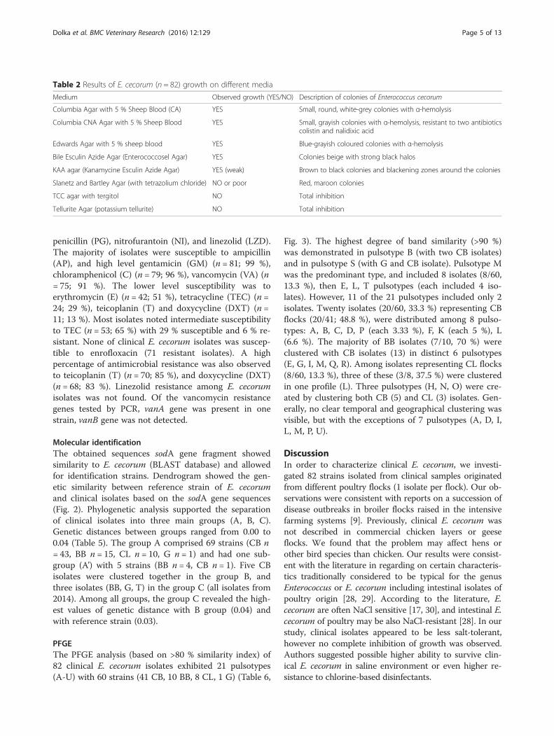
ResultsPhenotypic characterizationTable 1 shows results of conventional tests and effects ofdifferent temperatures on the growth and survival of E.cecorum strains. Bacterial growth was characterized on 7different microbiological media (Table 2).
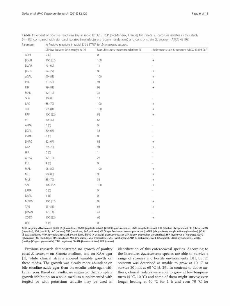
Biochemical testsThe strains were identified as E. cecorum with the APIrapid ID 32 STREP and Biolog system. API revealed per-fect identification profile (ID 99.9 %, T 0.83) for 40(49 %) E. cecorum strains, very good identification (ID99.9 %, T 0.67) for 21 (26 %) strains, good identification(ID 99.8 %, T 0.38) for 2 (2 %) strains, doubtful profile(99.9 %, T 0.4) for 16 (20 %) strains, and unacceptableprofile for 3 (4 %) strains. Among perfect identificationprofiles for E. cecorum, the code 6717–4607–131 was re-corded the most often. Based on the analysis of 82 ob-tained profiles in API (each with 32 tests), we definedone common code 2317–4607–111 for clinical strainswhich gives perfect identification as E. cecorum with theAPI database. Biochemical results obtained in API werepresented in Table 3. The vast majority of isolates was
positive in tests for βGLU, RAF, SAC, MβDG, CDEX(100 %), αGAL, RIB, TRE (99 %), MAL, MEL (98 %). Allisolates were completely negative for ADH, APPA, HIP,PYRA, LARA. The discrepancies among tested and con-trol isolates or recommendations for E. cecorum werenoted in 6 tests: βGAR, MAN, VP, βGAL, GLYG,βMAN.All of examined isolates were identified as E. cecorum in
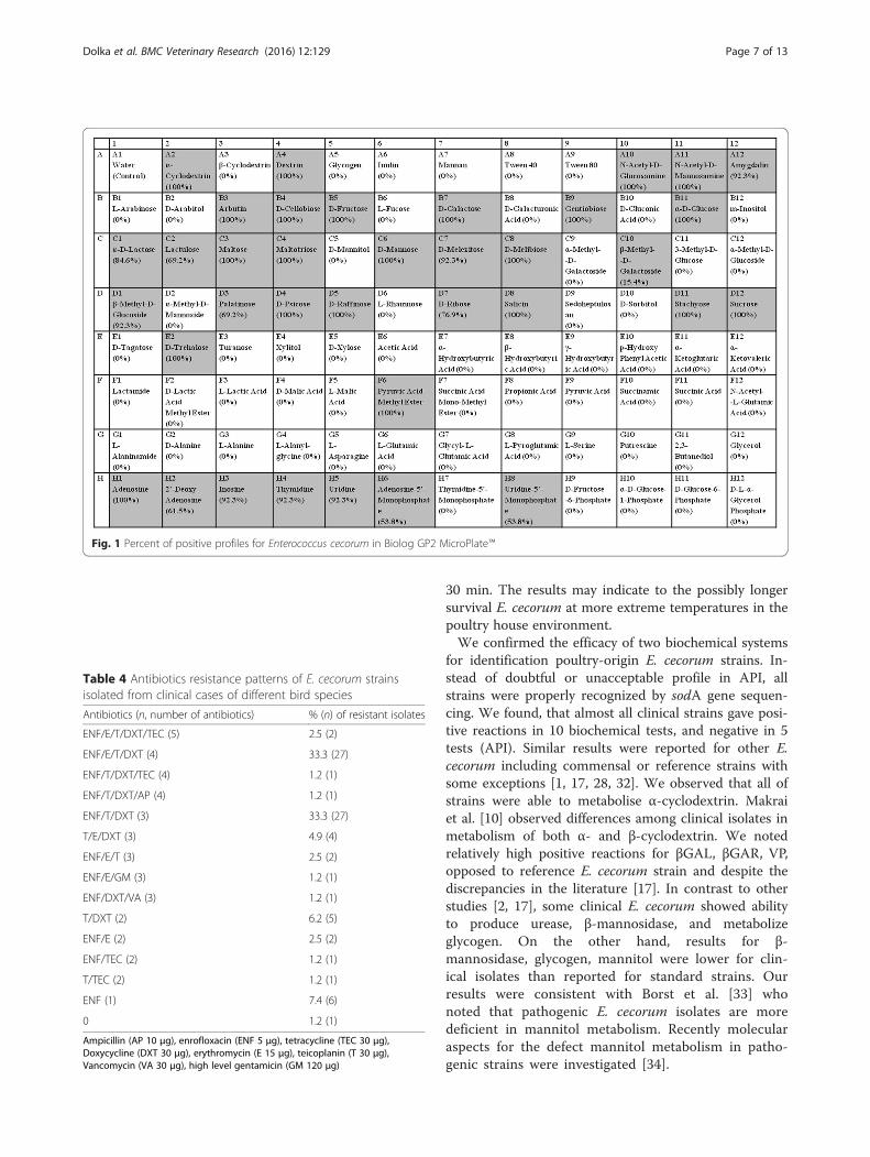
Biolog system (index: probability 91.7 %, similarity 0.806).The metabolic fingerprinting for E. cecorum was showedin Fig. 1. All of the examined isolates were able tometabolise 22 carbon sources (α-cyclodextrin, dextrin, N-acetyl-D-glucosamine, N-acetyl-D-mannosamine, arbutin,D-cellobiose, D-fructose, D-galactose, gentiobiose, α-D-glucose, maltose, maltotriose, D-mannose, D-melibiose,D-psicose, D-raffinose, salicin, stachyose, sucrose, D-trehalose, pyruvic acid methyl ester, adenosine). Not all ofexamined isolates were able to metabolise 14 carbonsources: amygdalin, D-melezitose, β-methyl-D-glucoside,inosine, thymidine, uridine (metabolised by 92.3 %strains), α-D-lactose (84.6 %), D-ribose (76.9 %), lactulose,palatinose (69.2 %), 2’-deoxy-adenosine (61.5 %),adenosine-5’-monophosphate, uridine-5’-monophosphate(53.8 %), β-methyl-D-galactoside (15.4 %). Further 59 car-bon sources present in the GP2 microplate were not uti-lised by E. cecorum.
Virulence factorsOf all 82 E. cecorum strains, 22 (26.8 %) were positivefor asa1, 21 (25.6 %) for gelE, 12 (14.6 %) for ace, 11(13.4 %) for efaA. The cylA and esp PCR amplificationyielded positive results in 4 (4.9 %) and 2 (2.4 %) E.cecorum strains. The hyl gene was not detected in anystrain. The isolates from CB were positive for asa1(24.5 %), gelE (22.4 %), ace (14.3 %), efaA (14.3 %), cylA(2.1 %). The isolates from BB were positive for asa1(20 %), gelE (20 %), ace (15 %), esp (10 %), cylA (10 %).The isolates from CL were positive for asa1 (60 %), gelE(60 %), efaA (20 %), ace (20 %). None of 7 virulence fac-tors was found in isolates from G and T flocks. Most ofvirulence-gene positive isolates (11; 13.4 %) contained 2of tested 7 virulence genes, then 6 (7.3 %) E. cecorumcontained 4 virulence genes, 5 (6.1 %) harbored 1 viru-lence gene, while 3 (3.7 %) carried 3 virulence genes. Intwo isolates (2.4 %) 6 virulence genes were identified.None of isolates carried 5 or 7 virulence genes. Pheno-typically, non of isolates produced gelatinase despite be-ing gelE-positive in PCR.
Antibiotic susceptibilityOne (0.82 %) out of the 82 clinical E. cecorum was sus-ceptible to 13 antibiotics tested, the rest were resistantto one or more antibiotics (Table 4). All isolates weresusceptible to amoxicillin/clavulanic acid (AUG) and
Table 1 Test or characteristic for E. cecorum isolates (n = 82)
Test or characteristic E. cecorum isolates from clinical cases
Hemolysis α (strong)
Gram-staining Gram-positive
Cell morphology ovoid cocci (single, double or short chains)
Catalase-production negative
Oxidase-production negative
Yellow pigment-production negative
Lancefield group D negative
Motility negative
Halotolerance (6.5 % NaCl) limited growth
Growth at: % positive (n)
4 °C 100 % (82)
10 °C 98.8 % (81)
45 °C 74.4 % (61)
Survival at 60 °C for: % positive (n)
15 min 76.8 % (63)
30 min 64.6 % (53)
1 h 54.9 % (45)
Survival at 70 °C for: % positive (n)
15 min 36.6 % (30)
30 min 15.9 % (13)
1 h 0 % (0)
Dolka et al. BMC Veterinary Research (2016) 12:129 Page 4 of 13
penicillin (PG), nitrofurantoin (NI), and linezolid (LZD).The majority of isolates were susceptible to ampicillin(AP), and high level gentamicin (GM) (n = 81; 99 %),chloramphenicol (C) (n = 79; 96 %), vancomycin (VA) (n= 75; 91 %). The lower level susceptibility was toerythromycin (E) (n = 42; 51 %), tetracycline (TEC) (n =24; 29 %), teicoplanin (T) and doxycycline (DXT) (n =11; 13 %). Most isolates noted intermediate susceptibilityto TEC (n = 53; 65 %) with 29 % susceptible and 6 % re-sistant. None of clinical E. cecorum isolates was suscep-tible to enrofloxacin (71 resistant isolates). A highpercentage of antimicrobial resistance was also observedto teicoplanin (T) (n = 70; 85 %), and doxycycline (DXT)(n = 68; 83 %). Linezolid resistance among E. cecorumisolates was not found. Of the vancomycin resistancegenes tested by PCR, vanA gene was present in onestrain, vanB gene was not detected.
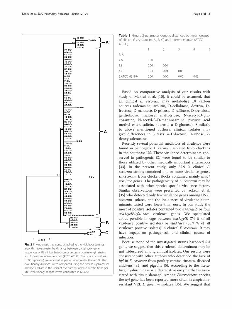
Molecular identificationThe obtained sequences sodA gene fragment showedsimilarity to E. cecorum (BLAST database) and allowedfor identification strains. Dendrogram showed the gen-etic similarity between reference strain of E. cecorumand clinical isolates based on the sodA gene sequences(Fig. 2). Phylogenetic analysis supported the separationof clinical isolates into three main groups (A, B, C).Genetic distances between groups ranged from 0.00 to0.04 (Table 5). The group A comprised 69 strains (CB n= 43, BB n = 15, CL n = 10, G n = 1) and had one sub-group (A’) with 5 strains (BB n = 4, CB n = 1). Five CBisolates were clustered together in the group B, andthree isolates (BB, G, T) in the group C (all isolates from2014). Among all groups, the group C revealed the high-est values of genetic distance with B group (0.04) andwith reference strain (0.03).
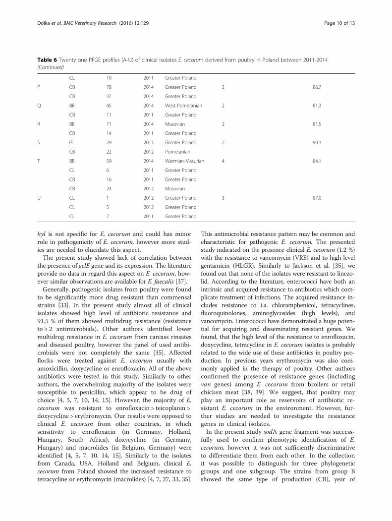
PFGEThe PFGE analysis (based on >80 % similarity index) of82 clinical E. cecorum isolates exhibited 21 pulsotypes(A-U) with 60 strains (41 CB, 10 BB, 8 CL, 1 G) (Table 6,
Fig. 3). The highest degree of band similarity (>90 %)was demonstrated in pulsotype B (with two CB isolates)and in pulsotype S (with G and CB isolate). Pulsotype Mwas the predominant type, and included 8 isolates (8/60,13.3 %), then E, L, T pulsotypes (each included 4 iso-lates). However, 11 of the 21 pulsotypes included only 2isolates. Twenty isolates (20/60, 33.3 %) representing CBflocks (20/41; 48.8 %), were distributed among 8 pulso-types: A, B, C, D, P (each 3.33 %), F, K (each 5 %), L(6.6 %). The majority of BB isolates (7/10, 70 %) wereclustered with CB isolates (13) in distinct 6 pulsotypes(E, G, I, M, Q, R). Among isolates representing CL flocks(8/60, 13.3 %), three of these (3/8, 37.5 %) were clusteredin one profile (L). Three pulsotypes (H, N, O) were cre-ated by clustering both CB (5) and CL (3) isolates. Gen-erally, no clear temporal and geographical clustering wasvisible, but with the exceptions of 7 pulsotypes (A, D, I,L, M, P, U).
DiscussionIn order to characterize clinical E. cecorum, we investi-gated 82 strains isolated from clinical samples originatedfrom different poultry flocks (1 isolate per flock). Our ob-servations were consistent with reports on a succession ofdisease outbreaks in broiler flocks raised in the intensivefarming systems [9]. Previously, clinical E. cecorum wasnot described in commercial chicken layers or geeseflocks. We found that the problem may affect hens orother bird species than chicken. Our results were consist-ent with the literature in regarding on certain characteris-tics traditionally considered to be typical for the genusEnterococcus or E. cecorum including intestinal isolates ofpoultry origin [28, 29]. According to the literature, E.cecorum are often NaCl sensitive [17, 30], and intestinal E.cecorum of poultry may be also NaCl-resistant [28]. In ourstudy, clinical isolates appeared to be less salt-tolerant,however no complete inhibition of growth was observed.Authors suggested possible higher ability to survive clin-ical E. cecorum in saline environment or even higher re-sistance to chlorine-based disinfectants.
Table 2 Results of E. cecorum (n = 82) growth on different media
Medium Observed growth (YES/NO) Description of colonies of Enterococcus cecorum
Columbia Agar with 5 % Sheep Blood (CA) YES Small, round, white-grey colonies with α-hemolysis
Columbia CNA Agar with 5 % Sheep Blood YES Small, grayish colonies with α-hemolysis, resistant to two antibioticscolistin and nalidixic acid
Edwards Agar with 5 % sheep blood YES Blue-grayish coloured colonies with α-hemolysis
Bile Esculin Azide Agar (Enterococcosel Agar) YES Colonies beige with strong black halos
KAA agar (Kanamycine Esculin Azide Agar) YES (weak) Brown to black colonies and blackening zones around the colonies
Slanetz and Bartley Agar (with tetrazolium chloride) NO or poor Red, maroon colonies
TCC agar with tergitol NO Total inhibition
Tellurite Agar (potassium tellurite) NO Total inhibition
Dolka et al. BMC Veterinary Research (2016) 12:129 Page 5 of 13
Previous research demonstrated no growth of poultrycecal E. cecorum on Slanetz medium, and on KAA agar[1], while clinical strains showed variable growth onthese media. The growth was clearly more abundant onbile esculine azide agar than on esculin azide agar withkanamycin. Based on results, we suggested that completegrowth inhibition on a solid medium supplemented withtergitol or with potassium tellurite may be used in
identification of this enterococcal species. According tothe literature, Enterococcus species are able to survive arange of stresses and hostile environments [31], but E.cecorum was described as unable to grow at 10 °C orsurvive 30 min at 60 °C [5, 29]. In contrast to above au-thors, clinical isolates were able to grow at low tempera-tures (4 °C, 10 °C) and some of them might survive evenlonger heating at 60 °C for 1 h and even 70 °C for
Table 3 Percent of positive reactions (%) in rapid ID 32 STREP (bioMérieux, France) for clinical E. cecorum isolates in this study(n = 82) compared with standard isolates (manufacturers recommendations) and control strain (E. cecorum ATCC 43198)
Parameter % Positive reactions in rapid ID 32 STREP for Enterococcus cecorum
Clinical isolates (this study) % (n) Manufacturers recommendations % Reference strain E. cecorum ATCC 43198 (+/-)
ADH 0 (0) 0 -
βGLU 100 (82) 100 +
βGAR 73 (60) 11 -
βGUR 94 (77) 88 +
αGAL 99 (81) 100 +
PAL 71 (58) 94 +
RIB 99 (81) 98 +
MAN 12 (10) 38 -
SOR 10 (8) 11 -
LAC 88 (72) 100 +
TRE 99 (81) 100 +
RAF 100 (82) 88 +
VP 60 (49) 66 -
APPA 0 (0) 0 -
βGAL 80 (66) 33 -
PYRA 0 (0) 0 -
βNAG 82 (67) 88 +
GTA 89 (73) 94 +
HIP 0 (0) 1 -
GLYG 12 (10) 27 -
PUL 4 (3) 0 -
MAL 98 (80) 100 +
MEL 98 (80) 98 +
MLZ 88 (72) 55 +
SAC 100 (82) 100 +
LARA 0 (0) 0 -
DARL 1 (1) 0 -
MβDG 100 (82) 98 +
TAG 65 (53) 64 +
βMAN 17 (14) 41 -
CDEX 100 (82) 66 +
URE 6 (5) 0 -
ADH (arginine dihydrolase), βGLU (β-glucosidase), βGAR (β-galactosidase), βGUR (β-glucuronidase), αGAL (α-galactosidase), PAL (alkaline phosphatase), RIB (ribose), MAN(mannitol), SOR (sorbitol), LAC (lactose), TRE (trehalose), RAF (rafinose), VP (Voges Proskauer, aceton production), APPA (alanyl-phenylalanyl-proline arylamidase), βGAL(β-galactosidase), PYRA (pyroglutamic acid arylamidase), βNAG (N-acetyl-β-glucosaminidase), GTA (glycyl-tryptophan arylamidase), HIP (hydrolysis of hipurate), GLYG(glycogen), PUL (pullulane), MAL (maltose), MEL (melibiose), MLZ (melezitose), SAC (saccharose), LARA (L-arabinose), DARL (D-arabitol), CDEX (cyclodextrin), MβDG(methyl-βD-glucopyranoside), TAG (tagatose), βMAN (β-mannosidase), URE (urease)
Dolka et al. BMC Veterinary Research (2016) 12:129 Page 6 of 13
30 min. The results may indicate to the possibly longersurvival E. cecorum at more extreme temperatures in thepoultry house environment.We confirmed the efficacy of two biochemical systems
for identification poultry-origin E. cecorum strains. In-stead of doubtful or unacceptable profile in API, allstrains were properly recognized by sodA gene sequen-cing. We found, that almost all clinical strains gave posi-tive reactions in 10 biochemical tests, and negative in 5tests (API). Similar results were reported for other E.cecorum including commensal or reference strains withsome exceptions [1, 17, 28, 32]. We observed that all ofstrains were able to metabolise α-cyclodextrin. Makraiet al. [10] observed differences among clinical isolates inmetabolism of both α- and β-cyclodextrin. We notedrelatively high positive reactions for βGAL, βGAR, VP,opposed to reference E. cecorum strain and despite thediscrepancies in the literature [17]. In contrast to otherstudies [2, 17], some clinical E. cecorum showed abilityto produce urease, β-mannosidase, and metabolizeglycogen. On the other hand, results for β-mannosidase, glycogen, mannitol were lower for clin-ical isolates than reported for standard strains. Ourresults were consistent with Borst et al. [33] whonoted that pathogenic E. cecorum isolates are moredeficient in mannitol metabolism. Recently molecularaspects for the defect mannitol metabolism in patho-genic strains were investigated [34].
Fig. 1 Percent of positive profiles for Enterococcus cecorum in Biolog GP2 MicroPlate™
Table 4 Antibiotics resistance patterns of E. cecorum strainsisolated from clinical cases of different bird species
Antibiotics (n, number of antibiotics) % (n) of resistant isolates
ENF/E/T/DXT/TEC (5) 2.5 (2)
ENF/E/T/DXT (4) 33.3 (27)
ENF/T/DXT/TEC (4) 1.2 (1)
ENF/T/DXT/AP (4) 1.2 (1)
ENF/T/DXT (3) 33.3 (27)
T/E/DXT (3) 4.9 (4)
ENF/E/T (3) 2.5 (2)
ENF/E/GM (3) 1.2 (1)
ENF/DXT/VA (3) 1.2 (1)
T/DXT (2) 6.2 (5)
ENF/E (2) 2.5 (2)
ENF/TEC (2) 1.2 (1)
T/TEC (2) 1.2 (1)
ENF (1) 7.4 (6)
0 1.2 (1)
Ampicillin (AP 10 μg), enrofloxacin (ENF 5 μg), tetracycline (TEC 30 μg),Doxycycline (DXT 30 μg), erythromycin (E 15 μg), teicoplanin (T 30 μg),Vancomycin (VA 30 μg), high level gentamicin (GM 120 μg)
Dolka et al. BMC Veterinary Research (2016) 12:129 Page 7 of 13
Based on comparative analysis of our results withstudy of Makrai et al. [10], it could be assumed, thatall clinical E. cecorum may metabolise 18 carbonsources (adenosine, arbutin, D-cellobiose, dextrin, D-fructose, D-mannose, D-psicose, D-raffinose, D-trehalose,gentiobiose, maltose, maltotriose, N-acetyl-D-glu-cosamine, N-acetyl-β-D-mannosamine, pyruvic acidmethyl ester, salicin, sucrose, α-D-glucose). Similarlyto above mentioned authors, clinical isolates maygive differences in 3 tests: α-D-lactose, D-ribose, 2-deoxy adenosine.Recently several potential mediators of virulence were
found in pathogenic E. cecorum isolated from chickensin the southeast US. These virulence determinants con-served in pathogenic EC were found to be similar tothose utilized by other medically important enterococci[33]. In the present study, only 32.9 % clinical E.cecorum strains contained one or more virulence genes.E. cecorum from chicken flocks contained mainly asa1/gelE/ace genes. The pathogenicity of E. cecorum may beassociated with other species-specific virulence factors.Similar observations were presented by Jackson et al.[35] who detected only few virulence genes among US E.cecorum isolates, and the incidences of virulence deter-minants tested were lower than ours. In our study themost of positive isolates contained two asa1/gelE or fourasa1/gelE/efaA/ace virulence genes. We speculatedabout possible linkage between asa1/gelE (74 % of allvirulence positive isolates) or efaA/ace (33.3 % of allvirulence positive isolates) in clinical E. cecorum. It mayhave impact on pathogenesis and clinical course ofinfection.Because none of the investigated strains harbored hyl
gene, we suggest that this virulence determinant may benot widespread among clinical isolates. Our results wereconsistent with other authors who described the lack ofhyl in E. cecorum from poultry carcass rinsates, diseasedchickens [35] and pigeons [5]. According to the litera-ture, hyaluronidase is a degradative enzyme that is asso-ciated with tissue damage. Among Enteroccocus speciesthe hyl gene has been reported more often in ampicillin-resistant VRE E. faecium isolates [36]. We suggest that
Fig. 2 Phylogenetic tree constructed using the Neighbor-Joiningalgorithm to evaluate the distance between partial sodA genesequences of 82 clinical Enterococcus cecorum poultry-origin strainsand E. cecorum reference strain (ATCC 43198). The bootstrap values(1000 replicates) are reported as percentage greater than 60 %. Theevolutionary distances were computed using the Kimura 2-parametermethod and are in the units of the number of base substitutions persite. Evolutionary analyses were conducted in MEGA6
Table 5 Kimura 2-parameter genetic distances between groupsof clinical E. cecorum (A, A’, B, C) and reference strain (ATCC43198)
1 2 3 4 5
1. A
2.A’ 0.00
3.B 0.00 0.01
4.C 0.03 0.04 0.03
5.ATCC (43198) 0.00 0.00 0.00 0.03
Dolka et al. BMC Veterinary Research (2016) 12:129 Page 8 of 13
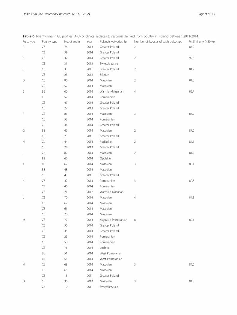
Table 6 Twenty one PFGE profiles (A-U) of clinical isolates E. cecorum derived from poultry in Poland between 2011-2014
Pulsotype Poultry type No. of strain Year Poland’s voivodeship Number of isolates of each pulsotype % Similarity (>80 %)
A CB 76 2014 Greater Poland 2 84.2
CB 39 2014 Greater Poland
B CB 32 2014 Greater Poland 2 92.3
CB 31 2013 Świętokrzyskie
C CB 3 2011 Greater Poland 2 84.2
CB 23 2012 Silesian
D CB 80 2014 Masovian 2 81.8
CB 57 2014 Masovian
E BB 60 2014 Warmian-Masurian 4 85.7
CB 52 2014 Pomeranian
CB 47 2014 Greater Poland
CB 27 2013 Greater Poland
F CB 81 2014 Masovian 3 84.2
CB 53 2014 Pomeranian
CB 34 2014 Greater Poland
G BB 46 2014 Masovian 2 87.0
CB 2 2011 Greater Poland
H CL 44 2014 Podlaskie 2 84.6
CB 28 2013 Greater Poland
I CB 82 2014 Masovian 2 81.2
BB 66 2014 Opolskie
J BB 67 2014 Masovian 3 80.1
BB 48 2014 Masovian
CL 4 2011 Greater Poland
K CB 42 2014 Pomeranian 3 80.8
CB 40 2014 Pomeranian
CB 21 2012 Warmian-Masurian
L CB 70 2014 Masovian 4 84.3
CB 62 2014 Masovian
CB 61 2014 Masovian
CB 20 2014 Masovian
M CB 77 2014 Kuyavian-Pomeranian 8 82.1
CB 56 2014 Greater Poland
CB 35 2014 Greater Poland
CB 25 2014 Pomeranian
CB 58 2014 Pomeranian
CB 75 2014 Lodzkie
BB 51 2014 West Pomeranian
BB 55 2014 West Pomeranian
N CB 68 2014 Masovian 3 84.0
CL 65 2014 Masovian
CB 13 2011 Greater Poland
O CB 30 2013 Masovian 3 81.8
CB 19 2011 Świętokrzyskie
Dolka et al. BMC Veterinary Research (2016) 12:129 Page 9 of 13
hyl is not specific for E. cecorum and could has minorrole in pathogenicity of E. cecorum, however more stud-ies are needed to elucidate this aspect.The present study showed lack of correlation between
the presence of gelE gene and its expression. The literatureprovide no data in regard this aspect on E. cecorum, how-ever similar observations are available for E. faecalis [37].Generally, pathogenic isolates from poultry were found
to be significantly more drug resistant than commensalstrains [33]. In the present study almost all of clinicalisolates showed high level of antibiotic resistance and91.5 % of them showed multidrug resistance (resistanceto ≥ 2 antimicrobials). Other authors identified lowermultidrug resistance in E. cecorum from carcass rinsatesand diseased poultry, however the panel of used antibi-crobials were not completely the same [35]. Affectedflocks were treated against E. cecorum usually withamoxicillin, doxycycline or enrofloxacin. All of the aboveantibiotics were tested in this study. Similarly to otherauthors, the overwhelming majority of the isolates weresusceptible to penicillin, which appear to be drug ofchoice [4, 5, 7, 10, 14, 15]. However, the majority of E.cecorum was resistant to enrofloxacin > teicoplanin >doxycycline > erythromycin. Our results were opposed toclinical E. cecorum from other countries, in whichsensitivity to enrofloxacin (in Germany, Holland,Hungary, South Africa), doxycycline (in Germany,Hungary) and macrolides (in Belgium, Germany) wereidentified [4, 5, 7, 10, 14, 15]. Similarly to the isolatesfrom Canada, USA, Holland and Belgium, clinical E.cecorum from Poland showed the increased resistance totetracycline or erythromycin (macrolides) [4, 7, 27, 33, 35].
This antimicrobial resistance pattern may be common andcharacteristic for pathogenic E. cecorum. The presentedstudy indicated on the presence clinical E. cecorum (1.2 %)with the resistance to vancomycin (VRE) and to high levelgentamicin (HLGR). Similarly to Jackson et al. [35], wefound out that none of the isolates were resistant to linezo-lid. According to the literature, enterococci have both anintrinsic and acquired resistance to antibiotics which com-plicate treatment of infections. The acquired resistance in-cludes resistance to i.a. chloramphenicol, tetracyclines,fluoroquinolones, aminoglycosides (high levels), andvancomycin. Enterococci have demonstrated a huge poten-tial for acquiring and disseminating resistant genes. Wefound, that the high level of the resistance to enrofloxacin,doxycycline, tetracycline in E. cecorum isolates is probablyrelated to the wide use of these antibiotics in poultry pro-duction. In previous years erythromycin was also com-monly applied in the therapy of poultry. Other authorsconfirmed the presence of resistance genes (includingvan genes) among E. cecorum from broilers or retailchicken meat [38, 39]. We suggest, that poultry mayplay an important role as reservoirs of antibiotic re-sistant E. cecorum in the environment. However, fur-ther studies are needed to investigate the resistancegenes in clinical isolates.In the present study sodA gene fragment was success-
fully used to confirm phenotypic identification of E.cecorum, however it was not sufficiently discriminativeto differentiate them from each other. In the collectionit was possible to distinguish for three phylogeneticgroups and one subgroup. The strains from group Bshowed the same type of production (CB), year of
Table 6 Twenty one PFGE profiles (A-U) of clinical isolates E. cecorum derived from poultry in Poland between 2011-2014(Continued)
CL 10 2011 Greater Poland
P CB 78 2014 Greater Poland 2 86.7
CB 37 2014 Greater Poland
Q BB 45 2014 West Pomeranian 2 81.3
CB 11 2011 Greater Poland
R BB 71 2014 Masovian 2 81.5
CB 14 2011 Greater Poland
S G 29 2013 Greater Poland 2 90.3
CB 22 2012 Pomeranian
T BB 59 2014 Warmian-Masurian 4 84.1
CL 6 2011 Greater Poland
CB 16 2011 Greater Poland
CB 24 2012 Masovian
U CL 1 2012 Greater Poland 3 87.0
CL 5 2012 Greater Poland
CL 7 2011 Greater Poland
Dolka et al. BMC Veterinary Research (2016) 12:129 Page 10 of 13
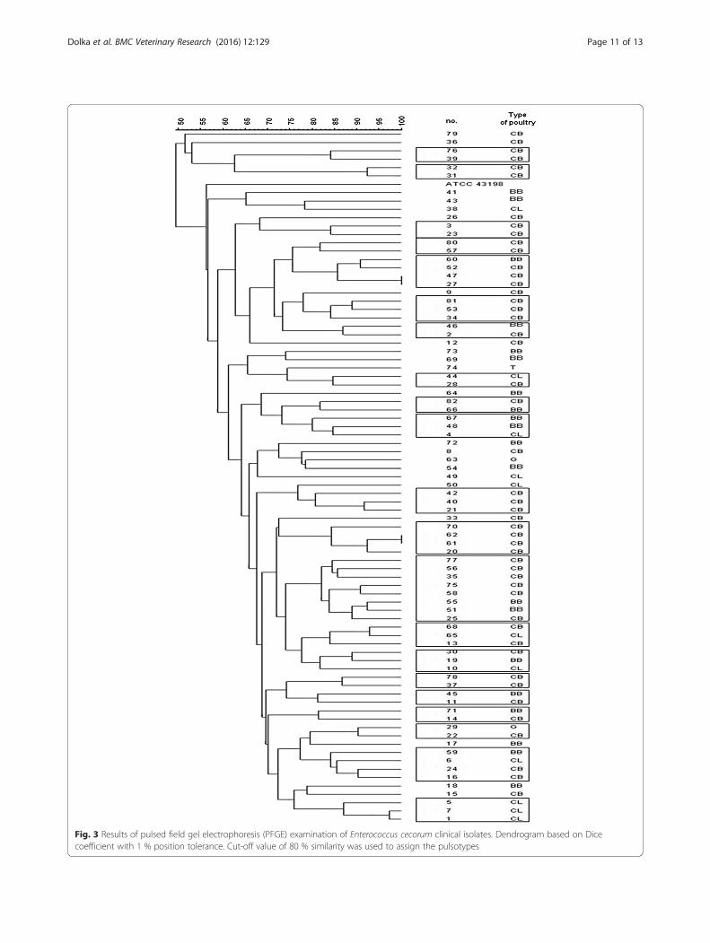
Fig. 3 Results of pulsed field gel electrophoresis (PFGE) examination of Enterococcus cecorum clinical isolates. Dendrogram based on Dicecoefficient with 1 % position tolerance. Cut-off value of 80 % similarity was used to assign the pulsotypes
Dolka et al. BMC Veterinary Research (2016) 12:129 Page 11 of 13
isolation, virulence determinants and multidrug resist-ance pattern, but different geographical origin; 80 % ofthem belong to pulsotype M. The strains of group Cshared only the same year of isolation, virulence andmultidrug resistance patterns. The low genetic distance(based on sodA gene sequences) indicated on the veryclose genetic relationships between clinical E. cecorum.No clear genetic differences were observed between clin-ical strains and reference strain.Recent data indicated that pathogenic E. cecorum from
the southeast US were clonal, however comparative gen-omic analysis revealed fundamental differences in their ge-nomes [34]. According to the previous report, isolatesrecovered from spinal abscesses were highly similar andcould be detected by using PFGE [33]. In our study, PFGEresults showed the genetic heterogeneity between clinicalE. cecorum isolates, that is consistent with the other stud-ies [18]. Therefore, the usage of PFGE in distinguishingpathogenic strains may be difficult and limited. This gen-etic diversity was seen between poultry flocks, howeversome clustering was visible in relation of type of produc-tion (CB, CL). Moreover, some temporal and geographicalclustering was visible. Many CB isolates from the sameyear and geographical origin were clustered together (pul-sotype A, D, L, P) indicating their close genetic relation-ship. Some CL isolates from the same location butdifferent years were grouped into a single pulsotype (U)indicating on the possible horizontal transmission amongCL flocks in this area. We found that CB and BB isolatesfrom the same year which were clustered together intoseparate pulsotypes (I, M). Based on the relatively close re-lationship between isolates from geese and chicken flocks,it could be assumed that isolates from the single clonallineage may cause outbreaks in different bird species. Theresults may suggest the transmission of potential disease-causing E. cecorum between flocks.
ConclusionsThese data indicate that several, widely distributed patho-genic E. cecorum clones seemed to be responsible for in-fection cases found in different poultry types. The isolatescausing infection in different CB flock in the same yearand region may be somewhat genetically distinct fromeach other and from those that cause disease in CL or BBflocks in the same year and region. Phenotypically, clinicalisolates were generally found to be very similar, howeversome properties or characteristics described in some iso-lates were not found in others. The study presented hereis the first in Poland as well as one of the few in Europewhich provides phenotypic and genotypic characterizationof E. cecorum isolates associated with disease outbreaks inpoultry flocks. Further research needs to focus on findingnew virulence determinants of E. cecorum and recognitionof transmission routes.
Additional files
Additional file 1: Alignment of partial sodA gene sequences from E.cecorum isolates and reference strain (ATCC 43198). Nucleotidedifferences are specified by the nucleotide, while dot represented nonucleotide changing. (TIF 12403 kb)
Additional file 2: Alignment of partial 16S rRNA gene sequences fromE. cecorum isolates and reference strain (ATCC 43198). Nucleotidedifferences are specified by the nucleotide, while dot represented nonucleotide changing. (TIF 60654 kb)
AbbreviationsBB, broiler breeder flocks; CA, Columbia agar with 5 % sheep blood; CB,chicken broiler flocks (commercial broilers); CL, commercial layer flocks; E.cecorum, Enterococcus cecorum; ES, enterococcal spondylitis; G, geese flocks;HLGR, high level gentamicin resistance; PFGE, Pulsed Field GelElectrophoresis; T, turkey flock; VRE Vancomycin-Resistant Enterococcus
AcknowledgementsThe authors thank mgr Beata Sienkiewicz for excellent technical assistance.The authors are very grateful veterinary laboratories and private practiceveterinarians for help in collecting strains: Microbiological Laboratory SGGW,SLW Biolab Ostróda, Lab-Vet Sp. z o.o. Tarnowo Podgórne, Vet-Lab Brudzew,Vetdiagnostica Solec Kujawski, Animal Pharma, Wet-Net s.c. Giżycko, GabinetWeterynaryjny “Gallus” Sylwester Barabasz, “Spec-Drób” Mariusz Lorek, andDVM: Anna Biegańska, Magdalena Wiczk, Tomasz Nowak, Ismaila Massaly,Mirosław Berezowski, Paweł Tubielewicz, Natalia Bednarz.
FundingThis work was financially supported by grant no. 505-10-023700-L00183-99 atDepartment of Pathology and Veterinary Diagnostics, Faculty of VeterinaryMedicine, Warsaw University of Life Sciences-SGGW, Poland. The analysis by Biologsystem was supported by Department of Microbiology and Infectious Diseases,Faculty of Veterinary Science, Szent István University in Budapest, Hungary.
Availability of data and materialThe datasets supporting our findings are included within the article and itsAdditional files 1 and 2.
Authors’ contributionsBD conceived and designed the study, collected the strains, performed allworks, analysis and interpretation of data, wrote manuscript. DCC input inPFGE. LM contributed to perform analysis in Biolog system. PS gaveconceptual advice and additional inputs in study project, contributedmaterials. All authors read and approved the final manuscript.
Competing interestsThe authors declare that they have no competing interests.
Consent to publicationNot applicable.
Ethics approval and consent to participatePoultry samples were collected for laboratory diagnosis by as part of theusual clinical practice on farms, and Polish ethical guidelines (Dz.U. 2015 poz.266) and animal welfare regulations were strictly respected. Veterinarianswho provided healthcare for poultry flocks were in contact with the owners,and gave an oral informed consent and acceptance for using obtainedisolates for research studies. As this study was focused on bacterial isolatescollected from routine samples, approval from Ethics Committee at theWarsaw University of Life Sciences University was not necessary.
Author details1Department of Pathology and Veterinary Diagnostics, Faculty of VeterinaryMedicine, Warsaw University of Life Sciences-SGGW, Nowoursynowska 159cSt., Warsaw 02-776, Poland. 2Department of Preclinical Sciences, Faculty ofVeterinary Medicine, Warsaw University of Life Sciences-SGGW, Ciszewskiego8 St., Warsaw 02-786, Poland. 3Department of Microbiology and InfectiousDiseases, Faculty of Veterinary Science, Szent István University, Hungária krt.23-25, Budapest H-1143, Hungary.
Dolka et al. BMC Veterinary Research (2016) 12:129 Page 12 of 13
Received: 6 February 2016 Accepted: 23 June 2016
References1. Devriese LA, Dutta GN, Farrow JAE, van de Kerckhove A, Phillips BA.
Streptococcus cecorum, a new species isolated from chickens. Int J SystBacteriol. 1983;33:772–6.
2. Greub G, Devriese LA, Pot B, Dominguez J, Bille J. Enterococcus cecorumsepticemia in a malnourished adult patient. Eur J Clin Microbiol Infect Dis.1997;16:594–8.
3. Warnke P, Köller T, Stoll P, Podbielski A. Nosocomial infection due toEnterococcus cecorum identified by MALDI-TOF MS and Vitek 2 from a bloodculture of a septic patient. Eur J Microbiol Immunol (Bp). 2015;5:177–9.
4. Devriese LA, Cauwerts K, Hermans K, Wood AM. Enterococcus cecorumsepticaemia as a cause of bone and joint lesions resulting in lameness inbroiler chickens. Flemish Vet J. 2002;71:219–21.
5. Jung A, Teske L, Rautenschlein S. Enterococcus cecorum infection in a racingpigeon. Avian Dis. 2014;58:654–8.
6. Wood AM, MacKenzie G, McGiliveray NC, Brown L, Devriese LA, Baele M.Isolation of Enterococcus cecorum from bone lesions in broiler chickens. VetRec. 2002;150:27.
7. De Herdt P, Defoort P, Van Steelant J, Swam H, Tanghe L, Van Goethem S,Vanrobaeys M. Enterococcus cecorum osteomyelitis and arthritis in broilerchickens. Vlaams Diergen Tijds. 2008;78:44–8.
8. Stalker MJ, Brash ML, Weisz A, Ouckama RM, Slavic D. Arthritis andosteomyelitis associated with Enterococcus cecorum infection in broiler andbroiler breeder chickens in Ontario, Canada. J Vet Diagn Invest. 2010;22:643–5.
9. Armour NK, Collett SR, Williams SM. Enterococcus cecorum – related arthritis andosteomyelitis in broilers and broiler breeders. Poult Inf Profess. 2011;17:1–7.
10. Makrai L, Nemes C, Simon A, Ivanics E, Dudás Z, Fodor L, Glávits R.Association of Enterococcus cecorum with vertebral osteomyelitis andspondylolisthesis in broiler parent chicks. Acta Vet Hung. 2011;59:11–21.
11. Dolka B, Szeleszczuk P. Enterococcal vertebral osteoarthritis in chickens. MedWeter. 2012;68:157–62.
12. Robbins KM, Suyemoto MM, Lyman RL, Martin MP, Barnes HJ, Borst LB. Anoutbreak and source investigation of enterococcal spondylitis in broilerscaused by Enterococcus cecorum. Avian Dis. 2012;56:768–73.
13. Szeleszczuk P, Dolka B, Żbikowski A, Dolka I, Peryga M. First case ofenterococcal spondylitis in broiler chickens in Poland. Med Weter. 2013;69:298–303.
14. Aitchison H, Poolman P, Coetzer M, Griffiths C, Jacobs J, Meyer M, BisschopS. Enterococcal-related vertebral osteoarthritis in South African broilerbreeders: A case report. J S Afr Vet Assoc. 2014;85:1077.
15. Jung A, Rautenschlein S. Comprehensive report of an Enterococcus cecoruminfection in a broiler flock in Northern Germany. BMC Vet Res. 2014;10:311.
16. Murray BE. The life and times of the Enterococcus. Clin Microbiol Rev. 1990;3:46–65.
17. Manero A, Blanch AR. Identification of Enterococcus spp. with a biochemicalkey. Appl Environ Microbiol. 1999;65:4425–30.
18. Wijetunge DS, Dunn P, Wallner-Pendleton E, Lintner V, Lu H, Kariyawasam S.Fingerprinting of poultry isolates of Enterococcus cecorum using threemolecular typing methods. J Vet Diagn Invest. 2012;24:1166–71.
19. Tenover FC, Arbeit RD, Goering RV, Mickelsen PA, Murray BE, Persing DH,Swaminathan B. Interpreting chromosomal DNA restriction patternsproduced by pulsed-field gel electrophoresis: criteria for bacterial straintyping. J Clin Microbiol. 1995;33:2233–9.
20. Kense MJ, Landman WJ. Enterococcus cecorum infections in broiler breedersand their offspring: molecular epidemiology. Avian Pathol. 2011;40:603–12.
21. Ke D, Picard FJ, Martineau F, Menard C, Roy PH, Ouellette M, Bergeron MG.Development of a PCR assay for rapid detection of enterococci. J ClinMicrobiol. 1999;37:3497–503.
22. Jackson CR, Fedoka – Cray PJ, Barett JB. Use of a genus- and species-specificmultiplex PCR for identication of enterococci. J Clin Microbiol. 2004;42:3558–65.
23. Tsai JC, Hsueh PR, Lin HM, Chang HJ, Ho SW, Teng LJ. Identification ofclinically relevant enterococcus species by direct sequencing of groES andspacer region. J Clin Microbiol. 2005;43:235–41.
24. Martín-Platero AM, Valdivia E, Maqueda M, Martínez-Bueno M.Characterization and safety evaluation of enterococci isolated from Spanishgoats’ milk cheeses. Int J Food Microbiol. 2009;132:24–32.
25. CLSI. Performance Standards for Antimicrobial Disk and DilutionSusceptibility Tests for Bacteria Isolated from Animals; Approved Standard –Fourth Edition. Wayne: Clinical and Laboratory Standards Institute; 2013. PACLSI document VET01-A4, supplement VET01-S2.
26. Van den Braak N, van Belkum A, van Keulen M, Vliegenthart J, Verbrugh HA,Endtz HP. Molecular characterization of vancomycin-resistant enterococcifrom hospitalized patients and poultry products in The Netherlands. J ClinMicrobiol. 1998;36:1927–32.
27. Boerlin P, Nicholson V, Brash M, Slavic D, Boyen F, Sanei B, Butaye P.Diversity of Enterococcus cecorum from chickens. Vet Microbiol. 2012;157:405–11.
28. Devriese LA, Hommez J, Wijfels R, Haesebrouck F. Composition of theenterococcal and streptococcal intestinal flora of poultry. J Appl Bacteriol.1991;71:46–50.
29. Devriese LA, Pot B, Collins MD. Phenotypic identification of the genusEnterococcus and differentiation of phylogenetically distinct enterococcalspecies and species groups. J Appl Bacteriol. 1993;75:399–408.
30. Domig KJ, Mayer HK, Kneifel W. Methods used for the isolation,enumeration, characterisation and identification of Enterococcus spp. 2.Pheno- and genotypic criteria. Int J Food Microbiol. 2003;55:165–88.
31. Fisher K, Phillips C. The ecology, epidemiology and virulence ofEnterococcus. Microbiology. 2009;155:1749–57.
32. Devriese LA, Ceyssens K, Haesebrouck F. Characteristics of Enterococcuscecorum strains from the intestines of different animal species. Lett ApplMicrobiol. 1991;12:137–9.
33. Borst LB, Suyemoto MM, Robbins KM, Lyman RL, Martin MP, Barnes HJ.Molecular epidemiology of Enterococcus cecorum isolates recovered fromenterococcal spondylitis outbreaks in the southeastern United States. AvianPathol. 2012;41:479–85.
34. Borst LB, Suyemoto MM, Scholl EH, Fuller FJ, Barnes HJ. Comparativegenomic analysis identifies divergent genomic features of pathogenicEnterococcus cecorum including a type IC CRISPR-Cas system, a capsulelocus, an epa-like locus, and putative host tissue binding proteins. PLoSOne. 2015;10:e0121294.
35. Jackson CR, Kariyawasam S, Borst LB, Frye JG, Barrett JB, Hiott LM, WoodleyTA. Antimicrobial resistance, virulence determinants and genetic profiles ofclinical and nonclinical Enterococcus cecorum from poultry. Lett ApplMicrobiol. 2015;60:111–9.
36. Vankerckhoven V, Van Autgaerden T, Vael C, Lammens C, Chapelle S, RossiR, Jabes D, Goossens H. Development of a multiplex PCR for the detectionof asa1, gelE, cylA, esp, and hyl genes in enterococci and survey forvirulence determinants among European hospital isolates of Enterococcusfaecium. J Clin Microbiol. 2004;42:4473–9.
37. Olsen RH, Schønheyder HC, Christensen H, Bisgaard M. Enterococcus faecalisof human and poultry origin share virulence genes supporting the zoonoticpotential of E. faecalis. Zoonoses Public Health. 2012;59:256–63.
38. Cauwerts K, Decostere A, De Graef EM, Haesebrouck F, Pasmans F. Highprevalence of tetracycline resistance in Enterococcus isolates from broilerscarrying the erm(B) gene. Avian Pathol. 2007;36:395–9.
39. Harada T, Kawahara R, Kanki M, Taguchi M, Kumeda Y. Isolation andcharacterization of vanA genotype vancomycin-resistant Enterococcuscecorum from retail poultry in Japan. Int J Food Microbiol. 2012;153:372–7.
• We accept pre-submission inquiries
• Our selector tool helps you to find the most relevant journal
• We provide round the clock customer support
• Convenient online submission
• Thorough peer review
• Inclusion in PubMed and all major indexing services
• Maximum visibility for your research
Submit your manuscript atwww.biomedcentral.com/submit
Submit your next manuscript to BioMed Central and we will help you at every step:
Dolka et al. BMC Veterinary Research (2016) 12:129 Page 13 of 13