Embed Size (px)
Citation preview
Genotypic and phenotypic spectrum of
mitochondrial diseases with focus on early onset
mitochondrial encephalopathies
Kalliopi Sofou
Department of Pediatrics
The Queen Silvia Children’s Hospital
Institute of Clinical Sciences
Sahlgrenska Academy at University of Gothenburg
Gothenburg, 2014
Cover illustration: Mitochondrion (greek: Μιτοχόνδριο). Βy Kalliopi Sofou.
Genotypic and phenotypic spectrum of mitochondrial diseases with focus on
early onset mitochondrial encephalopathies
© Kalliopi Sofou 2014
ISBN 978-91-628-9110-7
ISBN 978-91-628-9111-4 (e-publication)
Printed in Gothenburg, Sweden 2014
Ineko AB

To Nikolaos, Zoe and Foteini

ABSTRACT
Early-onset mitochondrial encephalopathies comprise a challenging group of
neurodegenerative disorders. This is due to their progressive nature, often
leading to major disability and premature death, as well as their diagnostic
complexity and lack of customized treatments.
The overall aim of the research presented in this thesis was to explore the
phenotypic and genotypic spectrum of childhood-onset mitochondrial
diseases with central nervous system involvement. The present thesis focuses
on early-onset mitochondrial encephalopathies with particular emphasis on
Alpers and Leigh syndromes.
We studied 19 patients with Alpers syndrome and showed specific genotype-
phenotype correlations depending on the presence or not of POLG1
mutations. We have further identified, with the help of whole exome
sequencing, mutations in NARS2 and PARS2 in two of our patients with
Alpers syndrome not associated to POLG1, being the first to link mutations
in these genes to human disease and to Alpers syndrome.
We also present the natural history data on a unique cohort of 130 patients
with Leigh syndrome, along with predictors of long-term outcomes. Disease
onset before six months of age, failure to thrive, brainstem lesions on
neuroimaging and intensive care treatment were associated with poorer
survival. Based on the findings from this study, we suggest revised diagnostic
criteria for Leigh syndrome.
We also studied the brain MRIs of 66 patients with mitochondrial disorders
with central nervous system involvement. We describe the optimal use of
brain neuroimaging in the diagnostic work-up of suspected mitochondrial
disorders, as well as its role in the differential diagnosis among mitochondrial
encephalopathies and from other diseases with similar features.
This thesis advances our knowledge of the phenotypic and genotypic
spectrum of early-onset mitochondrial encephalopathies and discusses the
applicable diagnostic methods, from the diagnostic criteria used to define
clinical syndromes, to the role of the traditional and modern methodologies in
the diagnostic work-up of these complex disorders. The study of patients with
Leigh syndrome is the first joint research work between eight centers from
six European countries specializing in mitochondrial diseases, creating a
strong platform for ongoing collaboration on mitochondrial research projects.
Keywords: mitochondrial encephalopathy, Alpers syndrome, Leigh
syndrome, neuroimaging, whole exome sequencing

Sammanfattning på svenska
Tidigt debuterande mitokondriella encefalopatier är en grupp av
neurodegenerativa sjukdomar som kännetecknas av ett progressivt förlopp
som oftast leder till grav funktionsnedsättning och för tidig död.
Diagnostiken av dessa sjukdomar är komplex och behandlingsmöjligheterna
är begränsade.
Forskningen som presenteras i denna avhandling har som övergripande mål
att studera det kliniskt uttrycksättet (fenotypen) och de genetiska orsakerna
(genotypen), för mitokondriella sjukdomar som engagerar det centrala
nervsystemet hos barn. Avhandlingen fokuserar på tidigt debuterande
mitokondriella encefalopatier och i synnerhet på Alpers och Leigh syndrom.
Vi studerade 19 patienter med Alpers syndrom och visar specifika genotyp-
fenotyp korrelationer beroende på närvaron eller frånvaron av POLG1
mutationer. Vi har dessutom, med hjälp av helexomsekvensering av två
patienter med Alpers syndrom, hittat mutationer i NARS2 respektive PARS2.
Mutationer i dessa gener har därigenom för första gången associerats till
sjukdom hos människa och till Alpers syndrom.
I avhandlingen presenteras naturalförlopp och riskfaktorer hos 130 patienter
med Leigh syndrom. Sjukdomsdebut före sex månaders ålder,
tillväxthämning, neuroradiologiska tecken till hjärnstampåverkan och
behandling inom intensivvård var kopplat till sämre överlevnad. Baserat på
studiens resultat, föreslår vi reviderade diagnostiska kriterier för Leigh
syndrom.
Vidare eftergranskade vi MR-undersökningar av hjärnan från 66 patienter
med mitokondriella encefalopatier. Vi beskriver hur neuroradiologiska
metoder kan användas vid diagnostik av misstänkt mitokondriell sjukdom
och vid differentialdiagnostik av olika former av mitokondriell encefalopati
och för att skilja dessa från andra sjukdomar med liknande bild.
Avhandlingen tillför ny kunskap om de fenotypiska formerna av och de
genetiska orsakerna till tidigt debuterande mitokondriella encefalopatier.
Diagnostiken av dessa sjukdomar diskuteras utifrån de kriterier som finns för
olika kliniska mitokondriella syndrom och utifrån traditionell och nyare
diagnostisk metodik. Studien om Leigh syndrom är den första som
genomförs inom ett kollaborativt nätverk av åtta centra från sex europeiska
länder som driver forksning om mitokondriella sjukdomar.
Nyckelord: mitokondriell encefalopati, Alpers syndrom, Leigh syndrom,
neuroradiologi, helexomsekvensering

LIST OF PAPERS
This thesis is based on the following papers, referred to in the text by their
Roman numerals.
I. Sofou K, Moslemi AR, Kollberg G, Bjarnadóttir I, Oldfors A,
Nennesmo I, Holme E, Tulinius M, Darin N.
Phenotypic and genotypic variability in Alpers syndrome. Eur J
Paediatr Neurol, 2012. 16(4): p. 379-89.
II. Sofou K, Kollberg G, Dávila M, Darin N, Gustafsson C, Holme E,
Oldfors A, Tulinius M, Asin-Cayuela J.
Whole exome sequencing reveals mutations in NARS2 and PARS2,
encoding the mitochondrial asparaginyl-tRNA synthetase and prolyl-
tRNA synthetase, in patients with Alpers Syndrome. Submitted.
III. Sofou K, De Coo IF, Isohanni P, Ostergaard E, Naess K, De
Meirleir L, Tzoulis C, Uusimaa J, De Angst IB, Lönnqvist T, Pihko
H, Mankinen K, Bindoff LA, Tulinius M, Darin N.
A multicenter study on Leigh syndrome: disease course and
predictors of survival. Orphanet J Rare Dis, 2014. 9(1): p. 52.
IV. Sofou K, Steneryd K, Wiklund LM, Tulinius M, Darin N.
MRI of the brain in childhood-onset mitochondrial disorders with
central nervous system involvement. Mitochondrion, 2013. 13(4): p.
364-71.

CONTENTS
ABBREVIATIONS ................................................................................................
1 SCIENTIFIC BACKGROUND ................................................................. 1
1.1 Introduction ........................................................................................... 1
1.2 Mitochondria: Structure and functions .................................................. 2
1.3 The genetics of mitochondrial disease .................................................. 4
1.4 Early-onset mitochondrial encephalopathies ........................................ 8
1.4.1 Alpers syndrome ............................................................................ 8
1.4.2 Leigh syndrome ............................................................................. 9
1.4.3 GRACILE syndrome ................................................................... 12
1.4.4 Hepatocerebral mtDNA depletion syndromes (MDS) not
associated to POLG ............................................................................... 12
1.4.5 Encephalomyopathic and other mtDNA depletion syndromes
(MDS) .................................................................................................... 13
1.4.6 Neonatal lactic acidosis with cardiomyopathy ............................ 14
1.4.7 Infantile onset leukoencephalopathy ........................................... 15
1.5 The diagnostics of mitochondrial disease ........................................... 16
1.5.1 Identifying the clinical phenotype ............................................... 16
1.5.2 Metabolic laboratory work-up ..................................................... 16
1.5.3 Neuroimaging .............................................................................. 17
1.5.4 Specific tissue biopsies ................................................................ 17
1.5.5 Molecular diagnostics .................................................................. 18
1.5.6 Post-mortem investigation ........................................................... 19
2 AIMS .......................................................................................................... 20
3 PATIENTS AND METHODS .......................................................................... 21
3.1 Patients ................................................................................................ 21
3.1.1 Patients in Papers I, II and IV ...................................................... 21
3.1.2 Patients in Paper III ..................................................................... 22
3.2 Methods ............................................................................................... 22
3.2.1 Methods in Papers I, II and IV .................................................... 22
3.2.2 Methods in Paper III .................................................................... 24
3.2.3 Ethical considerations ................................................................. 25
4 RESULTS.................................................................................................... 26
4.1 Alpers syndrome – Paper I and II ....................................................... 26
4.1.1 Genotypic spectrum ..................................................................... 26
4.1.2 Phenotypic spectrum and genotype-phenotype correlations ....... 26
4.2 Leigh syndrome – Paper III ................................................................ 30
4.2.1 Perinatal history ........................................................................... 30
4.2.2 Onset ........................................................................................... 31
4.2.3 Clinical course and outcome ....................................................... 31
4.2.4 Morphological, biochemical and histochemical findings ............ 32
4.2.5 Genetic findings .......................................................................... 33
4.2.6 Predictors of disease severity and long-term prognosis .............. 33
4.3 Neuroimaging in childhood-onset mitochondrial disorders with CNS
involvement – Paper IV .............................................................................. 34
4.3.1 Overview of the study population ............................................... 34
4.3.2 Predominant lesions in the cerebral cortex/limbic system .......... 34
4.3.3 Predominant lesions in the basal ganglia/diencephalon .............. 36
4.3.4 Predominant lesions in the white matter ..................................... 37
4.3.5 Predominant lesions in the posterior fossa .................................. 37
4.3.6 Diffuse lesions ............................................................................. 37
4.3.7 Normal MRI ................................................................................ 37
5 DISCUSSION .............................................................................................. 38
5.1 Genotype-phenotype correlations in Alpers syndrome ....................... 38
5.2 Neuropathological and neuroimaging findings in Alpers syndrome and
correlation to the genotype ......................................................................... 40
5.3 The differential diagnosis of Alpers syndrome ................................... 41
5.4 The diagnostic criteria of Leigh syndrome ......................................... 42
5.5 The natural history of Leigh syndrome and factors of disease severity
and survival ................................................................................................. 43

5.6 The differential diagnosis of Leigh syndrome .................................... 44
5.7 The role of neuroimaging in identifying mitochondrial
encephalopathies ......................................................................................... 44
6 CONCLUSIONS ........................................................................................... 46
7 FUTURE PROSPECTS .................................................................................. 47
8 REFERENCES ............................................................................................. 48
ACKNOWLEDGEMENT ............................. ERROR! BOOKMARK NOT DEFINED.
APPENDIX ............................................ ERROR! BOOKMARK NOT DEFINED.
PAPERS ................................................. ERROR! BOOKMARK NOT DEFINED.

ABBREVIATIONS
AARS2 Alanyl-tRNA synthetase 2
ADC Apparent diffusion coefficient
ATP Adenosine triphosphate
BBGD Biotin-responsive basal ganglia disease
CNS Central nervous system
COX Cytochrome C oxidase
CRF Case report form
CSF Cerebrospinal fluid
CT Computed tomography
DARS2 Aspartyl-tRNA synthetase 2
DWI Diffusion-weighted imaging
EDC Electronic data capture
FADH
FARS2
FLAIR
Flavin adenine dinucleotide
Phenylalanine-tRNA synthetase 2
Fluid attenuated inversion recovery
GAMT Guanidinoacetate methyltransferase
GRACILE Growth retardation, aminoaciduria, cholestasis, iron overload, lactic
acidosis, early death
IBSN
ILAE
Infantile bilateral striatal necrosis
International league against epilepsy
IOSCA Infantile onset spinocerebellar ataxia
IUGR Intrauterine growth restriction
KSS Kearns-Sayre syndrome
LDH
LHON
Lactate dehydrogenase
Leber hereditary optic neuropathy
LSBL Leukoencephalopathy with brainstem and spinal cord involvement
and lactate elevation
MCRN Mitochondrial Clinical and Research Network
MDS mtDNA depletion syndrome
MEGDEL 3-methylglutaconic aciduria with deafness, encephalopathy and
Leigh-like syndrome
MELAS Mitochondrial encephalomyopathy, lactic acidosis and stroke-like
episodes
MERRF Myoclonus epilepsy with ragged red fibers
MILS Maternally inherited Leigh syndrome
MIRAS Mitochondrial recessive ataxia syndrome
MRI Magnetic resonance imaging
MRS Magnetic resonance spectroscopy
MSCAE Mitochondrial spinocerebellar ataxia and epilepsy
mt-AARS Mitochondrial aminoacyl-tRNA synthetase
mtDNA Mitochondrial DNA
NADH Nicotinamide adenine dinucleotide
NARP Neuropathy, ataxia and retinitis pigmentosa
NARS2 Asparaginyl-tRNA synthetase 2
nDNA Nuclear DNA
OXPHOS Oxidative phosphorylation
PARS2 Prolyl-tRNA synthetase 2
PDHc Pyruvate dehydrogenase complex
PEO Progressive external ophthalmoplegia
POLG1 Polymerase gamma 1
RRFs Ragged red fibers
rRNA Ribosomal RNA
tRNA Transfer RNA
WES Whole-exome sequencing
Kalliopi Sofou
1
1 SCIENTIFIC BACKGROUND
1.1 Introduction
Mitochondria were first recognized as unique intracellular structures by
Altmann in 1890, described under the name ‘bioblasts’, as elementary
organisms living inside cells and carrying out vital functions. The name
mitochondrion was introduced in 1898 and originates from the Greek ‘mitos’
(thread) and ‘chondros’ (granule), a descriptive term of their morphology
during spermatogenesis [1]. Their role in the evolution of complex species
has been essential, as cells without mitochondria would have been dependent
exclusively upon anaerobic glycolysis for energy production, which is
unlikely to support complex multicellular organisms [2]. As a result of their
fundamental role in the evolution to the present-day species, mitochondria
have been the focus of intense morphological, biochemical and molecular
research.
Mitochondria were first linked to human disease in 1962 by the Swedish
endocrinologist Rolf Luft, who described a condition of childhood-onset
hypermetabolism with biochemical and histological findings of
mitochondrial dysfunction [3]. One year later, it was shown that
mitochondria carry their own genome, known currently as the mitochondrial
DNA (mtDNA) [4, 5]. However, it wasn’t until 1981 that the human
mitochondrial genome was fully sequenced [6]. In the following years, it was
shown that the mitochondrial structure and functions are under dual genomic
control, mitochondrial (mtDNA) and nuclear (nDNA). To date, mutations in
228 protein-encoding nDNA genes and 13 mtDNA genes have been linked to
human disorders, while novel genes are continuously being identified [7].
Mitochondrial disorders comprise a clinically and genetically heterogeneous
group of disorders caused by defects in mtDNA or nDNA, which impair the
cellular energy production [8]. Multiple organs and tissues can be affected,
but those with the highest aerobic demand, such as the brain and the skeletal
muscles are the most vulnerable [9]. Childhood-onset mitochondrial
disorders typically present with central nervous system (CNS) involvement,
often manifesting as diffuse encephalopathy, with a devastating and rapidly
progressive disease course [9, 10].
The present thesis reviews the current knowledge and provides novel data on
the phenotypic and genotypic spectrum of childhood-onset mitochondrial
disorders with a special focus on early-onset mitochondrial encephalopathies.
Mitochondrial encephalopathies in early childhood
2
We discuss further the diagnostic approach to mitochondrial
encephalopathies, from the diagnostic criteria used to define clinical
syndromes, to the role of the traditional and modern methodologies in the
diagnostic work-up.
1.2 Mitochondria: Structure and functions
Mitochondria are intracellular organelles that regulate critical cellular
processes, from energy production to apoptosis. They are remarkably mobile
and plastic, constantly changing shape through fusion and fission, and
forming networks, in response to the highly intricate relationship between
mitochondrial dynamics, structure and function [11]. Their number per cell
varies from a few hundred to several thousand depending upon the energy
requirements of the cell. Their structure is bounded by two phospholipid
bilayer, highly specialized membranes; an outer membrane with protein
channels permeable to molecules smaller than 5 kDa and a highly-convoluted
inner membrane that separates the intermembrane space from the matrix. The
mitochondrial respiratory chain is composed of multi-heteromeric protein
complexes in the inner membrane of the mitochondrion, known as complexes
I to V. The mitochondrial genome (mtDNA) resides in multiple copies in the
mitochondrial matrix. It is a 16.569 base pair, double-stranded, closed-
circular molecule that encodes 13 structural subunits and 24 RNAs – of
which 22 are transfer RNAs (tRNAs) and two are ribosomal RNAs (rRNAs)-,
that are essential for intramitochondrial protein synthesis. These 13 structural
subunits interact with approximately 79 nuclear-encoded subunits to form the
respiratory chain complexes I to V [12]. The mitochondrial matrix also
contains mitochondrial ribosomes, tRNAs and a large variety of enzymes,
including those required for the expression of mitochondrial genes, as well as
those that mediate the oxidation of pyruvate and fatty acids for the citric acid
cycle [2]. Approximately two thirds of the mitochondrial proteins are located
in the matrix [2].
Mitochondria are the major source of energy production in the form of
adenosine triphosphate (ATP). ATP is synthesized through the process of
oxidative phosphorylation (OXPHOS) carried out by the mitochondrial
respiratory chain. The entire process is driven by an electrochemical proton
gradient across the inner membrane, with electron transport, proton pumping,
and ATP formation occurring simultaneously. The electron donors are the
products of the Krebs cycle, nicotinamide adenine dinucleotide (NADH) and
flavin adenine dinucleotide (FADH2). The Krebs cycle takes place inside the
mitochondrial matrix to oxidize the acetyl-CoA which is produced from the
metabolism of pyruvate and fatty acids entering from the cytosol. For every
Kalliopi Sofou
3
molecule of acetyl-CoA, the Krebs cycle produces energy in the form of 3
NADH, 1 FADH2 and 1 ATP. The electrons from NADH are transferred
through complex I (NADH: ubiquinone oxidoreductase) to ubiquinone
(coenzyme Q10). The electrons from FADH2 are also transferred to
ubiquinone through complex II (succinate-ubiquinone oxidoreductase).
Subsequently, electrons are transferred to complex III (ubiquinol:cytochrome
c oxidoreductase) and then, through cytochrome C, to complex IV
(cytochrome C oxidase, COX), where these are eventually accepted by
oxygen atoms to form oxygen ions which in their turn form water. In parallel
to electron transport, protons are also pumped through the complexes I, III
and IV, from the matrix to the intermembrane space, creating an
electrochemical proton gradient. When these protons flow down the
concentration gradient through channels of the inner membrane, the ATP
synthase (complex V) uses this energy to further generate ATP. For each
molecule of glucose entering the cell, the aerobic cellular respiration
produces a total of 36 ATP, as summarized in the following three stages:
Glycolysis (cytosol): 1 glucose + 2 NAD+ + 2 ATP 2 pyruvates + 2
NADH + 4 ATP
Krebs cycle (matrix): 2 pyruvates + 8 NAD+ + 2 FAD + 2 ADP 6 CO2 + 8
NADH + 2 FADH2 + 2 ATP
OXPHOS (inner membrane): 6 O2 + 8 NADH + 4 FADH2 + 32 ADP 8
NAD+ + 4 FAD + 12 H20 + 32 ATP
Under anaerobic conditions, pyruvate is converted by lactate dehydrogenase
(LDH) to lactate according to the following reaction: Pyruvate + NADH ↔
Lactate + NAD+
The lactate-to-pyruvate ratio is therefore correlated with the cytoplasmic
NADH: NAD+ ratio and is used as a surrogate measure of oxidative
phosphorylation [13, 14]. In order to keep the NADH levels low, so that
pyruvate keeps on metabolizing to acetyl-CoA that continues into the Krebs
cycle, shuttles are used to assist the oxidative phosphorylation and the
oxidation of NADH back to NAD+ [15]. The malate–aspartate shuttle is the
principle mechanism. Impairment of the oxidative phosphorylation or
increased rate of glycolysis or both, result in an increased NADH: NAD+
ratio and a shift of the LDH equilibrium toward increased production of
lactate and increased lactate-to-pyruvate ratio [14, 15].
Mitochondrial encephalopathies in early childhood
4
1.3 The genetics of mitochondrial disease
As mitochondrial function is under dual genomic control, mitochondrial and
nuclear, genetic defects in either the mitochondrial or the nuclear genome
may give rise to a mitochondrial disease. The mitochondrial genome is
maternally inherited. The human cells contain between 100 and 10.000
copies of mtDNA. In the majority of cases, mtDNA copies share identical
sequence known as homoplasmy. As mtDNA is often subject to mutation, it
is common that the mutated mtDNA co-exists with the wild-type counterpart,
known as heteroplasmy. The relative proportion of mutant to wild-type
genome that causes a mitochondrial disease, known as threshold, varies
depending upon the type of mutation and the tissue. Pathogenic mtDNA
mutations may occur either as (i) point mutations or (ii) mtDNA
rearrangements (deletions and insertions).
The vast majority of pathogenic mtDNA point mutations occur in the tRNA
genes and they are typically heteroplasmic. Examples of mitochondrial
diseases due to mtDNA point mutations in tRNA genes are (i) mitochondrial
encephalomyopathy, lactic acidosis and stroke-like episodes (MELAS)
commonly caused by an A>G transition at m.3243 in tRNA-leucine, and (ii)
myoclonus epilepsy with ragged red fibers (MERRF), mainly caused by an
A>G transition at m.8344 in tRNA-lysine. Pathogenic mtDNA point
mutations may also occur in protein coding genes, affecting the subunits of
the respiratory chain complexes. An example is the mutation at m.8993 in the
gene encoding the subunit 6 of the ATP synthetase (complex V), which
depending on the level of heteroplasmy, may give rise to the maternally
inherited Leigh syndrome (MILS) or to a milder phenotype of neuropathy,
ataxia and retinitis pigmentosa (NARP) syndrome. Pathogenic mtDNA
rearrangements are typically large-scale deletions, which are mainly sporadic.
The major clinical phenotypes associated with mtDNA large-scale deletions
are (i) Kearns-Sayre syndrome (KSS), (ii) progressive external
ophthalmoplegia (PEO) and (iii) Pearson syndrome.
Mutations in the nuclear genome account for the majority of mitochondrial
disorders, as the nuclear genome encodes the majority of mitochondrial
proteins. The pattern of inheritance in this case is usually autosomal
recessive; however, autosomal dominant and occasionally X-linked patterns
of inheritance, are also found in nDNA-associated mitochondrial disorders.
Mutations in the nuclear genome may cause mitochondrial disease via five
distinct pathways, i.e. mutations affecting (i) the nuclear-encoded subunits of
the respiratory chain complexes; (ii) the biogenesis and regulation of
OXPHOS; (iii) the mtDNA replication, transcription and translation; (iv) the
Kalliopi Sofou
5
mtDNA stability and maintenance; and (v) the mitochondrial network
dynamics.
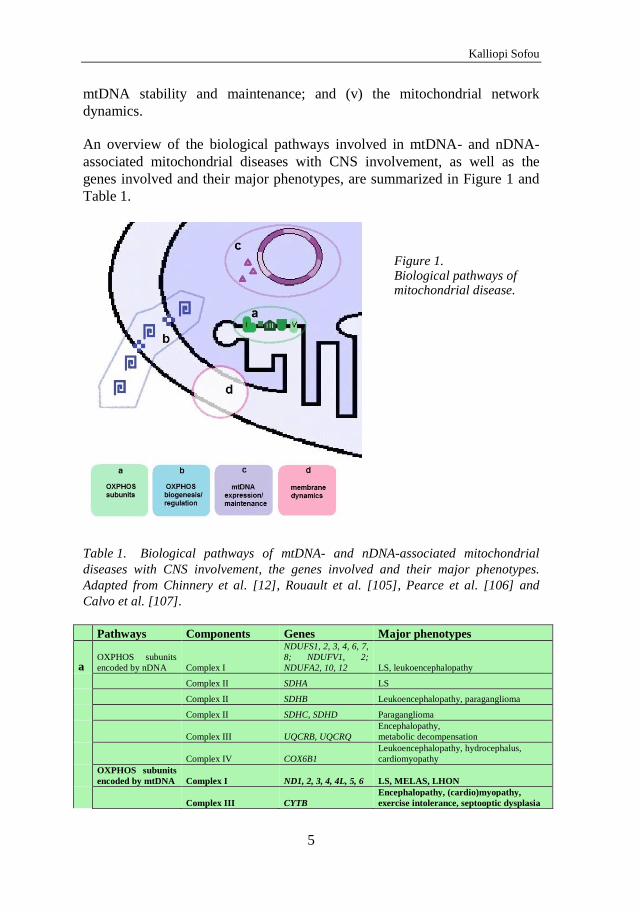
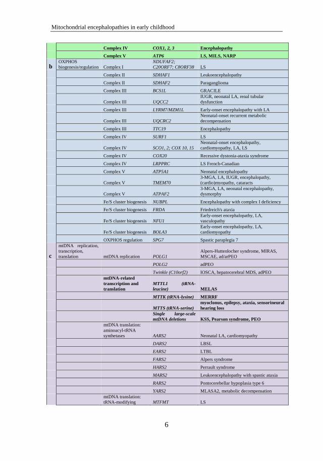
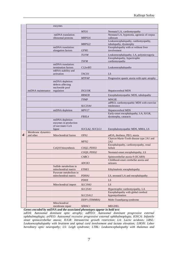
An overview of the biological pathways involved in mtDNA- and nDNA-
associated mitochondrial diseases with CNS involvement, as well as the
genes involved and their major phenotypes, are summarized in Figure 1 and
Table 1.
Figure 1. Biological pathways of mitochondrial disease.
Table 1. Biological pathways of mtDNA- and nDNA-associated mitochondrial
diseases with CNS involvement, the genes involved and their major phenotypes.
Adapted from Chinnery et al. [12], Rouault et al. [105], Pearce et al. [106] and
Calvo et al. [107].
Pathways Components Genes Major phenotypes
a OXPHOS subunits
encoded by nDNA Complex I
NDUFS1, 2, 3, 4, 6, 7,
8; NDUFV1, 2;
NDUFA2, 10, 12 LS, leukoencephalopathy
Complex II SDHA LS
Complex II SDHB Leukoencephalopathy, paraganglioma
Complex II SDHC, SDHD Paraganglioma
Complex III UQCRB, UQCRQ
Encephalopathy,
metabolic decompensation
Complex IV COX6B1
Leukoencephalopathy, hydrocephalus,
cardiomyopathy
OXPHOS subunits
encoded by mtDNA
Complex I
ND1, 2, 3, 4, 4L, 5, 6
LS, MELAS, LHON
Complex III CYTB
Encephalopathy, (cardio)myopathy,
exercise intolerance, septooptic dysplasia
Mitochondrial encephalopathies in early childhood
6
Complex IV COX1, 2, 3 Encephalopathy
Complex V ATP6 LS, MILS, NARP
b OXPHOS
biogenesis/regulation Complex I
NDUFAF2;
C20ORF7; C8ORF38 LS
Complex II SDHAF1 Leukoencephalopathy
Complex II SDHAF2 Paraganglioma
Complex III BCS1L GRACILE
Complex III UQCC2
IUGR, neonatal LA, renal tubular
dysfunction
Complex III LYRM7/MZM1L Early-onset encephalopathy with LA
Complex III UQCRC2
Neonatal-onset recurrent metabolic
decompensation
Complex III TTC19 Encephalopathy
Complex IV SURF1 LS
Complex IV SCO1, 2; COX 10, 15
Neonatal-onset encephalopathy,
cardiomyopathy, LA, LS
Complex IV COX20 Recessive dystonia-ataxia syndrome
Complex IV LRPPRC LS French-Canadian
Complex V ATP5A1 Neonatal encephalopathy
Complex V TMEM70
3-MGA, LA, IUGR, encephalopathy,
(cardio)myopathy, cataracts
Complex V ATPAF2
3-MGA, LA, neonatal encephalopathy,
dysmorphy
Fe/S cluster biogenesis NUBPL Encephalopathy with complex I deficiency
Fe/S cluster biogenesis FRDA Friedreich's ataxia
Fe/S cluster biogenesis NFU1
Early-onset encephalopathy, LA,
vasculopathy
Fe/S cluster biogenesis BOLA3
Early-onset encephalopathy, LA,
cardiomyopathy
OXPHOS regulation SPG7 Spastic paraplegia 7
c
mtDNA replication,
transcription,
translation mtDNA replication POLG1
Alpers-Huttenlocher syndrome, MIRAS,
MSCAE, ad/arPEO
POLG2 adPEO
Twinkle (C10orf2) IOSCA, hepatocerebral MDS, adPEO
mtDNA-related
transcription and
translation
MTTL1 (tRNA-
leucine) MELAS
MTTK (tRNA-lysine) MERRF
MTTS (tRNA-serine)
myoclonus, epilepsy, ataxia, sensorineural
hearing loss
Single large-scale
mtDNA deletions KSS, Pearson syndrome, PEO
mtDNA translation:
aminoacyl-tRNA
synthetases AARS2 Neonatal LA, cardiomyopathy
DARS2 LBSL
EARS2 LTBL
FARS2 Alpers syndrome
HARS2 Perrault syndrome
MARS2 Leukoencephalopathy with spastic ataxia
RARS2 Pontocerebellar hypoplasia type 6
YARS2 MLASA2, metabolic decompensation
mtDNA translation:
tRNA-modifying MTFMT LS
Kalliopi Sofou
7
enzymes
MTO1 Neonatal LA, cardiomyopathy
mtDNA translation:
ribosomal proteins MRPS16
Neonatal LA, hypotonia, agenesis of corpus
callosum
MRPS22
Leukoencephalopathy, cardiomyopathy,
tubulopathy, dysmorphy
mtDNA translation:
elongation factors GFM1
Encephalopathy with or without liver
involvement
TUFM Leukoencephalopathy, LA, polymicrogyria
TSFM
Encephalopathy, hypertrophic
cardiomyopathy
mtDNA translation:
termination factors C12orf65 Leukoencephalopathy
mRNA stability and
activation TACO1 LS
MTPAP Progressive spastic ataxia with optic atrophy
mtDNA maintenance
mtDNA depletion:
defects affecting
nucloeside pool
regulation DGUOK Hepatocerebral MDS
RRM2B Encephalomyopathic MDS, tubulopathy
TYMP MNGIE
SLC25A4
adPEO, cardiomyopathic MDS with exercise
intolerance
mtDNA depletion MPV17 Hepatocerebral MDS
FBXL4
Early-onset encephalopathy, LA, IUGR,
dysmorphy, cataracts
mtDNA depletion:
enzymes in production
of succinate-CoA
ligase SUCLA2, SUCLG1 Encephalomyopathic MDS, MMA, LS
d Membrane dynamics
and other Mitochondrial fusion OPA1 adOA, deafness, PEO, ataxia
MFN2
Charcot-Marie-Tooth disease type 2A1 and
2A2
CoQ10 biosynthesis COQ2, PDSS1
Encephalopathy, cardiomyopathy, renal
failure
COQ9, PDSS2 Neonatal-onset encephalopathy, LS
CABC1 Spinocerebellar ataxia-9 (SCAR9)
ADCK3
Childhood-onset cerebellar ataxia and
seizures
Sulfide metabolism in
mitochondrial matrix ETHE1 Ethylmalonic encephalopathy
Pyruvate metabolism in
mitochondrial matrix PDHA1 LS, neonatal LA and encephalopathy
PDHX LS
Mitochondrial import SLC19A3 LS
SLC25A3 Hypertrophic cardiomyopathy, LA
SLC25A12
Encephalopathy with global cerebral
hypomyelination
DDP1 (TIMM8A) Mohr-Tranebjaerg syndrome
Mitochondrial
membrane repair SERAC1 MEGDEL
Genes encoded by mtDNA and the associated phenotypes appear in bold text
adOA: Autosomal dominant optic atrophy; adPEO: Autosomal dominant progressive external ophthalmoplegia; arPEO: Autosomal recessive progressive external ophthalmoplegia; IOSCA: Infantile
onset spinocerebellar ataxia; IUGR: Intrauterine growth restriction; LA: Lactic acidosis; LBSL: Leukoencephalopathy with braistem and spinal cord involvement and lactate elevation; LHON: Leber
hereditary optic neuropathy; LS: Leigh syndrome; LTBL: Leukoencephalopathy with thalamus and
Mitochondrial encephalopathies in early childhood
8
brainstem involvement and high lactate; MDS: mtDNA depletion syndrome; MILS: Maternally inherited Leigh syndrome; MIRAS: Mitochondrial recessive ataxia syndrome; MLASA2: Myopathy, lactic acidosis
and sideroblastic anemia-2; MNGIE: Mitochondrial neurogastrointestinal encephalopathy; MMA:
Methylmalonic aciduria; MSCAE: Mitochondrial spinocerebellar ataxia and epilepsy; NARP: Neuropathy, ataxia and retinitis pigmentosa; PEO: Progressive external ophthalmoplegia; 3-MGA: 3-
methylglutaconic aciduria
1.4 Early-onset mitochondrial encephalopathies
Early-onset mitochondrial encephalopathies comprise a group of
mitochondrial disorders that present in infancy or early childhood and
primarily involve the CNS. An overview of the major phenotypes of early-
onset mitochondrial encephalopathies along with the associated genetic
defects, are displayed in Table 2.
1.4.1 Alpers syndrome
Alpers syndrome is a progressive neurodegenerative disorder of infancy and
early childhood that has over the years been designated various names, such
as diffuse progressive degeneration of the cerebral gray matter, progressive
(infantile) cerebral poliodystrophy, spongy glio-neuronal dystrophy, and
progressive neuronal degeneration of childhood. The syndrome was
neuropathologically described by Bernard Alpers in 1931 as diffuse,
progressive degeneration of the gray matter of the cerebrum [16]. This
cerebral degeneration has been shown to predominantly affect the cerebral
cortex and to a lesser degree the cerebellar gray matter, thalamus and basal
ganglia, while atrophy of the white matter is typically less striking. Alpers
syndrome initially presents with hypotonia, failure to thrive, epileptic
seizures and psychomotor regression, often deteriorating during infections.
The disease course is severe and often rapidly progressive, characterized by
intractable epilepsy, sever psychomotor regression, spasticity and cortical
blindness. Liver dysfunction has been associated with Alpers syndrome in
variable degree.
Historically, the neurodegenerative process underlying Alpers syndrome has
occasionally been attributed to causes other than genetic, such as extensive
perinatal cortical damage due to anoxia in relation to a complicated delivery,
postepileptic atrophy or inflammatory processes [17-19]. It was not until
2004, that mutations in POLG1, the gene encoding the gamma subunit of the
mtDNA polymerase, have been found to cause Alpers syndrome with hepatic
involvement, a syndrome that is better known as Alpers-Huttenlocher
syndrome [20-22]. Deficient polymerase gamma results in impaired mtDNA
replication, which may lead to reduction in mtDNA copy number, a condition
known as mtDNA depletion. Indeed, Alpers-Huttenlocher syndrome is one of
Kalliopi Sofou
9
the most common phenotypes of hepatocerebral mtDNA depletion
syndromes (MDS) [23]. The depleted mtDNA is apparent in the liver and
may be apparent in fibroblasts, whereas muscle mtDNA content is usually
normal [23]. Antiepileptic treatment with valproate has been associated with
fulminant liver failure in the presence of POLG1 mutations [24, 25].
The genetic etiology underlying Alpers syndrome is basically unknown.
Recently, mutations in FARS2, a gene encoding the mitochondrial
phenylalanine-tRNA synthetase, have been identified in two Finnish patients
with Alpers syndrome [26]. This enzyme belongs to the class II aminoacyl-
tRNA synthetase (mt-aaRS) family and is responsible for charging the
mitochondrial tRNA-phenylalanine. This function, like that of all the other
mitochondrial aminoacyl-tRNA synthetases, is essential for efficient
mitochondrial protein synthesis [27].
1.4.2 Leigh syndrome
Leigh syndrome or subacute necrotizing encephalomyelopathy, is a
progressive neurodegenerative disorder that is usually associated with defects
involving mitochondrial OXPHOS. Leigh syndrome primarily affects infants
and young children and is considered to be the most common distinct
phenotype among OXPHOS disorders in children [9]. It was first described
by Denis Leigh in 1951, as a distinct neuropathological entity with focal,
bilaterally symmetrical, subacute necrotic lesions extending from the
thalamus to the brainstem and the posterior columns of the spinal cord [28].
As opposed to the poliodystrophy in Alpers syndrome that mainly affects the
cerebral cortex, the lesions in Leigh syndrome mainly affect the central gray
matter, i.e. the basal ganglia, diencephalon, brainstem, cerebellum and/or
spinal cord [29, 30]. Onset of disease occurs typically between three and 12
months of age, with disease progression and death within two years. Later
onset and slower progression have also been reported [29-33]. Clinical
manifestations include psychomotor delay, hypotonia, dyskinesia, akinesia,
ataxia, dystonia and brainstem dysfunction, including respiratory
abnormalities, swallowing dysfunction, ophthalmological manifestations and
abnormal thermoregulation [29, 34].
Leigh syndrome is genetically heterogeneous and can be inherited as a
mitochondrial trait, as an autosomal recessive trait due to mutations in
nuclear genes encoding mitochondrial respiratory chain complex subunits or
complex assembly proteins [35] and X-linked related to defects in pyruvate
dehydrogenase complex (PDHc) due to mutations in the PDHA1 gene [36].
Mitochondrial encephalopathies in early childhood
10
Table 2. Early-onset mitochondrial encephalopathies: Major phenotypes, typical age of onset, cardinal features and associated genotypes.
Phenotypes Age of onset Cardinal features Genotypes
Alpers syndrome Neonatal,
infantile
Hypotonia, seizures,
psychomotor regression
FARS2
Alpers-
Huttenlocher
syndrome
Infantile,
occasionally
later
Hypotonia, refractory
seizures, epilepsia partialis
continua, psychomotor
regression, hepatic failure
POLG1
Leigh syndrome Infantile,
occasionally
later
Hypotonia, dystonia,
dyskinesia, ataxia,
brainstem dysfunction.
Phenotypic overlap with
GRACILE syndrome (see
below), biotin-responsive
basal ganglia disease and
MEGDEL
mtDNA
ND1, ND2, ND3,
ND4, ND5, ND6,
ATPase 6
nDNA
NDUFS1, NDUFS2,
NDUFS3, NDUFS4,
NDUFS7, NDUFS8,
NDUFV1,
NDUFA2,
NDUFA10,
NDUFA12,
NDUFAF2,
C20orf7, C8orf38,
SDHA, BCS1L,
SURF1, COX10,
COX15, TACO1,
LRPPRC, PDSS2,
PDHA1, PDHX,
SLC19A3, SUCLA2,
SUCLG1, MTFMT,
SERAC1
GRACILE Neonatal,
infantile
Growth retardation,
aminoaciduria, cholestasis,
iron overload, lactic
acidosis, early death.
Phenotypic overlap with
Leigh syndrome.
BCS1L
Hepatocerebral
MDS
Infantile Hypotonia, failure to thrive,
hepatopathy, psychomotor
delay/regression
DGUOK
MPV-17
Infantile,
occasionally
later
Ataxia, hypotonia,
athetosis, ophthalmoplegia.
Also known as infantile
onset spinocerebellar ataxia
(IOSCA)
C10orf2
Infantile,
occasionally
later
POLG1-associated, also
known as Alpers-
Huttenlocher syndrome (see
above)
POLG1
Kalliopi Sofou
11
Encephalo-
myopathic MDS
Infantile Hypotonia, psychomotor
delay/regression, dystonia,
hearing impairment,
respiratory distress.
Phenotypic overlap with
Leigh syndrome (see above)
SUCLA2, SUCLG1
Neonatal,
infantile
Hypotonia, lactic acidosis,
progressive muscle
weakness, seizures,
tubulopathy, respiratory
distress
RRM2B
Other MDS Neonatal,
infantile
IUGR, hypotonia,
microcephaly, craniofacial
abnormalities, cataracts
FBXL4
Neonatal lactic
acidosis with
cardiomyopathy
Neonatal Lactic acidosis, hypotonia,
failure to thrive, heart
disease, psychomotor delay,
myopathy, respiratory
distress, oculomotor
findings
AARS2, MTO1,
TMEM70, SCO2
Infantile-onset
leukoencephalopa
thy
Neonatal,
infantile
Psychomotor delay, ataxia,
spasticity
SDHAF1, COX6B1,
DARS2, NDUFS1,
NDUFV1
The main genetic defects affecting the respiratory chain complexes that have
been associated with Leigh syndrome are summarized below:
Complex I: (i) mutations affecting mtDNA-encoded subunits, i.e. MT-
ND1, MT-ND2, MT-ND3, MT-ND4, MT-ND5, MT-ND6 [10, 37, 38]; (ii)
mutations affecting nDNA-encoded subunits, i.e. NDUFS1, NDUFS2,
NDUFS3, NDUFS4, NDUFS7, NDUFS8, NDUFV1, NDUFA2,
NDUFA10, NDUFA12 [10, 39-41]; (iii) mutations affecting nDNA-
encoded assembly factors, i.e. NDUFAF2, C20orf7, C8orf38 [10]
Complex II: mutations in the SDHA encoding flavoprotein subunit A [10]
Complex III: mutations in BCS1L gene, encoding an assembly factor, see
detailed description in paragraph 1.4.3
Complex IV: mutations in nDNA-encoded COX assembly factors, i.e.
SURF1, COX10, COX15 and mRNA translation activator for COX
subunit I, i.e. TACO1 [10, 42-45]
Complex V: mutations in mtDNA affecting the ATP synthetase 6, the
most frequent being T8993G [10]
Mitochondrial encephalopathies in early childhood
12
The French Canadian (or Saguenay-Lac-Saint-Jean) type of Leigh
syndrome with tissue-specific COX deficiency is caused by mutations in
the LRPPRC gene, which encodes for a mitochondrial protein involved in
mtDNA expression and in mRNA stability and processing [46]
Mutations in genes affecting the biosynthesis of coenzyme Q10, i.e.
PDSS2 [47]
Besides defects in PDHc, other genetic mitochondrial pathways indirectly
affecting OXPHOS have been associated with Leigh syndrome, such as: (i)
mutations in the thiamine transporter SLC19A3 impair the import of thiamine
in mitochondria, leading to thiamine metabolism dysfunction syndrome-2 or
biotin-responsive basal ganglia disease (BBGD) [48]; (ii) mutations in
nDNA-encoded enzymes for the production of succinate-CoA ligase,
SUCLA2 and SUCLG1, may cause Leigh syndrome with methylmalonic
aciduria, often in the context of an encephalomyopathic MDS, see also
paragraph 1.4.5; (iii) mutations in MTFMT affect the initiation and elongation
of mitochondrial translation [49]; (iv) mutations in SERAC1 affect the
phosphatidylglycerol remodeling in mitochondria, causing 3-
methylglutaconic aciduria with deafness, encephalopathy and Leigh-like
syndrome (MEGDEL) [50].
1.4.3 GRACILE syndrome
GRACILE syndrome is a fatal, neurodegenerative disease characterized by
fetal growth retardation, aminoaciduria, cholestasis with iron overload in the
liver, lactic acidosis and early death [51, 52]. This syndrome is caused by
mutations in the BCS1L gene, which is the chaperone needed to incorporate
the catalytic subunit, Rieske iron-sulfur protein, into complex III at the final
stage of its assembly. The resulting phenotype of infantile onset
encephalopathy with lesions in the basal ganglia and brainstem overlaps with
Leigh syndrome [52].
1.4.4 Hepatocerebral mtDNA depletion syndromes (MDS)
not associated to POLG
The POLG1-related hepatocerebral MDS is better known as Alpers-
Huttenlocher syndrome and is presented separately in paragraph 1.4.1. Other
hepatocerebral MDS presenting early in childhood include those related to
pathogenic mutations in (i) DGUOK, (ii) MPV-17 and (iii) C10orf2 genes.
The DGUOK gene encodes for the deoxyguanosine kinase, a kinase
responsible for the salvage of deoxyribonucleotides for mtDNA replication
inside mitochondria. DGUOK- and MPV-17-related hepatocerebral MDS
Kalliopi Sofou
13
have overlapping phenotypes, both characterized by infantile onset
hepatopathy, often rapidly progressive to liver failure, and neonatal or
infantile onset of hypotonia, failure to thrive and psychomotor delay [53-55].
Hypoglycemia, lactic acidosis and cholestasis are typically present at onset
[53, 54]. Abnormal ophthalmological findings –mainly nystagmus- and
seizures are less common [53]. Lesions in the reticular formation and globus
pallidi have been found in both DGUOK- and MPV-17-related MDS [53],
while white matter lesions and initial normal brain MRI have also been
reported [55].
C10orf2-related hepatocerebral MDS is a rare cause of early-onset
encephalopathy. The C10orf2 gene encodes the mtDNA helicase Twinkle,
which works in close connection with POLG in mtDNA replication.
Recessive C10orf2 mutations give rise to a hepatocerebral MDS, known as
infantile onset spinocerebellar ataxia (IOSCA) [56, 57]. This syndrome
presents with ataxia, hypotonia, athetoid movements and loss of deep tendon
reflexes in infancy [56, 57]. Age of onset is typically between six and 14
months [57]. Ophthalmoplegia and hearing deficit develop soon after the
onset of the disease. Sensory axonal neuropathy, psychomotor delay,
migraine-like headaches, psychotic episodes and catastrophic epilepsy
develop later in the disease course [58]. Liver involvement occurs early, but
is typically less striking than in the other hepatocerebral MDS and not
associated with valproate treatment [58]. Neuropathologically, the syndrome
is characterized by spinocerebellar neurodegeneration, with moderate atrophy
of the brain stem and the cerebellum and severe atrophy of the dorsal roots,
the posterior columns and the posterior spinocerebellar tracts [56, 57]. The
clinical and neuropathological findings in IOSCA are typically milder than
those seen in Alpers-Huttenlocher syndrome [58].
1.4.5 Encephalomyopathic and other mtDNA depletion
syndromes (MDS)
The encephalomyopathic MDS present two different phenotypes, one related
to mutations in SUCLA2 and SUCLG1 genes and another related to mutations
in RRM2B. SUCLA2 and SUCLG1 are the two subunits of succinyl-
coenzyme A ligase, the enzyme that catalyzes the reversible conversion of
succinyl-coenzyme A to succinate in the Krebs cycle. The phenotype in
SUCLA2- and SUCLG1-related MDS is characterized by infantile onset
hypotonia and psychomotor delay, dystonia, choreoathetosis, feeding and
sucking difficulties, sensorineural hearing impairment and respiratory
insufficiency [59, 60]. The patients may also develop abnormal
ophthalmological findings and seizures, while neonatal lactic acidosis and
Mitochondrial encephalopathies in early childhood
14
elevated methylmalonic acid in the urine are characteristic findings [59-61].
Bilateral lesions in the basal ganglia are a common neuroimaging finding,
resulting in a phenotypic overlap with Leigh syndrome [62].
The RRM2B gene encodes the R2 subunit of the p53-controlled
ribonucleotide reductase (p53R2) that catalyzes the biosynthesis of
deoxyribonucleotides for mtDNA replication. The RRM2B-related MDS is
characterized by neonatal or infantile onset hypotonia, lactic acidosis and
progressive muscle weakness [63, 64]. The majority of patients develop renal
proximal tubulopathy, seizures and respiratory distress during infancy [63,
64].
Another phenotype of MDS was recently reported, owing to mutations in
FBXL4, encoding an F-box protein important for the maintenance of mtDNA
[65]. Affected patients develop fatal mitochondrial encephalopathy and lactic
acidosis with typical onset within the first year of life. Intrauterine growth
restriction, microcephaly, hypotonia, craniofacial abnormalities and cataracts
are common clinical features. Severe mtDNA depletion has been found in the
muscle resulting in combined respiratory chain enzyme deficiencies [65].
The phenotype and natural course of MDS with encephalopathy are believed
to depend upon the severity of the causative mutations, which also reflects to
the severity of the mtDNA depletion and the subsequent impairment of
OXPHOS [64, 65].
1.4.6 Neonatal lactic acidosis with cardiomyopathy
Neonatal lactic acidosis in combination with cardiomyopathy or heart failure
is seen in neonates with severe, usually fatal, mitochondrial disorders, while
encephalopathy develops in those patients who survive a fatal outcome early
in infancy. The phenotypic spectrum of these disorders is overlapping and
includes a combination of features, such as severe myopathy, central
hypotonia, failure to thrive, respiratory distress, psychomotor delay, ataxia
and oculomotor findings. An overview of the most common genotypes
implicated in these disorders is summarized below.
Mutations in AARS2, encoding the mitochondrial alanyl-tRNA synthetase,
have been associated with neonatal lactic acidosis and hypertrophic
cardiomyopathy resulting in infantile cardiac failure [66].
MTO1 encodes one of the two subunits of the enzyme that catalyzes the 5-
carboxymethylaminomethylation (mnm5s2U34) of the uridine base in the
Kalliopi Sofou
15
mitochondrial tRNAs specific to glutamine, glutamic acid, lysine, leucine,
and possibly tryptophan. Mutations in MTO1 have been associated with
neonatal lactic acidosis, hypertrophic cardiomyopathy, hypotonia, muscle
weakness and failure to thrive, while features of encephalopathy, dystonia
and optic atrophy have been found in patients surviving into early childhood
[67, 68].
Mutations in TMEM70, a gene that encodes the structural subunits of ATP
synthase (complex V) have been associated with neonatal lactic acidosis,
respiratory distress, hypotonia, cardiomyopathy and psychomotor delay [69,
70].
SCO2 is a gene which together with SCO1 code for metallochaperones that
deliver copper to a subunit in the catalytic core of cytochrome c oxidase.
Mutations in SCO2 have been associated with neonatal lactic acidosis and
cardiomyopathy with fatal outcome in infancy [71, 72].
Mutations in two genes involved in the biogenesis of the Fe-S clusters in
mitochondria, BOLA3 and NFU1 have been linked to neonatal
encephalopathy with lactic acidosis, hyperglycinemia and fatal outcome in
infancy [73-75]. Cardiomyopathy has been seen in BOLA3 mutations, while
patients with NFU1 mutations suffer from pulmonary hypertension owing to
obstructive vasculopathy [75].
1.4.7 Infantile onset leukoencephalopathy
The appearance of clinical and neuroimaging findings that predominantly
involve the cerebral white matter occurs in a heterogeneous group of
mitochondrial disorders known as leukoencephalopathies. Infantile onset
leukoencephalopathy is often associated with defects of complex I or
complex II. The most common genotypes underlying this disorder are: (i)
defects in SDHAF1, the gene encoding for the SDH assembly factor 1, causes
an infantile leukoencephalopathy with accumulation of lactate and succinate
in the white matter [76]; (ii) mutations in COX6B1 have been linked to
infantile onset leukodystrophic encephalopathy, myopathy and growth
retardation [77]; (iii) leukoencephalopathy with brainstem and spinal cord
involvement and lactate elevation (LBSL) due to mutations in the
mitochondrial aspartyl-tRNA synthetase DARS2 present a broad phenotypic
spectrum, including a more severe phenotype of infantile onset [78]; (iv)
mutations in the nDNA-encoded subunits NDUFS1 and NDUFV1 have been
shown to cause infantile onset cavitating leukoencephalopathy associated
with complex I deficiency [79, 80].
Mitochondrial encephalopathies in early childhood
16
1.5 The diagnostics of mitochondrial disease
The genotypes of childhood-onset mitochondrial disorders is extremely
heterogeneous and the resulting phenotypes extremely broad and
overlapping. As a consequence, the diagnosis of mitochondrial diseases is a
multi-step process with molecular diagnostics being the verification step of
the entire process. The diagnosis of a mitochondrial disease is clear cut when
a pathogenic mutation is found along with compatible clinical findings. In the
absence of identified pathogenic mutations, the diagnosis rests upon the
combination of characteristic phenotypic, morphological and biochemical
findings. The major steps in the diagnostic approach of mitochondrial
disorders are described below.
1.5.1 Identifying the clinical phenotype
The diagnostic process starts upon clinical suspicion. As the phenotypic
spectrum of mitochondrial disorders is broad and complex, the physical
examination should include a thorough neurological examination of both the
central and peripheral nervous system, as well as evaluation of the
involvement of other systems or organs, suggestive of a mitochondrial
disorder. In order to facilitate the clinical evaluation, a number of ‘red-flags’
have been proposed, as the clinical features that should prompt a diagnostic
work-up for suspected mitochondrial disease [81]. The clinical suspicion is
strengthened by a positive family history, which assists in recognizing the
inheritance pattern; a negative family history, however, does not exclude the
presence of mitochondrial disease.
1.5.2 Metabolic laboratory work-up
The laboratory work-up aims to identify biochemical markers suggestive of
mitochondrial disease, such as the lactate levels, to evaluate involvement of
other organs, such as measurement of liver and renal function and to help
differentiate from other metabolic disorders with similar phenotypes, i.e. urea
cycle defects, aminoacidopathies, organic acidopathies and fatty acid
oxidation disorders. The measurement of lactate and occasionally pyruvate in
the blood and the cerebrospinal fluid (CSF) is included in the initial work-up,
along with the estimation of the lactate-to-pyruvate ratio. Despite their lack
of specificity, an elevated plasma lactate or pyruvate level can be suggestive
of mitochondrial disease. CSF lactate levels are considered more reliable as
they are less influenced by external factors, such as the collection technique.
In some patients, the levels of lactate and/or pyruvate rise only during
episodes of metabolic decompensation and are otherwise normal [81].
Kalliopi Sofou
17
1.5.3 Neuroimaging
Both structural magnetic resonance imaging (MRI) and functional brain
imaging methods, e.g. magnetic resonance spectroscopy (MRS), diffusion-
weighted imaging (DWI) and perfusion MRI, have helped to increase our
knowledge of mitochondrial disorders by allowing a good assessment of the
anatomic lesions, metabolism and hemodynamics of the brain [82]. MRI
signal abnormalities, as acquired with T1- and T2-weighted sequences, can
reveal specific or ‘signature’ disease features, non-specific features or
leukodystrophy-like features suggesting a mitochondrial disorder [83-86].
However, a brain MRI may occasionally be normal [83-86]. MRS provides a
unique in vivo evaluation of brain metabolism and may also be used to
monitor disease progression. Lactate accumulation and N-acetyl-aspartate
(NAA) reduction are the most prominent MRS signal abnormalities in
mitochondrial disorders. DWI, on the other hand, detects the random
Brownian motion of water protons in the brain. The diffusion of water
protons can be quantitated by a parameter known as the apparent diffusion
coefficient (ADC). The combination of DWI and ADC helps differentiate
between acute and chronic ischemia and between cytotoxic and vasogenic
edema [84]. Combined with clinical indices, neuroimaging of the brain can
assist in identifying and differentiating between mitochondrial disorders [87].
This is of great importance in non-syndromic mitochondrial disorders, where
brain imaging features are characteristically diverse and non-specific [88].
1.5.4 Specific tissue biopsies
In general, a biopsy has higher chance to provide a representative sample for
diagnostic testing when performed in the tissues that are most profoundly
affected by the disease. In mitochondrial encephalopathies, the most affected
organ is usually the brain and the second most affected organ is the muscle.
The skeletal muscle biopsy is an essential diagnostic tool, as it provides
optimal sampling for morphological, biochemical and molecular testing.
Portions of the biopsy are utilized for histochemical, immunohistochemical
and electron microscopy studies to evaluate for morphological evidence of
primary mitochondrial disease and assist in the differential diagnosis from
other neuromuscular diseases [82]. Morphological findings highly suggestive
of a mitochondrial disease are the identification of ragged red fibers (RRFs),
decreased SDH reaction suggesting complex II deficiency, and decreased
COX reaction suggesting complex IV deficiency. Muscle tissue is also used
for biochemical analysis of the respiratory chain enzyme activities and for the
extraction of DNA for genetic testing. Biochemical analysis includes
polarographic studies of oxygen consumption and spectrophotometric
analysis of the mitochondrial respiratory chain enzymes in cultured cells,
Mitochondrial encephalopathies in early childhood
18
tissue homogenates or whole cells. To help distinguish primary electron
transport chain defects from secondary deficiencies, the enzyme activity
measurements are reported relative to a marker enzyme, such as citrate
synthase, a mitochondrial matrix enzyme considered to be a good indicator of
the mitochondrial mass [82].
In mitochondrial encephalopathies, the absence of pathognomonic
morphological or biochemical findings from the skeletal muscle does not
exclude a mitochondrial etiology. A skin biopsy is usually performed at the
time of muscle biopsy and the cultured skin fibroblasts can also be used for
biochemical and molecular analysis. When affected, the liver is a biopsy site
that allows not only for morphological and biochemical analysis, but also for
detection of mtDNA depletion in the case of hepatocerebral MDS. A heart
biopsy can be informative when signs of mitochondrial cardiomyopathy are
present.
1.5.5 Molecular diagnostics
The molecular diagnosis of suspected mitochondrial disease has evolved
rapidly over the past two decades. Genetic testing of mtDNA, that was once
limited to a panel of common mtDNA point mutations underlying well-
recognized syndromic mitochondrial disorders, such as MELAS, MERRF
and NARP, has now evolved to whole mtDNA genome sequencing; the latter
permits identification of all known and potentially novel disease-causing
mutations in a single platform [89]. When the phenotype, family history and
biochemical findings raise the suspicion of maternally inherited
mitochondrial disease, the testing of mtDNA is usually the first step towards
a molecular diagnosis. The increasing number of nuclear genes causing
primary mitochondrial disease, now counting 1500 nuclear genes, has made
targeted sequencing of nuclear genes on an individual step-wise basis
expensive, time-consuming and of low diagnostic yield [89].
The development of Next Generation Sequencing (NGS) has revolutionized
the diagnostic approach of mitochondrial diseases. The highly advanced NGS
technology is capable of sequencing a group of target genes in parallel and is
therefore an ideal approach for the diagnosis of complex dual genome
mitochondrial disorders [90]. Whole-exome sequencing (WES) is the
application of the next-generation technology to determine the variations of
all coding regions, or exons, of known genes. WES provides coverage of
more than 95% of the exons, which contains 85% of disease-causing
mutations in Mendelian disorders [91]. WES can identify not just known
mitochondrial disease genes, but also mutations in a wide range of genetic

Kalliopi Sofou
19
disorders with overlapping clinical manifestations that may directly or
indirectly cause secondary mitochondrial dysfunction, thus offering an
excellent diagnostic tool in the demanding field of mitochondrial disorders
[89, 90].
1.5.6 Post-mortem investigation
Post-mortem investigation is pivotal both for diagnostic and research
purposes, as it discloses the underlying neuropathology in mitochondrial
encephalopathies. Both Alpers syndrome and Leigh syndrome were first
described as neuropathological entities and later shown to be genetic
disorders. The poliodystrophy seen in Alpers syndrome is a characteristic
finding seen on brain biopsy. The role of post-mortem investigation is also
essential, as it may reveal tissue-specific findings in the absence of muscle
pathology, such as in hepatocerebral MDS, where the mtDNA depletion may
only be seen in the brain or the liver.
Mitochondrial encephalopathies in early childhood
20
2 AIMS
The overall aim of this thesis was to explore the genotypic and phenotypic
spectrum of childhood-onset mitochondrial diseases with CNS involvement,
with focus on early-onset mitochondrial encephalopathies. The present work
describes this spectrum with emphasis on Alpers and Leigh syndrome.
The basic aims of this thesis were:
i) To present the genotypic and phenotypic spectrum of Alpers syndrome and
to identify genotype-phenotype correlations – Paper I
ii) To present novel genetic defects underlying Alpers syndrome along with
the respective phenotypes – Paper II
iii) To present the genotypic and phenotypic spectrum of Leigh syndrome, to
characterize the clinical course and identify predictors of survival – Paper III
iv) To present the brain MRI findings in childhood-onset mitochondrial
disorders with CNS involvement – Paper IV
Kalliopi Sofou
21
3 PATIENTS AND METHODS
3.1 Patients
3.1.1 Patients in Papers I, II and IV
The patients who participated in our studies were collected from a total of
approximately 170 patients who were investigated for suspected
mitochondrial disease at the Queen Silvia’s Children Hospital between
January 1st 1984 and December 31st 2012 and were found to meet the
diagnostic criteria of mitochondrial encephalopathy, as follows [9]:
Presence of known pathogenic mutations of mtDNA or nDNA, together with
compatible clinical findings;
Or, in the absence of other known disorders with secondary effects on the
mitochondrial respiratory chain, at least two out of four criteria should be
fulfilled:
1. Oximetry: Decreased respiratory rates indicating isolated or combined
complex deficiencies, as follows: Respiratory rates below the control range in
the presence of the nicotinamide adenine dinucleotide (NAD)-linked
substrates pyruvate and glutamate but with normal rates in the presence of
succinate and ascorbate plus N,N,N’,N’-tetra methyl-p-phenylene-diamine
(TMPD), indicating a deficiency of reduced NAD (NADH)-Co Q reductase
(complex I). Respiratory rates below the control range in the presence of
pyruvate, glutamate, and succinate but with normal rates in the presence of
ascorbate plus TMPD, indicating a deficiency of Co Q-cytochrome c
reductase (complex III). Decreased respiratory rates in the presence of all
substrates tested, indicating a deficiency of cytochrome c oxidase (COX,
complex IV).
2. Spectrophotometry: enzyme activities below the control range of NADH
ferricyanide reductase (complex I), succinate-cytochrome c reductase
(complex II and/or III) or COX (complex IV).
3. Histochemical evidence of COX deficiency.
4. Abundant, ultra-stucturally abnormal mitochondria.
Patients with Alpers syndrome were considered those who in addition to the
above mentioned diagnostic criteria, exhibited early-onset diffuse progressive
Mitochondrial encephalopathies in early childhood
22
cerebral atrophy with cortical predominance, based on their clinical features
in combination with neuroimaging and/or neuropathological findings.
Siblings to study participants that had presented similar clinical features and
disease course were also included.
3.1.2 Patients in Paper III
The patients who participated in the study presented in Paper III arose from a
collaboration between eight centers specializing in mitochondrial diseases in
Europe; Gothenburg, Rotterdam, Helsinki, Copenhagen, Stockholm,
Brussels, Bergen and Oulu. We included patients with Leigh syndrome that
were diagnosed and followed at the participating centers and that fulfilled
both of the following inclusion criteria: (i) clinical features compatible with
Leigh syndrome, such as psychomotor regression, dystonia, ataxia and/or
brainstem dysfunction and (ii) MRI or CT or neuropathological findings of
Leigh syndrome, as follows: bilateral symmetrical lesions in the basal
ganglia, and/or thalamus, and/or brainstem. Patients were excluded from this
study if they had known syndromic mitochondrial phenotypes other than
Leigh syndrome.
3.2 Methods
This research work was performed as a retrospective analysis of longitudinal
data, including clinical, laboratory, morphological, histochemical and
(neuro)pathological data. The author performed neurological examination on
only a few patients. All patients had been clinically assessed by at least one
of the neurologists involved in the studies. A set of tools and procedures were
applied to ensure that all study-related data were collected in a standardized
and valid manner, as described below.
3.2.1 Methods in Papers I, II and IV
3.2.1.1 Clinical evaluation, laboratory investigations, investigations of the muscle and post-mortem investigations
The clinical evaluation and investigations performed in our clinic for
suspected mitochondrial disease follow a specific protocol, which has been
implemented in collaboration with a research team of experts in
mitochondrial diseases at the Sahlgrenska University Hospital. The clinical
and laboratory findings for our studies were collected from the patients’
medical records with the help of a standardized case report form (CRF). The
biochemical, morphological and histochemical investigations of the muscle
samples were performed in collaboration with the research team at
Kalliopi Sofou
23
Sahlgrenska University Hospital according to standard protocols, essentially
as described previously [92]. The pathologists involved in the studies
reported the pathological findings from specimens obtained post-mortem.
3.2.1.2 Genetic investigations
The methodology applied for the genetic investigations is described in detail
in the original publications.
DNA extraction
Total genomic DNA was extracted from skeletal muscle tissues or peripheral
blood using standard commercial extraction kits. Total DNA was extracted
from frozen skeletal muscle tissues and post-mortem tissues using
commercially available kits.
POLG1 analysis
All patients with Alpers syndrome were investigated for POLG1 mutations.
The only exception was the siblings to patients who were found negative for
mutations in POLG1 and thus they were considered to be negative as well.
Mitochondrial DNA analysis
Muscle mtDNA was analyzed by Southern blotting to detect large-scale
deletions and duplications after cleavage with the restriction enzymes PvuII
or BamHI, and for the point mutations A3243G, T8993C/G and A8344G.
mtDNA was also analyzed for the presence of large-scale deletions by long
expand PCR and depletion using real-time quantitative PCR in selected
patients. The ratio of mtDNA copy number to nDNA copy number in each
patient was compared to the mean relative ratio in a control group of children
(n=8) younger than two years of age and without evidence of neuromuscular
disease. Values below 0.2 were considered as mtDNA depletion. Complete
mtDNA sequence analysis was performed in all patients with Alpers
syndrome without POLG1 mutations (Paper I). Identified mutations
considered to be potentially pathogenic were further investigated in the
mothers’ mtDNA. The primer sequences and PCR conditions are available
upon request.
Whole exome sequencing
Whole exome sequencing (WES) was performed in selected patients with
Alpers syndrome without POLG1 mutations. The results in two of these
Mitochondrial encephalopathies in early childhood
24
patients are presented in Paper II. WES in these two patients was carried out
using high-throughput sequencing technology, as described in detail in Paper
II. Quality assessment of the sequence reads was performed by generating
QC statistics with FastQC (http://www.bioinformatics.bbsrc.ac.uk/
projects/fastqc). For the identification of potentially pathogenic variants, the
Ingenuity® Variant AnalysisTM software was used
(www.ingenuity.com/variants). All mutations identified by WES were
verified by Sanger sequencing using primers amplifying the exons
harbouring the mutations in NARS2 and PARS2. Sequencing analysis was
performed using an ABI PRISM® 3100 Genetic analyzer and the BigDye
Terminator v.1.1 Cycle Sequencing Kit (Applied Biosystems). A description
of the sequencing data is provided in Paper II. Primer sequences and PCR
conditions are available upon request.
3.2.1.3 Neuroimaging investigations
All brain MRI scans, performed either at our hospital or elsewhere, were re-
evaluated by the author together with a pediatric neuroradiologist, who were
both blinded to the patients’ diagnosis, as well as a pediatric neurologist.
Since this was a retrospective study, patients’ MRI scans were not performed
under a standardized protocol. Axial and sagittal T1-weighted images were
assessed in particular for the presence of cortical atrophy,
agenesis/hypoplasia of the corpus callosum, enlargement/dilatation of the
ventricles and/or subarachnoid spaces. Axial and coronal T2-weighted
images, including fluid attenuated inversion recovery (FLAIR) images, were
studied to identify signal intensity changes. Myelination was assessed with
the aid of conventional MRI images. Myelination was compared to age-
matched healthy controls and classified as described in detail in Paper IV.
Additional imaging sequences, such as DWI, ADC and MRS, were evaluated
where available.
For the diagnostic approach used in Paper IV, i.e. from the neuroanatomical
and/or neurofunctional lesion to disease identification, the abnormal MRI
findings were categorized on the basis of their predominant location in the
brain as follows: lesion predominance in the cerebral cortex/limbic system;
white matter; basal ganglia/diencephalon; posterior fossa; diffuse lesions
(more than one anatomic region involved with no apparent predominance).
3.2.2 Methods in Paper III
Paper III is the first collaborative research work within the Mitochondrial
Clinical and Research Network (MCRN). This network was established in
2011 and constitutes a European network of research centers specializing in
Kalliopi Sofou
25
mitochondrial diseases. The centers that participated in this study were from
Gothenburg, Rotterdam, Helsinki, Copenhagen, Stockholm, Brussels, Bergen
and Oulu. Our center in Gothenburg had the leadership in the design and
coordination of this research project.
For the collection of patient data, an electronic-Case Report Form (e-CRF)
was designed, which is shown in paper form in the Appendix. The design of
the CRF was a multi-step procedure involving quarterly meetings between all
investigators from the participating sites. The development of the electronic
database for the electronic data capture (EDC) was undertaken by the
Contract Research Organisation (CRO) Qualitis Ltd (http://www.qualitis.gr/).
Qualitis’ assigned personnel in collaboration with the author, set the
validation rules and interim monitoring procedures to ensure the validity,
completeness and integrity of the collected data and provided comprehensive
training to all designated investigators prior to study initiation. Data were
entered electronically via the e-CRF using web-based secure server by the
designated investigators. The e-CRF was completed by one designated
investigator at each center. Prior to study initiation, a detailed data
management plan was issued describing the process to be followed for
validating, processing and cleaning all study data. When all data were
properly validated and the quality control procedure had been completed, the
database was locked. The data management plan, the data validation plan and
the standard operating procedures (SOPs) that were applied for quality
control are available upon request. A detailed list with the definitions of the
terms used in this study was provided as an attachment in the e-CRF (see
Appendix).
3.2.3 Ethical considerations
The present research work is part of ongoing studies on childhood-onset
mitochondrial diseases undertaken by the department of Child Neurology at
the Queen Silvia Children’s Hospital in collaboration with the Sahlgrenska
University Hospital with the approval of the ethics committee at the
University of Gothenburg. Additionally, for the multicenter study on Leigh
syndrome (Paper III), each participating center received approval by the local
ethical committee, as per the standing regulatory requirements, prior to the
initiation of the study. The web-based EDC application (e-CRF) that was
specifically designed for the needs of the multicenter study shown in Paper
III adhered to all applicable data protection regulations and requirements with
regard to electronic records.
Mitochondrial encephalopathies in early childhood
26
4 RESULTS
4.1 Alpers syndrome – Paper I and II
4.1.1 Genotypic spectrum
Nineteen patients with Alpers syndrome were studied, among them four non-
twin sibling pairs. Six patients were found to have pathogenic mutations in
POLG1 in compound heterozygosity as shown in Table 3. In two of the 13
patients without POLG1 mutations, whole exome sequencing revealed
mutations in two mitochondrial aminoacyl-tRNA synthetases (mt-aaRS), as
follows: patient AS-13 was found to have a homozygous mutation
(c.641C>T, p.P214L) in NARS2, encoding the mitochondrial asparaginyl-
tRNA synthetase, while patient AS-15 was compound heterozygous for two
mutations (c.1130dupC, p.K378fs*1 / c.836C>T, p.S279L) in PARS2, a gene
encoding the mitochondrial prolyl-tRNA synthetase. The results were
verified by Sanger sequencing and the parents of patients AS-13 and AS-15
were shown to be heterozygous for the respective mutations. The underlying
cause for the remaining 11 patients with Alpers syndrome remains elusive.
4.1.2 Phenotypic spectrum and genotype-phenotype
correlations
4.1.2.1 Onset
Patients with POLG1 mutations had a median disease onset at 6.5 months of
age, while patients without POLG1 mutations exhibited a median disease
onset in the perinatal period. The most common initial symptoms among all
patients were seizures (13/19) and failure to thrive (5/19). Patient AS-13 with
mutations in NARS2 presented in early infancy with inconsolable crying,
tendency to opisthotonus posturing and delayed head control. The patient’s
psychomotor development reached its maximum level at the age of six
months, which corresponded to a developmental age of three months. Then,
the patient showed the first signs of psychomotor regression, with loss of
previously acquired motor skills and feeding difficulties. Patient AS-15 with
mutations in PARS2 was admitted to the hospital at 2.5 months of age
because of feeding difficulties following an upper respiratory tract infection.
During hospitalization, the patient developed seizures and signs of
psychomotor regression.
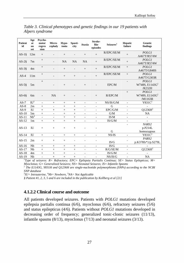
Kalliopi Sofou
27
Table 3. Clinical phenotypes and genetic findings in our 19 patients with Alpers syndrome
Patient
id
Age
at
on
set
Psycho-
motor
regres
sion
Micro-
cephaly
Hypo-
tonia
Spasti-
city Ataxia
Stroke-
like
episodes
Seizures1 Hepatic
failure
Genetic
findings
AS-1§ 12m + - + - + + R/EPC/SE/M
+ POLG1
A467T/R574W
AS-2§ 7m +
- NA NA NA + R/EPC/SE/M
+ POLG1
A467T/R574W
AS-3§ 4m +
- + - + + R/EPC/SE/M + POLG1
A467T/G848S
AS-4 11m +
- + + - + R/EPC/SE/M + POLG1
A467T/G303R
AS-5§ 5m
+
- + - + - EPC/M
- POLG1
W748S, E1143G2 /R232H
AS-6§ 6m - NA + - - + R/EPC/M
+ POLG1
W748S, E1143G2 /M1163R
AS-7 IU3 - + + + - - NS/IS/G/M - Y831C2
AS-8 2m + - + + - - G - -
AS-9 IU + + + + - - EPC/G/M - Q1236H2
AS-10 5m + - + + - - G/M - NA
AS-11 Nb4 - - + + - - IS/M - -
AS-12 1m + + + + - - IS/G/M - -
AS-13 IU + + + + - -
G
- NARS2
p.P214L
homozygous
AS-14 IU + - + + - - NS/IS - Y831C2
AS-15 2m + + + - - - IS/G
- PARS2
p.K378fs*1/p.S279L
AS-16 Nb + + + + - - IS/G - -
AS-17 Nb + + + + - - R/G/SE/M - Q1236H2
AS-18 4m + + + - - - IS/G/M - -
AS-19 Nb - + + + - - NS/IS/G - NA 1Type of seizures: R= Refractory; EPC= Epilepsia Partialis Continua; SE= Status Epilepticus; M= Myoclonus; G= Generalized Seizures; NS= Neonatal Seizures; IS= Infantile Spasms 2 The E1143G, Y831H and Q1236H are single-nucleotide polymorphisms (SNPs) according to the NCBI
SNP database 3IU= Intrauterine, 4Nb= Newborn, 5NA= Not Applicable
§ Patient #1, 2, 3, 5 and 6 are included in the publication by Kollberg et al [21]
4.1.2.2 Clinical course and outcome
All patients developed seizures. Patients with POLG1 mutations developed
epilepsia partialis continua (6/6), myoclonus (6/6), refractory seizures (5/6)
and status epilepticus (4/6). Patients without POLG1 mutations developed in
decreasing order of frequency; generalized tonic-clonic seizures (11/13),
infantile spasms (8/13), myoclonus (7/13) and neonatal seizures (3/13).
Mitochondrial encephalopathies in early childhood
28
All patients with POLG1 mutations developed hepatic dysfunction. In the
majority of them, hepatic dysfunction led to liver failure (5/6) and was related
to preceding valproate treatment (4/6). Most of the patients without POLG1
mutations developed hepatic dysfunction (9/13), regardless of valproate
treatment, which mainly presented as abnormal liver function tests and did
not lead to liver failure.
The majority of patients with Alpers syndrome developed muscular
hypotonia (18/19), psychomotor regression (15/19) and failure to thrive
(15/19). Ataxia (3/19) and stroke-like episodes (5/19) were only seen in
patients with POLG1 mutations. Progressive microcephaly and spasticity
were more common in patients without POLG1 mutations.
Cardiomyopathy was found in one patient with POLG1 mutations and in four
patients without POLG1 mutations. One of these patients was patient AS-15
with PARS2 mutations, who at the age of two years developed dilated
cardiomyopathy and left ventricular hypertrophy and died of heart failure
shortly afterwards.
Renal impairment was found in two patients with POLG1 mutations and in
one patient without POLG1 mutations, patient AS-13 with NARS2 mutations.
This patient developed hypochloremic metabolic alkalosis and increased
urinary glucose and salt excretion due to renal tubulopathy.
Our patients with Alpers syndrome were followed up for a period of seven
years (range: 4m-18y). At the time of data analysis, nine patients had died
(five with POLG1 mutations and four without) at a median age of two years
(range: 1y-16y). Patients with POLG1 mutations exhibited a shorter life span,
as five of them had died at a median age of 15 months (range: 1y-4.3y). The
median age of death for the four patients without POLG1 mutations was 10
years (range: 2y-16y). Patients AS-13 and AS-15 are among those four
patients who had died. The survival analysis for all 19 patients and separately
for those with versus without POLG1 mutations is depicted in Figure 2.
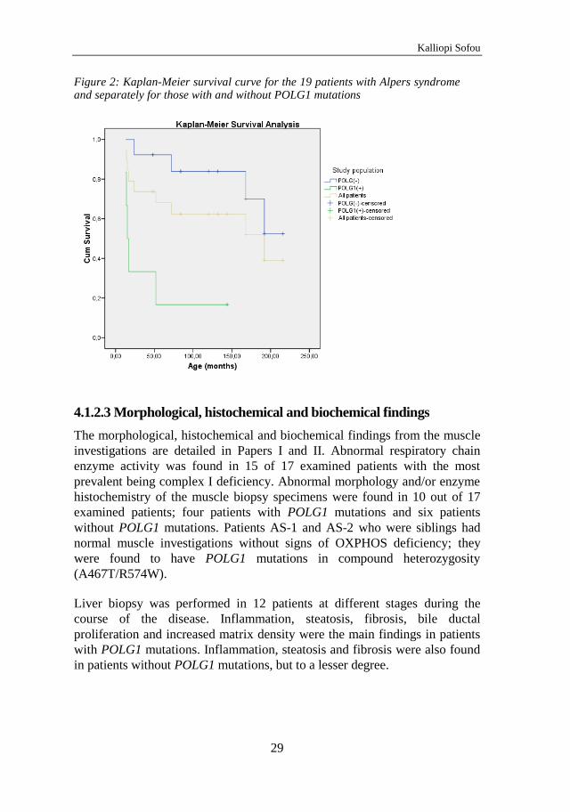
Kalliopi Sofou
29
Figure 2: Kaplan-Meier survival curve for the 19 patients with Alpers syndrome and separately for those with and without POLG1 mutations
4.1.2.3 Morphological, histochemical and biochemical findings
The morphological, histochemical and biochemical findings from the muscle
investigations are detailed in Papers I and II. Abnormal respiratory chain
enzyme activity was found in 15 of 17 examined patients with the most
prevalent being complex I deficiency. Abnormal morphology and/or enzyme
histochemistry of the muscle biopsy specimens were found in 10 out of 17
examined patients; four patients with POLG1 mutations and six patients
without POLG1 mutations. Patients AS-1 and AS-2 who were siblings had
normal muscle investigations without signs of OXPHOS deficiency; they
were found to have POLG1 mutations in compound heterozygosity
(A467T/R574W).
Liver biopsy was performed in 12 patients at different stages during the
course of the disease. Inflammation, steatosis, fibrosis, bile ductal
proliferation and increased matrix density were the main findings in patients
with POLG1 mutations. Inflammation, steatosis and fibrosis were also found
in patients without POLG1 mutations, but to a lesser degree.
Mitochondrial encephalopathies in early childhood
30
4.1.2.4 Neuroimaging findings
CT and/or MRI-investigations of the brain showed progressive atrophy of the
cerebral cortex (17/19); in addition, these patients had lesions in the white
matter (9/19), cerebellum (4/19), basal ganglia (3/19), thalamus (2/19) and
agenesis/hypoplasia of the corpus callosum (2/19). Two patients with POLG1
mutations had no atrophy on brain neuroimaging. One of them had a normal
CT of the brain but experienced a detrimental disease progress that led to
death shortly afterwards. The other patient presented with a unilateral
cerebral lobar infarction and died shortly afterwards.
4.1.2.5 Post-mortem findings
The major neuropathological finding was a profound degeneration of the
cerebral cortex, characterized by neuronal loss, spongiosis and gliosis
frequently accompanied by signs of capillary proliferation, as described in
detail in Paper I. Basal ganglia, thalamus and cerebellum were involved to
variable extent in several patients. Microscopic vacuolation in the thalamus
and basal ganglia was prominent in most patients with POLG1 mutations.
Several patients without POLG1 mutations showed severe brain atrophy,
which might be related to disease duration before death.
Post-mortem examination of the kidneys in patient AS-13 with NARS2
mutations revealed an increased number of sclerotic glomeruli and occasional
focal segmental sclerosis, along with a few atrophic tubules, without signs of
increased interstitial fibrosis.
Post-mortem examination of the heart in patient AS-15 with PARS2
mutations showed marked enlargement with dilation of the ventricles and
increased thickness of the ventricular walls. There was both macroscopic and
microscopic interstitial fibrosis of variable degree.
4.2 Leigh syndrome – Paper III
4.2.1 Perinatal history
A total of 130 patients with Leigh syndrome were enrolled into the study,
among whom 11 sibling pairs. 19 patients (14.6%) were born preterm. Most
pregnancies were uneventful (77.7%); the most common causes of
complicated pregnancy were preeclampsia and oligohydramnios. 13 patients
(10.0%) were born small for gestational age, while two patients presented
intrauterine growth restriction. Microcephaly was evident at birth in six
Kalliopi Sofou
31
patients (4.6%). The median Apgar score at 1, 5 and 10 minutes was 9-9-10
(Q25: 7-9-9; Q75: 9-10-10). The most common pathological signs at birth in
decreasing order of frequency were respiratory difficulties (n=9),
hypotonia/floppiness (n=7), cardiac complications (n=5), lactic acidosis
(n=4) and feeding/sucking difficulties (n=3).
4.2.2 Onset
The median age of disease onset was 7 months (range: intrauterine-19y), with
80.8% presenting by the age of 2 years. Perinatal onset of disease was
reported in 17 patients (13.1%), while three patients were reported to have
intrauterine onset. Leigh syndrome presented initially with abnormal motor
findings in the vast majority of patients (82.8%), with the most common
being hypotonia (59.2%), abnormal tendon reflexes (14.6%) and ataxia
(12.3%). Other presenting features were abnormal ocular findings (25.0%),
feeding/sucking difficulties (14.1%), epileptic seizures (13.3%) and failure to
thrive (10.2%). The clinical features at onset in relation to age of onset are
shown in detail in Paper III.
4.2.3 Clinical course and outcome
4.2.3.1 Clinical features throughout the disease course
The vast majority of patients exhibited abnormal motor findings (99.2%),
with the most common being hypotonia (74.6%), abnormal tendon reflexes
(47.7%) and dystonia (44.6%). Dystonia, spasticity, hypertonia and
choreoathetosis were less frequent at disease onset but developed later in the
disease course.
Abnormal ocular findings were present in 79 patients (60.8%), the most
prevalent being nystagmus (23.8%), followed by strabismus (19.2%), visual
impairment (16.2%), optic atrophy (14.6%), ptosis (13.1%) and
ophthalmoplegia (12.3%).
Epileptic seizures were reported in 51 patients (39.2%) and were classified
according to ILAE as follows: generalized seizures (22.3%), focal seizures
(14.6%) and epileptic spasms (6.1%). Resistance to antiepileptic treatment
was reported in 16 patients.
Respiratory dysfunction was present in 37.7% with hyperventilation and/or
abnormal breathing pattern being the most prevalent type (20.0%), followed
by apnoea (16.1%), obstructive or restrictive respiratory disease (13.8%) and
central hypoventilation (10.0%).
Mitochondrial encephalopathies in early childhood
32
Cardiac dysfunction was present in 23 patients (17.7%), with more than half
having hypertrophic cardiomyopathy (9.2%). Arrhythmia/conduction defects
were reported in five patients and dilated cardiomyopathy was reported in
two patients.
59 patients manifested feeding difficulties (45.4%) sufficient to necessitate
tube feeding and/or gastrostomy. Mental retardation was found in 48 patients
(36.9%). The severity of mental retardation was classified as mild in 11
patients, moderate in 17, severe in 15, profound in three and unspecified in
two patients. Hearing impairment was identified in 25 patients and was
sensorineural in 22, conductive in two and mixed in one patient. Hepatic
dysfunction was present in 16 patients (12.3%), with elevated liver
transaminases in 12; structural abnormalities defined by ultrasound or biopsy,
including liver steatosis and/or fibrosis in four; severe liver failure in two and
hepatomegaly in two patients. Microcephaly was present in 15 patients
(11.5%).
4.2.3.2 Acute exacerbations and survival outcome
The study population was followed up for a median time of 9.6 years from
disease onset. In total, 56.9% of patients experienced at least one acute
exacerbation requiring hospitalisation during their disease course, 43.8%
during the previous year. Of these, one fourth had at least three exacerbations
during the previous year. Intensive care was required in 39.2% of
hospitalized patients. The main cause of acute exacerbation was infection
(60.8%); other causes included respiratory complications (13.5%), stroke-like
episodes (4.0%) and poor nutrition or dehydration (4.0%).
53 patients were alive at the time of data analysis (40.8%), 51 were dead
(39.2%) and 26 (20.0%) were lost to follow-up. Median age at death was 2.4
years (range: 1 month – 21 years). The elapsed median time from disease
onset to death was 1.8 years. Main causes of death were respiratory
complications (51.0%), progression of Leigh syndrome (17.6%) or infection
(17.6%). The survival analysis for the entire population is seen in Figure 3.
4.2.4 Morphological, biochemical and histochemical
findings
Abnormal respiratory chain enzyme activity was found in 70% of examined
patients, with the most prevalent being complex I deficiency. In 24 of 57
patients with abnormal histopathology of the muscle, at least one of the
following was found: cytochrome c oxidase deficiency, succinate
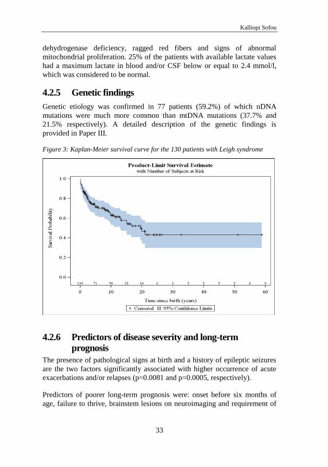
Kalliopi Sofou
33
dehydrogenase deficiency, ragged red fibers and signs of abnormal
mitochondrial proliferation. 25% of the patients with available lactate values
had a maximum lactate in blood and/or CSF below or equal to 2.4 mmol/l,
which was considered to be normal.
4.2.5 Genetic findings
Genetic etiology was confirmed in 77 patients (59.2%) of which nDNA
mutations were much more common than mtDNA mutations (37.7% and
21.5% respectively). A detailed description of the genetic findings is
provided in Paper III.
Figure 3: Kaplan-Meier survival curve for the 130 patients with Leigh syndrome
4.2.6 Predictors of disease severity and long-term
prognosis
The presence of pathological signs at birth and a history of epileptic seizures
are the two factors significantly associated with higher occurrence of acute
exacerbations and/or relapses (p=0.0081 and p=0.0005, respectively).
Predictors of poorer long-term prognosis were: onset before six months of
age, failure to thrive, brainstem lesions on neuroimaging and requirement of
Mitochondrial encephalopathies in early childhood
34
intensive care treatment. The respective figures and p-values are included in
detail in Paper III.
25% of patients had normal lactate levels throughout the disease course; 10
of them had genetically verified disease. Elevated lactate in CSF (> 2.4
mmol/l) was associated with early onset of Leigh syndrome before six
months of age (p=0.013), presence of hypotonia (p=0.002), acute
exacerbations and/or relapses (p=0.014), brainstem lesions on neuroimaging
(p=0.002) and absence of dystonia (p=0.011).
4.3 Neuroimaging in childhood-onset mitochondrial disorders
with CNS involvement – Paper IV
4.3.1 Overview of the study population
In this study, we re-evaluated the brain MRIs from 66 patients who were
diagnosed with the following mitochondrial disorders: 19 patients with Leigh
syndrome; 10 patients with Alpers syndrome; nine patients with
leukoencephalopathy; six patients with MELAS; three patients with MERRF;
three patients with KSS; two patients with Leber hereditary optic neuropathy
(LHON); one patient with GRACILE; seven patients with congenital lactic
acidosis; and six patients with mitochondrial encephalomyopathy with
isolated or multiple respiratory chain enzyme disorders.
4.3.2 Predominant lesions in the cerebral cortex/limbic
system
Sixteen patients had abnormal MRI findings predominantly affecting the
cerebral cortex, including all patients with MELAS (6/6), six patients with
Alpers syndrome (6/10), one patient with MERRF (1/3), one with congenital
lactic acidosis (1/7) and two with non-syndromic mitochondrial
encephalomyopathy (2/6). The majority of MELAS patients had bilateral
findings on MRI, representing acute and chronic stroke-like lesions with
variable involvement of the subcortical white matter. One of the patients with
MELAS showed abnormal MRI findings mainly on DWI, with bilateral
occipital increased diffusion and low attenuation on ADC, while only a
discrete hyperintense signal was present on conventional T2-weighted
sequence.
The main MRI findings in Alpers syndrome were moderate to severe
cerebrocortical atrophy with variable degree of accompanying atrophy of the

Kalliopi Sofou
35
white matter, basal ganglia and cerebellum (Figure 4). Two patients with
Alpers syndrome had a first normal MRI performed at the age of two days
and one week respectively, with an abnormal follow-up MRI, eight and 13
months later.
Figure 4. Patient AS-1 with Alpers syndrome due to POLG1 mutations: This patient had a normal axial T2-weighted image at 1.5 years of age (a). Follow-up MRI at 3 years of age showed a swollen left occipital cortex on T2-weighted image (b) with decreased diffusion (c). Follow-up MRI at 3.5 years of age showed atrophy of the occipital cortex at T2-weighted image (d).
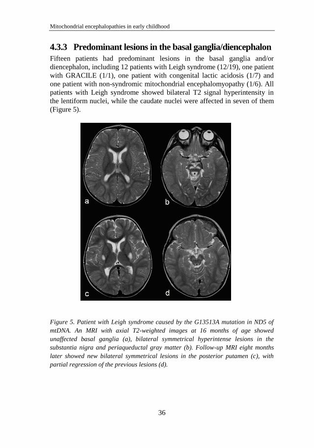
Mitochondrial encephalopathies in early childhood
36
4.3.3 Predominant lesions in the basal ganglia/diencephalon
Fifteen patients had predominant lesions in the basal ganglia and/or
diencephalon, including 12 patients with Leigh syndrome (12/19), one patient
with GRACILE (1/1), one patient with congenital lactic acidosis (1/7) and
one patient with non-syndromic mitochondrial encephalomyopathy (1/6). All
patients with Leigh syndrome showed bilateral T2 signal hyperintensity in
the lentiform nuclei, while the caudate nuclei were affected in seven of them
(Figure 5).
Figure 5. Patient with Leigh syndrome caused by the G13513A mutation in ND5 of
mtDNA. An MRI with axial T2-weighted images at 16 months of age showed
unaffected basal ganglia (a), bilateral symmetrical hyperintense lesions in the
substantia nigra and periaqueductal gray matter (b). Follow-up MRI eight months
later showed new bilateral symmetrical lesions in the posterior putamen (c), with
partial regression of the previous lesions (d).
Kalliopi Sofou
37
4.3.4 Predominant lesions in the white matter
Fifteen patients had pathological MRI findings predominantly affecting the
white matter, including all patients with leukoencephalopathy (9/9), one
patient with KSS (1/3) and five patients with congenital lactic acidosis (5/7).
Patients with leukoencephalopathy showed signal abnormalities of the white
matter with abnormal myelination, cystic lesions (4/9) and abnormal MRS in
all examined patients (6/9). The patients with congenital lactic acidosis had in
their majority, moderate-to-marked reduction in the periventricular white
matter with enlargement of the subarachnoid spaces and ventricles,
hypoplasia or complete agenesis of the corpus callosum and delayed
myelination. The patient with KSS had signal hyperintensities in T2-
weighted sequences in the subcortical white matter both supra- and
infratentorially, as well as in the deep gray matter.
4.3.5 Predominant lesions in the posterior fossa
Three patients with Leigh syndrome presented with MRI lesions
predominantly affecting the posterior fossa; marked signal abnormalities in
the tectum (3/19), pons and medullar involvement (2/19), as well as
involvement of the diencephalon and basal ganglia (2/19).
4.3.6 Diffuse lesions
Diffuse lesions were seen in four patients with Leigh syndrome (4/19), three
patients with Alpers syndrome (3/10), one patient with KSS (1/3) and one
patient with non-syndromic mitochondrial encephalomyopathy (1/6).
4.3.7 Normal MRI
Eight patients presented with a normal MRI, as follows: two patients with
MERRF with a mutation in A8344G tRNALys (2/3); two patients with
LHON (2/2); one patient with KSS due to an mtDNA-deletion (1/3); one
patient with Alpers syndrome with mutations in POLG1 (1/10); two patients
with non-syndromic mitochondrial encephalomyopathy, with COX
deficiency and complex I and V deficiencies respectively (2/6).

Mitochondrial encephalopathies in early childhood
38
5 DISCUSSION
Early-onset mitochondrial encephalopathies comprise a challenging group of
neurodegenerative disorders. This is due to their progressive nature, often
leading to major disability and premature death, as well as their diagnostic
complexity and lack of customized treatments.
In this work, we present our findings from studying 19 patients with Alpers
syndrome. Our studies describe the clinical spectrum of Alpers syndrome,
along with the neuroimaging and pathological features, and present specific
genotype-phenotype correlations depending on the presence or not of POLG1
mutations. We have further identified, with the help of WES, mutations in
NARS2 and PARS2 in two of our patients with Alpers syndrome, being the
first to link mutations in these genes to human disease and to Alpers
syndrome.
We also present natural history data on a unique cohort of 130 patients with
Leigh syndrome. This study is the first joint research work between eight
centers from six European countries specializing in mitochondrial diseases,
creating a strong platform for ongoing collaboration on mitochondrial
research projects. This study helps define the natural history of Leigh
syndrome and identify novel factors that predict outcome and long-term
prognosis.
Finally, we studied the brain MRIs of 66 patients with mitochondrial
disorders with CNS involvement. The diagnostic approach used in this study
— from the neuroanatomical/ neurofunctional lesion to disease identification
— assists the physician in the use of brain neuroimaging early in the
diagnostic work-up of suspected mitochondrial encephalopathies.
5.1 Genotype-phenotype correlations in Alpers syndrome
In our study, six patients had pathogenic mutations in POLG1, a syndrome
better known as Alpers-Huttenlocher [20-22]. These patients exhibited
infantile disease onset at around six months of age, followed by a severe and
rapidly progressive disease course, characterized by psychomotor regression,
ataxia, refractory seizures, epilepsia partialis continua, convulsive status
epilepticus and stroke-like episodes [93]. All patients developed moderate to
severe hepatic dysfunction, which was mainly related to valproate treatment
[93]. All but one of the patients with Alpers-Huttenlocher syndrome died at a
median age of 15 months [93].
Kalliopi Sofou
39
We also showed that mutations in POLG1 only explain the mitochondrial
respiratory chain deficiency in some patients with Alpers syndrome, while
the genetic cause of the disorder in the majority still remains obscure.
Patients with Alpers syndrome with no mutations in POLG1 present
symptoms earlier in life, often perinatally, and exhibit a more protracted
disease course, characterized by neonatal seizures, infantile spasms or
generalized seizures, while they usually develop spasticity and progressive
microcephaly with accompanying severe mental retardation [93]. Liver
dysfunction may also be present in Alpers syndrome not associated to
POLG1 mutations, but is usually mild, not associated to valproate treatment,
and sometimes spontaneously resolving [93]. The absence of POLG1
mutations was associated with better survival outcomes in these patients [93].
Alpers syndrome not associated to POLG1 mutations has been recently
linked to mutations in FARS2, a gene encoding the mitochondrial
phenylalanyl-tRNA synthetase [26]. With the help of WES, we identified
mutations in mt-aaRS in two of our patients with Alpers syndrome.
Patient AS-13 presented a homozygous mutation in NARS2. This gene
encodes 2 isoforms, named isoform 1 and isoform 2. The mutation is located
in the 3’-UTR region of isoform 2 (c.641C>T), whereas in isoform 1 it
causes a change of amino acid affecting a conserved residue (c.641C>T,
p.P214L). Based on the crystal structure of the protein homolog in
Pyrococcus horikoshii [94], we can predict that the affected proline is located
in the stem of a loop from which two conserved aromatic residues (tyrosine
and/or phenylalanine) protrude and physically participate in monomer-
monomer interaction. The structural change caused by the mutation in this
loop can therefore be expected to affect the activity of the protein by
destabilizing the interaction between the two monomers, still leaving some
residual activity.
Patient AS-13 also developed renal disease in the form of focal segmental
glomerulosclerosis and tubulopathy with accompanying hypochloremic
metabolic alkalosis. Renal disease has been associated with another mt-aaRS,
SARS2, the gene encoding the mitochondrial seryl-tRNA synthetase. Patients
with mutations in SARS2 show infantile onset of a multiorgan syndrome
known as HUPRA syndrome. The renal disease in HUPRA syndrome is
characterized by distal tubular dysfunction and renal failure [95]. Renal
manifestations in mitochondrial disorders are uncommon and have been
reported mainly in patients with mtDNA-associated mitochondrial disorders,
such as MELAS, MERRF, KSS and Pearson syndrome [96]. To our

Mitochondrial encephalopathies in early childhood
40
knowledge, POLG1 mutations have not been associated with kidney disease
so far.
Patient AS-15 presented compound heterozygosity for two mutations in
PARS2. The first mutation (c.1130dupC, p.K378fs*1) is an one base insertion
at codon 377 which creates a premature stop codon at position 378. This
mutation is predicted to generate a truncated protein 98 residues shorter than
the wild type form. The predicted missing part contains motif 3, which is part
of the active site and part of the dimer interface. Even assuming that the
truncated protein is stable, it can be predicted that the lack of such motifs
totally abolishes the enzymatic activity of the protein. The second mutation
(c.836C>T, p.S279L) is a missense mutation, predicted by SIFT to be
damaging. It is located in a highly conserved motif which shares homology
with a fragment of the bacterial-type prolyl-tRNA synthetase editing domain.
This fragment is flanked by CXXC motifs, which is characteristic of
functional domains inserted in larger proteins [97]. From this evidence it has
been postulated that mitochondrial prolyl-tRNA synthetases first acquired
and then lost their cis-editing domain at some point during evolution [98]. It
is therefore tempting to speculate that the function of the cluster in which the
mutation is located is somehow related to editing, perhaps by interacting with
a free standing editing domain in trans-.
Patient AS-15 developed mitochondrial cardiomyopathy at two years of age.
This patient had increased lactate levels at disease onset, but did not develop
lactic acidosis. The disease course was milder than the one described in the
mitochondrial disorders with neonatal lactic acidosis and cardiomyopathy in
infancy.
Even though there is in silico evidence to propose that the mutations
identified in our patients are responsible for their respective clinical
phenotypes, further experimental work needs to be carried out to confirm
these observations. It is important to further investigate whether these
phenotypes are specific for the identified candidate genes NARS2 and PARS2.
5.2 Neuropathological and neuroimaging findings in Alpers
syndrome and correlation to the genotype
The neuropathology is pathognomonic in Alpers syndrome and pertains to
major degenerative changes in the cerebral cortex, characterized by neuronal
loss, spongiosis and gliosis. The deep gray matter, the white matter and the
cerebellum may be involved to a variable degree. However, the extent of the
Kalliopi Sofou
41
brain lesions and accompanying atrophy are considered to be related to
disease duration. These findings are uniform in Alpers syndrome regardless
of the underlying genotype. In our study, patients with POLG1 mutations
also had vacuolation in the thalamus and basal ganglia. This finding was
absent in those patients with Alpers syndrome with no mutations in POLG1.
Brain neuroimaging plays a pivotal role in capturing the neurodegenerative
changes, with the sensitivity increasing with disease duration. The
progressive degeneration of the cerebral cortex in Alpers syndrome is mainly
seen as cortical atrophy on brain MRI. However, depending on the duration
of the disease, the MRI findings vary from normal, early in the disease
course, to generalized cortical or diffuse atrophy at later stages [99]. As a
result, patients develop progressive microcephaly. Furthermore, the brain
MRI of patients with POLG1 mutations may show features compatible with
stroke-like episodes, similar to those seen in MELAS [99].
5.3 The differential diagnosis of Alpers syndrome
The differential diagnosis of Alpers syndrome includes other progressive
neurodegenerative disorders characterized by early-onset encephalopathy and
similar neuroimaging and/or neuropathological findings. The early-onset
mitochondrial encephalopathies that pose the greatest challenge in the
differential diagnosis from Alpers syndrome are IOSCA and Leigh
syndrome. IOSCA and Alpers-Huttenlocher syndrome share similar age of
onset and clinical features, making the differential diagnosis on clinical
grounds difficult (see also paragraph 1.4.4). The neuropathology of IOSCA
typically affects the posterior fossa, while the poliodystrophy in Alpers-
Huttenlocher syndrome predominates in the cerebral cortex. However, both
syndromes may present with stroke-like cortical lesions. Both POLG1
mutations in Alpers-Huttenlocher syndrome and Twinkle mutations in
IOSCA cause tissue specific mtDNA depletions. The differential diagnosis
from Leigh syndrome is discussed separately in paragraph 5.6.
Late-onset Alpers syndrome is uncommon. Other POLG1-related
encephalopathies with typical onset in adolescence or adulthood are
mitochondrial spinocerebellar ataxia and epilepsy (MSCAE) and
mitochondrial recessive ataxia syndrome (MIRAS), which both present with
ataxia, epilepsy –often epilepsia partialis continua-, ptosis and/or external
ophthalmoplegia, sensory peripheral neuropathy and exacerbation episodes of
rapidly progressive encephalopathy. The MRI findings suggestive of these
syndromes are cerebellar cortical atrophy with or without white matter
Mitochondrial encephalopathies in early childhood
42
involvement, and thalamic lesions, while the basal ganglia are typically
spared. The stroke-like lesions seen in POLG1-related encephalopathies are
similar to those seen in MELAS. The latter is caused by mtDNA point
mutations, mostly in the tRNA gene for leucine (MT-TL), but also in other
tRNAs or MT-ND5. These stroke-like episodes, which are typical for
MELAS, are seen as stroke-like cortical lesions on brain MRI, often affecting
the occipital and temporal lobes. There is often a co-existence of acute and
chronic lesions in MELAS patients [99]. Calcifications of the basal ganglia
and dentate nuclei may also be seen in MELAS, but are not typically found in
POLG1-associated encephalopathies [100].
5.4 The diagnostic criteria of Leigh syndrome
Almost two decades have passed since Rahman and colleagues proposed
diagnostic criteria for Leigh syndrome [30]. Based on our findings and the
findings from other studies performed since then, we believe that the
following set of criteria is more appropriate to capture the wide phenotypic
and genotypic spectrum of Leigh syndrome [101, 102]:
a) clinical features of a progressive neurodegenerative disorder resulting from
affected deep gray matter structures and/or the brainstem, i.e. psychomotor
delay, disturbed muscle tone (hypotonia, hypertonia, dystonia), disturbed
movement (dyskinesia, akinesia, chorea, athetosis), ataxia and brainstem
dysfunction (dysfunction of swallowing/sucking, ophthalmological
manifestations, abnormal respiration and thermoregulation);
b) neuroimaging or neuropathological findings of symmetrically affected
deep gray matter structures and/or the brainstem;
c) defects in mitochondrial function shown on laboratory or molecular
testing, in the absence of inflammatory, autoimmune, vascular, toxic or other
metabolic etiology.
As Leigh syndrome is an umbrella term of an increasing number of
genetically identifiable mitochondrial diseases, we believe that the term
‘Leigh-like syndrome’, which is widely used in the literature, is not a suitable
term to an already wide spectrum of diseases. We therefore suggest the use of
the term ‘suspected Leigh syndrome’ in those patients that do not fulfill all
three of the above mentioned criteria.
Kalliopi Sofou
43
5.5 The natural history of Leigh syndrome and factors of
disease severity and survival
In our study, four of five patients with Leigh syndrome presented with
symptoms by the age of two years. This is in accordance with other studies
and it is the age that we propose as a cut-off age between early- and late-
onset Leigh syndrome [101-104]. The survival rate in our patients was better
than in previous studies, with 41% of patients being alive at a median follow-
up of 11.4 years from birth. However, in the group of patients that had died,
an early mortality was found at a median age of 2.4 years.
Predictors of survival in Leigh syndrome have not previously been assessed,
probably due to the small number of patients per study. Factors associated
with poorer survival were found to be: disease onset before six months of
age, failure to thrive, brainstem lesions on neuroimaging and intensive care
treatment. The negative effect of early disease onset on survival has been
previously reported, not only in Leigh syndrome, but in mitochondrial
disorders in general [9, 101]. In our study, respiratory complications were
found to be the leading cause of death. In view of the significant correlation
between the presence of brainstem lesions on neuroimaging and poor
survival, we believe that brainstem involvement may also be responsible for
the respiratory complications seen in patients with Leigh syndrome [102].
The presence of pathological signs at birth and a history of epileptic seizures
were associated with higher risk of acute exacerbations and/or relapses. The
relatively high incidence of pathological signs at birth, even if not fully
expressing Leigh syndrome, suggests that an ongoing energy failure linked to
mitochondrial dysfunction may already have started in utero, and as a
consequence, these patients might be more susceptible to external stress
factors, such as infection, poor nutrition and dehydration, that may lead to
acute exacerbations later in the disease course.
The presence of increased levels of lactate in CSF was significantly
correlated to a more severe disease course in our patients, characterized by
early disease onset before six months of age, acute exacerbations and/or
relapses, as well as brainstem involvement. Therefore, we suggest that the
presence of increased lactate levels in CSF may be used as a prognostic factor
of disease severity in Leigh syndrome.
Mitochondrial encephalopathies in early childhood
44
5.6 The differential diagnosis of Leigh syndrome
As previously discussed, Leigh syndrome encompasses an increasing number
of phenotypically and genetically variable mitochondrial disorders and as
such, this syndrome should be differentiated from other mitochondrial
encephalopathies with similar phenotypic spectrum, as well as other disorders
with degeneration of the deep gray matter structures.
Leigh syndrome shares phenotypical similarities with Alpers syndrome, as
both syndromes are typically characterized by early-onset encephalopathy,
abnormal muscle tone, psychomotor delay, epilepsy and involvement of other
organs of high-energy demands. The neuropathology in both syndromes
results from mitochondrial energy failure, but the lesion sites, often
delineated first on neuroimaging, differ; in Leigh syndrome, the lesions affect
predominantly the deep gray matter and/or brainstem, while in Alpers
syndrome, the cerebral cortex is primarily affected. This site specificity may
weaken with disease duration, often leading to diffuse, generalized atrophy in
Alpers syndrome. However, the cerebral cortex in Leigh syndrome is usually
better preserved [99].
Leigh syndrome should be differentiated from other diseases associated with
degeneration of the basal ganglia in the pediatric population, such as carbon
monoxide intoxication, Wilson’s disease, juvenile Huntington’s chorea,
neurodegeneration with brain iron accumulation, familial degeneration of the
striatum, non-familial infantile bilateral striatal necrosis (IBSN), and GAMT
(guanidinoacetate methyltransferase) deficiency. These diseases can
generally be ruled out on the basis of laboratory tests, history of exposure to
toxins and family history.
5.7 The role of neuroimaging in identifying mitochondrial
encephalopathies
Neuroimaging of the brain is a pivotal examination when suspecting a
mitochondrial encephalopathy. Our study shows that a combination of the
conventional MRI sequences with DWI and MRS provide a good indicator of
the underlying neuropathology, which can be missed in conventional
sequences alone. This is particularly important at early stages of disease,
where the lesions may be captured only on DWI.
Kalliopi Sofou
45
We have previously discussed the role of brain MRI in the differential
diagnosis between Alpers syndrome and Leigh syndrome (see paragraph 5.6).
Neuroimaging helps differentiate not only among mitochondrial
encephalopathies, but also from other metabolic, inflammatory, vascular or
toxic disorders with similar features. Furthermore, MRS is an important
adjuvant diagnostic tool to traditional brain MRI, as it provides useful
diagnostic information especially in the context of metabolic white matter
disorders.
Mitochondrial encephalopathies in early childhood
46
6 CONCLUSIONS
POLG1 mutations only explain the mitochondrial respiratory chain
deficiency in some patients with Alpers syndrome, a disorder better known as
Alpers-Huttenlocher syndrome. Alpers syndrome not associated to POLG1
mutations presents phenotypical differences from Alpers-Huttenlocher
syndrome.
With the help of whole exome sequencing, we have identified mutations in
NARS2 and PARS2 in two of our patients with Alpers syndrome. Mutations
in these genes have not previously been associated with human disease or
Alpers syndrome. We suggest that these are candidate genes for Alpers
syndrome not associated to POLG1 mutations.
Leigh syndrome is an umbrella term used to describe mitochondrial disorders
of progressive, neurodegenerative nature resulting from affected deep gray
matter structures and/or the brainstem. Disease onset before six months of
age, failure to thrive, brainstem lesions on neuroimaging and intensive care
treatment are associated with poorer survival of patients with Leigh
syndrome. The presence of pathological signs at birth and a history of
epileptic seizures increase the risk for acute exacerbations and/or relapses.
Increased lactate in CSF may be used as a predictor of disease severity.
If a mitochondrial disorder is suspected, neuroimaging should include
conventional MRI sequences in combination with DWI, ADC and MRS.
When a mitochondrial disorder is suspected on clinical or neuroimaging
grounds, mitochondrial investigations should be performed. A follow-up
brain MRI should be considered, particularly if the first MRI investigation
has been normal.
Kalliopi Sofou
47
7 FUTURE PROSPECTS
Some of our results from the study on Leigh syndrome have been reported
and discussed in this thesis, mainly pertaining to the natural history and
predictors of long-term outcomes. Upon completion of our work, we hope to
show specific associations between genotype and phenotype in Leigh
syndrome.
Moreover, through our collaboration with MCRN, we aim to study the brain
MRIs of our patients with Leigh syndrome and further develop MRI-based
pattern recognition algorithms.
The genetic causes of Alpers and Leigh syndrome in some of our patients
still remain unknown. Future projects will include the application of WES to
uncover the underlying genetic causes and study potential relationships to the
respective phenotypes.
Mitochondrial encephalopathies in early childhood
48
8 REFERENCES
1. Ernster L, Schatz G. Mitochondria: a historical review. The Journal of cell
biology. 1981;91(3 Pt 2):227s-55s.
2. Alberts BJ, A.; Lewis, J.; et al. The Mitochondrion. New York: Garland Science;
2002.
3. Luft R, Ikkos D, Palmieri G, Ernster L, Afzelius B. A case of severe
hypermetabolism of nonthyroid origin with a defect in the maintenance of
mitochondrial respiratory control: a correlated clinical, biochemical, and
morphological study. The Journal of clinical investigation. 1962;41:1776-804.
4. Nass S, Nass MM. Intramitochondrial Fibers with DNA Characteristics. Ii.
Enzymatic and Other Hydrolytic Treatments. The Journal of cell biology.
1963;19:613-29.
5. Nass MM, Nass S. Intramitochondrial Fibers with DNA Characteristics. I. Fixation
and Electron Staining Reactions. The Journal of cell biology. 1963;19:593-611.
6. Anderson S, Bankier AT, Barrell BG, de Bruijn MH, Coulson AR, Drouin J, et al.
Sequence and organization of the human mitochondrial genome. Nature.
1981;290(5806):457-65.
7. Koopman WJ, Willems PH, Smeitink JA. Monogenic mitochondrial disorders. The
New England journal of medicine. 2012;366(12):1132-41.
8. Wallace DC, Fan W, Procaccio V. Mitochondrial energetics and therapeutics.
Annual review of pathology. 2010;5:297-348.
9. Darin N, Oldfors A, Moslemi AR, Holme E, Tulinius M. The incidence of
mitochondrial encephalomyopathies in childhood: clinical features and
morphological, biochemical, and DNA anbormalities. Annals of neurology.
2001;49(3):377-83.
10. Uziel G, Ghezzi D, Zeviani M. Infantile mitochondrial encephalopathy. Seminars
in fetal & neonatal medicine. 2011;16(4):205-15.
11. Chen H, Chan DC. Physiological functions of mitochondrial fusion. Annals of
the New York Academy of Sciences. 2010;1201:21-5.
12. Chinnery PF, Hudson G. Mitochondrial genetics. British medical bulletin.
2013;106:135-59.
13. Robinson B. Lactic acidemia. 8th edition. ed. eds. SCBASWVD, editor:
McGraw-Hill New York. ; 2001.
14. Debray FG, Mitchell GA, Allard P, Robinson BH, Hanley JA, Lambert M.
Diagnostic accuracy of blood lactate-to-pyruvate molar ratio in the differential
diagnosis of congenital lactic acidosis. Clinical chemistry. 2007;53(5):916-21.
15. Phypers B, Pierce, T. Lactate physiology in health and disease. Continuing
Education in Anaesthesia, Critical Care & Pain. 2006;6(3).
Kalliopi Sofou
49
16. Alpers B. Diffuse progressive degeneration of the gray matter of the cerebrum.
Arch Neurol Psychiatry. 1931;25:469-505.
17. Courville CB. Widespread softening of the cerebral gray matter in infancy. Its
probable etiology in a paranatal circulatory disorder. Bulletin of the Los Angeles
Neurological Society. 1960;25:72-88.
18. Greenhouse AH, Neubuerger KT. The Syndrome of Progressive Cerebral
Poliodystrophy. Archives of neurology. 1964;10:47-57.
19. Dreifuss FE, Netsky MG. Progressive Poliodystrophy. The Degenerations of
Cerebral Gray Matter. American journal of diseases of children. 1964;107:649-56.
20. Huttenlocher PR, Solitare GB, Adams G. Infantile diffuse cerebral degeneration
with hepatic cirrhosis. Archives of neurology. 1976;33(3):186-92.
21. Kollberg G, Moslemi AR, Darin N, Nennesmo I, Bjarnadottir I, Uvebrant P, et al.
POLG1 mutations associated with progressive encephalopathy in childhood. Journal
of neuropathology and experimental neurology. 2006;65(8):758-68.
22. Naviaux RK, Nguyen KV. POLG mutations associated with Alpers' syndrome
and mitochondrial DNA depletion. Annals of neurology. 2004;55(5):706-12.
23. Rotig A, Poulton J. Genetic causes of mitochondrial DNA depletion in humans.
Biochimica et biophysica acta. 2009;1792(12):1103-8.
24. Ferrari G, Lamantea E, Donati A, Filosto M, Briem E, Carrara F, et al. Infantile
hepatocerebral syndromes associated with mutations in the mitochondrial DNA
polymerase-gammaA. Brain : a journal of neurology. 2005;128(Pt 4):723-31.
25. Isohanni P, Hakonen AH, Euro L, Paetau I, Linnankivi T, Liukkonen E, et al.
POLG1 manifestations in childhood. Neurology. 2011;76(9):811-5.
26. Elo JM, Yadavalli SS, Euro L, Isohanni P, Gotz A, Carroll CJ, et al.
Mitochondrial phenylalanyl-tRNA synthetase mutations underlie fatal infantile
Alpers encephalopathy. Human molecular genetics. 2012;21(20):4521-9.
27. Suzuki T, Nagao A, Suzuki T. Human mitochondrial tRNAs: biogenesis,
function, structural aspects, and diseases. Annual review of genetics. 2011;45:299-
329.
28. Leigh D. Subacute necrotizing encephalomyelopathy in an infant. Journal of
neurology, neurosurgery, and psychiatry. 1951;14(3):216-21.
29. Montpetit VJ, Andermann F, Carpenter S, Fawcett JS, Zborowska-Sluis D,
Giberson HR. Subacute necrotizing encephalomyelopathy. A review and a study of
two families. Brain : a journal of neurology. 1971;94(1):1-30.
30. Rahman S, Blok RB, Dahl HH, Danks DM, Kirby DM, Chow CW, et al. Leigh
syndrome: clinical features and biochemical and DNA abnormalities. Annals of
neurology. 1996;39(3):343-51.
Mitochondrial encephalopathies in early childhood
50
31. Dahl HH. Getting to the nucleus of mitochondrial disorders: identification of
respiratory chain-enzyme genes causing Leigh syndrome. American journal of
human genetics. 1998;63(6):1594-7.
32. Goldenberg PC, Steiner RD, Merkens LS, Dunaway T, Egan RA, Zimmerman
EA, et al. Remarkable improvement in adult Leigh syndrome with partial cytochrome
c oxidase deficiency. Neurology. 2003;60(5):865-8.
33. Huntsman RJ, Sinclair DB, Bhargava R, Chan A. Atypical presentations of leigh
syndrome: a case series and review. Pediatric neurology. 2005;32(5):334-40.
34. Thorburn DR, Rahman S. Mitochondrial DNA-Associated Leigh Syndrome and
NARP. In: Pagon RA, Adam MP, Ardinger HH, Bird TD, Dolan CR, Fong CT, et al.,
editors. GeneReviews(R). Seattle (WA)1993.
35. Moslemi AR, Darin N. Molecular genetic and clinical aspects of mitochondrial
disorders in childhood. Mitochondrion. 2007;7(4):241-52.
36. Matthews PM, Marchington DR, Squier M, Land J, Brown RM, Brown GK.
Molecular genetic characterization of an X-linked form of Leigh's syndrome. Annals
of neurology. 1993;33(6):652-5.
37. Moslemi AR, Darin N, Tulinius M, Wiklund LM, Holme E, Oldfors A.
Progressive encephalopathy and complex I deficiency associated with mutations in
MTND1. Neuropediatrics. 2008;39(1):24-8.
38. Hadzsiev K, Maasz A, Kisfali P, Kalman E, Gomori E, Pal E, et al.
Mitochondrial DNA 11777C>A mutation associated Leigh syndrome: case report
with a review of the previously described pedigrees. Neuromolecular medicine.
2010;12(3):277-84.
39. Marin SE, Mesterman R, Robinson B, Rodenburg RJ, Smeitink J, Tarnopolsky
MA. Leigh syndrome associated with mitochondrial complex I deficiency due to
novel mutations In NDUFV1 and NDUFS2. Gene. 2013;516(1):162-7.
40. Hoefs SJ, van Spronsen FJ, Lenssen EW, Nijtmans LG, Rodenburg RJ, Smeitink
JA, et al. NDUFA10 mutations cause complex I deficiency in a patient with Leigh
disease. European journal of human genetics : EJHG. 2011;19(3):270-4.
41. Ostergaard E, Rodenburg RJ, van den Brand M, Thomsen LL, Duno M, Batbayli
M, et al. Respiratory chain complex I deficiency due to NDUFA12 mutations as a
new cause of Leigh syndrome. Journal of medical genetics. 2011;48(11):737-40.
42. Coenen MJ, van den Heuvel LP, Ugalde C, Ten Brinke M, Nijtmans LG, Trijbels
FJ, et al. Cytochrome c oxidase biogenesis in a patient with a mutation in COX10
gene. Annals of neurology. 2004;56(4):560-4.
43. Darin N, Moslemi AR, Lebon S, Rustin P, Holme E, Oldfors A, et al. Genotypes
and clinical phenotypes in children with cytochrome-c oxidase deficiency.
Neuropediatrics. 2003;34(6):311-7.
Kalliopi Sofou
51
44. Moslemi AR, Tulinius M, Darin N, Aman P, Holme E, Oldfors A. SURF1 gene
mutations in three cases with Leigh syndrome and cytochrome c oxidase deficiency.
Neurology. 2003;61(7):991-3.
45. Oquendo CE, Antonicka H, Shoubridge EA, Reardon W, Brown GK. Functional
and genetic studies demonstrate that mutation in the COX15 gene can cause Leigh
syndrome. Journal of medical genetics. 2004;41(7):540-4.
46. Sasarman F, Brunel-Guitton C, Antonicka H, Wai T, Shoubridge EA, Consortium
L. LRPPRC and SLIRP interact in a ribonucleoprotein complex that regulates
posttranscriptional gene expression in mitochondria. Molecular biology of the cell.
2010;21(8):1315-23.
47. Lopez LC, Schuelke M, Quinzii CM, Kanki T, Rodenburg RJ, Naini A, et al.
Leigh syndrome with nephropathy and CoQ10 deficiency due to decaprenyl
diphosphate synthase subunit 2 (PDSS2) mutations. American journal of human
genetics. 2006;79(6):1125-9.
48. Gerards M, Kamps R, van Oevelen J, Boesten I, Jongen E, de Koning B, et al.
Exome sequencing reveals a novel Moroccan founder mutation in SLC19A3 as a
new cause of early-childhood fatal Leigh syndrome. Brain : a journal of neurology.
2013;136(Pt 3):882-90.
49. Tucker EJ, Hershman SG, Kohrer C, Belcher-Timme CA, Patel J, Goldberger
OA, et al. Mutations in MTFMT underlie a human disorder of formylation causing
impaired mitochondrial translation. Cell metabolism. 2011;14(3):428-34.
50. Wortmann S, Rodenburg RJ, Huizing M, Loupatty FJ, de Koning T, Kluijtmans
LA, et al. Association of 3-methylglutaconic aciduria with sensori-neural deafness,
encephalopathy, and Leigh-like syndrome (MEGDEL association) in four patients
with a disorder of the oxidative phosphorylation. Molecular genetics and metabolism.
2006;88(1):47-52.
51. Fellman V, Rapola J, Pihko H, Varilo T, Raivio KO. Iron-overload disease in
infants involving fetal growth retardation, lactic acidosis, liver haemosiderosis, and
aminoaciduria. Lancet. 1998;351(9101):490-3.
52. Visapaa I, Fellman V, Vesa J, Dasvarma A, Hutton JL, Kumar V, et al.
GRACILE syndrome, a lethal metabolic disorder with iron overload, is caused by a
point mutation in BCS1L. American journal of human genetics. 2002;71(4):863-76.
53. Al-Hussaini A, Faqeih E, El-Hattab AW, Alfadhel M, Asery A, Alsaleem B, et
al. Clinical and molecular characteristics of mitochondrial DNA depletion syndrome
associated with neonatal cholestasis and liver failure. The Journal of pediatrics.
2014;164(3):553-9 e1-2.
54. Scaglia F, Dimmock D, Wong LJ. DGUOK-Related Mitochondrial DNA
Depletion Syndrome, Hepatocerebral Form. In: Pagon RA, Adam MP, Ardinger HH,
Bird TD, Dolan CR, Fong CT, et al., editors. GeneReviews(R). Seattle (WA)1993.
Mitochondrial encephalopathies in early childhood
52
55. El-Hattab AW, Li FY, Schmitt E, Zhang S, Craigen WJ, Wong LJ. MPV17-
associated hepatocerebral mitochondrial DNA depletion syndrome: new patients and
novel mutations. Molecular genetics and metabolism. 2010;99(3):300-8.
56. Lonnqvist T, Paetau A, Nikali K, von Boguslawski K, Pihko H. Infantile onset
spinocerebellar ataxia with sensory neuropathy (IOSCA): neuropathological features.
Journal of the neurological sciences. 1998;161(1):57-65.
57. Hakonen AH, Isohanni P, Paetau A, Herva R, Suomalainen A, Lonnqvist T.
Recessive Twinkle mutations in early onset encephalopathy with mtDNA depletion.
Brain : a journal of neurology. 2007;130(Pt 11):3032-40.
58. Lonnqvist T, Paetau A, Valanne L, Pihko H. Recessive twinkle mutations cause
severe epileptic encephalopathy. Brain : a journal of neurology. 2009;132(Pt
6):1553-62.
59. Ostergaard E, Schwartz M, Batbayli M, Christensen E, Hjalmarson O, Kollberg
G, et al. A novel missense mutation in SUCLG1 associated with mitochondrial DNA
depletion, encephalomyopathic form, with methylmalonic aciduria. European journal
of pediatrics. 2010;169(2):201-5.
60. Ostergaard E, Hansen FJ, Sorensen N, Duno M, Vissing J, Larsen PL, et al.
Mitochondrial encephalomyopathy with elevated methylmalonic acid is caused by
SUCLA2 mutations. Brain : a journal of neurology. 2007;130(Pt 3):853-61.
61. Sakamoto O, Ohura T, Murayama K, Ohtake A, Harashima H, Abukawa D, et al.
Neonatal lactic acidosis with methylmalonic aciduria due to novel mutations in the
SUCLG1 gene. Pediatrics international : official journal of the Japan Pediatric
Society. 2011;53(6):921-5.
62. Carrozzo R, Dionisi-Vici C, Steuerwald U, Lucioli S, Deodato F, Di
Giandomenico S, et al. SUCLA2 mutations are associated with mild methylmalonic
aciduria, Leigh-like encephalomyopathy, dystonia and deafness. Brain : a journal of
neurology. 2007;130(Pt 3):862-74.
63. Bornstein B, Area E, Flanigan KM, Ganesh J, Jayakar P, Swoboda KJ, et al.
Mitochondrial DNA depletion syndrome due to mutations in the RRM2B gene.
Neuromuscular disorders : NMD. 2008;18(6):453-9.
64. Kollberg G, Darin N, Benan K, Moslemi AR, Lindal S, Tulinius M, et al. A novel
homozygous RRM2B missense mutation in association with severe mtDNA
depletion. Neuromuscular disorders : NMD. 2009;19(2):147-50.
65. Bonnen PE, Yarham JW, Besse A, Wu P, Faqeih EA, Al-Asmari AM, et al.
Mutations in FBXL4 cause mitochondrial encephalopathy and a disorder of
mitochondrial DNA maintenance. American journal of human genetics.
2013;93(3):471-81.
66. Gotz A, Tyynismaa H, Euro L, Ellonen P, Hyotylainen T, Ojala T, et al. Exome
sequencing identifies mitochondrial alanyl-tRNA synthetase mutations in infantile
mitochondrial cardiomyopathy. American journal of human genetics.
2011;88(5):635-42.
Kalliopi Sofou
53
67. Ghezzi D, Baruffini E, Haack TB, Invernizzi F, Melchionda L, Dallabona C, et
al. Mutations of the mitochondrial-tRNA modifier MTO1 cause hypertrophic
cardiomyopathy and lactic acidosis. American journal of human genetics.
2012;90(6):1079-87.
68. Baruffini E, Dallabona C, Invernizzi F, Yarham JW, Melchionda L, Blakely EL,
et al. MTO1 mutations are associated with hypertrophic cardiomyopathy and lactic
acidosis and cause respiratory chain deficiency in humans and yeast. Human
mutation. 2013;34(11):1501-9.
69. Cizkova A, Stranecky V, Mayr JA, Tesarova M, Havlickova V, Paul J, et al.
TMEM70 mutations cause isolated ATP synthase deficiency and neonatal
mitochondrial encephalocardiomyopathy. Nature genetics. 2008;40(11):1288-90.
70. Diodato D, Invernizzi F, Lamantea E, Fagiolari G, Parini R, Menni F, et al.
Common and Novel TMEM70 Mutations in a Cohort of Italian Patients with
Mitochondrial Encephalocardiomyopathy. JIMD reports. 2014.
71. Papadopoulou LC, Sue CM, Davidson MM, Tanji K, Nishino I, Sadlock JE, et al.
Fatal infantile cardioencephalomyopathy with COX deficiency and mutations in
SCO2, a COX assembly gene. Nature genetics. 1999;23(3):333-7.
72. DiMauro S, Tanji K, Schon EA. The many clinical faces of cytochrome c oxidase
deficiency. Advances in experimental medicine and biology. 2012;748:341-57.
73. Haack TB, Rolinski B, Haberberger B, Zimmermann F, Schum J, Strecker V, et
al. Homozygous missense mutation in BOLA3 causes multiple mitochondrial
dysfunctions syndrome in two siblings. Journal of inherited metabolic disease.
2013;36(1):55-62.
74. Navarro-Sastre A, Tort F, Stehling O, Uzarska MA, Arranz JA, Del Toro M, et
al. A fatal mitochondrial disease is associated with defective NFU1 function in the
maturation of a subset of mitochondrial Fe-S proteins. American journal of human
genetics. 2011;89(5):656-67.
75. Mayr JA, Feichtinger RG, Tort F, Ribes A, Sperl W. Lipoic acid biosynthesis
defects. Journal of inherited metabolic disease. 2014.
76. Ohlenbusch A, Edvardson S, Skorpen J, Bjornstad A, Saada A, Elpeleg O, et al.
Leukoencephalopathy with accumulated succinate is indicative of SDHAF1 related
complex II deficiency. Orphanet journal of rare diseases. 2012;7:69.
77. Massa V, Fernandez-Vizarra E, Alshahwan S, Bakhsh E, Goffrini P, Ferrero I, et
al. Severe infantile encephalomyopathy caused by a mutation in COX6B1, a nucleus-
encoded subunit of cytochrome c oxidase. American journal of human genetics.
2008;82(6):1281-9.
78. Steenweg ME, van Berge L, van Berkel CG, de Coo IF, Temple IK, Brockmann
K, et al. Early-onset LBSL: how severe does it get? Neuropediatrics. 2012;43(6):332-
8.
79. Ferreira M, Torraco A, Rizza T, Fattori F, Meschini MC, Castana C, et al.
Progressive cavitating leukoencephalopathy associated with respiratory chain
Mitochondrial encephalopathies in early childhood
54
complex I deficiency and a novel mutation in NDUFS1. Neurogenetics.
2011;12(1):9-17.
80. Zafeiriou DI, Rodenburg RJ, Scheffer H, van den Heuvel LP, Pouwels PJ,
Ververi A, et al. MR spectroscopy and serial magnetic resonance imaging in a patient
with mitochondrial cystic leukoencephalopathy due to complex I deficiency and
NDUFV1 mutations and mild clinical course. Neuropediatrics. 2008;39(3):172-5.
81. Haas RH, Parikh S, Falk MJ, Saneto RP, Wolf NI, Darin N, et al. Mitochondrial
disease: a practical approach for primary care physicians. Pediatrics.
2007;120(6):1326-33.
82. Mitochondrial Medicine Society's Committee on D, Haas RH, Parikh S, Falk MJ,
Saneto RP, Wolf NI, et al. The in-depth evaluation of suspected mitochondrial
disease. Molecular genetics and metabolism. 2008;94(1):16-37.
83. Barragan-Campos HM, Vallee JN, Lo D, Barrera-Ramirez CF, Argote-Greene M,
Sanchez-Guerrero J, et al. Brain magnetic resonance imaging findings in patients
with mitochondrial cytopathies. Archives of neurology. 2005;62(5):737-42.
84. Bianchi MC, Sgandurra G, Tosetti M, Battini R, Cioni G. Brain magnetic
resonance in the diagnostic evaluation of mitochondrial encephalopathies. Bioscience
reports. 2007;27(1-3):69-85.
85. Lebre AS, Rio M, Faivre d'Arcier L, Vernerey D, Landrieu P, Slama A, et al. A
common pattern of brain MRI imaging in mitochondrial diseases with complex I
deficiency. Journal of medical genetics. 2011;48(1):16-23.
86. Valanne L, Ketonen L, Majander A, Suomalainen A, Pihko H. Neuroradiologic
findings in children with mitochondrial disorders. AJNR American journal of
neuroradiology. 1998;19(2):369-77.
87. Saneto RP, Friedman SD, Shaw DW. Neuroimaging of mitochondrial disease.
Mitochondrion. 2008;8(5-6):396-413.
88. Dinopoulos A, Cecil KM, Schapiro MB, Papadimitriou A, Hadjigeorgiou GM,
Wong B, et al. Brain MRI and proton MRS findings in infants and children with
respiratory chain defects. Neuropediatrics. 2005;36(5):290-301.
89. McCormick E, Place E, Falk MJ. Molecular genetic testing for mitochondrial
disease: from one generation to the next. Neurotherapeutics : the journal of the
American Society for Experimental NeuroTherapeutics. 2013;10(2):251-61.
90. Wong LJ. Challenges of bringing next generation sequencing technologies to
clinical molecular diagnostic laboratories. Neurotherapeutics : the journal of the
American Society for Experimental NeuroTherapeutics. 2013;10(2):262-72.
91. Rabbani B, Tekin M, Mahdieh N. The promise of whole-exome sequencing in
medical genetics. Journal of human genetics. 2014;59(1):5-15.
92. Tulinius MH, Holme E, Kristiansson B, Larsson NG, Oldfors A. Mitochondrial
encephalomyopathies in childhood. I. Biochemical and morphologic investigations.
The Journal of pediatrics. 1991;119(2):242-50.
Kalliopi Sofou
55
93. Sofou K, Moslemi AR, Kollberg G, Bjarnadottir I, Oldfors A, Nennesmo I, et al.
Phenotypic and genotypic variability in Alpers syndrome. European journal of
paediatric neurology : EJPN : official journal of the European Paediatric Neurology
Society. 2012;16(4):379-89.
94. Iwasaki W, Sekine S, Kuroishi C, Kuramitsu S, Shirouzu M, Yokoyama S.
Structural basis of the water-assisted asparagine recognition by asparaginyl-tRNA
synthetase. Journal of molecular biology. 2006;360(2):329-42.
95. Belostotsky R, Ben-Shalom E, Rinat C, Becker-Cohen R, Feinstein S, Zeligson S,
et al. Mutations in the mitochondrial seryl-tRNA synthetase cause hyperuricemia,
pulmonary hypertension, renal failure in infancy and alkalosis, HUPRA syndrome.
American journal of human genetics. 2011;88(2):193-200.
96. O'Toole JF. Renal manifestations of genetic mitochondrial disease. International
journal of nephrology and renovascular disease. 2014;7:57-67.
97. Grishin NV. Treble clef finger--a functionally diverse zinc-binding structural
motif. Nucleic acids research. 2001;29(8):1703-14.
98. Ahel I, Korencic D, Ibba M, Soll D. Trans-editing of mischarged tRNAs.
Proceedings of the National Academy of Sciences of the United States of America.
2003;100(26):15422-7.
99. Sofou K, Steneryd K, Wiklund LM, Tulinius M, Darin N. MRI of the brain in
childhood-onset mitochondrial disorders with central nervous system involvement.
Mitochondrion. 2013;13(4):364-71.
100. Sidiropoulos C, Moro E, Lang AE. Extensive intracranial calcifications in a
patient with a novel polymerase gamma-1 mutation. Neurology. 2013;81(2):197-8.
101. Naess K, Freyer C, Bruhn H, Wibom R, Malm G, Nennesmo I, et al. MtDNA
mutations are a common cause of severe disease phenotypes in children with Leigh
syndrome. Biochimica et biophysica acta. 2009;1787(5):484-90.
102. Sofou K, De Coo IF, Isohanni P, Ostergaard E, Naess K, De Meirleir L, et al. A
multicenter study on Leigh syndrome: disease course and predictors of survival.
Orphanet journal of rare diseases. 2014;9(1):52.
103. Yang YL, Sun F, Zhang Y, Qian N, Yuan Y, Wang ZX, et al. Clinical and
laboratory survey of 65 Chinese patients with Leigh syndrome. Chinese medical
journal. 2006;119(5):373-7.
104. Lee HF, Tsai CR, Chi CS, Lee HJ, Chen CC. Leigh syndrome: clinical and
neuroimaging follow-up. Pediatric neurology. 2009;40(2):88-93.
105. Rouault TA. Biogenesis of iron-sulfur clusters in mammalian cells: new insights
and relevance to human disease. Disease models & mechanisms. 2012;5(2):155-64.
106. Pearce S, Nezich CL, Spinazzola A. Mitochondrial diseases: translation matters.
Molecular and cellular neurosciences. 2013;55:1-12.
107. Calvo SE, Mootha VK. The mitochondrial proteome and human disease. Annual
review of genomics and human genetics. 2010;11:25-44.