Embed Size (px)
Citation preview

on April 28, 2018http://rsif.royalsocietypublishing.org/Downloaded from
rsif.royalsocietypublishing.org
ReviewCite this article: Shaw CJ, ter Haar GR, Rivens
IH, Giussani DA, Lees CC. 2014 Pathophysiolo-
gical mechanisms of high-intensity focused
ultrasound-mediated vascular occlusion and
relevance to non-invasive fetal surgery.
J. R. Soc. Interface 11: 20140029.
http://dx.doi.org/10.1098/rsif.2014.0029
Received: 10 January 2014
Accepted: 6 March 2014
Subject Areas:biophysics, medical physics
Keywords:high-intensity focused ultrasound, prenatal,
blood vessels, pathophysiology
Author for correspondence:C. C. Lees
e-mail: [email protected]
& 2014 The Author(s) Published by the Royal Society. All rights reserved.
Pathophysiological mechanisms of high-intensity focused ultrasound-mediatedvascular occlusion and relevance tonon-invasive fetal surgery
C. J. Shaw1,2, G. R. ter Haar3, I. H. Rivens3, D. A. Giussani2 and C. C. Lees1,4
1Imperial College London, Hammersmith Campus, Du Cane Road, London W12 0HS, UK2Department of Physiology, Development and Neuroscience, University of Cambridge, Cambridge CB2 3EG, UK3Joint Department of Physics, Institute of Cancer Research: Royal Marsden NHSF Trust, Downs Road, Sutton,Surrey SM2 5PT, UK4Department of Obstetrics and Gynaecology, University Hospitals Leuven, Campus Gasthuisberg, KU Leuven,Belgium
High-intensity focused ultrasound (HIFU) is a non-invasive technology,
which can be used occlude blood vessels in the body. Both the theory
underlying and practical process of blood vessel occlusion are still under
development and relatively sparse in vivo experimental and therapeutic data
exist. HIFU would however provide an alternative to surgery, particularly in
circumstances where serious complications inherent to surgery outweigh the
potential benefits. Accordingly, the HIFU technique would be of particular
utility for fetal and placental interventions, where open or endoscopic surgery
is fraught with difficulty and likelihood of complications including premature
delivery. This assumes that HIFU could be shown to safely and effectively
occlude blood vessels in utero. To understand these mechanisms more fully,
we present a review of relevant cross-specialty literature on the topic of vascu-
lar HIFU and suggest an integrative mechanism taking into account clinical,
physical and engineering considerations through which HIFU may produce
vascular occlusion. This model may aid in the design of HIFU protocols to
further develop this area, and might be adapted to provide a non-invasive
therapy for conditions in fetal medicine where vascular occlusion is beneficial.
1. IntroductionSurgical techniques can be used to create permanent arterial or venous occlusion,
either for therapeutic benefit or prior to ligation of an artery or vein, and require
close proximity to or contact with the vessels to be occluded. High-intensity
focused ultrasound (HIFU) is a non-invasive alternative method of occlusion
which may avoid complications inherent to surgery. HIFU could be of particular
use in fetal surgery, were it shown to occlude blood vessels safely and effectively.
This would potentially offer an in-utero treatment for fetal diseases such as
twin-to-twin transfusion syndrome (TTTS), twin-reversed arterial perfusion
(TRAP) sequence, bronchopulmonary sequestration (BPS) or sacrococcygeal
teratoma (SCT).
HIFU involves the production of alternating high positive (compression)
and negative (rarefaction) pressures which cause tissue heating (with the poten-
tial to boil interstitial water) and acoustic cavitation bubbles to form, which can
have extremely energetic locally destructive activity. In addition, ultrasound
has an acoustic radiation force (non-ionizing), which in a high power focused
beam can have a significant impact on exposed tissues. How each of these inter-
acts with tissue, and in particular blood vessels, is not yet fully understood. In
the current absence of specific experimental work designed to understand these
mechanisms more fully, we present a review of relevant literature on the topic
of vascular HIFU and suggest an integrative mechanism taking into account
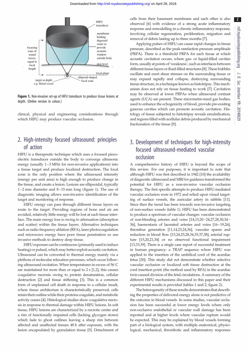
focusingzone ofsoundwaves,equal to
focallength
target at depthe.g. blood vessel
ellipsoid-shapedfocal zone
focal plane
skin level
membraneholdingdegassedwater toprovideacousticwindowoutside body
HIFUtransducer
Figure 1. Non-invasive set-up of HIFU transducer to produce tissue lesions atdepth. (Online version in colour.)
rsif.royalsocietypublishing.orgJ.R.Soc.Interface
11:20140029
2
on April 28, 2018http://rsif.royalsocietypublishing.org/Downloaded from
clinical, physical and engineering considerations through
which HIFU may produce vascular occlusion.
2. High-intensity focused ultrasound: principlesof action
HIFU is a therapeutic technique which uses a focused piezo-
electric transducer outside the body to converge ultrasonic
energy (usually 1–3 MHz for non-invasive applications) into
a tissue target and produce localized destruction. The focal
zone is the only position where the ultrasound intensity
(energy per unit area) is high enough to produce change in
the tissue, and create a lesion. Lesions are ellipsoidal, typically
1–2 mm diameter and 8–15 mm long (figure 1). The use of
diagnostic imaging allows non-invasive identification of the
target and monitoring of response.
HIFU energy can pass through different tissue layers en
route to the target. Providing regions of bone and air are
avoided, relatively little energy will be lost at each tissue inter-
face. The main energy loss is owing to attenuation (absorption
and scatter) within the tissue layers. Alternative techniques
such as radio frequency ablation (RFA), laser photocoagulation
and microwave energy have poor tissue penetration so use
invasive methods to destroy deep tissue.
HIFU exposure can be continuous (primarily used to induce
heating) or pulsed, which may better exploit acoustic cavitation.
Ultrasound can be converted to thermal energy mainly via a
plethora of molecular relaxation processes, which occur follow-
ing ultrasound excitation. When temperatures in excess of 568Care maintained for more than or equal to 2 s [1,2], this causes
coagulative necrosis owing to protein denaturation, cellular
destruction [2] and tissue stiffening [3]. This is a common
form of unplanned cell death in response to a cellular insult,
where tissue architecture is characteristically preserved, cells
retain their outline while their proteins coagulate, and metabolic
activity ceases [4]. Histological studies show coagulative necro-
sis in response to thermal damage within HIFU lesions. In soft
tissue, HIFU lesions are characterized by a necrotic centre and
a rim of functionally impaired cells (lacking glycogen stores)
which fade to ghost outlines leaving a sharp rim between
affected and unaffected tissues 48 h after exposure, with the
lesion encapsulated by granulation tissue [5]. Detachment of
cells from their basement membrane and each other is also
observed [6] with evidence of a strong acute inflammatory
response and remodelling in a chronic inflammatory response,
involving cellular regeneration, proliferation, migration and
removal of debris lasting up to three months [7].
Applying pulses of HIFU can cause rapid changes in tissue
pressure, described as the peak rarefaction pressure amplitude
(PRPA). There is a threshold PRPA for each tissue at which
acoustic cavitation occurs, where gas- or liquid-filled cavities
form, usually at points of ‘weakness’, such as interfaces between
different tissue layers or fluid-filled structures [8]. These bubbles
oscillate and exert shear stresses on the surrounding tissue or
may expand rapidly and collapse, destroying surrounding
tissue structure, in a technique known as histotripsy. This mech-
anism does not rely on tissue heating to work [7]. Cavitation
may be observed at lower PRPAs when ultrasound contrast
agents (UCA) are present. These micrometre-sized gas bodies,
used to enhance the echogenicity of blood, provide pre-existing
gaseous cavities which can promote acoustic cavitation. His-
tology of tissue subjected to histotripsy reveals emulsification,
and regions filled with acellular debris produced by mechanical
fractionation of the tissue [9].
3. Development of techniques for high-intensityfocused ultrasound-mediated vascularocclusion
A comprehensive history of HIFU is beyond the scope of
this review. For our purposes, it is important to note that
although HIFU was first described in 1942 [10] the availability
of diagnostic ultrasound and MRI for guidance transformed the
potential for HIFU as a non-invasive vascular occlusion
therapy. The first specific attempts to produce HIFU-mediated
vascular occlusion were in 1972 and relied upon visual target-
ing of surface vessels, the auricular artery in rabbits [11].
Since then the trend has been towards non-invasive targeting
of non-surface vessels (table 1). HIFU has been demonstrated
to produce a spectrum of vascular changes: vascular occlusion
of non-bleeding arteries and veins [14,15,20–24,27,28,30,34–
36], haemostasis of lacerated arteries and veins [16–19,26],
thrombus generation [11,14,23,24,36], vascular spasm and
reduction in blood flow [15,24,25,28,34,35,37,38], arterial rup-
ture [15,20,21,34] or no observed functional impairment
[13,33,39]. There is a single case report of successful treatment
in human pregnancy: a TRAP sequence where HIFU was
applied to the insertion of the umbilical cord of the acardiac
fetus [30]. This study did not demonstrate whether selective
vascular occlusion or localized soft tissue destruction at the
cord insertion point (the method used by RFA) in the acardiac
twin caused division of the fetal circulations. A summary of the
different HIFU mechanisms discussed in this paper and their
experimental results is provided (tables 1 and 2; figure 2).
The heterogeneity of these results demonstrates that describ-
ing the properties of delivered energy alone is not predictive of
the outcome in blood vessels. In some studies, vascular occlu-
sion has been successful at lower energy levels where only
non-occlusive endothelial or vascular wall damage has been
reported and at higher levels where vascular rupture would
be expected. This may be explained by blood vessels forming
part of a biological system, with multiple anatomical, physio-
logical, mechanical, thrombotic and inflammatory responses

Tabl
e1.
Sum
mar
yof
mec
hani
sms
used
and
expe
rimen
tal
outco
mes
inan
imal
mod
elsan
dhu
man
studi
esex
amin
ing
cont
inuo
usHI
FU-m
ediat
edva
scul
aroc
clusio
n.Ke
y:o,
vasc
ular
effe
ctob
serv
ed;
x,va
scul
aref
fect
not
obse
rved
;-,
vasc
ular
effe
ctno
trep
orte
d/in
vesti
gate
d.
invi
vom
odel
s—co
ntin
uous
HIFU
refe
renc
esm
odel
vess
el
anim
als
(n)
expo
sure
s
(n)
vess
el
diam
eter
(mm
)
non-
inva
sive?
targ
etin
g
inte
nsity
(Wcm
22 )
frequ
ency
(MHz
)
foca
l
leng
th
(mm
)
no.
expo
sure
s
expo
sure
time
(s)
[11]
rabb
itau
ricul
arar
tery
1228
0.7–
1.0
Yvis
ual
25–
1500
112
51
0.5–
720
[12]
dog
unsp
ec.v
ein19
-NR
YA-
mod
eNR
3NR
NRNR
man
varic
ose
legve
ins
11-
NRY
visua
lNR
3NR
NRNR
[13]
rabb
itao
rta,I
VC24
-NR
Nvis
ual
1500
4NR
205
[14]
ratfe
mor
alve
in6
-1.
5N
A-m
ode
167
7.31
204–
73
[15]
rabb
itfe
mor
alar
t.,ve
in19
261.
0–1.
3Y
MRI
4400
–88
001.
49NR
5–8
1
[15]
rabb
itre
nala
rtery
9-
0.6
YM
RI65
0028
001.
5NR
12–
1610
[16]
rabb
itliv
er micr
ovas
culat
urea
827
,0.
5N
visua
l30
003.
340
11–
2
[17]
pig
mus
cular
arte
riesa
541
3–10
Nvis
ual
3100
3.5
551
10–
20
[18]
pig
mus
cular
arte
riesa
389
3–10
NDo
ppler
2000
3.5
551
5–20
[19]
pig
mus
cular
arte
ries
476
3–10
NDo
ppler
3000
3.5
551
17–
25
[20]
ratfe
mor
alar
t.,ve
in10
-0.
5–1.
5N
visua
l46
601.
715
01
2
[21]
ratfe
mor
alar
t.,ve
in16
-0.
5–1.
5N
visua
l16
90–
4660
1.7
150
15
[22]
ratfe
mor
alar
tery
1836
0.5
NRNR
800
1NR
15–
10
1000
03
15–
10
[23]
rabb
itau
ricul
arve
ina
15-
1.0
Yvis
ual
750
3.9
351–
33
[24]
ratfe
mor
alar
tery
23-
0.5
YDo
ppler
530–
2750
3.3
NR1
5
[25]
shee
put
erin
ear
tery
7-
NRN
Dopp
lerNR
1.05
100
6–7
8–10
[26]
rabb
itfe
mor
alar
tery
25-
1.5
Nvis
ual
3000
9.6
25NR
5–10
[27]
rabb
itum
bilic
alar
tery
11-
0.4–
0.8
YDo
ppler
1400
–55
002.
2670
3–15
5
[28]
rabb
itre
nala
rtery
8-
0.5
YDo
ppler
4000
2.2
602–
105
[29]
hum
an (inut
ero)
umbi
lical
arte
ry1
-NR
YDo
ppler
2300
1.71
NRNR
10
[30]
hum
an (inut
ero)
umbi
lical
arte
ry1
-NR
YDo
ppler
4600
1.71
60NR
10
rsif.royalsocietypublishing.orgJ.R.Soc.Interface
11:20140029
3
on April 28, 2018http://rsif.royalsocietypublishing.org/Downloaded from

invi
vom
odel
s—co
ntin
uous
HIFU
refe
renc
esm
odel
vess
el
tissu
e
tem
p(88888
C)ca
vita
tion
vasc
ular
occlu
sion
haem
osta
sisth
rom
bus
endo
thel
ial
dam
age
vasc
ular
spas
m
vasc
ular
rupt
ure
vasc
ular
wal
ldam
age
leng
thof
follo
w-u
p
[11]
rabb
itau
ricul
arar
tery
38.0
–66
.7NR
x-
oo
--
o72
hour
s
[12]
dog
unsp
ec.v
einNR
NR-
-o
--
-o
none
man
varic
ose
legve
ins
NRNR
--
o-
--
ono
ne
[13]
rabb
itao
rta,I
VC68
.0–
80.1
NRx
--
-x
xo
sixm
onth
s
[14]
ratfe
mor
alve
in,
45x
o-
oo
--
-28
days
[15]
rabb
itfe
mor
alar
t.,ve
inNR
oo
--
-o
o-
14da
ys
[15]
rabb
itre
nala
rtery
64o
o-
--
oo
-7
days
[16]
rabb
itliv
er micr
ovas
culat
urea
59–
86o
oo
xo
--
ono
ne
[17]
pig
mus
cular
arte
riesa
NRNR
xo
oo
o-
ono
ne
[18]
pig
mus
cular
arte
riesa
NRNR
xo
--
-o
-no
ne
[19]
pig
mus
cular
arte
ries
NRNR
oo
oo
--
ono
ne
[20]
ratfe
mor
alar
t.,ve
inNR
NRo
o-
--
o-
none
[21]
ratfe
mor
alar
t.,ve
inNR
NRo
--
--
o-
none
[22]
ratfe
mor
alar
tery
46NR
x-
--
--
-no
ne
98NR
o-
--
--
o12
days
[23]
rabb
itau
ricul
arve
ina
NRNR
oo
oo
--
o28
days
[24]
ratfe
mor
alar
tery
98NR
o-
oo
o-
o3
days
[25]
shee
put
erin
ear
tery
NRo
x-
-o
o-
oNR
[26]
rabb
itfe
mor
alar
tery
NRNR
oo
oo
--
o60
days
(Con
tinue
d.)
rsif.royalsocietypublishing.orgJ.R.Soc.Interface
11:20140029
4
on April 28, 2018http://rsif.royalsocietypublishing.org/Downloaded from

Tabl
e1.
(Con
tinue
d.)
invi
vom
odel
s—co
ntin
uous
HIFU
refe
renc
esm
odel
vess
el
tissu
e
tem
p(88888
C)ca
vita
tion
vasc
ular
occlu
sion
haem
osta
sisth
rom
bus
endo
thel
ial
dam
age
vasc
ular
spas
m
vasc
ular
rupt
ure
vasc
ular
wal
ldam
age
leng
thof
follo
w-u
p
[27]
rabb
itum
bilic
alar
tery
NRNR
o-
oo
-x
ono
ne
[28]
rabb
itre
nala
rtery
NRNR
o-
--
oo
o7
days
[29]
hum
an (inut
ero)
umbi
lical
arte
ryNR
NRx
--
--
--
thre
ewe
eks
[30]
hum
an (inut
ero)
umbi
lical
arte
ryNR
NRo
--
--
--
20we
eks
exvi
voex
perim
enta
lmod
els—
cont
inuo
usHI
FU
refe
renc
esm
odel
vess
el
anim
als
(n)
expo
sure
s
(n)
vess
el
diam
eter
(mm
)
non-
inva
sive?
targ
etin
g
inte
nsity
(Wcm
22 )
frequ
ency
(MHz
)
foca
l
leng
th
(mm
)
num
ber
of
expo
sure
s
expo
sure
time
(s)
[31]
dog (e
xviv
o)
coro
nary
arte
ry6
-NR
N/A
visua
lNR
NRNR
115
[32]
rat(e
xviv
o)fe
mor
alar
tery
1-
0.5
N/A
visua
l23
0–14
001.
5438
220
[33]
pig
(ex
vivo)
hepa
ticar
t.,po
rtal
vein
20-
4N/
AB-
mod
e
USS
NR0.
9414
0NR
10
refe
renc
esm
odel
vess
el
tissu
e
tem
p(88888
C)ca
vita
tion
vasc
ular
occlu
sion
haem
osta
sisth
rom
bus
endo
thel
ial
dam
age
vasc
ular
spas
m
vasc
ular
rupt
ure
vasc
ular
wal
ldam
age
leng
thof
follo
w-u
p
[31]
dog (e
xviv
o)
coro
nary
arte
ryNR
NRN/
AN/
AN/
Ao
N/A
N/A
-N/
A
[32]
rat(e
xviv
o)fe
mor
alar
tery
NRNR
N/A
N/A
N/A
-N/
AN/
Ao
N/A
[33]
pig
(ex
vivo)
hepa
ticar
t.,po
rtal
vein
NRNR
N/A
N/A
N/A
oN/
AN/
Ao
N/A
a Lace
rated
vess
els.
rsif.royalsocietypublishing.orgJ.R.Soc.Interface
11:20140029
5
on April 28, 2018http://rsif.royalsocietypublishing.org/Downloaded from

Tabl
e2.
Sum
mar
yof
mec
hani
sms
and
expe
rimen
talo
utco
mes
inan
imal
mod
elsan
dhu
man
studi
esex
amin
ing
pulse
dHI
FU-m
ediat
edva
scul
aroc
clusio
n.Ke
y:o,
vasc
ular
effe
ctob
serv
ed;X
,vas
cular
effe
ctno
tob
serv
ed;-
,vas
cular
effe
ctno
trep
orte
d/in
vesti
gate
d.
invi
vom
odel
s—pu
lsed
HIFU
refe
renc
em
odel
vess
elno
.ani
mal
s
no.
expo
sure
s
vess
el
diam
eter
(mm
)
non-
inva
sive?
targ
etin
g
inte
nsity
(Wcm
22 )
PRPA
(MPa
)
frequ
ency
(MHz
)PR
F(H
z)
burs
tle
ngth
(ms)
expo
sure
time
(s)
[34]
rabb
itau
ricul
arar
tery
,
vein
22-
1.0
yes
visua
lNR
4–37
0.68
–2.
025–
2010
10–
180
[40]
rabb
itau
ricul
arve
in16
-1.
0ye
svis
ual
NR1–
91.
171
NR1–
120
[37]
ratao
rta, fem
oral
arte
ry
9-
1no
Dopp
ler57
8–17
34NR
11
506
[35]
rabb
itau
ricul
arve
in21
-1
yes
visua
l23
91.
17NR
NR60
refe
renc
em
odel
vess
el
tissu
ete
mp
(88888C)
cavi
tatio
n
vasc
ular
occlu
sion
haem
osta
sisth
rom
bus
endo
thel
ial
dam
age
vasc
ular
spas
m
vasc
ular
rupt
ure
vasc
ular
wal
l
dam
age
leng
thof
follo
w-u
p
addi
tiona
lIV
ther
apy
[34]
rabb
itau
ricul
arar
tery
,
vein
NRo
o-
--
oo
o3
h-
[40]
rabb
itau
ricul
arve
inNR
oX
-o
o-
-o
1h
UCA
[37]
ratao
rta,f
emor
al
arte
ry
29–
30o
XX
XX
oX
Xim
med
iate
sacri
fice
UCA
[35]
rabb
itau
ricul
arve
inNR
oo
-o
o-
XX
14da
ysUC
Aþ
fibrin
ogen rsif.royalsocietypublishing.org
J.R.Soc.Interface11:20140029
6
on April 28, 2018http://rsif.royalsocietypublishing.org/Downloaded from

endothelial damagevessel wall damagevascular occlusionvascular rupture
inte
nsity
(W
cm
–2)
6000
5000
4000
3000
2000
1000
0 2 4 6 8 10 12time (s)
14 16 18 20 22
Figure 2. Summary of vascular responses for intensity against time for theexposure conditions listed in table 1. No differentiation has been madebetween intensities reported as spatial peak or spatial average.
rsif.royalsocietypublishing.orgJ.R.Soc.Interface
11:20140029
7
on April 28, 2018http://rsif.royalsocietypublishing.org/Downloaded from
to the absorption of ultrasound energy. How these biological
responses may best interact with the properties of HIFU to
produce vascular occlusion is the focus of this review.
Neither coagulative necrosis nor tissue emulsification alone
are likely to be effective mechanisms of vascular occlusion.
HIFU can destroy soft tissue microvasculature by coagulative
necrosis even when not deliberately targeted at the vessels,
occluding vessels up to a diameter of 0.5 mm [41], working
with limited efficacy on lacerated vessels of 0.5–2.5 mm and
being ineffective on vessels more than 2.5 mm [16]. However,
both damaged [42] and undamaged [6] un-occluded blood
vessels are often seen surrounding soft tissue HIFU lesions,
resulting in circumferential haemorrhage [43], with greater
haemorrhage in more vascular regions [44]. Changes sugges-
tive of coagulative necrosis are also seen in vessel walls
exposed to HIFU without loss of structure or function [13,16].
In emulsified tissue, there are also margins which demonstrate
circumferential haemorrhage around the lesioned area [45].
Pulsed HIFU has been shown to produce oedema and disrup-
tion of the extracellular matrix and collagen fibrils of vessels,
without occlusion [13,16,46], acoustic cavitation in tissue is
associated with unintended vascular rupture [8,47] and may
therefore be used to increase vascular permeability.
4. Effect of heating on vessels and high-intensity focused ultrasound-mediatedtissue fusion
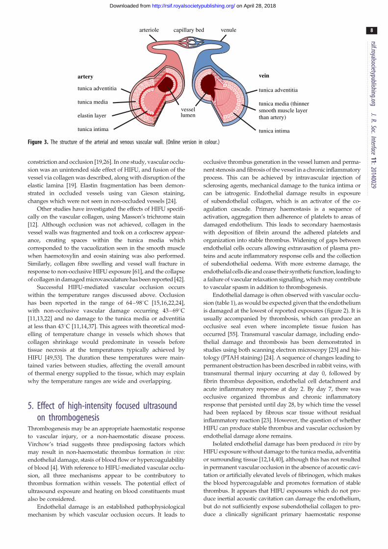
Arteries and veins have a common basic structure (figure 3),
comprising the tunica intima, an inner layer of endothelium
on a basement membrane. The tunica media is an intermediate
layer of smooth muscle, thicker in arteries than veins and
absent in capillaries. The tunica adventitia is an external
supportive layer and may be continuous with surrounding col-
lagenous connective tissue, absent in capillaries. Arteries also
contain elastin layers [48]. Capillaries, arterioles and venules
(d � 0.3 mm) comprise tissue microvasculature. Theoretical
modelling of vascular thermal damage suggests that the first
vessel changes are caused by thermal denaturation and shrink-
age of collagen fibres while vessel integrity is maintained by
elastic laminae, if present, and smooth muscle [49].
Tissue fusion is a common method of therapeutic vascular
occlusion. The vessel is mechanically compressed and occluded,
while energy is converted to heat at the point of occlusion, which
seals the vessel closed by tissue fusion. Tissue fusion requires
partial denaturation of collagen (and elastin if present) such
that the fractured structure is able to reform bonds outside the
laminar structure (bridging), effectively fusing the many layers
of the vessel wall together. Arteries and veins become more dif-
ficult to occlude by tissue fusion as their diameter, lumenal
blood flow or pulse pressure increases. Collagen is a prominent
feature of the tunica media and adventitia. Heating collagen to
above its denaturation threshold of 62–678C [50] unravels its
rope-like structure. Collagen undergoes isovolumetric shrinkage
of up to 60% of its length when heated above this threshold, the
majority of which occurs within 1 s [51]. Such vessel shrinkage
has been demonstrated ex vivo where the luminal cross-sectional
area of a non-perfused vessel was reduced by up to 96% [32,52],
although this effect is much less pronounced in perfused vessels
in vivo [38]. Heating of collagen beyond this threshold leads to
hyalinization and fracturing of its structure, which contributes
to tissue stiffness, and is a possible explanation for why vessels
subjected to multiple HIFU exposures stiffen and become
prone to rupture [18]. During the initial phase of heating (less
than 1 s), collagen is able to form bridging bonds to other par-
tially denatured collagen fibres and fuse tissue [53]. However,
the free ends of collagen must be physically adjacent to form
bridging bonds, usually requiring a form of external com-
pression. Capillaries, arterioles and venules have sufficiently
narrow lumens that a combination of collagen shrinkage and cel-
lular dehydration will result in vessel collapse and coagulation
with minimal attendant cell damage or haemorrhage [54], the
likely mechanism of HIFU-mediated microvascular destruction.
Vascular occlusion does not invariably result from heating
a vessel. Heating to temperatures above 1008C with laser or
diathermy, where the water in cells vaporizes and ruptures
cellular membranes, produces a weak coagulum of denatured
cells, collagen debris and carbonized tissue, unable to with-
stand physiological blood pressure [55], and can cause
vascular rupture owing to vessel wall necrosis and elastin
destruction [56]. A similar tissue vaporization is seen in
HIFU tissue emulsification, although tissue carbonization is
less likely to occur as desiccated tissue cannot propagate ultra-
sound, and further heating does not occur beyond this point.
Successful vascular occlusion by collagen fusion occurs in
temperature range 73–878C [57], while temperatures below
548C caused no structural changes in collagen [57].
The electrothermal bipolar vessel sealing system is
approved for clinical occlusion of arteries (d � 6 mm) and
veins (d � 12 mm) [58]. It uses mechanical compression com-
bined with electrical heating to cause collagen bridging
between vessel walls [59]. The resultant occlusion has a burst
pressure well in excess of physiological pressures [60]. The
system is computer controlled, monitoring electrical impe-
dance to allow careful control of the energy supplied and
prevent tissue under or overheating [59].
While there are no studies on HIFU-mediated vascular
occlusion that investigate the presence of tissue fusion, several
studies indirectly suggest that it occurs. Disruption of vascular
and perivascular collagen has been observed with vascular
occlusion using histology and electron microscopy [22,24,27].
The collagen changes can extend beyond the tunica media to
the tunica adventitia and adjacent connective tissue, and
shrinkage in these outer layers can result in lumenal
artery
tunica adventitia
tunica media
elastin layer
tunica intima
venulecapillary bed
vessellumen
arteriole
vein
tunica adventitia
tunica media (thinner smooth muscle layer than artery)
tunica intima
Figure 3. The structure of the arterial and venous vascular wall. (Online version in colour.)
rsif.royalsocietypublishing.orgJ.R.Soc.Interface
11:20140029
8
on April 28, 2018http://rsif.royalsocietypublishing.org/Downloaded from
constriction and occlusion [19,26]. In one study, vascular occlu-
sion was an unintended side effect of HIFU, and fusion of the
vessel via collagen was described, along with disruption of the
elastic lamina [19]. Elastin fragmentation has been demon-
strated in occluded vessels using van Gieson staining,
changes which were not seen in non-occluded vessels [24].
Other studies have investigated the effects of HIFU specifi-
cally on the vascular collagen, using Masson’s trichrome stain
[12]. Although occlusion was not achieved, collagen in the
vessel walls was fragmented and took on a corkscrew appear-
ance, creating spaces within the tunica media which
corresponded to the vacuolization seen in the smooth muscle
when haemotoxylin and eosin staining was also performed.
Similarly, collagen fibre swelling and vessel wall fracture in
response to non-occlusive HIFU exposure [61], and the collapse
of collagen in damaged microvasculature has been reported [42].
Successful HIFU-mediated vascular occlusion occurs
within the temperature ranges discussed above. Occlusion
has been reported in the range of 64–988C [15,16,22,24],
with non-occlusive vascular damage occurring 43–698C[11,13,22] and no damage to the tunica media or adventitia
at less than 438C [11,14,37]. This agrees with theoretical mod-
elling of temperature change in vessels which shows that
collagen shrinkage would predominate in vessels before
tissue necrosis at the temperatures typically achieved by
HIFU [49,53]. The duration these temperatures were main-
tained varies between studies, affecting the overall amount
of thermal energy supplied to the tissue, which may explain
why the temperature ranges are wide and overlapping.
5. Effect of high-intensity focused ultrasoundon thrombogenesis
Thrombogenesis may be an appropriate haemostatic response
to vascular injury, or a non-haemostatic disease process.
Virchow’s triad suggests three predisposing factors which
may result in non-haemostatic thrombus formation in vivo:
endothelial damage, stasis of blood flow or hypercoagulability
of blood [4]. With reference to HIFU-mediated vascular occlu-
sion, all three mechanisms appear to be contributory to
thrombus formation within vessels. The potential effect of
ultrasound exposure and heating on blood constituents must
also be considered.
Endothelial damage is an established pathophysiological
mechanism by which vascular occlusion occurs. It leads to
occlusive thrombus generation in the vessel lumen and perma-
nent stenosis and fibrosis of the vessel in a chronic inflammatory
process. This can be achieved by intravascular injection of
sclerosing agents, mechanical damage to the tunica intima or
can be iatrogenic. Endothelial damage results in exposure
of subendothelial collagen, which is an activator of the co-
agulation cascade. Primary haemostasis is a sequence of
activation, aggregation then adherence of platelets to areas of
damaged endothelium. This leads to secondary haemostasis
with deposition of fibrin around the adhered platelets and
organization into stable thrombus. Widening of gaps between
endothelial cells occurs allowing extravasation of plasma pro-
teins and acute inflammatory response cells and the collection
of subendothelial oedema. With more extreme damage, the
endothelial cells die and cease their synthetic function, leadingto
a failure of vascular relaxation signalling, which may contribute
to vascular spasm in addition to thrombogenesis.
Endothelial damage is often observed with vascular occlu-
sion (table 1), as would be expected given that the endothelium
is damaged at the lowest of reported exposures (figure 2). It is
usually accompanied by thrombosis, which can produce an
occlusive seal even where incomplete tissue fusion has
occurred [55]. Transmural vascular damage, including endo-
thelial damage and thrombosis has been demonstrated in
studies using both scanning electron microscopy [23] and his-
tology (PTAH staining) [24]. A sequence of changes leading to
permanent obstruction has been described in rabbit veins, with
transmural thermal injury occurring at day 0, followed by
fibrin thrombus deposition, endothelial cell detachment and
acute inflammatory response at day 2. By day 7, there was
occlusive organized thrombus and chronic inflammatory
response that persisted until day 28, by which time the vessel
had been replaced by fibrous scar tissue without residual
inflammatory reaction [23]. However, the question of whether
HIFU can produce stable thrombus and vascular occlusion by
endothelial damage alone remains.
Isolated endothelial damage has been produced in vivo by
HIFU exposure without damage to the tunica media, adventitia
or surrounding tissue [12,14,40], although this has not resulted
in permanent vascular occlusion in the absence of acoustic cavi-
tation or artificially elevated levels of fibrinogen, which makes
the blood hypercoagulable and promotes formation of stable
thrombus. It appears that HIFU exposures which do not pro-
duce inertial acoustic cavitation can damage the endothelium,
but do not sufficiently expose subendothelial collagen to pro-
duce a clinically significant primary haemostatic response

rsif.royalsocietypublishing.orgJ.R.Soc.Interface
11:20140029
9
on April 28, 2018http://rsif.royalsocietypublishing.org/Downloaded from
in vivo. This is supported by in vitro studies which show that
although platelet aggregation occurs with HIFU exposure,
adherence requires collagen substrate [62]. Where inertial cavi-
tation is promoted by UCA, this results in a higher proportion
of the endothelial surface area being damaged than at the
same energy level without microbubbles, and in the generation
of thrombus [40]. However, even with increased endothelial
damage, elevated circulating levels of fibrinogen were required
to produce occlusive thrombus [35].
Blood hypercoagulability has been described as the primary
cause of ‘acoustic haemostasis’ and has also been suggested
as a method of thrombogenesis [62,63]. This describes the
expression of adhesion molecules on the surface of platelets
(activation) in response to HIFU exposure and clumping (aggre-
gation) in the absence of triggers from endothelial damage,
occurring up to 50 s after initial HIFU exposure. It is known
that platelets can be activated in the absence of tissue damage
by shear stresses on their surface [64], and in the studies by
Poliachik, aggregation occurred in response to acoustic cavita-
tion. This is thought to be due to microstreaming causing
shear stresses on the surface of the platelets which are sufficient
to cause activation and aggregation but insufficient to cause dis-
ruption of the membrane [65]. However, adherence has not
been observed without an artificial collagenous substrate or
endothelial damage, meaning the resultant thrombus would
be non-adherent in vivo. Given the nature of the continuous
flow of the circulatory system, such aggregated platelets are
more likely to result in distant emboli than occlusive thrombus.
Heating platelets has not been directly studied with
reference to HIFU. It is known that platelets are optimally
activated at body temperature, 36–388C and are functionally
impaired at temperatures more than 458C, showing an
inability to activate or aggregate [66]. Hence it is unlikely
that platelets heated by HIFU have any role in vascular occlu-
sion, other than as cellular debris in organized thrombus.
Similarly, red blood cells haemolyse at more than 458C [67].
Slowing of blood flow has been observed to contribute to
HIFU-mediated vascular occlusion as unintended occlusion
occurred more commonly at points of vessel compression in
one study where the aim was to seal lacerated vessels [19].
Stasis of red blood cells is a known effect of low intensity
unfocused ultrasound (intensities � 12 W cm22), with separ-
ation of blood into bands of cellular aggregates and plasma
owing to the ultrasound standing waves [68], with normal
circulation restored after exposure [69]. To form a standing
wave, strong reflection of the plane of the wave is required,
which is unlikely to occur in vivo. To date, this effect has
not been observed in vessels exposed to HIFU. This remains
an area for further investigation in HIFU.
This evidence suggests that endothelial damage and phys-
iological primary haemostasis are the key features in thrombus
generation resulting from HIFU exposure of blood vessels.
However, there is minimal evidence that this mechanism
alone can produce permanent vascular occlusion. It is more
likely that thrombosis has a contributory role in maintaining
and organizing HIFU-mediated vascular occlusion.
6. Effect of high-intensity focused ultrasound onblood flow
Vascular spasm is a recognized response of vessels to injury.
The smooth muscle of the vessel contracts, as a physiological
method of reducing blood flow, improving the success of pri-
mary haemostasis and ultimately reducing blood loss [70]. It
is seen in vessels exposed to HIFU, both in the presence and
the absence of vascular damage, and can be monitored non-
invasively by colour Doppler measurements of peak systolic
velocity (PSV). Induction of vascular spasm by HIFU to pro-
duce temporary cessation of blood flow has been shown [15]
although the intensities used were above the threshold for
acoustic cavitation causing vascular rupture. Regardless, this
represents a means by which lumenal narrowing can occur.
Vascular spasm has been seen in vivo at HIFU intensities
which produce transmural vascular damage [15,25,28,34] but
also in exposures which do not produce vascular damage
[24,52] and is not reliant on the occurrence of tissue heating
[37]. Although the effect is described as transient, it persists
for sufficient time post HIFU exposure to allow repeat PSV
measurements taken by Doppler ultrasonography even in
the absence of evidence of vascular damage on histology [37].
Vascular spasm is likely augmented by the loss of
vascular relaxation in response to endothelial damage and
cessation of endothelial cell synthetic function. In a series of
ex vivo experiments in canine coronary arteries, vascular
relaxation was lost as a result of endothelial damage and
disruption of the nitric oxide signalling pathways, but mech-
anisms of relaxation remain intact in the vascular smooth
muscle and can be activated by the administration of exogen-
ous nitric oxide donors, isolating the cause as endothelial
damage [31].
The radiation force of high-intensity sound waves can
create localized streaming of liquids away from the focal
point [71], even overcoming the physiological pressure gradi-
ent normally controlling blood flow in vessels. This has been
observed in practice, where reversal of the arterial jet and
pulsatile flow was observed in lacerated femoral, axillary
and carotid arteries in pigs independent of mechanical com-
pression exerted by the transducer being pressed against the
vessel, which alone was insufficient to obstruct flow even tem-
porarily [17]. The lumens of arteries are maintained patent by
the pressure gradient of blood flow through them, with a
typical mean arterial pressure (MAP) in the range of
85–125 mmHg in humans. Arteries such as the femoral, axil-
lary and carotid artery will lose lumenal patency when the
MAP � 50 mmHg [72]. This suggests the possibility that
acoustic streaming could disrupt the local pressure gradient
in the artery sufficiently to cause it to collapse. Veins and the
microvasculature are much more easily collapsible and so
would also be expected to collapse by this mechanism.
A final hypothesized mechanical effect by which HIFU
could occlude blood flow is by temporary compression of
the vessel owing to its acoustic radiation force. When exposed
to HIFU, tissue cannot respond fast enough to the changes
between positive and negative pressures, meaning its
motion becomes out of phase with the acoustic wave and
energy is transferred to the tissue. This transfer of momentum
to the tissue, in the direction of wave propagation, results in
tissue displacement. The amount of pressure delivered will
vary with both the intensity of the HIFU source and the
attenuation of the overlying tissue, but will be magnified
by the small area over which it is delivered.
Although much speculated upon, acoustic radiation force
has not been observed to collapse blood vessels in vivo, how-
ever soft tissue deformation and displacement of 1–3 mm has
been observed in vivo [73]. Given the recognized difficulties

rsif.royalsocietypublishing.orgJ.R.Soc.Interface
11:20140029
10
on April 28, 2018http://rsif.royalsocietypublishing.org/Downloaded from
in making non-invasive in situ pressure measurements [52], it
is not unreasonable that in vivo effects of acoustic radiation
force have not been fully demonstrated. Synchronized sys-
tems to enable use of diagnostic Doppler ultrasound during
HIFU exposure have been proposed [74,75], but these
would only demonstrate ‘no flow’ in a vessel, not a definitive
cause, and B-mode ultrasound imaging would likely lack the
spatial resolution to determine reliable changes in diameter of
vessels. In an experimental system to study isolated blood
vessels in vitro, acoustic radiation force was sufficient to dis-
place vessels out of the field of view of the microscope
intended to monitor changes in vessel diameter [52].
Greater deformation and displacement of the proximal wall
of the blood vessel compared to the distal wall would probably
be expected, promoting collapse, as the blood within the vessel
is much less stiff than surrounding soft tissues. Even without
this difference, unidirectional pressure can occlude arterial
blood flow at normal pulse pressures (e.g. pressing on a
pulse point) and can be likened to the effect of momentum in
the direction of HIFU wave propagation. Blood vessels are
also maintained patent by the pulse pressure of blood flow,
as previously discussed, and if this pressure has been reduced
by acoustic streaming as observed experimentally [17], then
lower pressures would be needed to produce such collapse
or deformation. This is an area which would benefit from
further experimental evaluation as whether compression and
deformation of blood vessels by acoustic radiation force
occurs is of relevance to the design of HIFU systems.
7. Contribution of the inflammatory responseand repair mechanisms
There has been minimal work specifically on the contribution of
inflammatory responses to vascular occlusion. The harmonic
scalpel, an invasive system for producing vascular occlusion
which uses ultrasound to create vibrational forces in the
tissue, creates a more pronounced acute inflammatory response
than electrocautery, despite resulting in a smaller area of injured
tissue [59]. HIFU produces extensive oedema, disruption to
tissue, inflammatory cell infiltration and activation of the cyto-
kine cascade. Inertial cavitation induced damage produces a
comparable activation of the acute inflammatory response,
though this response does not convert into a chronic inflamma-
tory response in the absence of tissue damage [7]. Extracellular
oedema is known to reduce blood flow, especially in smaller
vessels, and perivascular oedema resulting from HIFU exposure
has been cited as contributing to lumenal constriction [32].
The role of the inflammatory response in controlling and
organizing the physiological response to vascular damage
cannot be discounted. Collateral circulation was seen to arise
quickly following vascular occlusion [20], so while a distribut-
ing artery may be blocked the area is not automatically
devascularized. Ultimately, the nature of the chronic inflamma-
tory response will determine whether the vessel becomes patent
again or remains permanently occluded and fibrosed [14,23].
8. An integrated mechanism of actionConsideration of the theoretical and experimental evidence of
the responses of vessels exposed to HIFU demonstrates that
there are many potential mechanisms through which the
physical properties of HIFU may interact with a living
system, and that contribute to vascular occlusion. Vascular
occlusion cannot be considered simply as a side effect of
vessel wall cellular destruction, and protocols for HIFU-
mediated vascular occlusion should be designed to maximize
this plethora of effects.
There are various ways in which the effects described above
could be combined but as time and temperature appear to be
important factors in distinguishing acute from more delayed
vascular occlusion, a multi-stage process can be envisioned
taking place along a typical heating–time curve of HIFU
(figure 4). Working out the exact timescale and thermal doses
to which these stages may correspond is a challenge for further
experimental work.
In the initial stage, we consider the effects taking place
before temperatures reach the 64–988C range where tissue
fusion occurs, which for HIFU could mean from less than
1 s up to approximately 20 s. These effects are temporary,
reversible or repairable, and alone have not been shown to
produce permanent vascular occlusion. This stage relies on
lumenal narrowing achieved through combination of the
mechanical effects of ultrasound (tissue displacement and
acoustic streaming), with tissue response such as vascular
spasm, cell shrinkage, endothelial damage and accumulation
of subendothelial oedema. This stage does not necessarily
represent lumenal collapse but features processes through
which vessel wall apposition could be produced.
In the next stage, temperatures will reach a range in which
tissue fusion can occur, and the energy deposition will be suffi-
cient to produce irreversible damage, with denaturation and
shrinkage of collagen and fracturing of elastic laminae. There
is also the persistent effect of vascular spasm, obstructed
blood flow and the increasing endothelial destruction leads to
a loss of vascular relaxation responses and activates platelets.
The final stage is dominated by tissue response that persists
beyond the end of the HIFU heating. The mechanical effects of
HIFU are removed and so perivascular oedema is the only
potential method of weak external compression. In this stage,
the strength of tissue fusion and/or thrombotic occlusion is
tested by the return of physiological blood pressure. The
vessel may be fused in a fully or partially occluded position,
or not at all. Activation of secondary haemostatic mechanisms
form occlusive, stable thrombus that can withstand the physio-
logical flow pressures involved. Progression from acute to
chronic inflammatory responses remodel the damaged vessel,
which ultimately determines whether permanent occlusion of
the vessel occurs over the following period, up to 28 days.
9. Potential application to fetal medicineThere are several diseases in fetal medicine where prenatal
occlusion of abnormal blood vessel(s) is either curative or con-
fers improvement in prognosis. Such treatments are invasive,
requiring entry to the intrauterine cavity and amniotic sac
and have inherent risks of preterm pre-labour rupture of mem-
branes (pPROM), preterm labour (PTL) and extreme
prematurity, chorioamonitis, chorio-amniotic membrane sep-
aration and maternal operative risks [76]. Hence, despite
identification through antenatal screening, the mainstay of
management for these conditions is expectant, monitoring for
severe fetal compromise, a tipping point where the associated
risks of invasive treatment become less than the potential

direction of blood flowreversed direction of blood flow
acoustic streaming effect
radiation force ofsound wave
compresses lumen
diameterof lumen
vascular spasm of tunica mediaresults in lumenal narrowing
endothelial damage leading to subendothelial oedema andinitiation of acute inflammatory response. There is insufficient
damage to activate platelets or lose vascular relaxation signalling
non-thermal mechanisms oflumenal narrowing
persistant vascular spasm and perivascular oedemamaintain lumenal narrowing
evolving acuteinflammatory
responseconversion tochronic inflammatory
reaction day 3–7
fibrin deposition and erythrocyte deposition result fromactivated platelet adherence to area of endothelialdamage produce an organized, occlusive thrombus
tunica adventitia (connective tissue, collagen)tunica media (smooth muscle, collagen)elastic laminatunica intima (endothelium, basement membrane)path of HIFU ultrasound wave propagation
organization of vascular damage
persistant effects— radiation force— acoustic streaming— vascular spasm elastin fragmentation and refusion
tissue fusion— denaturation of collagen— formation of bridging bonds— vacuolization of smooth muscle
collagen shrinkage
increased endothelial damagesufficient to activate plateletsand loss of vascular relaxation(loss of NOx synthesis)
augmentation of effectof vascular spasm and
lumenal narrowing
vessel heating and tissue fusion
absent blood flow absent blood flow
activated plateletsadhere to exposed
subendothelial tissue
fibrosis and obliteration of thevessel (permanent occlusion)
Figure 4. A suggested multi-stage integrated mechanism for HIFU-mediated vascular occlusion, presented as progression through time and temperature changes.
rsif.royalsocietypublishing.orgJ.R.Soc.Interface
11:20140029
11
on April 28, 2018http://rsif.royalsocietypublishing.org/Downloaded from
benefit. Using such therapies as ‘salvage methods’ reduces
their success rate. Development of non-invasive alternatives,
such as HIFU, could not only reduce the risks associated
with salvage therapies but may provide a therapeutic modality
for less severely compromised fetuses in utero, or even
represent a potential preventative treatment.
10. Twin-reversed arterial perfusion sequenceThis affects 1% of monochorionic twin pregnancies [77]. The
heart of one twin either fails to develop or is rudimentary,
and the heart of the healthy ‘pump’ twin perfuses both
bodies through placental arterio-arterial anastomoses or a

rsif.royalsocietypublishing.orgJ.R.Soc.Interface
11:20140029
12
on April 28, 2018http://rsif.royalsocietypublishing.org/Downloaded from
direct connection between the umbilical cords. This can result
in cardiac failure and death in the healthy twin, with an
untreated mortality rate of 55% [77] and spontaneous resol-
ution in less than or equal to 21% of cases [78]. The aim of
treatment is to divide the circulations by umbilical cord occlu-
sion of the acardiac twin. Bipolar diathermy, fetoscopic laser
and RFA have all been used. Bipolar diathermy cord
occlusion has been superseded by minimally invasive tech-
niques, as the risks of pPROM and PTL were elevated
(27%, 22%, respectively, survival 85%) [76]. Fetoscopic laser
reports a 80% survival with pPROM in 18% and PTL in 3%
within 28 days of procedure [79]; RFA reports a 70–92% sur-
vival rate [76,80] with a pPROM and preterm labour rate of
14% and 7%, respectively [76]. Hence, minimally invasive
techniques reduce but do not remove the risks, without chan-
ging survival rates. Although TRAP may be diagnosed from
11 to 13 weeks gestation, treatment is delayed until after 16
weeks gestation to allow time for fusion of the amnion and
chorion, by which time intrauterine death of the pump
twin occurs in up to one-third of cases [78]. Non-invasive
HIFU has been used to successfully produce umbilical cord
occlusion from 13 weeks gestation [30] in one of the two
reported cases without complications [29,30]. Although the
authors report technical challenges related to achieving
sufficient focal depth, HIFU is possible therapy for TRAP.
11. Twin-to-twin transfusion syndromeTTTS affects 9–15% of monochorionic diamniotic twins
[81,82], or 1 : 2500 livebirths, and is the most important
cause of death and handicap in monochorionic twins [83].
It results from unequal sharing of blood supply between
twins owing to placental vascular anastomoses, present in
95% monochorionic pregnancies [84,85]. Anastomoses allow
‘vascular steal’ or imbalance between circulations, a relative
over and under-perfusion of the recipient and donor twin,
respectively [86]. There is grading of disease progression
from mild to severe (Quintero stages 1–4) [87] and while
mild disease may not require treatment, severe disease left
untreated has approaching 90% mortality [88]. In severe
cases, the gold standard treatment is fetoscopic laser ablation
of vascular anastomoses [89], either by selective ablation as
they cross between the two ‘halves’ of the placenta, or by
ablating the entire equator between the two halves of the
placenta to create a functionally dichorionic placenta [90].
Success rates are quoted at single twin survival of 76–87%
and double twin survival of 50–60% with a 5–25% double
fetal loss rate [91] and an 11% rate of neurological impair-
ment in survivors [92]. Fetal losses are attributed to either
the complications of the procedure or recurrent disease,
which occurs in up to 16% of cases and is associated with
lower rates of survival and neurologically intact infants
[93]. Five per cent to 30% of anastomoses can remain post
ablation [94,95] and are recognized as causing recurrent
disease [96] or a related condition, twin anaemia polycythae-
mia syndrome (TAPS). Iatrogenic TAPS is more common after
ablation than spontaneously (13 versus 5%) [81,97]. HIFU
may be an alternative to invasive therapy in TTTS. The
energy levels needed to ablate placental vasculature [98]
and the focal depths required are within the HIFU range of
operation, and the reduction in complication rate may
allow treatment of less severe disease. Either selective
vascular ablation or equatorial division of the placenta
could be performed, as points or confluent planes of abla-
tion can be achieved with HIFU [20]. HIFU may reduce
rates of recurrent TTTS and TAPS, as residual anastomoses
would be identified by Doppler ultrasound [99] integrated
into the HIFU system.
12. Bronchopulmonary sequestrationBPS is rare, characterized by a non-functional lung mass with
an abnormal systemic arterial rather than pulmonary blood
supply, typically diagnosed in the second trimester and
reaching peak size by 28–30 weeks gestation. Masses are
variable: some spontaneously regress, while others increase
in size, causing pulmonary hypoplasia, mediastinal shift, car-
diac failure, pleural effusions and hydrops [100]. The
presence of pleural effusion and hydrops are poor prognostic
features with untreated perinatal mortality of 95% [101]. Var-
ious therapies are used, and the more successful focus on
ablation of the abnormal blood supply.
The sequestration can be removed by open fetal surgery,
with 54% survival [102]. It carries additional maternal surgi-
cal risks and creates a uterine scar. Doppler ultrasound
guided fetoscopic laser vessel ablation is a minimally inva-
sive alternative, with survival rates in excess of 87.5%
[101,103]; sclerosants injected into the aberrant vessel have
shown high success rates, but trials have small numbers
[104,105]. Complication rates are not quoted. Thoraco-amniotic
shunting to drain pleural effusions is complicated by shunt
blockage [106] and chest wall deformity in 77% cases [107].
Again, the energy levels, focal depth and suitable targeting
method for BPS vasculature are well within the scope of a
HIFU system integrated with Doppler ultrasound. In one
animal model, the targeting of intra-thoracic structures led
to skin burns in the fetus owing to reflection of the ultrasonic
energy by the calcified rib cage [43], however in our experi-
ence it is possible to identify the aberrant vasculature using
a sagittal view which would avoid transmission of ultrasonic
energy through the rib cage.
13. Sacrococcygeal teratomaSCT is the most common neoplasm in the fetus, occurring in
1 : 22 000 livebirths [108]. SCT is typically benign, though
requiring removal postnatally to avoid malignant transform-
ation [109]. They are attached into the coccyx and may
extend into the pelvis and abdomen. They are identified and
monitored prenatally by ultrasound and Doppler can assess
vascularity. The antenatal course is usually uncomplicated,
but highly vascular or rapidly growing tumours can cause
high output cardiac failure, anaemia and hydrops. These are
poor prognostic features [110]. Without cardiac compromise
or hydrops, 88% survival is quoted [111]. Perinatal mortality
results from tumour avulsion, rupture or haemorrhage at
delivery [112,113]. With cardiac compromise or hydrops mor-
tality approaches 100% [111]. Reducing tumour vascularity can
slow its growth and the impact of ‘vascular steal’ effect. Open
fetal surgery and resection of the teratoma is possible [114]
though it has a higher risk of haemorrhage than other fetal sur-
gery [115]. Minimally invasive devascularization of the
tumour via percutaneous laser ablation [116–118] is possible
although only superficial vessels can be occluded as the

rsif.royalsocietypublishing.orgJ.R.Soc.Interface
11:20140029
13
on April 28, 2018http://rsif.royalsocietypublishing.org/Downloaded from
maximum penetration of laser is 2–3 mm. Both methods report
mixed results. RFA has a 50% survival rate with significant
morbidity reported owing to spread of thermal energy into
adjacent soft tissues [119,120]. Alcohol sclerosis has been
attempted but specific results are not reported [116].
While it is unlikely that the entire tumour could be ablated
by HIFU exposure to the soft tissue, especially as larger tumours
are more likely to require treatment, it has been demonstrated
that destruction of tumour vasculature increases the volume
of tissue destroyed for the amount of energy supplied
[121,122]. The vasculature of the SCT could be pre-mapped by
colour Doppler ultrasound or MR angiography and used to
target HIFU. Ultrasound energy is recognized to have a lower
‘thermal spread’ than other applications [59] so may produce
less thermal injury in adjacent soft tissues. Reduction in vascu-
larity may also reduce risks associated with haemorrhage at
time of delivery. Animal studies have demonstrated that
HIFU lesions in the fetal pelvis can be made despite the restric-
tions of the bony pelvis on the acoustic window [123], and that
survival is compatible with the generation of necrotic tissue
within the fetus [43,44]. HIFU represents a potential non-
invasive therapy for SCT in the antenatal period, especially of
fetuses with significant compromise in utero.
14. Physics and engineering requirementsHIFU treatment comprises a number of components—target-
ing, energy delivery and monitoring of its effects. Targeting is
most usually achieved by either ultrasound or magnetic res-
onance imaging (MRI). Therapy ultrasound beams are
usually generated from a transducer that is focused using
either a bowl configuration, a plane transducer/lens combi-
nation or a multi-element array with which focusing is
achieved by appropriate application of phase and amplitude
to each element. Where ultrasound is used to monitor HIFU
ablation of soft tissue, a successful treatment is assessed in
terms of the appearance of a bright echo, whereas with
MRI thermometry sequences are used to calculate thermal
dose [124]. If the spatial precision of HIFU damage is to be
harnessed to its fullest extent, it is important to account for
tissue motion when placing the lesions. This presents a
major challenge to successful targeting and treatment deliv-
ery and may be achieved, for example, by synchronizing
the HIFU exposure with cardiac or respiratory motion, and
specifically for fetal medicine, accounting for movement of
the fetus and umbilical cord within the amniotic sac.
In obstetrics, ultrasound image guidance is likely to be the
method of choice for reasons of safety, clinical and patient fam-
iliarity, targeting efficacy and real-time responsiveness to fetal
movement. To successfully occlude a selected blood vessel it
needs to be identified clearly for targeting, currently best done
using Doppler ultrasound techniques. Vessels of more than
1 mm diameter are routinely identified in clinical practice by
Doppler ultrasound [125]. The main disadvantage of diagnostic
ultrasound is that, unlike MRI, it does not permit the measure-
ment of absolute temperature rise during exposure, both
owing to interference of the therapy beam with the diagnostic
information and to a lack of consistent sound speed dependence
on temperature. Should accurate thermal dosimetry become a
key factor then MRI may be more suitable.
The choice of appropriate HIFU transducer geometry and
exposure frequency depends on the clinical application and,
more specifically, the depth of vessel to be occluded. In choos-
ing the frequency to be used, a trade-off is necessary between
the amount of energy absorbed within the target volume
(which increases with frequency) and the ability to get suffi-
cient energy to the target depth (which decreases with
increasing frequency owing to attenuation by the overlying
tissues). It has been shown that a ‘rule of thumb’ for determin-
ing the optimum frequency is to allow the total attenuation in
the overlying tissue to be around 10 dB [126]. For typical soft
tissues with an attenuation of 0.7 dB cm21 MHz21, this
would imply an optimum frequency of around 2.6 MHz for a
target depth of 5 cm and 1.4 MHz for 10 cm. It should also
be remembered that the focal region is smaller at higher fre-
quencies, if all other source geometries remain the same. The
inclusion of a central aperture in the HIFU transducer design
allows an US imaging probe to be inserted into the treatment
head, thus ensuring a fixed geometry between targeting and
treatment components, most efficiently with coincident acous-
tic axes. This also facilitates the monitoring of a treatment
immediately after cessation of the HIFU exposure.
15. Undesirable effects of high-intensity focusedultrasound applied to tissue and vessels
The main specific complication of vessel exposure to HIFU is
rupture and haemorrhage. Exposure to higher intensities in
the experimental range has been associated with rupture of
vessels, potentially owing to acoustic cavitation [15,20,21] as
has multiple exposures of the vessel wall which cause overheat-
ing and tissue stiffening by a cumulative effect [19,34,38]. This
effectively represents an upper exposure limit above which the
application of HIFU becomes counterproductive. Therefore,
HIFU appears to differ from several other occlusive techniques,
particularly laser and electrocautery, which rely on multiple
exposures of blood vessels to ensure permanent and stable
occlusion. It is possible that it would be unsafe to use HIFU
in this manner, although varying the exposure site in larger
vessels may mitigate this effect.
Paradoxically, the non-invasive nature of HIFU also rep-
resents a risk of complications. Invasive methods have
greater control over where the energy (thermal and mechan-
ical) they supply is delivered as they are visually targeted
and controlled. Excessive soft tissue destruction or thermal
spread to adjacent structures may cause unintended damage
and therefore complications. This is of particular significance
where the targets are small, or hard to locate, and the margin
of error is limited. Injuries to bowel [15,44], nerves [20] or adja-
cent vessels [14] have been reported in fetal and vascular HIFU
experiments, with or without associated vascular occlusion.
Mistargeting is also an issue as not only does it fail to occlude
the target vessels, but also repeat exposure is likely to be pre-
cluded until such time as tissue healing has been allowed to
occur. However, accuracy of vascular targeting has been
shown to be improved with the use of colour flow Doppler
ultrasound over even surgical exposure and visual targeting
in the application of HIFU [18].
If an inappropriate acoustic window is used, or there are
elements in the acoustic window which scatter or reflect,
rather than transmitting, the ultrasonic energy, heating to
unpredictable locations may occur, causing burns, typically
on the skin because this has a higher absorption coefficient
compared with most other soft tissues. In some animal

rsif.royalsoc
14
on April 28, 2018http://rsif.royalsocietypublishing.org/Downloaded from
experiments, skin burns were also caused by poor acoustic
coupling to the animal’s skin owing to trapped air pockets in
the animal’s fur. The incidence of this is reduced with more
extensive shaving of animal models and application of depila-
tory creams, and should not be a significant issue in humans.
ietypublishing.orgJ.R.Soc.In
16. ConclusionWe present an integrated multi-stage mechanism through
which HIFU-mediated vascular occlusion could occur. If
the integrated technique described here is to be implemented,
it is important to optimize treatment delivery, and more
detailed experimental research must be carried out to validate
it and establish the exposure parameters that will provide the
most efficient vascular occlusion for the clinical applications
being considered. This will involve optimization of transdu-
cer design as well as exposure regimes. We propose that
should this optimization occur, HIFU has the potential to
be adapted to provide a non-invasive therapy for conditions
in fetal medicine where vascular occlusion is required.
Funding statement. The authors acknowledge funding from Action Medi-cal Research grant no. GN2052, the Isaac Newton Trust and the GenesisResearch Trust. C.C.L. is supported by the National Institute for HealthResearch (NIHR) Biomedical Research Centre based at Imperial Col-lege Healthcare NHS Trust and Imperial College London. The viewsexpressed are those of the author(s) and not necessarily those of theNHS, the NIHR or the Department of Health. G.R.tH. and I.H.R. arefunded by the Engineering and Physical Sciences Research Council(EPSRC) grant no. EP/F0-25750.
terface11:2
References0140029
1. Cline HE, Schenck JF, Hynynen K, Watkins RD, SouzaSP, Jolesz FA. 1992 MR-guided focused ultrasoundsurgery. J. Comput. Assist. Tomogr. 16, 956 – 965.(doi:10.1097/00004728-199211000-00024)
2. Clarke RL, ter Haar GR. 1997 Temperature riserecorded during lesion formation by high-intensityfocused ultrasound. Ultrasound Med. Biol. 23,299 – 306. (doi:10.1016/S0301-5629(96)00198-6)
3. Shi X, Martin RW, Rouseff D, Vaezy S, Crum LA.1999 Detection of high-intensity focused ultrasoundliver lesions using dynamic elastometry. Ultrason.Imaging 21, 107 – 126. (doi:10.1177/016173469902100203)
4. Underwood JCEBR. 2004 General and systematicpathology, 4th edn. New York, NY: ChurchillLivingstone.
5. ter Haar GR, Robertson D. 1993 Tissue destructionwith focused ultrasound in vivo. Eur. Urol. 23(Suppl. 1), 8 – 11.
6. Susani M, Madersbacher S, Kratzik C, Vingers L,Marberger M. 1993 Morphology of tissuedestruction induced by focused ultrasound. Eur.Urol. 23(Suppl. 1), 34 – 38.
7. Burks SR, Ziadloo A, Hancock HA, Chaudhry A, DeanDD, Lewis BK, Frenkel V, Frank JA. 2011Investigation of cellular and molecular responses topulsed focused ultrasound in a mouse model. PLoSONE 6, e24730. (doi:10.1371/journal.pone.0024730)
8. Fry FJ, Kossoff G, Eggleton RC, Dunn F. 1970Threshold ultrasonic dosages for structural changesin the mammalian brain. J. Acoust. Soc. Am.48(Suppl. 2), 1413. (doi:10.1121/1.1912301)
9. Winterroth F, Xu Z, Wang TY, Wilkinson JE, FowlkesJB, Roberts WW, Cain CA. 2011 Examining andanalyzing subcellular morphology of renal tissuetreated by histotripsy. Ultrasound Med. Biol. 37,78 – 86. (doi:10.1016/j.ultrasmedbio.2010.10.002)
10. Lynn JG, Zwemer RL, Chick AJ, Miller AE. 1942A new method for the generation and use offocused ultrasound in experimental biology. J. Gen.Physiol. 26, 179 – 193. (doi:10.1085/jgp.26.2.179)
11. Fallon JT, Stehbens WE, Eggleton RC. 1972 Effect ofultrasound on arteries. Arch. Pathol. 94, 380 – 388.
12. Schultz-Haakh H, Li JK, Welkowitz W, Rosenberg N.1989 Ultrasonic treatment of varicose veins.Angiology 40, 129 – 137. (doi:10.1177/000331978904000208)
13. Yang R et al. 1992 Feasibility of using high intensityfocused ultrasound for treatment of unresectableretroperitoneal malignancies. J. Ultrasound Med.11, 1.
14. Delon-Martin C, Vogt C, Chignier E, Guers C,Chapelon JY, Cathignol D. 1995 Venous thrombosisgeneration by means of high-intensity focusedultrasound. Ultrasound Med. Biol. 21, 113 – 119.(doi:10.1016/0301-5629(94)00095-6)
15. Hynynen K, Colucci V, Chung A, Jolesz F. 1996Noninvasive arterial occlusion using MRI-guidedfocused ultrasound. Ultrasound Med. Biol. 22,1071 – 1077. (doi:10.1016/S0301-5629(96)00143-3)
16. Vaezy S et al. 1997 Liver hemostasis using high-intensity focused ultrasound. Ultrasound Med. Biol.23, 1413 – 1420. (doi:10.1016/S0301-5629(97)00143-9)
17. Vaezy S et al. 1998 Hemostasis of punctured bloodvessels using high-intensity focused ultrasound.Ultrasound Med. Biol. 24, 903 – 910. (doi:10.1016/S0301-5629(98)00050-7)
18. Martin RW, Vaezy S, Kaczkowski P, Keilman G,Carter S, Caps M, Beach K, Plett M, Crum L. 1999Hemostasis of punctured vessels using Doppler-guided high-intensity ultrasound. Ultrasound Med.Biol. 25, 985 – 990. (doi:10.1016/S0301-5629(99)00027-7)
19. Vaezy S et al. 1999 Use of high-intensity focusedultrasound to control bleeding. J. Vasc. Surg. 29,533 – 542. (doi:10.1016/S0741-5214(99)70282-X)
20. Rivens IH, Rowland IJ, Denbow M, Fisk NM, ter HaarGR, Leach MO. 1999 Vascular occlusion usingfocused ultrasound surgery for use in fetalmedicine. Eur. J. Ultrasound 9, 89 – 97. (doi:10.1016/S0929-8266(99)00008-7)
21. Denbow ML, Rivens IH, Rowland IJ, Leach MO, FiskNM, ter Haar GR. 2000 Preclinical development ofnoninvasive vascular occlusion with focusedultrasonic surgery for fetal therapy. Am. J. Obstet.
Gynecol. 182, 387 – 392. (doi:10.1016/S0002-9378(00)70229-8)
22. Fujiwara R, Sasaki K, Ishikawa T, Suzuki M,Umemura S-I, Kushima M, Okai T. 2002 Arterialblood flow occlusion by high intensity focusedultrasound and histologic evaluation of its effect onarteries and surrounding tissues. J. Med. Ultrasonics29, 85 – 90. (doi:10.1007/BF02481229)
23. Hwang JH, Vaezy S, Martin RW, Cho MY, Noble ML,Crum LA, Kimmey MB. 2003 High-intensity focusedUS: a potential new treatment for GI bleeding.Gastrointest. Endosc. 58, 111 – 115. (doi:10.1067/mge.2003.322)
24. Ishikawa T, Okai T, Sasaki K, Umemura S, FujiwaraR, Kushima M, Ichihara M, Ichizuka K. 2003Functional and histological changes in rat femoralarteries by HIFU exposure. Ultrasound Med. Biol. 29,1471 – 1477. (doi:10.1016/S0301-5629(03)00951-7)
25. Nizard J, Pessel M, De Keersmaecker B, Barbet JP,Ville Y. 2004 High-intensity focused ultrasound inthe treatment of postpartum hemorrhage: ananimal model. Ultrasound Obstet. Gynecol. 23,262 – 266. (doi:10.1002/uog.1007)
26. Zderic V, Keshavarzi A, Noble ML, Paun M, ShararSR, Crum LA, Martin RW, Vaezy S. 2006Hemorrhage control in arteries using high-intensityfocused ultrasound: a survival study. Ultrasonics 44,46 – 53. (doi:10.1016/j.ultras.2005.08.002)
27. Ichizuka K et al. 2007 Application of high-intensityfocused ultrasound for umbilical artery occlusion ina rabbit model. Ultrasound Obstet. Gynecol. 30,47 – 51. (doi:10.1002/uog.4008)
28. Ichihara M, Sasaki K, Umemura S, Kushima M, OkaiT. 2007 Blood flow occlusion via ultrasound image-guided high-intensity focused ultrasound and itseffect on tissue perfusion. Ultrasound Med. Biol. 33,452 – 459. (doi:10.1016/j.ultrasmedbio.2006.08.016)
29. Ichizuka K, Hasegawa J, Nakamura M, Matsuoka R,Sekizawa A, Okai T, Umemura S. 2012 High-intensity focused ultrasound treatment for twinreversed arterial perfusion sequence. UltrasoundObstet. Gynecol. 40, 476 – 478. (doi:10.1002/uog.11114)

rsif.royalsocietypublishing.orgJ.R.Soc.Interface
11:20140029
15
on April 28, 2018http://rsif.royalsocietypublishing.org/Downloaded from
30. Okai T, Ichizuka K, Hasegawa J, Matsuoka R,Nakamura M, Shimodaira K, Sekizawa A, KushimaM, Umemura S. 2013 First successful case of non-invasive in-utero treatment of twin reversed arterialperfusion sequence by high-intensity focusedultrasound. Ultrasound Obstet. Gynecol. 42,112 – 114. (doi:10.1002/uog.12466)
31. Discigil B, King RM, Pearson PJ, Capellini VK,Rodrigues AJ, Schaff HV, Evora PRB. 2008 High-frequency ultrasonic waves cause endothelialdysfunction on canine epicardial coronary arteries.Rev. Bras. Cir. Cardiovasc. 23, 190 – 196. (doi:10.1590/S0102-76382008000200007)
32. Henderson PW, Lewis GK, Shaikh N, Sohn A,Weinstein AL, Olbricht WL, Spector JA. 2010 Aportable high-intensity focused ultrasounddevice for noninvasive venous ablation.J. Vasc. Surg. 51, 707 – 711. (doi:10.1016/j.jvs.2009.10.049)
33. Jiao J, Wu F, Zou J, Li F, Liu F, Zhao X, Wang Q.2013 Effect of ablations by pulsed versus continuoushigh-intensity focused ultrasound on isolatedperfused porcine liver. Nan Fang Yi Ke Da Xue XueBao. 33, 230 – 234.
34. Mahoney K, Martin H, Hynynen K. 2000 Focusedultrasound effects on blood vessels in vivo-limits forvascular interventions. In Ultrasonics Symposium,2000 IEEE, 22 Oct 2000 – 25 Oct 2000, San Juan,Puerto Rico, vol. 2, pp. 1405 – 1408. (doi:10.1109/ULTSYM.2000.921585)
35. Hwang JH, Zhou Y, Warren C, Brayman AA, Crum LA.2010 Targeted venous occlusion using pulsedhigh-intensity focused ultrasound. IEEE Trans.Biomed. Eng. 57, 37 – 40. (doi:10.1109/TBME.2009.2029865)
36. Yue Y, Chen W, Wang Z. 2010 The impact ofmicrobubbles-mediated intermitten HIFU on bloodflow in femoral artery of rabbit. Sheng Wu Yi XueGong Cheng Xue Za Zhi. 27, 58 – 61.
37. Yang FY, Chiu WH, Liu SH, Lin GL, Ho FM. 2009Functional changes in arteries induced by pulsedhigh-intensity focused ultrasound. IEEE Trans.Ultrason. Ferroelectr. Freq. Control. 56, 2643 – 2649.(doi:10.1109/TUFFC.2009.1355)
38. Hynynen K, Chung AH, Colucci V, Jolesz FA. 1996Potential adverse effects of high-intensity focusedultrasound exposure on blood vessels in vivo.Ultrasound Med. Biol. 22, 193 – 201. (doi:10.1016/0301-5629(95)02044-6)
39. Koruth JS et al. 2011 Safety and efficacy of high-intensity focused ultrasound atop coronary arteriesduring epicardial catheter ablation. J. Cardiovasc.Electrophysiol. 22, 1274 – 1280. (doi:10.1111/j.1540-8167.2011.02084.x)
40. Hwang JH, Tu J, Brayman AA, Matula TJ, Crum LA.2006 Correlation between inertial cavitation doseand endothelial cell damage in vivo. UltrasoundMed. Biol. 32, 1611 – 1619. (doi:10.1016/j.ultrasmedbio.2006.07.016)
41. Yang R et al. 1991 High-intensity focused ultrasoundin the treatment of experimental liver cancer. ArchSurg. 126, 1002 – 1009 (discussion 9 – 10). (doi:10.1001/archsurg.1991.01410320088012)
42. Wu F, Chen W-Z, Bai J, Zou J-Z, Wang Z-L, Zhu H,Wang ZB. 2001 Pathological changes in humanmalignant carcinoma treated with high-intensityfocused ultrasound. Ultrsound. Med. Biol. 27,1099 – 1106. (doi:10.1016/S0301-5629(01)00389-1)
43. Paek BW et al. 2003 Tissue ablation using high-intensity focused ultrasound in the fetal sheepmodel: potential for fetal treatment. Am. J. Obstet.Gynecol. 189, 702 – 705. (doi:10.1067/S0002-9378(03)00664-1)
44. Kim Y et al. 2011 Non-invasive pulsed cavitationalultrasound for fetal tissue ablation: feasibility studyin a fetal sheep model. Ultrasound Obstet. Gynecol.37, 450 – 457. (doi:10.1002/uog.8880)
45. Owens GE, Miller RM, Owens ST, Swanson SD, IvesK, Ensing G, Gordon D, Xu Z. 2012 Intermediate-term effects of intracardiac communications creatednoninvasively by therapeutic ultrasound (histotripsy)in a porcine model. Pediatr. Cardiol. 33, 83 – 89.(doi:10.1007/s00246-011-0094-6)
46. Hancock HA, Smith LH, Cuesta J, Durrani AK,Angstadt M, Palmeri ML, Kimmel E, Frenkel V. 2009Investigations into pulsed high-intensity focusedultrasound-enhanced delivery: preliminary evidencefor a novel mechanism. Ultrasound Med. Biol. 35,1722 – 1736. (doi:10.1016/j.ultrasmedbio.2009.04.020)
47. Lele PP. 1967 Production of deep focal lesions byfocused ultrasound—current status. Ultrasonics 5,105 – 112. (doi:10.1016/S0041-624X(67)80011-8)
48. Burkitt HGYBHJWWPR. 1993 Wheater’s functionalhistology: a text and colour atlas. Edinburgh, UK:Churchill Livingstone.
49. Agah R, Pearce JA, Welch AJ, Motamedi M. 1994Rate process model for arterial tissue thermaldamage: implications on vessel photocoagulation.Lasers Surg. Med. 15, 176 – 184. (doi:10.1002/lsm.1900150205)
50. Florkin MSEH. 1971 Comprehensive biochemistry.26C: extracellular and supporting structures.Amsterdam: Elsevier.
51. Chen SS, Wright NT, Humphrey JD. 1997 Heat-induced changes in the mechanics of a collagenoustissue: isothermal free shrinkage. J. Biomech. Engg.119, 372 – 378. (doi:10.1115/1.2798281)
52. Tokarczyk A, Rivens I, van Bavel E, Symonds-TaylerR, ter Haar G. 2013 An experimental system for thestudy of ultrasound exposure of isolated blood.Phys. Med. Biol. 58, 2281 – 2304. (doi:10.1088/0031-9155/58/7/2281)
53. Moros EG. 2013 Physics of thermal therapy:fundamentals and clinical applications. Boca Raton,FL: Taylor & Francis.
54. DeLia JE, Rogers JG, Dixon JA. 1985 Treatment ofplacental vasculature with a neodymium –yttrium – aluminum – garnet laser via fetoscopy.Am. J. Obstet. Gynecol. 151, 1126 – 1127. (doi:10.1016/0002-9378(85)90395-3)
55. White RA, Kopchok G, Peng SK, Fujitani R, White G,Klein S, Uitto J. 1987 Laser vascular welding—howdoes it work? Ann. Vasc. Surg. 1, 461 – 464.
56. Banga I. 1966 Structure and function of elastin andcollagen. Budapest: Akademiai Kiado.
57. Martinot VL, Mordon SR, Mitchell VA, Pellerin PN,Brunetaud JM. 1994 Determination of efficientparameters for argon laser-assisted anastomoses inrats: macroscopic, thermal, and histologicalevaluation. Lasers Surg. Med. 15, 168 – 175. (doi:10.1002/lsm.1900150204)
58. Landman J, Kerbl K, Rehman J, Andreoni C, HumphreyPA, Collyer W, Olweny E, Sundaram C, Clayman RV.2003 Evaluation of a vessel sealing system, bipolarelectrosurgery, harmonic scalpel, titanium clips,endoscopic gastrointestinal anastomosis vascularstaples and sutures for arterial and venous ligation in aporcine model. J. Urol. 169, 697 – 700. (doi:10.1016/S0022-5347(05) 63995-X)
59. Manouras A, Markogiannakis HE, Kekis PB,Lagoudianakis EE, Fleming B. 2008 Novelhemostatic devices in thyroid surgery:electrothermal bipolar vessel sealing system andharmonic scalpel. Expert Rev. Med. Devices 5,447 – 466. (doi:10.1586/17434440.5.4.447)
60. Harold KL, Pollinger H, Matthews BD, Kercher KW,Sing RF, Heniford BT. 2003 Comparison of ultrasonicenergy, bipolar thermal energy, and vascular clipsfor the hemostasis of small-, medium-, and large-sized arteries. Surg. Endosc. 17, 1228 – 1230.(doi:10.1007/s00464-002-8833-7)
61. Jiang F, He M, Liu YJ, Wang ZB, Zhang L, Bai J.2013 High intensity focused ultrasound ablation ofgoat liver in vivo: pathologic changes of portal veinand the ‘heat-sink’ effect. Ultrasonics 53, 77 – 83.(doi:10.1016/j.ultras.2012.04.001)
62. Poliachik SL, Chandler WL, Mourad PD, Ollos RJ,Crum LA. 2001 Activation, aggregation andadhesion of platelets exposed to high-intensityfocused ultrasound. Ultrasound Med. Biol. 27,1567 – 1576. (doi:10.1016/S0301-5629(01)00444-6)
63. Poliachik SL, Chandler WL, Ollos RJ, Bailey MR, CrumLA. 2004 The relation between cavitation and plateletaggregation during exposure to high-intensityfocused ultrasound. Ultrasound Med. Biol. 30,261 – 269. (doi:10.1016/j.ultrasmedbio.2003.10.012)
64. Sakariassen KS, Holme PA, Orvim U, Barstad RM,Solum NO, Brosstad FR. 1998 Shear-induced plateletactivation and platelet microparticle formation innative human blood. Thromb. Res. 92, S33 – S41.(doi:10.1016/S0049-3848(98)00158-3)
65. Williams AR, O’Brien Jr WD, Coller BS. 1976Exposure to ultrasound decreases the recalcificationtime of platelet rich plasma. Ultrasound Med. Biol.2, 113 – 118. (doi:10.1016/0301-5629(76)90019-3)
66. Rao GH, Smith 2nd CM, Escolar G, White JG. 1993Influence of heat on platelet biochemistry, structure,and function. J. Lab. Clin. Med. 122, 455 – 464.
67. Gershfeld NL, Murayama M. 1988 Thermalinstability of red blood cell membrane bilayers:temperature dependence of hemolysis. J. Membr.Biol. 101, 67 – 72. (doi:10.1007/BF01872821)
68. Dyson M, Pond JB, Woodward B, Broadbent J. 1974The production of blood cell stasis and endothelialdamage in the blood vessels of chick embryostreated with ultrasound in a stationary wave field.Ultrasound Med. Biol. 1, 133 – 148. (doi:10.1016/0301-5629(74)90003-9)
rsif.royalsocietypublishing.orgJ.R.Soc.Interface
11:20140029
16
on April 28, 2018http://rsif.royalsocietypublishing.org/Downloaded from
69. ter Haar G, Dyson M, Smith SP. 1979 Ultrastructuralchanges in the mouse uterus brought about byultrasonic irradiation at therapeutic intensities instanding wave fields. Ultrasound Med. Biol. 5,167 – 179. (doi:10.1016/0301-5629(79)90085-1)
70. Quick AJ. 1969 Hemostasis in surgical procedures.Surg. Gynecol. Obstet. 128, 523 – 532.
71. Nyborg WLM, Warren PM. 1965 11—Acousticstreaming. Properties of polymers and nonlinearacoustics, vol. 2, pp. 265 – 331. New York, NY:Academic Press.
72. American College of Surgeons CoT. 2008 ATLS,advanced trauma life support for doctors. Chicago,IL: American College of Surgeons.
73. Laughner JI, Sulkin MS, Wu Z, Deng CX, Efimov IR.2012 Three potential mechanisms for failure of highintensity focused ultrasound ablation in cardiactissue. Circ. Arrhythmia Electrophysiol. 5, 409 – 416.(doi:10.1161/CIRCEP.111.967216)
74. Chen WS et al. 2011 Single-element ultrasoundtransducer for combined vessel localization andablation. IEEE Trans. Ultrason. Ferroelectr. Freq. Control58, 766 – 775. (doi:10.1109/TUFFC.2011.1869)
75. Vaezy S, Shi X, Martin RW, Chi E, Nelson PI, BaileyMR, Crum LA. 2001 Real-time visualization of high-intensity focused ultrasound treatment usingultrasound imaging. Ultrasound Med. Biol. 27,33 – 42. (doi:10.1016/S0301-5629(00)00279-9)
76. Bebbington MW, Danzer E, Moldenhauer J, KhalekN, Johnson MP. 2012 Radiofrequency ablation vsbipolar umbilical cord coagulation in themanagement of complicated monochorionicpregnancies. Ultrasound Obstet. Gynecol. 40,319 – 324. (doi:10.1002/uog.11122)
77. Moore TR, Gale S, Benirschke K. 1990 Perinataloutcome of forty-nine pregnancies complicated byacardiac twinning. Am. J. Obstet. Gynecol. 163,907 – 912. (doi:10.1016/0002-9378(90)91094-S)
78. Lewi L, Valencia C, Gonzalez E, Deprest J, NicolaidesKH. 2010 The outcome of twin reversed arterialperfusion sequence diagnosed in the first trimester.Am. J. Obstet. Gynecol. 203, 213.e1 – 213.e4.(doi:10.1016/j.ajog.2010.04.018)
79. Hecher K, Lewi L, Gratacos E, Huber A, Ville Y,Deprest J. 2006 Twin reversed arterial perfusion:fetoscopic laser coagulation of placentalanastomoses or the umbilical cord. UltrasoundObstet. Gynecol. 28, 688 – 691. (doi:10.1002/uog.3816)
80. Lee H, Wagner AJ, Sy E, Ball R, Feldstein VA,Goldstein RB, Farmer DL. 2007 Efficacy ofradiofrequency ablation for twin-reversed arterialperfusion sequence. Am. J. Obstet. Gynecol. 196,459.e1 – 459.e4.
81. Lewi L et al. 2008 The outcome of monochorionicdiamniotic twin gestations in the era of invasivefetal therapy: a prospective cohort study.Am. J. Obstet. Gynecol. 199, 514.e1 – 514.e8.
82. Sebire NJ, Snijders RJ, Hughes K, Sepulveda W,Nicolaides KH. 1997 The hidden mortality ofmonochorionic twin pregnancies. Br. J. Obstet.Gynaecol. 104, 1203 – 1207. (doi:10.1111/j.1471-0528.1997.tb10948.x)
83. Mahieu-Caputo D et al. 2005 Paradoxic activation ofthe renin – angiotensin system in twin – twintransfusion syndrome: an explanation forcardiovascular disturbances in the recipient. Pediatr.Res. 58, 685 – 688. (doi:10.1203/01.PDR.0000180558.03164.E8)
84. Denbow ML, Cox P, Taylor M, Hammal DM, Fisk NM.2000 Placental angioarchitecture in monochorionictwin pregnancies: relationship to fetal growth,fetofetal transfusion syndrome, and pregnancyoutcome. Am. J. Obstet. Gynecol. 182, 417 – 426.(doi:10.1016/S0002-9378(00)70233-X)
85. Lewi L et al. 2007 Placental sharing, birthweightdiscordance, and vascular anastomoses inmonochorionic diamniotic twin placentas.Am. J. Obstet. Gynecol. 197, 587.e1 – 587.e8.(doi:10.1016/j.ajog.2007.05.009)
86. Saunders NJ, Snijders RJ, Nicolaides KH. 1991Twin – twin transfusion syndrome during the 2ndtrimester is associated with small intertwinhemoglobin differences. Fetal Diagn. Ther. 6,34 – 36. (doi:10.1159/000263622)
87. Quintero RA, Morales WJ, Allen MH, Bornick PW,Johnson PK, Kruger M. 1999 Staging of twin – twintransfusion syndrome. J Perinatol. 19, 550 – 555.(doi:10.1038/sj.jp.7200292)
88. Haverkamp F, Lex C, Hanisch C, Fahnenstich H,Zerres K. 2001 Neurodevelopmental risks in twin-to-twin transfusion syndrome: preliminary findings.Eur. J. Paediatr. Neurol. 5, 21 – 27. (doi:10.1053/ejpn.2001.0400)
89. Senat M-V, Deprest J, Boulvain M, Paupe A, WinerN, Ville Y. 2004 Endoscopic laser surgery versusserial amnioreduction for severe twin-to-twintransfusion syndrome. N Engl. J. Med. 351,136 – 144. (doi:10.1056/NEJMoa032597)
90. Baschat AA, Barber J, Pedersen N, Turan OM,Harman CR. 2013 Outcome after fetoscopic selectivelaser ablation of placental anastomoses vsequatorial laser dichorionization for the treatmentof twin-to-twin transfusion syndrome. Am. J. Obstet.Gynecol. 209, 234.e1 – 234.e8. (doi:10.1016/j.ajog.2013.05.034)
91. Rossi AC, D’Addario V. 2009 The efficacy of Quinterostaging system to assess severity of twin – twintransfusion syndrome treated with laser therapy: asystematic review with meta-analysis. Am. J. Perinatol.26, 537– 544. (doi:10.1055/s-0029-1215430)
92. Rossi AC, Vanderbilt D, Chmait RH. 2011Neurodevelopmental outcomes after laser therapyfor twin – twin transfusion syndrome: a systematicreview and meta-analysis. Obstet. Gynecol. 118,1145 – 1150. (doi:10.1097/AOG.0b013e318231827f )
93. Walsh CA, McAuliffe FM. 2012 Recurrent twin – twintransfusion syndrome after selective fetoscopic laserphotocoagulation: a systematic review of theliterature. Ultrasound Obstet. Gynecol. 40, 506 – 512.(doi:10.1002/uog.11105)
94. Chmait RH, Assaf SA, Benirschke K. 2010 Residualvascular communications in twin – twin transfusionsyndrome treated with sequential laser surgery:frequency and clinical implications. Placenta 31,611 – 614. (doi:10.1016/j.placenta.2010.04.006)
95. Lopriore E, Slaghekke F, Middeldorp JM, Klumper FJ,Oepkes D, Vandenbussche FP. 2009 Residualanastomoses in twin-to-twin transfusion syndrometreated with selective fetoscopic laser surgery:localization, size, and consequences. Am. J. Obstet.Gynecol. 201, 66.e1 – 66.e4.
96. Lewi L et al. 2006 Intertwin anastomoses inmonochorionic placentas after fetoscopic lasercoagulation for twin-to-twin transfusion syndrome:is there more than meets the eye? Am. J. Obstet.Gynecol. 194, 790 – 795. (doi:10.1016/j.ajog.2005.08.062)
97. Robyr R, Lewi L, Salomon LJ, Yamamoto M, BernardJP, Deprest J, Ville Y. 2006 Prevalence andmanagement of late fetal complications followingsuccessful selective laser coagulation of chorionicplate anastomoses in twin-to-twin transfusionsyndrome. Am. J. Obstet. Gynecol. 194, 796 – 803.(doi:10.1016/j.ajog.2005.08.069)
98. DeLia JE, Cukierski MA, Lundergan DK, KochenourNK. 1989 Neodymium: yttrium – aluminum – garnetlaser occlusion of rhesus placental vasculature viafetoscopy. Am. J. Obstet. Gynecol. 160, 485 – 489.(doi:10.1016/0002-9378(89)90477-8)
99. Taylor MJ, Farquharson D, Cox PM, Fisk NM. 2000Identification of arterio-venous anastomoses in vivoin monochorionic twin pregnancies: preliminaryreport. Ultrasound Obstet. Gynecol. 16, 218 – 222.(doi:10.1046/j.1469-0705.2000.00227.x)
100. Khalek N, Johnson MP. 2013 Management ofprenatally diagnosed lung lesions. Semin. Pediatr.Surg. 22, 24 – 29. (doi:10.1053/j.sempedsurg.2012.10.005)
101. Cavoretto P, Molina F, Poggi S, Davenport M,Nicolaides KH. 2008 Prenatal diagnosis and outcomeof echogenic fetal lung lesions. Ultrasound Obstet.Gynecol. 32, 769 – 783. (doi:10.1002/uog.6218)
102. Adzick NS. 2010 Open fetal surgery for life-threatening fetal anomalies. Semin. Fetal NeonatalMed. 15, 1 – 8. (doi:10.1016/j.siny.2009.05.003)
103. Ruano R, da Silva MM, Salustiano EM, Kilby MD,Tannuri U, Zugaib M. 2012 Percutaneous laserablation under ultrasound guidance for fetalhyperechogenic microcystic lung lesions withhydrops: a single center cohort and a literaturereview. Prenat. Diagn. 32, 1127 – 1132. (doi:10.1002/pd.3969)
104. Nicolini U, Cerri V, Groli C, Poblete A, Mauro F. 2000A new approach to prenatal treatment of extralobarpulmonary sequestration. Prenat. Diagn. 20,758 – 760. (doi:10.1002/1097-0223(200009)20:9,758::AID-PD899.3.0.CO;2-A)
105. Bermudez C, Perez-Wulff J, Bufalino G, Sosa C,Gomez L, Quintero RA. 2007 Percutaneousultrasound-guided sclerotherapy for complicatedfetal intralobar bronchopulmonary sequestration.Ultrasound Obstet. Gynecol. 29, 586 – 589. (doi:10.1002/uog.3944)
106. Witlox RS, Lopriore E, Oepkes D. 2011 Prenatalinterventions for fetal lung lesions. Prenat. Diagn.31, 628 – 636. (doi:10.1002/pd.2778)
107. Merchant AM, Peranteau W, Wilson RD, JohnsonMP, Bebbington MW, Hedrick HL, Flake AW, Adzick

rsif.royalsocietypublishing.orgJ.R.Soc.Interface
11:20140029
17
on April 28, 2018http://rsif.royalsocietypublishing.org/Downloaded from
NS. 2007 Postnatal chest wall deformities after fetalthoracoamniotic shunting for congenital cysticadenomatoid malformation. Fetal Diagn. Ther. 22,435 – 439. (doi:10.1159/000106350)
108. Swamy R, Embleton N, Hale J. 2008 Sacrococcygealteratoma over two decades: birth prevalence,prenatal diagnosis and clinical outcomes.Prenat. Diagn. 28, 1048 – 1051. (doi:10.1002/pd.2122)
109. Donnellan WL, Swenson O. 1969 Benign andmalignant sacrococcygeal teratomas. CA Cancer J. Clin.19, 344 – 351. (doi:10.3322/canjclin.19.6.344)
110. Okada T, Sasaki F, Cho K, Honda S, Naito S, HirokataG, Todo S. 2008 Management and outcome inprenatally diagnosed sacrococcygeal teratomas.Pediatr. Int. 50, 576 – 580. (doi:10.1111/j.1442-200X.2008.02703.x)
111. Bond SJ et al. 1990 Death due to high-outputcardiac failure in fetal sacrococcygeal teratoma.J. Pediatr. Surg. 25, 1287 – 1291. (doi:10.1016/0022-3468(90)90535-H)
112. Hedrick HL, Flake AW, Crombleholme TM, Howell LJ,Johnson MP, Wilson RD, Adzick NS. 2004Sacrococcygeal teratoma: prenatal assessment, fetalintervention, and outcome. J. Pediatr. Surg. 39,430 – 438 (discussion-8). (doi:10.1016/j.jpedsurg.2003.11.005)
113. Yamaguchi Y, Tsukimori K, Hojo S, Nakanami N,Nozaki M, Masumoto K, Taguchi T, Wake N. 2006Spontaneous rupture of sacrococcygeal teratomaassociated with acute fetal anemia. UltrasoundObstet. Gynecol. 28, 720 – 722. (doi:10.1002/uog.3821)
114. Adzick NS, Crombleholme TM, Morgan MA, QuinnTM. 1997 A rapidly growing fetal teratoma. Lancet349, 538. (doi:10.1016/S0140-6736(97)80088-8)
115. Rychik J et al. 2004 Acute cardiovascular effects offetal surgery in the human. Circulation 110, 1549 –1556. (doi:10.1161/01.CIR.0000142294.95388.C4)
116. Makin EC, Hyett J, Ade-Ajayi N, Patel S, NicolaidesK, Davenport M. 2006 Outcome of antenatallydiagnosed sacrococcygeal teratomas: single-centerexperience (1993 – 2004). J. Pediatr. Surg. 41,388 – 393. (doi:10.1016/j.jpedsurg.2005.11.017)
117. Ruano R, Duarte S, Zugaib M. 2009 Percutaneouslaser ablation of sacrococcygeal teratoma in ahydropic fetus with severe heart failure—too latefor a surgical procedure? Fetal Diagn. Ther. 25,26 – 30. (doi:10.1159/000188663)
118. Ding J, Chen Q, Stone P. 2010 Percutaneous laserphotocoagulation of tumour vessels for thetreatment of a rapidly growing sacrococcygealteratoma in an extremely premature fetus.J. Matern. Fetal Neonatal Med. 23, 1516 – 1518.(doi:10.3109/14767051003678085)
119. Paek BW, Jennings RW, Harrison MR, Filly RA, TacyTA, Farmer DL, Albanese CT. 2001 Radiofrequencyablation of human fetal sacrococcygeal teratoma.Am. J. Obstet. Gynecol. 184, 503 – 507. (doi:10.1067/mob.2001.110446)
120. Ibrahim D, Ho E, Scherl SA, Sullivan CM. 2003Newborn with an open posterior hip dislocationand sciatic nerve injury after intrauterineradiofrequency ablation of a sacrococcygealteratoma. J. Pediatr. Surg. 38, 248 – 250.(doi:10.1053/jpsu.2003.50055)
121. Voogt MJ, van Stralen M, Ikink ME, Deckers R,Vincken KL, Bartels LW, Mali WPTM, Bosch MAAJ.2012 Targeted vessel ablation for more efficientmagnetic resonance-guided high-intensity focusedultrasound ablation of uterine fibroids. Cardiovasc.Intervent Radiol. 35, 1205 – 1210. (doi:10.1007/s00270-011-0313-9)
122. Wu F, Chen W-Z, Bai J, Zou J-Z, Wang Z-L, Zhu H,Wang ZB. 2002 Tumor vessel destruction resultingfrom high-intensity focused ultrasound in patientswith solid malignancies. Ultrasound Med. Biol. 28,535 – 542. (doi:10.1016/S0301-5629(01)00515-4)
123. Aoki H, Ichizuka K, Ichihara M, Matsuoka R, HasegawaJ, Okai T, Umemura S. 2013 Application of high-intensity focused ultrasound for fetal therapy:experimental study using an animal model of lowerurinary tract obstruction. J. Med. Ultrasonics 40,107 – 10. (doi:10.1007/s10396-012-0398-z)
124. Sapareto SA, Dewey WC. 1984 Thermal dosedetermination in cancer therapy. Int. J. Radiat.Oncol. Biol. Phys. 10, 787 – 800. (doi:10.1016/0360-3016(84)90379-1)
125. Evans N, Kluckow M, Simmons M, Osborn D. 2002Which to measure, systemic or organ blood flow?Middle cerebral artery and superior vena cava flowin very preterm infants. Arch. Dis. Child FetalNeonatal Ed. 87, F181 – F184. (doi:10.1136/fn.87.3.F181)
126. Hill CR, Rivens I, Vaughan MG, ter Haar GR.1994 Lesion development in focused ultrasoundsurgery: a general model. Ultrasound Med.Biol. 20, 259 – 269. (doi:10.1016/0301-5629(94)90066-3)