Embed Size (px)
Citation preview
on July 27, 2018http://rsif.royalsocietypublishing.org/Downloaded from
rsif.royalsocietypublishing.org
ReviewCite this article: Kreit E, Mathger LM, Hanlon
RT, Dennis PB, Naik RR, Forsythe E, Heikenfeld
J. 2013 Biological versus electronic adaptive
coloration: how can one inform the other? J R
Soc Interface 10: 20120601.
http://dx.doi.org/10.1098/rsif.2012.0601
Received: 28 July 2012
Accepted: 5 September 2012
Subject Areas:biophysics, biotechnology
Keywords:adaptive coloration, reflective coloration,
cephalopod, reflective displays
Author for correspondence:Jason Heikenfeld
& 2012 The Author(s) Published by the Royal Society. All rights reserved.
Biological versus electronic adaptivecoloration: how can one inform the other?
Eric Kreit1, Lydia M. Mathger2, Roger T. Hanlon2, Patrick B. Dennis3,Rajesh R. Naik3, Eric Forsythe4 and Jason Heikenfeld1
1Novel Devices Laboratory, School of Electronic and Computing Systems,University of Cincinnati, Cincinnati, OH 45221, USA2Marine Biological Laboratory, Woods Hole, MA 02543, USA3Materials and Manufacturing Directorate, Air Force Research Laboratory,Wright Patterson Air Force Base, Dayton, OH 45433, USA4Sensors and Electron Devices Directorate, Army Research Laboratory,Adelphi, MD 20783, USA
Adaptive reflective surfaces have been a challenge for both electronic paper
(e-paper) and biological organisms. Multiple colours, contrast, polarization,
reflectance, diffusivity and texture must all be controlled simultaneously with-
out optical losses in order to fully replicate the appearance of natural surfaces
and vividly communicate information. This review merges the frontiers of
knowledge for both biological adaptive coloration, with a focus on cephalo-
pods, and synthetic reflective e-paper within a consistent framework of
scientific metrics. Currently, the highest performance approach for both
nature and technology uses colourant transposition. Three outcomes are envi-
sioned from this review: reflective display engineers may gain new insights
from millions of years of natural selection and evolution; biologists will benefit
from understanding the types of mechanisms, characterization and metrics
used in synthetic reflective e-paper; all scientists will gain a clearer picture of
the long-term prospects for capabilities such as adaptive concealment and
signalling.
1. IntroductionIn the scientific record, some of the earliest writings regarding adaptive coloration
were by Aristotle [1], who wrote extensively about octopus tuneable coloration.
Throughout our recorded history, there have always been diverse examples of tune-
able coloration in nature for the purposes of both adaptive concealment and
information communication [2,3]. Only in the past decade has humanity begun
to master adaptive coloration for its own purposes, primarily in the form of reflec-
tive electronic paper (e-paper) devices such as the Amazon Kindle. This initial
launch of e-paper products has stimulated investment in research and develop-
ment, and a torrent of technological progress in more than a dozen disparate
technologies vying for applications ranging from e-readers, to signage, to tuneable
colour mobile phone casings [4,5]. However, e-paper still lags behind biological sys-
tems in optical performance, especially in colour generation, which is not surprising
because biology has had more than a 100 million year head start. As a result, some
e-paper technologies are now attempting to emulate optical effects already per-
fected in nature. Therefore, e-paper engineers should be examining equivalent
biological systems in greater detail. On the other hand, intense e-paper research
and development have now given us a mature understanding of the optics of adap-
tive coloration with synthetic materials, and the advanced ‘measurement
standards’ required for scientific involvement. Although this framework now
exists, it is far underused for analysis of biological adaptive coloration. It seems
that now is an appropriate time for biologists and engineers to better inform each
other, and therefore advance the state of the art across the wide spectrum of
disciplines and applications.
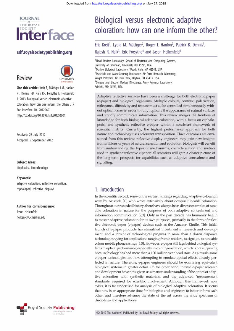
organism/system level
biol
ogic
alm
an-m
ade
octopus
AmazonKindle (E ink)
Kent displaysChLC films
cephalopodskin
pigment granules
primaryinfoldings
and pouches
radialmusclefibres
mitochondriamuscle cellnerve
axon
glialcell
chromatophorestructure
nucleus cytoelasticsacculus
capsuleswith chargedpigment
black state
+++
+++++++
++ +
++ oil
++
++++
++ +
–––
– – –––
–––––
– –– – – –––
–––
white state
E inkpixels transparent
electrode
control electrodes
cellular/pixel and materials levelorgan/device level
(d) ( f )
(c)(b)
(a)
(e) (g)
E inkfilm
Figure 1. Hierarchical levels for biological and synthetic adaptive coloration: organism/system (a) octopus rapidly transitioning out of concealment, (b) Kent Displays’multilayer cholesteric display, (c) Amazon Kindle e-reader using E ink film; organ/device (d ) cephalopod skin, (e) E ink film; cellular/pixel, ( f ) chromatophorestructure [6] and (g) E ink pixels.
rsif.royalsocietypublishing.orgJR
SocInterface10:20120601
2
on July 27, 2018http://rsif.royalsocietypublishing.org/Downloaded from
This review aims to merge the frontiers of knowledge for
both biological adaptive coloration and synthetic reflective e-
paper within a consistent framework of scientific metrics. Mer-
ging these disparate fields is challenging, and our chosen
approach for this review aims to develop common ground
between biology and materials science. First, we review in a
generic framework the optics of adaptive coloration. Next,
because, out of the whole of the animal kingdom, the mollus-
can class Cephalopoda (squid, cuttlefish and octopus) is the
most renowned for rapid adaptive coloration used for a variety
of communication and camouflage tasks, we review the mech-
anisms of their adaptive coloration. This review includes
detailed subsections on the chromatophore organs, irido-
phores and leucophore cells within the skin. Each of these
structures will then be compared with synthetic technologies
that provide similar functionality. Biological systems are too
complex to compare directly with a single synthetic technol-
ogy; for example, comparing a squid and an Amazon Kindle
e-reader is not highly meaningful. However, important com-
parisons can be made between the function and performance
of biological pigments and reflectors, and similarly functioning
synthetic pixels and components.
The best approaches to adaptive colour generation in
nature and artifice share two common aspects: adaptive
colour is changed by compacting or spreading pigment (i.e.
colourant transposition), and efficient reflection is achieved
by optical interference/diffraction. The major outcome from
this review is the foundation of a consistent set of scientific par-
ameters that allows for a better understanding of how display
engineers and biologists can efficiently coordinate a bio-
inspired approach to advanced materials and devices.
2. A hierarchical view of adaptive colorationA common, or at least interchangeable, set of terminology is
needed for this review. There are three hierarchical levels that
will be used in our analysis (figure 1). At the apex, there is
the ‘organism’ or ‘system’ level that contains everything
needed for self-sufficient operation. Even though an e-reader
(figure 1c) is not truly autonomous, it is grouped with auton-
omous organisms (figure 1a) because it has all the features
necessary to respond to stimuli and achieve an appropriate
adaptation in colour or information communicated. One level
deeper is the ‘organ’ or ‘device’ level. For organisms, the adap-
tive coloration ‘organ’ is often the skin, including the vascular
and nervous system features within it. In electronic systems,
there is no organ but instead the analogous ‘device’ that actu-
ally modulates the reflected colour. For example, an E ink
‘device’ has microcapsules of electronically switchable ink,
and an array of electrodes for switching the ink capsules.
Lastly, there is the ‘cellular’ or ‘pixel’ level, both of which also
inherently require consideration of the materials used. The cel-
lular level is where the basic biology and optical physics that
enable adaptive coloration can be discussed in their greatest
physical detail. The comparisons in this review will focus on
this cellular/pixel level.
3. The optics of adaptive colorationBefore we focus on the optics specific to the organisms/tech-
nologies of this review, we will introduce the reader to
adaptive reflective coloration in its broadest context. The
optics for reflective adaptive colour are unlike transmissive or
emissive display approaches. In emissive or transmissive
colour generation, there is an internal source to generate
light, and optical inefficiency can be overcome by simply
increasing the electrical power to the internal light generation.
In the reflective mode, the only way to achieve proper color-
ation is through high optical efficiency for all layers and
materials. Consider, for example, a conventional liquid-crystal
display (LCD) in a laptop computer. The panel is designed for
optimum efficiency; however, it emits a very small percentage
of the light incident on the display (low optical efficiency). This
is why the panel appears black if the backlight is turned off.
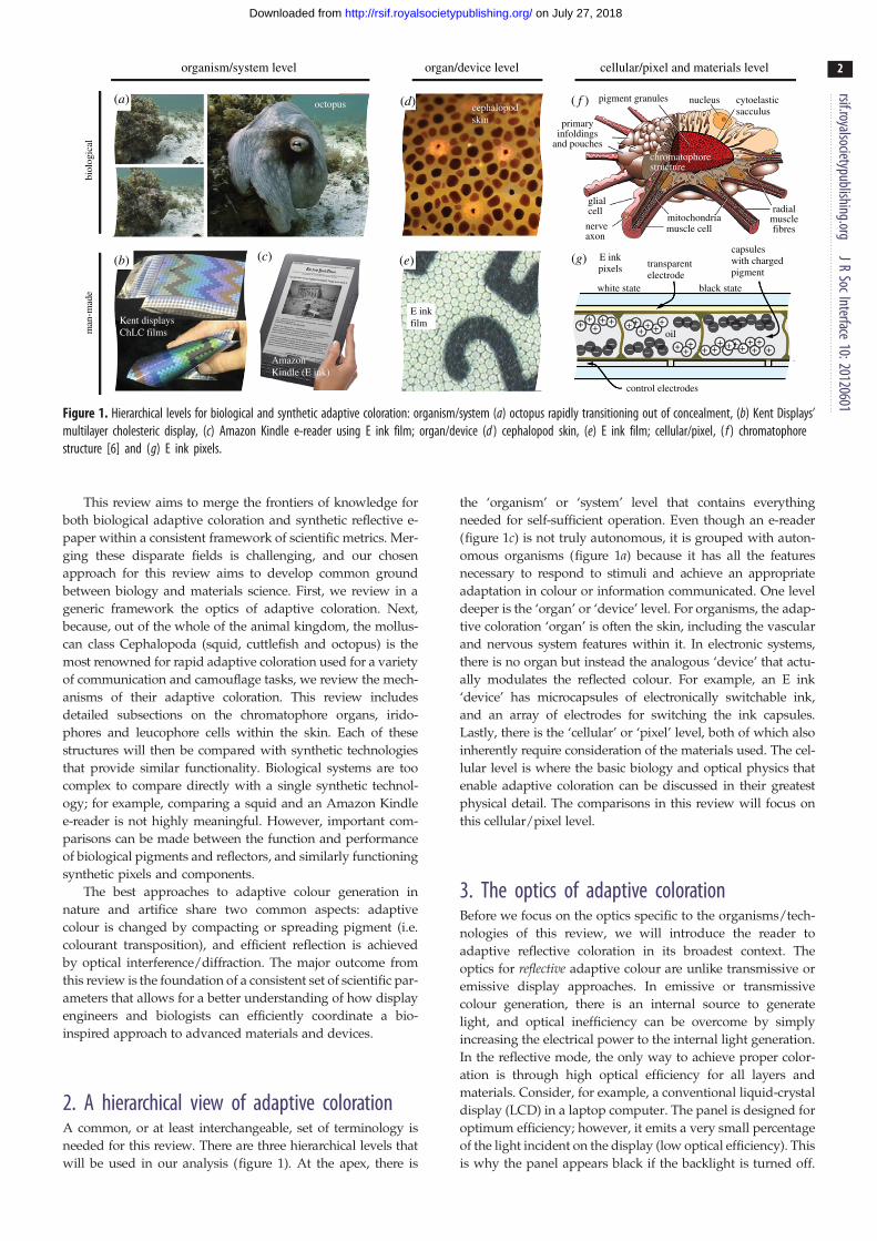
reflect/absorb(a)
(i) (ii)
(b)
(c) reflect/transmit
transmit/absorb
mediumreflects
reflector reflector
medium absorbs(black or colour)
medium absorbs(black or colour)
absorberabsorber
incident lightFresnelreflections
mediumtransmits
mediumreflects
mediumtransmits
transparentprotectivelayer(s)
Figure 2. Fundamental approaches for reflective adaptive coloration. (a) Medium can switch from a reflective mode (i) to an absorbing mode (ii); (b) medium canswitch from a transmitting mode with a reflector behind it (i) to an absorbing mode (ii); (c) medium can switch from a reflective mode with an absorber behind it(i) to a transmitting mode where the absorber prevents light from reflecting out of the device (ii).
rsif.royalsocietypublishing.orgJR
SocInterface10:20120601
3
on July 27, 2018http://rsif.royalsocietypublishing.org/Downloaded from
Considering the low optical efficiency in LCDs (despite being
designed for optimum performance), it is all the more amazing
that biological organisms are able to achieve bright adaptive
coloration solely in the reflective mode. Simply stated, animal
pigments and structurally coloured reflectors are very efficient
at using available light. There are well-established visual stan-
dards for reflective efficiency (brightness) and colour [4].
Whether biological or electronic, the optical efficiency of
adaptive coloration involves several chronological steps that
are common regardless of which approach is used (figure 2).
(i) Light must be effectively coupled into the device or organ. Gener-
ally, this means that various layers of the organ or device
should have low optical absorption and as small a refractive
index mismatch as possible. Layers with different refractive
indices can cause Fresnel reflection [7] which reflects the light
before it can couple (transmit) into the device or organ. (ii)
Light must be efficiently reflected inside the pixel. A mechanism
for efficient reflection is needed, and generally the reflection
must be at least semi-diffuse (semi-Lambertian) to appear like
natural surfaces. (iii) The diffusely reflected light must be outcoupled.
Once the incident light is diffused, it must also be outcoupled
(i.e. escape the device or organ). Some light does not escape
due to total internal reflection [7] and must be diffusely reflected
again, introducing further optical loss. For example, an organ or
device that is internally 80 per cent reflective and has a refractive
index of n � 1.5 can lose more than 20 per cent of the light owing
to total internal reflection [7].
Three important optical parameters for comparison include
contrast ratio, colour gamut and the number of grey-scale
levels. Contrast ratio is the optical ratio between colour state
and black (or non-colour colour state). Colour gamut is defined
as the subset of complete colours that a surface can achieve.
Reflective colour is typically defined by the L*a*b* (labora-
tory colour space) coordinates that can be derived from the
wavelength spectrum of the reflected light, when compared
with a perfectly diffuse white reflector [4]. L* is an intensity
measurement that closely matches human perception of light-
ness. This is distinct from a reflectivity measurement in that
reflectivity measures the amount of photons reflected from a
surface that is not linear with the human visual system. a*
and b* are the colour-opponent dimensions. Greyscale relates
to the number or shades of a colour.
While one can use optical parameters for comparative dis-
cussion, importantly, a human-made system (e-paper) is
optimized for a fundamentally different purpose than the
evolved, biological adaptive colour systems, i.e. it is opti-
mized specifically for the human visual system. The
primate (e.g. human) visual system is distinct from many
other organisms. Humans possess three cone visual pigments
for conveying colour information that is said to allow
humans to be able to detect approximately 10 million
unique colours [8,9] but only distinguish about 30 shades
of grey [10]. With regard to spectral sensitivity, there are
enormous variations in visual abilities across the animal king-
dom. Some simpler animal eyes have only one visual
pigment; others, such as mantis shrimp, have as many as
12, covering the human visible spectrum as well as UV and
IR [11–13]. While there are several methods available to
(c)
2 cm 2 cm
(a)
(c)
(b)
rsif.royalsocietypublishing.orgJR
SocInterface10:20120601
4
on July 27, 2018http://rsif.royalsocietypublishing.org/Downloaded from
analyse how colours are perceived from the perspective of the
human visual system (e.g. CIE 1931 XYZ colour space), there
have to date been very few attempts to extend these methods
to include animal visual systems.
Nevertheless, various modelling methods have
been developed [14–17] that allow us to assess how particu-
lar colours are perceived by a given animal’s visual system.
This is particularly important because the (L*a*b) method is
devised for human vision and is not applicable to other
visual systems. These modelling methods [14–17] take into
consideration how many photons (i.e. light reflected from a
surface) are absorbed by a given set of photoreceptors in
the retina, and how these photon catches are represented in
a specific colour space. However, precise knowledge of the
ratio of different photoreceptor types present in the retina
of di-, tri- or even tetra-chromats are required for these
models to give an accurate estimation of what an animal
sees. While such models are helpful when assessing species
for which these data exist, for many others, these modelling
techniques remain speculation.
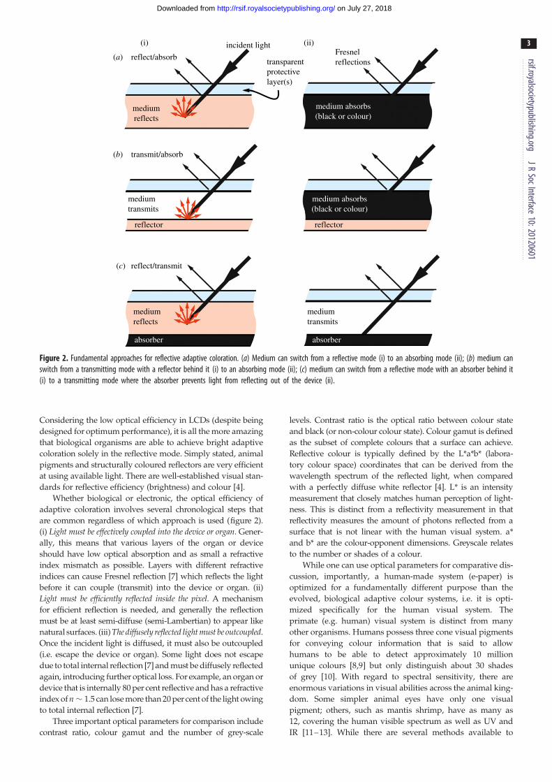
Figure 3. Cuttlefish, Sepia officinalis, showing (a) mottle and (b) disruptivecamouflage. (c) Low magnification and close-up photographs of a hatchlingblue-ringed octopus (Hapalochlaena lunulata), credit: Roy Caldwell.
4. The cephalopod system for adaptive colorationOf all of the organisms in the animal kingdom capable of
colour modulation, cephalopods (squid, cuttlefish and octo-
pus) are able to produce the widest range of colours and
patterns to help them adapt to their visually diverse marine
environments as well as signal and communicate with their
own species and others. There are more than 700 species of
cephalopods, and many of the organisms within this class
are able to adapt their coloration to their environment to var-
ious degrees (figure 3). Cephalopods are the focus in this
review, although other organisms (such as chameleons)
[18–20] have similar mechanisms for adaptive colour and
will be briefly discussed in a later section. The cephalopod
is best presented in terms of the organism/organ/cellular
hierarchy (figure 1). Organism—for the cephalopod organism
to modulate skin colour, it first needs to sense its surround-
ings to determine what colours and patterns it needs to
create. This sensing is done by the visual system of the cepha-
lopod [21]. The visual data are processed by the brain, which
then sends control signals to the skin, which is the Organ that
enables adaptive coloration. The skin contains three distinct
structures—chromatophores, iridophores and leucophores—
that contribute to the colour and pattern adaptation. The
skin is also capable of flattening or wrinkling on demand,
providing surface texturing (see octopus in figure 1a). As
shown in figure 3, the epidermis and dermis are transparent,
allowing light to pass through to impact all three skin struc-
tures before light is modified through interaction at a Cellularlevel. At the cellular level, the chromatophore pigments
impart variable optical transmission, and the iridophores and
leucophores serve as a rear reflector (similar to the basic
approach of figure 2b). The chromatophore has a pigment-
filled sac that has dozens of radial muscle fibres attached
around its periphery. As shown in the photographs of figure
3, these structures work in combination to produce stark
changes in the organisms’ coloration and patterning (via
colour transposition).
The cumulative mechanism behind adaptive coloration for
cephalopods is more sophisticated than anything synthetic
(figure 4). There are separate mechanisms for the physics of
how the cephalopod’s colour modulation works. The pigmen-
ted chromatophores are punctate when retracted (i.e. barely
visible) and are expanded into a thin disc of colour by the
radial muscle fibres. This allows the pigments to go from
almost invisible to full coverage on the organism.
The function of chromatophores is to reflect, block and transmit
light, as well as act as a spectral filter. A red-pigmented chroma-
tophore, for example, absorbs all other wavelengths except red.
The iridophores function differently: they produce structural
colour based on constructive interference (i.e. there is no
absorption of light). There is a difference in refractive index
between the reflectin protein in each iridophore platelet and
the interplatelet spaces within the entire iridophore cell [22–
24]. Moreover, the platelet thicknesses and adjacent spaces
have precise and periodic arrangement that influence which
wavelengths are reflected (a dielectric mirror) [25]. The plate
spacing for some iridophores can also be controlled by the
organism, thereby varying the peak reflected wavelength [26].
Recently, it has been discovered that not only the iridophore
plate spacing can be controlled but also the arrangement of
the reflectin proteins through a reversible phosphorylation pro-
cess [23]. The rearrangement of these proteins also results in
changes to the peak reflected wavelength. Cuttlefish leuco-
phores (leuco means ‘white’) also reflect light, but from
wavelengths of 300 to 900 nm giving them a diffuse white
appearance (like paper). In some skin areas, reflectance is as
high as 70 per cent, and the intensity of the reflected white
light is the same, regardless of the viewing and incident light
angles (similar to a Lambertian surface). Cuttlefish
leucophores are composed of spherical protein assemblages
of varying diameters (200–2000 nm) that scatter light whether
the surrounding medium is water or air. Moreover, these leuco-
phores are very flexible: they do not lose their optical properties
when mechanically deformed. They are physiologically pas-
sive: with no associated musculature or innervation, their
energy requirement is nil [27].
The sophistication of biological adaptive coloration can be
fully appreciated by a detailed examination of the ray traces
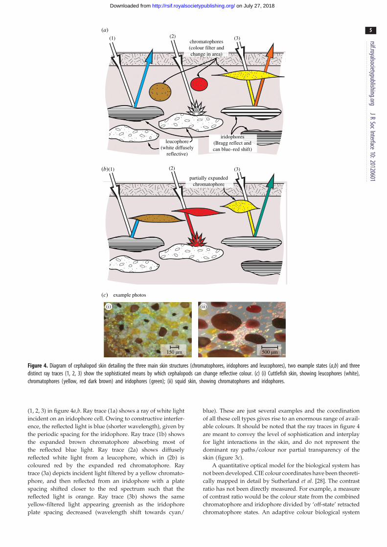
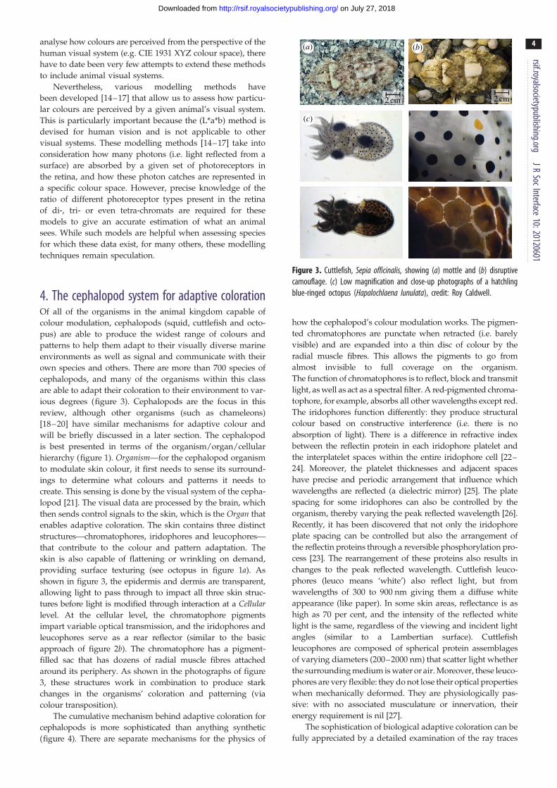
chromatophores(colour filter andchange in area)
(1)
(1) (2) (3)
(a)
(b)
(2) (3)
iridophores(Bragg reflect andcan blue–red shift)
leucophore(white diffusely
reflective)
partially expandedchromatophore
150 µm 500 µm
(c) example photos
(i) (ii)
Figure 4. Diagram of cephalopod skin detailing the three main skin structures (chromatophores, iridophores and leucophores), two example states (a,b) and threedistinct ray traces (1, 2, 3) show the sophisticated means by which cephalopods can change reflective colour. (c) (i) Cuttlefish skin, showing leucophores (white),chromatophores (yellow, red dark brown) and iridophores (green); (ii) squid skin, showing chromatophores and iridophores.
rsif.royalsocietypublishing.orgJR
SocInterface10:20120601
5
on July 27, 2018http://rsif.royalsocietypublishing.org/Downloaded from
(1, 2, 3) in figure 4a,b. Ray trace (1a) shows a ray of white light
incident on an iridophore cell. Owing to constructive interfer-
ence, the reflected light is blue (shorter wavelength), given by
the periodic spacing for the iridophore. Ray trace (1b) shows
the expanded brown chromatophore absorbing most of
the reflected blue light. Ray trace (2a) shows diffusely
reflected white light from a leucophore, which in (2b) is
coloured red by the expanded red chromatophore. Ray
trace (3a) depicts incident light filtered by a yellow chromato-
phore, and then reflected from an iridophore with a plate
spacing shifted closer to the red spectrum such that the
reflected light is orange. Ray trace (3b) shows the same
yellow-filtered light appearing greenish as the iridophore
plate spacing decreased (wavelength shift towards cyan/
blue). These are just several examples and the coordination
of all these cell types gives rise to an enormous range of avail-
able colours. It should be noted that the ray traces in figure 4
are meant to convey the level of sophistication and interplay
for light interactions in the skin, and do not represent the
dominant ray paths/colour nor partial transparency of the
skin (figure 3c).
A quantitative optical model for the biological system has
not been developed. CIE colour coordinates have been theoreti-
cally mapped in detail by Sutherland et al. [28]. The contrast
ratio has not been directly measured. For example, a measure
of contrast ratio would be the colour state from the combined
chromatophore and iridophore divided by ‘off-state’ retracted
chromatophore states. An adaptive colour biological system
rsif.royalsocietypublishing.org
6
on July 27, 2018http://rsif.royalsocietypublishing.org/Downloaded from
has evolved to optimize colour contrast ratio relative to their
environment. As such, a comparison between the biological
optical parameters and environment is equally important.
For example, for concealment, biological systems should gener-
ally resemble the contrast ratio of environment or a dark
shadow, and the colour coordinates for the majority of environ-
ment colours. For signalling, the contrast ratio is usually high
so that the signal is highly conspicuous, but of course colour
plays a role in some species by targeting certain wavelengths
in the receiver’s visual system [29].
JRSocInterface
10:20120601
5. Cephalopod adaptive coloration for signallingThe most well-known application of adaptive coloration in
cephalopods is predator avoidance through camouflage.
However, cephalopods also use the same skin elements
used in camouflage to create visual patterns of high contrast
for vivid unambiguous signalling and communication
[30,31]. Biological signalling is an important topic, because
it parallels the foremost human use of adaptive coloration,
which is for visual communication of information. The leuco-
phores can play a particularly important role here because
their highly reflective whiteness provides the strongest con-
trast to the overlying dark brown/black pigmented
chromatophores (figure 4) [32,33]. For example, the Zebra
display, shown by cuttlefish males during reproductive be-
haviour, is created by maximally expanding the black
chromatophores to achieve a dark striped pattern, while sim-
ultaneously retracting the chromatophores in-between and
allowing the bright white scattering from leucophores to pro-
duce maximum contrast with the dark stripes. In addition,
the cuttlefish amplifies the signal with arm postures that
maximize visibility of the pattern. Other more subtle signals,
including the use of polarized reflections, may use
the iridophores to achieve displays of varying con-
spicuousness [30,31,33]. It is worth noting that synthetic
technologies also have the ability to reflect polarized light
but owing to limitations of the human visual system, it is
of little use to humanity, unless technological aid is provided.
6. Synthetic adaptive colour technologyThere has been a plethora of ‘bio-inspired’ technology develop-
ment, most of which has only a weak connection to the actual
optics of cephalopod skin [34,35]. Therefore, we have judi-
ciously selected the very few technologies that are more
closely biomimetic to the cephalopod skin. Next, detailed sub-
sections on the chromatophore, iridophore and leucophore
structures within the skin will be compared with synthetic
technologies that provide similar functionality. The skin
in cephalopods is more complex and sophisticated than any
synthetic technology. Therefore, meaningful comparisons
must be made at the cellular level (biological) to the pixel
level (synthetic).
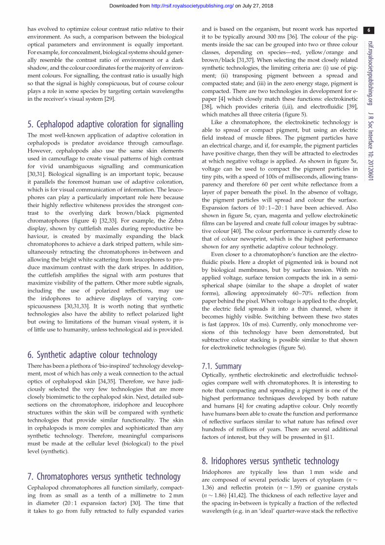
7. Chromatophores versus synthetic technologyCephalopod chromatophores all function similarly, compact-
ing from as small as a tenth of a millimetre to 2 mm
in diameter (20 : 1 expansion factor) [30]. The time that
it takes to go from fully retracted to fully expanded varies
and is based on the organism, but recent work has reported
it to be typically around 300 ms [36]. The colour of the pig-
ments inside the sac can be grouped into two or three colour
classes, depending on species—red, yellow/orange and
brown/black [31,37]. When selecting the most closely related
synthetic technologies, the limiting criteria are: (i) use of pig-
ment; (ii) transposing pigment between a spread and
compacted state; and (iii) in the zero energy stage, pigment is
compacted. There are two technologies in development for e-
paper [4] which closely match these functions: electrokinetic
[38], which provides criteria (i,ii), and electrofluidic [39],
which matches all three criteria (figure 5).
Like a chromatophore, the electrokinetic technology is
able to spread or compact pigment, but using an electric
field instead of muscle fibres. The pigment particles have
an electrical charge, and if, for example, the pigment particles
have positive charge, then they will be attracted to electrodes
at which negative voltage is applied. As shown in figure 5a,
voltage can be used to compact the pigment particles in
tiny pits, with a speed of 100s of milliseconds, allowing trans-
parency and therefore 60 per cent white reflectance from a
layer of paper beneath the pixel. In the absence of voltage,
the pigment particles will spread and colour the surface.
Expansion factors of 10 : 1–20 : 1 have been achieved. Also
shown in figure 5a, cyan, magenta and yellow electrokinetic
films can be layered and create full colour images by subtrac-
tive colour [40]. The colour performance is currently close to
that of colour newsprint, which is the highest performance
shown for any synthetic adaptive colour technology.
Even closer to a chromatophore’s function are the electro-
fluidic pixels. Here a droplet of pigmented ink is bound not
by biological membranes, but by surface tension. With no
applied voltage, surface tension compacts the ink in a semi-
spherical shape (similar to the shape a droplet of water
forms), allowing approximately 60–70% reflection from
paper behind the pixel. When voltage is applied to the droplet,
the electric field spreads it into a thin channel, where it
becomes highly visible. Switching between these two states
is fast (approx. 10s of ms). Currently, only monochrome ver-
sions of this technology have been demonstrated, but
subtractive colour stacking is possible similar to that shown
for electrokinetic technologies (figure 5a).
7.1. SummaryOptically, synthetic electrokinetic and electrofluidic technol-
ogies compare well with chromatophores. It is interesting to
note that compacting and spreading a pigment is one of the
highest performance techniques developed by both nature
and humans [4] for creating adaptive colour. Only recently
have humans been able to create the function and performance
of reflective surfaces similar to what nature has refined over
hundreds of millions of years. There are several additional
factors of interest, but they will be presented in §11.
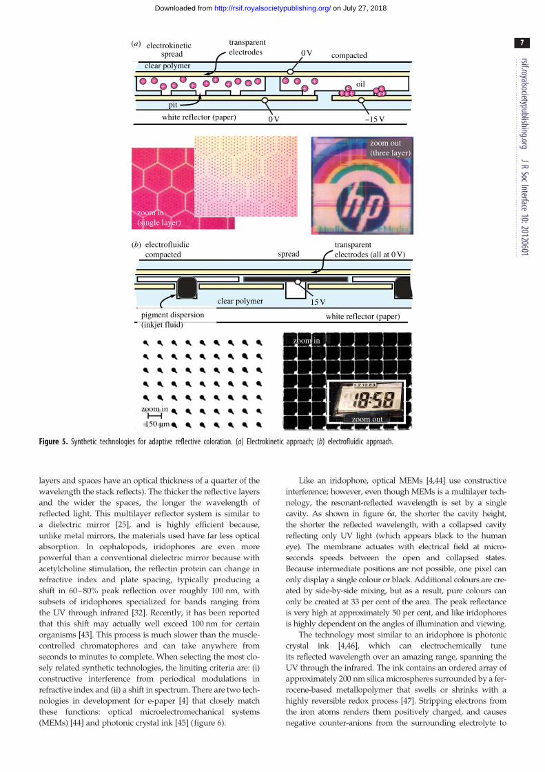
8. Iridophores versus synthetic technologyIridophores are typically less than 1 mm wide and
are composed of several periodic layers of cytoplasm (n �1.36) and reflectin protein (n � 1.59) or guanine crystals
(n � 1.86) [41,42]. The thickness of each reflective layer and
the spacing in-between is typically a fraction of the reflected
wavelength (e.g. in an ‘ideal’ quarter-wave stack the reflective
(a)
(b) electrofluidiccompacted spread
transparentelectrodes (all at 0 V)
transparentelectrodes
15 V
–15 V0 V
0 V
oil
compactedclear polymer
+ ++ +
++ +
++ + + + + +
++
+ + + ++++
+
pit
white reflector (paper)
clear polymer
pigment dispersion(inkjet fluid)
white reflector (paper)
zoom in
zoom in(single layer)
zoom out(three layer)
zoom in
150 µmzoom out
spreadelectrokinetic
Figure 5. Synthetic technologies for adaptive reflective coloration. (a) Electrokinetic approach; (b) electrofluidic approach.
rsif.royalsocietypublishing.orgJR
SocInterface10:20120601
7
on July 27, 2018http://rsif.royalsocietypublishing.org/Downloaded from
layers and spaces have an optical thickness of a quarter of the
wavelength the stack reflects). The thicker the reflective layers
and the wider the spaces, the longer the wavelength of
reflected light. This multilayer reflector system is similar to
a dielectric mirror [25], and is highly efficient because,
unlike metal mirrors, the materials used have far less optical
absorption. In cephalopods, iridophores are even more
powerful than a conventional dielectric mirror because with
acetylcholine stimulation, the reflectin protein can change in
refractive index and plate spacing, typically producing a
shift in 60–80% peak reflection over roughly 100 nm, with
subsets of iridophores specialized for bands ranging from
the UV through infrared [32]. Recently, it has been reported
that this shift may actually well exceed 100 nm for certain
organisms [43]. This process is much slower than the muscle-
controlled chromatophores and can take anywhere from
seconds to minutes to complete. When selecting the most clo-
sely related synthetic technologies, the limiting criteria are: (i)
constructive interference from periodical modulations in
refractive index and (ii) a shift in spectrum. There are two tech-
nologies in development for e-paper [4] that closely match
these functions: optical microelectromechanical systems
(MEMs) [44] and photonic crystal ink [45] (figure 6).
Like an iridophore, optical MEMs [4,44] use constructive
interference; however, even though MEMs is a multilayer tech-
nology, the resonant-reflected wavelength is set by a single
cavity. As shown in figure 6a, the shorter the cavity height,
the shorter the reflected wavelength, with a collapsed cavity
reflecting only UV light (which appears black to the human
eye). The membrane actuates with electrical field at micro-
seconds speeds between the open and collapsed states.
Because intermediate positions are not possible, one pixel can
only display a single colour or black. Additional colours are cre-
ated by side-by-side mixing, but as a result, pure colours can
only be created at 33 per cent of the area. The peak reflectance
is very high at approximately 50 per cent, and like iridophores
is highly dependent on the angles of illumination and viewing.
The technology most similar to an iridophore is photonic
crystal ink [4,46], which can electrochemically tune
its reflected wavelength over an amazing range, spanning the
UV through the infrared. The ink contains an ordered array of
approximately 200 nm silica microspheres surrounded by a fer-
rocene-based metallopolymer that swells or shrinks with a
highly reversible redox process [47]. Stripping electrons from
the iron atoms renders them positively charged, and causes
negative counter-anions from the surrounding electrolyte to
(b) photonic crystal ink
P-ink (Opalux)
optical film stack
resonant air gap
prototype by Qualcomm
– – – – –+++++
reflective membrane
transparent electrode
polymer withsilica beads
pixel electrodes
green reflect
green reflect
red reflect
red reflectIR (black) UV, black
no reflect(UV, black)
blue reflect
blue reflect
optical MEMs(a) transparentelectrode
electrolyte
Figure 6. Synthetic technologies for adaptive iridescence. (a) Optical microelectromechanical systems; (b) photonic crystal ink.
rsif.royalsocietypublishing.orgJR
SocInterface10:20120601
8
on July 27, 2018http://rsif.royalsocietypublishing.org/Downloaded from
enter and swell the film. Wider bead spacing causes reflections
at longer wavelengths (figure 6b). Currently, peak performance
is 60–70% reflectance with approximately 100 ms switching
speed. There are additional technologies that leverage the use
of structural coloration that include altering the localized con-
centration of colloidal particles in a fluid medium [48,49] and
self-assembling block copolymers [35]. However, these technol-
ogies are still early in their development.
The colours and intensity produced by constructive inter-
ference techniques using regularly spaced arrays (as
discussed in this section) are angularly dependent (will
appear different as viewing angle changes). However, not
all forms of constructive interference produce angularly
dependent colours and intensity. Amorphous or quasi-
ordered nanostructures (as can be found in bird feathers)
[50] produce a non-iridescent structural coloration. While
the iridescent form of structural coloration is desirable for
applications like banknotes and identification documents, it
imposes an extra challenge for displays where appearance
should not vary with viewing angle [4].
8.1. SummaryLike chromatophores, optically synthetic technology appears
to outperform iridophores. However, all synthetic technologies
exhibit numerous limitations that will be highlighted in §11.
Moreover, the synthetic technologies mentioned in this section
have the ability to reflect UV and/or IR wavelengths. Many
organisms (including cephalopods) use these wavelengths for
a variety of signalling activities. Thus, technology can actually
replicate the appearance of some biological organisms.
9. Leucophores versus synthetic technologyLeucophores also use protein to reflect light. However, in leu-
cophores, the reflection is not from layered sheets but from
spherical granules with varying diameters (200–2000 nm)
inside the cell. This reflection is similar in appearance and
function to diffuse white paint (which typically contains
approx. 100 nm-sized granules of TiO2). This reflection
involves significant scattering, and is far less efficient than
rsif.royalsocietypublishing.orgJR
SocInterface10:2012060
9
on July 27, 2018http://rsif.royalsocietypublishing.org/Downloaded from
reflection due to multilayer interference. Therefore, thicker
sheets of cells (approx. 100–250 mm) are needed to increase
the total reflectance to approximately 70 per cent (as in the
Sepia cuttlefish fin spots). The function of the leucophore is
assumed to be in aiding both wavelength and intensity
matching at a localized level within the skin [27,51].
There are numerous synthetic examples of highly efficient
white reflectors, including the scattering properties of bleached
wood fibre in paper, microvoided polymer films and polymer :
TiO2 composite films. For example, 3M has developed less than
60 mm thick films of multilayer bi-refringent polyethylene ter-
ephthalate or polyethylene naphthalate that reflect the entire
visible spectrum (white light) with more than 99 per cent
efficiency over nearly all angles and polarizations [52].
9.1. SummaryIn terms of diffuse reflection, humanity clearly outperforms
nature. However, again, nature has several advantages that
will be highlighted in §11.
110. Other biological colour-changing organismsCephalopods are not the only organisms capable of
colour modulation. In fact, chameleons have historically been
the most well-known organism capable of adaptive coloration.
The mechanisms of colour change in the chameleon differ
greatly from the cephalopod. In cephalopods, colour change
occurs under direct neural control and is therefore fast (millise-
conds to seconds). In lizards, such as the chameleon, colour
change is regulated by a neuro-hormonal mechanism, which
generally takes longer (seconds to minutes, and in some species
hours or days) [18]. The structures responsible for colour
change also differ between the two animal groups. Lizards
also have structures called chromatophores, but they are dis-
tinct from those of cephalopods. Lizard chromatophores are
cells, not organs. Three types of lizard chromatophores are
known: melanophores, guanophores and xanthophores. Mela-
nophores are large cells deep in the dermis. They have
numerous long dendritic processes (like small channels) that
run towards the skin surface. Melanin granules travel along
these processes to darken the skin when they are at the skin sur-
face, or lighten it, when they are concentrated at the centre of
the cell. Guanophores are above the melanophores and contain
guanine crystals that reflect light by constructive interference
and are responsible for blue coloration. Xanthophores are the
most superficial cells and lack dendritic processes. They con-
tain red and yellow pigments. Similar to cephalopods, the
final skin coloration is the end result of the interactions
among all skin structures [18–20]. It is worth noting that the
function of the melanophores is similar to vertical electrophor-
etic displays [4,5] used in the Amazon Kindle: coloured
particles move either closer to, or farther away from, the
viewer to elicit a change in reflection.
Chameleons change colour for a variety of reasons, such
as thermoregulation, camouflage and communication. In
the dwarf chameleon, it has been shown that the camouflage
body patterns are even tailored to the visual system of the
predator they are aiming to avoid [53,54]. However, adapt-
able colour change of cephalopods is much more diverse in
patterning, speed and optical effects [21,33].
Other biological organisms, such as birds [55,56] and
insects, [55,57] commonly produce coloration through the
use of submicron structures and pigmentation. These color-
ation strategies can also be described as coherent (i.e.
structural interference/diffraction) and incoherent (i.e. scat-
tering) [58,59]. Which means are used is dependent on the
specific species of interest. Although not tuneable like the
cephalopod system, the structural means of coloration used
by these organisms are equally sophisticated and can achieve
a similarly wide gamut of colours.
11. Discussion: who can inform whom?Synthetic technology was shown to equal or outperform biologi-
cal systems (cephalopods) when making direct comparison with
chromatophores, iridophores and leucophores.
This is only a very recent achievement for humanity (past
several years) [4]. The current applications of synthetic technol-
ogy more closely match the signalling/communication
functionality of the cephalopod system than camouflage. How-
ever, the cephalopod skin systems are far more efficient at
handling ambient light, whereas many synthetic technologies
have thus far relied on large amounts of electrical power to
create emissive light and there has been much less focus on look-
ing for reflective materials that may produce comparable results.
Regarding signalling, the requirements for electronic sig-
nage and e-readers are still very different from cephalopod
signalling and communication. The primary difference is in
the desired colour gamut and level of information content.
Synthetic technology often displays information-rich and
dense groupings of high-resolution symbols and characters,
while cephalopods and other animals achieve signalling
using simple vivid static or flashing patterns.
Regarding camouflage, few synthetic technologies aim to
replicate the mechanisms and functions found in biological sys-
tems. However, robust adaptive coloration is the basic enabler
of high performance in all applications (camouflage, signalling
and communication). For example, having good dark states,
bright maximum light states and ability to adapt quickly
benefits nearly all uses for both technology and cephalopods.
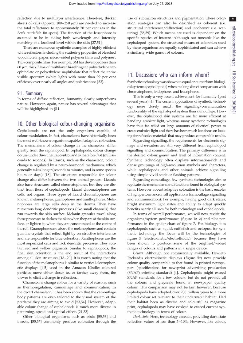
In terms of overall performance, we will now revisit the
organisms/system performance (figure 1a–c) and plot per-
formance in the spider chart of figure 7. For biology, it is
cephalopods such as squid, cuttlefish and octopus, for syn-
thetic technology the focus will be the technologies of
figure 5 (electrokinetic/electrofluidic), because they have
been shown to produce some of the brightest/widest
ranges of colours and patterns in a single device.
Colour. Although not commercially available, Hewlett-
Packard’s electrokinetic displays (figure 5a) now provide
colour quality comparable to that found in printed newspa-
pers (specifications for newsprint advertising production
(SNAP) printing standard) [4]. Cephalopods might exceed
SNAP standards for a few colours, but do not provide all
the colours and greyscale found in newspaper quality
colour. This comparison may not be fair, however, because
cephalopods have adapted over 200 million years to a more
limited colour set relevant to their underwater habitat. Had
their habitat been as diverse and colourful as magazine
print, cephalopods may have evolved to exceed current syn-
thetic technology in terms of colour.
Dark state. Here, technology exceeds, providing dark state
reflection values of less than 5–10%. However, like colour,
colour
man-madetech
cephalopodsflexible
surfacetexture
speed required energy
magazine
conf
orm
al
rolla
ble
news
paper
limited
yes
10 ms low
prov
enposs
ible
moderate100 ms
1 s
dark
dumb
dark state
integrationand Sophis.
adapt toenvironment
black
full
Figure 7. Spider chart comparing the important metrics to both cephalopod (green) and synthetic (blue) adaptive coloration. The newspaper/magazine metrics inthe colour category refer to ‘SNAP and specifications for web offset publications (SWOP)’ standards, respectively [4]. Regarding the dark state metric, cephalopods arecapable of changing their colour to dark brown, but not to black.
rsif.royalsocietypublishing.orgJR
SocInterface10:20120601
10
on July 27, 2018http://rsif.royalsocietypublishing.org/Downloaded from
this comparison may be unfair as most cephalopods do not
require adaptation to a pitch black background.
Integration and sophistication. Cephalopods far outperform
technology in this category, not only in terms of intelligence
(compared with a computer that lacks intelligence), but also
in elegant integration of numerous cell types in a single
organ. Humanity has never developed anything as complex
nor sophisticated as the biology and physics of cephalopod
skin. For optical integration, biological systems have one
major advantage, in that tissue layers can be extremely clear
and have a low and homogeneous refractive index such that
light is very efficiently coupled into and out of the tissue.
Adapt to environment. Although in theory, technology
could use sensors and computing to adapt in coloration to
its local environment, this requires numerous additional elec-
tronic components and integration of all such components.
Compared with cephalopods, colour adaptation to the
environment with technology is unproven.
Required energy. Both synthetic and biological systems
exhibit very low power consumptions; however, biological
systems are superior in how they are self-reliant for gathering
energy compared with synthetic systems, which typically
require batteries. However, a few displays have had solar-
cells integrated into them to harvest energy [60,61].
Speed. Ideally, speed of adaptation should be close to or
exceed the response time of biological vision systems (typically
10 s of ms). Here, technology is far superior to cephalopods.
Surface texture. Displaying texture on an e-reader or
laptop screen is most always noticeably fake. Texture pro-
vides additional light scattering and shadowing that is
difficult to dynamically display with technology. However,
cephalopods have developed the ability to selectively adapt
or ‘crinkle’ their skin to match a variety of textures.
Flexible. Technology companies have prototypes that are
not mass-produced and sold, including flexible electrophor-
etic displays [62–64], and electrokinetic displays [38] to
name a few. These flexible display technologies currently
conform in one-dimension to adapt to the printed-paper tech-
nologies, which is far from the flexible capability that
cephalopods can achieve. Several groups have demonstrated
electronics that conform (stretch) in two-dimensions, which
has allowed synthetic electronics to be integrated on human
skin [65]. By contrast, biological organisms have adapted to
a highly conformal environment that requires a system that
can stretch many times the original size.
Scalable. For an adaptive reflective surface to be scalable, it
must be able to be increased in size while keeping an acceptably
low number of defects. In this respect, cephalopods also outper-
form technology. While humans have been able to scale up rigid
panels (10000 rigid panels are being manufactured) flexible scal-
ability has been far less (less than 1000). Cephalopods have been
successful at scaling up their fully conformal method of adap-
tive coloration over a wide range of sizes of organisms.
It is noteworthy that although not included in figure 7, the
ratio of the colour state to dark state (contrast ratio) is very
important. A good dark state will make a colour state look
brighter, and vice versa. There are other additional metrics,
like polarization of light, resolution (points per inch) and abil-
ity to self-generate light, to name a few. These metrics are
beyond the scope of this review but may be worthy of con-
sideration based on application.
So, how can nature better inform the development of improvedsynthetic technology for adaptive coloration? For some appli-
cations, nature has already influenced synthetic technology.
For example, biological coloration strategies used by cephalo-
pods have inspired engineering approaches to create devices
that exhibit tuneable optical properties. One example is that
the use of layered materials has resulted in the fabrication
of Bragg mirrors that have a rapid reversible optical reflec-
tance [66]. This is analogous to the colour-changing
mechanism used by cephalopods, using the change in thick-
ness of iridophore platelets. Another synthetic device
rsif.royalsocietypublishing.orgJR
SocInterface10:20120601
11
on July 27, 2018http://rsif.royalsocietypublishing.org/Downloaded from
inspired by cephalopods is the electrochemically tuneable
block copolymer full colour pixels demonstrated by
Thomas and co-workers [35]. Again, this is analogous to
the iridophore platelets in that changing the distance in the
optical elements (platelets in the cephalopod) results in tune-
able optical properties. The iridophores are not the only
component of the cephalopod that humans have taken inspi-
ration from. Recently, a group at the University of Bristol
used inspiration from the underlying mechanisms of
chromatophore actuation to create artificial elastomeric chro-
matophores that undergo optical modulation in response to
electrical stimuli [67]. In addition, a simple soft machine con-
structed from polymers was equipped with microfluidics to
change the color and pattern of the small robot; this work
was inspired by cephalopods and other animals with adap-
tive coloration [68]. Inspiration has also been derived from
organisms other than cephalopods. For example, the struc-
tural coloration created by butterfly wings has been used
by L’Oreal for cosmetics [69].
For the existing applications of e-paper, such as
e-readers, humanity has explored most of the physics
relevant to reflective coloration. Furthermore, the performance
of emerging e-paper technologies is now superior in optical
performance to biological adaptive colour. However, there
are emergent designs for applications where humanity is lack-
ing technology that nature has mastered. For example, consider
reconfigurable keyboards used in touch-based smart phones
and tablets. It would be highly desirable to provide both
visual display of the keyboard and texture/tactile feedback.
The ability of cephalopods to modulate the texture of their
skin could, in theory, provide a form of tactile feedback. Fur-
thermore, there is the desire to have adaptive colour, and
reconfigurable input capability, on compound curves of elec-
tronics. Although the development of rollable displays is well
underway [70], conformal technology is much farther behind
[65]. In addition, humanity can learn from the many other
performance deficiencies described in figure 7. We note that
our speculations certainly do not represent a complete set
of opportunities.
So, how can technology better inform our understanding ofadaptive coloration in nature? Firstly, it is difficult to isolate
and analyse cellular components of adaptive coloration.
Work has been done in direct acetylcholine stimulation of iri-
dophores [71] and sophisticated spectroscopic models of
chromatophore/iridophore layers have been developed [72].
However, synthetic devices can be easily controlled electroni-
cally and cycled millions if not billions of times, and, in some
cases, might provide suitable biomimetic measurements. Sec-
ondly, there is a clear lack of measurement standards, metrics
and advanced measurement techniques in analysis of natural
adaptive colour. Synthetic e-paper is part of a much larger
display industry that has developed sunlight, diffuse light
and other illumination standards. In addition, measurement
techniques are now quite advanced for diffuse versus specu-
lar reflection, and optical models for light-outcoupling are
now available [73]. Furthermore, biologists could go beyond
simple plots of ‘reflected intensity versus angle’ to cephalo-
pod equivalents of L* and a*,b* colour space [4] (caveat,
these are for human perception of colour; suitable colour-
space models for other organisms need further development
[74]). It seems that the value that technology can provide to
nature is in optical characterization of natural adaptive
coloration.
12. ConclusionsAdaptive reflective surfaces have been developed through
natural selection by biological organisms for hundreds of
millions of years for the purposes of adaptive coloration
and communication. Only recently have synthetic technol-
ogies (e-paper) attempted to achieve similar adaptive
reflective properties. In order to achieve the most robust
adaptive reflective platforms, both biological organisms and
synthetic technologies have to control patterns, textures, col-
ours, contrast, diffuseness, reflectance and polarization all
while minimizing optical losses. Because biology has a sig-
nificant head start on humanity in terms of adaptive
reflectance, it is imperative to study biology closely to help
direct the development of technology.
Owing to the complexity of biological systems, it is not
easy to make a direct comparison between an organism
and a synthetic technology. By breaking down both the
organism and technology into a functional hierarchy, much
more can be learned. Cephalopods are useful candidates for
study, as they achieve the widest range of adaptive color-
ation. From studying cephalopods, two main outcomes
about adaptive reflectance can be derived. The first is that
the best method for changing coloration is by compacting
and spreading out pigments. The second is that efficient
reflection and wavelength modulation can be achieved by
thin-film interference. Most importantly, animal systems
such as cephalopods that have the most diverse and change-
able skin patterning always use a combination of pigments
and reflectors in various combinations of layers, and study-
ing these systems will certainly yield new ideas about how
to engineer synthetic systems.
Technologies exist that begin to imitate the individual
colour adapting structures of cephalopods and outperform
them in terms of speed (microseconds to milliseconds), color-
ation (SNAP achieved) and dark states (,5% reflective in
dark state). However, cephalopods have major advantages
in terms of flexibility (fully conformal), texturing (technology
has yet to commercially prove this capability), adapting to
environments, integration and scalability. As a result,
humanity can learn much from biology in terms of how to
increase the sophistication of their technology, but
implemented in a simple and self-reliant system. On the
other hand, science as a whole can learn from the various
standards and methods for optical characterization that
have been developed for measuring the performance of syn-
thetic technology. By using these methods and metrics, a
more complete database of information regarding adaptive
coloration in organisms can be created.
E.K., J.H. and E.F. created content on technological (synthetic) aspectsof adaptive coloration while L.M.M., R.T.H., P.B.D. and R.R.N.focused on the biological content. E.K. organized and compiled thesections into the main paper, and worked with J.H. on the compari-sons section at the end. All authors discussed the informationpresented in the paper at all stages, with the exception of E.F. whomade significant contributions at later stages. The University of Cin-cinnati authors gratefully acknowledge partial support from AFRL(contract no. 5408-25-SC-0003) NSF Career award (no. 0640964; Uni-versity of Cincinnati), NSF IHCS award (no. 1001141) and ARL(grant no. W9111NF-09-2-0034). MBL authors acknowledge supportfrom AFOSR grant no. FA9550-09-0346, ARL grant no. W911NF-09-2-0043, DARPA (DSO) grant no. W911NF-10-1-0113 and ONRgrant no. N00014-10-1-0989. R.R.N. acknowledges supportfrom AFOSR.
rsif.r
12
on July 27, 2018http://rsif.royalsocietypublishing.org/Downloaded from
References
oyalsocietypublishing.orgJR
SocInterface10:20120601
1. Aristotle. 1883 Aristotle’s history of animals. In tenbooks. London, UK: George Bell.
2. Cott HB. 1940 Adaptive coloration in animals.London, UK: Methuen & Co. Ltd.
3. Thayer GH. 1896 The law which underlies protectivecoloration. Auk 13, 124 – 129.
4. Heikenfeld J, Drzaic P, Yeo JS, Koch T. 2011 Reviewpaper: a critical review of the present and futureprospects for electronic paper. J. SID 19, 129 – 156.(doi:10.1889/JSID19.2.129)
5. Comiskey B, Albert JD, Yoshizawa H, Jacobson J. 1998An electrophoretic ink for all-printed reflectiveelectronic displays. Nature 394, 253 – 255.(doi:10.1038/28349)
6. Cloney RA, Florey E. 1968 Ultrastructure ofcephalopod chromatophore organs. Zeitschrift furZellforschung 89, 250 – 280. (doi:10.1007/BF00347297)
7. Yang S, Hagedon M, Heikenfeld J. 2011 Light out-coupling for reflective displays: simple geometricalmodel, MATLAB simulation, and experimentalvalidation. J. Display Technol. 7, 473 – 477. (doi:10.1109/JDT.2011.2130510)
8. Judd DB, Wyszecki G. 1975 Color in business, scienceand industry, 3rd edn. New York, NY: WileyInterscience.
9. Sharma G, Trussell HJ. 1997 Digital color imaging.IEEE Trans. Image Process 6, 901 – 932. (doi:10.1109/83.597268)
10. Glover DM, Jenkins WJ, Doney SC. 2011 Scientificvisualization. In Modeling methods for marine science,p. 398. New York, NY: Cambridge University Press.
11. Kelber A, Vorobyev M, Osorio D. 2003 Colour visionin animals: behavioural tests and physiologicalconcepts. Biol. Rev. 78, 81 – 118. (doi:10.1017/S1464793102005985)
12. Lythgoe JN, Partridge JC. 1989 Visual pigments and theacquisition of visual information. J. Exp. Biol. 146, 1 – 20.
13. Land MF, Nilsson DE. 2012 Animal eyes, 2nd edn.Oxford, UK: Oxford University Press.
14. Vorobyev M, Brandt R, Peitsch D, Laughlin SB,Menzel R. 2001 Colour tresholds and receptor noise:behaviour and physiology compared. Vision Res. 41,639 – 653. (doi:10.1016/S0042-6989(00)00288-1)
15. Vorobyev M, Marshall J, Osorio D, de Ibarra NH,Menzel R. 2001 Colourful objects through animaleyes. Color Res. Appl. 26, S214 – S217. (doi:10.1002/1520-6378(2001)26:1+,::AID-COL45.3.0.CO;2-A)
16. Vorobyev M, Osorio D. 1998 Receptor noise as adeterminant of colour thresholds. Proc. R. Soc. Lond.B 265, 351 – 358. (doi:10.1098/rspb.1998.0302)
17. Stoddard MC, Prum RO. 2008 Evolution of avianplumage color in a tetrahedral color space: aphylogenetic analysis of New World buntings. Am.Nat. 171, 755 – 776.
18. Bagnara JT, Hadley ME. 1973 Chromatophores andcolor change: the comparative physiology of animalpigmentation. Englewood Cliffs, NJ: Prentice-Hall.
19. Fox DL. 1976 Animal biochromes and structuralcolours. Berkeley, CA: University of California Press.
20. Necas P. 2001 Chameleons: nature’s hidden jewels.Malabar, FL: Krieger Publishing.
21. Hanlon RT. 2007 Cephalopod dynamic camouflage.Curr. Biol. 17, R400 – R404. (doi:10.1016/j.cub.2007.03.034)
22. Crookes WJ, Ding L, Huang QL, Kimbell JR, HorwitzJ, Mcfall-Ngai MJ. 2004 Reflectins: the usualproteins of squid reflective tissues. Science 303,235 – 238. (doi:10.1126/science.1091288)
23. Izumi M. et al. 2009 Changes in reflectin proteinphosphorylation are associated with dynamiciridescence in squid. J. R. Soc. Interface 7, 549 – 560.(doi:10.1098/rsif.2009.0299)
24. Kramer RM, Crookes-Goodson WJ, Naik RR. 2007The self-organizing properties of squid reflectinprotein. Nat. Mater. 6, 533 – 538. (doi:10.1038/nmat1930)
25. Hecht E. 2002 Optics, 4th edn. Reading, MA:Addison-Wesley.
26. Cooper KM, Hanlon RT, Budelmann BU. 1990Physiological color change in squid iridophores. II.Ultrastructural mechanisms in Lolliguncula brevis.Cell Tissue Res. 259, 15 – 24. (doi:10.1007/BF00571425)
27. Mathger LM et al. In preparation. Bright whitepassive diffusion from sorft organic spheres in colorchanging cuttlefish.
28. Sutherland RL, Mathger LM, Hanlon RT, Urbas AM,Stone MO. 2008 Cephalopod coloration model.I. Squid chromatophores and iridophores. J. Opt. Soc.Am. A 25, 588 – 599. (doi:10.1364/JOSAA.25.000588)
29. Endler JA, Westcott DA, Madden JR, Robson T. 2005Animal visual systems and the evolution of colorpatterns: sensory processing illuminates signalevolution. Evolution 59, 1795 – 1818.
30. Hanlon RT. 1982 The functional organization ofchromatophores and iridescent cells in the bodypatterning of Loligo plei (Cephalopoda: Myopsida).Malacologia 23, 89 – 119.
31. Hanlon RT, Messenger JB. 1996 Cephalopodbehaviour. Cambridge, UK: Cambridge UniversityPress.
32. Mathger LM, Hanlon RT. 2007 Malleable skincoloration in cephalopods: selective reflectance,transmission and absorbance of light bychromatophores and iridophores. Cell Tissue Res.329, 179 – 186. (doi:10.1007/s00441-007-0384-8)
33. Mathger LM, Denton EJ, Marshall NJ, Hanlon RT. 2009Mechanisms and behavioral functions of structuralcolouration in cephalopods. J. R. Soc. Interface 6, S149 –S163. (doi:10.1098/rsif.2008.0311)
34. Manakasettharn S, Taylor JA, Krupenkin TN. 2011Bio-inspired artificial iridophores based on capillaryorigami: fabrication and device characterization.Appl. Phys. Lett. 99, 144102. (doi:10.1063/1.3646394)
35. Walish JJ, Kang Y, Mickiewicz RA, Thomas EL. 2009Bioinspired electrochemically tunable blockcopolymer full color pixels. Adv. Mater. 21,3078 – 3081. (doi:10.1002/adma.200900067)
36. Mathger LM, Bell G, Kuzirian AM, Allen JJ, HanlonRT. In press. How does the blue-ringedoctopus (Hapalochlaena lunulata) flash its bluerings? J. Exp. Biol.
37. Messenger JB. 2001 Cephalopodchromatophores: neurobiology and natural history.Biol. Rev. 76, 473 – 528. (doi:10.1017/S1464793101005772)
38. Yeo JS et al. 2010 Novel flexible reflective colormedia integrated with transparent oxide TFTbackplane. SID Symp. Digest 41, 1041. (doi:10.1889/1.3499827)
39. Heikenfeld J, Zhou K, Kreit E, Raj B, Yang S, Sun B,Milarcik A, Clapp L, Schwartz R. 2009 Electrofluidicdisplays using Young – Laplace transposition ofbrilliant pigment dispersions. Nat. Photonics 3,292 – 296. (doi:10.1038/nphoton.2009.68)
40. Koch TR, Liu Q, Benson B, Mabeck J, Hoffman R,Mourey D, Combs G, Zhou Z-L, Henze D. Reflectivefull color electrokinetic displays. In Proc. of the 18thInt. Display Workshops (IDW ‘11). 7 – 9 December2011, Nagoya, Japan.
41. Romney AK. 2008 Relating reflectance spectra spaceto Munsell color appearance space. J. Opt. Soc. Am.A 25, 658 – 666. (doi:10.1364/JOSAA.25.000658)
42. Levy-Lior A, Shimoni E, Schwartz O, Gavish-Regev E,Oron D, Oxford G, Weiner S, Addadi L. 2010Guanine-based biogenic photonic-crystal arrays infish and spiders. Adv. Funct. Mater. 20, 320 – 329.(doi:10.1002/adfm.200901437)
43. Tao AR, DeMartini DG, Izumi M, Sweeney AM, Holt AL,Morse DE. 2010 The role of protein assembly indynamically tunable bio-optical tissues. Biomaterials31, 793 – 801. (doi:10.1016/j.biomaterials.2009.10.038)
44. Miles MW. 1997 A new reflective FPD technologyusing interferometric modulation. J. SID 5, 379 –382. (doi:10.1889/1.1985183)
45. Arsenault AC, Puzzo DP, Manners I, Ozin GA. 2007Photonic-crystal full-colour displays. Nat. Photonics1, 468 – 472. (doi:10.1038/nphoton.2007.140)
46. Wang H, Kerins F, Kamp U, Bonifacio L, ArsenaultAC, Ozin GA. 2011 Photonic-crystal display materials.Inf. Display 7, 26 – 29.
47. Kulbaba K et al. 2001 Polyferrocenylsilane and magneticceramic microspheres. Adv. Mater. 13, 732–736. (doi:10.1002/1521-4095(200105)13:10,732::AID-ADMA732.3.0.CO;2-2)
48. Lee I et al. 2010 Quasi-amorphous colloidalstructures for electrically tunable full-color photonicpixels with angle-independency. Adv. Mater. 22,4973 – 4977. (doi:10.1002/adma.201001954)
49. Harun-Ur-Rashid M, Imran AB, Seki T, Ishii M,Nakamura H, Takeoka Y. 2010 Angle-independentstructural color in colloidal amorphous arrays.
rsif.royalsocietypublishing.orgJR
SocInterface10:20120601
13
on July 27, 2018http://rsif.royalsocietypublishing.org/Downloaded from
ChemPhysChem 11, 579 – 583. (doi:10.1002/cphc.200900869)
50. Saranathan V, Forster JD, Noh H, Liew SF, MochrieSGJ, Cao H, Dufresne ER, Prum RO. 2012 Structureand optical function of amorphous photonicnanostructures from avian feather barbs: acomparative small angle X-ray scattering (SAXS)analysis of 230 bird species. J. R. Soc. Interface 9,2563 – 2580. (doi:10.1098/rsif.2012.0191)
51. Froesch D, Messenger JB. 1978 On leucophores andchromatic unit of Octopus vulgaris. J. Zool. Lond. 186,163 – 173. (doi:10.1111/j.1469-7998.1978.tb03363.x)
52. Weber MF, Stover CA, Gilbert LR, Nevitt TJ,Ouderkirk AJ. 2000 Giant birefringent optics inmultilayer polymer mirrors. Science 287, 2451 –2456. (doi:10.1126/science.287.5462.2451)
53. Stuart-Fox D, Moussalli A. 2009 Camouflage,communication and thermoregulation: lessons fromcolour changing organisms. Phil. Trans. R. Soc. B364, 463 – 470. (doi:10.1098/rstb.2008.0254)
54. Stuart-Fox D, Moussalli A, Whiting MJ. 2008Predator-specific camouflage in chameleons. Biol.Lett. 4, 326 – 329. (doi:10.1098/rsbl.2008.0173)
55. Vukusic P, Sambles JR. 2003 Photonic structures inbiology. Nature 424, 852 – 855. (doi:10.1038/nature01941)
56. Prum RO, Dufresne ER, Quinn T, Waters K. 2009Development of colour-producing beta-keratinnanostructures in avian feather barbs. J. R. Soc.Interface 6(Suppl. 2), S253 – S265. (doi:10.1098/rsif.2008.0466.focus)
57. Srinivasarao M. 1999 Nano-optics in the biologicalworld: beetles, butterflies, birds, and moths. Chem.Rev. 99, 1935 – 1962. (doi:10.1021/cr970080y)
58. Shawkey MD, Morehouse NI, Vukusic P. 2009 Aprotean palette: colour materials and mixing inbirds and butterflies. J. R. Soc. Interface 6, S221 –S231. (doi:10.1098/rsif.2008.0459.focus)
59. Vukusic P, Sambles JR, Lawrence CR, Wootton RJ. 1999Quantified interference and diffraction in singlemorpho butterfly scales. Proc. R. Soc. Lond. B. 266,1403 – 1411. (doi:10.1098/rspb.1999.0794)
60. Nozawa T. 2011 TechOn. See http://techon.nikkeibp.co.jp/english/NEWS_EN/20111027/199890/.
61. Heikenfeld J. 2010 IEEE spectrum. See http://spectrum.ieee.org/computing/hardware/the-electronic-display-of-the-future/0.
62. Gelinck GH et al. 2006 A rollable, organicelectrophoretic QVGA display with field-shieldedpixel architecture. J. SID 14, 113 – 118. (doi:10.1889/1.2176112)
63. Johnson MT, Zhou G, Zehner R, Amundson K,Henzen A, Kamer Jvd. 2006 High-quality images onelectrophoretic displays. J. Soc. Info. Display 14,175 – 180. (doi:10.1889/1.2176120)
64. O’Rourke SM et al. 2008 Direct fabrication of a-Si:Hthin film transistor arrays on flexible substrates: criticalchallenges and enabling solutions. ECS Transactions 16,49 – 54.
65. Kim DH et al. 2011 Epidermal electronics. Science333, 838 – 843. (doi:10.1126/science.1206157)
66. Karaman M, Kooi SE, Gleason KK. 2008Vapor deposition of hybrid organic-inorganicdielectric Bragg mirrors having rapid and reversiblytunable optical reflectance. Chem. Mater. 20,2262 – 2267. (doi:10.1021/cm703107d)
67. Rossiter J, Yap B, Conn A. 2012 Biomimeticchromatophores for camouflage and soft active
surfaces. Bioinspiration Biomimetics 7, 036009.(doi:10.1088/1748-3182/7/3/036009)
68. Morin SA, Shepherd RF, Kwok SW, Stokes AA,Nemiroski A, Whitesides GM. 2012 Camouflage anddisplay for soft machines. Science 337, 828 – 832.(doi:10.1126/science.1222149)
69. Vukusic P. 2010 Contact lens spectrum. See http://www.clspectrum.com/printarticle.aspx?articleID=104164 (accessed 19 July 2012).
70. Huitema E, van Veenendaal E, van Aerle N,Touwslager F, Hamers J, van Lieshout P. 2008Rollable displays: a technology developmentenabling breakthrough mobile devices. SID Symp.Digest 39, 927. (doi:10.1889/1.3069827)
71. Mathger LM, Collins TFT, Lima PA. 2004 The role ofmuscarinic receptors and intercellular Ca2þ in thespectral reflectivity changes of squid iridophores. J.Exp. Biol. 207, 1759 – 1769. (doi:10.1242/jeb.00955)
72. Sutherland RL, Mathger LM, Hanlon RT, UrbasAM, Stone MO. 2008 Cephalopod colorationmodel. II. Multiple layer skin effects. J. Opt. Soc.Am. A 25, 2044 – 2054. (doi:10.1364/JOSAA.25.002044)
73. Yang S et al. 2011 Electrofluidic displays:fundamental platforms and unique performanceattributes. J. SID 19, 608 – 613. (doi:10.1889/JSID19.9.608)
74. Chiao CC, Wickiser JK, Allen JJ, Genter B,Hanlon RT. 2011 Hyperspectral imaging ofcuttlefish camouflage indicates good color matchin the eyes of fish predators. Proc. Natl Acad.Sci. USA 108, 9148 – 9153. (doi:10.1073/pnas.1019090108)