Embed Size (px)
Citation preview

Alterations in Lipid Signaling Underlie LipodystrophySecondary to AGPAT2 MutationsAngela R. Subauste,
1Arun K. Das,
1Xiangquan Li,
1Brandon Elliot,
1Charles Evans,
1
Mahmoud El Azzouny,2Mary Treutelaar,
1Elif Oral,
1Todd Leff,
3and Charles F. Burant
1,4
Congenital generalized lipodystrophy (CGL), secondary toAGPAT2 mutation is characterized by the absence of adipocytesand development of severe insulin resistance. In the currentstudy, we investigated the adipogenic defect associated withAGPAT2 mutations. Adipogenesis was studied in muscle-derivedmultipotent cells (MDMCs) isolated from vastus lateralis biopsiesobtained from controls and subjects harboring AGPAT2 muta-tions and in 3T3-L1 preadipocytes after knockdown or over-expression of AGPAT2. We demonstrate an adipogenic defectusing MDMCs from control and CGL human subjects withmutated AGPAT2. This defect was rescued in CGL MDMCs witha retrovirus expressing AGPAT2. Both CGL-derived MDMCs and3T3-L1 cells with knockdown of AGPAT2 demonstrated anincrease in cell death after induction of adipogenesis. Lack ofAGPAT2 activity reduces Akt activation, and overexpression ofconstitutively active Akt can partially restore lipogenesis.AGPAT2 modulated the levels of phosphatidic acid, lysophos-phatidic acid, phosphatidylinositol species, as well as theperoxisome proliferator–activated receptor g (PPARg) inhibitorcyclic phosphatidic acid. The PPARg agonist pioglitazone par-tially rescued the adipogenic defect in CGL cells. We concludethat AGPAT2 regulates adipogenesis through the modulationof the lipome, altering normal activation of phosphatidylinositol3-kinase (PI3K)/Akt and PPARg pathways in the early stages ofadipogenesis.
Lipodystrophy and lipoatrophy syndromes arecharacterized by congenital or acquired decreasesin adipose tissue, which are associated with se-vere metabolic consequences (1). Two pheno-
types, congenital generalized lipodystrophy (CGL) andfamilial partial lipodystrophy, are recognized with dif-ferent degrees of loss of body fat. CGL has been linkedwith mutations in the BSL2, CAV1, and AGPAT2 genes(2–4). AGPAT2 is one of a family of 11 related proteinswith acyl transferase activity, with AGPAT2 shown tomediate acylation of lysophosphatidic acid (LPA) to formphosphatidic acid (PA), which serves as a precursor fortriacylglycerol and phospholipid synthesis (5). Structure-function studies of AGPAT2 mutations identified in CGLpatients demonstrated reduced conversion of LPA to PAafter overexpression in CHO cells, suggesting that reduced
AGPAT2 enzymatic activity underlies the CGL clinicalphenotype (6).
AGPAT2 expression is upregulated in a number oftumors, and small-molecule inhibitors have been de-veloped that specifically inhibit AGPAT2, but not AGPAT1,activity (7,8). Treatment of tumor cell lines with theseagents results in the attenuation of a number of signalingpathways, including both the Ras/Raf/extracellular signal–related kinase (Erk) and phosphatidylinositol 3-kinase(PI3K)/Akt pathways, and results in cell death. Studieshave suggested that AGPAT2 may regulate adipogenesis,but, to date, the mechanism by which AGPAT2 may reg-ulate this process has not been defined (10).
Mesenchymal progenitor cells can differentiate alongeither adipogenic or myogenic pathways. In particular, ithas been shown that in vitro mouse satellite cells can di-rectly differentiate into adipocytes (11–13). In this study,we used muscle-derived multipotent cells (MDMCs) frompatients with CGL together with 3T3-L1 cells to study themechanisms by which AGPAT2 supports adipogenesis. Wedemonstrate that human cells carrying the AGPAT2 mu-tation have disrupted adipogenesis with cell death. Similarresults were obtained in 3T3-L1 cells with AGPAT2 lossof function. The defect in adipogenesis was associatedwith disruption of PI3K/Akt signaling and peroxisomeproliferator–activated receptor g (PPARg) transactivation,likely through the modulation of the lipome early in thedifferentiation process.
RESEARCH DESIGN AND METHODS
Human muscle biopsies and MDMC isolation. The institutional reviewboards of the University of Michigan approved the study protocol, and allsubjects gave written informed consent. A percutaneous muscle biopsy wasobtained from the lateral portion of the vastus lateralis. The biopsy (;100 mg)was minced and digested in collagenase-dispase (10 and 1 mg/mL, re-spectively) for 30 min. Nondigested tissue was allowed to sediment, and thesupernatant was filtered (70 mm). The supernatant was centrifuged and pre-plated on type I collagen–coated dishes for 4 h and transferred to collagen-coated dishes (14).Cell culture and induction of differentiation. MDMCs were maintained inan undifferentiated state in Ham-F10 media/20% FBS/0.5% chicken embryo withantibiotic and antifungals. 3T3-L1 preadipocytes were propagated and main-tained in Dulbecco’s modified Eagle’s medium containing 10% (volume forvolume) FBS with antibiotic and antifungals. Differentiation of 3T3-L1 cellswas as previously described (15). To induce differentiation of human MDMCs,2-day postconfluent cells were fed Dulbecco’s modified Eagle’s medium withinsulin (I), dexamethasone (D), and 3-isobutyl-1-methylxanthine (M) and 10%FBS. On day 3, cells were incubated in I media for 2 days and then in IDM for 2days. This process was repeated for three cycles, until day 21. Oil Red Ostaining was performed as previously described (15).
3T3-L1 cells were transfected with 20 nmol/L AGPAT1 or AGPAT2 smallinterfering RNA (siRNA) SMARTpools (Dharmacon, Lafayette, CO) orsiCONTROL nontargeting siRNA using Dharmafect 3 transfection reagent. Forcells undergoing differentiation, transfection was performed on day 22 ofdifferentiation. Relative AGPAT mRNA levels were determined after 48 h. Foroverexpression experiments, cells were infected with retrovirus expressingeither green fluorescent protein (GFP) or GFP-AGPATs and selected with
From the 1Department of Internal Medicine, University of Michigan, AnnArbor, Michigan; the 2Department of Chemistry, University of Michigan,Ann Arbor, Michigan; the 3Department of Pathology, Wayne State Univer-sity, Detroit, Michigan; and the 4Department of Molecular and IntegrativePhysiology, University of Michigan, Ann Arbor, Michigan.
Corresponding author: Charles F. Burant, [email protected] 4 January 2012 and accepted 24 May 2012.DOI: 10.2337/db12-0004This article contains Supplementary Data online at http://diabetes
.diabetesjournals.org/lookup/suppl/doi:10.2337/db12-0004/-/DC1.� 2012 by the American Diabetes Association. Readers may use this article as
long as the work is properly cited, the use is educational and not for profit,and the work is not altered. See http://creativecommons.org/licenses/by-nc-nd/3.0/ for details.
diabetes.diabetesjournals.org DIABETES 1
ORIGINAL ARTICLE
Diabetes Publish Ahead of Print, published online August 28, 2012

G418 for 1 week. For transient expression, cells were transfected withV5-tagged AGPAT1, AGPAT2, or empty vector.Reverse transcriptase PCR analysis. cDNA was synthesized using randomhexamers (Promega Biosciences) from 1 mg of cellular RNA. Expression of thespecific genes was assessed by quantitative reverse transcriptase PCR, aspreviously described (15), using 18s mRNA to normalize expression.Analysis of cell death by flow cytometry. MDSCs and 3T3-L1 cells weredifferentiated as previously described, harvested at defined intervals, fixedwith 90% ethanol, and incubated with a staining solution containing RNase A(30 mg/mL) and propidium iodide (50 mg/mL) in PBS. Cellular DNA contentwas analyzed by flow cytometry using a Becton Dickinson laser-based flowcytometer. At least 1,000 events were used for each analysis.Western blot. Cells were lysed in a solubilizing buffer and processed forWestern blot analysis as previously described.Immunostaining and immunofluorescence microscopy. Cells were fixed in4% formaldehyde in PBS for 15 min at room temperature, permeabilized, andblocked with 0.03% Triton X-100 and 10% normal goat serum for 30 min atroom temperature, followed by incubation with specific antibodies in 10%normal goat serum overnight at 4°C and developed with secondary florescent-conjugated antibodies.Phosphoinositide analysis. For phosphatidylinositol 4-phosphate (PIP)analyses, lipids were extracted from cultured cells (100-mm plate) and eluted aspreviously described (16,17).Reverse-phase liquid chromatography mass spectrometry for PIP
analysis. PIP extracts were separated on an XBridge C8 column (Waters,Milford, MA). Mobile phase A was methanol/water/70% ethylamine (50:50:0.13).Mobile phase Bwas 2-propanol/70% ethylamine (100:0.13). The gradient was 95%/5% (A/B) to 10%/90% (A/B) over 15 min, after which it was held for 1 min, beforereturning to 95%/5% (A/B) over 1 min, followed by holding for 10 min forreequilibration. The flow rate was 30mL/min at 25°C using an LTQ Orbitrap massspectrometer (Thermo-Fisher Scientific, Waltham, MA). Ion spray voltage wasset to 24.0 kV in negative ion mode. Phospholipids were measured by a fullscan of parent ion (MS1) within charge/mass ratio (m/z) 38021500 in negativeion mode using an Orbitrap FTMS (Fourier transform mass spectrometry) an-alyzer with a resolution of 60,000. Mass accuracy was within 3 ppm. MS/MS(MS2) measurements at a collision energy setting of 35% were performed withan LTQ-IT (ion trap) analyzer when needed. PIP1- and PIP2-specific fragments(m/z 321 and 401, respectively) observed in MS2 were used for identification.Cyclic phosphatidic acid/LPA/lysophosphatidylcholine assay. Cells wereextracted with methanol:chloroform (2:1) containing 1 nmol/L 13C16 16:0 LPA(18). The organic layer was dried under nitrogen and redissolved in 200 mL of0.1 mol/L ammonium acetate in methanol. Cyclic phosphatidic acid (CPA) andLPA were analyzed by LC-ESI-MS/MS (liquid chromatography coupled totandem MS with electrospray ionization) using a modified protocol of Shanet al. (19). High-performance liquid chromatography separation was per-formed using an XBridge C18 column (50 3 2.1 mm, 3 mm; Waters). Mobilephase A consisted of 5 mmol/L ammonium acetate in water, adjusted to pH 9.9with ammonium hydroxide. Mobile phase B was 60% acetonitrile and 40%isopropanol with a linear increase from 20% B to 99.5% B over 15 min, fol-lowed by a 5-min hold at 99.5% B. Online tandem MS analysis was performedusing negative ion electrospray. Optimum multiple reaction monitoring (MRM)parameters were determined by flow-injection analysis of authentic standardsand are listed in Supplementary Table 1. The parent ion for LPA and CPAspecies is the deprotonated molecular anion (M-H). Daughter ions for all LPAspecies were a common fragment at m/z 153.1. CPA species gave daughterions, which were characterized by a common loss of 136. For lysophospha-tidylcholine (LPC) analysis, samples were extracted by a mixture of methanol:chloroform and analyzed as previously described (20) using an Ascentis C18column 15 cm 3 2.1 mmol/L 3 3 mm (Supelco) and a TOF-MS (Agilent) op-erated in negative ion mode. LPC 16:0 was detected as a formic acid adductwith accurate mass of 540.3306 (,1 ppm error). Thin layer chromatographyfor PA and LPA analysis was performed as previously described (18).Luciferase assay. NIH3T3 cells (5 3 106) were electroporated with vectorscontaining PPARg1, AGPAT2, RXRa, and FATP-luciferase (1 mg each) and1 mg of either empty vector, Agpat1, or Agpat2 expression plasmids. Afterplating, the cells were treated with the vehicle or rosiglitazone for 18 h. Lu-ciferase activity was normalized to b-gal.Statistics. Data are expressed as mean 6 one SEM. Statistical significancewas determined using a noncorrected two-tailed Student t test, unpaired as-suming equal variance or ANOVA, as appropriate. A P value of ,0.05 wasconsidered significant.
RESULTS
Intact myogenesis but impaired adipogenesis inMDMCs from CGL subjects. Expanded single-cellMDMC isolates from vastus lateralis muscle biopsies from
two control individuals were differentiated with either adi-pogenic (MDI) or myogenic (2% horse serum) media to de-termine their lineage potential (Supplementary Fig. 1).About 30% of the cultures showed bipotential differentiation,
FIG. 1. Defective adipogenesis in CGL MDMCs. A: Formation of multi-nucleated cells in culture of CGL MDMCs (arrows). Cells were in-cubated in myocytic differentiation media (2% horse serum) for 10 days.Inset shows MF-20 staining. B: Expression of myosin heavy chain in CGLMDMCs after myocytic differentiation. *, P < 0.05. C: Oil Red O stainingof control and CGL MDMC culture after 12 days of differentiation inadipogenic media (103). D: mRNA profiles of control and CGL MDMCsafter the addition of adipogenic media. Expression levels were normal-ized to 18s mRNA. C/EBPa and PPARg were not detected in CGL-derivedMDMC cultures. Points represent an average of two experiments. E:Rescue of adipogenesis by expression of GFP-AGPAT2 in CGL MDMCs.a: Phase contrast image demonstrating the accumulation of lipid drop-lets (arrows). b: Cells showing GFP expression under fluorescent mi-croscopy (original magnification 340). F: mRNA expression in CGLMDMCs after expression of GFP-AGPAT2. (A high-quality digital rep-resentation of this figure is available in the online issue.)
UNDERLYING MECHANISM FOR CGL1
2 DIABETES diabetes.diabetesjournals.org
as evidenced by the formation of multinucleated cellsstaining for the myogenic marker Myf20 or the accumulationof lipid droplets positive for perilipin. Approximately 20% ofthe cells underwent adipogenesis only, 40–50% showed onlymyogenic differentiation, and a small percentage of thesingle-cell cultures could not be differentiated. Thus, in-dividual human MDMCs undergo both myogenic andadipocytic differentiation.
MDMC cultures were obtained from two CGL subjectsharboring defined AGPAT2 mutations (subjects 1 and 2)(Supplementary Table 2) (3). MDMC cultures from bothcontrol and CGL subjects were able to undergo myo-genesis after exposure to 2% horse serum, demonstratedby the appearance of elongated, multinucleate cells(Fig. 1A) and increased expression of myosin heavychain mRNA (Fig. 1B). In contrast to controls, CGLMDMC cultures exposed to MDI showed no accumula-tion of lipid droplets (Fig. 1C). C/EBPb and C/EBPdmRNA rose in both control and CGL-derived culture (Fig.1D) after 1 day of MDI treatment, and by 3 and 21 days,control culture showed induction of C/EBPa and PPARgwhereas CGL cells showed no (threshold cycle . 40)PPARg or C/EBPa expression (Fig. 1D), indicating anability to initiate adipogenesis while displaying a block interminal differentiation.
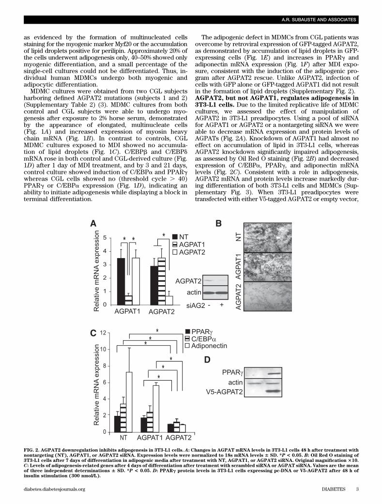
The adipogenic defect in MDMCs from CGL patients wasovercome by retroviral expression of GFP-tagged AGPAT2,as demonstrated by accumulation of lipid droplets in GFP-expressing cells (Fig. 1E) and increases in PPARg andadiponectin mRNA expression (Fig. 1F) after MDI expo-sure, consistent with the induction of the adipogenic pro-gram after AGPAT2 rescue. Unlike AGPAT2, infection ofcells with GFP alone or GFP-tagged AGPAT1 did not resultin the formation of lipid droplets (Supplementary Fig. 2).AGPAT2, but not AGPAT1, regulates adipogenesis in3T3-L1 cells. Due to the limited replicative life of MDMCcultures, we assessed the effect of manipulation ofAGPAT2 in 3T3-L1 preadipocytes. Using a pool of siRNAfor AGPAT1 or AGPAT2 or a nontargeting siRNA we wereable to decrease mRNA expression and protein levels ofAGPATs (Fig. 2A). Knockdown of AGPAT1 had almost noeffect on accumulation of lipid in 3T3-L1 cells, whereasAGPAT2 knockdown significantly impaired adipogenesis,as assessed by Oil Red O staining (Fig. 2B) and decreasedexpression of C/EBPa, PPARg, and adiponectin mRNAlevels (Fig. 2C). Consistent with a role in adipogenesis,AGPAT2 mRNA and protein levels increase markedly dur-ing differentiation of both 3T3-L1 cells and MDMCs (Sup-plementary Fig. 3). When 3T3-L1 preadipocytes weretransfected with either V5-tagged AGPAT2 or empty vector,
FIG. 2. AGPAT2 downregulation inhibits adipogenesis in 3T3-L1 cells. A: Changes in AGPAT mRNA levels in 3T3-L1 cells 48 h after treatment withnontargeting (NT), AGPAT1, or AGPAT2 siRNA. Expression levels were normalized to 18s mRNA levels 6 SD. *P < 0.05. B: Oil Red O staining of3T3-L1 cells after 7 days of differentiation in adipogenic media after treatment with NT, AGPAT1, or AGPAT2 siRNA. Original magnification 310.C: Levels of adipogenesis-related genes after 4 days of differentiation after treatment with scrambled siRNA or AGPAT siRNA. Values are the meanof three independent determinations 6 SD. *P < 0.05. D: PPARg protein levels in 3T3-L1 cells expressing pc-DNA or V5-AGPAT2 after 48 h ofinsulin stimulation (300 nmol/L).
A.R. SUBAUSTE AND ASSOCIATES
diabetes.diabetesjournals.org DIABETES 3
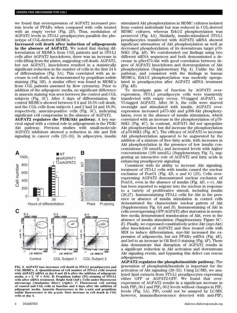
we found that overexpression of AGPAT2 increased pro-tein levels of PPARg when compared with cells treatedwith an empty vector (Fig. 2D). Thus, modulation ofAGPAT2 levels in 3T3-L1 preadipocytes parallels the phe-notype of CGL-derived MDMCs.Increased cell death after induction of adipogenesisin the absence of AGPAT2. We noted that during dif-ferentiation of MDMCs from CGL patients and in 3T3-L1cells after AGPAT2 knockdown, there was an increase incells lifting from the plates, suggesting cell death. AGPAT2,but not AGPAT1, knockdown resulted in a statisticallysignificant reduction in the number of cells in the first 24 hof differentiation (Fig. 3A). This correlated with an in-crease in cell death, as demonstrated by propidium iodidestaining (Fig. 3B). A similar effect was found in MDMCsfrom CGL patients assessed by flow cytometry. Prior toaddition of the adipogenic media, no significant differencein annexin staining was seen between the control and CGLsubjects (Fig. 3C). After 3 days of differentiation, thecontrol MDMCs showed between 9.4 and 10.5% cell death,and the CGL cells from subjects 1 and 2 had 24 and 18.3%,respectively, annexin-positive cells (Fig. 3C), indicatingsignificant cell compromise in the absence of AGPAT2.AGPAT2 regulates the PI3K/Akt pathway. A key sur-vival signal with a central role in adipogenesis is the PI3K/Akt pathway. Previous studies with small-moleculeAGPAT2 inhibitors showed a reduction in Akt and Erksignaling in cancer cells (21–24). In adipocytes, insulin
stimulated Akt phosphorylation in MDMC cultures isolatedfrom control individuals but was reduced in CGL-derivedMDMC cultures, whereas Erk1/2 phosphorylation waspreserved (Fig. 4A). Similarly, insulin-stimulated 3T3-L1preadipocytes transfected with AGPAT2 siRNA showedsignificant attenuation of Akt phosphorylation as well asdecreased phosphorylation of its downstream target p70-S6K1 (Fig. 4B). We corroborated our findings using twodifferent siRNA sequences and both demonstrated a de-crease in pSer473-Akt with good correlation between de-gree of AGPAT2 knockdown and downregulation of Aktphosphorylation (Supplementary Fig. 4). Unlike the Aktpathway, and consistent with the findings in humanMDMCs, Erk1/2 phosphorylation was modestly upregu-lated in preadipocytes after AGPAT2 siRNA treatment(Fig. 4B).
To investigate gain of function by AGPAT2 over-expression, 3T3-L1 preadipocyte cells were transientlytransfected with empty vector, V5-tagged AGPAT1, orV5-tagged AGPAT2. After 36 h, the cells were starvedovernight and stimulated with insulin. AGPAT2 over-expression increased p473-Akt and p308-Akt phosphory-lation, even in the absence of insulin stimulation, whichcorrelated with an increase in the phosphorylation of p70-S6K1 (Fig. 4C). In contrast, AGPAT1 did not upregulateAkt phosphorylation but did increase the phosphorylationof p70-S6K1 (Fig. 4C). The efficacy of AGPAT2 to increaseAkt phosphorylation appeared to be augmented by theaddition of a mixture of free fatty acids, with increases inAkt phosphorylation in the presence of low insulin con-centrations (50 nmol/L) and increased levels with higherconcentrations (100 nmol/L) (Supplementary Fig. 5), sug-gesting an interactive role of AGPAT2 and fatty acids inenhancing preadipocyte signaling.
Consistent with its ability to increase Akt signaling,treatment of 3T3-L1 cells with insulin caused the nuclearexclusion of FoxO1 (Fig. 4D, a and b) (25). Cells over-expressing AGPAT2 demonstrated nuclear exclusion ofFoxO1, even in the absence of insulin (Fig. 4D, c–e). Akthas been reported to migrate into the nucleus in responseto a variety of proliferative stimuli, including insulin(26,27). Immunostaining 3T3-L1 cells for Akt in the pres-ence or absence of insulin stimulation in control cellsdemonstrated the characteristic nuclear pattern of Akt(Supplementary Fig. 6A and B). Immunostaining of Akt incells overexpressing GFP-AGPAT2 after starvation in serum-free media demonstrated translocation of Akt, even in theabsence of insulin stimulation (Supplementary Figure 6C–E). Finally, we expressed constitutively active Akt (myr-Akt)after knockdown of AGPAT2 and then treated cells withMDI to induce differentiation. myr-Akt increased the ex-pression of adiponectin, but not PPARg mRNA (Fig. 4E),and led to an increase in Oil Red O staining (Fig. 4F). Thesedata demonstrate that disruption of AGPAT2 results ina significant reduction in Akt activation and downstreamAkt signaling events, and bypassing this defect can rescueadipogenesis.AGPAT2 regulates the phosphoinositide pathway. Thegeneration of phosphatidylinositols is important for theactivation of Akt signaling (28–32). Using LC/MS, we ana-lyzed lipid extracts from 3T3-L1 preadipocytes expressingeither GFP or AGPAT2-GFP. We found that over-expression of AGPAT2 results in a significant increase inboth PIP2 36:1 and PIP2 36:2 levels without changes in PIP1levels (Fig. 5A). PIP3 could not be assayed by LC/MS;however, immunofluorescence detected with anti-PIP3
FIG. 3. AGPAT2 loss increases cell death in 3T3-L1 preadipocytes andCGL MDMCs. A: Quantification of cell number of 3T3-L1 cells treatedwith AGPAT2 siRNA at day 0 and 48 h after the addition of adipogenicmedia. n = 3. *P < 0.01. B: Propidium iodine (PI) staining of 3T3-L1cells after siRNA treatment. Bright field (left). Cells under fluorescentmicroscopy (rhodamine filter) (right). C: Fluorescent cell sortingof control and CGL cells at baseline and 4 days after the addition ofadipogenic media. Annexin fluorescence is the x-axis and propidiumiodide fluorescence is the y-axis. Note increase in cell death in CGLcells at day 4.
UNDERLYING MECHANISM FOR CGL1
4 DIABETES diabetes.diabetesjournals.org
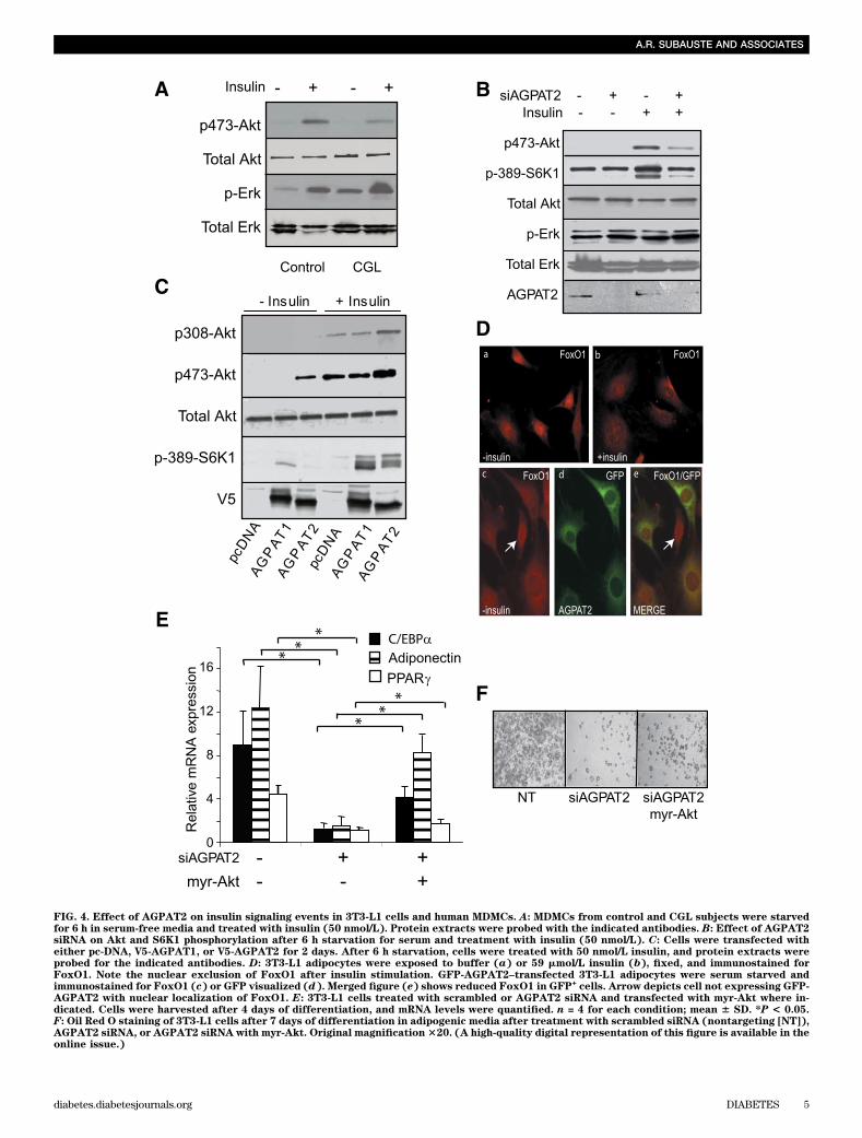
FIG. 4. Effect of AGPAT2 on insulin signaling events in 3T3-L1 cells and human MDMCs. A: MDMCs from control and CGL subjects were starvedfor 6 h in serum-free media and treated with insulin (50 nmol/L). Protein extracts were probed with the indicated antibodies. B: Effect of AGPAT2siRNA on Akt and S6K1 phosphorylation after 6 h starvation for serum and treatment with insulin (50 nmol/L). C: Cells were transfected witheither pc-DNA, V5-AGPAT1, or V5-AGPAT2 for 2 days. After 6 h starvation, cells were treated with 50 nmol/L insulin, and protein extracts wereprobed for the indicated antibodies. D: 3T3-L1 adipocytes were exposed to buffer (a) or 59 mmol/L insulin (b), fixed, and immunostained forFoxO1. Note the nuclear exclusion of FoxO1 after insulin stimulation. GFP-AGPAT2–transfected 3T3-L1 adipocytes were serum starved andimmunostained for FoxO1 (c) or GFP visualized (d). Merged figure (e) shows reduced FoxO1 in GFP
+cells. Arrow depicts cell not expressing GFP-
AGPAT2 with nuclear localization of FoxO1. E: 3T3-L1 cells treated with scrambled or AGPAT2 siRNA and transfected with myr-Akt where in-dicated. Cells were harvested after 4 days of differentiation, and mRNA levels were quantified. n = 4 for each condition; mean 6 SD. *P < 0.05.F: Oil Red O staining of 3T3-L1 cells after 7 days of differentiation in adipogenic media after treatment with scrambled siRNA (nontargeting [NT]),AGPAT2 siRNA, or AGPAT2 siRNA with myr-Akt. Original magnification320. (A high-quality digital representation of this figure is available in theonline issue.)
A.R. SUBAUSTE AND ASSOCIATES
diabetes.diabetesjournals.org DIABETES 5

antibody was increased after AGPAT2 overexpression andwas confirmed by quantification using direct detection onnitrocellulose membranes (Fig. 5B and C). Consistent witha role for AGPAT2 regulating the upstream signaling toAkt, we found that in serum-starved 3T3-L1 preadipocytes,AGPAT2 overexpression resulted in increased Akt phos-phorylation, which was completely abrogated by the PI3Kinhibitor LY294002 (Fig. 5D). The findings support a rolefor AGPAT2 as regulator of phosphatidylinositol levels inpreadipocytes, which in turn modulates Akt signaling.Effect of AGPAT1 and AGPAT2 on PPARg activityand glycerolipid levels. LPA has been described as anendogenous PPARg agonist, whereas CPA was recentlyidentified as a PPARg antagonist with nanomolar affinity(33,34). CPA is a naturally occurring analog of LPA, andalthough both share a similar structure, their biologicaleffects differ significantly (35). We assessed the effect ofAGPAT1 and AGPAT2 on PPARg transactivation and en-dogenous levels of these bioactive lipids. When NIH3T3cells were transfected with AGPAT1 or AGPAT2, onlyAGPAT2 increased expression of a PPARg luciferasepromoter driven by a canonical PPAR-responsive element
(PPRE) (36), both in the absence and presence of thePPARg ligand rosiglitazone (Fig. 6A). This was not asso-ciated with a significant increase in PPARg protein levelsor migration, suggesting that AGPAT2 overexpressionmodulates the transactivation of PPARg (Fig. 6B). It waspossible that the effect of AGPAT2 overexpression onPPARg activity was dependent on the activation of thePI3K/Akt pathway. Cells treated with the PI3K inhibitorLY294002 demonstrated ;50% downregulation of lucifer-ase activity (Fig. 6B). However, AGPAT2 overexpressionincreased the luciferase expression in both control andLY294002-treated cells, suggesting that the effect ofAGPAT2 was independent of the direct activation of Akt.
Knockdown of AGPAT2 resulted in an increase in dif-ferent LPA species (Fig. 7A), and, surprisingly, over-expression of AGPAT1 and AGPAT2 also resulted in anincrease in the levels of LPA (Fig. 7B) while simultaneouslydecreasing PA. Conversely, knockdown of AGPAT2 resultedin increased levels of the PPARg inhibitor CPA, in particular
FIG. 5. AGPAT2 increases PIP2 and PIP3. A: 3T3-L1 cells expressingGFP or GFP-AGPAT2 were harvested under basal conditions. Lipidextracts were analyzed with LC/MS. n = 5 for each condition; mean 6SD. *P < 0.05. B: Immunostaining for PIP3 of cells after 6 h starvationand insulin stimulation for the indicated times. C: PIP3 blot from lipidextracts of the same cells in serum-replete media. D: 3T3-L1 cellstransfected with either empty vector or AGPAT2 were starved for 6 h inserum-free media and treated with LY294002 10 nmol/L for 20 h, whereindicated. Protein extracts were probed for the indicated antibodies.
FIG. 6. AGPAT2 increases PPARg transactivation independent of PI3K.A: NIH3T3 cells were transfected with empty vector, AGPAT1, orAGPAT2 and PPREx3-LUC reporter, RXRa, and PPARg. Cells weretreated with rosiglitazone for 20 h, and relative luciferase activity wasquantified. n = 4 for each condition; mean 6 SD. *P < 0.05. B: PPARgprotein levels at 48 h of transfection. b-actin was used as loadingcontrol. C: NIH3T3 cells were transfected with empty vector orAGPAT2 and PPREx3-LUC reporter, RXRa, and PPARg. Cells weretreated with LY294002 (10 nmol/L) for 20 h, and relative luciferaseactivity was quantified. n = 3 for each condition; mean 6 SD. *P < 0.05.
UNDERLYING MECHANISM FOR CGL1
6 DIABETES diabetes.diabetesjournals.org
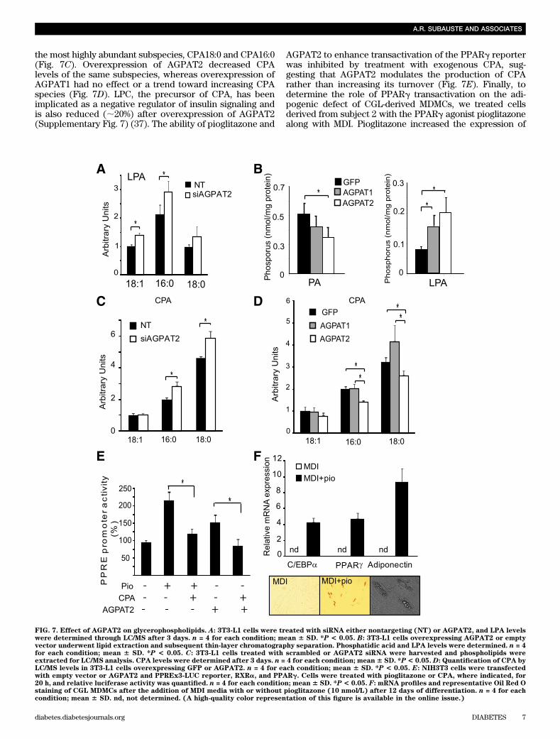
the most highly abundant subspecies, CPA18:0 and CPA16:0(Fig. 7C). Overexpression of AGPAT2 decreased CPAlevels of the same subspecies, whereas overexpression ofAGPAT1 had no effect or a trend toward increasing CPAspecies (Fig. 7D). LPC, the precursor of CPA, has beenimplicated as a negative regulator of insulin signaling andis also reduced (;20%) after overexpression of AGPAT2(Supplementary Fig. 7) (37). The ability of pioglitazone and
AGPAT2 to enhance transactivation of the PPARg reporterwas inhibited by treatment with exogenous CPA, sug-gesting that AGPAT2 modulates the production of CPArather than increasing its turnover (Fig. 7E). Finally, todetermine the role of PPARg transactivation on the adi-pogenic defect of CGL-derived MDMCs, we treated cellsderived from subject 2 with the PPARg agonist pioglitazonealong with MDI. Pioglitazone increased the expression of
FIG. 7. Effect of AGPAT2 on glycerophospholipids. A: 3T3-L1 cells were treated with siRNA either nontargeting (NT) or AGPAT2, and LPA levelswere determined through LC/MS after 3 days. n = 4 for each condition; mean 6 SD. *P < 0.05. B: 3T3-L1 cells overexpressing AGPAT2 or emptyvector underwent lipid extraction and subsequent thin-layer chromatography separation. Phosphatidic acid and LPA levels were determined. n = 4for each condition; mean 6 SD. *P < 0.05. C: 3T3-L1 cells treated with scrambled or AGPAT2 siRNA were harvested and phospholipids wereextracted for LC/MS analysis. CPA levels were determined after 3 days. n = 4 for each condition; mean6 SD. *P< 0.05. D: Quantification of CPA byLC/MS levels in 3T3-L1 cells overexpressing GFP or AGPAT2. n = 4 for each condition; mean 6 SD. *P < 0.05. E: NIH3T3 cells were transfectedwith empty vector or AGPAT2 and PPREx3-LUC reporter, RXRa, and PPARg. Cells were treated with pioglitazone or CPA, where indicated, for20 h, and relative luciferase activity was quantified. n = 4 for each condition; mean6 SD. *P < 0.05. F: mRNA profiles and representative Oil Red Ostaining of CGL MDMCs after the addition of MDI media with or without pioglitazone (10 nmol/L) after 12 days of differentiation. n = 4 for eachcondition; mean 6 SD. nd, not determined. (A high-quality color representation of this figure is available in the online issue.)
A.R. SUBAUSTE AND ASSOCIATES
diabetes.diabetesjournals.org DIABETES 7
terminal differentiation markers C/EBPa and PPARg, andthis associated with the accumulation of lipid droplets, asshown by Oil Red O staining (Fig. 7F), though this effectwas still significantly reduced compared with the differ-entiation observed in cells from control individuals.
DISCUSSION
AGPAT2 mutations have been found in a subset ofpatients with CGL, and it was presumed that these sub-jects were unable to generate triglycerides, resulting in“empty adipocytes” (3). However, earlier studies sug-gested that AGPAT2 is involved in adipogenesis. Ourwork in MDSCs from humans with AGPAT2 mutationsas well as in 3T3-L1 cells shows that AGPAT2 plays a cen-tral role in adipogenesis by generating a proadipogeniclipome, leading to the activation of Akt signaling while
simultaneously regulating the production of CPA, a PPARginhibitor.
AGPAT2 is upregulated in a number of tumors, includingovarian cancer (38,39) and small-molecule inhibitors havebeen developed that specifically inhibit AGPAT2. Theseagents result in the attenuation of a number of signalingpathways, including both the Ras/Raf/Erk and PI3K/Aktpathways (9). Our work demonstrates that gain and lossof AGPAT2, but not AGPAT1, has concordant effects onthe phosphorylation of Akt and its downstream targets.Further, we find that constitutively active Akt can partiallyrescue the adipogenic defect, demonstrating that loss ofAGPAT2 is working proximal to Akt activation. In the ab-sence of AGPAT2, differentiating CGL-derived progenitorcells had increased cell death, similar to AGPAT2-expressingtumor cells after treatment with an AGPAT2-specific small-molecule inhibitor, suggesting that apoptosis of differentiating
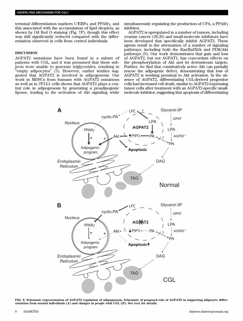
FIG. 8. Schematic representation of AGPAT2 regulation of adipogenesis. Schematic of proposed role of AGPAT2 in supporting adipocyte differ-entiation from normal individuals (A) and changes in people with CGL (B). See text for details.
UNDERLYING MECHANISM FOR CGL1
8 DIABETES diabetes.diabetesjournals.org
adipocytes may play a role in the severe lipodystrophicphenotype seen in CGL patients. Both Akt and PPARg areinvolved in adipocyte survival (40). A provocative obser-vation is that addition of fatty acids to cells overexpressingAGPAT2 further increases Akt phosphorylation, suggest-ing a physiological mechanism whereby increased flux offatty acids may participate in the stimulation of the adi-pogenic program. Further work on the specificity of fattyacids and concentration and duration of exposure will beimportant to define these relationships.
We continue to try to understand the apparently para-doxical effects of AGPAT2 on PA and LPA levels in cells.Ovarian cancer cells in which AGPAT2 is highly expressedshow an increase in LPA levels, and knockdown ofAGPAT2 in OP9 cells results in increases in PA levels,identical to our findings (41). As with AGPAT2, disruptionof the Lipin-1 gene results in lipodystrophy in mice (42,43). PA results in Lipin-1 exclusion from the nucleus andaccumulation in the endoplasmic reticulum (44,45). Onepotential explanation for the paradoxical PA changes isthat the PA pool affected by AGPAT2 is not reflected intotal cells extracts. In preliminary studies, we have foundthat AGPAT2 overexpression causes the nuclear exclusionof Lipin-1 (A.R.S., unpublished observation), which isconsistent with a localized increase of PA in the endo-plasmic reticulum. Further studies will be needed to sortout the complex changes in lipid metabolism that result inunexpected changes in bulk PA and LPA levels in cellsafter alterations in AGPAT2 expression.
Although AGPAT2 mRNA is detected in 3T3-L1 pre-adipocytes prior to differentiation, there is a significantincrease after induction of adipogenesis, paralleling that ofPPARg, and it is also increased in vivo after treatment withthe PPARg agonist pioglitazone (46), placing AGPAT2 asa PPARg-responsive gene. Sequence analysis showsa PPAR direct repeat at2723 from the transcriptional startsite. The finding that AGPAT2 reduces CPA levels in pre-adipocytes could suggest that the progressive increase inPPARg with differentiation is accompanied by reduction inCPA levels, allowing activation of PPARg. These findingssupport a model in which AGPAT2 joins PPARg andc/EBPa as part of a positive feedback loop promoting themature adipocyte phenotype.
Treatment of CGL-derived MDMCs with pioglitazonewas able to increase the expression of adipocyte terminaldifferentiation markers and generate lipid droplets, al-though this effect was markedly attenuated comparedwith MDMCs derived from control individuals. Despitethe almost complete absence of adipose tissue, subject 3(Supplementary Table 2) responded to therapy withPPARg agonists. Her HbA1c was reduced from 14.6 to9.7% and her triglycerides decreased from 727 mg/dL to213 mg/dL after 6 months of treatment (47). Consideringthe in vitro response of MDMCs to pioglitazone, it istempting to suggest that generation of adipocytes fromprecursor cells helped alleviate some of the metabolicderangements in this patient, although it is possible thatthiazolidinedione exerted its metabolic effect in-dependent of adipose tissue (48) (49,50). AGPAT2 andPPARg are both expressed in other insulin-responsivetissues, including liver and skeletal muscle. This wouldraise the possibility that AGPAT2 is important for theregulation of PPARg and PI3K/Akt in tissues, whichcontributes to the severe insulin resistance in CGLpatients. Although AGPAT2 is expressed in liver, the he-patic steatosis seen in CGL subjects appears to be
secondary to the insulin resistance and not to a directeffect of AGPAT2 on hepatic lipogenesis (51).
In summary, this study provides a further un-derstanding of the pathogenesis of lipodystrophy inpatients with AGPAT2 mutations. Our findings demon-strate that CGL secondary to AGPAT2 mutation is nota disease of empty adipocytes, rather a disease of de-fective adipose tissue development due to a disruption inthe modulation of the lipome during adipogenesis(Fig. 8). Normally, upon activation of the differentiationprogram, AGPAT2 expression increases and reducescyclic PA levels, possibly by directing LPA generatedfrom LPC to PA. This allows endogenously derivedPPARg activators to increase PPARg-mediated activationof the adipogenic program. The increased flux to PAcould be the basis for increasing the generation of PIP3,which is important for activation of Akt. Because of theparadoxical change in LPA in cells with AGPAT2 ma-nipulation, we believe that the PA generated by AGPAT2is compartmentalized from that generated by AGPAT1(Fig. 8A, dashed circle). In patients with CGL due toAGPAT2 mutations, cyclic PA increases and inhibitsPPARg, and the reduced generation of PA could beleading to reduced PIP3 levels, impaired activation ofAkt, and an increase in apoptosis (Fig. 8B). Un-derstanding AGPAT2 actions in adipogenesis in relationto the modulation of the lipome provides the potential forunderstanding the intimate relationship between lipids,adipogenesis, lipid metabolism, and insulin resistance incommon obesity.
ACKNOWLEDGMENTS
This work was supported by the Michigan NutritionObesity Research Center (DK-089503) MetabolomicsCore and Pilot Grant Program and by the Robert C. andVeronica Atkins Research Foundation.
No potential conflicts of interest relevant to this articlewere reported.
A.R.S. designed and performed the experiments andwrote the manuscript. A.K.D., X.L., B.E., C.E., M.E.A., andM.T. performed the experiments. E.O. and T.L. contributedto the experiments and manuscript preparation. C.F.B.contributed to experimental design and writing and editingthe manuscript. C.F.B. and A.R.S. are the guarantors of thiswork and, as such, had full access to all the data in thestudy and take responsibility for the integrity of the dataand the accuracy of the data analysis.
REFERENCES
1. Huang-Doran I, Sleigh A, Rochford JJ, O’Rahilly S, Savage DB. Lipodys-trophy: metabolic insights from a rare disorder. J Endocrinol 2010;207:245–255
2. Cao H, Alston L, Ruschman J, Hegele RA. Heterozygous CAV1 frameshiftmutations (MIM 601047) in patients with atypical partial lipodystrophy andhypertriglyceridemia. Lipids Health Dis 2008;7:3
3. Agarwal AK, Arioglu E, De Almeida S, et al. AGPAT2 is mutated in con-genital generalized lipodystrophy linked to chromosome 9q34. Nat Genet2002;31:21–23
4. Miranda DM, Wajchenberg BL, Calsolari MR, et al. Novel mutations of theBSCL2 and AGPAT2 genes in 10 families with Berardinelli-Seip congenitalgeneralized lipodystrophy syndrome. Clin Endocrinol (Oxf) 2009;71:512–517
5. Leung DW. The structure and functions of human lysophosphatidic acidacyltransferases. Front Biosci 2001;6:D944–D953
6. Simha V, Garg A. Phenotypic heterogeneity in body fat distributionin patients with congenital generalized lipodystrophy caused by
A.R. SUBAUSTE AND ASSOCIATES
diabetes.diabetesjournals.org DIABETES 9
mutations in the AGPAT2 or seipin genes. J Clin Endocrinol Metab 2003;88:5433–5437
7. Bonham L, Leung DW, White T, et al. Lysophosphatidic acid acyltransferase-beta: a novel target for induction of tumour cell apoptosis. Expert Opin TherTargets 2003;7:643–661
8. Hideshima T, Chauhan D, Hayashi T, et al. Antitumor activity of lyso-phosphatidic acid acyltransferase-beta inhibitors, a novel class of agents,in multiple myeloma. Cancer Res 2003;63:8428–8436
9. Coon M, Ball A, Pound J, et al. Inhibition of lysophosphatidic acid acyl-transferase beta disrupts proliferative and survival signals in normal cellsand induces apoptosis of tumor cells. Mol Cancer Ther 2003;2:1067–1078
10. Gale SE, Frolov A, Han X, et al. A regulatory role for 1-acylglycerol-3-phosphate-O-acyltransferase 2 in adipocyte differentiation. J Biol Chem2006;281:11082–11089
11. Wada MR, Inagawa-Ogashiwa M, Shimizu S, Yasumoto S, Hashimoto N.Generation of different fates from multipotent muscle stem cells. De-velopment 2002;129:2987–2995
12. Williams JT, Southerland SS, Souza J, Calcutt AF, Cartledge RG. Cellsisolated from adult human skeletal muscle capable of differentiating intomultiple mesodermal phenotypes. Am Surg 1999;65:22–26
13. Asakura A, Komaki M, Rudnicki M. Muscle satellite cells are multipotentialstem cells that exhibit myogenic, osteogenic, and adipogenic differentia-tion. Differentiation 2001;68:245–253
14. Cossu G, De Angelis L, Borello U, et al. Determination, diversification andmultipotency of mammalian myogenic cells. Int J Dev Biol 2000;44:699–706
15. Subauste AR, Burant CF. Role of FoxO1 in FFA-induced oxidative stress inadipocytes. Am J Physiol Endocrinol Metab 2007;293:E159–E164
16. Ogiso H, Taguchi R. Reversed-phase LC/MS method for polyphosphoinositideanalyses: changes in molecular species levels during epidermal growth factoractivation in A431 cells. Anal Chem 2008;80:9226–9232
17. Bligh EG, Dyer WJ. A rapid method of total lipid extraction and purifica-tion. Can J Biochem Physiol 1959;37:911–917
18. Subauste AR, Elliott B, Das AK, Burant CF. A role for 1-acylglycerol-3-phosphate-O-acyltransferase-1 in myoblast differentiation. Differentiation2010;80:140–146
19. Shan L, Li S, Jaffe K, Davis L. Quantitative determination of cyclic phos-phatidic acid in human serum by LC/ESI/MS/MS. J Chromatogr B AnalytTechnol Biomed Life Sci 2008;862:161–167
20. Sato Y, Nakamura T, Aoshima K, Oda Y. Quantitative and wide-rangingprofiling of phospholipids in human plasma by two-dimensional liquidchromatography/mass spectrometry. Anal Chem 2010;82:9858–9864
21. Zhang HH, Huang J, Düvel K, et al. Insulin stimulates adipogenesis throughthe Akt-TSC2-mTORC1 pathway. PLoS ONE 2009;4:e6189
22. Garg A, Agarwal AK. Lipodystrophies: disorders of adipose tissue biology.Biochim Biophys Acta 2009;1791:507–513
23. Baudry A, Yang ZZ, Hemmings BA. PKBalpha is required for adipose dif-ferentiation of mouse embryonic fibroblasts. J Cell Sci 2006;119:889–897
24. Longo KA, Kennell JA, Ochocinska MJ, Ross SE, Wright WS, MacDougaldOA. Wnt signaling protects 3T3-L1 preadipocytes from apoptosis throughinduction of insulin-like growth factors. J Biol Chem 2002;277:38239–38244
25. Nakae J, Kitamura T, Kitamura Y, Biggs WH 3rd, Arden KC, Accili D. Theforkhead transcription factor Foxo1 regulates adipocyte differentiation.Dev Cell 2003;4:119–129
26. Meier R, Alessi DR, Cron P, Andjelković M, Hemmings BA. Mitogenic ac-tivation, phosphorylation, and nuclear translocation of protein kinaseBbeta. J Biol Chem 1997;272:30491–30497
27. Meier R, Hemmings BA. Regulation of protein kinase B. J Recept SignalTransduct Res 1999;19:121–128
28. Magun R, Burgering BM, Coffer PJ, et al. Expression of a constitutivelyactivated form of protein kinase B (c-Akt) in 3T3-L1 preadipose cellscauses spontaneous differentiation. Endocrinology 1996;137:3590–3593
29. Sakaue H, Ogawa W, Matsumoto M, et al. Posttranscriptional control ofadipocyte differentiation through activation of phosphoinositide 3-kinase.J Biol Chem 1998;273:28945–28952
30. Xia X, Serrero G. Inhibition of adipose differentiation by phosphatidylinositol3-kinase inhibitors. J Cell Physiol 1999;178:9–16
31. Kohn AD, Summers SA, Birnbaum MJ, Roth RA. Expression of a consti-tutively active Akt Ser/Thr kinase in 3T3-L1 adipocytes stimulates glucoseuptake and glucose transporter 4 translocation. J Biol Chem 1996;271:31372–31378
32. Sopasakis VR, Liu P, Suzuki R, et al. Specific roles of the p110alpha iso-form of phosphatidylinsositol 3-kinase in hepatic insulin signaling andmetabolic regulation. Cell Metab 2010;11:220–230
33. McIntyre TM, Pontsler AV, Silva AR, et al. Identification of an intracellularreceptor for lysophosphatidic acid (LPA): LPA is a transcellular PPAR-gamma agonist. Proc Natl Acad Sci USA 2003;100:131–136
34. Tsukahara T, Tsukahara R, Fujiwara Y, et al. Phospholipase D2-dependentinhibition of the nuclear hormone receptor PPARgamma by cyclic phos-phatidic acid. Mol Cell 2010;39:421–432
35. Tania M, Khan MA, Zhang H, Li J, Song Y. Autotaxin: a protein with twofaces. Biochem Biophys Res Commun 2010;401:493–497
36. Hegele RA, Cao H, Frankowski C, Mathews ST, Leff T. PPARG F388L,a transactivation-deficient mutant, in familial partial lipodystrophy. Di-abetes 2002;51:3586–3590
37. Han MS, Lim YM, Quan W, et al. Lysophosphatidylcholine as an effector offatty acid-induced insulin resistance. J Lipid Res 2011;52:1234–1246
38. Burton A. LPAAT-beta identifies aggressive ovarian cancer. Lancet Oncol2006;7:893
39. Springett GM, Bonham L, Hummer A, et al. Lysophosphatidic acidacyltransferase-beta is a prognostic marker and therapeutic target ingynecologic malignancies. Cancer Res 2005;65:9415–9425
40. Imai T, Takakuwa R, Marchand S, et al. Peroxisome proliferator-activatedreceptor gamma is required in mature white and brown adipocytes fortheir survival in the mouse. Proc Natl Acad Sci USA 2004;101:4543–4547
41. Pua TL, Wang FQ, Fishman DA. Roles of LPA in ovarian cancer de-velopment and progression. Future Oncol 2009;5:1659–1673
42. Péterfy M, Phan J, Xu P, Reue K. Lipodystrophy in the fld mouse resultsfrom mutation of a new gene encoding a nuclear protein, lipin. Nat Genet2001;27:121–124
43. Zhang P, Takeuchi K, Csaki LS, Reue K. Lipin-1 phosphatidic acid phos-phatase activity modulates phosphatidic acid levels to promote inductionof peroxisome proliferator-activated receptor gamma (PPARgamma) geneexpression during adipogenesis. J Biol Chem 2012;287:3485–3494
44. Ren H, Federico L, Huang H, et al. A phosphatidic acid binding/nuclear local-ization motif determines lipin1 function in lipid metabolism and adipogenesis.Mol Biol Cell 2010;21:3171–3181
45. Antonescu CN, Danuser G, Schmid SL. Phosphatidic acid plays a regula-tory role in clathrin-mediated endocytosis. Mol Biol Cell 2010;21:2944–2952
46. Yao-Borengasser A, Rassouli N, Varma V, et al. Stearoyl-coenzyme A de-saturase 1 gene expression increases after pioglitazone treatment and isassociated with peroxisomal proliferator-activated receptor-gamma re-sponsiveness. J Clin Endocrinol Metab 2008;93:4431–4439
47. Arioglu E, Duncan-Morin J, Sebring N, et al. Efficacy and safety oftroglitazone in the treatment of lipodystrophy syndromes. Ann InternMed 2000;133:263–274
48. Burant CF, Sreenan S, Hirano K, et al. Troglitazone action is independentof adipose tissue. J Clin Invest 1997;100:2900–2908
49. Norris AW, Chen L, Fisher SJ, et al. Muscle-specific PPARgamma-deficientmice develop increased adiposity and insulin resistance but respond tothiazolidinediones. J Clin Invest 2003;112:608–618
50. Hevener AL, He W, Barak Y, et al. Muscle-specific Pparg deletion causesinsulin resistance. Nat Med 2003;9:1491–1497
51. Agarwal AK, Sukumaran S, Cortés VA, et al. Human 1-acylglycerol-3-phosphate O-acyltransferase isoforms 1 and 2: biochemical characterizationand inability to rescue hepatic steatosis in Agpat2(-/-) gene lipodystrophicmice. J Biol Chem 2011;286:37676–37691
UNDERLYING MECHANISM FOR CGL1
10 DIABETES diabetes.diabetesjournals.org