Embed Size (px)
Citation preview
Egypt. J. Chem. Environ. Health, 2 (2):167 - 182 (2016) On line ISSN: 2536-9164
761
Molecular Characterization and Hemato Biochemical Studies of
Reovirus in Ismailia Farms
Neven M. Ramzy1, Hala N. Ibrahim
2, Seham F. ElHadad
3
Institute of Animal health research, Dokki, Egypt. Department of Virology1, Department of Clinical
pathology2
Ismailia branch, Department of Pathology3 ,
Tanta branch
Abstract
The avian reoviruse (AVR) induces various manifestations in chickens. They
are associated with disease conditions including malabsorption syndrome and
tenosynovitis. RT-PCR is a rapid, sensitive and specific diagnostic test for ARV
detection and avoiding economic losses. Three broiler poultry farms in Ismailia with
reovirus like symptoms were screened for Reoviruses in SPF embryonated chicken
eggs. In these farms, there were 20-25% morbidities at 24th day of age and 3-4%
morbidities at 14th day of age, and 5-6% mortalities at 24th day, while 2-3 % is at
the 14th day. Three isolates of Reovirus were obtained and characterized and
identified by RT-PCR. Electrophoretic pattern showed a specific band at 399 bp
positive at lane 1, 3 and negative at lane 2. Results of haematological investigation
showed low levels of total erythrocytic count (TEC), haemoglobin (Hb), and packed
cell volume (PCV) but significant increase in level of total leukocytic count (TLC),
absolute lymphocyte count (ALC) and absolute monocyte count (AMC) in infected
chickens compared to apparently healthy. Biochmical estimation revealed marked
elevation of total serum proteins, AST; ALT. Pathological changes in internal organs
of the infected chicks were also described. The proventriculus revealed marked
hyperplasia of lymphoid aggregates and the small intestine revealed marked loss of
villi with replacement by fibrin, necrotic debris, high numbers of macrophages and
lymphocytes .All these alterations were suggested to interfere with the immunity and
normal digestive processes resulting in poor weight gain and stunting of chicks.
Key words: Reovirus- PCR- sequence- chicken- hematobiochemical alteration-Pathology
Introduction
Reoviruses belong to the genus Orthoreovirus, in the family Reoviridae. Virus
particles measure 70 nm to 80 nm, are non-enveloped and have icosahedral symmetry
with a double-shelled arrangement of surface protein. The virus contains double-
stranded ribonucleic acid which has ten segments. The genome can be separated into
three size classes, namely: L (large), M (medium) and S (small). Protein coding
assignments of all ten genome segments of strain S I 133 have been determined
(varela and Benavente, 1994). Both vertical and horizontal transmission of avian
reoviruses is recognised. Egg transmission has been confirmed after experimental
Egypt. J. Chem. Environ. Health, 2 (2):167 - 182 (2016) On line ISSN: 2536-9164
761
infection (Vander Heide et al., 1974, Menendez N.A.et al., 1975, Al-Mufarred et
al., 1996), but the rate of transmission is probably very low in nature. The
transmission of the infection to susceptible chickens is realized horizontally. The
vertical route of transmission is also proved. Reoviruses could persist in infected birds
for more than 40 weeks. Avian reoviruses are ubiquitous among poultry flocks.
Although infection is usually present without disease, reoviruses may occasionally be
involved in several disease syndromes of which viral arthritis/tenosynovitis in
chickens is the most important, particularly in broiler breeds. Diagnosis depends on
detection of the virus in clinical samples, although the presence of the virus does not
necessarily confirm that this is the cause of the disease, except where reoviruses are
detected in affected joints. Serological tests are usually difficult to interpret in view of
widespread and frequently harmless reovirus infection. Many vaccines are based on
the S1133 strain isolated in the United States of America, but these may not be
effective against antigenic variants (Varela and Benavente, 1994).
The stunting syndrome in broilers is associated with a reovirus infection but according
to some studies, the role of the reovirus is probably secondary. It is characterized by a
considerably reduced live weight in affected birds and a various degree of uniformity
in the flock varying from 5-10% to 40-50%, usually seen after the age of 14 days.
Reoviruses are highly resistant to a number of environmental factors such as
temperature, pH etc. Reoviruses are shed with faeces and could contaminate the egg
shells. The polymerase chain reaction (PCR) as a diagnostic technique is generally
known as a very sensitive, specific, and rapid tool for detection of viruses. It has been
shown that reverse transcription (RT)-PCR can be used in the detection of avian
reovirus (Lee et al., 1998; Liu et al., 1999a, 2004; Bruhn et al., 2005). However,
conventional PCR tests can be hampered by the high risk of contamination by
previously amplified materials.
Haematological examination of infected birds shows lower levels of haemoglobin
(Hb), total erythrocyte count (TEC) and total leucocyte count (TLC) as compared to
normal . Absolute lymphocyte count (ALC), absolute monocyte count (AMC) and
absolute eosinophil count were also found lower than normal as stated by
(Muhammad et al., 2014). A marked elevation of total serum proteins with
corresponding increase of serum albumin level in infected chickens was observed,
(Singh et al., 2005). All these changes may interfere with normal digestive processes
and normal body functions resulting in poor weight gain and retarded growth or
stunting chicks.
Histopathological changes due to reovirus infection including viral arthritis /
tenosynovitis, stunting syndrome, respiratory disease , enteric disease,
immunosuppression and malabsorbtion syndrome (Rosenberger and Olson 1997,
sterner et al , 1989, Van der Heide, 1996, 2000) . Pericarditis, myocarditis and
immunosuppression also occur (Mc Nuly 1993).
Egypt. J. Chem. Environ. Health, 2 (2):167 - 182 (2016) On line ISSN: 2536-9164
761
This study aimed to detect the degree of spreading of AVR infection between some
poultry farms in Ismailia governorate using the real –time RT-PCR as a rapid,
sensitive and specific test. Also, the changes in hematological and some biochemical
parameters of infected chickens were investigated as indication on the effect on and
immunity, and for avoiding the economic losses by dealing with such parameters.
Materials and Methods
Isolation of Reovirus:
The broiler ARV field strain was isolated from ten farms, ten samples from farm. The
observed symptoms were diarrhea, malabsorption, respiratory and intestinal disorders.
The organs (intestine, proventriculus, lung, liver and spleen) were frozen-thawed for 2-
3 times samples which collected from sick chickens (slaughter chickens) were
homogenized in sterile phosphate-buffered saline (PBS, pH 7.2) to give a 20%
suspension (w/v). After centrifugation at 3000 _ g for 20 min, the supernatants were
filtered through 0.2 mm syringe filters. The filtered suspension was injected into the
allantoic sac of 9-day-old chick embryos (0.2 ml/embryo). Embryos were candled daily
for 7 days and the amino-allantoic fluids of the infected embryos were collected for
further passage in the embryos of chickens and specific pathogen free (SPF). (Schat
and Purchase, 1989). After third passage embryo showed stunting in growth.
RNA extraction: RNA extraction from samples was performed using the QIAamp
viral RNA Mini kit (Qiagen, Germany, GmbH). Briefly, 140 µl of the sample
suspension was incubated with 560 µl of AVL lysis buffer and 5.6 µl of carrier RNA
at room temperature for 10 min. After incubation, 560 µl of 100% ethanol was added
to the lysate. The sample was then washed and centrifuged following the
manufacturer’s recommendations. Nucleic acid was eluted with 60 µl of elution buffer
provided in the kit.
Oligonucleotide Primers:
supplied from (Metabion Germany) are listed in table (1).
Egypt. J. Chem. Environ. Health, 2 (2):167 - 182 (2016) On line ISSN: 2536-9164
711
Table (1). Primers sequences, target genes, amplicon sizes
PCR amplification: Was done according to (Bruhn et al 2005)Primers were utilized
in a 25- µl reaction containing 12.5 µl of Quantitect probe rt-PCR buffer (QIAgen,
Gmbh), 1 µl of each primer of 20 pmol concentration, 0.25 µl of rt-enzyme 4.25 µl of
water, and 6 µl of template. The reaction was performed in a Biometra thermal cycler.
Reverse transcription was applied at 50 O
C for 30 min, a primary denaturation step was
done at 95 O
C for 5 min, followed by 35 cycles of 94OC for 30 sec., 55
OC for 45 sec.
and 72OC for 45 sec. min. A final extension step was done at 72
OC for 10 min.
Analysis of the PCR Products:
The products of PCR were separated by electrophoresis on 1.5% agarose gel
(Applichem, Germany, GmbH) in 1x TBE buffer at room temperature using gradients
of 5V/cm. For gel analysis, 15 µl of the products was loaded in each gel slot. A 100bp
DNA ladder (Qiagen, Germany, GmbH) was used to determine the fragment sizes.
The gel was photographed by a gel documentation system (Alpha Innotech, Biometra)
and the data was analyzed through computer software.
Hematological and biochemical studies:
1-Haematological studies:
Blood samples were taken from diseased and apparently healthy birds ( 10 samples of each )
two samples one without anticoagulant for serum separation and other one with anticoagulant
for evaluation of red blood corpuscles (RBCs 106/mm
3), heamoglobin (Hbgm/dl), packed cell
volume (PCV %) and blood indices (mean corpuscular volume MCV fl, mean corpuscular
haemoglbin MCH pg and mean corpuscular haemoglobin concentration MCHC %), total
leukocytic count (WBCs 103/mm
3) ,and differential leukocytic count were determined
according to routine haematological examination and standard blood smear (Jain., 2000) .
2-Serum biochemical parameters:-
The level of alanine aminotransferase (ALT), aspartate aminotransferase (AST), total
protein, albumin, uric acid, creatinine, were estimated by using auto analyzeospital
Hitachi 912 in Suez Canal University Hospital. Estimation of globulin was difference
between total protein and albumin (Kaneko et al.., 1997).
Length of
amplified
product (bp)
Primer sequence
(5'-3')
Target
gene
Primer
399 bp CCC ATG GCA ACG ATT TC
S2
REO-F
TTC GGC CAG GTC TCA AC
REO-R
Egypt. J. Chem. Environ. Health, 2 (2):167 - 182 (2016) On line ISSN: 2536-9164
717
3-Statisttical Analysis:
Data collected from haematological and serum biochemical results of different groups
of chickens were statistically analyzed for the mean and standard deviation of analysis
were performed according to ( Sndecor and Cochran.,1982).
4-Pathological examination:
Postmortem examination was done immediately after slaughtering and tissue
specimens from liver, kidney, lung, proventriculs and intestine were collected and
fixed in 10% neutral buffered formalin. They were routinely processed by standard
paraffin embedding technique. Section at 4 micron, stained with Hematoxylin and
Eosin (Bancroft and Gamble 2002)
Result and Discussion
The clinical signs were characterisitic for reovirus infection in the present study.
Affected chickens showed depression. Respiratory disorders, weight loss and diarrhea.
The demonstrated morbidity did not involve all chickens in the farms under study. .
Reovirus infection caused 30% morbidity and 20% mortality in Israeli poultry farms
and the disease first appeared at 10 days of age and persisted in an affected Flock until
six weeks of age. A reliable reverse transcriptase polymerase chain Reaction (RT-
PCR) method was developed to detect ARV contaminations in poultry vaccines.
Because ARVs exhibit diversity and heterogeneity in their genome (Liu et al., 1999a,
2004), in recent years, several attempts have been made to detect ARV in chicken
tissues or cultured cells by conventional RTPCR (Lee et al., 1998; Liu et al., 1999a,
1999b, 2004; Bruhn et al., 2005). Reovirus infection caused 30% morbidity and 20%
mortality in Israeli poultry farms and the disease first appeared at 10 days of age and
persisted in an affected Flock until six weeks of age. In the present study reovirus
infection caused 20-25% morbidities at 24th day of age and 3-4% morbidities at 14th
day of age, and 5-6% mortalities at 24th day, while 2-3 % is at 14th day. So, the
mortality rate at the younger ages seemed to be non-significant in the field of poultry
industry. Rapid diagnostic detection of reovirus is particularly important to the poultry
industry in order to prevent spreading of the disease and to limit economic losses. PCR
has in addition benefits of being cost effective, time saving, specific and sensitive;
furthermore, it has been used for screening and surveillance of poultry flocks (Pang.
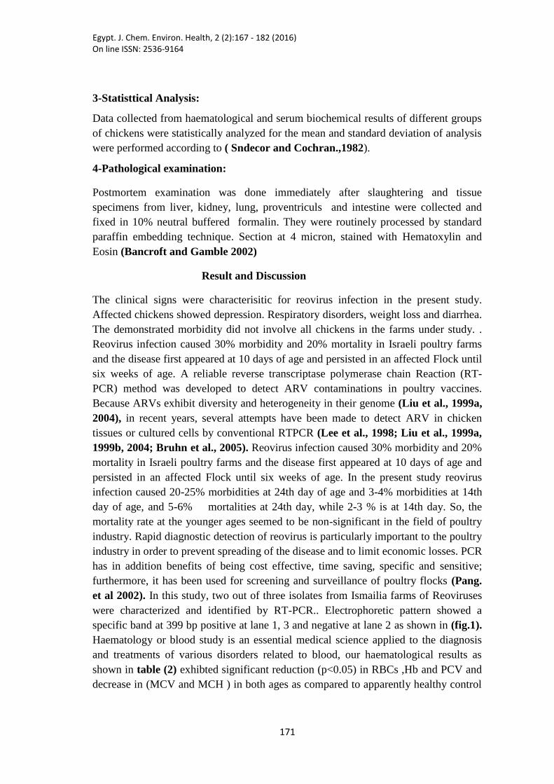
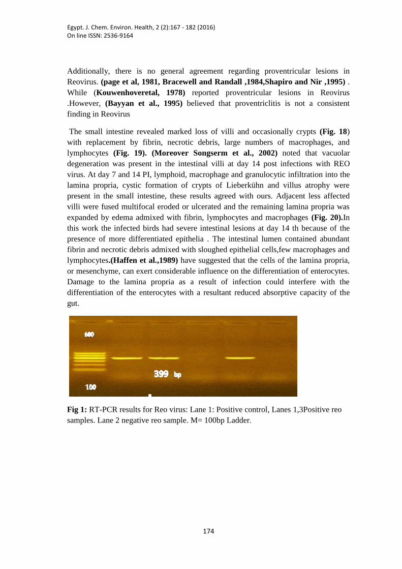
et al 2002). In this study, two out of three isolates from Ismailia farms of Reoviruses
were characterized and identified by RT-PCR.. Electrophoretic pattern showed a
specific band at 399 bp positive at lane 1, 3 and negative at lane 2 as shown in (fig.1).
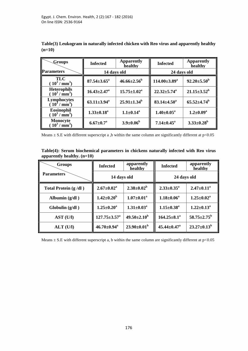
Haematology or blood study is an essential medical science applied to the diagnosis
and treatments of various disorders related to blood, our haematological results as
shown in table (2) exhibted significant reduction (p<0.05) in RBCs ,Hb and PCV and
decrease in (MCV and MCH ) in both ages as compared to apparently healthy control
Egypt. J. Chem. Environ. Health, 2 (2):167 - 182 (2016) On line ISSN: 2536-9164
711
chickens these findings illustrated anaemic changes (microcytic hypochromic
anaemia) which may be caused by malabsorptionsyndrom,our results come agree
with(Singh et al., 2005), (Nilli et al., 2007) and (Rani et al., 2011). Total leukocytic
count (TLC), absolute lymphocyte count (ALC),and absolute monocyte count (AMC)
significantly increased (p<0.05) in infected chickens 14 days and 24 days old as
shown in table (3) such findings are partially in agreement with (Rani et al., 2011)
who reported leukocytosis with monocytosis but lymphopenia had occuared.
Leukocytosis could be due to many reasons including stress and infection, (Otto et al.,
2006).Significant increase of monocytes in response of invading micro-organisms,
inflammation by migration into the tissues and necrotic materials, (Seller et al., 2010).
The lymphocytes increased number was seen during viral infection, they can also play
part in immunological defense mechanism (Young and Heath. 2000). A marked
elevation of total Serum proteins with corresponding increase in albumin in 14 days
old infected chickens (table 4) which come in agreement with (Singh et al., 2005).The
increased level of total proteins in 14 days old chickens may be contributed to diarrhea
catarrhal enteritis which is sign of illness that lead to dehydration and relative increase
of total proteins (Dustan, 2009). Some plasma biochemical parameters have been
examined in stunted broiler chickens by some researchers.(Sinclair et al., 1984)
reported that there were no significant differences between stunted and non-stunteded
birds in the concentration of plasma proteins. Concerning liver enzymes the elevation
of Aspertate aminotransferase (AST) and Alanineaminotransferase (ALT) in both ages
parallel the magnitude of hepatocellular damage (Kaneko et al., 1997).
Gross Pathological examination of suspected cases showed that the liver slightly
enlarged with multiple white small foci, the kidneys were swollen and pale with few
small foci. The lungs were dark-red suggesting pneumonia. Mild catarrhal enteritis
was also found in the small intestine.
Histopathological Examination:
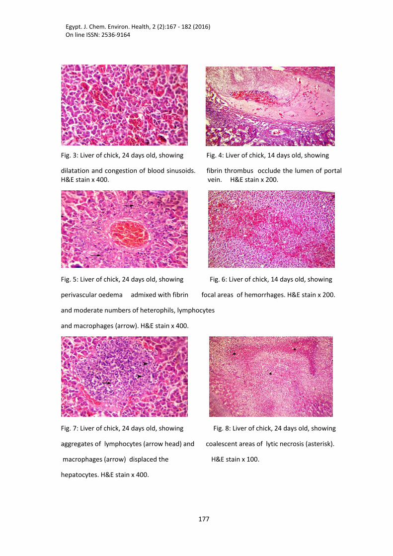
The livers of the chicks showed congestion and dilatation of central and portal
veins, the blood sinusoids were occasionally dilated and congested (Fig. 3). Fibrin
thrombi were frequently observed in the lumen of portal vein (Fig. 4). The
perivascular interstitial was expanded by edema admixed with fibrin and moderate
numbers of heterophils, lymphocytes and macrophages (Fig. 5). (Saskia and Eva-
Maria, 2007) noted that multifocal or confluent necrosis with or without evidence of
heterophils infiltration in the liver of infected birds. These findings agreed with ours
Liver of the chicken is considered to be one of the target organs for reovirus infection
(McFerran et al, 19976).In this work , Lesions increased in Infected chickens at 14th
day than 24th day. Occasionally, focal areas of hemorrhages (Fig. 6) and aggregates
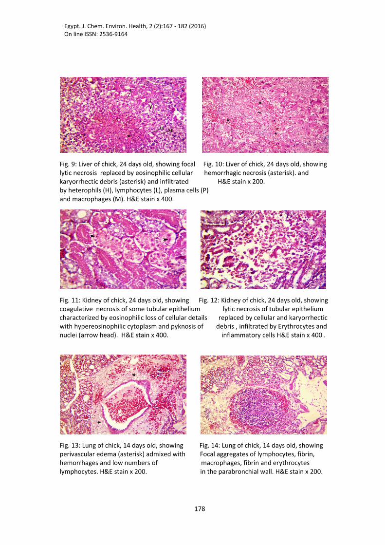
of lymphocytes and macrophages displaced the hepatocytes (Fig. 7). There were
multifocal to coalescent areas of lytic necrosis (Fig. 8) replaced by eosinophilic
Egypt. J. Chem. Environ. Health, 2 (2):167 - 182 (2016) On line ISSN: 2536-9164
711
cellular and karyorrhectic debris and infiltrated by heterophils, lymphocytes, plasma
cells and macrophages (Fig. 9). Occasionally, hemorrhagic necrosis was seen (Fig.
10). Rarely, the vacant space of necrotic hepatocytes was replaced by edema and
erythrocytes. Moreover, the portal areas were expanded by moderate numbers of
inflammatory cells mainly lymphocytes and fewer macrophages. The bile ducts
revealed mild biliary hyperplasia with eosinophilic debris in the lumen and periductal
fibrous tissue proliferation admixed with small numbers of lymphocytes and
macrophages, these lesions were due to infected organisms which found with damaged
tissues as a result of immunosupretion of the bird. (Sharma et al., 1994; Pertile et al.,
1996; Sanchez-Cordon et al., 2002).
The examined kidneys revealed congestion of the renal blood vessels and
intertubular capillaries with occasional fibrin thrombosis of some blood vessels. The
interstitium of renal cortex was expanded by aggregates of lymphocytes. Multifocal
coagulative necrosis of some tubular epithelium characterized by loss of cellular
details with hypereosinophilic cytoplasm and pyknosis of nuclei was observed (Fig.
11). Occasionally, there was lytic necrosis of tubular epithelium replaced by
eosinophilic cellular and karyorrhectic debris and infiltrated by erythrocytes,
heterophils, few lymphocytes, plasma cells and macrophages (Fig. 12). Additionally,
homogenous eosinophilic casts were observed in the lumen of some renal tubules.
The lung showed congestion of pulmonary blood vessels with occasional thickening
of the wall due to fibrous tissue proliferation. Fibrin thrombi were frequently seen in
the lumen of blood vessels. Multifocal, perivascular and interlobular septa were
markedly expanded by fibrin, edema, hemorrhages and lymphocytes (Fig. 13). Foci of
aggregates of lymphocytes, macrophages, fibrin and erythrocytes were occasionally
seen in the Para bronchial wall (Fig. 14). Lesions in birds at 14 th day were increased
than birds at 24 th day .( Jones and Georgiou ,1984), suggested that the age
associated susceptibility may be related to the inability of young birds to develop an
effective immune response . Reoviruses are suggested to have an immunosuppressive
activity which is most probably caused by lymphodeplesion mediated by factors
relased by macrophages (Sharma et al, 1994,; Pertile et al, 1996; Sanchez-Cordon
et al, 2002).This immunosuppresioncould facilitate growth of bacteria and parasites
and allow other viruses to co-infect the bird. The interatrial septa were also expanded
by edema admixed with mononuclear inflammatory cells. The bronchial mucosa
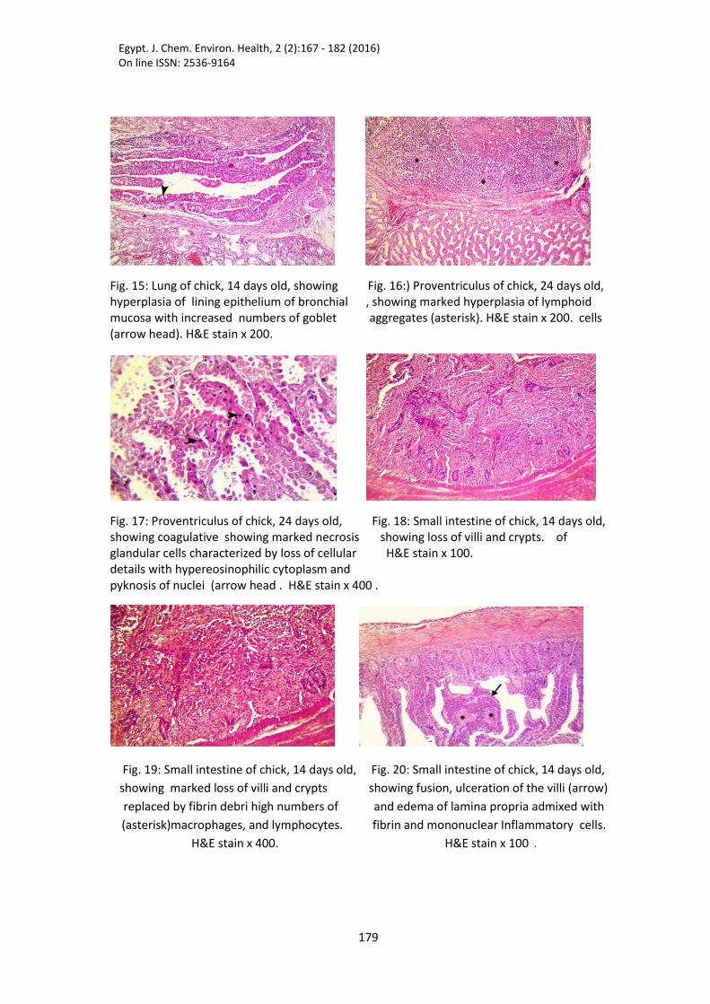
exhibited hyperplasia of lining epithelium with increased numbers of goblet cells (Fig.
15). Furthermore, subpleural edema admixed with fibrin, hemorrhage, and
lymphocytes were also detected.
The proventriculus revealed marked hyperplasia of lymphoid aggregates (Fig. 16).
Multifocal, glandular cells exhibited coagulative necrosis characterized by loss of
cellular details with hypereosinophilic cytoplasm and pyknosis of nuclei (Fig. 17).
Egypt. J. Chem. Environ. Health, 2 (2):167 - 182 (2016) On line ISSN: 2536-9164
711
Additionally, there is no general agreement regarding proventricular lesions in
Reovirus. (page et al, 1981, Bracewell and Randall ,1984,Shapiro and Nir ,1995) .
While (Kouwenhoveretal, 1978) reported proventricular lesions in Reovirus
.However, (Bayyan et al., 1995) believed that proventriclitis is not a consistent
finding in Reovirus
The small intestine revealed marked loss of villi and occasionally crypts (Fig. 18)
with replacement by fibrin, necrotic debris, large numbers of macrophages, and
lymphocytes (Fig. 19). (Moreover Songserm et al., 2002) noted that vacuolar
degeneration was present in the intestinal villi at day 14 post infections with REO
virus. At day 7 and 14 PI, lymphoid, macrophage and granulocytic infiltration into the
lamina propria, cystic formation of crypts of Lieberkühn and villus atrophy were
present in the small intestine, these results agreed with ours. Adjacent less affected
villi were fused multifocal eroded or ulcerated and the remaining lamina propria was
expanded by edema admixed with fibrin, lymphocytes and macrophages (Fig. 20).In
this work the infected birds had severe intestinal lesions at day 14 th because of the
presence of more differentiated epithelia . The intestinal lumen contained abundant
fibrin and necrotic debris admixed with sloughed epithelial cells,few macrophages and
lymphocytes.(Haffen et al.,1989) have suggested that the cells of the lamina propria,
or mesenchyme, can exert considerable influence on the differentiation of enterocytes.
Damage to the lamina propria as a result of infection could interfere with the
differentiation of the enterocytes with a resultant reduced absorptive capacity of the
gut.
Fig 1: RT-PCR results for Reo virus: Lane 1: Positive control, Lanes 1,3Positive reo
samples. Lane 2 negative reo sample. M= 100bp Ladder.
Egypt. J. Chem. Environ. Health, 2 (2):167 - 182 (2016) On line ISSN: 2536-9164
711
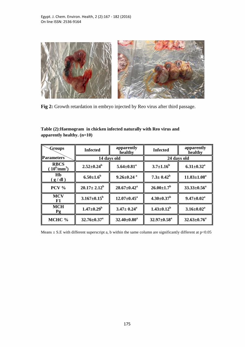
Fig 2: Growth retardation in embryo injected by Reo virus after third passage.
Table (2):Haemogram in chicken infected naturally with Reo virus and
apparently healthy. (n=10)
Means ± S.E with different superscript a, b within the same column are significantly different at p<0.05
Groups
Parameters
Infected apparently
healthy Infected
apparently healthy
14 days old 24 days old
RBCS ( 10
6/mm
3)
2.52±0.24b
5.64±0.81a
3.7±1.16b
6.31±0.32a
Hb ( g / dl )
6.50±1.6b 9.26±0.24
a 7.3± 0.42
b 11.83±1.08
a
PCV % 20.17± 2.12b
28.67±0.42a
26.00±1.7b
33.33±0.56a
MCV F1
3.167±0.15b
12.07±0.45a
4.30±0.37b
9.47±0.02a
MCH Pg
1.47±0.29b
3.47± 0.24a
1.43±0.12b
3.16±0.02a
MCHC % 32.76±0.37a 32.40±0.80
a 32.97±0.58
a 32.63±0.76
a
Egypt. J. Chem. Environ. Health, 2 (2):167 - 182 (2016) On line ISSN: 2536-9164
716
Table(3) Leukogram in naturally infected chicken with Reo virus and apparently healthy
(n=10)
Groups
Parameters
Infected Apparently
healthy Infected
Apparently healthy
14 days old 24 days old
TLC ( 10
3 / mm
3)
87.54±3.65a 46.66±2.56
b 114.00±3.89
a 92.20±5.50
b
Heterophils ( 10
3 / mm
3)
16.43±2.47a 15.75±1.02
a 22.32±5.74
a 21.15±3.52
b
Lymphocytes ( 10
3 / mm
3)
63.11±3.94a 25.91±1.34
b 83.14±4.50
a 65.52±4.74
b
Eosinophil ( 10
3 / mm
3)
1.33±0.18a 1.1±0.14
a 1.40±0.05
a 1.2±0.09
a
Monocyte ( 10
3 / mm
3)
6.67±0.7a 3.9±0.06
b 7.14±0.45
a 3.33±0.28
b
Means ± S.E with different superscript a ,b within the same column are significantly different at p<0.05
Table(4): Serum biochemical parameters in chickens naturally infected with Reo virus
apparently healthy. (n=10)
Means ± S.E with different superscript a, b within the same column are significantly different at p<0.05
Groups
Parameters
Infected apparently
healthy Infected
apparently healthy
14 days old 24 days old
Total Protein (g /dl ) 2.67±0.02a 2.38±0.02
b 2.33±0.35
a 2.47±0.11
a
Albumin (g/dl ) 1.42±0.20b 1.07±0.01
a 1.18±0.06
a 1.25±0.02
a
Globulin (g/dl ) 1.25±0.20a 1.31±0.03
a 1.15±0.38
a 1.22±0.13
a
AST (U/l) 127.75±3.57a 49.50±2.10
b 164.25±8.1
a 58.75±2.75
b
ALT (U/l) 46.70±0.94a 23.90±0.01
b 45.44±0.47
a 23.27±0.13
b
Egypt. J. Chem. Environ. Health, 2 (2):167 - 182 (2016) On line ISSN: 2536-9164
711
Fig. 3: Liver of chick, 24 days old, showing Fig. 4: Liver of chick, 14 days old, showing
dilatation and congestion of blood sinusoids. fibrin thrombus occlude the lumen of portal H&E stain x 400. vein. H&E stain x 200.
Fig. 5: Liver of chick, 24 days old, showing Fig. 6: Liver of chick, 14 days old, showing
perivascular oedema admixed with fibrin focal areas of hemorrhages. H&E stain x 200.
and moderate numbers of heterophils, lymphocytes
and macrophages (arrow). H&E stain x 400.
Fig. 7: Liver of chick, 24 days old, showing Fig. 8: Liver of chick, 24 days old, showing
aggregates of lymphocytes (arrow head) and coalescent areas of lytic necrosis (asterisk).
macrophages (arrow) displaced the H&E stain x 100.
hepatocytes. H&E stain x 400.
Egypt. J. Chem. Environ. Health, 2 (2):167 - 182 (2016) On line ISSN: 2536-9164
711
Fig. 9: Liver of chick, 24 days old, showing focal Fig. 10: Liver of chick, 24 days old, showing lytic necrosis replaced by eosinophilic cellular hemorrhagic necrosis (asterisk). and karyorrhectic debris (asterisk) and infiltrated H&E stain x 200. by heterophils (H), lymphocytes (L), plasma cells (P) and macrophages (M). H&E stain x 400.
Fig. 11: Kidney of chick, 24 days old, showing Fig. 12: Kidney of chick, 24 days old, showing coagulative necrosis of some tubular epithelium lytic necrosis of tubular epithelium characterized by eosinophilic loss of cellular details replaced by cellular and karyorrhectic with hypereosinophilic cytoplasm and pyknosis of debris , infiltrated by Erythrocytes and nuclei (arrow head). H&E stain x 400. inflammatory cells H&E stain x 400 .
Fig. 13: Lung of chick, 14 days old, showing Fig. 14: Lung of chick, 14 days old, showing perivascular edema (asterisk) admixed with Focal aggregates of lymphocytes, fibrin, hemorrhages and low numbers of macrophages, fibrin and erythrocytes lymphocytes. H&E stain x 200. in the parabronchial wall. H&E stain x 200.
Egypt. J. Chem. Environ. Health, 2 (2):167 - 182 (2016) On line ISSN: 2536-9164
711
Fig. 15: Lung of chick, 14 days old, showing Fig. 16:) Proventriculus of chick, 24 days old, hyperplasia of lining epithelium of bronchial , showing marked hyperplasia of lymphoid mucosa with increased numbers of goblet aggregates (asterisk). H&E stain x 200. cells (arrow head). H&E stain x 200.
Fig. 17: Proventriculus of chick, 24 days old, Fig. 18: Small intestine of chick, 14 days old, showing coagulative showing marked necrosis showing loss of villi and crypts. of glandular cells characterized by loss of cellular H&E stain x 100. details with hypereosinophilic cytoplasm and pyknosis of nuclei (arrow head . H&E stain x 400 .
Fig. 19: Small intestine of chick, 14 days old, Fig. 20: Small intestine of chick, 14 days old,
showing marked loss of villi and crypts showing fusion, ulceration of the villi (arrow)
replaced by fibrin debri high numbers of and edema of lamina propria admixed with
(asterisk)macrophages, and lymphocytes. fibrin and mononuclear Inflammatory cells.
H&E stain x 400. H&E stain x 100 .
Egypt. J. Chem. Environ. Health, 2 (2):167 - 182 (2016) On line ISSN: 2536-9164
711
Conclusion
The isolation of reovirus from three farms in Ismailia province indicates its economic
importance as one of the possible viral infections. The morbidity due to infection was
noticed to be higher than mortalities and both increase with age. Haematological and
biochemical changes in the examined birds might affect the immunity of chickens to
reovirus infection , the histological lesions present in the proventriculus, small
intestine and in the liver could interfere with the normal digestive processes
resulting in poor weight gain and delayed or retarded growth.
References
Al-Mufarred S.I., Savage C.E. and Jones R.C. (1996): Egg transmission of avian
reoviruses in chickens: comparison of a trypsin-sensitive and a trypsin-resistant strain.
Avian Pathol., 25 (3), 469-480.
Bancroft J.D. and Gamble (2002): Theory and Practice of histological technique. 6
Ed.,Churchill, Livingston , New York, London,San Francisco, Tokyo.
BayyanGR, Huffe WE, Balog JM, Rath NC, Beasly JN (1995) :Experimental
reproduction of proventriculitis using homogenates of proventricular tissue. Poultry
Sci 74:1799–1809.
Bracewell C,D. and Randall C.J. (1984): The infectious stunting syndrome. World’s
Poult Sci. J. 40:31–37.
Bruhn S., Bruckner L. and Ottiger H.,( 2005): Application of RT-PCR for the
detection of avian reovirus contamination in avian viral vaccines. J. Virol. Meth. 123,
179–186.
Dustan C. F. (2009): Avian Reovirus Infections AVIAN Advice newsletter (volume
5, number 3) –Center of Excellence for Poultry Science University of Arkanasa Cooperative
Extension Service – Division of Agriculture. Haffen K., Kedinger M., Bouziges F.and Simon-Assmann P.(1989): Mesenchyme–
endoderm interactions and enterocyte development.In: Smith MW, Sepulveda FV
(eds) Adap. and develop. of gastrointestinal function. Manchester Univ.,UK, p.92-102.
Jain N.C.,(2000): " Schalm's veterinary haematology"8th
Ed. Lea and Febiger,
Philadelphia, USA.
Jones R. C. and Georgiou k., (1984): Reovirus-induced tenosynovitis in chickens:
The influence of age at infection. Avian Pathol 13:441-457.
Kaneko J.J. John W.H. and Michael L.L.B., (1997): "Clinical Biochemistry of
Domestic Animals." 4th
ed – Academic Press, New York.
Kouwenhoven B., Davelar F.G. and Van Walsum J., (1978): Infectious
proventriculitis causing runting in broilers. Avian Pathol 7:183–187
Lee L.H., Shien J.H. and Shieh H.K.,( 1998): Detection of avian reovirus RNA and
comparison of a portion of genome segment S3 by polymerase chain reaction and
restriction enzyme fragment length polymorphism. Res. Vet. Sci. 65, 11.
Liu H.J., Chen J.H., Liao M.H., Lin M.Y. and Chang, G.N., (1999a.): Identification of the sigma C-encoded gene of avian reovirus by nested PCR and
restriction endonuclease analysis. J. Virol. Meth. 81, 83–90.
Egypt. J. Chem. Environ. Health, 2 (2):167 - 182 (2016) On line ISSN: 2536-9164
717
Liu, H.J., Liao, M.H.,Chang,C.D.,Chen, J.H., Lin,M.Y.,Tung, M.C.,(1999b):
Comparison of two techniques for the detection of avian reovirus in formalin-fixed
paraffin-embedded chicken tissues. J. Virol. Meth. 80, 197–201
Liu, H.J., Lee, L.H., Shih, W.L., Li, Y.J., Su, H.Y.,( 2004): Rapid characterization
of avian reoviruses using phylogenetic analysis, reverse transcription-PCR and
restriction enzyme fragmant length polymorphism. Avian Pathol. 33, 171–180.
McFerran J.B., McCracken R.M., Connor T.J. & Evans R.T. (1976):Isolation of
viruses from clinical outbreaks of inclusion body hepatitis. Avian Pathol, 5, 315-324.
Mc Nulty N. S., (1993): Reovirus; J.B.Mc Ferran, M.S.McNulty (Eds.), Virus
Infection of Birds, Elsevier Science Publishers, Amesterdam / London/ NewYourk/
Tokyo.pp.181-198.
Menendez N.A., Calnek B.W. and Cowen B.S. (1975): Localisation of avian
reovirus (FDO isolant) in tissues of mature chickens. Avian Dis., 19, 112-117.
Muhammad, F. Q.; Hina, A.; and Ayesha L. (2014): Haematological studies on
stunting syndrome in broilers. Res. J. Vet. Pract. 3 (1) : 19-24.
Nilli H,Jahantigh M, and Nazifi S(2007):Clinical observation,pathology ,and serum
biochemical changes in infections stunting syndrome of broiler
chickens.Comp.Clin.Pathol.16(3):161-16.
Otto P., Liebler-tenorio E.M., Elschner M., Reetz J., Lohren and Diller R.,
(2006): Detection of rotaviruses and intestinal lesions in broiler chicks from flocks
with runting and stunting syndrome (RSS).Avian Dis.50(3):411-418.
Page RK, Fletcher OJ, Rowland GN, Gaudry D and Villegas P (1981): Malabsorption syndrome in broiler chickens. Avian Dis 26:618–624
Pang Y, Wang H, Girshick T, Xie Z and Khan MI.(2002): Development and
application of a multiplex polymerase chain reaction for avian respiratory agents.
Avian Dis;46:691–9.
Pertile, T.L., Karaca, K., Walser., M.M. and Sharma, J.M. (1996): Suppressor
macrophages mediate depressed lymphoproliferation in chickens infected with avian
reovirus. Veterinary Immunology and Immunopathology, 53, 129-45.
Rani MP,Ahmad NN,Prasad and E,Latha SC(2011):.Haematological and
biochemical changes of stunting syndrome in broiler chicken .Vet.World.4(3):124-
125.
Rosenberger, J. K. and N. O. Olson. (1997): Viral arthritis. InB. W.
Calnek, H. J. Barnes, C. W. Beard, L. R. McDougald, and Y. M. Saif
(eds.). Diseases of Poultry, 10th ed. Iowa StateUniversity Press; Ames,
IA, 711—718. Sanchez-Cordon, P.J., Hervas, J., Chacon de Lara, F., Jahn, J., Salguero, F.J.
and Gomez-Villamandos, J.C. (2002). Reovirus infection in Psittacine birds
(Psittacus erithacus): Morphologic and immunohistochemical study. Avian Diseases,
46, 485-492. Saskia van de Zande and Eva-Maria Kuhn (2007): Central nervous system signs in
chickens caused by a new avian reovirus strain: A pathogenesis study. Veterinary
Microbiology. 149(3–4): 422–429.
Egypt. J. Chem. Environ. Health, 2 (2):167 - 182 (2016) On line ISSN: 2536-9164
711
Schat, K.A.and Purchase, G.H., (1989): Cell culture methods. In: Purchase, H.G.,
Arp, L.H., Domermuth, C.H., Pearson, J.E. (Eds.), A laboratory manual for the
isolation and identification of avian pathogens.American Assoc. of Avian Path,
Kennett Square, PA, pp. 167–175.
Sellers, H., Linneman E., Icard AH and Mundt E (2010): Apurified recombinant
baculovirus expressed capsid protein of a new astrovirus provides partial protection to
runting- stunting syndrome in chickens. Vaccine. 28(5): 1253- 1263.
Shapiro F and Nir I (1995): Stunting syndrome in broilers: physical, physiological
and behavioral aspect. Poultry Sci 74:33–34.
Sharma, J.M., Karaca, K. and Pertile (1994). Virus-induced immunosuppression in
chickens. Poultry Science, 73, 1082-1086.
Sinclair AJ, Embury DH, Smart IJ, Barr DA, Reece RL, Hooper PT and Gould
JA (1984): Pancreatic degeneration in broilers with runting and stunting syndrome.
Vet Rec 115: 485-488.
Singh R., Agrawal D.K., and Chauhan R.S. (2005): Haemato-biochimical
alterations in chickens with experimental avian reovirus infection, 40 (3) : 296-298.
Snedecor,G.W. and Cochran,W.G.(1982):Statistical Method 7 Ed. Lowa, U.S.A.
Songserm oTh., Pl J. ., Avan M.Roozelaar D.,Kok G.L., Wagenaar F.and ter
Huurne A. (2000): A comparative study of the pathogenesisof malabsorption
syndrome in broilers, Avian Dis. 44 :556-567..
Songserm, T.H.; Engel, B.; Van Roozelaar, D.J; Kok, G.L.; Pijpers, A.; Pol,
J.M.A. and Ter Huurne, A.A.H.M. (2002): Cellular immune response in the small
intestine of two broiler chicken lines orally inoculated with malabsorption syndrome
homogenates. Veterinary Immunology and Immunopathology. 85(1-2): 51-62.
Sterner, F. J., J. K. Rosenberger, A. Margolin, and M. D. Ruff.
( 1989): In vitro and in vivo characterization of avianreoviruses. II
Clinical evaluation of chickens infected withtwo avian reovirus
pathotypes. Avian Dis 22:545—554. Vander Heide, L., Geissler, J.and Bryant, E.S., (1974): Infectious tenosynovitis:
serologic and histopathologic response after experimental infection with aConnecticut
isolate. Avian Dis. 18, 289–296.
Van der Heide, L. (1996):. Introduction on avian reovirus.
Proc.International Symposium on Adenovirus andReovirus Infections in
Poultry, Rauischholzhausen, Germany,138—142.
Van der Heide, L.( 2000.): The history of avian reovirus. AvianDis
44:638—641. Varela, R.and Benavente, J., (1994): Protein coding assignment of avian reovirus
strain S1133. J. Virol. 68, 6775–6777.
Young B,and Health JW(2000): Wheather's functional histology:atext and colour
atlas.4th
ed.Churchill Livingstone,New York.Pp.46-64.