Embed Size (px)
Citation preview

MLAB 1415- Hematology
Keri Brophy-Martinez
Chapter 9: Iron Metabolism and Hypochromic Anemias

Iron Metabolism
Primary function Oxygen transport and storage
Distribution Types of iron-containing compounds
• Functional, assisting in enzymatic and metabolic functions
• Transportation or storage Location
• RBCs- majority here
• Macrophages of spleen& liver- where destruction of RBC occurs, liberating iron
• Hepatocytes and enterocytes- storage of iron

Iron Metabolism
Iron absorption and storage is influenced by: The amount and type of available iron in the diet
• Is it a nutritional deficiency Incomplete absorption due to GI tract problems Current iron stores Increased demand (pregnancy, the growth years) Excessive loss due to acute or chronic hemorrhage
• menstrual period for women of childbearing years, GI bleeding for men

Forms of Iron
NonhemeIonic or ferricFound in vegetables and whole grains
HemeFound in red meatsEasily absorbed

Iron Metabolism
Transport Transferrin
• Transports iron to bone marrow to be used in hgb synthesis
• Synthesized in the liver Storage
Primarily in the liver Ferritin
• Soluble iron, quick release for heme synthesis Hemosiderin
• Partially degraded iron, slow release

Iron Balance
Loss of ironSecretions of urine, bile , sweat and
exfoliation of intestinal epithelial cells of GI tract
Approx. 1 mg/ day Regulation of iron
Delicate balance between loss and absorption

Laboratory Assessment of Iron
Serum iron Total iron binding capacity (TIBC) Percent saturation Serum ferritin

Clinical Syndromes of Iron Metabolism
Iron Deficiency Anemia (IDA)AKA Sideropenic anemiaThis is the most common form of
anemia.IDA occurs when the iron stores
in the body are inadequate to preserve homeostasis.

Causes of IDA
Dietary Blood Loss Hemodialysis Malabsorption
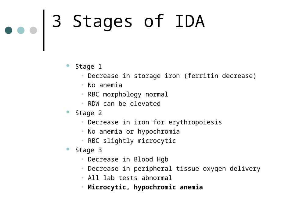
3 Stages of IDA
Stage 1• Decrease in storage iron (ferritin decrease)• No anemia• RBC morphology normal• RDW can be elevated
Stage 2• Decrease in iron for erythropoiesis• No anemia or hypochromia• RBC slightly microcytic
Stage 3 • Decrease in Blood Hgb• Decrease in peripheral tissue oxygen delivery• All lab tests abnormal• Microcytic, hypochromic anemia

Clinical Features of IDA
SOB Lethargy Pallor Gastritis Pica Koilonychia

IDA: Lab features
Decreased RBC, Hgb, Hct, MCV,
MCH, MCHC
Normal to decreased Retic
Peripheral blood smear microcytic-hypochromic Targets, elliptocytes,
teardrops If IDA is caused by
bleeding, leukocytosis and thrombocytosis are possible.

IDA: Lab Findings
Bone marrow Decrease in stainable iron Decrease in erythroid hyperplasia M:E ratio decreased
Chemistry Decrease in serum iron and ferritin Increased TIBC

Anemia of Chronic Disease (ACD)
Anemia that occurs in patients with chronic infections, chronic inflammatory disorders, trauma, organ failure or neoplasms
Occurs due to biochemical changes during inflammation
Hallmark is normal iron stores but low serum iron

Anemia of Chronic Disease
MechanismsBlock in release of iron from
macrophages due to increased cytokines
Cytokine inhibition of EPO productionCytokine inhibition of erythropoiesisShortened erythrocyte survival

ACD: Lab Features
Typical lab findings Decreased
• RBC, Hgb, Hct, MCV, MCH, serum iron Increased
• ferritin Normal
• MCHC Normal to decreased
• Retic, TIBC Peripheral blood smear
• normocytic-normochromic• Targets, elliptocytes, teardrops
Bone marrow• M:E ratio increased

Anemia’s Associated with Abnormal Heme Synthesis
Sideroblastic Anemia Lead Poisoning Porphyrias

Sideroblastic Anemia (SA)
First step in heme synthesis is affected
Characterized by:Increase in total body ironPresence of ringed sideroblasts in
bone marrowHypochromic anemia

Sideroblastic anemia
Classification Hereditary Acquired
• 2 Forms• Idiopathic • Secondary type
• Certain therapeutic drugs• Chronic transfusions (for aplastic anemia, leukemia,
thalassemia)• Alcoholism and food fads• Use of iron utensils or increased iron in water
.

Sideroblastic anemia
MechanismAdequate iron but it can not be
incorporated into hgb synthesis.Iron enters mitochrondria of
metarubricyte, but accumulates leading to formation of ringed sideroblasts
Eventually, mitochrondria rupture

Lead poisoning
Lead interferes with iron storage in the mitochondria
Lead damages the activity of enzymes used for heme synthesis
Basophilic stippling pronounced

Lead Poisoning

Lab features of SA
Peripheral blood Pappenheimer bodies Hypochromic,
normochromic RBCs Normal to increased
platelets Chemistry
Increased serum iron, ferritin

Hemochromatosis
Condition caused by increased iron absorption which deposits in vital organs such as the liver, spleen and pancreas which then becomes fibrotic
Hyperpigmentation of skin
Therapy consists of iron removal by therapeutic phlebotomy or chelation

Porphyrias
Excessive production of porphyrins in the bone marrow (or liver) Rare disease caused by accumulation of
porphyrins in developing RBC’s Defect in one or more of the enzymes in
heme synthesis pathway Characterized by dermal photosensitivity and
rash caused by the sun. The original werewolf was probably a person with erythropoietic porphyria.

References
• Harmening, D. M. (2009). Clinical Hematology and Fundamentals of Hemostasis. Philadelphia: F.A Davis.
• McKenzie, S. B., & Williams, J. L. (2010). Clinical Laboratory Hematology . Upper Saddle River: Pearson Education, Inc.
• http://healthmap.wordpress.com/2008/11/