Embed Size (px)
Citation preview
Mammary gland 1
Mammary gland
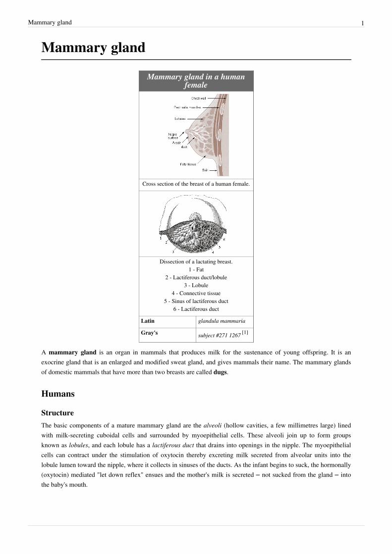
Mammary gland in a humanfemale
Cross section of the breast of a human female.
Dissection of a lactating breast.1 - Fat
2 - Lactiferous duct/lobule3 - Lobule
4 - Connective tissue5 - Sinus of lactiferous duct
6 - Lactiferous duct
Latin glandula mammaria
Gray's subject #271 1267 [1]
A mammary gland is an organ in mammals that produces milk for the sustenance of young offspring. It is anexocrine gland that is an enlarged and modified sweat gland, and gives mammals their name. The mammary glandsof domestic mammals that have more than two breasts are called dugs.
Humans
StructureThe basic components of a mature mammary gland are the alveoli (hollow cavities, a few millimetres large) linedwith milk-secreting cuboidal cells and surrounded by myoepithelial cells. These alveoli join up to form groupsknown as lobules, and each lobule has a lactiferous duct that drains into openings in the nipple. The myoepithelialcells can contract under the stimulation of oxytocin thereby excreting milk secreted from alveolar units into thelobule lumen toward the nipple, where it collects in sinuses of the ducts. As the infant begins to suck, the hormonally(oxytocin) mediated "let down reflex" ensues and the mother's milk is secreted – not sucked from the gland – intothe baby's mouth.
Mammary gland 2
All the milk-secreting tissue leading to a single lactiferous duct is called a "simple mammary gland"; a "complexmammary gland" is all the simple mammary glands serving one nipple. Humans normally have two complexmammary glands, one in each breast, and each complex mammary gland consists of 10–20 simple glands. Thepresence of more than two nipples is known as polythelia and the presence of more than two complex mammaryglands as polymastia.To keep the correct polarized morphology of the lactiferous duct tree requires another essential component -mammary epithelial cells extracellular matrix (ECM), which together with adipocytes, fibroblast, inflammatory cellsetc. constitute mammary stroma.[2] Mammary epithelial ECM mainly contains myoepithelial basement membraneand the connective tissue. They not only help to support mammary basic structure, but also serve as acommunicating bridge between mammary epithelials and their local and global environment throughout this organ'sdevelopment.[3]
Development and hormonal controlMammary glands develop all along during different growth cycles. They exist in both sexes during embryonic stage,forming only a rudimentary duct tree at birth. In this stage, mammary gland development is systemic hormoneindependent,[2] but under the regulation of paracrine communication between neighboring epithelial andmesenchymal cells by parathyroid hormone-related protein(PTHrP).[4] This local secreted factor gives rise to a seriesof outside-in and inside-out positive feedback between these two types of cells, so that mammary bud epithelial cellscan get to proliferate and sprout down into the mesenchymal layer until they reach the fat pad to begin the first roundof branching.[2] At the same time, the embryonic mesenchymal cells around the epithelial bud get secrecting factorsactivated by PTHrP, such as BMP4, can transform into a dense, mammary-specific mesenchyme, which laterdevelop into connective tissue with fibrous threads, forming blood vessels and the lymph system.[5] Basementmembrane, mainly containing laminin and collagen, formed thereafter by defferentiated myoepithelial cells keeps thepolarity of this primary duct tree.Secondary duct tree development occurs in females in response to circulating ovarian hormones from puberty.Estrogen promotes branching differentiation,[6] whereas in males testosterone inhibits it. A mature duct tree reachingthe limit of the fat pad of the mammary gland comes into being by bifurcation of duct terminal end buds (TEB),secondary branches sprouting from primary ducts[3] [7] and proper duct lumen formation. These processes are tightlymodulated by components of mammary epithelial ECM interacting with systemic hormones and local secretingfactors. However, for each mechanism the epithelial cells' "niche" can be delicately unique with different membranereceptor profiles and basement membrane thickness from specific branching area to area, so as to regulate cellgrowth or differentiation sub-locally.[8] Important players include beta-1 integrin, epidermal growth factor receptor(EGFR), laminin-1/5, collagen-IV, matrix metalloproteinase(MMPs), heparan sulfate proteoglycans etc. Elevatedcirculating level of growth hormone and estrogen get to multipotent cap cells on tip of TEB through a leaky thinlayer of basement membrance and promote specific gene expression. Hence cap cells can differentiate intomyoepithelial and luminal (duct) epithelial cells, and the increased amount of activated MMPs can degradesurrounding ECM helping duct buds to reach further in the fat pads.[9] [10] Lumen is formed when branching by innerbody cells apoptosis for lack of survival signals. On the other hand, basement membrane along the mature mammaryducts is thicker with strong adhesion to epithelial cells via binding to integrin and non-integrin receptors. When sidebranches develop, it is a much more “pushing-forward” working process including extending through myoepithelialcells, degrading basement membrane and then invading into a periductal layer of fibrous stromal tissue.[3] Degradedbasement membrane fragments (laminin-5) roles to lead the way of mammary epithelial cells migration.[11] Whereas,laminin-1 interacts with non-integrin receptor dystroglycan negatively regulates this side branching process in caseof cancer.[12] These complex "Yin-yang" balancing crosstalks between mammary ECM and epithelial cells "instruct"healthy mammary gland development until adult.
Mammary gland 3
True secretory alveoli only develop in pregnancy, when rising levels of estrogen and progesterone cause furtherbranching, together with an increase in adipose tissue and a richer blood flow. In gestation, serum progesteroneremains at a stably high concentration so signaling through its receptor is continuously activated. As one of thetranscribed genes, Wnts secreted from mammary epithelial cells act paracrinely to induce more neighboring cellsbranching.[13] [14] When the lactiferous duct tree is almost ready, "leaves" alveoli are differentiated from luminalepithelial cells and added at the end of each branch. In late pregnancy and for the first few days after giving birth,colostrum is secreted. Milk secretion (lactation) begins a few days later due to reduction in circulating progesteroneand the presence of another important hormone prolactin, which mediates further alveologenesis and milk proteinproduction. Laminin and collagen in myoepithelial basement membrane interacting with beta-1 integrin on epithelialsurface again, is essential in this process.[15] [16] Their binding ensures correct placement of prolactin receptors onbasal lateral side of alveoli cells and directional secretion of milk into lactiferous ducts.[15] [16] Suckling of the babycauses release of hormone oxytocin which stimulates contraction of the myoepithelial cells. In this way of combinedcontrol from ECM and systemic hormones, milk secretion can be reciprocally amplified so as to provide enoughnutrition for the baby.After lactation, decreased prolactin level and stop of baby suckling cause mammary involution. All alveoli andsecretory duct structure collapse by programmed cell death (apoptosis) and autophagy for lack of growth promotingfactors either from the ECM or circulating hormones.[17] [18] At the same time, apoptosis of blood capillaryendothelial cells speeds up the regression of lactation ductal beds. Shrinkage of the mammary duct tree and ECMremodeling by various proteinase is under the control of somatostatin and other growth inhibiting hormones andlocal factors.[19] This big structure change leads loose fat tissue to fill up the empty space thereafter. But a functionallactiferous duct tree can be formed again when a female is pregnant again.
Breast cancerTumorigenesis in mammary glands can be induced biochemically by abnormal expression level of circulatinghormones or local ECM components,[20] or from a mechanical change in the tension of mammary stroma.[21] Undereither of the two circumstances, mammary epithelial cells would grow out of control and eventually result in cancer.Almost all instances of breast cancer originate in the lobules or ducts of the mammary glands.
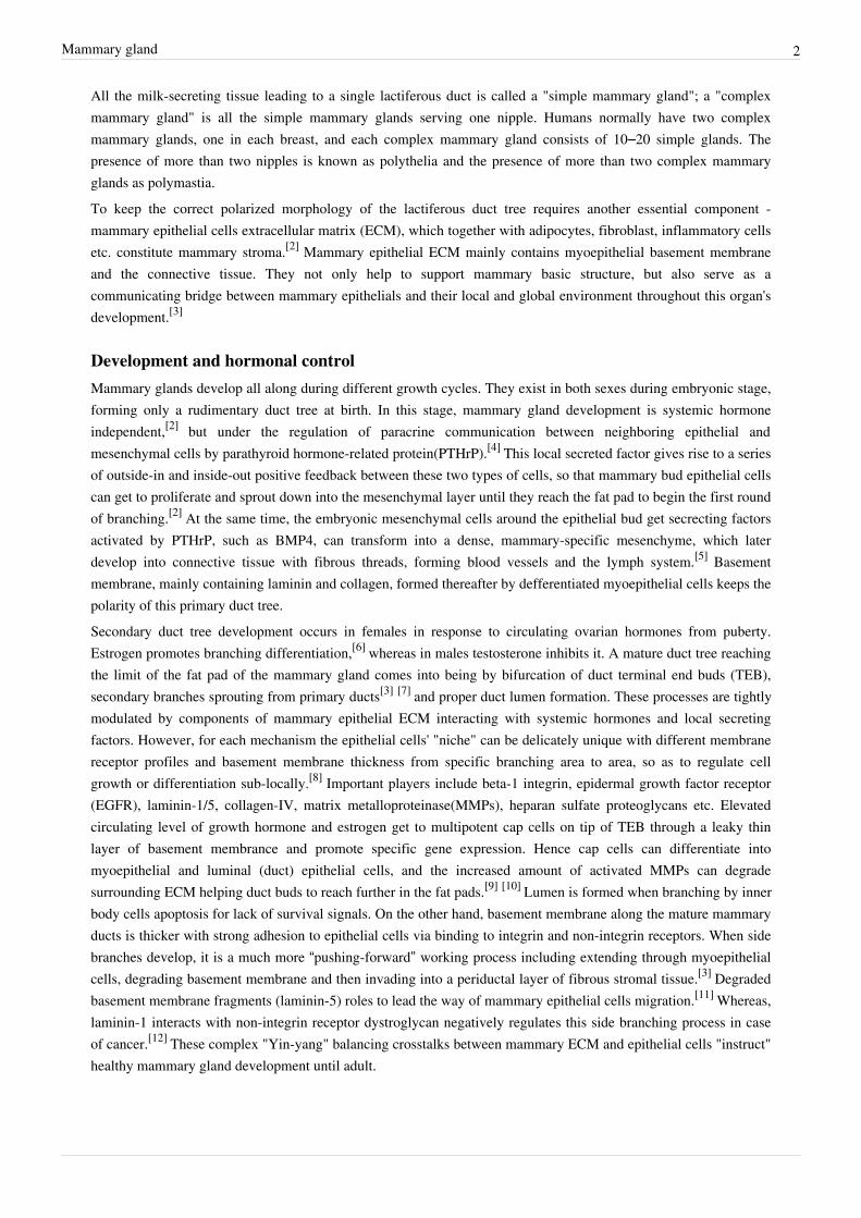
Other mammalsThe constantly protruding breasts of the adult human female, unusually large relative to body size, are a uniqueevolutionary development whose purpose is not yet fully known (see breasts); other mammals tend to have lessconspicuous mammary glands that protrude only while actually filling with milk. The number and positioning ofcomplex and simple mammary glands varies widely in different mammals. The nipples and glands can occuranywhere along the two milk lines, two roughly-parallel lines along the ventral aspect of the body. In general mostmammals develop mammary glands in pairs along these lines, with a number approximating the number of youngtypically birthed at a time. The number of nipples varies from 2 (in most primates) to 18 (in pigs). The VirginiaOpossum has 13, one of the few mammals with an odd number.[22] [23] The following table lists the number andposition of glands normally found in a range of mammals:
Mammary gland 4
Species [24] Anterior
(thoracic)Intermediate(abdominal)
Posterior(inguinal)
Total
Goat, sheep, horseguinea pig
0 0 2 2
Cattle 0 0 4 4
Cat 2 2 4 8
Dog [25] 4 2 2 or 4 8 or 10
Mouse 6 0 4 10
Rat 6 2 4 12
Pig 6 6 6 18
Elephants, primates 2 0 0 2
Male mammals typically have rudimentary mammary glands and nipples, with a few exceptions: male mice don'thave nipples, and male horses lack nipples and mammary glands. The male Dayak fruit bat has lactating mammaryglands;[26] male lactation occurs infrequently in some species, including humans.Mammary glands are true protein factories, and several companies have constructed transgenic animals, mainlygoats and cows, in order to produce proteins for pharmaceutical use. Complex glycoproteins such as monoclonalantibodies or antithrombin cannot be produced by genetically engineered bacteria, and the production in livemammals is much cheaper than the use of mammalian cell cultures.
EvolutionIt is believed that the mammary gland is a transformed sweat gland, more closely related to Apocrine sweatglands.[27] There are many theories of how they evolved, but since they do not fossilize well, supporting suchtheories is difficult. Many of the current theories are based off of comparisons between lines of living mammals-monotremes, marsupials and eutherians. One theory proposes that mammary glands evolved from glands that wereused to keep the eggs of early mammals moist[28] [29] and free from infection[30] [31] (monotremes still lay eggs).Other theories propose that early secretions were used directly by hatched young,[32] or that the secretions were usedby young to help them orient to their mothers.[33]
Gallery
Cattle Cat Pig Sheep
Mammary gland 5
Goat Elephant Mouse Human
See also• Breastfeeding• Mammary tumor• Gynecomastia• Teat• Udder• Witch's milk
References[1] http:/ / education. yahoo. com/ reference/ gray/ subjects/ subject?id=271#p1267[2] Watson, C.J. & Khaled, W.T. Mammary development in the embryo and adult: a journey of morphogenesis and commitment. Development
135, 995-1003 (2008)[3] Wiseman, B.S. & Werb, Z. Stromal effects on mammary gland development and breast cancer. Science 296, 1046-9 (2002)[4] Wysolmerski, J.J. et al. Rescue of the parathyroid hormone-related protein knockout mouse demonstrates that parathyroid hormone-related
protein is essential for mammary gland development. Development 125, 1285-94 (1998)[5] Hens, J.R. & Wysolmerski, J.J. Key stages of mammary gland development: molecular mechanisms involved in the formation of the
embryonic mammary gland. Breast Cancer Res 7, 220-4 (2005)[6] Sternlicht, M.D. Key stages in mammary gland development: the cues that regulate ductal branching morphogenesis. Breast Cancer Res 8,
201 (2006)[7] Sternlicht, M.D., Kouros-Mehr, H., Lu, P. & Werb, Z. Hormonal and local control of mammary branching morphogenesis. Differentiation 74,
365-81 (2006)[8] Fata, J.E., Werb, Z. & Bissell, M.J. Regulation of mammary gland branching morphogenesis by the extracellular matrix and its remodeling
enzymes. Breast Cancer Res 6, 1-11 (2004).[9] Wiseman, B.S. et al. Site-specific inductive and inhibitory activities of MMP-2 and MMP-3 orchestrate mammary gland branching
morphogenesis. J Cell Biol 162, 1123-33 (2003)[10] Koshikawa, N., Giannelli, G., Cirulli, V., Miyazaki, K. & Quaranta, V. Role of cell surface metalloprotease MT1-MMP in epithelial cell
migration over laminin-5. J Cell Biol 148, 615-24 (2000)[11] Dogic, D., Rousselle, P. & Aumailley, M. Cell adhesion to laminin 1 or 5 induces isoform-specific clustering of integrins and other focal
adhesion components. J Cell Sci 111 ( Pt 6), 793-802 (1998)[12] Muschler, J. et al. A role for dystroglycan in epithelial polarization: loss of function in breast tumor cells. Cancer Res 62, 7102-9 (2002)[13] Robinson, G.W., Hennighausen, L. & Johnson, P.F. Side-branching in the mammary gland: the progesterone-Wnt connection. Genes Dev
14, 889-94 (2000)[14] Brisken, C. et al. Essential function of Wnt-4 in mammary gland development downstream of progesterone signaling. Genes Dev 14, 650-4
(2000)[15] Streuli, C.H., Bailey, N. & Bissell, M.J. Control of mammary epithelial differentiation: basement membrane induces tissue-specific gene
expression in the absence of cell-cell interaction and morphological polarity. J Cell Biol 115, 1383-95 (1991)[16] Streuli, C.H. et al. Laminin mediates tissue-specific gene expression in mammary epithelia. J Cell Biol 129, 591-603 (1995)[17] Zarzynska J, Motyl T.Apoptosis and autophagy in involuting bovine mammary gland. J Physiol Pharmacol. 2008 Dec;59 Suppl 9:275-88[18] Fadok, V.A., Clearance: the last and often forgotten stage of apoptosis. J Mammary Gland Biol Neoplasia, 1999. 4(2): p. 203-11[19] Motyl, T., et al., Apoptosis and autophagy in mammary gland remodeling and breast cancer chemotherapy. J Physiol Pharmacol, 2006. 57
Suppl 7: p. 17-32[20] Gudjonsson, T. et al. Normal and tumor-derived myoepithelial cells differ in their ability to interact with luminal breast epithelial cells for
polarity and basement membrane deposition. J Cell Sci 115, 39-50 (2002)[21] Provenzano, P.P. et al. Collagen density promotes mammary tumor initiation and progression. BMC Med 6, 11 (2008)[22] With the Wild Things - Transcripts (http:/ / digitalcollections. fiu. edu/ wild/ transcripts/ possums1. htm)
Mammary gland 6
[23] Raising Orphaned Baby Opossums (http:/ / www. awrc. org/ Baby Opossums. htm)[24] Merle Cunningham, Animal Science and Industry ISBN 9780130462565[25] Dog breeds vary in the number of mammary glands: larger breeds tend to have 5 pairs, smaller breeds have 4 pairs.[26] Francis, Charles M.; Anthony, Edythe L. P.; Brunton, Jennifer A.; Kunz, Thomas H. (24 February 1994). "Lactation in male fruit bats"
(http:/ / www. nature. com/ nature/ journal/ v367/ n6465/ abs/ 367691a0. html). Nature (Nature Publishing Group) 367: 691–692.doi:10.1038/367691a0. . Retrieved 2008-05-14.
[27] Oftedal, O.T. (2002). The origin of lactation as a water source for parchment-shelled eggs=Journal of Mammary Gland Biology andNeoplasia. 7. pp. 253–266.
[28] http:/ / nationalzoo. si. edu/ ConservationAndScience/ SpotlightOnScience/ oftedalolav20030714. cfm[29] Oftedal, O.T. (2002). "The mammary gland and its origin during synapsid evolution". Journal of Mammary Gland Biology and Neoplasia 7
(3): 225–252. doi:10.1023/A:1022896515287. PMID 12751889.[30] http:/ / scienceblogs. com/ pharyngula/ 2006/ 05/ breast_beginnings. php[31] Vorbach C., Capecchi M.R., Penninger J.M. (2006). "Evolution of the mammary gland from the innate immune system?". Bioessays (28):
606–616.[32] Lefèvre C.M., Sharp J.A., Nicholas K.R. (2010). "Evolution of lactation: ancient origin and extreme adaptations of the lactation system".
Annual Review of Genomics and Human Genetics (11): 219–238.[33] Graves B.M and Duvall D. (1983). "A role for aggregation pheremones in the evolution of mammallike reptile lactation". The American
Naturalist 122 (6): 835–839. doi:10.1086/284177.
External links• Comparative Mammary Gland Anatomy (http:/ / web. archive. org/ web/ 20051201011719/ http:/ / classes. aces.
uiuc. edu/ AnSci308/ anatomycompar. html) by W. L. Hurley• On the anatomy of the breast (http:/ / jdc. jefferson. edu/ cooper/ 61/ ) by Sir Astley Paston Cooper (1840).
Numerous drawings, in the public domain.• mammary+gland (http:/ / www. emedicinehealth. com/ script/ main/ srchcont_dict. asp?src=mammary+ gland) at
eMedicine Dictionary
Article Sources and Contributors 7
Article Sources and ContributorsMammary gland Source: http://en.wikipedia.org/w/index.php?oldid=396286443 Contributors: Ackerman22, Adrian J. Hunter, Alex.tan, Andrewmc123, Arcadian, AxelBoldt,Batsnumbereleven, Beth 84, BrainyBabe, Brian Crawford, BullRangifer, CTho, CWii, CarTick, Caulfieldholden, Ccevo2010, Celllist, Cgingold, Chanlyn, Chaser, Chris G, Chrisn0113, CiaPan,ClamDip, Clawed, Clngre, Cointyro, CommonsDelinker, ConfuciusOrnis, Craig Pemberton, Dan100, Daniel Case, Darth Panda, Diberri, Dina, Dynzmoar, ENeville, Ealdgyth, EdC, Emijrp,Eras-mus, Favonian, Flarn2006, Flewis, Fratrep, Gilliam, Ginsengbomb, Gogo Dodo, Graham87, Grakk, Gvw686, Henrygb, HiEv, HiMyNameIsDick, I liek breasts, Illnab1024, Itai, JackAidley,Jackhynes, Jfurr1981, Jlcarter2, Joelmills, Julian Mendez, Jwissick, Karada, Kartano, Keenan Pepper, Keilana, Kerowyn, Latka, LeyteWolfer, Lmbhull, Louis Waweru, Luigifan, MONGO,Matt26, Mentifisto, Millw001, Mmxx, MrCheeseBasket, Naturehead, NawlinWiki, Neelix, Nhsnoboarder17, Nishkid64, NotAnonymous0, Notonegoro, Oneiros, Orlandoturner, Osm agha, PaulErik, Pecolee, QueenCake, Raymondwinn, Rich Farmbrough, Rickyrab, Rockpocket, Roleplayer, Rory096, Rune X2, SDC, Salvio giuliano, Scottalter, Scottandrewhutchins, Search255,Septegram, Sp3000, Star Trek Man, Steverapaport, Tabletop, Takagi, Techman224, Template namespace initialisation script, The Anome, The Thing That Should Not Be, The Wednesday Island,TimVickers, Timc, Tinman11, Torchiest, Toyalla, Ulric1313, Ursasapien, Van helsing, WLU, WerewolfHunter65, Westvoja, Why Not A Duck, WinterSpw, Woohookitty, Zigger, 125anonymous edits
Image Sources, Licenses and ContributorsImage:illu_breast_anatomy.jpg Source: http://en.wikipedia.org/w/index.php?title=File:Illu_breast_anatomy.jpg License: Public Domain Contributors: Lennert B, Maksim, Mattes, 2anonymous editsImage:Dissected lactating breast gray1172.png Source: http://en.wikipedia.org/w/index.php?title=File:Dissected_lactating_breast_gray1172.png License: unknown Contributors: AxelBoldt,The HonorableImage:Bezerro mamando REFON .jpg Source: http://en.wikipedia.org/w/index.php?title=File:Bezerro_mamando_REFON_.jpg License: Creative Commons Attribution 2.5 Contributors:User:ReynaldoImage:White Cat Nursing Four Kittens HQ.jpg Source: http://en.wikipedia.org/w/index.php?title=File:White_Cat_Nursing_Four_Kittens_HQ.jpg License: unknown Contributors:Kilom691, Rune X2, 1 anonymous editsImage:Piglets1.jpg Source: http://en.wikipedia.org/w/index.php?title=File:Piglets1.jpg License: Creative Commons Attribution 2.0 Contributors: FlickrLickr, FlickreviewR, Juiced lemon,Kersti Nebelsiek, Para, RanveigImage:Baby sheep feeding.JPG Source: http://en.wikipedia.org/w/index.php?title=File:Baby_sheep_feeding.JPG License: Creative Commons Attribution 2.5 Contributors: Gabriel PollardImage:Kid feeding on mothers milk.jpg Source: http://en.wikipedia.org/w/index.php?title=File:Kid_feeding_on_mothers_milk.jpg License: unknown Contributors: Fir0002, Kersti NebelsiekImage:Elephant_breastfeading.jpg Source: http://en.wikipedia.org/w/index.php?title=File:Elephant_breastfeading.jpg License: Creative Commons Attribution-Sharealike 2.0 Contributors:rkimpeljrImage:Mousesuckling.jpg Source: http://en.wikipedia.org/w/index.php?title=File:Mousesuckling.jpg License: GNU Free Documentation License Contributors: User:RockpocketImage:Breastfeeding_infant.jpg Source: http://en.wikipedia.org/w/index.php?title=File:Breastfeeding_infant.jpg License: Public Domain Contributors: Ken Hammond ()
LicenseCreative Commons Attribution-Share Alike 3.0 Unportedhttp:/ / creativecommons. org/ licenses/ by-sa/ 3. 0/