Embed Size (px)
Citation preview
Aquaporin Water Channels in the Mammary Gland:From Physiology to Pathophysiology and Neoplasia
Ali Mobasheri & Richard Barrett-Jolley
Received: 14 August 2013 /Accepted: 13 November 2013 /Published online: 13 December 2013# The Author(s) 2013. This article is published with open access at Springerlink.com
Abstract Aquaporins are membrane proteins that play fun-damental roles in water and small solute transport acrossepithelial and endothelial barriers. Recent studies suggest thatseveral aquaporin proteins are present in the mammary gland.Immunohistochemical techniques have confirmed the pres-ence of aquaporin 1 (AQP1) and AQP3 water channels inrat, mouse, bovine and human mammary glands. Studiessuggest that in addition to AQP1 and AQP3 AQP4, AQP5and AQP7 proteins are expressed in different locations in themammary gland. Aquaporins play key roles in tumor biologyand are involved in cell growth, migration and formation ofascites via increased water permeability of micro-vessels.Emerging evidence suggests that expression of these proteins
is altered in mammary tumors and in breast cancer cell linesalthough it is not yet clear whether this is a cause or aconsequence of neoplastic development. This review analyzesthe expression and potential functional roles of aquaporinwater channels in the mammary gland. The physiologicalmechanisms involved in the transport of water and smallsolutes across mammary endothelial and epithelial barriersare discussed in the context of milk production and lactation.This paper also reviews papers from the recent cancer litera-ture that implicate aquaporins in mammary neoplasia.
Keywords Mammary gland .Milk production . Lactation .
Aquaporin .Water channel . Immunohistochemistry . AQP1 .
AQP3 . Neoplasia . Breast cancer
Abbreviations
5’-DFUR 5’-deoxy-5-fluorouridineAQP AquaporinATP Adenosine triphosphateBRCA Breast cancer susceptibility proteinbZIP Basic leucine zipper domainC/EBP CCAAT/enhancer binding proteinCHIP28 Channel-forming integral protein 28CHTN Cooperative Human Tissue NetworkCMF Cyclophosphamide methotrexate
fluorouracilCO2 Carbon dioxideER Estrogen receptorFGF-2 Fibroblast growth factor 2FGFR-ERK Fibroblast growth factor receptor-
extracellular signal-regulated kinaseFGFR-PI3K Fibroblast growth factor receptor-
phosphatidylinositide 3-kinaseGLUT1 Glucose transporter 1HER2 Human epidermal growth factor receptor 2
A. Mobasheri (*)School of Pharmacy, University of Bradford, Richmond Road,Bradford BD7 1DP, UKe-mail: [email protected]
A. Mobasherie-mail: [email protected]
A. Mobasherie-mail: [email protected]
A. MobasheriSchool of Life Sciences, University of Bradford, Richmond Road,Bradford BD7 1DP, UK
A. MobasheriSchool of Medicine, Faculty of Medicine and Health Sciences,Queen’s Medical Centre, Nottingham NG7 2UH, UK
A. MobasheriCenter of Excellence in Genomic Medicine Research (CEGMR),King AbdulAziz University, Jeddah 21589, Kingdom of SaudiArabia
R. Barrett-JolleyInstitute of Ageing and Chronic Disease, Faculty of Health & LifeSciences, University of Liverpool, Liverpool L69 3GA, UK
J Mammary Gland Biol Neoplasia (2014) 19:91–102DOI 10.1007/s10911-013-9312-6
MCF-7 Michigan Cancer Foundation-7Na, K-ATPase Sodium-potassium-ATPaseNKCC1 Na/K/2Cl cotransporterNO Nitric oxideRT-PCR Reverse transcription polymerase
chain reactionSNP Single-nucleotide polymorphismSprr2A Small proline-rich protein 2ATARP Tissue Array Research ProgramTMA Tissue microarrayV2-R Arginine vasopressin receptor 2
Introduction
The mammary gland is a milk-producing organ that is charac-teristic of all female mammals and its overriding function is tosynthesize and deliver milk to the newborn offspring. It is alsopresent in a rudimentary and nonfunctional form in males. Themammary gland is a unique and dynamic organ that undergoesepithelial expansion and invasion during puberty and cycles ofbranching and lobular morphogenesis, secretory differentiation,
and regression during pregnancy, lactation, and involution [1].Embryonically, the mammary gland is derived from the ecto-derm. Development begins with invasion of the underlying fatpad by a rudimentary ductal structure [2]. The epithelial nod-ules become buried in the mesenchyme, where they undergodifferentiation under the influence of paracrine signals from themesenchyme. These later develop into a tubuloalveolar struc-ture that becomes functional in response to the hormonalchanges associated with parturition and regulated by the endo-crine system. Postnatal growth occurs in two phases: ductalgrowth and early alveolar development during estrous cycles,and cycles of cell proliferation, differentiation, and death thatoccur with each pregnancy, lactation, and involution.
In terms of structure, themammary gland is essentially similarto amodified sweat gland. In humans each fully developed breastis composed of 15–25 secretory lobes embedded in adiposetissue. Each secretory lobe is a compound tubular acinar gland.The acini lead to ducts, which are lined by cuboidal or columnarepithelial cells that are surrounded by myoepithelial cells. Theducts from each lobule empty into a lactiferous duct that leads tothe nipple in the ampulla. The development, proliferation anddifferentiation of the mammary gland involve the concertedactions of a variety of hormones and growth factors including
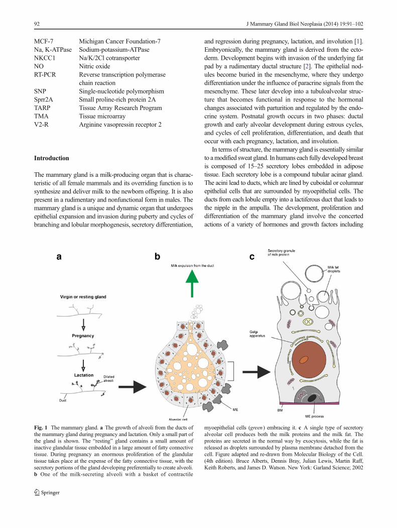
Fig. 1 The mammary gland. a The growth of alveoli from the ducts ofthe mammary gland during pregnancy and lactation. Only a small part ofthe gland is shown. The “resting” gland contains a small amount ofinactive glandular tissue embedded in a large amount of fatty connectivetissue. During pregnancy an enormous proliferation of the glandulartissue takes place at the expense of the fatty connective tissue, with thesecretory portions of the gland developing preferentially to create alveoli.b One of the milk-secreting alveoli with a basket of contractile
myoepithelial cells (green) embracing it. c A single type of secretoryalveolar cell produces both the milk proteins and the milk fat. Theproteins are secreted in the normal way by exocytosis, while the fat isreleased as droplets surrounded by plasma membrane detached from thecell. Figure adapted and re-drawn from Molecular Biology of the Cell.(4th edition). Bruce Alberts, Dennis Bray, Julian Lewis, Martin Raff,Keith Roberts, and James D. Watson. New York: Garland Science; 2002
92 J Mammary Gland Biol Neoplasia (2014) 19:91–102
estrogen, progesterone, and prolactin. In addition to these regu-latory endocrine factors, normal mammary development andlactation require cellular communication and cell-cell interactionsbetween the stromal and parenchymal elements of the mammarygland [3]. Figure 1 outlines the growth of alveoli from the ductsof the mammary gland during pregnancy and lactation. Thisfigure also highlights the differences between “resting”, “preg-nancy” and “lactating” states and illustrates the functional unit ofthe mammary gland consisting of milk-secreting alveoli with abasket of contractile myoepithelial cells embracing it.
Mammary lobes are comprised of secretory acini, whichare formed from cuboidal epithelial cells, responsible for thesynthesis and secretion of milk, surrounded by myoepithelial
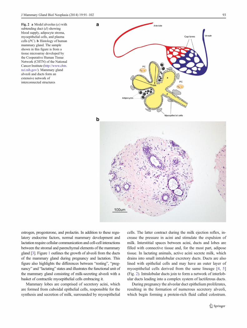
cells. The latter contract during the milk ejection reflex, in-crease the pressure in acini and stimulate the expulsion ofmilk. Interstitial spaces between acini, ducts and lobes arefilled with connective tissue and, for the most part, adiposetissue. In lactating animals, active acini secrete milk, whichdrains into small intralobular excretory ducts. Ducts are alsolined with epithelial cells and may have an outer layer ofmyoepithelial cells derived from the same lineage [4, 5](Fig. 2). Intralobular ducts join to form a network of interlob-ular ducts leading into a complex system of lactiferous ducts.
During pregnancy the alveolar duct epithelium proliferates,resulting in the formation of numerous secretory alveoli,which begin forming a protein-rich fluid called colostrum.
Fig. 2 a Model alveolus (a) withsubtending duct (d) showingblood supply, adipocyte stroma,myoepithelial cells, and plasmacells (PC). b Histology of humanmammary gland. The sampleshown in this figure is from atissue microarray developed bythe Cooperative Human TissueNetwork (CHTN) of the NationalCancer Institute (http://www.chtn.nci.nih.gov/). Mammary glandalveoli and ducts form anextensive network ofinterconnected structures
J Mammary Gland Biol Neoplasia (2014) 19:91–102 93
The formation of milk is one of the key functions of themammary gland. Milk consists of simple sugars, lipids, pro-teins, vitamins and minerals dissolved in water, which makesup to 88 % of its volume. The alveolar epithelial cells thatsynthesize milk are highly specialized, polarized and differ-entiated cells. Their function is to synthesize, package andexport the key constituents of milk. At least five pathways areinvolved in the formation of milk in the tubuloalveolar struc-tures of the mammary gland. The first, which is particularlyrelevant to the topic of this review article, is the secretion ofmonovalent cations and water. Current understanding is thatwater is secreted across the mammary epithelium in a trans-cellular manner, in response to an osmotic gradient producedlargely by the lactose content of the milk [6, 7]. Water isessentially drawn across the alveolar cells by the concentra-tion gradient created by osmotically active milk sugars. Thisprocess is followed by the transport of immunoglobulins, milklipids, milk proteins and other buffers and electrolytes. Theprocesses involved in milk production are discussed in thefollowing section.
Milk Production
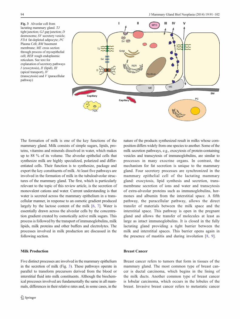
Five distinct processes are involved in themammary epitheliumin the secretion of milk (Fig. 3). These pathways operate inparallel to transform precursors derived from the blood orinterstitial fluid into milk constituents. Although the biochem-ical processes involved are fundamentally the same in all mam-mals, differences in their relative rates and, in some cases, in the
nature of the products synthesized result in milks whose com-position differs widely from one species to another. Some of themilk secretion pathways, e.g., exocytosis of protein-containingvesicles and transcytosis of immunoglobulins, are similar toprocesses in many exocrine organs. In contrast, themechanism for fat secretion is unique to the mammarygland. Four secretory processes are synchronized in themammary epithelial cell of the lactating mammarygland: exocytosis, lipid synthesis and secretion, trans-membrane secretion of ions and water and transcytosisof extra-alveolar proteins such as immunoglobulins, hor-mones and albumin from the interstitial space. A fifthpathway, the paracellular pathway, allows the directtransfer of materials between the milk space and theinterstitial space. This pathway is open in the pregnantgland and allows the transfer of molecules at least aslarge as intact immunoglobulins. It is closed in the fullylactating gland providing a tight barrier between themilk and interstitial spaces. This barrier opens again inthe presence of mastitis and during involution [8, 9].
Breast Cancer
Breast cancer refers to tumors that form in tissues of themammary gland. The most common type of breast can-cer is ductal carcinoma, which begins in the lining ofthe milk ducts. Another common type of breast canceris lobular carcinoma, which occurs in the lobules of thebreast. Invasive breast cancer refers to metastatic cancer
Fig. 3 Alveolar cell fromlactating mammary gland. TJtight junction;GJ gap junction;Ddesmosome; SV secretory vesicle;FDA fat-depleted adipocyte; PCPlasma Cell; BM basementmembrane; ME cross sectionthrough process of myoepithelialcell; RER rough endoplasmicreticulum. See text forexplanation of secretory pathwaysI (exocytosis), II (lipid), III(apical transport), IV(transcytosis) and V (paracellularpathway)
94 J Mammary Gland Biol Neoplasia (2014) 19:91–102
that has spread from breast ducts or lobules to sur-rounding normal tissues. The global burden of breastcancer exceeds all other cancers and the incidence ratesof breast cancer are constantly increasing. Breast canceris the second most common cancer worldwide after lungcancer, the fifth most common cause of cancer death,and the leading cause of cancer death in women. Al-though breast cancer affects both men and women, theincidence of male breast cancer is very rare. Up-to-datestatistics from the National Institutes of Health suggestthat in the United States there were 232,340 (female)and 2,240 (male) new cases resulting in 39,620 deathsin females and 410 in males.
This article does not focus on the usual suspects inbreast cancer. We do not discuss the recent data onBRCA mutations, the expression of HER2, the estrogenreceptor, the progesterone receptor or new adjuvanttherapies. The aim of this article is to discuss the roleof aquaporins in mammary gland physiology and reviewthe recent information on aquaporins in cancer.
Aquaporin Water Channels
The fact that water has the ability to cross the hydrophobicmembrane and enter (and leave cells) had been known fordecades, but the mechanism was somewhat of a paradox. Theparadox was solved by the discovery of a family of 28 kDamembrane transporters with high permeability for water [10].The first of these proteins to be discovered was initially called“CHIP28” but is now known as aquaporin 1 (AQP1) [10, 11].Transfection of cells with AQP1 increased membrane perme-ability to water by 50x. The discovery of aquaporins led to theaward of the Nobel Prize in chemistry to Dr. Peter Agre in2003.1, 2 A transcript of his Nobel lecture was published inBioscience Reports in 2004 [12]. Following the initial discov-ery a number of advancements have been made in the field.Aquaporins are a family of membrane bound proteins that arebelieved to be ubiquitously expressed in cellular and intracel-lular membranes. They are extensively distributed in micro-organisms [13], animals [14–16] and plants [17–19]. They aresmall transmembrane proteins that are expressed in a varietyof epithelial tissues where they are responsible for regulatingrapid water movement across epithelial barriers driven byosmotic gradients. Initially, the main role of aquaporins wasbelieved to be water and small solute transport across epithe-lial and endothelial barriers [20, 21]. It is now known thataquaporins are found in a wide range of non-epithelial/
endothelial cells from acinar cells to chondrocytes [22–24],leukocytes to astrocytes, and in a wide range of reproductiveorgans [25–27]. Thus aquaporins appear to be present in most,if not all, cell types. Furthermore, aquaporins serve importantfunctions in a wide range of processes such as cell division,cell migration, cellular volume regulation and apoptosis. The-se are clearly of importance when considering the pathogen-esis of carcinomas. Despite this ubiquitous expression andfundamental importance in cell biology, genetic deletion ofAQP genes has a less dramatic effect on animal survival thanmay be expected. Transgenic mice lacking AQP1 water chan-nels survive but they have severely impaired urinary concen-trating ability [28] and lack of a functional AQP2 gene leads toa rare form of nephrogenic diabetes insipidus [29]. Survival inthe absence of specific aquaporins may be because of thephenotypic adaption that takes place; for example, geneticknock-out of AQP1 leads to up-regulation of other aquaporinssuch as AQP4, AQP7 and AQP8, at least in some tissues [30].Mammalian aquaporins are located at strategic membranesites in endothelia and a variety of epithelia, most of whichhave well-defined physiological functions in fluid absorptionor secretion [31]. To date, 13 members of the aquaporin genefamily have been identified in humans: AQP0-AQP12 [32].Animal genome projects have also confirmed the presence ofmultiple aquaporin genes encoding distinct protein isoforms.The proteins encoded by aquaporin genes have been classifiedinto two major groups based on their substrate permeabilities:1) the classical water permeable aquaporins are permeated bywater and include AQP1, AQP2, AQP4, AQP5 and AQP8[14]; 2) the water and small solute permeable aquaglyceropo-rins exhibit permeability to water and a range of small neutralsolutes including substances such as glycerol and urea. Aqua-glyceroporins include AQP3, AQP7, AQP9 and AQP10 [14,33]. Several aquaporins, including (AQP1, AQP4, AQP5 andAQP8) have also been proposed to facilitate entry of gasessuch as CO2, NO and ammonia to cells, but this importanttopic is discussed and reviewed elsewhere [34, 35]. The morecontroversial suggestion that some aquaporins (AQP1 forexample) conduct small cations (e.g., Na+ and K+ [36, 37])is disputed by the groups’ of Agre and Pohl [34, 38].
Hormone Regulation of Aquaporins
The physiological advantage of membrane expression of ionchannel and porin proteins compared to the evolution of“leaky” membranes is that membrane permeability can beprecisely and dynamically controlled by variations in proteinexpression and activity. This allows for changing membranewater permeability in response to changing osmotic environ-ment or the physiological need. This is achieved in two ways,firstly the permeability of the aquaporin channels themselvesis variable, being strongly dependent on the molecular weight
1 The Nobel Prize in Chemistry 2003 was awarded “for discoveriesconcerning channels in cell membranes” jointly with one half to PeterAgre “for the discovery of water channels” and with one half to RoderickMacKinnon “for structural and mechanistic studies of ion channels”.2 http://www.nobelprize.org/nobel_prizes/chemistry/laureates/2003/
J Mammary Gland Biol Neoplasia (2014) 19:91–102 95
of the osmolytes in the aqueous solution to which the channelis exposed [39]. Secondly, functional expression can be dy-namically regulated. Although little data exists on this inmammary gland tissue, there are several examples of thisphenomenon elsewhere. The most commonly observed ex-ample is in the kidney collecting duct water (principle cell)where permeability increase is induced by arginine-vasopressin induced phosophorylation via V2-R receptors[40–43]. This system is a key step in regulation of re-absorption and thus urinary volume control and the regulationof body water balance. The cellular mechanism is understoodin some detail and involves the translocation of protein to thecellular membrane, a mechanism, which is significantly fasterthan protein synthetic generation of new protein de novo.During brain edema and in joint diseases such as osteoarthri-tis, synovitis and rheumatoid arthritis aquaporin expressionchanges as an apparent adaptation to changes in the localosmotic environment [24, 44–46]. Furthermore, steroid hor-mones increase AQP1 expression in both peribronchiolar anduterine vascular endothelial tissues as a part of tissue fluidmanagement [47, 48].We will also discuss, below, howAQP1is thought to be involved in estrogen mediated angiogenesis inthe mammary gland and outline how increases in AQP3
expression promote FGF-2 stimulated migration of breastcancer cell lines.
Immunolocalization of Aquaporins in Rat, Mouse,Human and Bovine Mammary Glands
Until recently nothing was known about the expression ofaquaporins in the mammary glands of rodents, humans anddairy animals. Recent immunohistochemical studies of aqua-porins in the rat and mouse mammary gland have confirmedthe presence of AQP1 and AQP3 proteins in both species [49].Matsuzaki and co-workers also used RT-PCR to study theexpression of AQP1, AQP2, AQP3, AQP4, AQP5, AQP6and AQP7 in the rat mammary gland [49]. In addition toAQP1 and AQP3 they found evidence for the presence ofAQP4, AQP5, AQP7 and AQP9 transcripts in the rat mam-mary gland. However, they did not use antibodies to localizethese proteins to specific tissues within the breast.
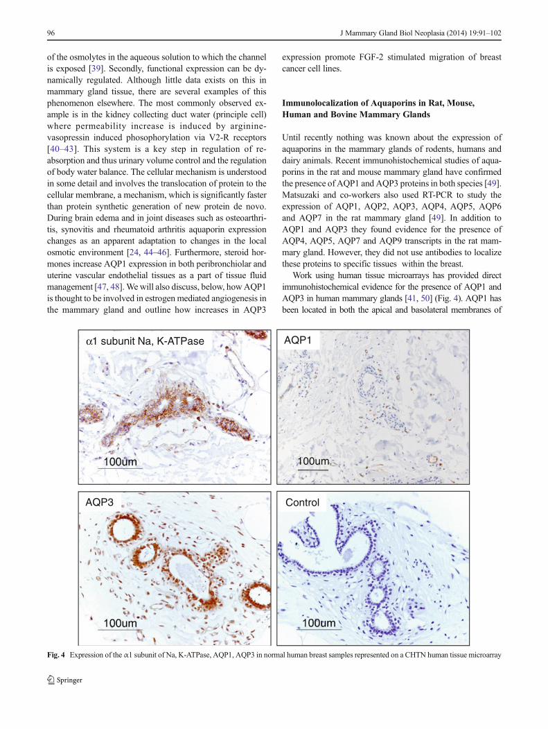
Work using human tissue microarrays has provided directimmunohistochemical evidence for the presence of AQP1 andAQP3 in human mammary glands [41, 50] (Fig. 4). AQP1 hasbeen located in both the apical and basolateral membranes of
α1 subunit Na, K-ATPase
AQP3 Control
AQP1
100um
Fig. 4 Expression of the α1 subunit of Na, K-ATPase, AQP1, AQP3 in normal human breast samples represented on a CHTN human tissue microarray
96 J Mammary Gland Biol Neoplasia (2014) 19:91–102
capillary endothelia in the rodent mammary gland [49]. AQP3has been located immunohistochemically in basolateral mem-branes of secretory epithelial cells and intralobular and interlob-ular duct epithelial cells in rat and mouse mammary tissue [49].Although the expression of mRNA transcripts encoding AQP4,AQP5, AQP7 and AQP9was demonstrated in rodent mammaryby RT-PCR, the presence of the corresponding proteins was notinvestigated and the presence of aquaporins 4, 5 and 7 in therodent mammary gland was not studied using immunohisto-chemical methods [49].
Physiological Relevance of Aquaporins to MilkProduction
Milk is a secreted body fluid that consists of water plus lipids,electrolytes, vitamins, sugars and specific milk proteins. Thedelivery ofwater to themammary gland by the circulatory systemand the movement of water across endothelial and epithelialbarriers are critical for milk synthesis and secretion in lactatinganimals. There is published information on sodium and chloridetransport in mammary epithelia [51]. However, the functionalanatomy of the mammary gland has not been extensively studiedin the context of water transport across endothelial and epithelialbarriers and bulk fluid movement in lactating animals. Conse-quently, very little published information is available; AQP1 andAQP3 proteins have been shown to be present in the capillaryendothelia and mammary epithelial cells, respectively [49]. Sim-ilar observations suggest that AQP1 and AQP3 proteins are alsopresent in non-lactating human mammary glands [41, 50]. Datarecently published on aquaporins in the bovine mammary gland[52] confirm some of the recent observations made by [49] inrodent mammary glands and similar observations in humanmammary glands [41, 50]. Bovine data [52] also confirm thepresence of these proteins in the bovine mammary gland, al-though in distinct cellular locations. A striking and previouslyunreported expression of AQP1, at high abundance, is alsoobservable in myoepithelial cells underlying teat duct epithelia[52]. The physiological reason for this is at present unknown, buthigh AQP1 levels in this location may contribute to increasedpermeability of teat duct epithelia in the lactating bovine mam-mary gland. The relative abundance of the protein also appears torepresent a distinct aspect of differentiation of myoepithelial cellsin this location, compared with those surrounding the acini.
Goats’ milk is known to contain numerous cell fragmentsknown as “christiesomes” which originate from secretory epi-thelial cells of themammary gland [53, 54]. These cell fragmentsare known to contain intact and well-preserved endoplasmicreticulum, mitochondria and lipid droplets. Furthermore, theyare capable of triglyceride synthesis. Although cows’ milk hasbeen shown to contain very few cellular fragments, it doescontain different and denser particles containing fewer vesiclesand numerous microvillus-like protrusions on one side—known
as “sunbursts” [53]. Although it has been suggested that theseparticles are residues of dead cells, it is possible that thesefragmented cytoplasm-containing entities contain membraneproteins, which are targeted to the apical membranes of mam-mary secretory epithelial cells. Recent studies have providedevidence for urinary excretion of AQP2 water channel proteinsduring pregnancy [55, 56]. Biochemical analysis of“christiesomes” and “sunbursts” should reveal if these cellularfragments contain membrane proteins of the apical membranesof alveolar epithelial cells. Comparing this data with publisheddata from rodent studies clearly indicates that some of theaquaporins expressed in the lactating mammary tissues werefound in expected anatomical locations; immunohistochemicallabeling in the rat mammary gland suggests that AQP1 is local-ized to the capillaries and AQP3 is localized to the basolateralmembranes of the alveolar secretory cells. These results suggestthat aquaporins are present in lactating mammary glands andmay be participants in the control of milk water content bydiluting the sugar, protein and lipid contents of milk to anisotonic solution as it descends through the teat duct system.This data has provided interesting new information about thepossible regulation of water homeostasis in the mammary gland.Milk yield is ofmajor economic importance to the dairy industry.Therefore, it is necessary to understand the underlyingmolecularphysiology involved in fluid movement in lactation.
Aquaporins and Cancer
Aquaporins are more than just passive water and small solutechannels in biological membranes [57]. They are activelyregulated and increasingly implicated in a number of impor-tant clinical disease states [58]. Recent evidence suggests thataquaporins are also involved in cell migration [59], angiogen-esis [60], and tumor growth [61]. These water channel pro-teins are strongly expressed in tumor cells of different origins,particularly aggressive tumors [62]. AQP1 is ubiquitouslyexpressed in endothelia of many human tissues [41]. Immu-nohistochemical and histomorphometric studies have con-firmed the presence of AQP1 in endothelial barriers of almostall human tissues as well as many epithelial barriers involvedin absorptive and secretory functions [41]. AQP1 is alsopresent in tumor vascular endothelium [62]. Interestingly,targeted AQP1 gene disruption impairs angiogenesis and cellmigration and AQP1-null mice show defective tumor angio-genesis resulting from impaired endothelial cell migration [62,63]. AQP-expressing cancer cells show enhanced migrationin vitro and greater local tumor invasion, tumor cell extrava-sation, andmetastases in vivo thanAQP1-null transgenicmice[62]. AQP-dependent cell migration may involve AQP-facilitated water influx into lamellipodia at the front edge ofmigrating cells [63]. High throughput studies using tissuemicroarrays (TMAs) have shown that AQP1 is an excellent
J Mammary Gland Biol Neoplasia (2014) 19:91–102 97
marker of microvasculature but it is heterogeneouslyexpressed in different human tumors and is not necessarilyexpressed in all neoplastic cells [64]. It has been proposed thatincreased AQP1 expression in some human adenocarcinomasmay be a consequence of angiogenesis and important for theformation or clearance of tumor edema [64]. There is growingdata in the literature concerning the involvement of AQP1 andAQP4 in human brain tumor growth and edema formation[61, 65]. These studies suggest that the presence of AQP1, butnot AQP4, enhances glioma growth and migration, whileAQP4 enhances cell adhesion highlighting differential biolog-ical roles for AQP1 and AQP4 in gliomas [66].
Aquaporins have therefore been proposed as novel pharma-cological targets in cancer and associated edematous states [67].Potentially, block of aquaporins could limit angiogenesis andinhibit tumor growth and metastasis. Therefore it has beensuggested that novel therapeutic strategies approaches may bedeveloped by antagonizing their biological activity [65]. Itshould be noted that pharmacological inhibition of aquaporinscould have a greater protective effect than that resulting fromgenetic deletion, because there is less opportunity for adult cellsto exhibit the phenotypic switch of aquaporin expressiondiscussed above in APQ1 knock-out studies. Consequently,there is growing interest in aquaporin-based diagnostics [68]and the development of small-molecule aquaporin modulatorsfor treating various clinical states including brain edema, neuro-inflammation, glaucoma, epilepsy, cancer, pain, and obesity [58].
Aquaporins in Breast Cancer
Studies on the role of CCAAT/enhancer binding protein (C/EBP) family of bZIP transcription factors (particularly C/EBPβ) in mammary gland development and breast cancer haveshown that targeted deletion of C/EBPβ isoforms results insevere inhibition of lobuloalveolar development, blocks func-tional differentiation, and induces changes in ductal morphogen-esis. The loss of C/EBPβ isoforms results in altered expressionof a number of membrane proteins, transporters and molecularmarkers, including the progesterone, estrogen, prolactin recep-tors and several transporter proteins (NKCC1 and AQP5) [69].This was one of the early studies that implicated AQP5 inmammary gland development [69]. Further studies investigatedthe distribution of AQP1 in tumors of the prostate, colon, lung,breast and ovary using TARP multi-tumor Tissue TMAs andCHTN (Cooperative Human Tissue Network) TMAs [64].AQP1 expression was higher in advanced mammary and colo-rectal carcinomas where AQP1 immunoreactivity was also seenin some neoplastic tumor cells [64]. Subsequent studies fromour laboratory investigated expression of AQP1, the GLUT1glucose transporter and Na, K-ATPase in canine mammaryglands and mammary tumors [70]. We investigated the expres-sion of these proteins in normal canine mammary glands and in
benign and malignant mammary tumors, using immunohisto-chemistry and semi-quantitative histomorphometry. Interesting-ly, AQP1 immunoreactivity was absent from the majority ofcanine tumor specimens studied. However, this could have beendue to the lack of antibody cross-reactivity. The antibodies usedin the study were raised against rat AQP1 and could have cross-reacted poorly with their with canine AQP1 counterparts [70].
Otterbach and co-workers used immunohistochemicaltechniques to investigate the expression of AQP1 in 203invasive breast carcinomas with long-term follow up data[71]. AQP1 expression was seen in 11 tumors (5.4 %) andshowed a highly significant correlation with high tumor grade,medullary-like histology, “triple-negativity”, cytokeratin 14and smooth muscle actin expression. The authors used uni-variate analysis to show that AQP1 was significantly associ-ated with poor prognosis. They also usedmultivariate analysisto demonstrate that AQP1 protein expression is an indepen-dent prognostic marker if the tumors are properly stratified byage, tumor size, lymph node status, histological grade, ERstatus and CMF therapy. Based on these results the investiga-tors suggested that AQP1 expression is a characteristic featureof aggressive basal-like breast carcinomas [71].
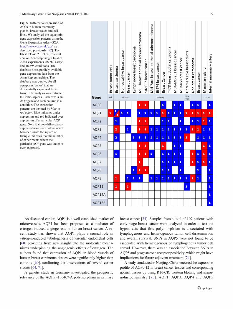
No genome wide transcriptomic studies have specificallyinvestigated changes in aquaporins in breast cancer, however,most of the common microarray RNA chips, includingAffymetrix, Agilent and Illumina include multiple probe setsfor the aquaporins. Analysis over a range of different breastcancer cell lines and experiments listed in the Gene ExpressionAtlas [72] reveals changes in transcript abundance ofmany of theaquaporins (see Fig. 5). Many of these are decreased in expres-sion in cancerous tissue, perhaps revealing that some of theobserved increases in aquaporins protein expression could resultfrom post-translational changes. Casey et al. [73] performed alarge microarray analysis of genome-wide expression in normal(breast reduction) and breast cancer patients. Both epithelialtumor and stromal tumor samples were analyzed using the pop-ular Affymetrix Human Genome U133A 2.0 Array. The raw andprocessed data are publically available in the Gene ExpressionAtlas (E-GEOD10797) and the Gene Expression Omnibus data-base (Accession no. GSE10797: http://www.ncbi.nlm.nih.gov/geo/geo2r/). Expression data is presented for many of thepossible aquaporins isoforms (AQP1, AQP2, AQP3, AQP4,AQP5, AQP6, AQP7, AQP8 and AQP9). There is considerablevariation between individual patients, with some showing verylarge increases in some isoforms of channel. The most notableobservation, however, is the differences between epithelial andstromal tumor tissue; whilst there are no significant differencesbetween any of the aquaporin transcripts in normal breast tissue;AQP1, AQP8, AQP4, AQP6 and AQP9 are all significantlyhigher in stromal tumor tissue, implying a considerable changein cellular phenotype. There is no evidence from thistranscriptomic data of a phenotypic switch from one isoform toanother to that observed with genetic deletion [30].
98 J Mammary Gland Biol Neoplasia (2014) 19:91–102
As discussed earlier, AQP1 is a well-established marker ofmicrovessels. AQP1 has been proposed as a mediator ofestrogen-induced angiogenesis in human breast cancer. A re-cent study has shown that AQP1 plays a crucial role inestrogen-induced tubulogenesis of vascular endothelial cells[60] providing fresh new insight into the molecular mecha-nisms underpinning the angiogenic effects of estrogen. Theauthors found that expression of AQP1 in blood vessels ofhuman breast carcinoma tissues were significantly higher thancontrols [60], confirming the observations of several earlierstudies [64, 71].
A genetic study in Germany investigated the prognosticrelevance of the AQP5 -1364C>A polymorphism in primary
breast cancer [74]. Samples from a total of 107 patients withearly stage breast cancer were analyzed in order to test thehypothesis that this polymorphism is associated withlymphogenous and hematogenous tumor cell disseminationand overall survival. SNPs in AQP5 were not found to beassociated with hematogenous or lymphogenous tumor cellspread. However, there was an association between SNPs inAQP5 and progesterone receptor positivity, which might haveimplications for future adjuvant treatment [74].
A study conducted inNanjing, China screened the expressionprofile of AQP0-12 in breast cancer tissues and correspondingnormal tissues by using RT-PCR, western blotting and immu-nohistochemistry [75]. AQP1, AQP3, AQP4 and AQP5
Fig. 5 Differential expression ofAQPs in human mammaryglands, breast tissues and celllines. We analysed the aquaporingene expression patterns using theGene Expression Atlas (GXA:http://www.ebi.ac.uk/gxa) asdescribed previously [72]. Thelatest release 2.0.21.3 (Ensemblversion 72) comprising a total of2,841 experiments, 88,280 assaysand 16,398 conditions. Thedatabase hosts publicly availablegene expression data from theArrayExpress archive. Thedatabase was queried for allaquaporin ‘genes’ that aredifferentially expressed breasttissue. The analysis was restrictedto Homo sapiens. Each row is anAQP gene and each column is acondition. The expressionpatterns are denoted by blue orred color. Blue indicates underexpression and red indicated overexpression of a particular AQPgene. Note that non-differentiallyexpressed results are not included.Number inside the square ortriangle indicates that the numberof experiments where theparticular AQP gene was under orover expressed.
J Mammary Gland Biol Neoplasia (2014) 19:91–102 99
exhibited differential expression compared to normal tissue.Interestingly, this study found that AQP5 was expressed mainlyin cell membranes of mammary carcinomas, with AQP5 beingnot detectable in normal breast tissues. Furthermore, expressionofAQP5was found to be associatedwith cellular differentiation,lymph node invasion, and tumor staging [75].
Subsequent studies on breast cancer cell lines have shownthat AQP5 is a marker protein for proliferation and migrationof human breast cancer cells [76]. Jung and co-workers de-tected AQP5 mRNA and protein in the human breast cancercell lines MCF7 and MDA-MB-231 by RT-PCR and immu-noblotting. Immunolabeling of AQP5 was seen in ductalepithelial cells of human breast tissues but the apical polarityof AQP5 in ducts was lost. The prominent expression ofAQP5 and the loss of polarity of ductal epithelial cells wereassociated with the progression of breast carcinoma. Theauthors concluded that AQP5 over-expression plays a role incell growth and metastasis in human breast cancer [76].
Several studies have also reported on the expression andfunctional role of AQP3 in breast cancer cell lines. A veryrecent study has shown that AQP3 is required for FGF-2-induced migration of human breast cancers [77]. The authorsused MDA-MB-231 and Bcap-37 cell lines as models andinhibited the expression of AQP3 expression using lentiviralconstructs that stably express shRNA against the mRNAencoding AQP3. Stimulation with FGF-2 treatment increasedAQP3 expression and induced cell migration. Silencing AQP3expression inhibited FGF-2 induced cell migration. The studyconcluded that AQP3 is required for FGF-2-induced cell mi-gration in cultured human breast cancer cells. These findingsalso highlight the importance of FGFR-PI3K and FGFR-ERKsignaling in FGF-2-induced AQP3 expression.
Finally, Trigueros-Motos et al., [78] used the capecitabinecatabolite 5′-deoxy-5-fluorouridine (5′-DFUR) (a nucleosideanalog used in the chemotherapy of solid tumors) to demon-strate that AQP3 is required for cytotoxic activity of 5′-DFURin the breast cancer cell line MCF7 and that this aquaporin isimplicated in cell volume increase and cell cycle arrest. Thus,it would appear that AQP3 ismore than a marker and a passivechannel. Its function is actually important for chemotherapy.
Conclusions
The mammary gland remains an enigma. The distinctive devel-opmental aspects and complex regulation by hormones andgrowth factors has made the study of the mammary glandfunction a major focus of research for scientists from manydifferent biological and biomedical disciplines. The increasedincidence of breast cancer is another important reason for study-ing the mammary gland. A number of studies have attempted toidentify specific genes required for the functional developmentof mammary epithelium [79]. However, very few studies have
examined the potential contribution of aquaporins to milk pro-duction. The altered expression of a number of molecularmarkers, including the progesterone, estrogen, and prolactinreceptors, the Na/K/2Cl cotransporter proteins (NKCC1) andaquaporin 5 (AQP5), and several markers of skin differentiation(Sprr2A and keratin 6) has been reported in breast cancer [69].The identification of AQP5 expression in neoplastic breastepithelium preceded subsequent reports of other aquaporins inrat, mouse [49] and bovine mammary glands [52]. The studies,cited in this review article, suggest that several aquaporin pro-teins collaborate in the production of milk in the mammarygland. There is also emerging evidence to suggest that aquapo-rins play important roles in breast cancer development.
From a physiological perspective, future studies will needto determine if additional aquaporins are involved at differentstages of lactation and correlate the expression of these withthe expression of milk protein genes and proteins. A betterunderstanding of the molecular mechanism involved in milkproduction will have significant benefits for animal breedingprogrammes.
From a pathophysiological perspective, there is an almostuniversal observation that AQP protein expression increasesin cancer. The biological implications of this are not reallycompletely clear but the information available supports thehypothesis that aquaporins are implicated in neoplastic trans-formation in the breast and other organs. This may be due tothe involvement of aquaporins in angiogenesis or cell migra-tion, but an additional intriguing possibility is thataquagylceroporins, in particular, could be coupled to the cel-lular metabolism of tumor-committed cells. Entry of glycerolthrough channels such as AQP3 can increase availability ofintracellular ATP in some cell types [80] and therefore itsinhibition could therefore slow proliferation. The role of aqua-glyceroporins in the mammary gland should be considered inthe context of lactation and neoplastic transformation. Therole of aquaglyceroporins may not be limited to cell prolifer-ation. The entry of glycerol into mammary epithelial cells isalso likely to be important for lipogenesis which is equallyimportant for mammary gland health and disease.
Further work is required to determine whether aquaporinsare viable therapeutic targets or reliable diagnostic and prog-nostic biomarkers, understand the functional roles of aquapo-rins in breast cancer and determine whether these proteins canbe targeted in new anti-cancer therapies.
Acknowledgments The authors’ research reviewed in this article re-ceived financial support from BSAS/Genesis Faraday. AM wishes to thankformer student members of the laboratory including Bryony Kendall, JudithMaxwell and Ami Sawran as well as long-term academic collaboratorsincluding Alexander German, Melissa Royal and David Marples.
Open Access This article is distributed under the terms of the CreativeCommons Attribution License which permits any use, distribution, andreproduction in any medium, provided the original author(s) and thesource are credited.
100 J Mammary Gland Biol Neoplasia (2014) 19:91–102
References
1. Masso-Welch PA, Darcy KM, Stangle-Castor NC, Ip MM. A devel-opmental atlas of rat mammary gland histology. J Mammary GlandBiol Neoplasia. 2000;5(2):165–85.
2. Richert MM, Schwertfeger KL, Ryder JW, Anderson SM. An atlas ofmouse mammary gland development. J Mammary Gland BiolNeoplasia. 2000;5(2):227–41.
3. Rillema JA. Development of the mammary gland and lactation.Trends Endocrinol Metab. 1994;5(4):149–54.
4. Pechoux C, Gudjonsson T, Ronnov-Jessen L, Bissell MJ, PetersenOW. Human mammary luminal epithelial cells contain progenitors tomyoepithelial cells. Dev Biol. 1999;206(1):88–99.
5. Gudjonsson T, Adriance MC, Sternlicht MD, Petersen OW, BissellMJ. Myoepithelial cells: their origin and function in breast morpho-genesis and neoplasia. J Mammary Gland Biol Neoplasia.2005;10(3):261–72.
6. Shennan DB, Peaker M. Transport of milk constituents by the mam-mary gland. Physiol Rev. 2000;80(3):925–51.
7. McManaman JL, Reyland ME, Thrower EC. Secretion and fluidtransport mechanisms in the mammary gland: comparisons with theexocrine pancreas and the salivary gland. J Mammary Gland BiolNeoplasia. 2006;11(3–4):249–68. doi:10.1007/s10911-006-9031-3.
8. Neville MC. Sampling and storage of human milk. In: Jensen RG,editor. Handbook ofMilk Composition. San Diego: Academic; 1995.p. 63–79.
9. Neville MC. Lactogenesis in women: a cascade of events revealed bymilk composition. San Diego: Academic; 1995.
10. Preston GM, Carroll TP, Guggino WB, Agre P. Appearance of waterchannels in Xenopus oocytes expressing red-cell chip28 protein.Science. 1992;256(5055):385–7.
11. Agre P, Preston GM, Smith BL, Jung JS, Raina S, Moon C, et al.Aquaporin CHIP: the archetypal molecular water channel. Am JPhysiol. 1993;265(4 Pt 2):F463–76.
12. Agre P. Nobel lecture. Aquaporin water channels. Biosci Rep.2004;24(3):127–63.
13. Calamita G. The Escherichia coli aquaporin-Z water channel. MolMicrobiol. 2000;37(2):254–62.
14. Agre P, King LS, Yasui M, Guggino WB, Ottersen OP, Fujiyoshi Y,et al. Aquaporin water channels–from atomic structure to clinicalmedicine. J Physiol. 2002;542(Pt 1):3–16.
15. King LS, Kozono D, Agre P. From structure to disease: the evolvingtale of aquaporin biology. Nat Rev Mol Cell Biol. 2004;5(9):687–98.
16. Agre P, Kozono D. Aquaporin water channels: molecular mecha-nisms for human diseases. FEBS Lett. 2003;555(1):72–8.
17. Schaffner AR. Aquaporin function, structure, and expression: arethere more surprises to surface in water relations? Planta.1998;204(2):131–9.
18. Chrispeels MJ, Maurel C. Aquaporins: the molecular basis of facil-itated water movement through living plant cells? Plant Physiol.1994;105(1):9–13.
19. Johansson I, Karlsson M, Johanson U, Larsson C, Kjellbom P. Therole of aquaporins in cellular and whole plant water balance. BiochimBiophys Acta. 2000;1465(1–2):324–42.
20. Verkman AS, Mitra AK. Structure and function of aquaporin waterchannels. Am J Physiol Renal Physiol. 2000;278(1):F13–28.
21. Verkman AS. Aquaporin water channels and endothelial cell func-tion. J Anat. 2002;200(6):617–27.
22. Ishibashi K, Kuwahara M, Gu Y, Tanaka Y, Marumo F, Sasaki S.Cloning and functional expression of a new aquaporin (AQP9)abundantly expressed in the peripheral leukocytes permeable to waterand urea, but not to glycerol. Biochem Biophys Res Commun.1998;244(1):268–74. doi:10.1006/bbrc.1998.8252.
23. Krane CM, Melvin JE, Nguyen HV, Richardson L, Towne JE,Doetschman T, et al. Salivary acinar cells from aquaporin 5-
deficient mice have decreased membrane water permeability andaltered cell volume regulation. J Biol Chem. 2001;276(26):23413–20. doi:10.1074/jbc.M008760200.
24. Vizuete ML, Venero JL, Vargas C, Ilundain AA, Echevarria M,Machado A, et al. Differential upregulation of aquaporin-4 mRNAexpression in reactive astrocytes after brain injury: potential role inbrain edema. Neurobiol Dis. 1999;6(4):245–58. doi:10.1006/nbdi.1999.0246.
25. StarowiczA,GrzesiakM,Mobasheri A, SzoltysM. Immunolocalizationof aquaporin 5 during rat ovarian follicle development and expansionof the preovulatory cumulus oophorus. Acta Histochem. 2013. doi:10.1016/j.acthis.2013.10.001.
26. Arrighi S, Aralla M, Fracassetti P, Mobasheri A, Cremonesi F.Aquaporin water channels in the canine gubernaculum testis. ActaHistochem. 2013;115(6):541–8. doi:10.1016/j.acthis.2012.12.001.
27. Aralla M, Mobasheri A, Groppetti D, Cremonesi F, Arrighi S.Expression of aquaporin water channels in canine fetal adnexa inrespect to the regulation of amniotic fluid production and absorption.Placenta. 2012;33(6):502–10. doi:10.1016/j.placenta.2012.02.017.
28. Ma T, Yang B, Gillespie A, Carlson EJ, Epstein CJ, Verkman AS.Severely impaired urinary concentrating ability in transgenic micelacking aquaporin-1 water channels. J Biol Chem. 1998;273(8):4296–9.
29. Wintour EM. Water channels and urea transporters. Clin ExpPharmacol Physiol. 1997;24(1):1–9.
30. Montiel V, Leon Gomez E, Bouzin C, Esfahani H, Romero Perez M,Lobysheva I, et al. Genetic deletion of aquaporin-1 results in micro-cardia and low blood pressure in mouse with intact nitric oxide-dependent relaxation, but enhanced prostanoids-dependent relaxa-tion. Pflugers Arch. 2013. doi:10.1007/s00424-013-1325-x.
31. BrownD, Katsura T, KawashimaM, Verkman AS, Sabolic I. Cellulardistribution of the aquaporins: a family of water channel proteins.Histochem Cell Biol. 1995;104(1):1–9.
32. Castle NA. Aquaporins as targets for drug discovery. Drug DiscovToday. 2005;10(7):485–93.
33. Hibuse T, Maeda N, Nagasawa A, Funahashi T. Aquaporins andglycerol metabolism. Biochim Biophys Acta. 2006;1758(8):1004–11.
34. Saparov SM, Liu K, Agre P, Pohl P. Fast and selective ammoniatransport by aquaporin-8. J Biol Chem. 2007;282(8):5296–301. doi:10.1074/jbc.M609343200.
35. Boron WF. Sharpey-Schafer lecture: gas channels. Exp Physiol.2010;95(12):1107–30. doi:10.1113/expphysiol.2010.055244.
36. Anthony TL, Brooks HL, Boassa D, Leonov S, Yanochko GM,Regan JW, et al. Cloned human aquaporin-1 is a cyclic GMP-gatedion channel. Mol Pharmacol. 2000;57(3):576–88.
37. Boassa D, StamerWD, Yool AJ. Ion channel function of aquaporin-1natively expressed in choroid plexus. J Neurosci. 2006;26(30):7811–9. doi:10.1523/JNEUROSCI.0525-06.2006.
38. Tsunoda SP, Wiesner B, Lorenz D, Rosenthal W, Pohl P. Aquaporin-1, nothing but a water channel. J Biol Chem. 2004;279(12):11364–7.doi:10.1074/jbc.M310881200.
39. Zeuthen T, Alsterfjord M, Beitz E, MacAulay N. Osmotic watertransport in aquaporins: evidence for a stochastic mechanism. JPhysiol. 2013;591(20):5017–29. doi:10.1113/jphysiol.2013.261321.
40. Nishimoto G, Zelenina M, Li D, Yasui M, Aperia A, Nielsen S, et al.Arginine vasopressin stimulates phosphorylation of aquaporin-2 inrat renal tissue. Am J Physiol. 1999;276(2 Pt 2):F254–9.
41. Mobasheri A, Shakibaei M, Marples D. Immunohistochemical local-ization of aquaporin 10 in the apical membranes of the human ileum:a potential pathway for luminal water and small solute absorption.HistochemCell Biol. 2004;121(6):463–71. doi:10.1007/s00418-004-0657-1.
42. Nielsen S, Chou CL,Marples D, Christensen EI, Kishore BK, KnepperMA. Vasopressin increases water permeability of kidney collectingduct by inducing translocation of aquaporin-CD water channels toplasma membrane. Proc Natl Acad Sci U S A. 1995;92(4):1013–7.
J Mammary Gland Biol Neoplasia (2014) 19:91–102 101
43. Hayashi M, Sasaki S, Tsuganezawa H, Monkawa T, Kitajima W,Konishi K, et al. Expression and distribution of aquaporin ofcollecting duct are regulated by vasopressin V2 receptor in rat kidney.J Clin Invest. 1994;94(5):1778–83. doi:10.1172/JCI117525.
44. Lewis R, May H, Mobasheri A, Barrett-Jolley R. Chondrocyte chan-nel transcriptomics: Do microarray data fit with expression andfunctional data? Channels (Austin). 2013;7(6). doi:10.4161/chan.26071.
45. Mobasheri A,Moskaluk CA,Marples D, Shakibaei M. Expression ofaquaporin 1 (AQP1) in human synovitis. Ann Anat. 2010;192(2):116–21. doi:10.1016/j.aanat.2010.01.001.
46. Trujillo E, Gonzalez T, Marin R, Martin-Vasallo P, Marples D,Mobasheri A. Human articular chondrocytes, synoviocytes and syno-vial microvessels express aquaporin water channels; upregulation ofAQP1 in rheumatoid arthritis. Histol Histopathol. 2004;19(2):435–44.
47. King LS, Nielsen S, Agre P. Aquaporins in complex tissues. I.Developmental patterns in respiratory and glandular tissues of rat.Am J Physiol. 1997;273(5 Pt 1):C1541–8.
48. Richard C, Gao J, Brown N, Reese J. Aquaporin water channel genesare differentially expressed and regulated by ovarian steroids during theperiimplantation period in the mouse. Endocrinology. 2003;144(4):1533–41.
49. Matsuzaki T,Machida N, Tajika Y, Ablimit A, Suzuki T, Aoki T, et al.Expression and immunolocalization of water-channel aquaporins inthe rat and mouse mammary gland. Histochem Cell Biol.2005;123(4–5):501–12.
50. Mobasheri A, Wray S, Marples D. Distribution of AQP2 and AQP3water channels in human tissue microarrays. J Mol Histol.2005;36(1–2):1–14. doi:10.1007/s10735-004-2633-4.
51. Blaug S, Hybiske K, Cohn J, Firestone GL, Machen TE, Miller SS.ENaC- and CFTR-dependent ion and fluid transport in mammaryepithelia. Am J Physiol Cell Physiol. 2001;281(2):C633–48.
52. Mobasheri A, Kendall BH, Maxwell JE, Sawran AV, German AJ,Marples D, et al. Cellular localization of aquaporins along the secretorypathway of the lactating bovine mammary gland: an immunohisto-chemical study. Acta Histochem. 2011;113(2):137–49. doi: 10.1016/j.acthis.2009.09.005.
53. Wooding FB, Morgan G, Craig H. “Sunbursts” and “christiesomes”:cellular fragments in normal cow and goat milk. Cell Tissue Res.1977;185(4):535–45.
54. Clegg RA. Lipoprotein lipase in “Christiesomes” from goats’milk: amembrane-bound enzyme [proceedings]. Biochem Soc Trans.1978;6(6):1205–7.
55. Buemi M, D’Anna R, Di Pasquale G, Floccari F, Ruello A, Aloisi C,et al. Urinary excretion of aquaporin-2 water channel during preg-nancy. Cell Physiol Biochem. 2001;11(4):203–8.
56. Schrier RW, Fassett RG, Ohara M, Martin PY. Pathophysiology ofrenal fluid retention. Kidney Int Suppl. 1998;67:S127–32.
57. Verkman AS. More than just water channels: unexpected cellularroles of aquaporins. J Cell Sci. 2005;118(Pt 15):3225–32. doi:10.1242/jcs.02519.
58. Verkman AS. Aquaporins in clinical medicine. Annu Rev Med.2012;63:303–16. doi:10.1146/annurev-med-043010-193843.
59. Papadopoulos MC, Saadoun S, Verkman AS. Aquaporins and cellmigration. Pflugers Arch. 2008;456(4):693–700. doi:10.1007/s00424-007-0357-5.
60. Zou LB, Shi S, Zhang RJ, Wang TT, Tan YJ, Zhang D, et al.Aquaporin-1 plays a crucial role in estrogen-induced tubulogenesisof vascular endothelial cells. J Clin Endocrinol Metab. 2013;98(4):E672–82. doi:10.1210/jc.2012-4081.
61. Nico B, Ribatti D. Role of aquaporins in cell migration and edemaformation in human brain tumors. Exp Cell Res. 2011;317(17):2391–6. doi:10.1016/j.yexcr.2011.07.006.
62. Verkman AS, Hara-Chikuma M, Papadopoulos MC. Aquaporins–new players in cancer biology. J Mol Med (Berl). 2008;86(5):523–9.doi:10.1007/s00109-008-0303-9.
63. Saadoun S, Papadopoulos MC, Hara-Chikuma M, Verkman AS.Impairment of angiogenesis and cell migration by targetedaquaporin-1 gene disruption. Nature. 2005;434(7034):786–92. doi:10.1038/nature03460.
64. Mobasheri A, Airley R, Hewitt SM, Marples D. Heterogeneous expres-sion of the aquaporin 1 (AQP1) water channel in tumors of the prostate,breast, ovary, colon and lung: a study using high densitymultiple humantumor tissue microarrays. Int J Oncol. 2005;26(5):1149–58.
65. Nico B, Ribatti D. Aquaporins in tumor growth and angiogenesis.Cancer Lett. 2010;294(2):135–8. doi:10.1016/j.canlet.2010.02.005.
66. McCoy E, Sontheimer H. Expression and function of water channels(aquaporins) in migrating malignant astrocytes. Glia. 2007;55(10):1034–43. doi:10.1002/glia.20524.
67. Yool AJ, Brown EA, Flynn GA. Roles for novel pharmacologicalblockers of aquaporins in the treatment of brain oedema and cancer.Clin Exp Pharmacol Physiol. 2010;37(4):403–9. doi:10.1111/j.1440-1681.2009.05244.x.
68. Verkman AS. Aquaporins: translating bench research to human dis-ease. J Exp Biol. 2009;212(Pt 11):1707–15. doi:10.1242/jeb.024125.
69. Grimm SL, Rosen JM. The role of C/EBPbeta in mammary glanddevelopment and breast cancer. J Mammary Gland Biol Neoplasia.2003;8(2):191–204.
70. Freeman A, Hetzel U, Cripps P,Mobasheri A. Expression of the plasmamembrane markers aquaporin 1 (AQP1), glucose transporter 1(GLUT1) andNa, K-ATPase in caninemammary glands andmammarytumours. Vet J. 2010;185(1):90–3. doi:10.1016/j.tvjl.2010.04.020.
71. Otterbach F, Callies R, Adamzik M, Kimmig R, Siffert W, Schmid KW,et al. Aquaporin 1 (AQP1) expression is a novel characteristic feature of aparticularly aggressive subgroup of basal-like breast carcinomas. BreastCancer Res Treat. 2010;120(1):67–76. doi:10.1007/s10549-009-0370-9.
72. Kapushesky M, Adamusiak T, Burdett T, Culhane A, Farne A,Filippov A, et al. Gene Expression Atlas update–a value-addeddatabase of microarray and sequencing-based functional genomicsexperiments. Nucleic Acids Res. 2012;40(Database issue):D1077–81. doi:10.1093/nar/gkr913.
73. Casey T, Bond J, Tighe S, Hunter T, Lintault L, Patel O, et al.Molecular signatures suggest a major role for stromal cells in devel-opment of invasive breast cancer. Breast Cancer Res Treat.2009;114(1):47–62. doi:10.1007/s10549-008-9982-8.
74. Kasimir-Bauer S, Heubner M, Otterbach F, Kimmig R, Siffert W,Adamzik M. Prognostic relevance of the AQP5–1364C>A polymor-phism in primary breast cancer. Mol Med Rep. 2009;2(4):645–50.doi:10.3892/mmr_00000151.
75. Shi Z, Zhang T, Luo L, Zhao H, Cheng J, Xiang J, et al. Aquaporinsin human breast cancer: identification and involvement in carcino-genesis of breast cancer. J Surg Oncol. 2012;106(3):267–72. doi:10.1002/jso.22155.
76. JungHJ, Park JY, JeonHS, Kwon TH.Aquaporin-5: a marker proteinfor proliferation and migration of human breast cancer cells. PLoSOne. 2011;6(12):e28492. doi:10.1371/journal.pone.0028492.
77. Cao XC, Zhang WR, Cao WF, Liu BW, Zhang F, Zhao HM, et al.Aquaporin3 is required for FGF-2-induced migration of humanbreast cancers. PLoS One. 2013;8(2):e56735. doi:10.1371/journal.pone.0056735.
78. Trigueros-Motos L, Perez-Torras S, Casado FJ, Molina-Arcas M,Pastor-Anglada M. Aquaporin 3 (AQP3) participates in the cytotoxicresponse to nucleoside-derived drugs. BMC Cancer. 2012;12:434.doi:10.1186/1471-2407-12-434.
79. Shillingford JM, Miyoshi K, Robinson GW, Bierie B, Cao Y, KarinM, et al. Proteotyping of mammary tissue from transgenic and geneknockout mice with immunohistochemical markers: a tool to definedevelopmental lesions. J Histochem Cytochem. 2003;51(5):555–65.
80. Hara-Chikuma M, Verkman AS. Prevention of skin tumorigenesisand impairment of epidermal cell proliferation by targeted aquaporin-3 gene disruption. Mol Cell Biol. 2008;28(1):326–32. doi:10.1128/MCB.01482-07.
102 J Mammary Gland Biol Neoplasia (2014) 19:91–102