Embed Size (px)
Citation preview
Leptin expression in the ovine mammary gland: Putative sequentialinvolvement of adipose, epithelial, and myoepithelial cells
during pregnancy and lactation1
M. Bonnet*2, I. Gourdou†, C. Leroux‡, Y. Chilliard*, and J. Djiane†
*INRA, Unite de Recherches sur les Herbivores, Equipe Tissu Adipeux et Lipides du Lait,63122 Saint-Genes-Champanelle, France and INRA, †Laboratoire de Biologie Cellulaire et Moleculaire and
‡Laboratoire de Genetique biochimique et de Cytogenetique, 78352 Jouy-en-Josas cedex, France
ABSTRACT: We examined the ability of the ovinemammary gland to synthesize leptin throughout preg-nancy and lactation. Leptin gene expression was as-sayed by real-time reverse transcription and polymer-ase chain reaction in mammary gland from ewes at 15,80, 106, 112, or 141 d of pregnancy and at 0 (30 minafter parturition), 3, 48, or 70 d of lactation. LeptinmRNA level was high at the beginning (the first 80 d)and at the end of pregnancy and was lower at mid-pregnancy and throughout lactation. Furthermore, dur-ing these periods of mammary leptin expression, the
Key Words: Lactation, Leptin, Mammary Tissue, Pregnancy, Sheep
2002 American Society of Animal Science. All rights reserved. J. Anim. Sci. 2002. 80:723–728
Introduction
Leptin is mainly, but not exclusively, produced byadipose tissue (Zhang et al., 1994; Ahima and Flier,2000) and contributes to the regulation of energy bal-ance by informing the brain about fat store levels, thenregulating food intake and energy expenditure in adultanimals (Houseknetch et al., 1998; Casanueva and Die-guez, 1999). Leptin, via its receptors located in mosttissues, has been implicated in numerous other roles,including modulation of reproduction, endocrine sys-tem, tissue metabolism, blood pressure, hematopoiesis,angiogenesis, brain and bone development, wound heal-ing, and cell differentiation and proliferation (Ahima
1The authors acknowledge A. Gertler for recombinant leptin, L.Belair for total RNA extraction and leptin antibody preparation, S.Taourit for DNA sequencing, B. Vigier, R. Boischard, A. Aubourg,M. Olivier-Bousquet, and M. Guillomot for advice and help in immu-nofluorescence analysis, F. Fort for photographic work, and K. Laudand P. Martin for helpful discussions.
2Correspondence: Centre de Recherches de Clermont-Ferrand/Theix, (phone: 33-4-73-62-47-01; Fax: 33-4-73-62-45-19; E-mail:[email protected]).
Received March 1, 2001.Accepted August 21, 2001.
723
location of leptin protein, as determined by immunohis-tochemical analysis, changed within mammary tissue.It was located in adipose cells during early stages ofpregnancy, in epithelial cells after full cell differentia-tion just before parturition, and in myoepithelial cellsafter parturition. These data, compared with publisheddata on leptin receptor gene expression, provide evi-dence that leptin could be produced by different celltypes of the mammary gland and could act as a para-crine factor on mammary cell growth and differentia-tion via adipose-epithelial cells and myoepithelial-epi-thelial cell interactions.
and Flier, 2000). The identification of leptin in human(Casabiell et al., 1997; Houseknetch et al., 1997; Smith-Kirwin et al., 1998), rat (Casabiell et al., 1997), murine(Aoki et al., 1999), bovine (Rosi et al., 2000), and porcine(Estienne et al., 2000) milk suggests that this hormonecould also be involved in the physiology of the neonate.However, the presence of leptin in milk opens the ques-tion of the mechanisms by which the epithelial cellstransfer leptin from the blood and(or) synthesize it. Atransfer of leptin from maternal blood to milk throughmammary epithelial cells was suggested by the detec-tion of [125I]-leptin in milk after intraperitoneal injec-tion of [125I]-leptin into lactating rats (Casabiell et al.,1997) and by the characterization of leptin receptormRNA in ovine mammary epithelial cells (Laud et al.,1999). However, the detection of leptin mRNA and(or)protein in human (Smith-Kirwin et al., 1998) and mu-rine (Aoki et al., 1999) mammary tissue suggests alsothat leptin could be produced in the mammary gland.To address the ability of the ovine mammary gland tosynthesize leptin, we quantified leptin mRNA levels byreal-time reverse transcription and polymerase chainreaction (RT-PCR) throughout pregnancy and lacta-tion. In addition, we used immunofluorescence detec-tion to localize the leptin protein among mammarycell types.
Bonnet et al.724
Materials and Methods
Tissue Samples
Animal care and use procedures were approved bythe French Ministry of Agriculture in agreement withFrench regulations for animal experimentation (guide-line 19/04/1988). Primiparous Prealpes du Sud ewes (n= 26) were allotted in eight groups according to theirpregnancy or lactation stage: 15, 80, 106, 112, or 141d of pregnancy and 0 (30 min after parturition), 3, and48 to 70 d of lactation (three to four animals per group).The diet distributed during the first 3 mo of pregnancyconsisted of 58% ammonia-treated straw, 19% barley,13% dehydrated alfalfa, and 10% peas. The diet distrib-uted from the 3rd mo to the end of pregnancy consistedof 46% ammonia-treated straw, 26% barley, 20% dehy-drated alfalfa, and 8% peas. The diet distributed tolactating ewes with one lamb consisted of 23% wheatand barley straw, 23% pea straw, 9% oats, 18% barley,23% dehydrated alfalfa, and 4% soybean meal. The dietdistributed to lactating ewes with two lambs consistedof 20% wheat and barley straw, 20% pea straw, 8%oats, 20% barley, 24% dehydrated alfalfa, and 8% soy-bean meal. Vitamin-mineral premix was added to thefeed at 15 or 30 g/d for the pregnancy and lactationstages, respectively. The body condition score was 2.4± 0.4 (on a 0-to-5 scale). The number of fetuses or lambswas one for 16 ewes and two for 10 ewes. Ewes wereslaughtered by exsanguination and samples of mam-mary tissue were immediately placed either in 2% para-formaldehyde-PBS buffer for immunohistochemicalanalysis or frozen in liquid nitrogen pending gene ex-pression analysis.
Quantification of Leptin mRNA Levelby Real-Time Quantitative RT-PCR Assay
Total RNA was prepared by the guanidium isothiocy-anate/phenol method as described by Puissant andHoudebine (1990). Quantification of leptin mRNA levelwas performed by real-time RT-PCR, using the fluores-cent TaqMan methodology and a 7700 Sequence Detec-tor System (PE Applied Biosystems, Courtaboeuf,France) according to a procedure described previously(Bonnet et al., 2000). Sense (5′-TCAGTGGATGGTCCC-TCGA-3′) and antisense primers (5′-GGGAAACC-CAAGCCTCCTC-3′) as well as TaqMan probe (5′-CAG-GACCAGCCCCCAGGAGCC-3′) (PE Applied Biosys-tems) were chosen (Figure 1) after characterizing 1,076bp of the leptin mRNA 3′untranslated region (3′UTR).This 1,076-bp cDNA fragment was reverse-transcribedand amplified by PCR with the forward (5′-CTTTGTTTCTACTGTGACTGACT-3′) and the reverse(5′-AGTGCAAGCAGGGTTAGCCTGTG-3′) primersand sequenced using an ABI 377A automated se-quencer (PE Applied Biosystems) as described pre-viously (Bonnet et al., 2000). The sequence accessionnumber of this 1,076-bp cDNA fragment is AF310264.
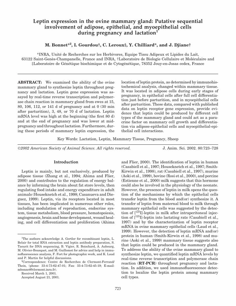
Figure 1. Partial nucleotide sequence of ovine leptincDNA and position of the primers and TaqMan probeused for the real-time reverse transcription and polymer-ase chain reaction assay. We characterized 1,076 bp of theovine leptin cDNA corresponding to a part of the leptinmRNA 3′untranslated region (accession numberAF310264). This ovine (o) fragment was aligned with itshuman (h) and pig (p) counterparts (sequence accessionnumbers U43653 and AF026976, respectively). Gaps (.)have been placed to maximize the similarity. Dashes (–)correspond to nucleotides that are identical to those ofthe ovine leptin sequence. The primers and TaqMan probeused for quantitative analysis of leptin mRNA level areshaded. Alignment was performed with the Clustalw pro-gram (Version 1.81). This ovine 1,076-bp sequence shows67 and 78% identity with the human and pig se-quences, respectively.
Leptin expression by the ovine mammary gland 725
The reverse transcription reaction (20 �L) of the real-time RT-PCR assay was performed using 4 �g of totalRNA, with 100 U of SuperScript reverse transcriptase(Gibco BRL, Life Technologies, Cergy Pontoise, France)and 10 pmol of oligo(dT)18. Amplification reactions (50�L) contained diluted (1:500 in water) cDNA sample(10 �L), 10 × PCR Master Mix (27.5 �L, PE AppliedBiosystem), 40 pmol of each primer, and 10 pmol ofTaqMan probe. The cycling conditions included 2 minat 50°C and 10 min at 95°C. Subsequently, thermalcycling proceeded with 45 cycles at 95°C for 15 s andat 60°C for 2 min. Each assay was performed in tripli-cate. The concentration of leptin mRNA was deter-mined from a calibration curve prepared by amplifying59,250, 29,625, 7,406, 1,851, and 592 copies of a recom-binant plasmid containing the 1,076-bp fragment de-scribed in Figure 1.
The leptin mRNA copy number was normalized bythe mRNA copy number of the constitutively expressedcyclophilin gene, quantified by real-time RT-PCR asdescribed previously (Bonnet et al., 2000).
Leptin Location by Indirect Immunofluorescence
Mammary fragments obtained after dissection werefixed with 2% paraformaldehyde in PBS buffer, pH 7.2,for 24 h. Fixed tissues were incubated overnight in 40%sucrose in PBS, frozen in liquid nitrogen vapors, andcut in 3-�m sections at −35°C with a Reichert Cryocut(Leica, Reuil-Malmaison, France).
Leptin antiserum was produced in rabbits by injec-tion of 1 mg of recombinant chicken leptin (Raver etal., 1998) solubilized in saline buffer and emulsified inFreund’s complete adjuvant. Three and six weeks laterthe rabbits were reimmunized with 1 mg of recombi-nant leptin solubilized in saline buffer and emulsifiedin Freund’s incomplete adjuvant. From the 6th wk afterinitial immunization, antiserum was collected weekly.
Serum and antibody dilutions were made in PBS con-taining 0.2% of fish gelatin.
For labeling of leptin protein, tissue sections (n =2 or 4 for tissues from pregnant and lactating ewes,respectively) were successively incubated with PBS-50mM NH4Cl (20 min), PBS (three times, 15 min eachtime), goat serum (1:10, 1 h) and rabbit anti-chickenleptin antiserum (1:100, 2 h); washed in PBS-0.2% fishgelatin; and then incubated with fluorescein isothiocya-nate (FITC)-conjugated goat anti-rabbit IgG (1:200 for2 h; Sanofi Diagnostics Pasteur, Marnes-La-Coquette,France). Sections were mounted on a drop of Vectas-hield (Vector Laboratories, Burlingame, CA) and ob-served with a Polyvar Reichert microscope (Leica). Con-trol sections were treated similarly with nonimmunerabbit serum or with omission of anti-chicken leptinantiserum. To check the specificity of the staining, sec-tions were incubated with anti-chicken leptin antise-rum fully adsorbed with chicken leptin. This adsorbedantiserum was prepared by incubating, for 2 h, chicken
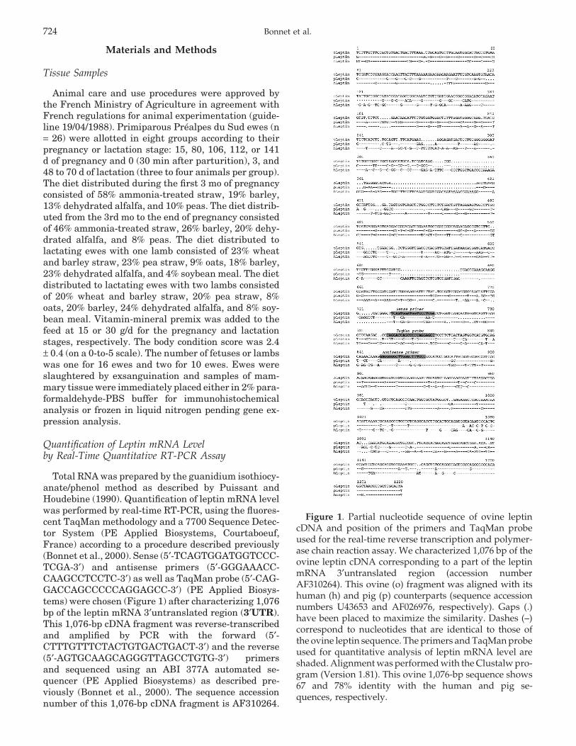
Figure 2. Expression of the leptin gene in mammarytissue throughout pregnancy and lactation determinedby real-time reverse transcription and polymerase chainreaction assay. Leptin and cyclophilin mRNA levels weremeasured from mammary gland tissue collected fromthree or four ewes at 15, 80, 106, 112, or 141 d of pregnancyand at 0, 3, 48, or 70 d of lactation. Leptin/cyclophilinmRNA ratios were calculated. a,b,c,dMeans (± SEM) withdifferent superscripts differ significantly (P < 0.05).
leptin (Raver et al., 1998) with anti-chicken leptin anti-serum (1:100) to make a final concentration of 1 �g/�L.
Double-labeling of both leptin protein and F-actinstructures was performed according to the same proce-dure, with the addition of 0.17 �mol of tetramethylrho-damine phalloidin (Molecular Probes, Eugene, OR) tothe incubation step with FITC-conjugated goat anti-rabbit IgG.
Statistical Analysis
Data were normalized by log transformation andwere submitted to an analysis of variance by the GLMprocedure of SAS (SAS Inst. Inc., Cary, NC). Since asignificant (P < 0.01) effect of the physiological statewas shown, the differences between two physiologicalstages were tested using Duncan’s test with a probabil-ity of 0.05.
Results
Temporal Expression of Leptin mRNA in OvineMammary Gland During Pregnancy and Lactation
Leptin mRNA level, normalized by the level ofcyclophilin mRNA, varied significantly (P < 0.01) de-pending on the pregnancy or lactation stage (Figure 2).During pregnancy, the leptin mRNA level decreasedstrongly between d 80 and d 106 or 112 (P < 0.05),before increasing slightly at d 141 (P < 0.05 for d 141vs d 106 of pregnancy) to levels similar to those assayedat d 15 and 80. Throughout lactation, leptin mRNAlevels did not vary significantly but were lower (P <
Bonnet et al.726
0.05) than the level assayed at d 80 of pregnancy. More-over, at d 3 of lactation, the level of leptin mRNA wassignificantly (P < 0.05) lower than those assayed at d15, 80, or 141 of pregnancy.
Immunofluorescent Location of Leptin in OvineMammary Gland During Pregnancy and Lactation
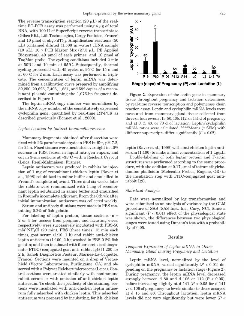
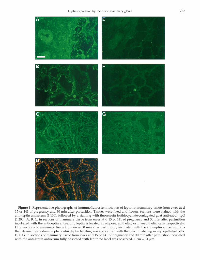
Immunofluorescence performed with the anti-leptinantiserum showed a labeling located in adipose, epithe-lial, or myoepithelial cells depending on the pregnancyor lactation stage. During early pregnancy (d 15), leptinimmunostaining was located in adipocytes, mainly intheir cytoplasm (Figure 3A). At the end of pregnancy(d 141), fluorescent labeling was detected on the apicalmembrane of the epithelial cells (Figure 3B). Just afterparturition (30 min), a leptin labeling was observed asa continuous fringe surrounding the acini (Figure 3C)and as a discontinuous fringe until 70 d of lactation(data not shown). Sections of mammary tissue from oneother ewe at each pregnancy stage and three other ewesat d 0 of lactation showed similar location of immuno-stainings. Just after parturition, the leptin labeling wascolocalized with the F-actin labeling, indicating thatleptin protein was mainly located in myoepithelial cells(Figure 3D). At all stages of pregnancy and lactation,leptin immunostaining was eliminated when anti-lep-tin antiserum was preadsorbed with leptin (Figures 3E,3F, 3G). No label was detectable when a non-immunerabbit serum was used instead of anti-leptin antiserumor when the second antibody alone was used (datanot shown).
Discussion
We report here the first evidence that ovine mam-mary tissue expresses leptin mRNA during lactation.This result is in agreement with the mammary synthe-sis of leptin previously reported in humans (Smith-Kir-win et al., 1998) and mice (Aoki et al., 1999).
We also report strong variations in ovine mammaryleptin gene expression depending on the stage of preg-nancy or lactation. Leptin mRNA was expressedthroughout pregnancy, with a strong decrease in theexpression at mid-pregnancy. During lactation the lep-tin mRNA levels were lower than or similar to thoseobserved during the end of pregnancy. One of the mostinteresting aspects of our study was the observationthat the cellular location of leptin changed during thedifferent phases of mammary gland development. Dur-ing early stages of development, leptin was exclusivelylocated in mammary adipocytes. In contrast, after fullcell differentiation, just before parturition, adipose tis-sue had completely regressed and leptin was presentin mammary epithelial cells, whereas during lactation,leptin was located in myoepithelial cells. Such a sequen-tial change in the leptin location between mammary celltypes suggests a new and complex scheme of mammaryleptin expression. Indeed, our results suggest a strong
synthesis of leptin by mammary adipocytes at the be-ginning of pregnancy that decreased to a lower levelduring the second part of pregnancy when adipocytesdisappeared. At the end of pregnancy, leptin mRNAlevel increased slightly and leptin protein was locatedin epithelial cells. A local production is likely becausea putative transfer of blood and(or) mammary adipocyteleptin would be reduced, due to the strong decrease inleptin receptor gene expression by the epithelial cellsat this stage (Laud et al., 1999). In addition, after partu-rition and throughout lactation, leptin mRNA was ex-pressed by the mammary tissue and leptin protein wasfound in myoepithelial cells. It could be hypothesizedthat leptin is produced exclusively by myoepithelialcells because leptin receptor is not expressed in thiscell type (Laud et al., 1999). Further studies, however,are needed to ascertain that leptin is synthesized byepithelial and myoepithelial cells around parturition.Nevertheless, in agreement with this hypothesis, leptinsynthesis was observed in a breast epithelial cell line(O’Brien et al., 1999) as well as in rat skeletal musclecells (Wang et al., 1999).
These sequential changes of leptin cellular location,together with the synthesis of leptin receptor exclu-sively by epithelial cells and mainly between 70 and106 d of pregnancy (Laud et al., 1999), suggest thatleptin could act as a paracrine factor in mammary glandgrowth, development, and function. Indeed, mammarygland growth and development during pregnancy arehighly dependent on steroids and protein hormonesfrom ovaries, placenta, and pituitary gland (Lyons,1958). However, in vitro studies indicate that thesehormone effects are largely indirect, being mediatedby growth factors synthesized by mammary adipocytes(Levine and Stockdale, 1984; Rudland et al., 1984;Woodward et al., 1998). Leptin could be one of thesesteroid-inducible proteins synthesized by mammary fatcells; the strong leptin gene expression that we observedat 80 d of pregnancy occurred concurrently with thestart of the increase in plasma estradiol concentration(Martinet and Houdebine, 1999). Moreover, the mainte-nance of alveolar structures during lactation is partlycontrolled by growth factors such as IGF-I, trans-forming growth factor-α and -β, and fibroblast growthfactor-1, 2, and 7, mainly synthesized by myoepithelialcells (Gomm et al., 1997; Plath et al., 1998; Martinetand Houdebine, 1999). Hence, the myoepithelial cellleptin observed in our study could participate in thecontrol of epithelial cell growth and survival(apoptosis). Finally, leptin gene expression by sheepmammary gland around parturition could be relatedto leptin secretion in colostrum and milk; it has beenobserved in women (Casabiell et al., 1997; Houseknetchet al., 1997; Smith-Kirwin et al., 1998), rats (Casabiellet al., 1997), cows (Rosi et al., 2000), pigs (Estienne etal., 2000), and mice, mainly during the first 2 d of lacta-tion (Aoki et al., 1999). It could be hypothesized thatleptin, as a colostral protein, may promote immunity
Leptin expression by the ovine mammary gland 727
Figure 3. Representative photographs of immunofluorescent location of leptin in mammary tissue from ewes at d15 or 141 of pregnancy and 30 min after parturition. Tissues were fixed and frozen. Sections were stained with theanti-leptin antiserum (1:100), followed by a staining with fluorescein isothiocyanate-conjugated goat anti-rabbit IgG(1:200). A, B, C: in sections of mammary tissue from ewes at d 15 or 141 of pregnancy and 30 min after parturitionincubated with the anti-leptin antiserum, leptin is located in adipose, epithelial, or myoepithelial cells, respectively.D: in sections of mammary tissue from ewes 30 min after parturition, incubated with the anti-leptin antiserum plusthe tetramethylrhodamine phalloidin, leptin labeling was colocalized with the F-actin labeling in myoepithelial cells.E, F, G: in sections of mammary tissue from ewes at d 15 or 141 of pregnancy and 30 min after parturition incubatedwith the anti-leptin antiserum fully adsorbed with leptin no label was observed. 1 cm = 31 �m.
Bonnet et al.728
(Lord et al., 1998) and(or) intestinal cell functionality(Morton et al., 1998) in newborn mammals.
Implications
We show for the first time that leptin is producedby the ovine mammary gland; its gene expression ismaximal at the beginning of pregnancy and, to a lesserextent, at the end of pregnancy. Moreover, we reporthere the presence of leptin protein in adipose, epithelial,or myoepithelial cells at the beginning or the end ofpregnancy, or after parturition, respectively. These re-sults suggest that leptin, besides being a colostral pro-tein, may be a paracrine factor acting on mammarygland growth, development, and function, via adipose-epithelial cells and myoepithelial-epithelial cell inter-actions favored by epithelial cell leptin receptors. Fur-ther studies are needed to clarify putative implicationsof leptin in the physiology of newborns as well as inmammogenesis around puberty and during pregnancy,and in mammary cell apoptosis during lactation. Thiswould help us to better understand the mechanisms forthe known effect of nutritional factors and body fatnesson peripubertal mammogenesis.
Literature Cited
Ahima, R. S., and J. S. Flier. 2000. Adipose tissue as an endocrineorgan. Trends Endocrinol. Metab. 11:327–332.
Aoki, N., M. Kawamura, and T. Matsuda. 1999. Lactation-dependentdown regulation of leptin production in mouse mammary gland.Biochim. Biophys. Acta 1427:298–306.
Bonnet, M., C. Leroux, Y. Faulconnier, J. F. Hocquette, F. Bocquier,P. Martin, and Y. Chilliard. 2000. Lipoprotein lipase activityand mRNA are up-regulated by refeeding in adipose tissue andcardiac muscle of sheep. J. Nutr. 130:749–756.
Casabiell, X., V. Pineiro, M. A. Tome, R. Peino, C. Dieguez, and F.F. Casanueva. 1997. Presence of leptin in colostrum and/orbreast milk from lactating mothers: a potential role in the regula-tion of neonatal food intake. J. Clin. Endocrinol. Metab.82:4270–4273.
Casanueva, F. F., and C. Dieguez. 1999. Neuroendocrine regulationand actions of leptin. Front. Neuroendocrinol. 20:317–363.
Estienne, M. J., A. F. Harpe, C. R. Barb, and M. J. Azain. 2000.Concentrations of leptin in serum and milk from sows that dif-fered in body condition at farrowing J. Anim. Sci. 78(Suppl.1):204 (Abstr.).
Gomm, J. J., P. J. Browne, R. C. Coope, G. S. Bansal, C. Yiangou,C. L. Johnston, R. Mason, and R. C. Coombes. 1997. A paracrinerole for myoepithelial cell-derived FGF2 in the normal humanbreast. Exp. Cell Res. 234:165–173.
Houseknecht, K. L., C. A. Baile, R. L. Matteri, and M. E. Spurlock.1998. The biology of leptin: A review. J. Anim. Sci. 76:1405–1420.
Houseknecht, K. L., M. K. McGuire, C. P. Portocarrero, M. A. McGu-ire, and K. Beerman. 1997. Leptin is present in human milkand is related to maternal plasma leptin concentration adiposity.Biochem. Bioph. Res. Commun. 240:742–747.
Laud, K., I. Gourdou, L. Belair, D. H. Keisler, and J. Djiane. 1999.Detection and regulation of leptin receptor mRNA in ovine mam-mary epithelial cells during pregnancy and lactation. FEBS Lett.463:194–198.
Levine, J. F., and F. E Stockdale. 1984. Cell-cell interactions promotemammary epithelial cell differentiation. Exp. Cell Res.151:112–122.
Lord, G. M., G. Matarese, J. K. Howard, R. J. Baker, S. R. Bloom,and R. I. Lechler. 1998. Leptin modulates the T-cell immuneresponse and reverses starvation-induced immunosuppression.Nature (Lond.) 394:897–901.
Lyons, W. R. 1958. Hormonal synergism in mammary growth. Proc.R. Soc. Lond. Ser. Biol. Sci. 149:303–325.
Martinet, J., and L. M. Houdebine. 1999. Mammary gland, mammo-genesis, growth factors, lactogenesis. In: J. Martinet, L. M.Houdebine, and H. Herbert (ed.) Biology of Lactation. pp 1–27.INRA Publications, Paris, France.
Morton, N. M., V. Emilsson, Y. L. Liu, and M. A. Cawthorne. 1998.Leptin action in intestinal cells. J. Biol. Chem. 273:26194–26201.
O’Brien, S. N., B. H. Welter, and T. M. Price. 1999. Presence of leptinin breast cell lines and breast tumors. Biochem. Biophys. Res.Commun. 259:695–698.
Plath, A., R. Einspanier, C. Gabler, F. Peters, F. Sinowatz, D. Gos-podarowicz, and D. Schams. 1998. Expression and localizationof members of the fibroblast growth factor family in the bovinemammary gland. J. Dairy Sci. 81:2604–2613.
Puissant, C., and L. M. Houdebine. 1990. An improvement of thesingle-step method of RNA isolation by acid guanidinium thiocy-anate-phenol-chloroform extraction. Biotechniques 8:148–149.
Raver, N., M. Taouis, S. Dridi, M. Derouet, J. Simon, B. Robinzon,J. Djiane, and A. Gertler. 1998. Large-scale preparation of biolog-ically active recombinant chicken obese protein (leptin). ProteinExpr. Purif. 14:403–408.
Rosi, F., V. Bontempo, R. Capalbo, and A. Baldi. 2000. Effects ofrumen-protected methionine on milk yield, milk leptin and milknitrogenous compounds during lacatation. In: A. Baldi and K.Stelwagen (ed.) Livestock Production Science, Special Issue. p178. Elsevier Science Publishers, Amsterdam, The Netherlands
Rudland, P. S., A. C. Twiston Davies, and S. W. Tsao. 1984. Ratmammary preadipocytes in culture produce a trophic agent formammary epithelia-prostaglandin E2. J. Cell. Physiol.120:364–376.
Smith-Kirwin, S. M., D. M. O’Connor, J. De Johnston, E. D. Lancey,S. G.Hassink, and V. L. Funanage. 1998. Leptin expression inhuman mammary epithelial cells and breast milk. J. Clin. Endo-crinol. Metab. 83:1810–1813.
Wang J., R. Liu, L. Liu, R. Chowdhury, N. Barzilai, J. Tan, and L.Rossetti. 1999. The effect of leptin on Lep expression is tissue-specific and nutritionally regulated. Nat. Med. 5:895–899.
Woodward, T. L, J. W. Xie, and S. Z. Haslam. 1998. The role ofmammary stroma in modulating the proliferative response toovarian hormones in the normal mammary gland. J. Mamm.Gland Biol. Neoplasia 3:117–131.
Zhang, Y., R. Proenca, M. Maffei, M. Barone, L. Leopold, and J. M.Friedman. 1994. Positional cloning of the mouse obese gene andits human homologue. Nature (Lond.) 372:425–432.


![Strategie Ovine [Compatibility Mode]](https://img.pdfslide.us/doc/110x75/577cd4611a28ab9e78985bbf/strategie-ovine-compatibility-mode.jpg)