Embed Size (px)
DESCRIPTION
Mammary Gland Digital Laboratory. It’s best to view this in Slide Show mode, especially for the quizzes. This module will take approximately 45 minutes to complete. After completing this exercise, you should be able to: - PowerPoint PPT Presentation
Citation preview
Mammary GlandDigital Laboratory
It’s best to view this in Slide Show mode, especially for the quizzes.
This module will take approximately 45 minutes to complete.
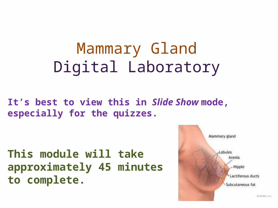
After completing this exercise, you should be able to:
•Distinguish, at the light microscope level, each of the following organs and their specific features:• Mammary gland
• Lobes and lobules • Acini • Ducts • Stroma
• Cells and structures • Secretory cells • Myoepithelial cells • (Plasma cells and other lymphocytes)
• Stages • Immature / Inactive • Mammary gland of pregnancy • Lactating mammary gland • Regressing (difficult to distinguish from poorly preserved tissue)
• Nipple • Lactiferous ducts / sinuses
•Distinguish, at the electron microscope level, each of the following organs and their specific features:• Mammary gland
• Acinar cells• Lipid product • Protein product
• Myoepithelial cells
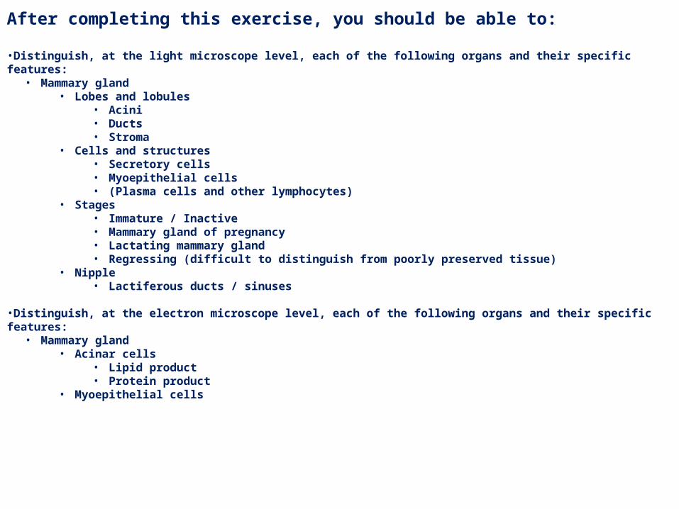
GROSS ANATOMY OF THE MAMMARY GLAND
Each breast contains 12-15 glands, each with a separate opening onto the nipple. The glands can be divided into lobes (blue outline) and lobules (black outline). These contain clusters of secretory cells surrounded by loose connective tissue, which lead to lactiferous ducts. Near the nipple, a dilation of the duct called the lactiferous sinus is a reservoir for milk between feedings.The nipple is a conical elevation which contains sebaceous glands, and smooth muscle for erection.
Here we’ll look at 4 different stages of mammary gland development1. Immature / inactive2. Mammary gland of pregnancy3. Mammary gland of
breastfeeding4. Regressing
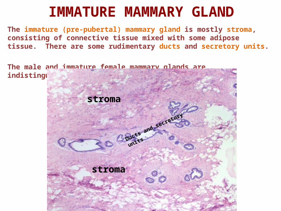
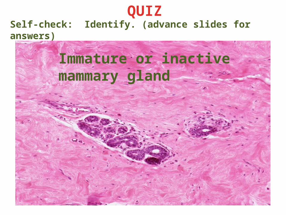
IMMATURE MAMMARY GLANDThe immature (pre-pubertal) mammary gland is mostly stroma, consisting of connective tissue mixed with some adipose tissue. There are some rudimentary ducts and secretory units.
The male and immature female mammary glands are indistinguishable.
stroma
stroma
Ducts and secretory units
Link to SL 149Be able to identify:
• Immature mammary gland• Rudimentary ducts and glands• Stroma
• Connective tissue (dense irregular and loose)• Adipose
Video showing immature mammary gland – SL149
IMMATURE MAMMARY GLAND
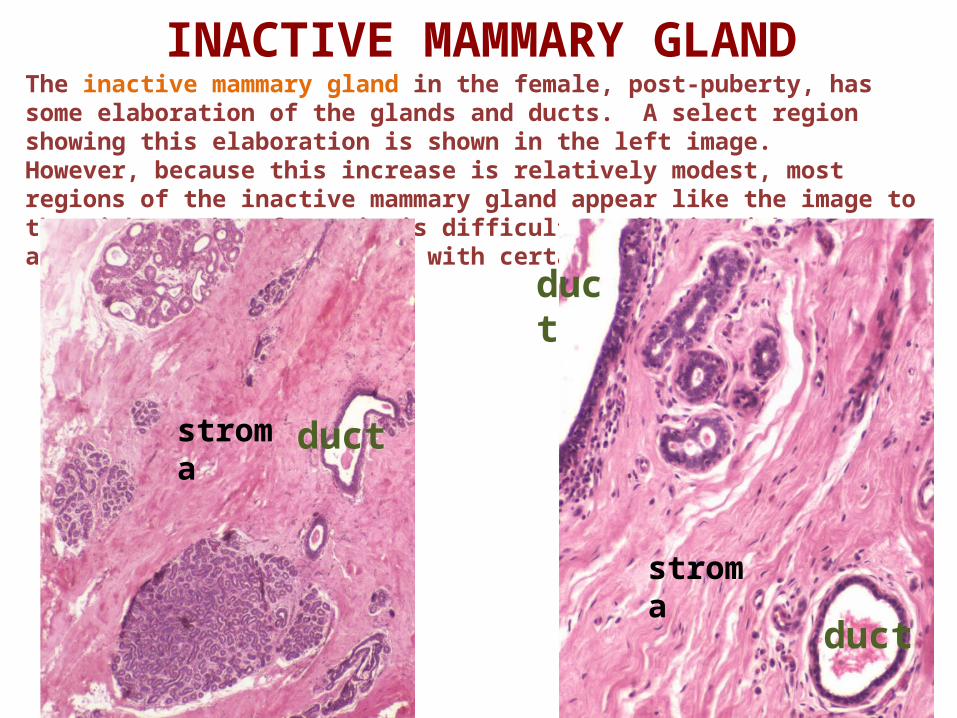
INACTIVE MAMMARY GLANDThe inactive mammary gland in the female, post-puberty, has some elaboration of the glands and ducts. A select region showing this elaboration is shown in the left image. However, because this increase is relatively modest, most regions of the inactive mammary gland appear like the image to the right. Therefore, it is difficult to distinguish immature and inactive mammary glands with certainty.
duct
duct
duct
stroma
stroma
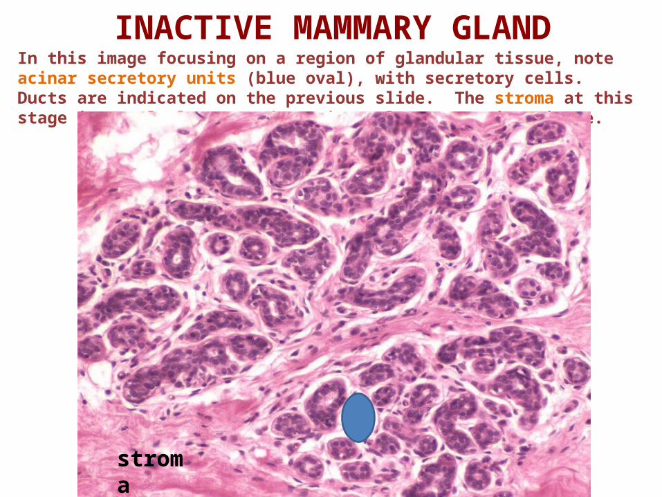
INACTIVE MAMMARY GLANDIn this image focusing on a region of glandular tissue, note acinar secretory units (blue oval), with secretory cells. Ducts are indicated on the previous slide. The stroma at this stage is mostly loose or dense irregular connective tissue.
stroma

Link to SL 150Be able to identify:
• Inactive mammary gland (indistinguishable from immature)
• Ducts and glands• Stroma
• Connective tissue (dense irregular and loose)• Adipose
INACTIVE MAMMARY GLAND
Video showing inactive mammary gland – SL150
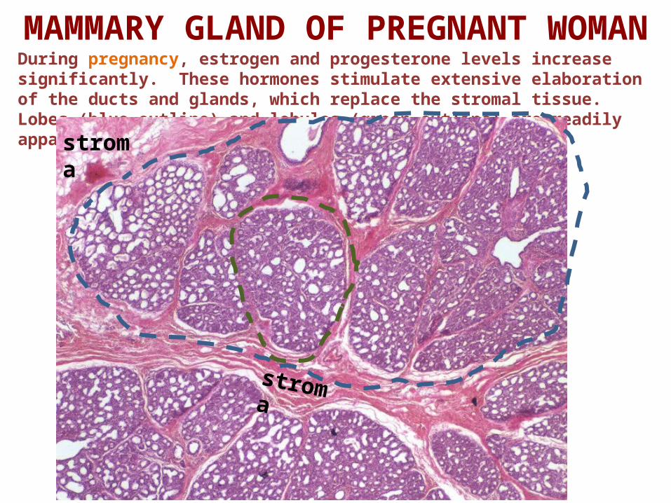
MAMMARY GLAND OF PREGNANT WOMANDuring pregnancy, estrogen and progesterone levels increase significantly. These hormones stimulate extensive elaboration of the ducts and glands, which replace the stromal tissue. Lobes (blue outline) and lobules (green outline) are readily apparent.
stroma
stroma
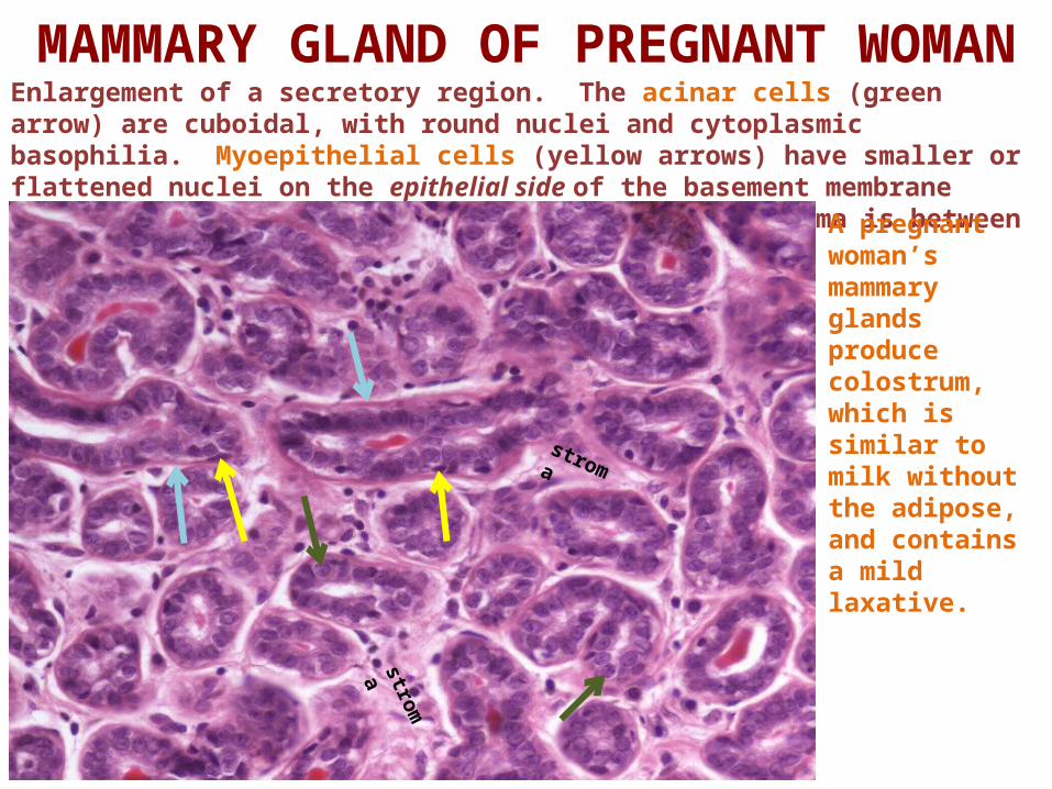
MAMMARY GLAND OF PREGNANT WOMANEnlargement of a secretory region. The acinar cells (green arrow) are cuboidal, with round nuclei and cytoplasmic basophilia. Myoepithelial cells (yellow arrows) have smaller or flattened nuclei on the epithelial side of the basement membrane (light blue arrows). Sparse connective tissue stroma is between the acini.
A pregnant woman’s mammary glands produce colostrum, which is similar to milk without the adipose, and contains a mild laxative.
stroma
stroma
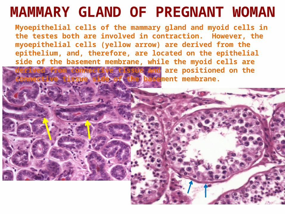
MAMMARY GLAND OF PREGNANT WOMANMyoepithelial cells of the mammary gland and myoid cells in the testes both are involved in contraction. However, the myoepithelial cells (yellow arrow) are derived from the epithelium, and, therefore, are located on the epithelial side of the basement membrane, while the myoid cells are derived from connective tissue and are positioned on the connective tissue side of the basement membrane.
Link to SL 151Be able to identify:
• Mammary gland from a pregnant woman• Ducts and glands• Epithelial cells
• Acinar cells• Myoepithelial cells
• Stroma• Plasma cells
MAMMARY GLAND OF PREGNANT WOMAN
Video showing mammary gland of pregnancy – SL151
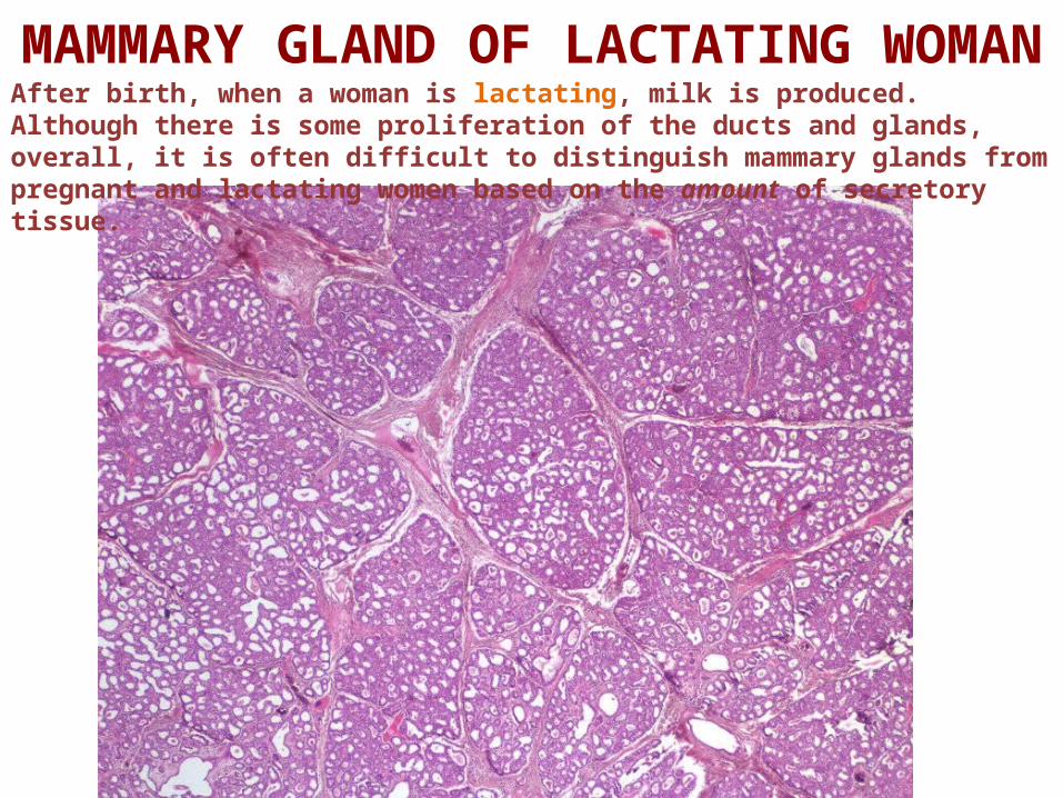
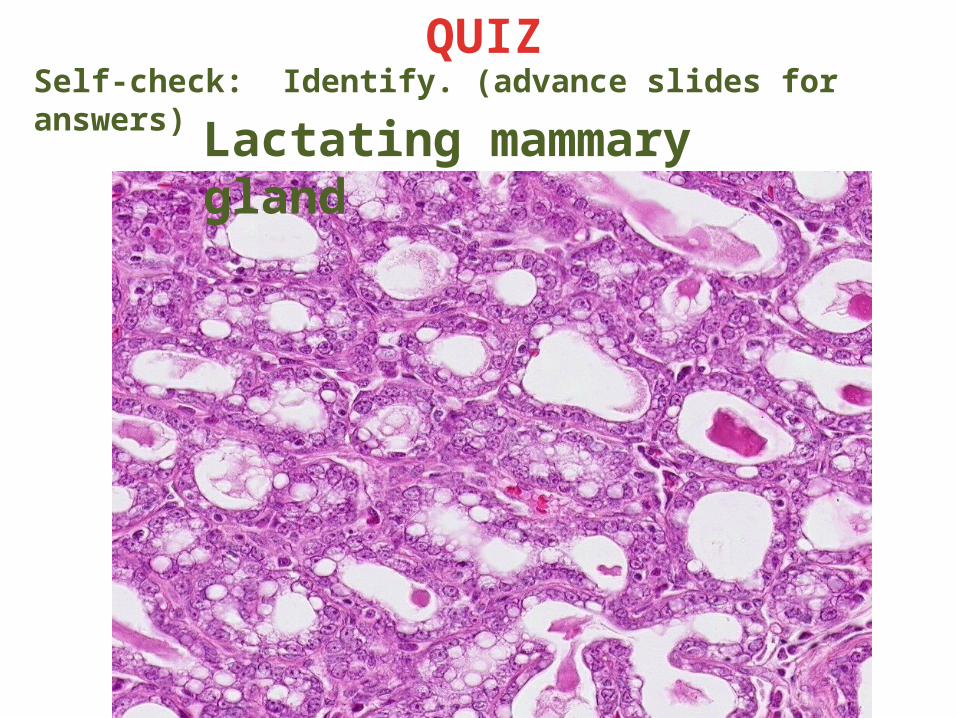
MAMMARY GLAND OF LACTATING WOMANAfter birth, when a woman is lactating, milk is produced. Although there is some proliferation of the ducts and glands, overall, it is often difficult to distinguish mammary glands from pregnant and lactating women based on the amount of secretory tissue.
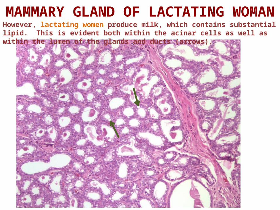
MAMMARY GLAND OF LACTATING WOMANHowever, lactating women produce milk, which contains substantial lipid. This is evident both within the acinar cells as well as within the lumen of the glands and ducts (arrows).
Link to SL 153Be able to identify:
• Mammary gland from a lactating woman• Ducts and glands• Epithelial cells
• Acinar cells• Myoepithelial cells
• Stroma• (Immune cells - you haven’t had the Infection and Immunity block yet,
but you have seen plasma cells a couple times now)
MAMMARY GLAND OF LACTATING WOMAN
Video showing mammary gland from a lactating woman – SL153

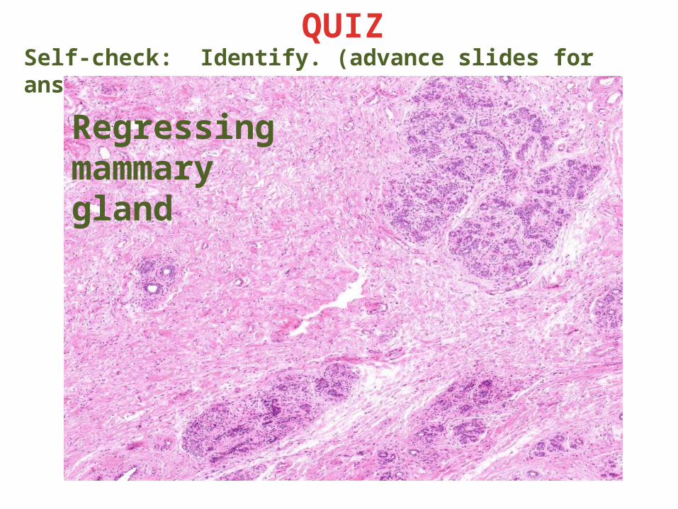
REGRESSING MAMMARYAfter lactation, the secretory units regress. Here, you can see the faint outlines of lobules, with sparse secretory tissue within them.
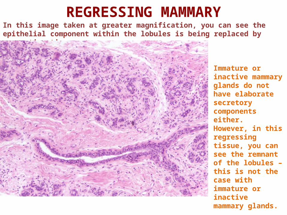
REGRESSING MAMMARYIn this image taken at greater magnification, you can see the epithelial component within the lobules is being replaced by connective tissue.
Immature or inactive mammary glands do not have elaborate secretory components either. However, in this regressing tissue, you can see the remnant of the lobules – this is not the case with immature or inactive mammary glands.
Link to SL 154Be able to identify:
• Regressing mammary gland
MAMMARY GLAND OF LACTATING WOMAN
Video showing regressing mammary gland – SL154
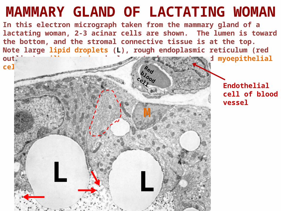
In this electron micrograph taken from the mammary gland of a lactating woman, 2-3 acinar cells are shown. The lumen is toward the bottom, and the stromal connective tissue is at the top. Note large lipid droplets (L), rough endoplasmic reticulum (red outline), milk proteins in lumen (red arrows), and myoepithelial cell (M).
MAMMARY GLAND OF LACTATING WOMAN
L L
M
Endothelial cell of blood vessel
Red blood cell
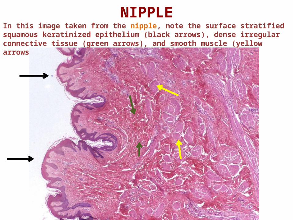
NIPPLEIn this image taken from the nipple, note the surface stratified squamous keratinized epithelium (black arrows), dense irregular connective tissue (green arrows), and smooth muscle (yellow arrows).
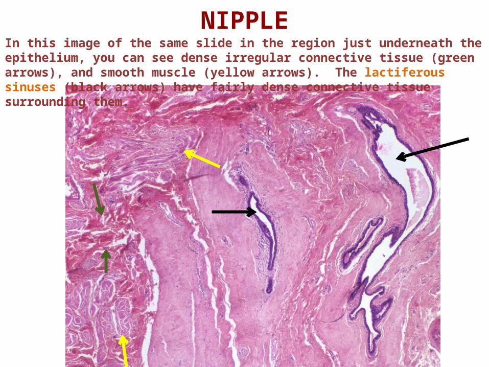
NIPPLEIn this image of the same slide in the region just underneath the epithelium, you can see dense irregular connective tissue (green arrows), and smooth muscle (yellow arrows). The lactiferous sinuses (black arrows) have fairly dense connective tissue surrounding them.
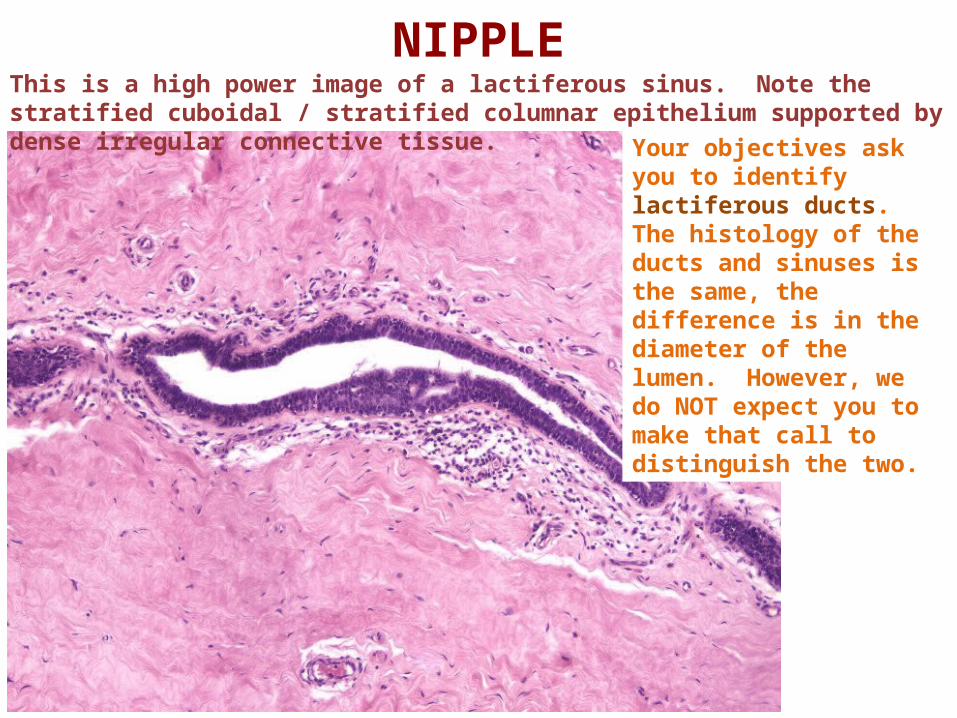
NIPPLEThis is a high power image of a lactiferous sinus. Note the stratified cuboidal / stratified columnar epithelium supported by dense irregular connective tissue.
Your objectives ask you to identify lactiferous ducts. The histology of the ducts and sinuses is the same, the difference is in the diameter of the lumen. However, we do NOT expect you to make that call to distinguish the two.
Link to SL 155Be able to identify:
• Nipple• Stratified squamous keratinized epithelium• Smooth muscle• Lactiferous ducts
NIPPLE OF BREAST
Video showing nipple of breast – SL155
The next set of slides is a final quiz for this module. You should review the structures covered in this module, and try to visualize each of these in light and electron micrographs.
•Distinguish, at the light microscope level, each of the following organs and their specific features:• Mammary gland
• Lobes and lobules • Acini • Ducts • Stroma
• Cells and structures • Secretory cells • Myoepithelial cells • (Plasma cells and other lymphocytes)
• Stages • Immature / Inactive • Mammary gland of pregnancy • Lactating mammary gland • Regressing (difficult to distinguish from poorly preserved tissue)
• Nipple • Lactiferous ducts / sinuses
•Distinguish, at the electron microscope level, each of the following organs and their specific features:• Mammary gland
• Acinar cells• Lipid product • Protein product
• Myoepithelial cells
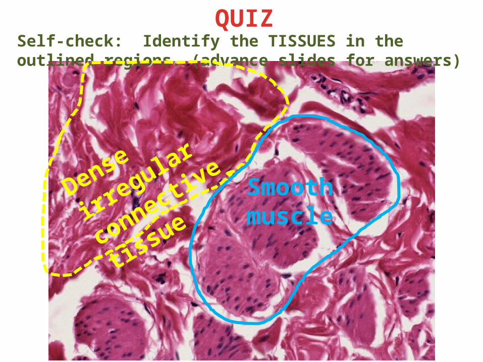
Self-check: Identify the TISSUES in the outlined regions. (advance slides for answers)
QUIZ
Smooth muscle
Dense irregular
connective tissue
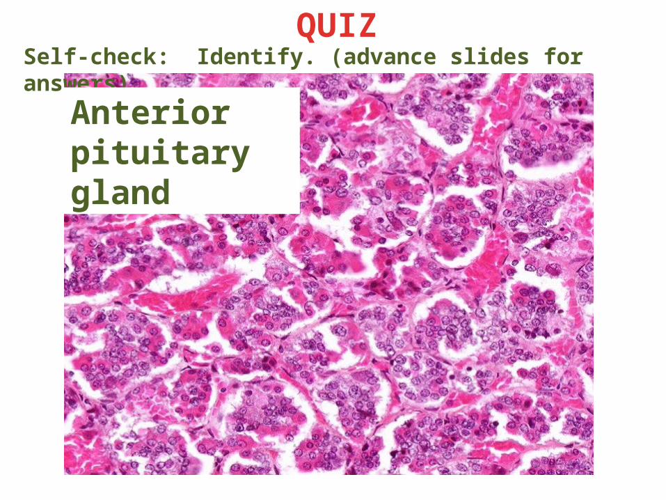
Self-check: Identify. (advance slides for answers)
QUIZ
Anterior pituitary gland
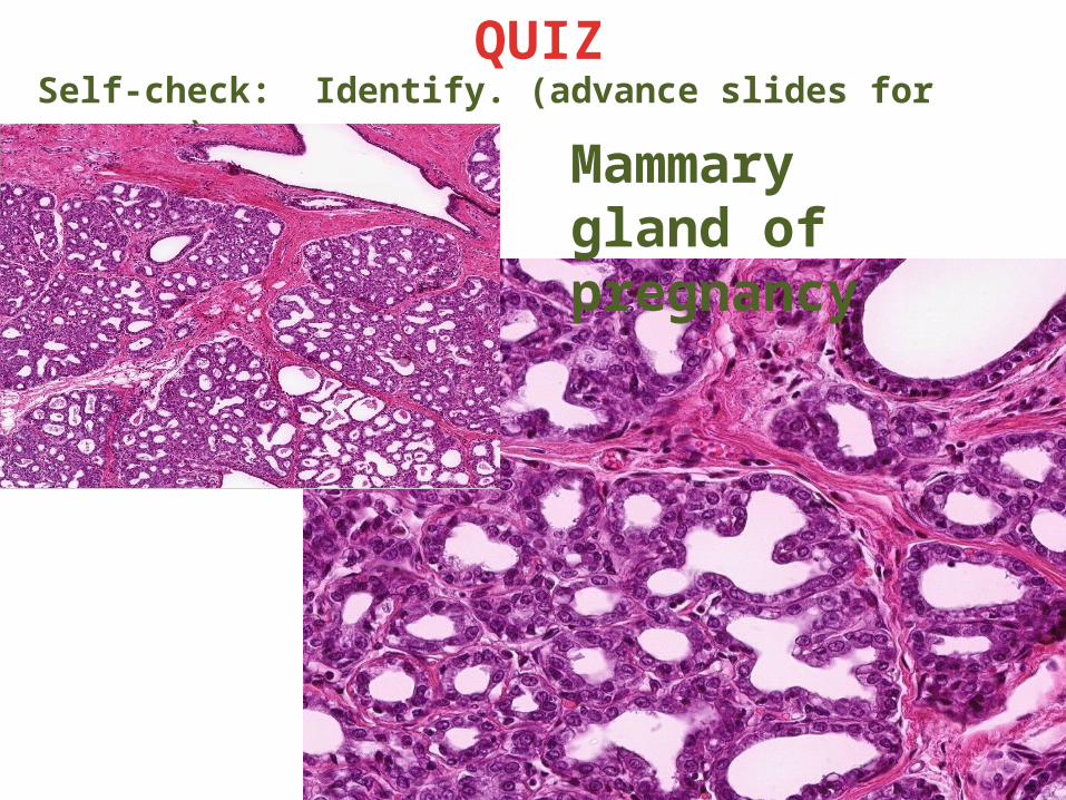
Self-check: Identify. (advance slides for answers)
QUIZ
Mammary gland of pregnancy
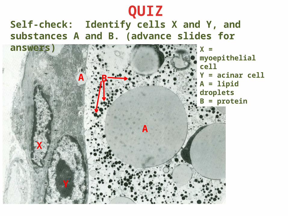
Self-check: Identify cells X and Y, and substances A and B. (advance slides for answers)
QUIZ
X = myoepithelial cellY = acinar cellA = lipid dropletsB = proteinB
A
Y
X
A
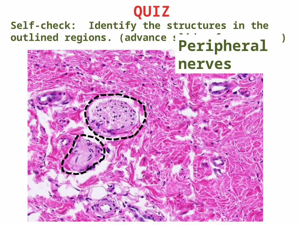
Self-check: Identify the structures in the outlined regions. (advance slides for answers)
QUIZ
Peripheral nerves
Self-check: Identify. (advance slides for answers)
QUIZ
Regressing mammary gland
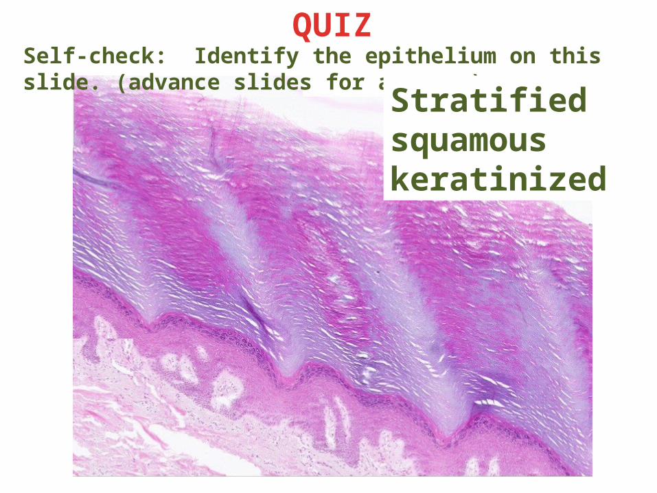
Self-check: Identify the epithelium on this slide. (advance slides for answers)
QUIZ
Stratified squamous keratinized
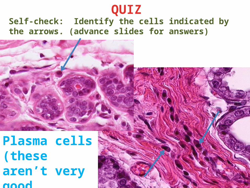
Self-check: Identify the cells indicated by the arrows. (advance slides for answers)
QUIZ
Plasma cells (these aren’t very good examples)
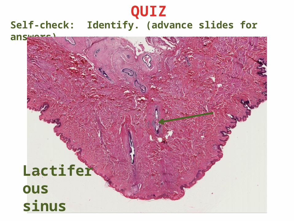
Self-check: Identify. (advance slides for answers)
QUIZ
Lactiferous sinus
Self-check: Identify. (advance slides for answers)
QUIZ
Lactating mammary gland
Self-check: Identify. (advance slides for answers)
QUIZ
Immature or inactive mammary gland
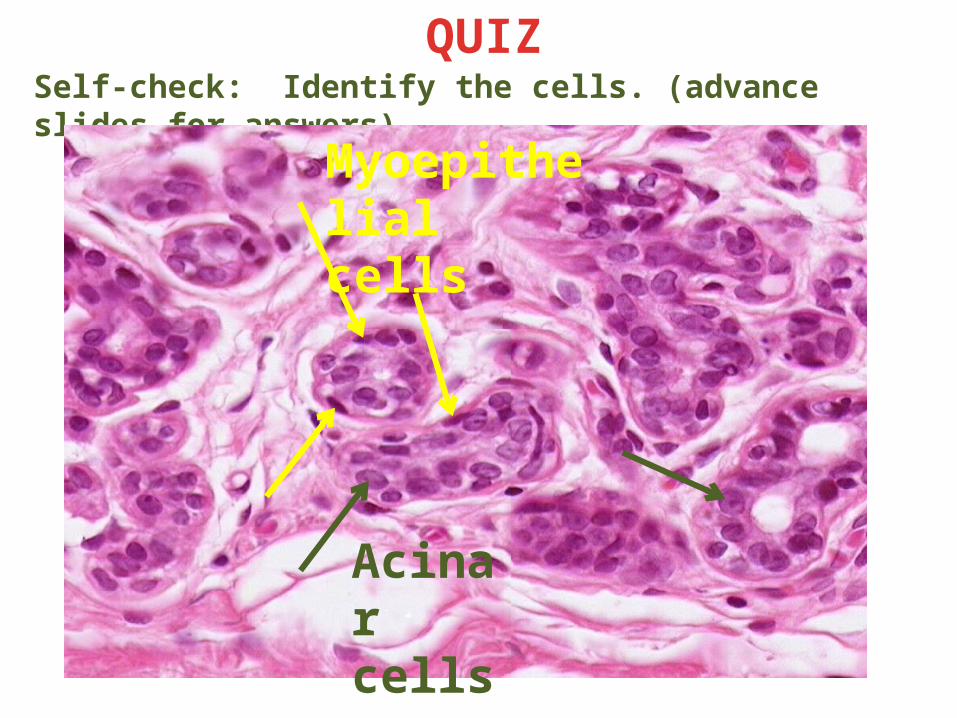
Self-check: Identify the cells. (advance slides for answers)
QUIZ
Myoepithelial cells
Acinar cells
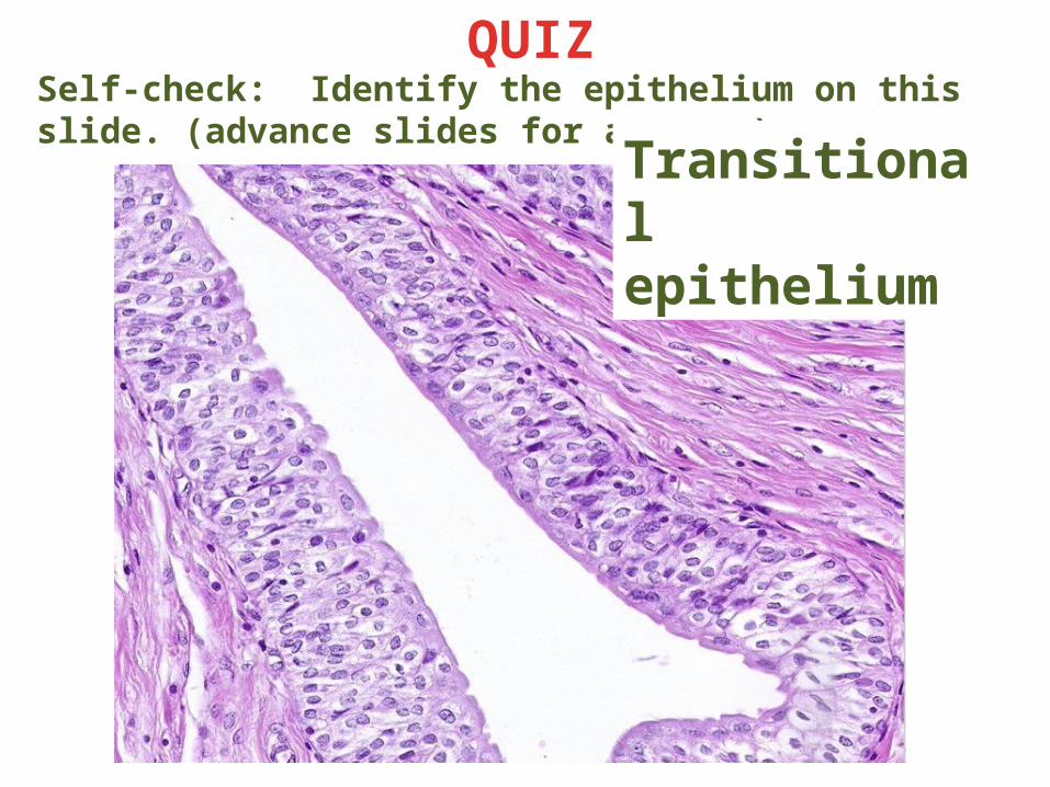
Self-check: Identify the epithelium on this slide. (advance slides for answers)
QUIZ
Transitional epithelium
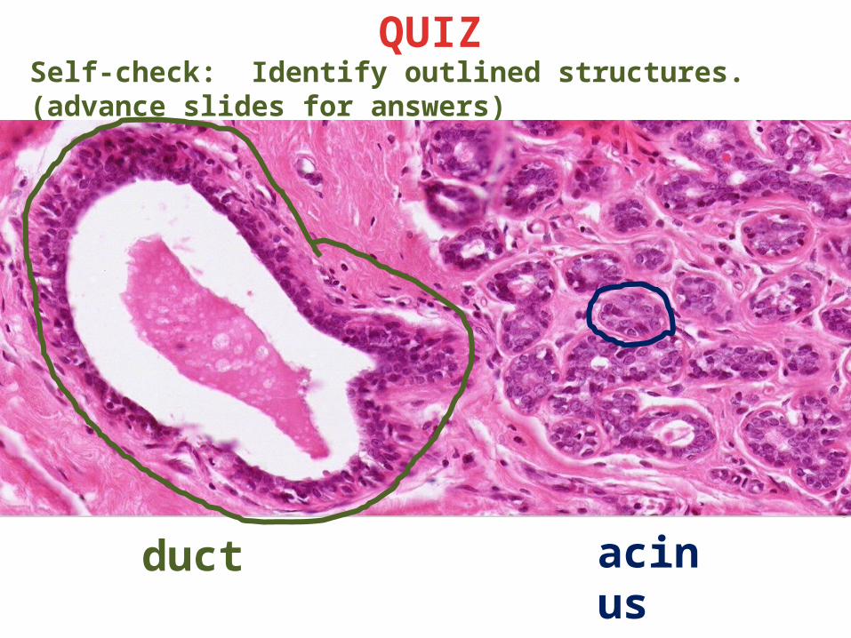
Self-check: Identify outlined structures. (advance slides for answers)
QUIZ
duct acinus
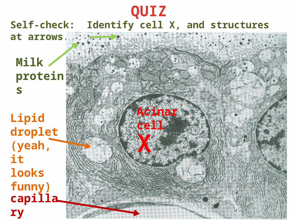
Self-check: Identify cell X, and structures at arrows. (advance slides for answers)
QUIZ
X
Milk proteins
Acinar cellLipid
droplet (yeah, it looks funny)
capillary
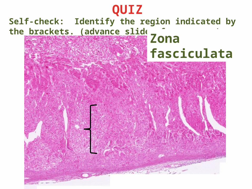
Self-check: Identify the region indicated by the brackets. (advance slides for answers)
QUIZ
Zona fasciculata
Link to SL 100A and SL 100B
NIPPLE OF BREASTSelf-check: This slide was from a dilation and curettage (D and C) procedure. Which phase of the menstrual cycle is represented on these slides? (they are the same)
Secretory phase
Link to SL 101
NIPPLE OF BREASTSelf-check: This slide was also taken from a dilation and curettage (D and C) procedure. What organ is represented on this slide? (don’t look at the title – cover upper left corner with your hand if you must))
cervix