Embed Size (px)
Citation preview
FEMALE REPRODUCTIVE SYSTEM
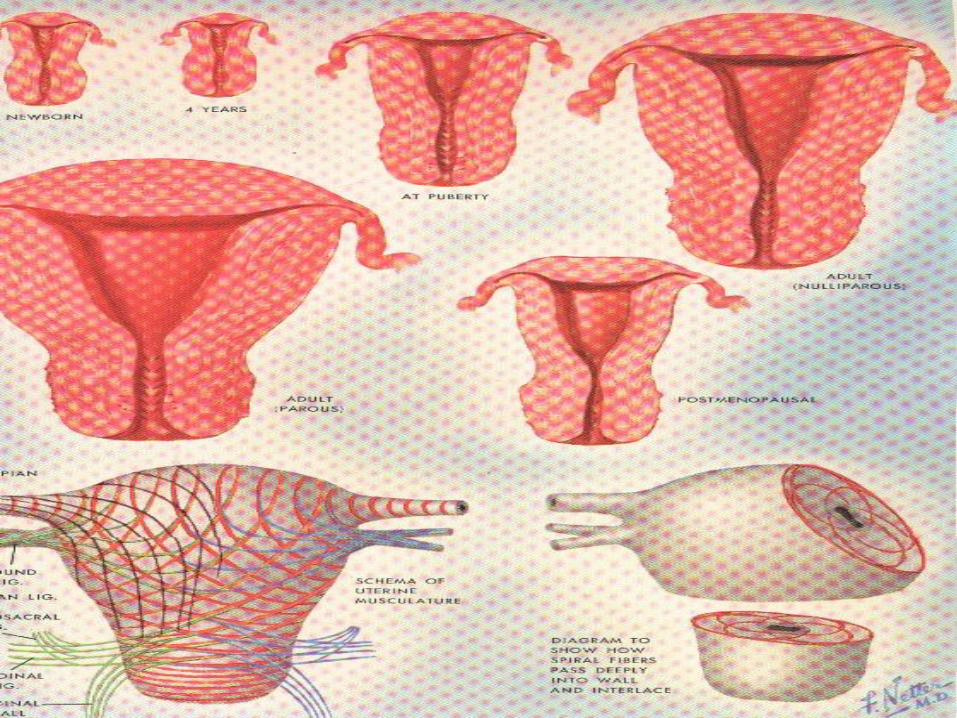
UTERUS
MAMMARY GLAND
PLACENTA
Prof.J.Anbalagan
Department of Anatomy

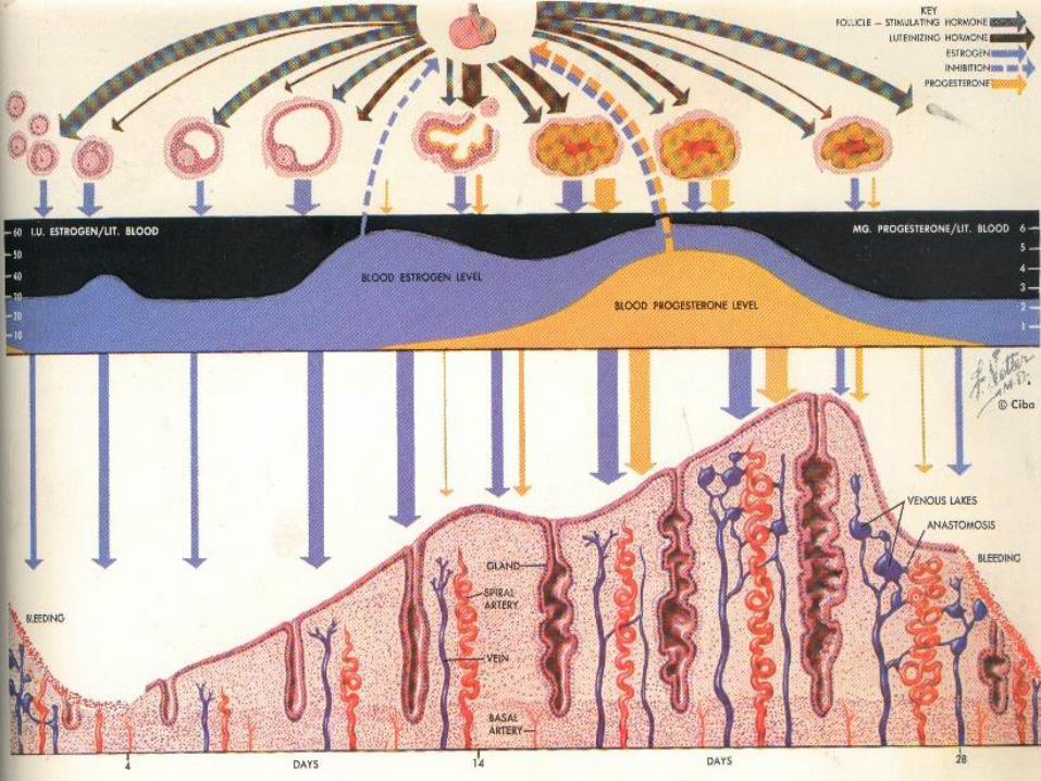
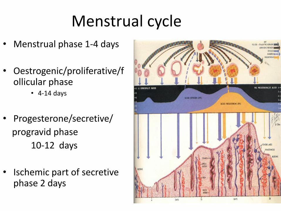
Menstrual cycle • Menstrual phase 1-4 days
• Oestrogenic/proliferative/follicular phase
• 4-14 days
• Progesterone/secretive/
progravid phase
10-12 days
• Ischemic part of secretive phase 2 days
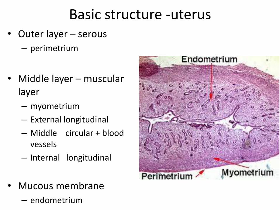
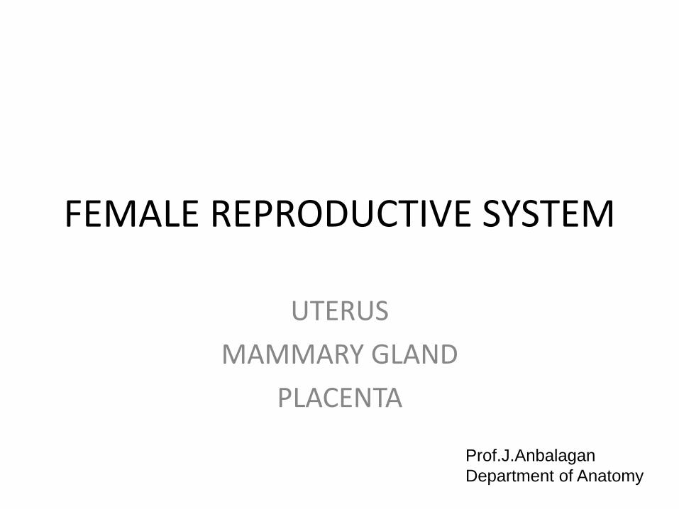
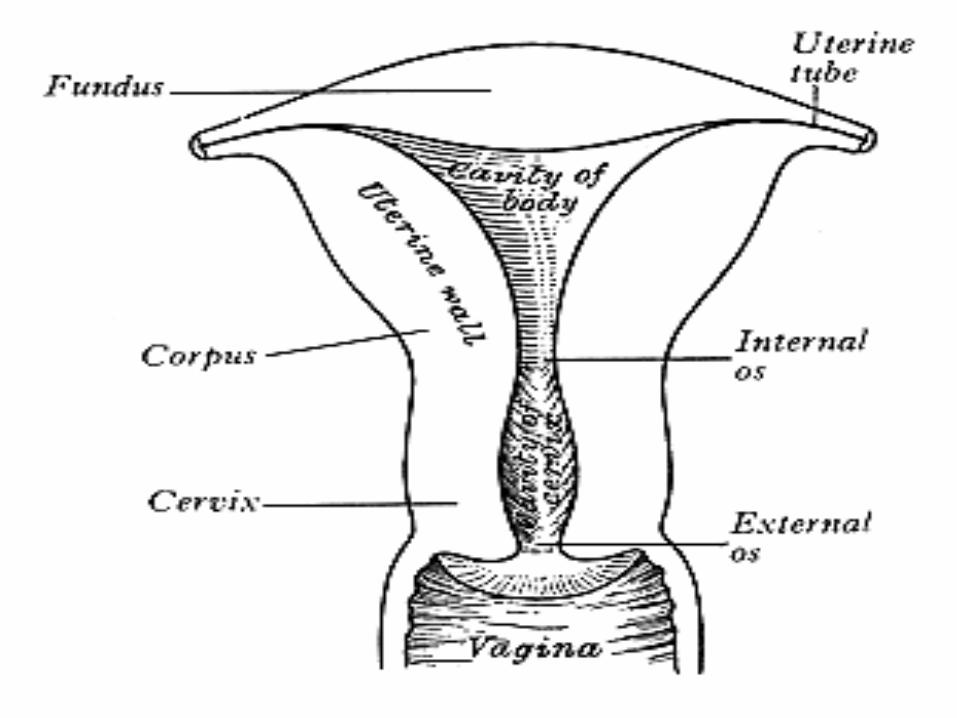
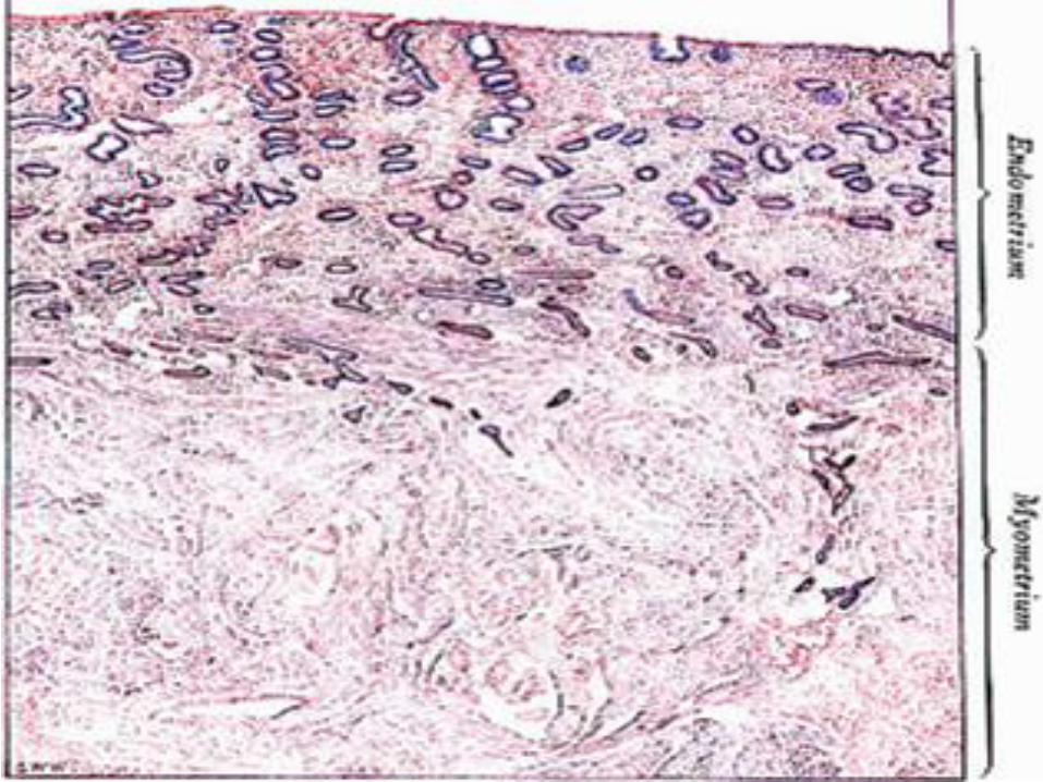
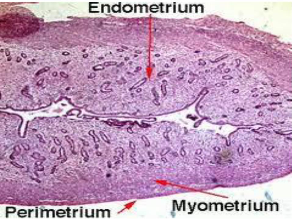
Basic structure -uterus • Outer layer – serous
– perimetrium
• Middle layer – muscular layer
– myometrium
– External longitudinal
– Middle circular + blood vessels
– Internal longitudinal
• Mucous membrane
– endometrium
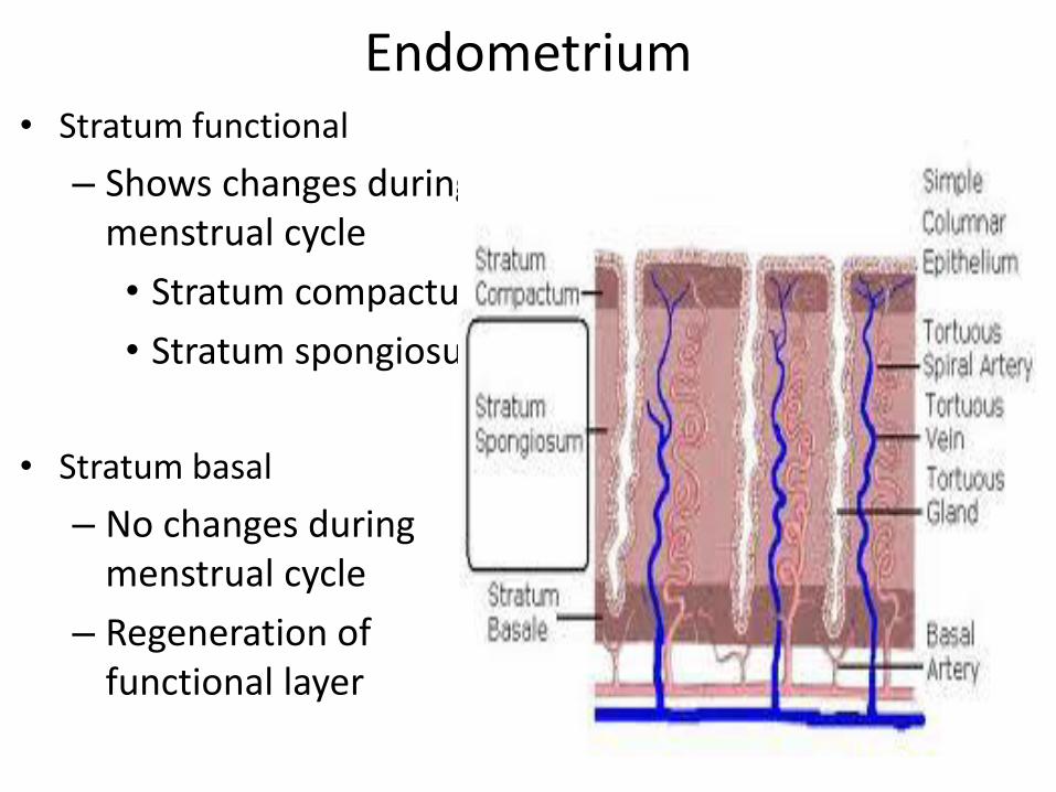
Endometrium • Stratum functional
– Shows changes during menstrual cycle
• Stratum compactum
• Stratum spongiosum
• Stratum basal
– No changes during menstrual cycle
– Regeneration of functional layer

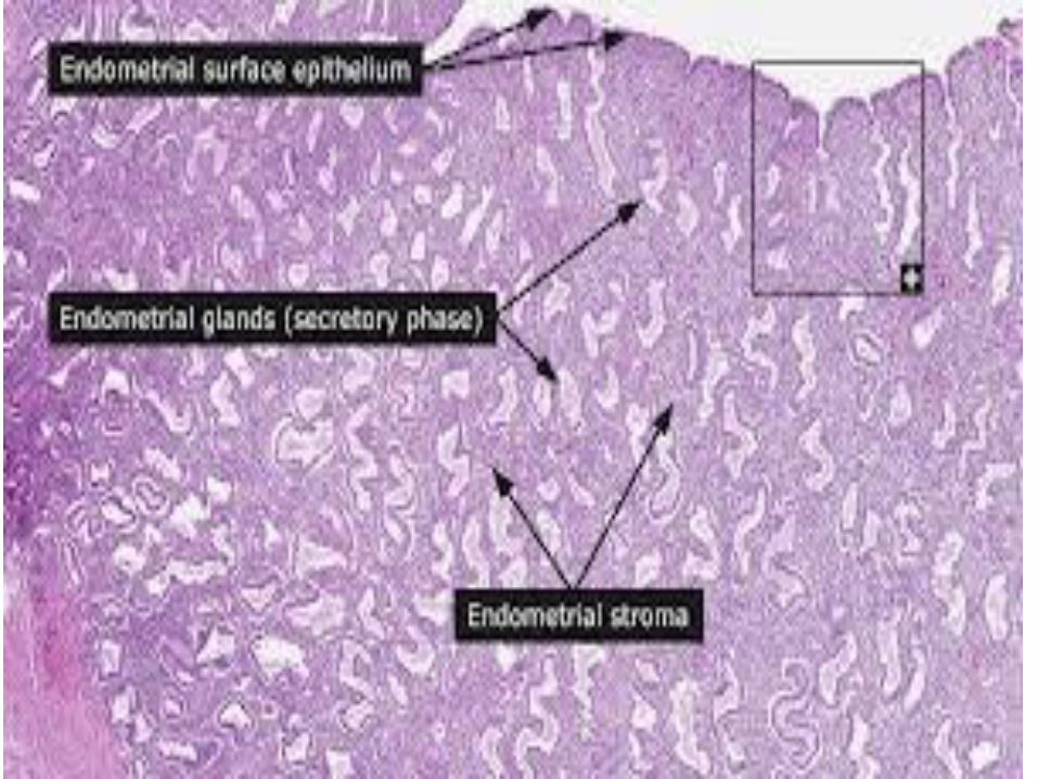
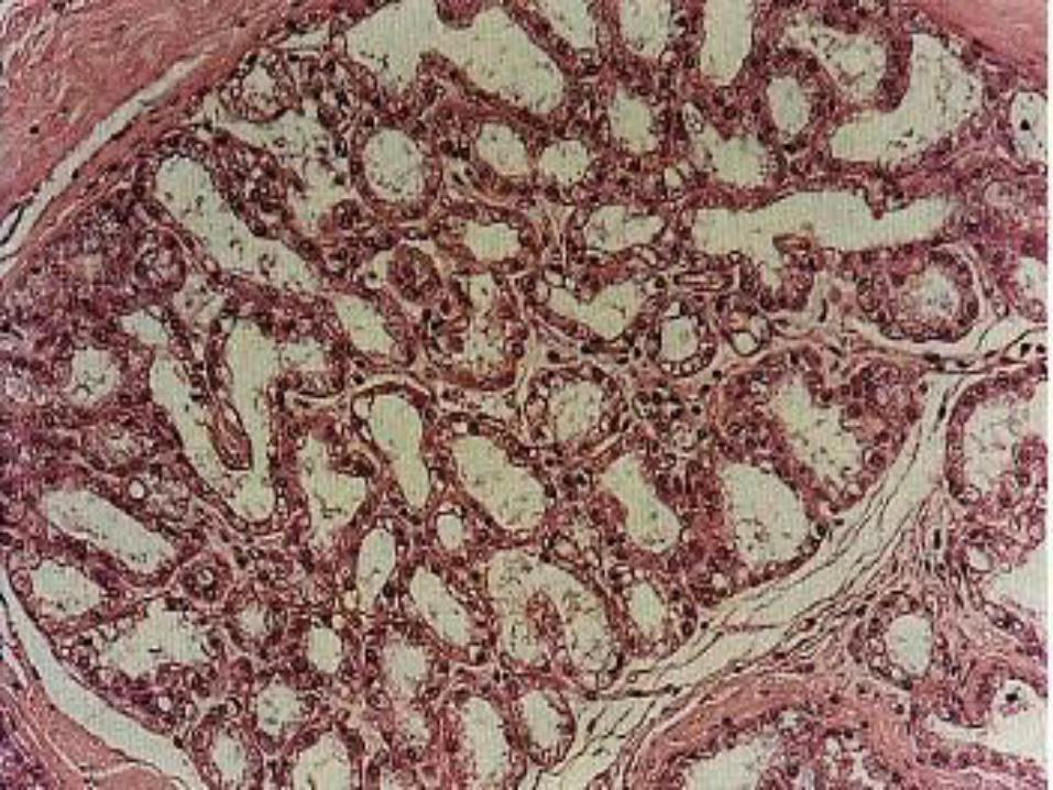
Mucous membrane/endometrium
• Epithelium – Columnar ciliated before puberty
– Simple columnar non ciliated in adult
• Lamina propria – Endometrial stroma
– Embryonic connective tissue • Mesenchymal cells
• Blood vessels – Coiled arteries supplies stratum
functional
– Straight arteries supplies stratum basal
• Long tubular glands lined by columnar cells
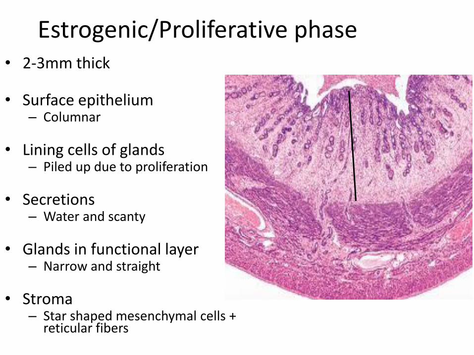
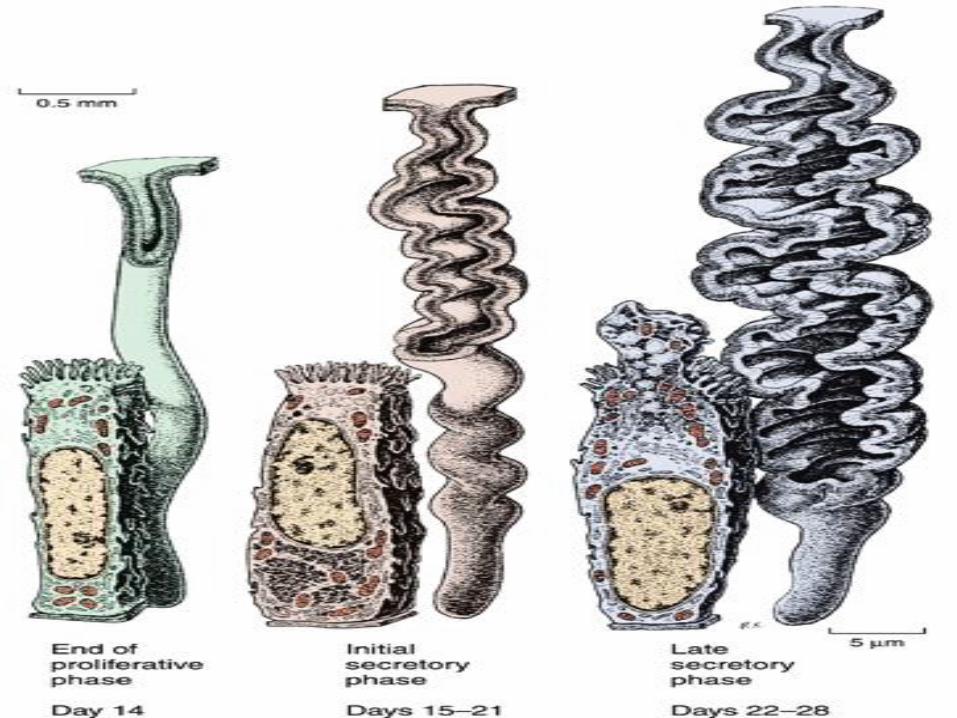
Estrogenic/Proliferative phase • 2-3mm thick
• Surface epithelium
– Columnar
• Lining cells of glands – Piled up due to proliferation
• Secretions
– Water and scanty
• Glands in functional layer – Narrow and straight
• Stroma
– Star shaped mesenchymal cells + reticular fibers
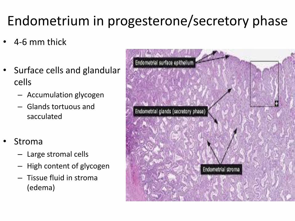
Endometrium in progesterone/secretory phase
• 4-6 mm thick
• Surface cells and glandular cells – Accumulation glycogen
– Glands tortuous and sacculated
• Stroma – Large stromal cells
– High content of glycogen
– Tissue fluid in stroma (edema)
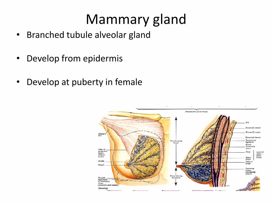
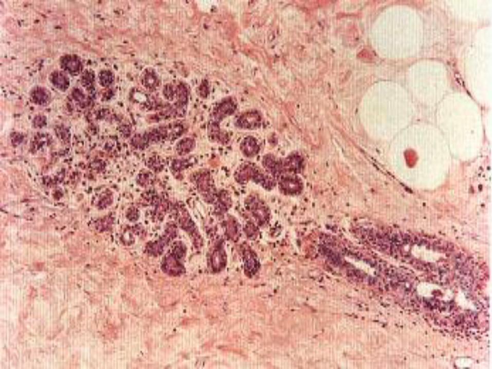
Mammary gland • Branched tubule alveolar gland
• Develop from epidermis
• Develop at puberty in female
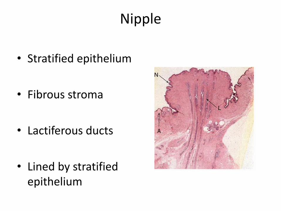
Nipple
• Stratified epithelium
• Fibrous stroma
• Lactiferous ducts
• Lined by stratified epithelium

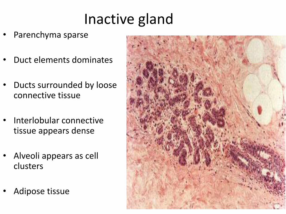
Inactive gland • Parenchyma sparse
• Duct elements dominates
• Ducts surrounded by loose connective tissue
• Interlobular connective tissue appears dense
• Alveoli appears as cell clusters
• Adipose tissue
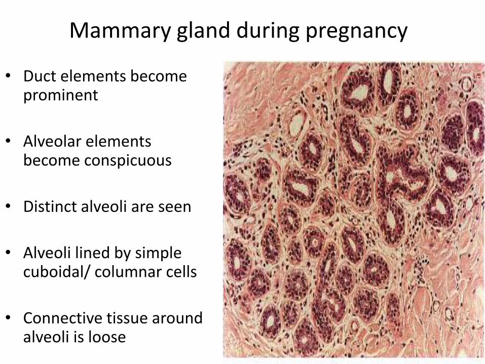
Mammary gland during pregnancy
• Duct elements become prominent
• Alveolar elements become conspicuous
• Distinct alveoli are seen
• Alveoli lined by simple cuboidal/ columnar cells
• Connective tissue around alveoli is loose
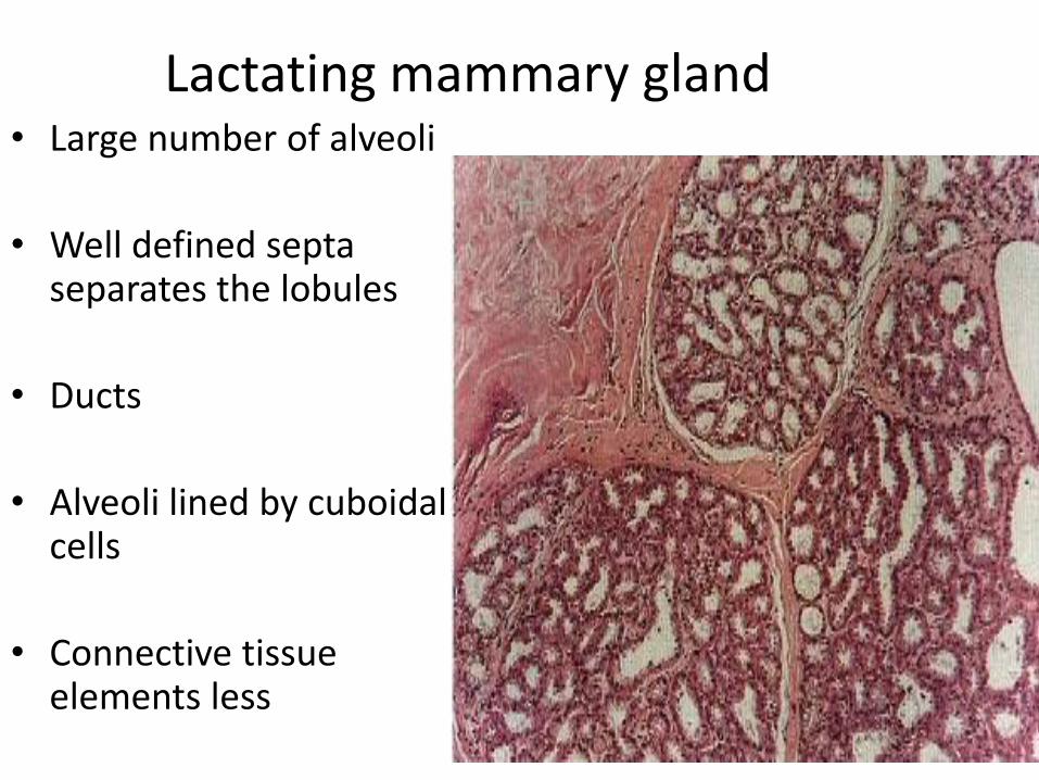
Lactating mammary gland • Large number of alveoli
• Well defined septa separates the lobules
• Ducts
• Alveoli lined by cuboidal cells
• Connective tissue elements less
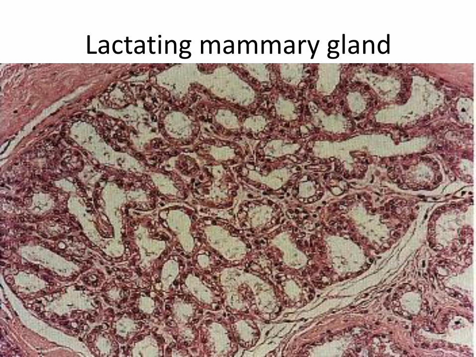
Lactating mammary gland
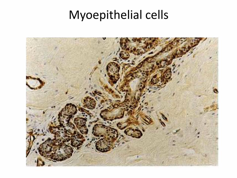
Myoepithelial cells
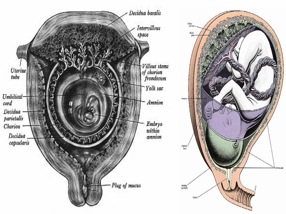
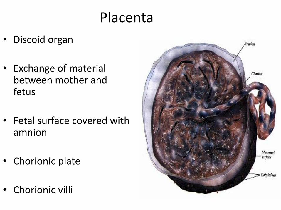
Placenta
• Discoid organ
• Exchange of material between mother and fetus
• Fetal surface covered with amnion
• Chorionic plate
• Chorionic villi
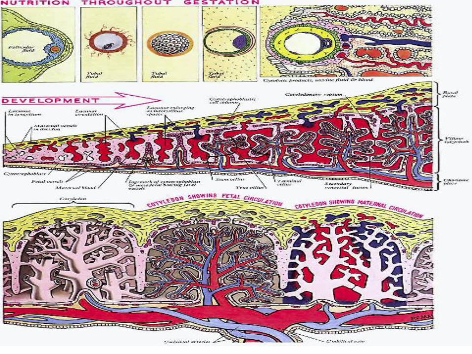
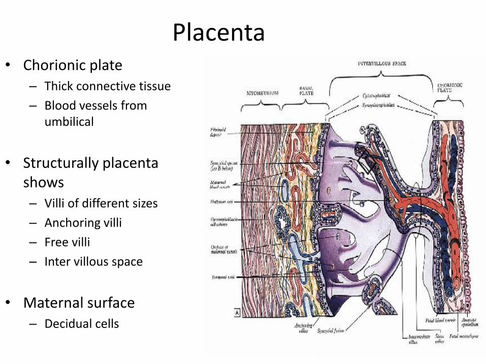
Placenta • Chorionic plate
– Thick connective tissue
– Blood vessels from umbilical
• Structurally placenta shows – Villi of different sizes
– Anchoring villi
– Free villi
– Inter villous space
• Maternal surface – Decidual cells

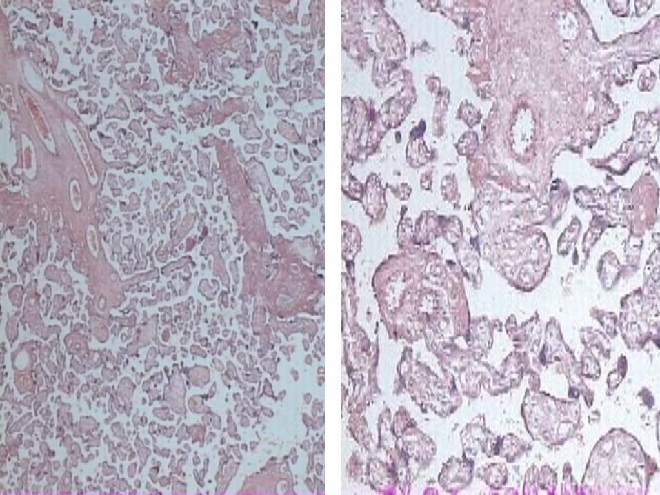
Structure of a villi • Chorionic villi
– External syncytotrophoblast
– Internal cytotrophoblast
– Mesoderm with blood vessels
– Hofbauer cells (phagocyte) in the CT
• Inter villus space with maternal blood


• Draw a labeled diagram showing the microscopic features of placenta. Mention its functions.
• Draw a labeled diagram shoving the structure of lobe of mammary gland. Discuss the histological changes during pregnancy and lactation
• Draw the histological features of uterus.
• Name the different layers of wall of uterus
• What is stratum functionale. Discuss the cyclic changes during menstrual cycle.


![PowerPoint Presentation · PDF fileattachment Placenta Uterus Placenta previa (complete) Placenta Cervix Umbilical Cord 4th week: 2mm long baby amnion forms [cushion] cord connects](https://img.pdfslide.us/doc/110x75/5a9f279f7f8b9a8e178c6556/powerpoint-presentation-placenta-uterus-placenta-previa-complete-placenta-cervix.jpg)