Embed Size (px)
Citation preview

LCA Brain/CNS Cancer Clinical Guidelines June 2014
LCA BRAIN/CNS CANCER CLINICAL GUIDELINES
Contents
Introduction ................................................................................................................................................ 4
Executive Summary .................................................................................................................................... 7
1 Early Diagnosis ..................................................................................................................................... 8
1.1 Criteria for referral .................................................................................................................... 8
1.2 Onward referral ......................................................................................................................... 9
2 Radiology – Guidelines for Imaging ................................................................................................... 11
2.1 Imaging protocols .................................................................................................................... 11
2.2 Referral pathways.................................................................................................................... 14
3 Pathology – Guidelines for Reporting Tumours ................................................................................. 16
3.1 Purpose of neuropathology services ....................................................................................... 16
3.2 Required neuropathological services ...................................................................................... 16
3.3 Provisional agreement between neuropathological centres for south and west London ...... 19
4 Multidisciplinary Team Membership and Function ........................................................................... 20
5 Inter-professional Communication between Secondary and Primary Care ...................................... 22
5.1 General principles .................................................................................................................... 22
5.2 Key communication points ...................................................................................................... 22
6 Neuro-oncology Clinical Nurse Specialist/Key Worker ...................................................................... 24
7 Therapeutic Radiographer ................................................................................................................. 25
8 Patient Information ............................................................................................................................ 27
9 Surgical Guidelines ............................................................................................................................. 28
9.1 Guidelines for low-grade glioma ............................................................................................. 28
9.2 Guidelines for high-grade glioma ............................................................................................ 29
10 Radiotherapy – Generic .................................................................................................................... 32
10.1 Basic principles ...................................................................................................................... 32
10.2 Radiotherapy planning .......................................................................................................... 32
10.3 Patient management during radiotherapy............................................................................ 34
11 Chemotherapy – Generic .................................................................................................................. 36
11.1 First-line treatment ............................................................................................................... 36
11.2 Chemotherapy treatment of recurrent disease .................................................................... 36
12 Tumour-specific Pathways ................................................................................................................ 38
12.1 High-grade gliomas ................................................................................................................ 38
12.2 Low-grade gliomas ................................................................................................................ 40
CONTENTS
3
12.3 Medulloblastoma: craniospinal radiation ............................................................................. 41
12.4 Spinal cord tumours .............................................................................................................. 44
12.5 Germ cell tumours ................................................................................................................. 45
13 Rehabilitation .................................................................................................................................... 48
13.1 Evidence ................................................................................................................................ 48
13.2 Prognosis and rehabilitation ................................................................................................. 48
13.3 Access and timing of rehabilitation ....................................................................................... 49
13.4 Rehabilitation assessment ..................................................................................................... 49
13.5 Rehabilitation intervention ................................................................................................... 49
13.6 Cancer/treatment secondary side effects and rehabilitation ............................................... 50
13.7 Multidisciplinary rehabilitation ............................................................................................. 50
13.8 Outcome measures ............................................................................................................... 51
13.9 Rehabilitation settings ........................................................................................................... 51
13.10 Service improvement, training and education ...................................................................... 52
14 Survivorship ...................................................................................................................................... 55
14.1 Symptom management ......................................................................................................... 55
14.2 Holistic needs assessment ..................................................................................................... 57
14.3 Additional support for brain tumour patients and carers ..................................................... 58
15 Palliative Care ................................................................................................................................... 59
15.1 Definition ............................................................................................................................... 59
15.2 Role and remit ....................................................................................................................... 59
15.3 Assessment ............................................................................................................................ 59
15.4 Consideration of palliative care needs .................................................................................. 60
16 Patient Experience/Satisfaction ........................................................................................................ 61
17 Management of Children, Teenagers and Young Adults with Diagnosed or Suspected Brain and Central
Nervous System Cancer .................................................................................................................... 63
18 Research and Innovation .................................................................................................................. 65
Appendix 1: Urgent Suspected Brain and Central Nervous System Cancers Referral Forms................... 66
Appendix 2: NCSI Treatment Summary .................................................................................................... 69
Appendix 3: LCA Key Worker Policy ......................................................................................................... 72
Appendix 4: Competencies for Key Worker Role ..................................................................................... 74
Appendix 5: LCA Holistic Needs Assessment Tool .................................................................................... 76
Appendix 6: Cancer Survivorship Guidelines ............................................................................................ 77
Appendix 7: Teenagers and Young Adults: Referral to Multidisciplinary Team Proforma ....................... 81
Appendix 8: LCA Specialist Palliative Care Referral Form......................................................................... 82
LCA BRAIN/CNS CANCER CLINICAL GUIDELINES
4
Introduction
Primary brain tumours account for 1.6% of all tumours diagnosed in the UK. It has been estimated that the
lifetime risk of developing brain and other central nervous system (CNS) cancer is 1 in 133 for men and 1 in
185 for women in the UK, and the incidence rates are rising slightly. In adults, most brain tumours are
supratentorial and high-grade gliomas and meningiomas predominate. Brain tumours can develop at any
age in adults but are most common in people aged between 50 and 70 (Brain and central nervous system
tumours – UK incidence statistics, Cancer Research UK).
Survival from brain tumours depends on many factors such as the type of tumour, its grade, position in the
brain, shape and size. Overall, for all types of malignant brain tumours in adults, more than 4 out of 10
people diagnosed (41.5%) live for at least a year. About 15 out of every 100 people (15%) live for more than
5 years after diagnosis. Just under 10 out of every 100 people diagnosed (10%) live for more than 10 years
after diagnosis (Cancer Research UK).
CNS malignancy accounts for approximately 2% of primary tumours, or about 6,500 cases per year in the
UK (6–7/100,000 per year). The catchment population for the LCA neuroscience centres (which includes
Kent and parts of Surrey and Sussex) is approximately 10 million.
The classification of brain tumours is complex and is being constantly reviewed. There are many subgroups
and the treatment approach differs for each. The WHO classification of brain tumours includes:
astrocytic tumours
oligodendroglial tumours and mixed gliomas
ependymal tumours
choroid plexus tumours
neuroepithelial tumours of uncertain origin
neural and mixed neuronal-glial tumours
pineal parenchymal tumours
embryonal tumours
peripheral neuroblastic tumours
tumours of the cranial and peripheral nerves
meningeal tumours
tumours of the haematopoietic system (lymphoma and histiocytic tumours)
germ cell tumours
tumours of the sella region (pituitary and craniopharyngioma)
metastatic tumours of the CNS.
CNS tumours can arise in any of the tissues within the CNS but are broadly grouped into intrinsic tumours,
tumours of the meninges and tumours of the pituitary and pineal glands. In the latest data available from
Cancer Research UK, 58% of all CNS tumours arose in the brain itself; 23% arose in the meninges; 11% in
the intracranial endocrine glands; and the remaining 8% in the other parts of the CNS (e.g. spinal cord).
INTRODUCTION
5
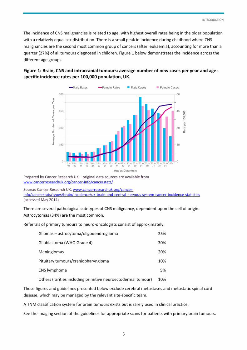
The incidence of CNS malignancies is related to age, with highest overall rates being in the older population
with a relatively equal sex distribution. There is a small peak in incidence during childhood where CNS
malignancies are the second most common group of cancers (after leukaemia), accounting for more than a
quarter (27%) of all tumours diagnosed in children. Figure 1 below demonstrates the incidence across the
different age groups.
Figure 1: Brain, CNS and intracranial tumours: average number of new cases per year and age-specific incidence rates per 100,000 population, UK.
Prepared by Cancer Research UK – original data sources are available from www.cancerresearchuk.org/cancer-info/cancerstats/
Source: Cancer Research UK, www.cancerresearchuk.org/cancer-info/cancerstats/types/brain/incidence/uk-brain-and-central-nervous-system-cancer-incidence-statistics (accessed May 2014)
There are several pathological sub-types of CNS malignancy, dependent upon the cell of origin.
Astrocytomas (34%) are the most common.
Referrals of primary tumours to neuro-oncologists consist of approximately:
Gliomas – astrocytoma/oligodendroglioma 25%
Glioblastoma (WHO Grade 4) 30%
Meningiomas 20%
Pituitary tumours/craniopharyngioma 10%
CNS lymphoma 5%
Others (rarities including primitive neuroectodermal tumour) 10%
These figures and guidelines presented below exclude cerebral metastases and metastatic spinal cord
disease, which may be managed by the relevant site-specific team.
A TNM classification system for brain tumours exists but is rarely used in clinical practice.
See the imaging section of the guidelines for appropriate scans for patients with primary brain tumours.
LCA BRAIN/CNS CANCER CLINICAL GUIDELINES
6
For those patients with suspected metastases, the appropriate clinical examination, blood tests and cross-
sectional imaging (usually a CT of chest abdomen and pelvis) are carried out. An 18FDG positron emission
tomography scan may be useful in some patients.
The management of patients with CNS tumours will discussed at multidisciplinary meetings and treatment
may include a combination of some form of surgery, radiotherapy (conventional or stereotactic) and
possibly chemotherapy.
The LCA Brain/CNS Cancer Clinical Guidelines encompass guidance from Improving Outcomes for People
with Brain and Other CNS Tumours (2006) and from the guidelines produced by the previous north west
London, south west London and south east London cancer networks. The Manual for Cancer Services: Brain
and CNS Measures (National Peer Review Programme, March 2014) was also taken into consideration.
The objective of these guidelines is to ensure that patients referred to any of the neuroscience
multidisciplinary team meetings throughout the London Cancer Alliance (LCA), with its existing integrated
cancer system, will be treated appropriately and in a timely manner. These guidelines are designed to
prevent or reduce any inequality of care within the LCA. All patients who live within the LCA should be
managed in an LCA centre to improve compliance with the LCA guidelines.
The LCA guidelines are designed to be used by all healthcare professionals in Trusts within the LCA who are
involved in the care of the brain and CNS cancer patient. They have been developed to take into account
the wide range of clinical experience of the user and the different clinical settings in which they work. The
guidelines are intended to assist in the initial assessment, investigation and management of patients.
Adoption of the LCA guidelines will allow widespread implementation of up-to-date and evidence-based
management of brain and CNS cancer patients, and will assist in the provision of a consistently high
standard of care across the LCA.
All Trusts are expected to be able to provide the standard of care detailed in these guidelines. These
guidelines will be reviewed on an annual basis in line with guidance from the National Institute for Health
and Care Excellence, the British Neuro-Oncology Society and other national and international guidance,
as well as significant new research publications, to ensure that they continue to reflect best practice.
With respect to the more rare forms of brain tumours, rather than use our ‘local’ guidelines we have
adopted the nationally agreed British Neuro-Oncology Society (BNOS) publications as a guide
(www.bnos.org.uk). The LCA has also developed operational policies for pituitary, anterior and lateral skull
base tumours, and which accompany this document.
I would like to thank the LCA Brain/CNS Pathway Group members who contributed greatly to the
development of these guidelines.
Dr Ron Beaney
Chair, LCA Brain/CNS Pathway Group
Consultant clinical oncologist, Guy’s and St Thomas’ NHS Foundation Trust
EXECUTIVE SUMMARY
7
Executive Summary
The LCA Brain/CNS Cancer Clinical Guidelines combine the best features of earlier network protocols and
have been developed in agreement with clinicians across the LCA. The guidelines combine evidence-based
and best practice recommendations with the aim of ensuring that there are equitable, high-quality services
across the LCA. The guidelines are multidisciplinary and cover early diagnosis, imaging, pathology, surgery,
radiotherapy, rehabilitation and survivorship.
Chapter 1 aims to reduce the time to diagnosis of primary brain tumours. This chapter outlines referral
criteria to the neuro-oncology multidisciplinary team (MDT) and emergency medicine department as well
as onward referrals from GPs and emergency medicine. It also provides guidance on increasing awareness
of brain tumours and access to imaging.
Chapter 2 provides guidance for imaging of brain tumours, including protocols for initial diagnosis of
primary and secondary brain and spinal tumours, follow-up and guidelines on metastatic spinal cord
compression.
Chapter 3 deals with neuropathology services, molecular and genetic markers, turnaround times and
provisional agreement between neuropathological services.
Chapters 4–7 set out the MDT structure in line with peer review requirements. This section also outlines
the role of the key worker and the neuro-oncology consultant therapeutic radiographer, essential
components of ensuring high-quality patient experience.
The patient information section in Chapter 8 provides a list of key areas to be discussed with each patient
and also looks at breaking bad news.
Chapters 9–12, on surgery, radiotherapy and chemotherapy, set out key generic principles for treating
brain and central nervous system (CNS) tumours as well as detailing tumour-specific treatment protocols.
Rehabilitation of brain/CNS patients is outlined in Chapter 13 and survivorship described in Chapter 14,
detailing the ongoing care for patients living with their condition during and beyond treatment.
Palliative care is presented in Chapter 15 and improving patient experience is outlined in Chapter 16.
Chapter 17 provides information for managing paediatric, teenage and young adult patients.
Alongside this, Chapter 18 stresses that there should be a continued emphasis on national clinical trial
leadership, proven to improve the standard of care for all patients.
Some of the recommendations in these guidelines will be challenging to implement, but as the role of the
LCA is to ensure that world-class quality of care is delivered for its patients with cancer, it is anticipated that
provider organisations within the LCA will use these guidelines as a tool to support change improvement.
During the coming months the clinicians will develop standards and measures against which organisations
can be assessed.
LCA BRAIN/CNS CANCER CLINICAL GUIDELINES
8
1 Early Diagnosis
There is an intention to reduce the time to diagnosis of primary brain tumours. This would offer the
opportunity to:
intervene earlier in the disease progress and reduce the risk of acquired neurological disability due to
tumour-related brain injury
improve prognosis with early and more extensive intervention
reduce the number of initial operations conducted as urgent or emergency procedures with associated
raised intracranial pressure and thus enhanced mortality and morbidity risks
reduce patients’ and their families’ anxieties about the consequences of avoidable delays in diagnosis,
whether they are due to patient and family delays, or physician and health system delay
enhance the public’s confidence in the health services.
1.1 Criteria for referral
1.1.1 Suspected brain tumour
Following is the guideline for the referral of adults admitted via their local A&E department or of patients
attending their GP with the symptoms listed below for imaging and onward referral to the neuro-oncology
multidisciplinary team (MDT):
patients with non-migrainous headaches of recent onset, accompanied by features suggestive of raised
intracranial pressure (e.g. woken by headache, vomiting, drowsiness)
patients who have a history of headaches who present complaining of an altered pattern or severity of
headaches, or with any focal neurological symptoms
patients who do not normally complain of headaches, now presenting with headache (particularly if
over 45 years)
patients presenting with a first fit.
Patients with the following should be referred via A&E or the on-call neurology or neurosurgical service
for immediate assessment and for urgent imaging:
acute/subacute neurological deficit developing over days and at most a few weeks (e.g. weakness,
sensory loss, dysarthria, ataxia)
new onset seizures characterised by one or more of the following:
- prolonged and repetitive focal seizures with or without secondary generalisation
- prolonged post-ictal focal deficit
- status epilepticus
- associated inter-ictal focal deficit
- patients with headache, vomiting and papilloedema
- cranial nerve palsy (e.g. double vision, visual failure including optician-defined visual field loss).
EARLY DIAGNOSIS
9
Patients not fitting these criteria or with other neurological symptoms should be referred to the local
neurology outpatient department.
1.1.2 Suspected spinal tumour
Refer to the LCA Acute Oncology Clinical Guidelines (September 2013, updated May 2014), section 9
Metastatic Spinal Cord Compression.
1.2 Onward referral
There will be protocols to ensure that patients with imaging showing a tumour who are referred by GPs,
A&E departments and radiologists will be incorporated into the neuro-oncology service, with a named
neuro-oncology clinical nurse specialist and consultant.
1.2.1 GPs
The intention is to increase the number of GPs with a specialist interest (GPwSIs) in neurology to act as a
referral source for local GPs for patients with suspected brain tumours. They would have additional
expertise in headache, first fit management and subacute neurological deficit so as to provide a filter
mechanism for the radiology service and be able to refer on appropriate cases to the neuro-oncology team
or to the local neurologist, as appropriate.
Patients should ideally be referred to their local GPwSI or into the neuro-oncology service by their GP
following local examination and clinical assessment. For the specific symptoms listed above, GPs should
have access to imaging (preferably through their local GPwSI if available) and then direct access either to
the local consultant neurologist (who can make the onward referral to the regional neuro-oncology MDT)
or directly to the regional neuro-oncology MDT. If there are acute symptoms, as listed above, then referral
should be through A&E or, alternatively, via the on-call neurosurgical or neurology service, where available.
If a patient is found to have a suspected tumour, and a scan has been performed in the patient’s local
hospital or region, then the images will be transferred through the Image Exchange Portal (IEP) for review
by the neuro-oncology MDT or by the on-call neurosurgical service in acute cases.
1.2.2 A&E departments
Any patient presenting to A&E with the specific symptoms listed above, particularly a first fit, should have
imaging while the patient remains in the A&E department; if the patient is found to have a suspected
tumour, then the A&E team should make the onward referral to the neuro-oncology MDT (ensuring that
the GP is informed and that the GP’s details are forwarded to the MDT) or, for patients with acute
symptoms, referral should be made to the on-call neurosurgical team.
First fit patients with negative imaging would then be referred to a ‘First Fit’ clinic or to the general
neurology or epilepsy clinic (depending on the availability of such clinics at the local hospital).
A standard protocol for imaging of first fits and patients with concerning acute and subacute neurological
symptoms will be devised by the LCA Brain/CNS Pathway Group and circulated to all A&E departments.
Increased awareness
Currently, only 1% of adult brain tumours are diagnosed following fast-track GP referral for suspected
cancer, according to the National Cancer Intelligence Network (2010); most present with an acute
LCA BRAIN/CNS CANCER CLINICAL GUIDELINES
10
presentation via A&E. The LCA Brain/CNS Pathway Group is collaborating with The Brain Tumour Charity to
help raise awareness of brain tumour symptoms in adults for the general public and for GPs, similar to the
HeadSmart campaign which was devised for the improved diagnosis of paediatric brain tumours.
Access to imaging
Of paramount importance is increased access to imaging by primary care and improved imaging through
A&E departments. The Department of Health has recently published a paper calling for increased direct
access to imaging for brain tumours by GPs.1
1 Department of Health (2012), Direct Access to Diagnostic Tests for Cancer. Best Practice Referral Pathways for General Practitioners. London: Department of Health.
RADIOLOGY – GUIDELINES FOR IMAGING
11
2 Radiology – Guidelines for Imaging
2.1 Imaging protocols
Neuroimaging is necessary in all patients with symptoms suggestive of intracranial or spinal tumour unless
deemed inappropriate on clinical grounds.
For intracranial tumours, magnetic resonance imaging (MRI) is superior to computerised tomography (CT)
for tumour detection, delineation, characterisation and differentiation from non-neoplastic lesions.
CT is frequently more readily available and convenient in acute presentations. It is usually sufficient for
excluding immediately life-threatening complications such as hydrocephalus, acute haemorrhage or
cerebral herniation. CT may also show tumour calcification or characterise bone involvement and thus aid
differential diagnosis.
For tumours of the spinal cord, MRI is necessary. CT myelography may be substituted if MRI is
contraindicated. MRI is required for full assessment of tumours of the vertebral column, unless
contraindicated. However, CT may give some indication of spinal canal compromise and may be useful for
assessment of bony integrity and surgical planning.
Contrast medium should be used in accordance with local protocols in patients with renal impairment,
history of allergy or other contraindications.
The following protocols are recommended:
2.1.1 Initial diagnosis of intracranial tumour (primary or secondary)
Patients with symptoms that may be due to an intracranial tumour should undergo MRI with a degree of
urgency according to clinical need. A non-contrast examination may be sufficient unless metastatic disease
is suspected, in which case the addition of contrast-enhanced scans increases sensitivity. If a tumour or
other mass is demonstrated, contrast-enhanced images are necessary unless contraindicated. Diffusion
weighted imaging should be acquired in all cases. It may contribute information about tumour grade and is
useful in differentiation from non-neoplastic lesions such as abscess, demyelination and infarcts.
CT may be used if MRI is unavailable or contraindicated, or if CT is deemed safer in an emergency setting.
Unenhanced scans should be acquired in the first instance, supplemented by post-contrast imaging if a
mass is confirmed or metastatic disease is suspected.
It is acknowledged that, in accordance with local resources, CT may be considered acceptable for early
diagnostic screening of patients with a low a priori likelihood of intracranial tumour (e.g. patients with
headaches uncomplicated by neurological signs or symptoms). However, CT incurs a small radiation dose
and offers lower sensitivity and specificity than MRI for smaller lesions and metastases. Therefore, where
possible, local services should work towards replacing CT with MRI in these pathways.
In patients newly diagnosed with intracranial metastatic disease, further imaging to identify a primary site
should be undertaken according to suspected primary diagnosis and clinical state, and guided by acute
oncology advice.

LCA BRAIN/CNS CANCER CLINICAL GUIDELINES
12
Contrast-enhanced diagnostic MRI should be performed in all patients prior to surgery or other active
treatment for intracranial tumour unless contraindicated or deemed otherwise inappropriate on
clinical grounds.
CT or MRI may be used for neurosurgical navigation according to local preference and availability.
According to local availability and practice, physiological and functional techniques such as perfusion
imaging, MR spectroscopy, diffusion tensor imaging (DTI), functional MRI (fMRI) and positron emission
tomography (PET) are sometimes used to inform treatment stratification or to assist surgical planning.
Intracranial MRI should be supplemented by whole spine imaging if a tumour known to disseminate
through cerebrospinal fluid (CSF) pathways such as a primitive neuroectodermal tumour (PNET) or
germinoma is diagnosed, or if there are spinal symptoms in patients with lymphoma, glioma or cerebral
metastases. For convenience, spinal imaging may be performed after intravenous (IV) contrast
administration when performed in conjunction with brain imaging and should include both T1 and T2
weighted sequences.
2.1.2 Follow-up imaging for intracranial tumour
MRI with contrast-enhanced sequences is necessary unless contraindicated, in which case CT pre- and post-
contrast may be substituted. Protocols may be tailored to diagnoses, for instance a limited protocol is
acceptable for monitoring of meningiomas. Physiological and quantitative MRI protocols including
perfusion and MR spectroscopy are sometimes incorporated in glioma follow-up protocols although local
practice varies and some techniques are currently partly investigational.
Following radical resection of high-grade (WHO Grade 3 and 4) glioma, patients should undergo an initial
contrast-enhanced MRI scan within 72 (ideally, 48) hours of surgery to determine the extent of resection
prior to the development of reactive enhancement of the surgical cavity margins. Early post-operative
scans may also be done following resection of other tumours on a case-by-case basis according to surgical
findings and local practice.
Frequency of subsequent follow-up imaging should be determined according to primary diagnosis and
multidisciplinary team (MDT) advice.
2.1.3 Skull base, pituitary fossa/sellar region, internal auditory canal/cerebellopontine angle, orbits
Dedicated protocols should be used including thin sections to increase anatomical resolution, and in some
cases contrast-enhanced and fat-attenuated sequences to optimise lesion conspicuity.
Skull base
Imaging of skull base tumours may include fat-suppressed sequences according to local preference. CT is
frequently useful to characterise bone involvement and may assist in surgical planning.
Pituitary fossa/sellar region
Imaging of the sellar region should include unenhanced T1 weighted sequences in coronal and sagittal
planes, the latter to assess for the normal hyperintense appearance of the posterior pituitary lobe and
integrity of the pituitary stalk. T2 weighted sequences are often also employed. Contrast-enhanced
sequences may be helpful in the detection of microadenomas causing Cushing’s disease or acromegaly.
RADIOLOGY – GUIDELINES FOR IMAGING
13
Contrast enhancement is not essential in the investigation of hyperprolactinaemia but may be performed
according to local practice.
On first examination of a pituitary macroadenoma, contrast-enhanced imaging may be helpful in
delineating anatomy and differentiating from mimics such as sellar meningioma.
Contrast enhancement is not usually necessary for follow-up imaging of pituitary adenomas. It is
sometimes useful for surgical follow-up if post-operative anatomy is distorted or in the early post-operative
period if it is necessary to differentiate residual tumour from haematoma.
CT may be helpful to demonstrate calcification in suspected craniopharyngioma. CT may also be used for
diagnosis or monitoring of pituitary fossa tumours if MRI is contraindicated. A spiral technique is usually
employed with multiplanar reformats.
Diabetes insipidus (DI) may be caused by lesions of the pituitary stalk or hypothalamus that are occult on
unenhanced MRI. Therefore, contrast-enhanced images should always be acquired for the investigation of
DI unless contraindicated. Whole head sequences should also be acquired if the differential diagnosis
includes disseminated conditions such as sarcoidosis. It is often necessary to do repeated follow-up imaging
if initial scans for DI are normal, as lesions such as germinoma may become symptomatic before they are
detectable on imaging.
Internal auditory canal/cerebellopontine angle
Outpatient screening examinations for tumours of the internal auditory canal/cerebellopontine angle
(IAC/CPA) are usually negative. Therefore, contrast-enhanced scans are not routinely required and heavily
T2 weighted 2D or 3D sequences are sufficient for screening purposes. If a tumour is demonstrated, it
should be characterised further using thin section T1 weighted sequences before and after IV contrast
administration. If MRI is contraindicated, high-resolution contrast-enhanced CT may be substituted
although it is inferior for detection of smaller tumours, in particular within the internal auditory canal.
Follow-up of non-operated vestibular tumours can be done using T2 weighted imaging alone. Following
surgery, contrast-enhanced MRI may be necessary for proper delineation of small volume residual disease.
Orbits
For orbital masses, T1 weighted imaging is used to exploit the natural contrast provided by orbital fat. T2
weighted sequences and post-contrast imaging should be performed with fat suppression.
CT may be used for the diagnosis and assessment of orbital masses and associated calcification or bone
involvement. It is the preferred modality in some centres but incurs a radiation dose that is avoided by
using MRI.
2.1.4 Initial diagnosis of spinal tumour
Spinal cord (primary or secondary)
MRI is required for proper delineation of intrinsic tumours of the spinal cord and differentiation from
mimics such as transverse myelitis or vascular malformations. Whole spine imaging should be performed
including pre- and post-contrast sequences. CT myelography may be substituted if MRI is absolutely
contraindicated but is less sensitive for intramedullary lesions.
LCA BRAIN/CNS CANCER CLINICAL GUIDELINES
14
Vertebral column (primary or secondary)
MRI is the preferred imaging modality and whole spine imaging should be performed. Sagittal T1 and T2
weighted sequences are usually sufficient to rule out spinal cord compression or threatened structural
integrity, particularly in an acute setting. Short T1 inversion recovery (STIR) or other fat-suppressed T2
weighted imaging may be helpful to demonstrate small deposits if time and patient compliance allow.
Where possible, axial images should be acquired through levels of actual or threatened neural compression
or vertebral collapse. Contrast-enhanced images are not usually necessary for vertebral infiltration but
should be obtained in suspected intradural disease, infection, primary tumour (of bone, nerve sheath or
meninges) or where the diagnosis is unclear.
CT may be required to assess bony integrity prior to surgery and if MRI is contraindicated.
Metastatic spinal cord compression (MSCC)
Patients with a known primary diagnosis of cancer who present with spinal pain suggestive of spinal
metastases should undergo MRI within a week. Imaging should be performed within 24 hours in the case of
spinal pain suggestive of spinal metastases and neurological signs or symptoms suggestive of metastatic
spinal cord compression (MSCC), and occasionally sooner if there is a pressing clinical need for emergency
surgery (see NICE (2008) Metastatic spinal cord compression, CG75).
MRI of the whole spine should be performed unless contraindicated. Sagittal T1 and T2 weighted
sequences are usually sufficient in the acute setting, supplemented by axial images through levels of neural
compression. Where time and patient compliance permit, STIR or other fat-suppressed T2 weighted
sequences may be helpful in detecting small metastases in disseminated disease.
If MRI is contraindicated, CT may be substituted. Spiral CT with multiplanar reformats may confirm spinal
tumour and spinal canal compromise. CT may be supplemented with myelography if necessary.
If MRI is unavailable on site at the time of presentation, the decision to transfer for urgent imaging or wait
until local imaging is available should be made on the basis of clinical findings and discussion with the
regional MSCC service.
2.1.5 Follow-up imaging for spinal tumour
MRI is preferred for follow-up imaging of tumours of the spinal cord or vertebral column. Frequency is
determined by primary diagnosis and individual patient management plan. CT may be used to follow up
vertebral tumour if MRI is contraindicated. CT is also useful to assess the instrumented spine.
2.2 Referral pathways
Patients presenting as an emergency with symptoms of raised intracranial pressure, depressed conscious
level, progressive neurological deficit or other pressing clinical features should undergo immediate
intracranial imaging, usually CT unless MRI is immediately available and considered safe. If tumour or other
life-threatening lesion such as acute hydrocephalus or abscess is suspected on imaging, the patient should
be discussed urgently with the regional on-call neurosurgical service according to local protocol.
CT scanning with radiology reporting (local or remote) should be available 24/7 in any hospital admitting
patients with possible intracranial tumour.
RADIOLOGY – GUIDELINES FOR IMAGING
15
All patients with confirmed or suspected primary intracranial or spinal primary tumour should be referred
to the regional brain/CNS MDT according to local protocol.
Patients with brain metastases should be referred to the brain/CNS MDT by the specialty MDT for the
primary cancer site (e.g. breast, lung) where advice is required on neurosurgical intervention, stereotactic
radiotherapy or other aspects of the management of brain metastases.
Suspected MSCC patients should be referred to the regional MSCC service via the MSCC coordinator
according to local protocol. MSCC patients should be discussed in the regional spinal or MSCC MDT, which
may be retrospective depending on the clinical urgency at the time of referral.
Image transfer via the Image Exchange Portal (IEP) to the regional neurosurgical centre should be available
24/7 to facilitate discussion of emergency patients. For non-emergency patients, IEP image transfer should
be done in timely fashion to allow adequate time for preparation of regional MDT meetings.
LCA BRAIN/CNS CANCER CLINICAL GUIDELINES
16
3 Pathology – Guidelines for Reporting Tumours
3.1 Purpose of neuropathology services
To standardise the classification and grading of tumours according to the current system of World
Health Organization (WHO) classification.
To provide adequate histological data for prognosis and planning treatment.
Standardised histological data is important in the communication between cancer centres, clinical audit
and stratification of patients in clinical trials.
The diagnosis of brain tumours has to be conducted by a medically qualified consultant neuropathologist
according to the National Institute for Health and Care Excellence (NICE) improving outcomes guidelines.
The guidelines define neuropathologists as accredited pathologists, registered as neuropathologists, or
histopathologists with specialist expertise in neuro-oncology who take part in the national external quality
assurance (EQA) scheme of neuropathology, organised by British Neuropathological Society.
3.2 Required neuropathological services
3.2.1 Intra-operative diagnosis (frozen sections and smear preparations)
The intra-operative diagnosis shows a good prediction of final histology. Although there is increasing
benefit from current imaging techniques, the intra-operative diagnosis is a well-established procedure and
valued by neurosurgeons to give an indication of the presence of diagnostic material, type of tumour and
possible grading. It can also be used to guide intra-operative adjuvant therapy in placement of local
chemotherapy wafer. Therefore, it is recommended by NICE to be available in neurosurgical centres. The
intra-operative diagnosis should be taken as provisional assessment but the final diagnosis, treatment and
planning and patient counselling should be based on the final record of paraffin histology.
3.2.2 Pathological specimen
Fresh specimen is necessary in cases where intra-operative diagnosis is needed or a specific molecular or
genetic analysis is recommended. However, most specimens are recommended to be received in fixative
(usually 10% neutral peppered formalin) and should be in an adequately sized specimen pot.
3.2.3 Clinical information
Clinical information is required on the specimen request form submitted by neurosurgeons or clinicians and
also recorded in the pathology report. Adequate clinical history is essential to ensure proper interpretation
of histological findings. Clinical information should include type of specimen-procedure, previous diagnosis
biopsy and therapy including radiotherapy, radiosurgical intervention, chemotherapy and others, site of
tumour and neuroradiological findings, and duration and nature of symptoms. Different containers should
be used if multiple specimens from different areas are taken.
3.2.4 Macroscopic examination of the specimen
Estimate of tumour size in three dimensions or volume of tumour tissue (if submitted piecemeal) or
provided tumour weight should be undertaken.
PATHOLOGY – GUIDELINES FOR REPORTING TUMOURS
17
3.2.5 Microscopic examination and histological classification
Central nervous system tumours are classified and graded according to the WHO grading system (currently
2007) but updated criteria should be followed if further WHO classification becomes available in the future.
The scheme is used in all neuropathology centres in the UK and its classification and grading scheme of
brain tumours is endorsed by the British Neuropathological Society and its national EQA scheme. It is also
widely used internationally, allowing comparison of data from European, North American and other
centres. Haematoxylin and eosin stained sections remain the cornerstone of histological evaluation but
should be further supplemented by immunohistochemical stains, the use of which, in general, aids the
classification. The use of immunohistochemistry should be subject to appropriate internal and external
quality control. This should involve the use of appropriate controls and the laboratory should be
participating in UK National External Quality Assessment Service for Immunocytochemistry (NEQAS-ICC).
Lymphomas may be presented as central nervous system disease and occur as primary tumour. Guidelines
on lymphoma reporting are available in the Royal College of Pathologists’ database for pathologists
reporting on lymphoma.1
Pituitary tumours are now classified according to sub-type based on hormone production with tumour
cells. This is generally determined by immunohistochemistry to conventional adenohypophyseal hormones
including adrenocorticotropic hormone (ACTH), luteinising hormone (LH), follicle-stimulating hormone
(FSH), alpha subunit, thyroid-stimulating hormone (TSH), prolactin and growth hormone. Ki-67 should also
be added to further characterise pituitary adenomas. In some cases, electron microscopy may contribute to
the diagnosis.
It is recommended that the report includes sections on the description of the pathological features of
tumour, in addition to sections of comment and then final conclusion and diagnosis. The latter should
include the tumour type, tumour sub-type relevant to grading and prognosis, tumour grade (WHO 2007)
and, for extra-axial tumours (particularly meningiomas), presence of brain invasion.
3.2.6 Molecular and genetic markers
These markers have become important and improve understanding and pathogenesis of brain tumours.
They have contributed towards classification of brain tumour, predicting prognosis and assisting in
treatment and management. The following are the recommended markers to be used in neurosurgical
centres. This list will certainly expand in the near future as more markers become available.
Co-deletion of chromosome arms 1p/19q. This is important in classifications of oligodendrogliomas
and other gliomas and can be assessed by fluorescent in-situ hybridisation (FISH), the polymerase chain
reaction based method for loss of heterogeneity or other methods (such as MLPA). A large number of
studies have suggested that loss of these chromosomal arms predict a better prognosis and better
response to chemotherapy and radiotherapy, suggesting that this is both a prognostic and predictive
factor. Testing for 1p/19q has become widely available and is the standard test for care of histological
analysis in brain tumours.
06 methylguanin-DNA methyltransferase (MGMT). This is a DNA repair enzyme which can repair the
damage induced by chemotherapeutic alkylating agents leading to chemo-resistance. Epigenetic
silencing of the MGMT gene by promoter methylation plays an important role in regulating MGMT
expression in gliomas. MGMT promoter methylation has shown value as a predictive marker for
temozolomide sensitivity and correlates with better progression-free and overall survival in
glioblastomas and anaplastic gliomas. Assessment of the percentage of methylation of MGMT is
LCA BRAIN/CNS CANCER CLINICAL GUIDELINES
18
recommended by pyrosequencing or other method. Immunohistochemical evaluation of MGMT should
be also be done as a complementary test and discrepancies between methylation-sensitive PCR and
immunostain should be noted in the report.
Mutation of isocitrate dehydrogenase 1 gene (IDH1 and the related IDH2). These gene mutations are
found in clinically and genetically distinct sub-types of gliomas, particularly astrocytoma and
oligodendroglioma of WHO Grade 2 and 3 and in secondary glioblastomas which evolved from lower-
grade tumours. Patients with mutation in these genes have a better outcome than those without
mutation. Assessment of IDH1 mutation is very reliable with the immunohistochemistry method which
should be available in all participating centres. Further tests for IDH1 and IDH2 gene sequencing are
recommended in glioma Grade 2 and 3 in which the IDH1 immunohistochemistry is negative.
KI1549-BRAF fusion gene. This gene occurs in over 70% of pilocytic astrocytomas and is emerging as an
important diagnostic marker and therapeutic target. This is easily identified by interface FISH but can
also be demonstrated in DNA from frozen tumour tissue. The identification of these genetic aberrations
may help to differentiate other tumour forms from pilocytic astrocytoma in difficult cases which have
significant implications for the prognosis and treatment of patients.
Molecular analysis should be carried out in laboratories participating in appropriate EQA schemes. A pilot
EQA for brain tumour molecular markers is now available in the UK.
Further molecular markers are predicted to be available in the future, and neuropathological centres
should follow and update their procedures accordingly.
3.2.7 Turn-around times
The turn-around times for diagnosing brain tumours and providing results for immunohistochemistry and
molecular markers should be agreed in each centre between neuropathologists, neurosurgeons and neuro-
oncologists. The following is the Royal College of Pathologists’ recommendation:
reporting central nervous system tumours (brain biopsies, surgical): 5 days (2 days if no special stains
are required)
pituitary biopsies: 4 days
epilepsy surgery: 15 days
1p/19q co-deletion: 15–20 days
MGMT methylation and BRAF-600 mutation: 10 days.
3.2.8 Multidisciplinary team meetings
Cases from brain tumours are recommended to be discussed in the context of multidisciplinary team (MDT)
meetings which are regarded in NICE guidelines as central to patient management. This forum should allow
for reviews of biopsies with clinical and neuroradiological information. This may be of particular value in
the assessment of small biopsies to ensure that the tissue is likely to be representative of the lesion. The
clinical, surgical, pathological and radiological findings can be compared and integrated for clinical
management purposes. In some cases, the final neuropathological report may need to be revised to
interpret the histological findings in the light of additional information. This may be useful in situations
where difficulties are encountered in finally categorising patients. In practice, assessment of borderline
tumours for prognostic and therapeutic purposes may be aided by discussion in MDT meetings where
PATHOLOGY – GUIDELINES FOR REPORTING TUMOURS
19
additional clinical and radiological factors such as patient’s age, tumour size, presence of contrast
enhancement, and rate of growth on serial scans provide additional information.
3.3 Provisional agreement between neuropathological centres for south and west London
Discussion has been conducted by the three neuropathological centres at King’s College Hospital, St
George’s Hospital and Charing Cross Hospital regarding provision of neuropathology services for brain
tumours across the LCA. The following are points of agreement:
The centres agreed that neuropathology services should follow and be co-located with the
neurosurgical services. This is important to provide intra-operative diagnosis, close discussion with the
neurosurgeons, neuroradiologists and neuro-oncologists and to conduct critical discussion in MDT
meetings. For these reasons it was felt that maintaining neuropathological services in each individual
centre is essential. It was recommend that each Trust should support neuropathology services and
provide sufficient cover for neuropathologists to provide the recommended neuropathology services.
The centres agreed to suggest a scheme of audit across all centres to ensure that the standard criteria
for neuropathological assessment are followed.
The centres agreed that clinical research activities should be conducted across all centres in
combination with activities of neurosurgery, neuro-oncology and neuroradiology.
The centres recommended that further funding and support should be targeted towards developing
state-of-the-art molecular biology services, as with other international centres, for the importance of
this subject in the current and future practices in classification, prognosis and directing of treatment of
brain tumours. It was recommended that the molecular biology services be integrated with the
neuropathology services.
1 www.rcpath.org/publications-media/publications/datasets/lymphoma.htm
LCA BRAIN/CNS CANCER CLINICAL GUIDELINES
20
4 Multidisciplinary Team Membership and Function
All adult patients with suspected primary brain and spinal tumours should be referred to the neurosurgical
multidisciplinary team (NS MDT) for discussion. The brain/CNS MDT should meet weekly.
Patients will be reviewed at the NS MDT meeting at the following points in the patient pathway:
post-initial radiological diagnosis, pre-histological confirmation
post-histological confirmation and/or post-definitive surgical procedure.
All patients with recurrent tumours managed by a neuro-oncology service will be reviewed at the NS MDT.
Brain metastases will be discussed at the NS MDT only if a surgical or radiosurgical opinion is required.
There is a single named lead clinician for the MDT, who is a core team member and has responsibilities
agreed by the lead clinician of the host Trust. The lead clinician is responsible for chairing the weekly
meeting or agreeing a rotating chair.
The MDT meets on a weekly basis (as a minimum) and discusses all patients with suspected brain and CNS
tumours who are formally referred via the neuro-oncology MDT coordinator.
The core members of the Brain and CNS MDT include:
two neurosurgeons
a neuroradiologist
two neuropathologists (Note: For the purpose of peer review, the neuropathologist is defined at
minimum as a consultant pathologist who is taking part in the external quality assurance (EQA) scheme
for neuropathology, organised by the British Neuropathological Society, or, for a pathologist acting only
as the pituitary tumour pathologist for a team, an EQA scheme for endocrine pathology)
a clinical oncologist
a neurologist with specified Direct Clinical Care PAs for the care of patients with the neurological
consequences of a CNS tumour and of its treatment
a clinical nurse specialist
a clinical neuropsychologist
a healthcare professional who is a core member of a specialist palliative care team
an allied health professional who is agreed to have responsibility for liaison with neuro-rehabilitation
services
a therapeutic radiographer
the team may choose to have specific allied health profession (AHP) representatives and liaise with
only one or some of the AHP subspecialties
an MDT coordinator/secretary.
A comprehensive list of all core members for each specialty and all extended members can be found in the
Manual for Cancer Services: Brain and CNS Measures, Version 2.0 (NCAT, 2014,
www.cquins.nhs.uk/download.php?d=resources/measures/Brain_and_CNS_March2014.pdf).

MULTIDISCIPLINARY TEAM MEMBERSHIP AND FUNCTION
21
Within the MDT there is a nominated member responsible for users’ and carers’ issues and information,
and a nominated member responsible for ensuring that recruitment into clinical trials and other well-
designed studies is integrated into the function of the MDT.
All patients who are discussed must have recent, relevant imaging. Patients who have suspected metastatic
disease from an unknown primary or an established diagnosis of cancer with new suspected metastatic
disease should undergo a computerised tomography (CT) scan of their chest, abdomen and pelvis to assess
the extent of disease. Details of other staging investigations that have been undertaken should be supplied
to the MDT to assist in appropriate treatment planning.
The MDT should provide specialist neuroradiology advice on patients with suspected brain/CNS tumours
but should not be used to provide specialist neuroradiology reporting.
During the meeting, the pathology for every patient who has undergone surgery should be discussed. In
some cases, additional molecular testing may be requested and should be brought back to a subsequent
meeting for discussion once the result has been formally agreed.
Clinicians will consider the potential entry of each patient into a trial.
The MDT is also an important forum in which to establish the specific needs of a patient. These needs
include input from hospital or community palliative care, inpatient therapies, formal cognitive assessment
and management, ongoing rehabilitation needs and community therapies. It is also an opportunity to
identify psychological, social and spiritual needs which need ongoing management through the network
MDT.
An agreed minimum dataset should be formally recorded during the MDT to reflect clinical workload,
accurate patient numbers and a breakdown of tumour types.
It is the responsibility of the referrer to communicate the outcome of the MDT to the patient and their GP
within 24 hours of the discussion and to ensure that a formal ongoing referral is instigated as instructed by
the MDT.
It is good practice for patients who require surgical intervention to be seen in an MDT clinic to discuss the
outcome of the meeting, to review scans and agree a treatment plan.
Patients who are not suitable for surgery but may benefit from oncological management should be
discussed with the oncologist and booked into the next available clinic.
The suitability of different radiotherapy modalities (e.g. 3D, intensity-modulated radiotherapy (IMRT),
helical modalities, stereotactic radiosurgery/stereotactic radiotherapy (SRS/SRT), protons) and proximity to
critical structures should be discussed so that dose and fractionation considerations can be planned for
within the radiotherapy service. The imaging reviewed at the MDT should also be evaluated to consider
whether this can be incorporated into the radiotherapy planning (fusion) or whether an additional imaging
referral is required.
An evaluation is required of patient-specific considerations, disease-specific considerations and treatment
modality options. This is the role of the neuro-oncology consultant radiographer/advanced practitioner so
that all referrals to radiotherapy modalities are dealt with in a timely manner to ensure a seamless
pathway.
All members of the team who have contact with patients at this point in the pathway should have formal
training in advanced communication skills.
LCA BRAIN/CNS CANCER CLINICAL GUIDELINES
22
5 Inter-professional Communication between Secondary and Primary Care
5.1 General principles
Communication needs to be timely and concise.
Use fax-back route/electronic means for urgent communications (meaning those that need to be with
the GP within 24 hours) and follow up with a call to confirm receipt.
Communications at key points along the patient journey must include:
what the patient has been told
who told the patient
who was there with the patient (e.g. named partner/friend)
what written/other information was offered
next steps – when the patient is being seen or their treatment started
actions for the GP – for information only or suggesting specific GP actions (including information for
Macmillan or district nursing colleagues)
named key worker in secondary/tertiary care and any planned changes in key worker
intent of treatment (curative/palliative)
any additional information required from the GP (e.g. co-morbidities status)
summary of medication and alterations to medication
contact details for further information/discussion
specialist assessment and intervention summary (e.g. allied health profession input)
treatment plan summary when created and when amended
written correspondence to be copied to all appropriate team members who have actions to be
undertaken.
5.2 Key communication points
Diagnosis
Multidisciplinary team (MDT) discussions
Clinic appointment reviews
On treatment reviews
Decision points for changes in care planning
Decision point for end of life care planning.
The LCA Survivorship Group has recommended the adoption of the National Cancer Survivorship Initiative
(NCSI) Treatment Summary. A copy of this document can be found at Appendix 2.

INTER-PROFESSIONAL COMMUNICATION BETWEEN SECONDARY AND PRIMARY CARE
23
It is recommended that all LCA providers refer to Improving Outcomes: A Strategy for Cancer. Third Annual
Report (Department of Health, 2013) for details about the MDT set-up.
LCA BRAIN/CNS CANCER CLINICAL GUIDELINES
24
6 Neuro-oncology Clinical Nurse Specialist/Key Worker
The LCA has produced a key worker policy document which should be read in conjunction with this
guidance. This document can be found at Appendix 3. A list of competencies for the key worker role can be
found at Appendix 4.
All patients seen within the LCA with a diagnosis of a brain or spinal tumour will be given the name and
contact details of a key worker. It is the responsibility of this key worker to refer on to a new key worker
when:
a patient’s care and follow-up is taken over by another hospital
a patient’s care is handed over to a community or hospital palliative care team.
All neurosurgical and cancer centres should have one or more trained neuro-oncology cancer clinical nurse
specialists to see patients before and after diagnosis, to provide continuing support, and to facilitate
communication between the secondary and tertiary care teams (including the MDT), the patient’s GP, the
community team and the patient. Patients may have joint key workers (one in the treating Trust and one in
the community).
All patients newly diagnosed with brain and spinal tumours should have access to a neuro-oncology clinical
nurse specialist who may act as the main key worker.
The neuro-oncology clinical nurse specialist is involved in all aspects of the disease journey, from diagnosis
to end of life care, in both out- and inpatient settings. The clinical nurse specialist has a pivotal role in the
MDT, ensuring that appropriate professionals are involved and working across boundaries in an effort to
provide seamless care and support for patients and carers.
The neuro-oncology clinical nurse specialist will play a key role in the following aspects of care:
the delivery of holistic care
coordination of care across sectors
onward referrals to appropriate allied health professions and local services
nurse-led outpatient care
information giving
holistic needs assessment
communicating significant news
pain control and symptom management in close collaboration with palliative care
carer support and assessment
referral for benefits and financial advice
end of life choices.
THERAPEUTIC RADIOGRAPHER
25
7 Therapeutic Radiographer
The transition points between primary, secondary and tertiary care, including radiotherapy, are recognised
as impeding continuity and are particular areas of concern.1 2 Improving Outcomes: A Strategy for Cancer3
states that the emphasis needs to be placed on patient-centred care carried out by appropriate highly
skilled teams. Radiotherapy requires highly specialist staff, including clinical oncologists, therapeutic
radiographers, medical physicists, medical scientists and technicians.4
It is recommended that therapeutic radiographers are present in Trusts to enhance patient care. This is the
only profession to specialise in radiotherapy from initial training and is therefore best positioned to provide
care from referral to follow-up within the radiotherapy pathway. It is recommended that all Trusts consider
employing neuro-oncology consultant therapeutic radiographers/advanced practitioners that have a
specialist understanding of the planning principles and the radiotherapy treatment modalities and are
therefore able to incorporate their clinical knowledge and skills into considering and evaluating disease
management issues.
The skills mix report published by the Department of Health5 identified the advanced and consultant tiers
of the skills mix model as reflecting “the requirements of clinical governance in respect of their contribution
to the continuous improvement of the service”. The Report from the National Radiotherapy Advisory
Group6 made recommendations for commissioners and service employers to fund implementation of
advanced and consultant-level posts because “where these roles have been introduced they have
demonstrated the potential to drive efficiency, reduce waiting times and refocus radiotherapy services
around the needs of patients”.
The neuro-oncology consultant therapeutic radiographer/advanced practitioner strategically manages the
patient pathway from referral to post-radiotherapy and long-term follow-up, and refines the patient
pathway according to both service provision and patient needs. The neuro-oncology patient’s management
plan within radiotherapy is often complex. Therapeutic radiographers have a unique role in supporting
patients throughout the journey. However, radiotherapy appointments are often short and under time
restraints; site-specific dedicated therapeutic radiographers are therefore important to ensure that the
needs of the patients are met and that service aims of individualised care are achieved.
The expectation is that the neuro-oncology consultant/advanced practitioner role would nominally
comprise 50% clinical work as well as work on research and development, audit, the education and training
of others, and policy and practice development. The practitioner performs an important and integral part in
the case management, providing an expert service for individual patients and ensuring a seamless service,
with continuity throughout the active treatment period.
Therapeutic radiographers working at consultant and advanced practitioner level provide:
an ‘expert practice’ function, delivering technical and cancer-site-specific expert knowledge
professional leadership and consultancy
an education and training role
development of the practice and the service
research and evaluation.
LCA BRAIN/CNS CANCER CLINICAL GUIDELINES
26
The role crosses traditional boundaries and improves the quality of patient care. It brings “strategic
direction, innovation and influence through practice, research and education, based on specialised
knowledge and skills”.7
Other sources of information include:
College of Radiographers (2009), Radiotherapy Moving Forward: Delivering new radiography staffing
models in response to the Cancer Reform Strategy.
Department of Health (2004), The NHS Cancer Plan and the New NHS: Providing a patient-centred service.
Department of Health (2007), The Cancer Reform Strategy.
Society of Radiographers (2008), Learning and Development Framework for Clinical Imaging and Oncology.
www.nhs.uk/NHSENGLAND/NSF/PAGES/CANCER.aspx
1 Society of Radiographers (2009), Implementing the Career Framework in Radiotherapy – Policy into Practice. London: Society of Radiographers.
2 College of Radiographers (2006), Positioning Therapeutic Radiographers within Cancer Services: Delivering Patient-Centred Care. London: College of Radiographers.
3 Department of Health, Public Health England and NHS England (2013), Improving Outcomes: A Strategy for Cancer, Third Annual Report. London: Department of Health.
4 The Royal College of Radiologists/The Society and College of Radiographers/The Institute of Physics and Engineering in Medicine, Commissioning arrangements for radiotherapy: A response. York/London: RCR/SCOR/IPEM.
5 Department of Health, Learning and Personal Development Division (2003), Radiography Skills Mix: A Report on the Four-tier Service Delivery Model. London: Department of Health.
6 National Radiotherapy Advisory Group (2007), Radiotherapy: Developing a World Class Service for England. Report to Ministers.
7 College of Radiographers (2005) Implementing Radiography Career Progression: Guidance for Managers. London: The College of Radiographers
PATIENT INFORMATION
27
8 Patient Information
Every patient and family/carer must receive information about their condition in an appropriate format.
Verbal and written information should be provided in a way that is clearly understood by patients and free
from jargon.
Information must be given in the most accessible format based on the patient’s cognitive/communication
needs. If there is concern about the patient’s cognition, there should be close liaison with allied health
professions, such as the occupational therapist, to facilitate a formal cognitive assessment. Speech and
language therapists should be involved to assist patients with cognitive and communication difficulties. If a
patient demonstrates cognitive impairment which may affect their capacity to understand and process the
information that is to be given, then the person responsible for giving that information should refer to the
Mental Capacity Act 2005.
Information may need to be sourced in the patient’s native language if English is not their first language
and delivered using the intervention of a translator. In some cases (with the exception of the
communication of bad news), Language Line may represent a useful tool and is widely available within NHS
Trusts. Audio and videotaped formats will also be considered.
Information to be given to the patient and families/carers should cover:
description of the disease
management of the disease
diagnostic procedures
treatment options and their effects (including potential adverse effects)
predicted outcome – any discussion of this with patients should take account of their requirements and
requests
drugs and other treatments
self-management and care
dietary and nutrition information
contact details of the patient’s allocated key worker
support organisations or internet resources recommended by the clinical team.
The National Cancer Action Team introduced information prescriptions (IPs) across the country to provide
standardised, personalised information for patients and their carers. This is a national resource accessed via
the NHS Choices website (although this initiative is not available for all rare brain and spinal tumours). In
those Trusts across the LCA which have access to IPs, this should be considered the written information of
choice. Individual Trusts will use leaflets when giving information for local procedures. Patients with brain
and spinal tumours should be provided with or signposted to local sources of support and written
information from appropriate brain and spinal tumour or cancer charities such as The Brain Tumour Charity
or Macmillan Cancer Support where IPs are not available.
LCA BRAIN/CNS CANCER CLINICAL GUIDELINES
28
9 Surgical Guidelines
9.1 Guidelines for low-grade glioma
These guidelines are based on the Guidelines on Management of Low-grade Gliomas: Report of an EFNS–
EANO Task Force, European Journal of Neurology (2010) 17: 1124–1133.
9.1.1 Background
Low-grade gliomas (LGGs) represent up to 30% of gliomas and affect patients at a younger age than high-
grade gliomas. LGGs are commonly located in or close to eloquent areas, i.e. those areas of the brain
involved in motor, language, visuospatial and memory function.
The 5-year overall and progression-free survival rates in randomised studies range from 58% to 72% and
37% to 55%, respectively.
Patients with LGGs may survive for up to 20 years, but these tumours grow continuously and tend to
progress to a higher grade, leading to neurological disability and ultimately to death.
9.1.2 Clinical features
Seizures are the most common presentation and may be partial or generalised. They occur in over 90% of
patients and are intractable in 50%. Seizures are more frequently associated with cortically based tumours,
particularly in frontal, temporal and insular/parainsular locations.
There is no clear association between severity of epilepsy and the behaviour of the tumour. Focal
neurological deficits are unusual, developing over many years.
Raised intracranial pressure is rare in patients with supratentorial tumours and is typically seen in posterior
fossa and intraventricular tumours. Intratumoural haemorrhage can occur.
9.1.3 Diagnostic pathway
Imaging
Diagnosis and staging includes imaging of the brain, ideally with conventional magnetic resonance
(MRI) including contrast enhancement.
Multimodal advanced imaging can be used to augment conventional MRI in specific circumstances:
guidance of stereotactic biopsy to the most aggressive regions of large lesions, especially those with
little or no contrast enhancement
treatment stratification in mass lesions of uncertain pre-surgical nature or grade
- in selected cases, functional MRI (fMRI, using motor and language tasks) and diffusion tensor MRI may be used to assess the relation of tumour to and invasion of functionally eloquent cortex and white matter tracts; these can be fused with structural datasets for neurosurgery planning and intra-operative neuronavigation to guide resection
- planning radiotherapy and monitoring treatment response.
SURGICAL GUIDELINES
29
9.1.4 Treatment pathway
Anti-epileptic therapy
Patients with no history of seizures have no benefit from anti-epileptic drugs (AEDs).
In patients with single seizures, immediate treatment with AEDs increases time to recurrent seizures
compared with delayed treatment, without differences with respect to quality of life or serious
complications.
AEDs should be individualised according to seizure type, co-medication, co-morbidity and patient
preferences.
In patients who need treatment with chemotherapeutics, non-enzyme inducing anti-epileptic drugs are
to be preferred.
9.1.5 Multidisciplinary team discussion
Patients are evaluated by a specialised multidisciplinary team (MDT) in the neurosciences brain/CNS
MDT meeting.
Special consideration is given to performance status and neurological function.
9.1.6 Surgery for newly diagnosed patients with suspected low-grade glioma
Surgery can provide tissue for distinguishing between the histological types, grading the malignancy
and assessing the molecular status of tumours using advanced molecular diagnostic profiling.
Total resection improves seizure control, particularly in patients with a long epileptic history and insular
tumours.
The use of brain mapping techniques, including awake surgery, should be available as an adjunct to
conventional surgery to maximise safe resection.
The use of visualisation technologies, including modern neuronavigation systems utilising advanced
image guidance, can improve the accuracy and extent of resection.
The extent of resection and determination of residual disease should be assessed using post-operative
MRI within 72 hours after surgery.
When resective surgery is not feasible (because of tumour location, extension or co-morbidities), a
biopsy (either stereotactic or open) should be considered to obtain a histological diagnosis.
Awake surgery has increased the safety of re-operation owing to mechanisms of brain plasticity.
The timing of surgery is controversial in patients who are young, present with an isolated seizure
(medically well controlled) and with small tumours.
Recurrent tumours
Patients with tumour recurrence should be discussed at the MDT and consideration be given to further
surgical intervention and second-line therapy.
9.2 Guidelines for high-grade glioma
These guidelines are based on Stupp et al. on behalf of the ESMO Guidelines Working Group (2010), ESMO
Clinical Practice Guidelines, Annals of Oncology 21 (Supplement 5): v190–v193.
LCA BRAIN/CNS CANCER CLINICAL GUIDELINES
30
9.2.1 Diagnostic pathway
Imaging
Diagnosis and staging includes imaging of the brain, ideally with conventional MRI including contrast
enhancement.
Multimodal advanced imaging can be used to augment conventional MRI in specific circumstances:
guidance of stereotactic biopsy to the most aggressive regions of large lesions, especially those with
little or no contrast enhancement
treatment stratification in mass lesions of uncertain pre-surgical nature or grade
in selected cases, functional MRI (fMRI, using motor and language tasks) and diffusion tensor MRI may
be used to assess the relation of tumour to and invasion of functionally eloquent cortex and white
matter tracts; these can be fused with structural datasets for neurosurgery planning and intra-
operative neuronavigation to guide resection
planning radiotherapy and monitoring treatment response.
9.2.2 Initial treatment pathway: concurrent medication
Corticosteroids
High doses of corticosteroids (usually dexamethasone 8–16mg/day) allow rapid reduction of tumour-
associated oedema and improve clinical symptoms; corticosteroid dose can be tapered according to
individual needs. The patient’s glucose levels must be monitored.
Steroids are not necessary in patients without increased intracranial pressure and absence of
neurological deficits.
There is no routine need for prolonged steroid therapy after tumour resection or prophylaxis during
radiotherapy but patients should be assessed on an individual basis according to needs.
Anti-epileptic therapy
Anti-epileptic therapy is indicated in patients presenting with an initial seizure; however, prophylactic
anti-epileptic therapy before or after surgery is not routinely needed.
After tumour resection, the indication for anti-seizure therapy should be revisited.
First-generation AEDs (phenytoin, carbamazepine, phenobarbital and their derivatives) are strong
inducers of the hepatic metabolism, and may interfere with medications including many commonly
used chemotherapy agents (but not with temozolomide). Third-generation agents such as lamotrigine,
levetiracetam or pregabalin are preferred.
9.2.3 Co-morbidities
Patients undergoing neurosurgery need to fulfil general fitness and anaesthetic criteria.
Any metabolic, haematological (particularly platelet) and clotting abnormalities should be corrected.
Anti-platelet drugs (e.g. aspirin, clopidogrel and dipyridamole) should be stopped at an appropriate
time before surgery except in high-risk cases (if in doubt discuss condition with
cardiologist/haematologist).

SURGICAL GUIDELINES
31
Anti-coagulants such as warfarin should be stopped except in high-risk patients such as those with
intracardiac thrombus, metallic heart valve or a pulmonary thrombus in the previous 6 months. In such
high-risk patients warfarin should be substituted for a suitable alternative as per local haematological
advice.
9.2.4 Multidisciplinary team discussion
Patients are evaluated by a specialised MDT in the neurosciences brain/CNS MDT meeting.
Special consideration is given to performance status and neurological function.
9.2.5 Surgery for newly diagnosed patients with suspected high-grade glioma
Surgery is commonly the initial therapeutic approach for tumour excision and obtaining tissue for
diagnosis, including the use of advanced molecular diagnostics.
Tumour resection is of prognostic value; it is beneficial to attempt maximal tumour resection provided
that neurological function is not compromised by the extent of resection.
The use of brain mapping techniques, including awake surgery, should be available as an adjunct to
conventional surgery to maximise safe resection.
The use of visualisation technologies, including modern neuronavigation systems utilising advanced
image guidance, can improve the accuracy and extent of resection. This can be further augmented with
the use of field visualisation techniques such as blue light fluorescence with or without the
administration of agents that increase tumour fluorophores such as porphyrins with 5-aminolevulinic
acid (5-ALA).
When microsurgical resection is not safely feasible (e.g. due to location of the tumour or impaired
clinical condition of the patient), a stereotactic biopsy can be considered to guide further treatment.
The extent of resection and determination of residual disease should be assessed using post-operative
MRI within 72 hours after surgery.
Implantation of chemotherapy-impregnated wafers (carmustine polymers, Gliadel) into the resection
cavity before radiotherapy has been shown to marginally improve median survival compared with
radiotherapy alone in patients who have radical tumour resection. However, subgroup analysis did not
show significant benefit for patients with glioblastoma.
The suitability for Gliadel wafer insertion should be discussed in the brain/CNS MDT. They are now
approved for use by NICE (TA121; www.nice.org.uk/TA121) for selected patients provided the following
criteria are satisfied:
- pre-operative MRI suggestive of high-grade glioma
- the case is discussed before surgery in a neuro-oncology MDT meeting
- surgery is performed by a specialist neuro-oncology surgeon
- the surgeon is able to remove more than 90% of the tumour
- the pathologist confirms that the tumour is high-grade glioma during surgery
- the ventricle (fluid space) in the brain is not widely opened.
Recurrent tumours
Patients with tumour recurrence should be discussed at the MDT and consideration be given to further
surgical intervention and second-line therapy.

LCA BRAIN/CNS CANCER CLINICAL GUIDELINES
32
10 Radiotherapy – Generic
10.1 Basic principles
In view of the varied types of tumours within the skull, the radiation treatment regimens vary.
Nevertheless, the basic principles – delivering a tumoricidal dose of radiation to the tumour and limiting
the dose to the normal, unaffected surrounding structures – are of paramount importance. This means that
immobilisation, whether by thermoplastic mask or another fixation device, is used to keep the patient’s
head steady during treatment. Cross-sectional imaging computerised tomography (CT), magnetic
resonance imaging (MRI) and, occasionally, positron emission tomography (PET) are important in
radiotherapy planning. In some cases, two or more co-registered imaging modalities are required for
optimum delineation of the tumour. For the more common intrinsic brain tumours, treatment is given in
several fractions – typically 25–33 fractions over approximately 6 weeks.
Some tumours and indeed benign lesions, for example arising from the skull base and pituitary region, or
some intra-cerebral tumours that are particularly resistant to conventionally fractionated radiotherapy,
may require stereotactic radiosurgery (SRS) or stereotactic radiotherapy (SRT). The term SRS is normally
reserved for single fraction treatment and SRT for a fractionated treatment – usually between 2 and 5
treatment fractions.
Stereotactic treatments, by virtue of their high individual fraction size, have to be delivered very accurately.
In principle there is little difference whether the radiation is delivered by Gamma Knife or CyberKnife or
indeed a linear accelerator-based system. There is, however, some difference in the intra-lesion dose and
the dose-rate delivered.
There may be a role for proton therapy in patients with clival chordomas and chondrosarcomas. Suitable
patients may be referred to the proton panel. At the moment a clinical oncologist must make this referral.
Prior to commencing any radiotherapy, the patient should be provided with information on the need for
this type of treatment along with the risks and benefits, and informed written consent should be obtained.
Any patient eligible for a National Cancer Research Network, European Organisation for Research and
Treatment of Cancer (EORTC) or locally approved clinical trial should be offered the chance to participate.
At this stage, the clinical nurse specialist should ensure that the support required for the patient and family
has been assessed and the patient is given the name and contact number of the key worker.
The patient’s signs and symptoms are determined by the size and location of primary tumour within the
central nervous system (CNS), and have been discussed in the early diagnosis section (see Chapter 1). In
some patients when there may be a need for assessment of vision, the following should be recorded:
visual acuity, formal visual field, colour vision and an assessment of the optic disc (either clinically,
photographically or by OCT).
10.2 Radiotherapy planning
Radiotherapy departments will have different hardware (linear accelerators), each requiring its associated
software. For this reason, local planning systems should be used.

RADIOTHERAPY – GENERIC
33
10.2.1 Position and immobilisation
The standard treatment position is supine with the head immobilised in a thermoplastic shell. The use
of head tilt angle may be discussed with the pre-treatment radiographer, dosimetrist or mould room
staff to ensure the optimum position for planning.
When appropriate, the MRI scan should be accurately fused with the CT scan.
These co-registered images will aid the delineation of the tumour and organs at risk. The MRI should
have as a minimum T1 preferably with contrast, T2 and FLAIR sequences. Diffusion weighted images
are sometimes helpful.
Most tumours are dealt with by 6–10MV photons.
Each department should be cognisant with ISO 9000 RT guidelines.
Craniospinal radiotherapy should be delivered preferably with a rotational or helical means, thereby
avoiding the need for matched spinal fields.
10.2.2 CT planning scan
The CT scan should be performed with the patient’s head in the appropriate position. The scan should be
done with 3mm slices from vertex to angle of mandible or to the C3 vertebral body if matched spinal fields
are to be applied.
Intravenous (IV) contrast is not necessary except where MRI co-registration is not possible. This will be
clearly indicated on the radiotherapy request form.
Target definition
See site-specific sections.
10.2.3 Tolerances/organs at risk
See QUANTEC (Quantitative Analysis of Normal Tissue Effects in the Clinic). As a rough guide, the tolerances
given below relate to a point max/0.3cc volume unless otherwise specified and the dose to these structures
will be displayed on a dose volume histogram:
Lens <7Gy
Lacrimal glands <20Gy (mean) will be outlined by clinician if appropriate
Optic nerves <50Gy–54Gy
Optic chiasm <55Gy
Spinal cord <50 Gy (0.2% risk; 60Gy – 6% risk)
Brainstem <54Gy
Pituitary <50Gy to 54Gy
Vestibulo-cochlear apparatus <45Gy less after cisplatin chemotherapy
Calculation of equivalent biological equivalent doses (BEDs) will be performed when doses higher than
2Gy/# are prescribed. Any member of the clinical team may order thermoluminescence dosimetry (TLD) or
diode measurements to record the actual lens dose if required.
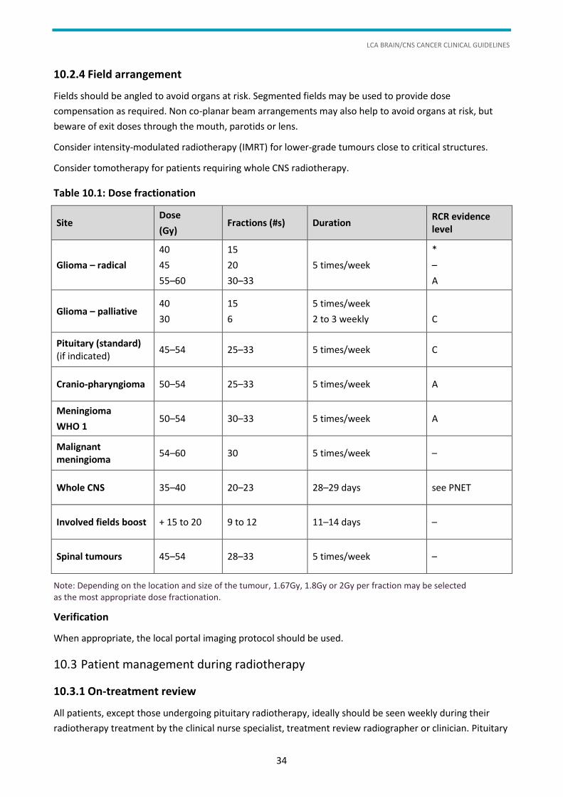
LCA BRAIN/CNS CANCER CLINICAL GUIDELINES
34
10.2.4 Field arrangement
Fields should be angled to avoid organs at risk. Segmented fields may be used to provide dose
compensation as required. Non co-planar beam arrangements may also help to avoid organs at risk, but
beware of exit doses through the mouth, parotids or lens.
Consider intensity-modulated radiotherapy (IMRT) for lower-grade tumours close to critical structures.
Consider tomotherapy for patients requiring whole CNS radiotherapy.
Table 10.1: Dose fractionation
Site Dose
(Gy) Fractions (#s) Duration
RCR evidence level
Glioma – radical
40
45
55–60
15
20
30–33
5 times/week
*
–
A
Glioma – palliative 40
30
15
6
5 times/week
2 to 3 weekly
C
Pituitary (standard) (if indicated)
45–54 25–33 5 times/week C
Cranio-pharyngioma 50–54 25–33 5 times/week A
Meningioma
WHO 1 50–54 30–33 5 times/week A
Malignant meningioma
54–60 30 5 times/week –
Whole CNS 35–40 20–23 28–29 days see PNET
Involved fields boost + 15 to 20 9 to 12 11–14 days –
Spinal tumours 45–54 28–33 5 times/week –
Note: Depending on the location and size of the tumour, 1.67Gy, 1.8Gy or 2Gy per fraction may be selected as the most appropriate dose fractionation.
Verification
When appropriate, the local portal imaging protocol should be used.
10.3 Patient management during radiotherapy
10.3.1 On-treatment review
All patients, except those undergoing pituitary radiotherapy, ideally should be seen weekly during their
radiotherapy treatment by the clinical nurse specialist, treatment review radiographer or clinician. Pituitary

RADIOTHERAPY – GENERIC
35
patients may be seen once at the end of treatment if they have no other concerns during their course of
treatment.
During the on-treatment review the following should be checked: appropriate clinical assessment, blood
tests if indicated and review of medication. Weekly full blood counts (FBCs), liver function tests (LFTs) and
urea and electrolytes (U&Es) are required for patients having concomitant temozolomide.
10.3.2 Treatment interruptions
Patients with medulloblastoma (primitive neuroectodermal tumour – PNET) are classed as Category 1 and
should therefore have all treatment interruptions accounted for. All other CNS patients being treated with
palliative intent are classed as Category 2 and should ideally have no more than two treatment
interruptions.
10.3.3 Patient follow-up
Patients are seen between 4 and 6 weeks after completing their radiotherapy.
Those patients undergoing adjuvant chemotherapy will be seen after 4 weeks, preferably with an up-to-
date MRI scan.
High-grade glioma patients are scanned every 3 months for the first year.
Low-grade glioma patients are scanned every 3 to 6 months for the first year
The scan intervals are usually increased in subsequent years depending on the patient’s status.
10.3.4 Clinical audit and quality review meeting
Regular audit and review of incident reports and variance forms should be undertaken and is presented in
the quality review meeting. This is done in accordance with local guidelines.
LCA BRAIN/CNS CANCER CLINICAL GUIDELINES
36
11 Chemotherapy – Generic
Valid consent and initiation of treatment (1st cycle) ideally should be performed by a consultant or
appropriately qualified specialty registrar (StR)/fellow. The commencement of each cycle will be based
upon clinical examination, full blood counts (FBCs, including differential white blood cell (WBC)
biochemistry) and any other test results specific to the regime in question. Subsequent cycles can be
authorised either by any clinician at StR level in the team or an appropriately trained and qualified clinical
nurse specialist who has passed the physical assessment and Independent Nurse Prescribers course.
11.1 First-line treatment
11.1.1 Temozolomide in newly diagnosed patients with glioblastoma multiforme
Concomitant with radiotherapy:
- temozolomide 75mg/m2 PO daily for 6 weeks
- (+ co-trimoxazole as PCP prophylaxis)
- discontinue concomitant temozolomide and co-trimoxazole if platelets less than 75 x 109/l.
Adjuvant temozolomide:
- 150mg/m2 daily for 5 days every 28 days for 1st cycle and commence 4–6 weeks post end of radiotherapy
- if adequate haematological recovery, increase to 200mg/m2 for the remaining 5 cycles
- total of 6 cycles as per NICE guidance TA121.
11.2 Chemotherapy treatment of recurrent disease
11.2.1 PCV (MRC protocol) for recurrent disease
PCV is the first line of choice in patients of good performance status (ECOG 0–2) with high-grade glioma
(HGG) who progressed on or after first-line treatment (either radiotherapy alone or radiotherapy and
temozolomide). It has been the gold standard regimen for anaplastic oligodendrogliomas.
Lomustine 100mg/m2 PO day 1
Vincristine 1.5mg/m2 IV day 1 (max 2mg)
Procarbazine 100mg/m2 PO daily for days 2–11 in 2 divided doses
Cycle repeated every 6 weeks (up to 6 cycles, stop if progressive disease).
Dose modification
If day 1 neutrophil count <1.5 x 10⁹/l or platelets <100 x 109/l: delay for 2 weeks and then give full dose
if count fully recovers.
If FBC low after initial 2-week delay: wait further 2 weeks and reduce dose by 25%.
If delay required for two consecutive cycles due to haematological toxicity: reduce dose by further 25%.
If LFTs >5 x ULN: reduce or omit vincristine and procarbazine.

CHEMOTHERAPY – GENERIC
37
11.2.2 Single agent alternatives to PCV at first recurrence
Lomustine 100–120mg/m2 PO once/cycle
q 6/52
Dose modifications as per PCV
Carmustine 80mg/m2 IV daily for 3 days or 200–240mg/m2 IV once
q 6/52
Dose modifications as per PCV
11.2.3 Temozolomide for recurrent disease
Indicated as per NICE guidance (TA23) after failure of a nitrosourea-based chemotherapy regime and good
performance patients (ECOG 0–1) or in patients with a contraindication to a nitrosourea-based
chemotherapy regime:
Standard dose: 200mg/m2 PO daily for 5 days (if no previous chemotherapy); 150mg/m2 PO daily for 5 days
at 1st cycle (if previous chemotherapy). If adequate haematological recovery after 4 weeks (neutrophil
count ≥1.0 x 10⁹/l and platelets ≥100 x 109/l), increase to standard dose of 200mg/m2 for the remaining
cycles.
11.2.4 Carboplatin/etoposide
Carboplatin and etoposide can be used in combination or etoposide can be used as a single agent as third-
or fourth-line treatment.
Carboplatin AUC 5 IV day 1 of 21
Etoposide 100mg/m2 IV day 1 of 21
Etoposide 200mg/m2 PO day 2, 3 of 21
Or
Etoposide 50mg/m2 PO (max 100mg) day 1 to 21 (inclusive)
11.2.5 Bevacizumab
Bevacizumab may be given as a single agent or in combination with irinotecan. Currently, this is not being
funded. We acknowledge that some patients may benefit with a marked reduction in perifocal oedema and
the ability to come off high-dose steroids.
Bevacizumab 5–7.5mg/kg can be administered every 2 or every 3 weeks
Bevacizumab 5mg/kg/week can be administered every 2 or every 3 weeks
Irinotecan/CPT11 340mg/m2 (enzyme inducing AEDs) day 1 of 21
Irinotecan/CPT11 125mg/m2 (non-enzyme inducing AEDs) day 1 of 21
11.2.6 Carboplatin as a single agent
Carboplatin may be used (AUC 4 to 6) depending on renal function and bone marrow reserve.
LCA BRAIN/CNS CANCER CLINICAL GUIDELINES
38
12 Tumour-specific Pathways
12.1 High-grade gliomas
12.1.1 Treatment policy
Unless there are exceptional (clinically indicated) circumstances, patients should only be offered active
treatment following tumour biopsy or debulking surgery.
Resection of a glioma may give rapid relief from symptoms (e.g. raised intracranial pressure – ICP),
especially if the tumour is in a non-eloquent area. A transient neurological deficit will occur in 10% of cases
post-operatively, and there is recovery of the neurological deficit in up to 50% of cases. The extent of
surgery (biopsy vs gross debulking) has been shown in a number of studies to affect the length of survival.
In a study by Ammirati et al., patients with high-grade gliomas who had a gross total resection had a 2-year
survival rate of 19%, while those with a subtotal resection had a 2-year survival rate of 0%.1
Bis-chloroethylnitrosourea (BCNU)-polymer wafers (Gliadel wafers) may be used by neurosurgeons in
selected patients with either primary or recurrent tumours, where a 90% tumour debulking is achievable.
Dural closure must be possible and peri-ventricular regions are best avoided. A phase III randomised trial
that included 240 patients compared surgery with the implantation of polymer wafers with BCNU into the
tumour bed, and demonstrated significant prolongation of survival compared with a placebo wafer. Both
groups received radiotherapy. The median survival was 13.9 months in the group treated with Gliadel
wafers and 11.6 months in the group treated with placebo. Level 1a
Age, performance status and the extent of surgery are strong predictors of survival, so radical radiotherapy
should be offered to patients <70 years and with good performance status of World Health Organization
(WHO) Grade 0–1. More frail or elderly patients may be suitable for palliative radiotherapy. Older patients
over 70 years of age who are fit with a good performance status should be considered for radical chemo-
radiotherapy.2
12.1.2 Delineation of target volumes
The gross tumour volume (GTV) is delineated on the contrast-enhancing edge of the tumour, preferably on
the post-operative gadolinium-enhanced T1-weighted magnetic resonance imaging/computerised
tomography (MRI/CT) fused images.
Clinical target volume (CTV) = GTV + 15mm to 25mm depending on the nature of the tumour.
The CTV may be modified to take into account the natural barriers to spread (e.g. bone, tentorium and falx)
as tumours tend to grow along the lines of least resistance, e.g. white matter tracts.
Gliomas generally do not grow across to the opposite hemisphere except via the corpus callosum;
therefore the CTV volume can often be trimmed up to 1cm over midline superiorly or inferiorly to this, in
order to reduce the treatment volume. In certain instances, for example where there is massive midline
shift, the tumour will cross the midline although it remains confined to one hemisphere.
Planning target volume (PTV) = CTV + 3mm to 5mm.
This 5mm margin is added to the CTV taking into account the departmental measurements for set-up
accuracy.
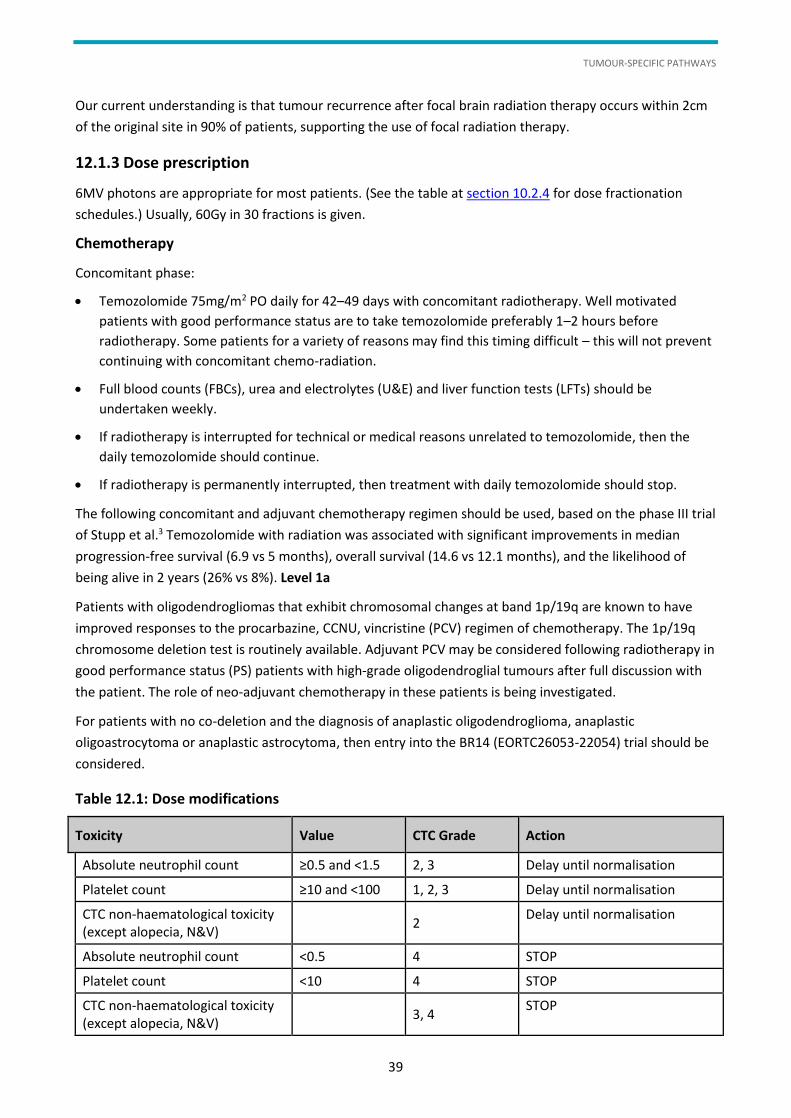
TUMOUR-SPECIFIC PATHWAYS
39
Our current understanding is that tumour recurrence after focal brain radiation therapy occurs within 2cm
of the original site in 90% of patients, supporting the use of focal radiation therapy.
12.1.3 Dose prescription
6MV photons are appropriate for most patients. (See the table at section 10.2.4 for dose fractionation
schedules.) Usually, 60Gy in 30 fractions is given.
Chemotherapy
Concomitant phase:
Temozolomide 75mg/m2 PO daily for 42–49 days with concomitant radiotherapy. Well motivated
patients with good performance status are to take temozolomide preferably 1–2 hours before
radiotherapy. Some patients for a variety of reasons may find this timing difficult – this will not prevent
continuing with concomitant chemo-radiation.
Full blood counts (FBCs), urea and electrolytes (U&E) and liver function tests (LFTs) should be
undertaken weekly.
If radiotherapy is interrupted for technical or medical reasons unrelated to temozolomide, then the
daily temozolomide should continue.
If radiotherapy is permanently interrupted, then treatment with daily temozolomide should stop.
The following concomitant and adjuvant chemotherapy regimen should be used, based on the phase III trial
of Stupp et al.3 Temozolomide with radiation was associated with significant improvements in median
progression-free survival (6.9 vs 5 months), overall survival (14.6 vs 12.1 months), and the likelihood of
being alive in 2 years (26% vs 8%). Level 1a
Patients with oligodendrogliomas that exhibit chromosomal changes at band 1p/19q are known to have
improved responses to the procarbazine, CCNU, vincristine (PCV) regimen of chemotherapy. The 1p/19q
chromosome deletion test is routinely available. Adjuvant PCV may be considered following radiotherapy in
good performance status (PS) patients with high-grade oligodendroglial tumours after full discussion with
the patient. The role of neo-adjuvant chemotherapy in these patients is being investigated.
For patients with no co-deletion and the diagnosis of anaplastic oligodendroglioma, anaplastic
oligoastrocytoma or anaplastic astrocytoma, then entry into the BR14 (EORTC26053-22054) trial should be
considered.
Table 12.1: Dose modifications
Toxicity Value CTC Grade Action
Absolute neutrophil count ≥0.5 and <1.5 2, 3 Delay until normalisation
Platelet count ≥10 and <100 1, 2, 3 Delay until normalisation
CTC non-haematological toxicity (except alopecia, N&V)
2 Delay until normalisation
Absolute neutrophil count <0.5 4 STOP
Platelet count <10 4 STOP
CTC non-haematological toxicity (except alopecia, N&V)
3, 4 STOP
LCA BRAIN/CNS CANCER CLINICAL GUIDELINES
40
Other prescriptions
Ondansetron 8mg (or equivalent anti-emetic) prior to first dose – then domperidone 10mg 8
hourly PRN
Co-trimoxazole Forte 960mg 3x per week (PCP prophylaxis) during the concomitant phase
Adjuvant temozolomide chemotherapy
Commence after a 4-week gap from the end of concomitant chemo-radiation.
Dose cycle 1: 150mg/m2 PO daily for 5 days out of 28 days. If this dose is tolerated, increase to 200 mg/m2
PO daily for cycle 2 to cycle 6. Further cycles may be given (at the discretion of the clinician) if there is
continued clinical and radiological improvement.
12.1.4 Recurrent high-grade gliomas
Options include:
best supportive care
further debulking surgery/Gliadel wafers
PCV chemotherapy if not received in the primary setting
temozolomide chemotherapy if not received in the primary setting or a re-challenge if appropriate time
interval
CEV chemotherapy (second-/third-line) – carboplatin, etoposide, vincristine or modified CEV (omitting
the vincristine)
a single nitrosourea may used
carboplatin as a single agent may be used
bevacizumab single agent or in combination with irinotecan for individual patients may be funded via
individual funding requests
referral for phase I/II trials (discuss with the clinical trials unit) EGFR inhibitors, imatinib, etc., only for
patients with WHO PS 0–1. Malignant gliomas often have amplification of the EGFR gene, amplification
of c-kit, and loss of PTEN.4
There is no proven role for stereotactic radiosurgery or Gamma Knife radiosurgery in recurrent high-
grade glioma. Nevertheless, in exceptional circumstances with a truly localised recurrence, stereotactic
radiosurgery/radiotherapy may be used. Partial brain radiotherapy re-treatment may be considered in
exceptional circumstances on an individual basis.
12.2 Low-grade gliomas
Patients with low-grade gliomas can receive radiotherapy either upfront after primary surgery, or at relapse
following a surveillance policy. Both options have a similar overall survival rate, although time to relapse is
greater with early radiotherapy. If indolent, and the patient is asymptomatic, a surveillance follow-up policy
is preferred with serial scanning. If symptomatic, a maximal safe resection and/or radiotherapy should be
offered. Delayed radiotherapy is preferred if resection is complete; however, in cases where only a biopsy
can be performed or resection is incomplete, or where there are atypical histological features, early
TUMOUR-SPECIFIC PATHWAYS
41
radiotherapy is preferred. Each case should be considered on its merits, and patient preference noted,
before deciding on early versus delayed radiotherapy.
12.2.1 Delineation of target volumes
The pre-operative GTV seen on T2 weighted MRI is outlined on the fused CT/MRI images.
CTV = GTV + 5mm to 25mm.
PTV = CTV + 3mm to 5mm.
Chemotherapy
There is no convincing evidence yet of a significant benefit to concomitant or adjuvant chemotherapy in
low-grade astrocytomas. Chemotherapy may have an adjuvant role in oligodendroglioma if incompletely
resected, and can be an alternative to radiotherapy in these patients. Choice of regimen is as for high-grade
glioma. 1p/19q and methylation status may be useful in decision making. With regard to neo-adjuvant
chemotherapy, currently there is no convincing evidence for its use but in exceptional cases it may be
considered with knowledge of the 1p/19q and methylation status.
12.2.2 Ependymoma
Maximal safe resection should be attempted if at all possible. These usually occur in posterior fossa or
spinal cord, but also supratentorially. The cure rate is approximately 50%. Craniospinal axis radiotherapy
(CSA-RT) does not improve the local control of ependymoma, therefore localised radiotherapy is the
preferred option in most cases.
Supratentorial, infratentorial or spinal (low grade)
If a complete resection has been achieved and brain and spinal MRI are clear and cerebrospinal fluid (CSF)
cytology is clear, limited field radiotherapy or observation (sub-ependymomas) can be used. If resection is
subtotal but MRI and CSF are clear, then offer local radiotherapy (50Gy to 55Gy). If MRI or CSF show
evidence of seeding, CSA-RT should be considered (for technique, see medulloblastoma, section 12.3).
Supratentorial (high grade)
If a complete resection has been achieved and post-operative brain and spinal MRI are clear and CSF
cytology is clear, local radiotherapy (55Gy/30#) may be used. If resection is subtotal but MRI and CSF are
clear, then local radiotherapy is probably sufficient. If MRI or CSF show evidence of seeding, CSA-RT should
be strongly considered (for technique, see medulloblastoma, section 12.3).
Note: Check for organs in exit beams, especially kidneys in lower T/upper L tumours. Whole kidney dose
must not exceed 15Gy. If clinically indicated, sperm banking or oophoropexy should be considered.
Chemotherapy
Chemotherapy can be used for ependymomas for inoperable cases recurrent after radiotherapy or for
metastatic disease. The regimens are generally derived from the paediatric studies and are cisplatin based.5
12.3 Medulloblastoma: craniospinal radiation
Medulloblastoma is a highly cellular malignant embryonal neoplasm classified as a primitive
neuroectodermal tumour (PNET). It accounts for 15% to 20% of all childhood primary central nervous
LCA BRAIN/CNS CANCER CLINICAL GUIDELINES
42
system (CNS) neoplasms. It is rare in adults. Standard management consists of complete or near complete
surgical resection followed by post-operative CSR-RT. (See British Neuro-oncology Society guidelines.)
12.3.1 Chemotherapy
The benefit of post-radiation chemotherapy, although not proven in a randomised controlled trial, has now
been generally accepted for all paediatric patients with medulloblastoma and should be discussed for all
young adult and adult patients. The standard treatment is the ‘Packer’ regimen.6 It consists of weekly
vincristine during treatment; commencing 6 weeks after radiation, patients receive cycles of CCNU,
cisplatin and vincristine. It is standard for 8 courses to be delivered but this is often modified because of
cisplatin toxicity.
12.3.2 Staging and pre-planning investigations
For patients to be ‘standard risk’ medulloblastoma, they must be non-metastatic and must meet the
following criteria:
histologically proven medulloblastoma
no CNS metastases on MRI or brain and spine (preferably performed before surgery)
no clinical evidence of extra-CNS metastases
no tumour cells on cytospin of lumbar CSF obtained at least 15 days following surgery.
Patients with metastases fall into the high-risk group. This includes Chang stage M1 disease in which
tumour cells are found free in the CSF without radiological evidence of metastasis; these patients have a
worse prognosis than those without evidence of such tumour spread and they are therefore included in the
high-risk group.
Residual posterior fossa disease
The patient would have to be discussed at the multidisciplinary team (MDT) meeting and the surgeons
asked to consider further surgery.
12.3.3 Radiotherapy principles
The aim of this treatment is to achieve homogenous irradiation to the whole CSF volume so as to eradicate
malignant cells free within the cerebrospinal fluid. In the cranium care must be taken to include:
the cribriform plate
the temporal lobe
the posterior orbit.
Patient position and immobilisation
Patients will be immobilised in a thermoplastic shell. Care must be taken to determine if the patient is to
have treatment on the tomotherapy unit or a conventional linear accelerator as this may determine the
patient’s position. The local guidelines should be followed.
CT Planning
The planning dataset is acquired from the CT scanner and if the patient is to have conventional whole CNS
radiotherapy it is then transferred into the planning system (see local guidelines).
TUMOUR-SPECIFIC PATHWAYS
43
If the patient is to have whole CNS radiotherapy on the tomotherapy unit, the CT dataset is transferred to
the tomotherapy planning system.
12.3.4 Normal tissue tolerances/gonad doses (see QUANTEC)
A dose volume histogram will be provided for the following structures and will require the clinician’s
approval.
In addition to the normal brain and brainstem organs at risk (e.g. supratentorial brain, posterior fossa,
cochlea, hypothalamus, pituitary and optic chiasm), the following should be considered:
kidneys
heart
lungs
gonads (consideration will be given to the location of these organs for dose estimation).
Posterior fossa boost
The CTV for the posterior fossa is determined from a planning CT and encompasses the entire posterior
fossa as follows:
superiorly – the tentorium
inferiorly – at least 2cm below the lower limit of the tumour and including at least the outer table of
the skull at the foramen magnum with a minimum 1cm margin
anteriorly – the posterior clinoid processes
posteriorly – the posterior extension of the meninges as far as the inner table of the skull; the CT
should include any herniation of the meninges through the craniotomy defect
laterally – the lateral extension of the meninges around the cerebellum.
PTV = CTV + 3mm to 5mm.
No attempt will be made to include the internal auditory meatus in the high-dose volume by extending the
CTV to include it.
Posterior fossa contents should be outlined on an MRI using image fusion or on a post-operative enhanced
CT scan.
It should be noted that some trials define posterior fossa target differently (e.g. HIT-SIOP PNET 4) and for
patients in studies the trial protocol should be studied. In addition, it is essential to have a pre-operative
scan available to ensure adequate margin.
12.3.5 Radiotherapy prescription
Localised disease
Brain – 35Gy in 21 daily fractions of 1.67Gy
Spine – 35Gy in 21 daily fractions of 1.67Gy
Primary tumour boost – 20Gy in 12 daily fractions of 1.67Gy
Total dose to primary – 55Gy in 33 fractions of 1.67Gy.
LCA BRAIN/CNS CANCER CLINICAL GUIDELINES
44
Modifications due to haematological toxicity
It is rare to have to interrupt radiotherapy but weekly FBCs need to be taken and patients reviewed in a
weekly on-treatment clinic. Patients will be monitored daily if their platelet count falls to 40 and
radiotherapy will be interrupted if the platelet count falls below 25 or the neutrophil count below 0.5. If
either of these occurs, then the craniospinal part of radiotherapy is interrupted and the posterior fossa
boost is commenced early. Granulocyte colony stimulating factor (G-CSF) should be given in consultation
with the pharmacologists to assist neutrophil recovery and platelet support may be required. Haemoglobin
levels should be maintained above 10gm/l by transfusion if necessary.
Portal images and lithium fluoride or diode dosimetry of the eyes
Patients being treated on a conventional linear accelerator will be imaged according to local guidelines.
Thermoluminscence dosimetry (TLD) or diodes are completed within the first two fractions of radiotherapy
to assess the eye dose. This is to be carried out via individual departmental policies.
12.4 Spinal cord tumours
Ependymomas, astrocytomas and meningiomas receive localised radiotherapy.
12.4.1 Spinal ependymoma
Surgical debulking should usually take place first. Complete resection is rare.
For certain tumours, e.g. Grade 1 myxopapillary ependymoma, surgery may be curative.
High grade (anaplastic) usually requires radiotherapy.
Initial investigations and assessment
Baseline tests as for glioma plus gadolinium-enhanced staging MRI of the spine.
Treatment policy
Completely excised myxopapillary ependymoma: no further treatment, surveillance only.
Incompletely excised myxopapillary ependymoma: discuss options of surveillance or local radiotherapy.
Residual or recurrent myxopapillary ependymoma with adjacent metastases: local radiotherapy.
Incompletely excised subependymoma (usually brain): surveillance.
Completely excised low-grade ependymoma: local radiotherapy/discuss.
Incompletely excised low-grade ependymoma and high-grade ependymoma: local radiotherapy.
Disseminated spinal ependymoma: craniospinal irradiation and boost to sites of macroscopic disease.
12.4.2 Spinal astrocytoma
Initial investigations and assessment
Baseline tests for glioma plus gadolinium-enhanced staging MRI of the spine.
Treatment policy
Low grade: surveillance or involved field radiotherapy.
High grade: involved field radiotherapy.
TUMOUR-SPECIFIC PATHWAYS
45
Treatment volume
The GTV is the extent of residual tumour on the post-operative MRI. Cover the spinal canal and/or nerve
roots with proximal and distal extension based on pre-operative MRI.
CTV = GTV + 20mm margin superiorly and inferiorly and 10mm laterally.
PTV = CTV + 5mm.
For lumbar tumours the CTV may need to be extended laterally to include the nerve roots.
Note: Check for organs in exit beams, especially kidneys in lower T/upper L tumours. Whole kidney dose
must not exceed 15Gy. If clinically indicated sperm banking or oophoropexy should be considered.
12.5 Germ cell tumours
12.5.1 Initial investigations and assessment
In addition to standard assessments, if not already performed:
serum tumour markers – FP, HCG
MRI spine
CSF cytology and tumour markers (FP, HCG) if no evidence of raised ICP
full endocrine assessment
formal visual assessment if clinically indicated.
12.5.2 Germinoma
Treatment policy
Post-pubertal male (localised or bifocal)
In fully staged patients with confirmed localised disease in typical locations (pineal/suprasellar):
whole ventricular system (all ventricles including 4th) + 1cm margin (WVRT) to 24Gy/15 #
boost to primary site plus 1cm margin 16Gy/10# (total dose 40Gy).
In incompletely staged patients or patients with non-typical primary locations (e.g. brainstem, thalamic):
craniospinal axis: 24Gy/15#
boost to primary site: 16Gy/10# (total dose 40Gy).
All females/pre-pubertal males (localised or bifocal)
Treatment should be individualised. The options include:
whole ventricular radiotherapy (24Gy in 15#) and boost to primary tumour (16Gy in 10#) (preferred
option)
carbo-PEI chemotherapy and WVRT and boost as per SIOP CNS GCT 96 protocol7 and the Children’s
Cancer and Leukaemia Group (CCLG) GCT group interim guidelines8
CSA-RT and boost to primary site (as for post-pubertal males above).
Metastatic

LCA BRAIN/CNS CANCER CLINICAL GUIDELINES
46
Craniospinal radiotherapy:
boost to primary and sites of macroscopic metastases (to total dose of 40Gy).
12.5.3 Non-germinomatous germ cell tumours
Treatment policy
Primary chemotherapy: PEI (cisplatin, etoposide and ifosfamide) as per SIOP CNS GCT 96 protocol (12
weeks).
Then focal radiotherapy in localised disease and CSA-RT and conformal boost in patients with
metastatic disease at diagnosis.
Allow for a 4–6 week gap between the end of chemotherapy and beginning of radiotherapy.
Chemotherapy – PEI protocol (used at The Royal Marsden)
Pre-hydration: NaCl 0.9% at 2L/m2 daily + 20mmol KCl/l + 12g mannitol/l for 3 hours.
Cisplatin 20mg/m2 in 500ml 5% dextrose over 60 minutes, days 1–5.
Etoposide 100mg/m2 IV, days 1–3.
Ifosfamide 1,500mg/m2 IV over 3 hours, days 1–5.
MESNA 1,875mg/m2 over 15 hours as a continuous infusion.
Post-hydration: NaCl 0.9% at 2L/m2 daily + 20mmol KCl/l + 12g mannitol/l for 9 hours.
Mannitol should only be administered to patients who exhibit no signs of diabetes insipidus.
Repeat every 3 weeks for a total of 4 cycles.
Notes
Ethylenediaminetetraacetic acid (EDTA) clearance before start of chemotherapy and repeat if EDTA
<100 after cycle 2.
MRI scan post-chemotherapy/pre-radiotherapy.
Consider surgical resection of residual disease in secreting germ cell tumour post completion of
chemotherapy.
Monitor tumour markers at each cycle of chemotherapy and at the start and end of radiotherapy.
Regular (at least twice daily) electrolyte measurements during chemotherapy.
Ensure and maintain regular review/input of endocrinology team during inpatient and outpatient
phases.
Table 12.2: Post-chemotherapy radiotherapy dose
Localised disease Primary tumour 54Gy/30F with conformal 3–6 field technique or IMRT/VMAT
Metastatic disease Whole brain 30Gy/20# (supine)
Whole spine 30Gy/20# (supine)
Boost to primary 24Gy/15# (total dose 54Gy)
Boost to macroscopic metastatic disease 24Gy/15# (20Gy if spine)
TUMOUR-SPECIFIC PATHWAYS
47
Radiotherapy technique
Phase I craniospinal axis – technique as in medulloblastoma/PNET.
Phase II GTV = tumour as visible on planning CT scan.
PTV = GTV + 0.8cm–1cm margin.
Patients needing CSA-RT should be referred for radiotherapy to a centre with a paediatric service.
Management at relapse
Given the rarity, treatment at relapse has to be individualised.
For these relatively rare tumours see BNOS guidelines, www.bnos.org.uk
CNS lymphoma
tumours of pineal region
optic pathway gliomas
adult PNET
1 Ammirati M et al. (1987), Reoperation in the treatment of recurrent intracranial malignant gliomas, Neurosurgery 21:607–614.
2 Hashem SA et al. (2012), Radiotherapy with concurrent or sequential temozolomide in elderly patients with glioblastoma multiforme, Journal of Medical Imaging and Radiation 56(2):204–210.
3 Stupp R et al. (2005), Radiotherapy plus concomitant and adjuvant temozolomide for glioblastoma, New England Journal of Medicine 352(10):987–996.
4 Prados MD et al. (2006), A Phase I trial of erlotinib with or without temozolomide in patients with recurrent malignant glioma, Neuro-Oncoogy 8(1):67–78.
5 Brandes AA et al. (2005), A multicenter retrospective study of chemotherapy for recurrent intracranial ependymal tumors in adults by the Gruppo Italiano Cooperativo di Neuro-Oncologia, Cancer 104(1):143–148.
6 Packer RJ et al. (1994), Outcome for children with medulloblastoma treated with radiation and cisplatin, CCNU, and vincristine chemotherapy, Journal of Neurosurgery 81(5):690–698.
7 Calaminus G et al. (2013), SIOP CNS GCT 96: final report of outcome of a prospective, multinational nonrandomized trial for children and adults with intracranial germinoma, comparing craniospinal irradiation alone with chemotherapy followed by focal primary site irradiation for patients with localized disease, Neuro-Oncology, http://neuro-oncology.oxfordjournals.org/content/early/2013/03/03/neuonc.not019.full.
8 CCLG (2008), Interim Guidelines for the Treatment of Intracranial Germ Cell Tumours, Version 2.1.
LCA BRAIN/CNS CANCER CLINICAL GUIDELINES
48
13 Rehabilitation
Cancer rehabilitation attempts to maximise patients’ ability to function, to promote their independence
and to help them adapt to their condition. It offers a major route to improving their quality of life, no
matter now long or short the timescale.1
Considerable medical advances in the treatment of primary brain and spinal tumours have translated into a
marked increase in the number of survivors who present with impairments and functional limitations.2,3
Learning to compensate for these deficits with a holistic rehabilitative approach that encompasses medical,
physical, social and psychological aspects of the disease is an important factor in improving the patient’s
quality of life.4
13.1 Evidence
Patients with brain tumours make significant functional gains after a course of rehabilitation.5,6,7,8,9,10
Inpatient multidisciplinary rehabilitation results in marked reduction in disability as measured by
various functional measurement tools (Functional Independence Measure, Barthel Index, Karnofsky
Performance Status Scale).5,11,12
The same principles of neuro-rehabilitation used in patients with traumatic brain injury and stroke are
equally applicable to patients with brain and spinal cord tumours.13
Treatments such as exercise and other therapy interventions that enhance physiological and functional
outcomes are known to improve the quality of life of cancer survivors.14,15,16,17
13.2 Prognosis and rehabilitation
Key elements, alongside generic principles of neuro-rehabilitation, should be considered in the
rehabilitation of brain and spinal tumours, such as:
- consideration of disease related functional fluctuations
- progressive decline
- difficult psychological adjustments to changing functional abilities
- uncertain prognosis
- depression, fatigue and medical fragility.13
Access to rehabilitation services should be offered to patients at curative stage, in long-term remission,
receiving active treatment, with advancing disease, or requiring support at the end of life.1
The approach in the rehabilitation of patients with a good long-term prognosis should focus on
reducing deficits over a medium to longer timeframe.18,19,20
It should be recognised that patients with a poor prognosis have a limited window of opportunity for
recovery of function and therefore should receive urgent access to rehabilitation services, including
inpatient settings, where goals should be clearly defined around maximising function and optimising
the quality of their remaining life.20
For other patients with poor prognosis, a compensatory rehabilitation approach may be more
appropriate to maintain a meaningful level of function for as long as possible.1
REHABILITATION
49
13.3 Access and timing of rehabilitation
Referral to rehabilitation and supportive care services should not be dependent on diagnosis, but on
patient need.20,21
The patient’s wishes in the rehabilitation referral process should be considered.
Rehabilitation should begin as early as possible to optimise rehabilitation opportunities and functional
participation in society.21
Clinicians must recognise that the principles of neuroplasticity and associated gains also apply to
patients with brain tumours.22,23,24,25
Consideration should be given to an individual’s diagnosis, prognosis and ongoing medical
management.
Patients receiving adjunct medical treatment such as radiotherapy or chemotherapy should still be
considered for rehabilitation.7,19
The timeliness of rehabilitation referral will be facilitated by comprehensive communication between
allied health professionals.20
13.4 Rehabilitation assessment
Assessment and interventions in rehabilitation should consider the impact of the disease at different
levels of the International Classification of Functioning, Disability and Health (ICF) framework such as
impairments, activity limitation, restriction in participation and contextual factors (environmental or
personal).26
Assessment areas should include: patients’ ideas, concerns and expectations; social history; levels of
function (including work, leisure, activities of daily living); impairments (including physical, cognitive,
perceptual, psychological, communication and nutritional status); and assessment of the carer’s ability
to actively participate in ongoing management.
Reassessment should be considered at any stage of the patient’s disease trajectory dependent on
clinical presentation of symptoms.1,20,27
13.5 Rehabilitation intervention
It is recommended that rehabilitation treatments be structured around the ICF framework to ensure a
holistic approach. For example, the activity and participation domain will address mobility, self-care
and domestic life; and environmental factors will consider transport, community access and so on.26
The intensity, frequency and content of therapy in multidisciplinary rehabilitation will vary and goal
setting should be individualised based on clinical needs.28
Rehabilitation approaches may be restorative or compensatory in nature and the content can include
functional training, physical reconditioning, task reacquisition strategies, cognitive and behavioural
therapy, psychological support, environmental adaptations, and vocational and recreational
programmes.20,29
Self-supported management is an important aspect in the rehabilitation of cancer patients applicable
to all settings.30
Education and counselling should be available at various point of the journey to address adjustment
issues such as self-worth, self-image and role reversal within the family.28,30
LCA BRAIN/CNS CANCER CLINICAL GUIDELINES
50
The emotional and spiritual needs of the patient, family and carers must be recognised and appropriate
services accessed.1,20
Access to and reliable information about complementary therapy services should be available.1
Brain and spinal cancer patients should have access to vocational rehabilitation or a similar form of
work support.30
Patients should be signposted to dedicated brain tumour-specific charities and non-profit organisations
at a time that is appropriate for them.
Patients discharged to the community are confronted with various adjustment issues. Ongoing
monitoring, education and counselling of the patient and family are therefore essential.28
Highly responsive community services to support rapid or significant changes in a patient’s condition
are key.
13.6 Cancer/treatment secondary side effects and rehabilitation
Rehabilitation should not be excluded where concurrent medical treatment such as chemotherapy or
radiotherapy is taking place2,20 as there is no proven detrimental effect on functional gains9,10,19 and no
significant difference in length of stay.31
Flexibility in rehabilitation in this population is important to promote the patient’s tolerance and
optimise rehabilitation outcomes.32
Rehabilitation should aim to improve the patient’s perception of cancer-related symptoms and
treatment side effects such as steroid-induced proximal myopathy, general deconditioning,13,33 poor
appetite, fatigue, insomnia, pain, anxiety, constipation and poor overall sense of wellbeing.34,35
13.7 Multidisciplinary rehabilitation
Rehabilitation should be delivered by a structured and coordinated multidisciplinary team
(MDT).1,20,27,36,37,38
The MDT’s goal should include improving patient symptoms, and optimising functional independence
and participation.26
The MDT should use a holistic model of care such as the ICF.26
The multidisciplinary approach should provide patients with the skills required to manage their own
care, to develop coping strategies and to maintain quality of life.39
Multidisciplinary rehabilitation should be provided, according to patient’s needs, by the following
professionals:20
specialist neuro-oncology medical team
physiotherapists
occupational therapists
speech and language therapists
dietitians
nurses
primary healthcare team/GPs
REHABILITATION
51
neuropsychologists, neuropsychiatrists and counsellors
social worker and social care team
orthotists
wheelchair and assistive technology teams
chaplaincy and bereavement services
complementary therapy services.
13.8 Outcome measures
The use of outcome measures is imperative in continuing to demonstrate the value of rehabilitation.
There is currently no agreed standard outcome measure that should specifically be used in the
rehabilitation of brain and spinal tumour patients. The clinician should therefore choose relevant
outcome measures based on the individual’s needs and goals.
In the literature, the following validated outcome measures were used in brain tumour research and
may be considered in the clinical setting:
- the Functional Independence Measure (FIM)40
- the Barthel Index41
- the Cancer Rehabilitation Evaluation System (CARES)42,43
- the Cancer Survivor Unmet Needs measure (CaSUN)44
- the Perceived Impact of Problem Profile (PIPP)45
- the Fatigue Impact Scale46
- the Depression Anxiety Stress Scale47
- the Work Instability scale.48
13.9 Rehabilitation settings
Rehabilitation and supportive intervention should be delivered in the most appropriate setting for each
individual.
Rehabilitation may be delivered by:
- specialist neuro-rehabilitation centres
- local inpatient rehabilitation units
- neuro-outpatient services
- community services
- hospice services with access to specialist neurological expertise.
Patients discharged to the community are confronted by various adjustments issues; families and
carers also often struggle to cope with the new demands associated with increased care needs and the
general limitation in the patient’s function. Ongoing monitoring, education and counselling of the
patient and family are therefore essential.28 Multidisciplinary rehabilitation in the community may
consist of integrated care delivered by health, social services and the voluntary sector. Highly
responsive community services to support rapid or significant changes in a patient’s condition are key.

LCA BRAIN/CNS CANCER CLINICAL GUIDELINES
52
13.10 Service improvement, training and education
Regular liaison and discussion between the clinical providers, specialists and local commissioners of
rehabilitation services are recommended in order to identify and meet the needs of patients with brain
and spinal tumours.
Inclusion of the rehabilitation needs of this patient group should be considered in the joint strategic
needs assessments of the local commissioning group.
Rapid access for advice and guidance on the rehabilitation needs and management from the specialist
teams should be available for the community services.
Regular meetings and discussions between specialist centres and local services are recommended to
improve links and develop skills, and to improve patient experience and outcomes.
Regular training and education days between the specialist service providers and local services,
including inpatient and community settings should be available.
The staff delivering rehabilitation to this patient group should be trained in the key principles of
rehabilitation identified in this document.
Monitoring of the progress of the referral and waiting times to access services is recommended.
Auditing of any gaps in rehabilitation services is recommended to inform commissioners and providers.
1 NICE (National Institute for Health and Care Excellence) (2004), Improving Supportive and Palliative Care for Adults with Cancer.
2 Huang ME, Silwa JA (2011), Inpatient rehabilitation of patients with cancer: efficacy and treatment considerations, Physical Medicine and Rehabilitation 3:746–757.
3 Poggi G, Liscio M, Pastore V, Adduci A, Galbiati S, Spreafico F, et al. (2009), Psychological intervention in your brain tumor survivors: the efficacy of the cognitive behavioural approach, Disability and Rehabilitation 31(13):1066–1073.
4 Korstjens I, Mesters I, van der Peet E, Gijsen B, van den Borne B (2006), Quality of life of cancer survivors after physical and psychosocial rehabilitation, European Journal of Cancer Prevention 15(6):541–5 47.
5 Geler-Kulcu D, Gulsen G, Buyukbaba E, Ozkan D (2009), Functional recovery of patients with brain tumor or acute stroke after rehabilitation; a comparative study, Journal of Clinical Neuroscience 16(1):74–78.
6 Greenberg E, Treger I, Ring H (2006), Rehabilitation outcomes in patients with brain tumors and acute stroke after rehabilitation: comparative study of inpatient rehabilitation, American Journal of Physical Medicine and Rehabilitation 85(7):568–573.
7 Huang ME, Wartella JE, Kreutzer JS (2001), Functional outcomes and quality of life in patients with brain tumors: a preliminary report, Archives of Physical Medicine and Rehabilitation 82(11):1540–1546.
8 Marciniak CM, Silwa JA, Spill G, Heinemann AW, Semik PE (2001), Functional outcomes of persons with brain tumors after inpatient rehabilitation, Archives of Physical Medicine and Rehabilitation 82(4):457–463.
9 O’Dell MW, Barr K, Spanier D, Warnick RE (1998), Functional outcome of inpatient rehabilitation in persons with brain tumors. Archives of Physical Medicine and Rehabilitation 79(12):1530–1534.
10 Tang V, Rathbone M, Park Dorsay J, Jiang S, Harvey D (2008), Rehabilitation in primary and metastatic brain tumours: impact of functional outcomes on survival, Journal of Neurology 255(6):820–827.
REHABILITATION
53
11 Bartolo M, Zucchell C, Pace A, Lanzetta G, Vecchione C (2012), Early rehabilitation after surgery improves functional outcome in inpatients with brain tumours, Journal of Neuro-Oncology 107:537–544.
12 Fu JB, Parsons HA, Shin KY, Guo Y, Konzen BS, Yadav RR, et al. (2010), Comparison of functional outcomes in low- and high-grade astrocytoma rehabilitation inpatients, American Journal of Physical Medicine and Rehabilitation 89(3):205–212.
13 Kirshblum S, O’Dell MW, Ho C, Barr K (2001), Rehabilitation of persons with central nervous system tumors, Cancer 92(Suppl 4):1029–1038.
14 MacVicar MG, Winningham ML (1986), Promoting functional capacity of cancer patients, Cancer Bulletin 38:235–239.
15 MacVicar MG, Winningham ML, Nickel JL (1989), Effects of aerobic interval training on cancer patients functional capacity, Nursing Research 38:348–351.
16 Markes, Brockow T, Resch KL (2006), Exercise for women receiving adjuvant therapy for breast cancer, Cochrane Database of Systematic Reviews 4.
17 McNeely ML, Campbell K, Opsina M, Rowe BH, Dabbs K, Klassen TP, et al. (2012), Exercise intervention for upper-limb dysfunction due to breast cancer treatment, Cochrane Database of Systematic Reviews 6.
18 Bell KR, O’Dell MW, Barr K, Yablon SA (1998), Rehabilitation of the patient with brain tumour, Archives Physical Medicine and Rehabilitation 79:S37–S46.
19 Marciniak CM, Silwa JA, Spill G, Heinemann AW, Semik PE (1996), Functional outcomes following rehabilitation of the cancer patients, Archives of Physical Medicine and Rehabilitation 77:54–57.
20 NICE (2006), Improving Outcomes for People with Brain and other CNS Tumours.
21 NCAT (2010) Rehabilitation Care Pathway: Brain CNS.
22 Duffau H (2006), New concepts in surgery of WHO grade II gliomas: functional brain mapping, connectionism and plasticity – a review. Journal of Neuro-Oncology 79:77–115.
23 Duffau H (2006), Brain plasticity: from pathophysiological mechanisms to therapeutic applications, Journal of Clinical Neuroscience 13:885–897.
24 Duffau H (2008), Brain plasticity and tumors, Advances and Technical Standards in Neurosurgery 33:3–33.
25 Duffau H, Taillandier L, Gatignol P, Capelle L (2006), The insular lobe and brain plasticity: lessons from tumour surgery, Clinical Neurology and Neurosurgery 108:543–548.
26 WHO (World Health Organization) (2001), International Classification of Functioning Disability and Health. Geneva: WHO.
27 NCAT (National Cancer Action Team)/Peer Review Team (2008), Cancer Rehabilitation Measures.
28 Khan F, Amatya B, Ng L, Drummond K, Olver J (2013), Multidisciplinary rehabilitation after primary brain tumour treatment (Review), Cochrane Database of Systematic Reviews 1.
29 Sherer M, Meyers CA, Bergloff P (1997), Efficacy of postacute brain injury rehabilitation for patients with primary malignant brain tumours, Cancer 2:250–257.
30 NCSI (National Cancer Survivorship Initiative). www.ncsi.org.uk.
31 Tay SS, Ng YS, Lim PA (2009), Functional outcomes of cancer patients in an inpatient rehabilitation setting, Annals, Academy of Medicine, Singapore 38:197–201.
32 Huang ME, Silwa JA (2011), Inpatient rehabilitation of patients with cancer: efficacy and treatment considerations, Physical Medicine and Rehabilitation 3:746–757.
LCA BRAIN/CNS CANCER CLINICAL GUIDELINES
54
33 Batchelor T, DeAngelis LM (1996), Medical management of cerebral metastases, Neurosurgery Clinics of North America 7(3):435–446.
34 Guo Y, Shin KY, Hainley S, Bruera E, Palmer JL (2011), Inpatient rehabilitation improved functional status in asthenic patients with solid and hematologic malignancies, American Journal of Physical Medicine and Rehabilitation 90:265–271.
35 Higginson IJ, Evans CJ (2010), What is the evidence that palliative care teams improve outcomes for cancer patients and their families?, Cancer Journal 16:423–435.
36 Khan F, Turner-Stokes L, Ng L, Kilpatrick T, Amatya B (2011), Multidisciplinary rehabilitation for adults with multiple sclerosis, Cochrane Database of Systematic Reviews 2.
37 Turner Stokes L, Nair A, Sedki I, Disler PB, Wade DT (2011), Multidisciplinary rehabilitation for acquired brain injury in adults of working age, Cochrane Database of Systematic Reviews 1.
38 Gabanelli P (2005), A rehabilitative approach to the patient with brain cancer, Neurological Sciences 26(Suppl 1):S51–2 .
39 Corner J, Richardson A (2007), Cancer survivorship briefing paper. www.macmillan.org.uk/Documents/GetInvolved/Campaigns/Campaigns/campaignsupdate/Cancer_survivorship_paper.pdf.
40 Granger CV (1998) The emerging science of functional assessment: our tool for outcome analysis, Archives of Physical Medicine and Rehabilitation 79:235–240.
41 Mahoney FI, Barthel D (1965), Functional evaluation: the Barthel Index, Maryland State Medical Journal 45:1456–1461.
42 Ganz PA, Shag CA, Lee TJ, Sim MS (1992), The CARES: a generic measure of health related quality of life for patients with cancer, Quality of Life Research 1:19–29.
43 Schag CA, Ganz PA, Heinrich RL (1991), Cancer rehabilitation evaluation system-short form (CARES-SF), Cancer 68:1406–1413.
44 Hodgkinson K, Butow P, Hunt G, Pendlebury S, Hobbs K, Wain G (2007), Breast cancer survivors supportive care needs 2–1 0 years after diagnosis, Supportive Care in Cancer 15:515–523.
45 Pallant JF, Misajon R, Bennett E, Manderson L (2006), Measuring the impact and distress of health problems form the individual’s perspective: development of the Perceived Impact of Problem Profile (PIPP), Health and Quality of Life Outcomes 4(1):36.
46 Fisk JD, Ritvo PG, Ross L, Haase DA, Marrie TJ, Schlech WF (1994), Measuring the functional impact of fatigue: initial validation of the fatigue impact scale, Clinical Infectious Disease 18(Suppl 1):S79–83.
47 Lovibond SH, Lovibond PF (1995), Manual for the Depression Anxiety Stress Scale (DASS). Sydney: Psychology Foundation of Australia.
48 Gilworth G, Chamberlain MA, Harvey A (2003), Development of a work instability scale for rheumatoid arthritis, Arthritis and Rheumatism 49:349–354.
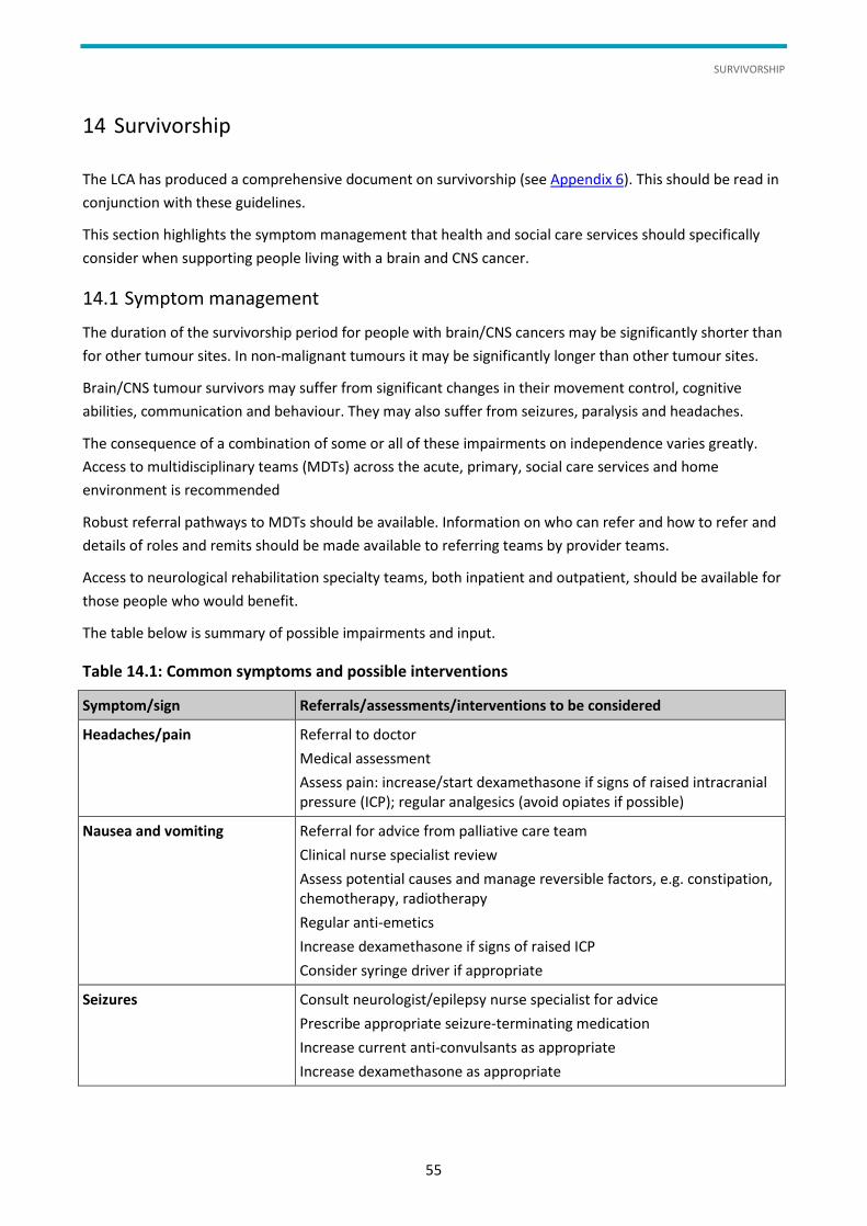
SURVIVORSHIP
55
14 Survivorship
The LCA has produced a comprehensive document on survivorship (see Appendix 6). This should be read in
conjunction with these guidelines.
This section highlights the symptom management that health and social care services should specifically
consider when supporting people living with a brain and CNS cancer.
14.1 Symptom management
The duration of the survivorship period for people with brain/CNS cancers may be significantly shorter than
for other tumour sites. In non-malignant tumours it may be significantly longer than other tumour sites.
Brain/CNS tumour survivors may suffer from significant changes in their movement control, cognitive
abilities, communication and behaviour. They may also suffer from seizures, paralysis and headaches.
The consequence of a combination of some or all of these impairments on independence varies greatly.
Access to multidisciplinary teams (MDTs) across the acute, primary, social care services and home
environment is recommended
Robust referral pathways to MDTs should be available. Information on who can refer and how to refer and
details of roles and remits should be made available to referring teams by provider teams.
Access to neurological rehabilitation specialty teams, both inpatient and outpatient, should be available for
those people who would benefit.
The table below is summary of possible impairments and input.
Table 14.1: Common symptoms and possible interventions
Symptom/sign Referrals/assessments/interventions to be considered
Headaches/pain Referral to doctor
Medical assessment
Assess pain: increase/start dexamethasone if signs of raised intracranial pressure (ICP); regular analgesics (avoid opiates if possible)
Nausea and vomiting Referral for advice from palliative care team
Clinical nurse specialist review
Assess potential causes and manage reversible factors, e.g. constipation, chemotherapy, radiotherapy
Regular anti-emetics
Increase dexamethasone if signs of raised ICP
Consider syringe driver if appropriate
Seizures Consult neurologist/epilepsy nurse specialist for advice
Prescribe appropriate seizure-terminating medication
Increase current anti-convulsants as appropriate
Increase dexamethasone as appropriate
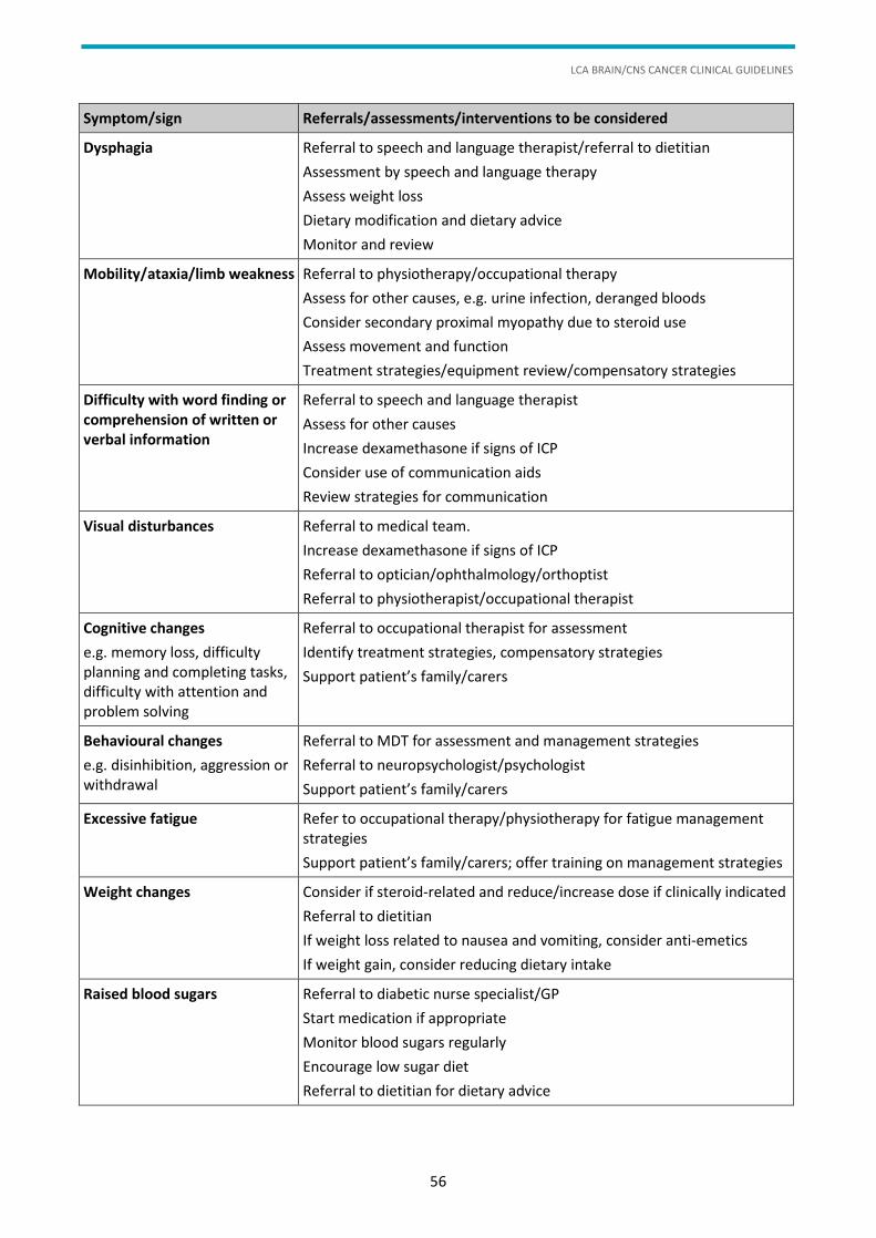
LCA BRAIN/CNS CANCER CLINICAL GUIDELINES
56
Symptom/sign Referrals/assessments/interventions to be considered
Dysphagia Referral to speech and language therapist/referral to dietitian
Assessment by speech and language therapy
Assess weight loss
Dietary modification and dietary advice
Monitor and review
Mobility/ataxia/limb weakness Referral to physiotherapy/occupational therapy
Assess for other causes, e.g. urine infection, deranged bloods
Consider secondary proximal myopathy due to steroid use
Assess movement and function
Treatment strategies/equipment review/compensatory strategies
Difficulty with word finding or comprehension of written or verbal information
Referral to speech and language therapist
Assess for other causes
Increase dexamethasone if signs of ICP
Consider use of communication aids
Review strategies for communication
Visual disturbances Referral to medical team.
Increase dexamethasone if signs of ICP
Referral to optician/ophthalmology/orthoptist
Referral to physiotherapist/occupational therapist
Cognitive changes
e.g. memory loss, difficulty planning and completing tasks, difficulty with attention and problem solving
Referral to occupational therapist for assessment
Identify treatment strategies, compensatory strategies
Support patient’s family/carers
Behavioural changes
e.g. disinhibition, aggression or withdrawal
Referral to MDT for assessment and management strategies
Referral to neuropsychologist/psychologist
Support patient’s family/carers
Excessive fatigue Refer to occupational therapy/physiotherapy for fatigue management strategies
Support patient’s family/carers; offer training on management strategies
Weight changes Consider if steroid-related and reduce/increase dose if clinically indicated
Referral to dietitian
If weight loss related to nausea and vomiting, consider anti-emetics
If weight gain, consider reducing dietary intake
Raised blood sugars Referral to diabetic nurse specialist/GP
Start medication if appropriate
Monitor blood sugars regularly
Encourage low sugar diet
Referral to dietitian for dietary advice
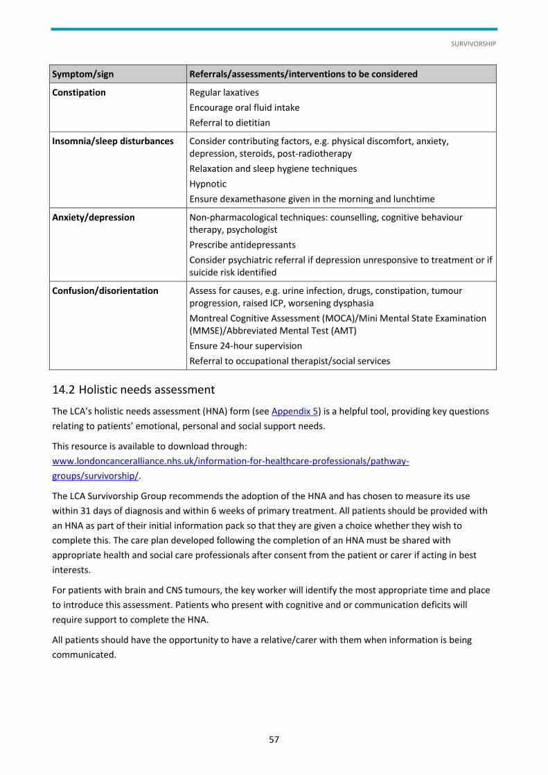
SURVIVORSHIP
57
Symptom/sign Referrals/assessments/interventions to be considered
Constipation Regular laxatives
Encourage oral fluid intake
Referral to dietitian
Insomnia/sleep disturbances Consider contributing factors, e.g. physical discomfort, anxiety, depression, steroids, post-radiotherapy
Relaxation and sleep hygiene techniques
Hypnotic
Ensure dexamethasone given in the morning and lunchtime
Anxiety/depression Non-pharmacological techniques: counselling, cognitive behaviour therapy, psychologist
Prescribe antidepressants
Consider psychiatric referral if depression unresponsive to treatment or if suicide risk identified
Confusion/disorientation Assess for causes, e.g. urine infection, drugs, constipation, tumour progression, raised ICP, worsening dysphasia
Montreal Cognitive Assessment (MOCA)/Mini Mental State Examination (MMSE)/Abbreviated Mental Test (AMT)
Ensure 24-hour supervision
Referral to occupational therapist/social services
14.2 Holistic needs assessment
The LCA’s holistic needs assessment (HNA) form (see Appendix 5) is a helpful tool, providing key questions
relating to patients’ emotional, personal and social support needs.
This resource is available to download through:
www.londoncanceralliance.nhs.uk/information-for-healthcare-professionals/pathway-
groups/survivorship/.
The LCA Survivorship Group recommends the adoption of the HNA and has chosen to measure its use
within 31 days of diagnosis and within 6 weeks of primary treatment. All patients should be provided with
an HNA as part of their initial information pack so that they are given a choice whether they wish to
complete this. The care plan developed following the completion of an HNA must be shared with
appropriate health and social care professionals after consent from the patient or carer if acting in best
interests.
For patients with brain and CNS tumours, the key worker will identify the most appropriate time and place
to introduce this assessment. Patients who present with cognitive and or communication deficits will
require support to complete the HNA.
All patients should have the opportunity to have a relative/carer with them when information is being
communicated.
LCA BRAIN/CNS CANCER CLINICAL GUIDELINES
58
14.3 Additional support for brain tumour patients and carers
Patients and carers may substantially benefit from early contact (as soon after diagnosis as possible) with
brain/spinal tumour-specific charities and not-for-profit organisations which offer face-to-face, telephone
and/or online support opportunities as well as a wide range of comprehensive, practical information
regarding brain and spinal tumours. Talking through the challenges of brain and spinal tumours with other
patients and carers who are on the same journey can provide a unique level of emotional support and
hope.
It is recommended that all brain/CNS centres set up local patient and user support groups. In addition,
patients may wish to access pan-London organisations or national charities. Some examples can be found in
the box below.
National organisations relating to brain tumours
www.patient.co.uk/support/British-Brain-Tumour-Association.htm
www.thebraintumourcharity.org/
www.braintumor.org/
http://meningiomauk.org/groups.html

PALLIATIVE CARE
59
15 Palliative Care
15.1 Definition
‘Palliation’ and ‘palliative treatment’ refer to interventions designed to relieve specific symptoms, not
merely to treatments that are not expected to be curative.
The World Health Organization (WHO) has defined palliative care as:
“an approach that improves the quality of life of patients and their families facing the
problems associated with life-threatening illness, through the prevention and relief of
suffering by means of early identification and impeccable assessment and treatment of
pain and other problems, physical, psychosocial and spiritual. [Palliative care] is
applicable early in the course of illness, in conjunction with other therapies that are
intended to prolong life, such as chemotherapy or radiation therapy, and includes those
investigations needed to better understand and manage distressing clinical
complications.”1
This philosophy is endorsed in the NHS Cancer Plan,2 the NICE Improving supportive and palliative care for
adults with cancer document3 and the Improving Outcomes for People with Brain and Other Central
Nervous System Tumours document.4
15.2 Role and remit
Palliative care specialists have a particular role in the management of ‘difficult-to-control’ symptoms, the
planning and provision of end of life care and supporting teams and patients in advanced care planning.
The remit of specialist palliative care services is discussed in detail in Improving supportive and palliative
care for adults with cancer, while the Department of Health’s guidance on end of life care is set out in detail
in the End of Life Care Strategy document.5 Other initiatives to improve end of life care planning include:
The Gold Standards Framework6
Coordinate My Care, a national initiative7
Advanced Care Planning8
Palliative care of the patient should be provided by general teams and specialist palliative care providers in
accordance with the NICE guidance.
Patients who would benefit from specialist palliative care services should be identified and seen without
delay (see Appendix 8 for LCA specialist palliative care referral form). Neuroscience and neuro-oncology
centres should have access to palliative care specialty staff for advice about symptom management, end of
life care and advanced care planning. A robust referral system to access palliative care is required. The
referral process should identify who can refer to the service and how. The palliative care service should
have clear descriptions of its role in symptom management, end of life care and advanced care planning.
15.3 Assessment
The management of difficult-to-control symptoms involves adequate assessment, appropriate treatment
and adequate reassessment (i.e. review of the efficacy and tolerability of the treatment). The objective of
assessment is to determine the aetiology of the symptom. Thus, many of the symptoms associated with
LCA BRAIN/CNS CANCER CLINICAL GUIDELINES
60
brain/CNS cancers are non-specific (e.g. headache, nausea and vomiting), and patients may also experience
these symptoms as a consequence of the anti-cancer treatment, the supportive care treatment, or a co-
existing medical condition.
15.4 Consideration of palliative care needs
There are key points in a patient’s illness when their palliative care needs may need to be specifically
considered and this may stimulate referral to multiprofessional specialist palliative care services. These key
points include:
pre-diagnosis if advanced disease is suspected
diagnosis
at commencement of definitive treatment of the disease
on completion of the primary treatment plan
on disease recurrence or relapse
at the point of recognition of incurability
end of life care
other times requested by a patient.
1 See www.who.int/cancer/palliative/definition/en/.
2 Department of Health (2000), The NHS cancer Plan.
3 NICE (2004) Improving supportive and palliative care for adults with cancer.
4 NICE (2006), Improving Outcomes for People with Brain and Other Central Nervous System Tumours, Guidance on Cancer Services.
5 Department of Health (2012), End of Life Care Strategy.
6 Gold Standards Framework, see www.goldstandardsframework.org.uk/.
7 Coordinate My Care, see www.coordinatemycare.co.uk/.
8 Royal College of Physicians, National End of Life Care Programme, 2012.
PATIENT EXPERIENCE/SATISFACTION
61
16 Patient Experience/Satisfaction
"Patient experience is only as good as the weakest point in the patient pathway."1
Cancer services should be patient-centred and should respond to patient and carer feedback. Excellent
communication between professionals and patients is particularly important to improve patient
satisfaction.
Positive patient experience is central to meeting the holistic care needs of neuro-oncology patients and
their families/carers. All neuro-oncology services should conduct an annual patient experience exercise.
Services could consider collecting feedback on the patient experience using patient focus and support
groups, one-to-one interviews and patient stories.
Key themes to explore include the patient pathway from first contact with the service through to onward
referral as appropriate. These should include:
pre-diagnosis
diagnosis
surgical
oncology
key worker
rehabilitation
community support
supportive and palliative care.
All health and social care staff working with patients, families and carers need training in communication
skills. The NICE guidelines2 identify four levels of communication training. There is evidence that all staff
involved benefit from level 1 training and this can have a positive impact on the patient’s experience, for
example by using the Sage and Thyme model for assisting people in distress.3
It is recommended that all key workers have level 2 advanced communication skills training.4
All services should ensure that they provide the following:
a local mechanism for collecting information from their patients regarding every aspect of the
patient pathway
a mechanism for reviewing and reflecting any information about the patient experience from local and
or national data
evidence of responding to patients’ feedback.
1 NHS (2010), A Model of Care for Cancer Services, Clinical paper.
2 NICE (2004), Supportive and palliative care for adults with cancer.

LCA BRAIN/CNS CANCER CLINICAL GUIDELINES
62
3 Connolly M, Perryman J et al (2010), Sage and Thyme: a model of training health and social professionals in patient focused support, Patient Education and Counselling 79 (910):87–93.
4 Moore PM, Wilkinson SSM, Rivera Mercado S (2004), Communication skills training for health care professionals working with cancer, Cochrane Database of Systematic Reviews (2):CD003751.

MANAGEMENT OF CHILDREN, TEENAGERS AND YOUNG ADULTS WITH DIAGNOSED OR SUSPECTED BRAIN AND CENTRAL NERVOUS SYSTEM CANCER
63
17 Management of Children, Teenagers and Young Adults with Diagnosed or Suspected Brain and Central Nervous System Cancer
Children below the age of 16 years with a diagnosis of cancer or suspected cancer must be referred to the
paediatric oncology team at the principal treatment centre (PCT) and must not be managed exclusively by
adult site-specific teams.
The joint PTC for children aged 1–16 years for South Thames is The Royal Marsden (Sutton)/St George’s
Hospital.
The PTC for North Thames (including North West London) is Great Ormond Street Hospital/University
College London Hospitals.
All patients <1 year should be referred to Great Ormond Street Hospital.
South Thames Principal Treatment Centre contacts
The Royal Marsden NHS Foundation Trust Lead Clinician – Dr Julia Chisholm
020 8661 3549
Paediatric oncology oncall registrar (new referrals)
020 8915 6248 (24h line)
North Thames Principal Treatment Centre contacts
Great Ormond Street Hospital
(patients aged <13 years)
Lead Clinician – Darren Hargrave
University College London Hospitals
(patients aged >13 years)
Lead Clinician – Dr Sara Stoneham
020 3447 9950
Teenagers aged 16–18 should be managed at a PTC for teenager and young adult (TYA) cancers. Young
adults aged 19–24 should be given the choice of being managed at a PTC or TYA designated hospital.
The PTC for TYA for South Thames is The Royal Marsden (Sutton).
The PTC for North Thames (including North West London) is University College London Hospitals.
All patients within this age range, regardless of place of care, should be referred to the TYA MDT at the
relevant PTC. Referral to the MDT should be made using the TYA referral form (see Appendix 7) which can
be found on the LCA website:
www.londoncanceralliance.nhs.uk/media/68982/TYA%20MDT%20proforma%20March%202014.doc.
Discussion at the TYA MDT is in addition to the SSMDT; key functions of the TYA MDT are to agree the
treatment plan of the SSMDT, ensure cancer registration and provide a psychosocial care plan. Members of
the SSMDT or TYA service at the PTC or TYA designated hospitals are invited to attend the TYA either
remotely or in person.
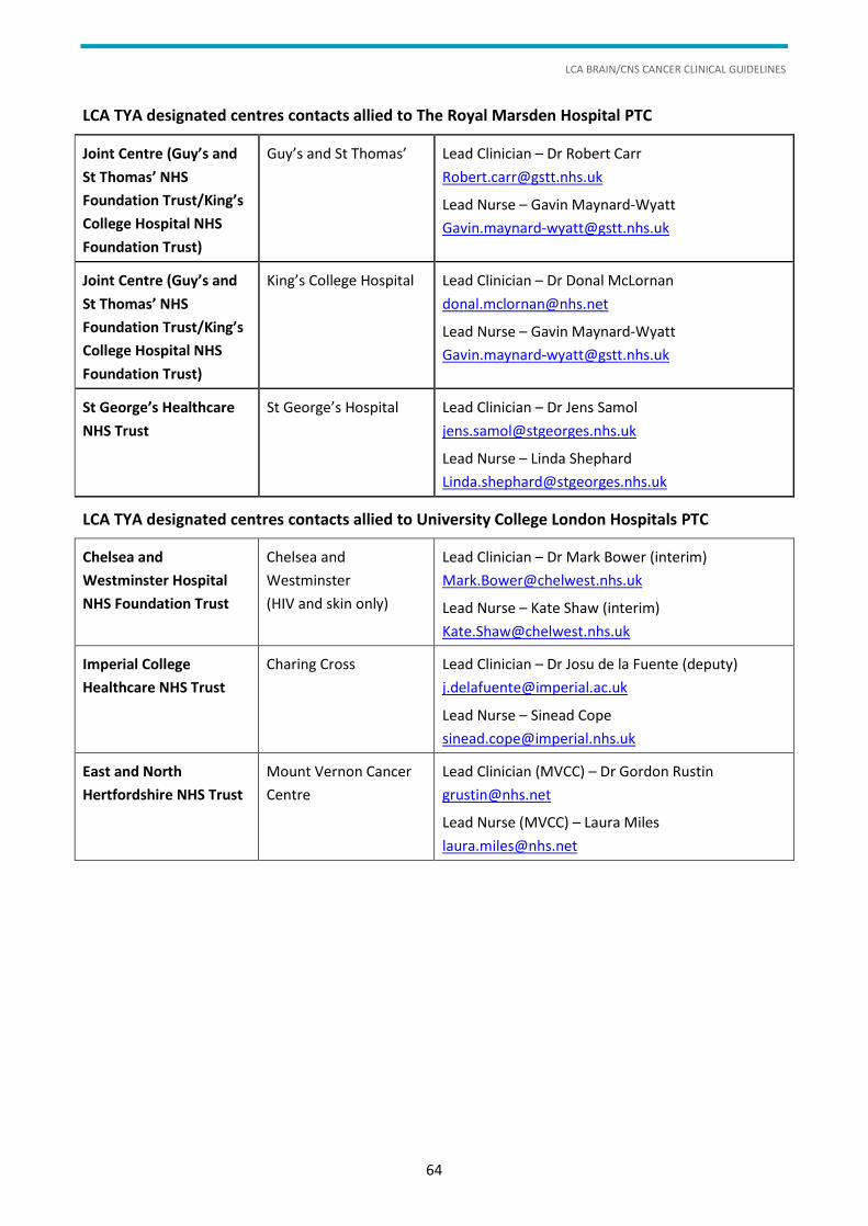
LCA BRAIN/CNS CANCER CLINICAL GUIDELINES
64
LCA TYA designated centres contacts allied to The Royal Marsden Hospital PTC
Joint Centre (Guy’s and
St Thomas’ NHS
Foundation Trust/King’s
College Hospital NHS
Foundation Trust)
Guy’s and St Thomas’ Lead Clinician – Dr Robert Carr
Lead Nurse – Gavin Maynard-Wyatt
Joint Centre (Guy’s and
St Thomas’ NHS
Foundation Trust/King’s
College Hospital NHS
Foundation Trust)
King’s College Hospital Lead Clinician – Dr Donal McLornan
Lead Nurse – Gavin Maynard-Wyatt
St George’s Healthcare
NHS Trust
St George’s Hospital Lead Clinician – Dr Jens Samol
Lead Nurse – Linda Shephard
LCA TYA designated centres contacts allied to University College London Hospitals PTC
Chelsea and
Westminster Hospital
NHS Foundation Trust
Chelsea and
Westminster
(HIV and skin only)
Lead Clinician – Dr Mark Bower (interim)
Lead Nurse – Kate Shaw (interim)
Imperial College
Healthcare NHS Trust
Charing Cross Lead Clinician – Dr Josu de la Fuente (deputy)
Lead Nurse – Sinead Cope
East and North
Hertfordshire NHS Trust
Mount Vernon Cancer
Centre
Lead Clinician (MVCC) – Dr Gordon Rustin
Lead Nurse (MVCC) – Laura Miles
RESEARCH AND INNOVATION
65
18 Research and Innovation
Patients with brain tumours continue to have a poor outcome, and there is recognition that research into
brain tumours is small, relative to their impact on patients.1
Given this, it seems logical to suggest that active research programmes remain key to improving patient
outcomes. In addition, there is some evidence that patients enrolled in clinical trials may have better
outcomes, regardless of the treatment they receive. In addition, the LCA provides an opportunity for units
to collaborate and coordinate research interests to improve recruitment and reduce duplication.
In order to achieve this, the LCA Brain/CNS Pathway Group will ensure that each centre provides and
maintains up-to-date lists of trials open to recruitment, and provides a list of patient groups for whom they
were unable to offer a clinical trial. In addition, the pathway group will ask the LCA informatics team to help
assemble data measuring trial recruitment across the LCA. The trial register will be held as a separate
document to allow for easy updating.
However, research in brain tumours cannot be limited to ensuring an optimal trial portfolio alone. In
addition to trial recruitment, the pathway group expects each neuro-oncology centre to provide plans, with
evidence of timelines, funding and outputs, for three other areas of research:
basic science/translational
quality of life and functional outcomes
non-glioma research.
The pathway group also expects each unit to submit a list of its academic publications on an annual basis,
so that it can present an LCA-wide neuro-oncology research output portfolio on an ongoing basis.
1 Burnet NG et al. (2005), Years of life lost (YLL) from cancer is an important measure of population burden – and should be considered when allocating research funds, British Journal of Cancer 92:241–245.
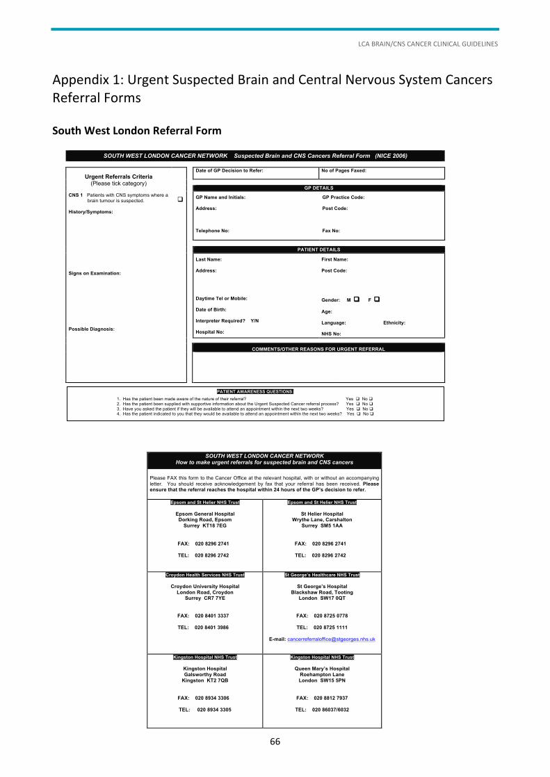
LCA BRAIN/CNS CANCER CLINICAL GUIDELINES
66
Appendix 1: Urgent Suspected Brain and Central Nervous System Cancers Referral Forms
South West London Referral Form
SOUTH WEST LONDON CANCER NETWORK Suspected Brain and CNS Cancers Referral Form (NICE 2006)
Urgent Referrals Criteria (Please tick category)
CNS 1 Patients with CNS symptoms where a brain tumour is suspected. History/Symptoms:
Signs on Examination:
Possible Diagnosis:
q
Date of GP Decision to Refer:
No of Pages Faxed:
GP DETAILS
GP Name and Initials: Address:
Telephone No:
GP Practice Code:
Post Code:
Fax No:
PATIENT DETAILS
Last Name:
Address:
Daytime Tel or Mobile:
Date of Birth:
Interpreter Required? Y/N
Hospital No:
First Name:
Post Code:
Gender: M q F q
Age:
Language: Ethnicity: NHS No:
COMMENTS/OTHER REASONS FOR URGENT REFERRAL
PATIENT AWARENESS QUESTIONS:
1. Has the patient been made aware of the nature of their referral? Yes q No q 2. Has the patient been supplied with supportive information about the Urgent Suspected Cancer referral process? Yes q No q 3. Have you asked the patient if they will be available to attend an appointment within the next two weeks? Yes q No q 4. Has the patient indicated to you that they would be available to attend an appointment within the next two weeks? Yes q No q
SOUTH WEST LONDON CANCER NETWORK How to make urgent referrals for suspected brain and CNS cancers
Please FAX this form to the Cancer Office at the relevant hospital, with or without an accompanying letter. You should receive acknowledgement by fax that your referral has been received. Please ensure that the referral reaches the hospital within 24 hours of the GP’s decision to refer.
Epsom and St Helier NHS Trust
Epsom General Hospital Dorking Road, Epsom
Surrey KT18 7EG
FAX: 020 8296 2741
TEL: 020 8296 2742
Epsom and St Helier NHS Trust
St Helier Hospital Wrythe Lane, Carshalton
Surrey SM5 1AA
FAX: 020 8296 2741
TEL: 020 8296 2742
Croydon Health Services NHS Trust
Croydon University Hospital
London Road, Croydon Surrey CR7 7YE
FAX: 020 8401 3337
TEL: 020 8401 3986
St George’s Healthcare NHS Trust
St George’s Hospital
Blackshaw Road, Tooting London SW17 0QT
FAX: 020 8725 0778
TEL: 020 8725 1111
E-mail: [email protected]
Kingston Hospital NHS Trust
Kingston Hospital
Galsworthy Road
Kingston KT2 7QB
FAX: 020 8934 3306
TEL: 020 8934 3305
Kingston Hospital NHS Trust
Queen Mary’s Hospital
Roehampton Lane
London SW15 5PN
FAX: 020 8812 7937
TEL: 020 86037/6032
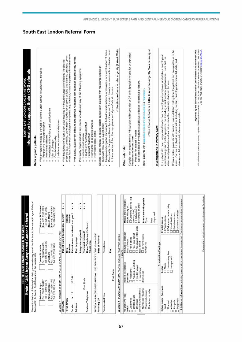
APPENDIX 1: URGENT SUSPECTED BRAIN AND CENTRAL NERVOUS SYSTEM CANCERS REFERRAL FORMS
67
South East London Referral Form
SO
UT
H E
AS
T L
ON
DO
N C
AN
CE
R N
ET
WO
RK
Bra
in /
CN
S U
rge
nt
Su
sp
ec
ted
Ca
nc
er
Re
ferr
al
Plea
se ti
ck th
e bo
x of
the
hosp
ital c
linic
you
are
refe
rring
to a
nd fa
x th
is fo
rm to
the
rele
vant
Urg
ent R
efer
ral
Team
with
in 2
4 ho
urs.
Gui
delin
es a
re o
n th
e re
vers
e si
de.
Pri
nc
es
s R
oy
al
Fax:
016
89 8
6318
7 Te
l: 01
689
8656
76
Qu
ee
n E
liza
be
th
Fax:
020
883
6 40
35
Tel:
020
8836
596
4/5
Gu
y’s
& S
t T
ho
ma
s’
Fax:
020
718
8 09
23
Tel:
020
7188
090
2
Kin
g’s
Co
lle
ge
Fax:
020
329
9 15
15
Tel:
020
3299
151
6 L
ew
ish
am
Fax:
020
833
3 34
51
Tel:
020
8333
345
0 Q
ue
en
Ma
ry’s
Fax:
020
830
8 92
64
Tel:
020
8308
323
0 S
EC
TIO
N 1
– P
AT
IEN
T I
NF
OR
MA
TIO
N.
PL
EA
SE
CO
MP
LE
TE
IN
BL
OC
K C
AP
ITA
LS
.
SU
RN
AM
E
Pa
tie
nt
vis
ite
d t
his
ho
sp
ita
l b
efo
re?
Y
/
N
FIR
ST
NA
ME
N
HS
Nu
mb
er
Ho
sp
ita
l
Nu
mb
er
Ge
nd
er
M
/ F
D
.O.B
. P
ati
en
t a
wa
re t
he
re
ferr
al
is u
rge
nt?
Y
/
N
Ad
dre
ss
P
os
t C
od
e
Fir
st
lan
gu
ag
e
Inte
rpre
ter
req
uir
ed
?
Y
/ N
Tra
ns
po
rt r
eq
uir
ed
?
Y
/ N
Da
yti
me
Te
lep
ho
ne
H
om
e T
ele
ph
on
e (i
f diff
eren
t) /
Mo
bil
e N
o.
SE
CT
ION
2 –
PR
AC
TIC
E I
NF
OR
MA
TIO
N.
US
E P
RA
CT
ICE
ST
AM
P I
F A
VA
ILA
BL
E.
Re
ferr
ing
GP
Da
te o
f R
efe
rra
l
Pra
cti
ce
Ad
dre
ss
Po
st
Co
de
Te
lep
ho
ne
Fa
x
SE
CT
ION
3 –
CL
INIC
AL
IN
FO
RM
AT
ION
. P
LE
AS
E T
ICK
TH
E R
EL
EV
AN
T B
OX
ES
. H
isto
ry
Pro
gre
ss
ive
fo
ca
l
de
fic
it
Ra
ise
d i
ntr
ac
ran
ial
pre
ss
ure
Se
izu
res
/ b
lac
ko
ut
Dat
e of
ons
et:
Me
nta
l s
tate
ch
an
ge
s –
sh
ort
his
tory
of…
Wea
knes
s U
nste
adin
ess
Uni
late
ral
sens
orin
eura
l dea
fnes
s N
umbn
ess
/ tin
glin
g
Vis
ual d
istu
rban
ce
Cra
nial
ner
ve p
alsy
Hea
dach
e
Nau
sea
/ Vom
iting
D
oubl
e vi
sion
In
term
itten
t dr
owsi
ness
Foc
al o
nset
P
ost-i
ctal
def
icit
Ass
ocia
ted
(inte
r-ict
al)
foca
l def
icit
d
e n
ovo
sta
tus
epile
ptic
us
Cog
nitiv
e de
clin
e (e
.g.
mem
ory
loss
) B
ehav
iour
or
pers
onal
ity c
hang
e P
rio
r c
an
ce
r d
iag
no
sis
Type
: D
ate
diag
nose
d:
Ex
am
ina
tio
n F
ind
ing
s
Hig
he
r m
en
tal
fun
cti
on
s
Lim
bs
C
ran
ial
ne
rve
s
Ale
rt O
rient
ed
Atte
ntiv
e F
orge
tful
D
ysph
asic
Ata
xia
Hem
isen
sory
loss
H
emip
ares
is
Pap
illoed
ema
Ext
raoc
ular
mus
cle
pals
y V
isua
l fie
ld lo
ss
Fac
ial w
eakn
ess
U
nila
tera
l dea
fnes
s A
dd
itio
na
l in
form
ati
on
- in
clud
ing
reas
ons
why
a b
rain
tum
our i
s su
spec
ted.
Con
tinue
on
sepa
rate
she
et if
requ
ired.
Plea
se a
ttach
pat
ient
com
pute
r rec
ord
sum
mar
y if
avai
labl
e.
SO
UT
H E
AS
T L
ON
DO
N C
AN
CE
R N
ET
WO
RK
Info
rma
tio
n t
o s
up
po
rt B
rain
/ C
NS
re
ferr
als
R
efe
r u
rge
ntl
y p
ati
en
ts:
·
With
sym
ptom
s re
late
d to
the
CN
S in
who
m a
bra
in tu
mou
r is
susp
ecte
d, in
clud
ing
o
Prog
ress
ive
neur
olog
ical
def
icit
o
New
-ons
et s
eizu
res
o
Hea
dach
es w
ith v
omiti
ng a
nd p
apillo
edem
a o
M
enta
l cha
nges
o
C
rani
al n
erve
pal
sy
o
Uni
late
ral s
enso
rineu
ral d
eafn
ess.
·
With
hea
dach
es o
f rec
ent o
nset
acc
ompa
nied
by
feat
ures
sug
gest
ive
of ra
ised
intra
cran
ial
pres
sure
(e.g
. vom
iting
, dro
wsi
ness
, hea
dach
es p
rese
nt o
n ly
ing
and
clea
ring
on s
ittin
g up
) or
othe
r foc
al /
non-
foca
l neu
rolo
gica
l sym
ptom
s (e
.g. b
lack
out,
chan
ge in
per
sona
lity
or m
emor
y).
·
With
a n
ew, q
ualit
ativ
ely
diffe
rent
, une
xpla
ined
hea
dach
e th
at b
ecom
es p
rogr
essi
vely
sev
ere.
·
Prev
ious
ly d
iagn
osed
with
any
can
cer w
ho d
evel
op a
ny o
f the
follo
win
g sy
mpt
oms:
o
Rec
ent-o
nset
sei
zure
o
Pr
ogre
ssiv
e ne
urol
ogic
al d
efic
it o
Pe
rsis
tent
hea
dach
es
o
New
men
tal o
r cog
nitiv
e ch
ange
s o
N
ew n
euro
logi
cal s
igns
. C
onsi
der u
rgen
t ref
erra
l to
an a
ppro
pria
te s
peci
alis
t in
patie
nts
with
rapi
d pr
ogre
ssio
n of
: ·
Suba
cute
foca
l neu
rolo
gica
l def
icit
·
Une
xpla
ined
cog
nitiv
e im
pairm
ent,
beha
viou
ral d
istu
rban
ce o
r slo
wne
ss, o
r a c
ombi
natio
n of
thes
e ·
Pers
onal
ity c
hang
es c
onfir
med
by
a w
itnes
s an
d fo
r whi
ch th
ere
is n
o re
ason
able
exp
lana
tion
even
in
the
abse
nce
of th
e ot
her s
ympt
oms
and
sign
s of
a b
rain
tum
our.
!
Us
e t
his
pro
form
a t
o r
efe
r u
rge
ntl
y (
2 W
ee
k W
ait
)
Oth
er
refe
rrals
: C
onsi
der n
on-u
rgen
t ref
erra
l / d
iscu
ssio
n w
ith s
peci
alis
t or G
P w
ith S
peci
al In
tere
sts
for u
nexp
lain
ed
head
ache
s of
rece
nt o
nset
·
Pres
ent f
or a
t lea
st 1
mon
th
·
Not
acc
ompa
nied
by
feat
ures
sug
gest
ive
of ra
ised
intra
cran
ial p
ress
ure.
R
efer
pat
ient
s w
ith s
uspe
cted
rece
nt-o
nset
sei
zure
s to
a n
euro
logi
st.
!
Us
e C
ho
os
e &
Bo
ok
or
a l
ett
er
to r
efe
r n
on
-urg
en
tly
/ t
o a
ne
uro
log
ist
Inve
sti
gati
on
s in
Pri
ma
ry C
are
: In
a p
atie
nt w
ith n
ew, u
nexp
lain
ed h
eada
ches
or n
euro
logi
cal s
ympt
oms,
und
erta
ke a
neu
rolo
gica
l ex
amin
atio
n gu
ided
by
the
sym
ptom
s, b
ut in
clud
ing
exam
inat
ion
for p
apill
oede
ma.
Not
e th
at th
e ab
senc
e of
pap
illoe
dem
a do
es n
ot e
xclu
de th
e po
ssib
ility
of a
bra
in tu
mou
r. W
hen
a pa
tient
pre
sent
s w
ith s
eizu
re, t
ake
a de
taile
d hi
stor
y fro
m th
e pa
tient
and
an
eyew
itnes
s to
the
even
t. C
arry
out
a p
hysi
cal e
xam
inat
ion,
incl
udin
g ca
rdia
c, n
euro
logi
cal a
nd m
enta
l sta
te, a
nd
deve
lopm
enta
l ass
essm
ent,
whe
re a
ppro
pria
te.
A
pp
rov
ed
by
th
e S
ou
th E
as
t L
on
do
n C
an
ce
r N
etw
ork
in
No
ve
mb
er
20
06
.
Fo
r co
mm
en
ts,
ad
ditio
na
l co
pie
s,
or
pa
tie
nt
info
rma
tio
n r
eso
urc
es f
or
GP
s t
o u
se
co
nta
ct
the
Ne
two
rk o
n T
el 0
20
71
88
70
90
/
Fa
x 0
20
71
88
71
20
, o
r vis
it o
ur
we
bsite
: w
ww
.se
lcn
.nh
s.u
k.
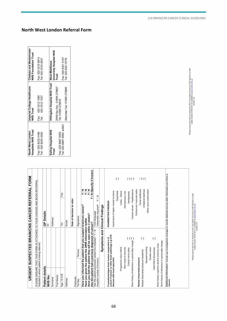
LCA BRAIN/CNS CANCER CLINICAL GUIDELINES
68
North West London Referral Form
Plea
se e
nsur
e th
is fo
rm is
rece
ived
in th
e Tr
ust w
ithin
24
hour
s of
GP
deci
sion
to re
fer
Late
st v
ersi
on o
f the
form
is a
vaila
ble
at w
ww
.nw
lcn.
nhs.
uk
vers
ion
3.9
UR
GE
NT
SU
SP
EC
TE
D B
RA
IN/C
NS
CA
NC
ER
RE
FE
RR
AL
FO
RM
PL
EASE
EN
SUR
E TH
AT T
HIS
FO
RM
IS A
TTAC
HED
TO
YO
UR
CH
OO
SE A
ND
BO
OK
REF
ERR
AL
Hos
pita
l to
whi
ch p
atie
nt is
bei
ng re
ferre
d:
Pa
tie
nt
de
tail
s
GP
De
tail
s
NH
S N
o:
D
r:
Surn
ame:
Addr
ess:
Firs
t Nam
e:
Ag
e / D
.O.B
:
Tel:
Fax:
Addr
ess:
Po
stco
de:
Emai
l:
Da
te o
f d
ec
isio
n t
o r
efe
r:
Tel d
ay:
Tel e
ve:
Sign
atur
e:
Have y
ou
in
form
ed
th
e p
ati
en
t th
at
yo
u s
usp
ect
bra
in/c
ns
can
cer?
Y
/ N
H
ave y
ou
giv
en
th
e p
ati
en
t th
e 2
WW
in
form
ati
on
le
afl
et
Y
/ N
H
ave y
ou
to
ld t
he p
ati
en
t th
ey w
ill b
e s
een
wit
hin
2 w
eek
s?
Y
/ N
H
as t
he p
ati
en
t h
ad
a p
revio
us d
iag
no
sis
of
can
cer?
Y
/ N
(S
pecif
y if
kn
ow
n)
Has
the
patie
nt p
revi
ousl
y vi
site
d th
is h
ospi
tal?
Y /
N
Hos
pita
l num
ber (
if k
no
wn
): Fi
rst l
angu
age:
In
terp
rete
r req
uire
d?
Y /
N
Sym
pto
ms
an
d C
lin
ica
l F
ind
ing
s
If s
ym
pto
ms
/sig
ns
bra
in t
um
ou
r s
us
pe
cte
d o
r if
u
ns
ure
if
tum
ou
r o
r o
the
r b
rain
pa
tho
log
y e
.g C
VA
dis
cu
ss
wit
h l
oc
al
sp
ec
iali
st
Prog
ress
ive
neur
o de
ficit
N
ew o
nset
of s
eizu
res
Cra
nial
ner
ve p
alsy
Shor
t his
tory
of b
ehav
iour
or p
erso
nalit
y ch
ange
Ra
ise
d i
ntr
ac
ran
ial
pre
ss
ure
Rai
sed
intra
cran
ial p
ress
ure
head
ache
Nau
sea/
vom
iting
Dou
ble
visi
on
Int
erm
itten
t dro
wsi
ness
Shor
t his
tory
cog
nitiv
e de
clin
e (m
emor
y lo
ss)
Shor
t his
tory
of b
ehav
iour
or p
erso
nalit
y ch
ange
EX
AM
INA
TIO
N F
IND
ING
S
Impa
irmen
t of h
ighe
r men
tal f
unct
ions
Dys
phas
ia
L
imbs
– A
taxi
a
H
emis
enso
ry lo
ss
H
emip
ares
is
C
rani
al n
erve
s –
Papi
lloed
ema
Ex
traoc
ular
mus
cula
r pal
sy
F
acia
l wea
knes
s
Uni
late
ral d
eafn
ess
Oth
er n
euro
exa
min
atio
n
Ad
dit
ion
al
Info
rma
tio
n:
Inc
lud
e a
ny
in
ve
sti
ga
tio
ns
arr
an
ge
d o
r re
su
lts
ob
tain
ed
an
d a
ny
oth
er
info
rma
tio
n y
ou
th
ink
is
rele
va
nt.
Plea
se e
nsur
e th
is fo
rm is
rece
ived
in th
e Tr
ust w
ithin
24
hour
s of
GP
deci
sion
to re
fer
Late
st v
ersi
on o
f the
form
is a
vaila
ble
at w
ww
.nw
lcn.
nhs.
uk
vers
ion
3.9
N
ort
h W
es
t L
on
do
n
Ho
sp
ita
ls N
HS
Tru
st
Fax:
020
823
5 41
88
Tel:
020
823
5 42
93
Imp
eri
al
Co
lle
ge H
ealt
hcare
N
HS
Tru
st
Fax:
0
20 3
312
1580
Te
l:
020
331
2 15
27
Ch
els
ea
an
d W
estm
inste
r N
HS
Fo
un
dati
on
Tru
st
Fax:
020
331
5 88
14
Tel:
020
3315
263
7
Ea
lin
g H
osp
ital
NH
S
Tru
st
Fax:
020
896
7 50
05
Tel:
020
8967
500
0, x
3921
Hilli
ng
do
n H
os
pit
al N
HS
Tru
st
2WW
fax
line
: 018
95 2
7980
7 Te
l: 01
895
2796
79
Alte
rnat
e Fa
x: 0
1895
279
890
West
Mid
dle
sex
Un
ivers
ity
Ho
sp
ita
l N
HS
T
rust
Fax:
020
832
1 51
57
Tel:
020
8321
677
6
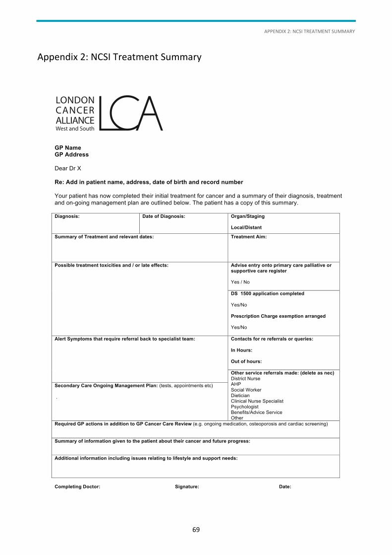
APPENDIX 2: NCSI TREATMENT SUMMARY
69
Appendix 2: NCSI Treatment Summary
GP Name GP Address
Dear Dr X Re: Add in patient name, address, date of birth and record number
Your patient has now completed their initial treatment for cancer and a summary of their diagnosis, treatment and on-going management plan are outlined below. The patient has a copy of this summary. Diagnosis:
Date of Diagnosis:
Organ/Staging Local/Distant
Summary of Treatment and relevant dates:
Treatment Aim:
Possible treatment toxicities and / or late effects: Advise entry onto primary care palliative or supportive care register Yes / No
DS 1500 application completed
Yes/No Prescription Charge exemption arranged Yes/No
Alert Symptoms that require referral back to specialist team:
Contacts for re referrals or queries:
In Hours: Out of hours:
Other service referrals made: (delete as nec) District Nurse AHP Social Worker Dietician Clinical Nurse Specialist Psychologist Benefits/Advice Service Other
Secondary Care Ongoing Management Plan: (tests, appointments etc) .
Required GP actions in addition to GP Cancer Care Review (e.g. ongoing medication, osteoporosis and cardiac screening) Summary of information given to the patient about their cancer and future progress:
Additional information including issues relating to lifestyle and support needs:
Completing Doctor: Signature: Date:
LCA BRAIN/CNS CANCER CLINICAL GUIDELINES
70
APPENDIX 2: NCSI TREATMENT SUMMARY
71
Actions required by the GP Actions required by the GP Tumour marker monitoring Xalqg Tumour marker monitoring 8A9.. PSA Xalqh PSA 43Z2. Osteoporosis monitoring XalSd Osteoporosis monitoring 66a.. Referral for specialist opinion Xalst
Advised to apply for free prescriptions
9D05 Entitled to free prescription 6616.
Cancer Care Review Xalyc Cancer Care Review 8BAV. Palliative Care Review XalG1 Palliative Care Plan Review 8CM3. Medication: Medication:
New medication started by specialist
XEOhn Medication given 8BC2.
Medication changed by specialist
8B316 Medication changed 8B316
Advice to GP to start medication XaKbF
Advice to GP to stop medication XaJC2
Information to patient: Information to patient:
DS1500 form claim XaCDx DS1500 completed 9EB5. Benefits counselling 6743. Benefits counselling 6743. Cancer information offered XalmL Cancer information offered 677H. Cancer diagnosis discussed XalpL Cancer diagnosis discussed 8CL0. Aware of diagnosis XaQly Unaware of prognosis XaVzE Carer aware of diagnosis XaVzA Miscellaneous: Miscellaneous: On GSF palliative care framework
XaJv2 On GSF Palliative Care Framework 8CM1.
GP OOH service notified Xaltp GP OOH service notified 9e0.. Carers details 9180. Carer details 9180.
LCA BRAIN/CNS CANCER CLINICAL GUIDELINES
72
Appendix 3: LCA Key Worker Policy
Definition
A key worker is a person who, with the patient’s consent and agreement, takes a key role in coordinating
the patient’s care and promoting continuity, ensuring the patient knows who to access for information and
advice in relation to their cancer diagnosis. In addition, the key worker will facilitate patients making
informed decisions about their treatment.
The implementation of the key worker role is a requirement of the National Cancer Peer Review
Programme and detailed in the Manual for Cancer Services, originally published by the National Cancer
Action Team (NCAT), and related site-specific Improving Outcomes Guidance, issued by the National
Institute for Health and Care Excellence (NICE).
Principles and responsibilities
Designation
1. The key worker is a named clinical member of the site-specific multidisciplinary team (MDT), and acts
as the point of contact between the patient and MDT.
2. The key worker is a healthcare professional.
3. The key worker is assigned by the core clinical nurse specialist of an MDT, agreed by the MDT and
recorded within the patient record and multidisciplinary meeting proforma.
4. The name of the key worker, designation and contact details will also be recorded in the patient
handheld record (PHR), if used, and included in all correspondence and in the patient medical records.
All entries in the medical notes will comply with the NHS Litigation Authority standards.
Access
5. All cancer patients will be made aware of their allocated key worker, but have the right to ask for an
alternative if they prefer. This will usually happen at diagnosis.
6. The key worker will provide a contact number to all the patients for whom they act as the key worker.
Multi-professional communication
7. If a more appropriate person is identified as a key worker at a point in the patient’s pathway, this will
be discussed and agreed by the patient and the new key worker, and recorded in the patient’s notes.
This situation is most likely to arise with referral to the palliative care team. In such cases the palliaitve
care clinical nurse specialist will check if a key worker has already been identified for the patient by the
relevant tumour MDT. The palliative care clinical nurse specialist will then negotiate and document care
responsibilites in the patient’s notes.
8. The key worker may change as patients pass through various stages of the care trajectory or when care
is transferred to a different Trust. It is the responsibility of the key worker to hand over to the next one,
to document this in the patient’s notes and to keep the patient informed.
APPENDIX 3: LCA KEY WORKER POLICY
73
9. The key worker will lead on patient communication issues and coordination of the pathway for patients
referred to the team.
10. The key worker will ensure that the patient pathway is coordinated and that all relevant information is
transferred to the appropriate professionals as the patient moves across care boundaries, e.g. on
admission to and discharge from institutions, when care is transferred between teams.
11. The key worker has responsibility for ensuring holistic needs assessments (HNAs) are
recorded/documented in patient records.
Patient communication and support
12. Where possible, the key worker will be available to support the patient on diagnosis to signpost and
provide them with information and contacts for the MDT, national information and support services,
self-help groups and associated site-specific support.
13. If the key worker is not available at the time of diagnosis, the person who is providing support at the
time will ensure that the patient is aware of the key worker role and provide the relevant contact
details.
14. The key worker will be accessible to the patient as a constant point of contact, handing over to
colleagues when unavailable and making sure that the patient has clear information about alternative
contacts and cover arrangements.
15. The key worker will provide information, care and support throughout the patient journey regardless
of the patient’s condition, liaising between health professionals to ensure continuity of care and a
seamless service.
Data/audit
16. The key worker will contribute to the audit of the key worker role in their organisation.
Annex A
NCAT peer review standard
There should be an operational policy whereby a single named key worker for the patient’s care at a given
time is identified by the MDT members for each individual patient and the name and contact number of the
current key worker is recorded in the patient’s case notes. The responsibility for ensuring that the key
worker is identified should be that of the nurse MDT member(s).
The above policy should have been implemented for patients who came under the MDT’s care after
publication of these measures and who are under their care at the time of the peer review visit.
Notes
According to the NICE supportive and palliative care guidance, a key worker is a person who, with the
patient’s consent and agreement, takes a key role in coordinating the patient’s care and promoting
continuity, e.g. ensuring that the patient knows who to access for information and advice. This is not
intended to have the same connotation as the key worker in social work.
It may be necessary to agree a single key worker across both a cancer site-specific MDT and the
specialist palliative care MDT for certain patients.
LCA BRAIN/CNS CANCER CLINICAL GUIDELINES
74
Appendix 4: Competencies for Key Worker Role
Work as an integral member of the multidisciplinary team (MDT) to ensure continuity of patient care.
Initiate and participate in case conferences with all professionals involved in the delivery of
patient care.
Communicate and coordinate information to patients and carers, evaluating their levels of
understanding and utilising a range of skills/techniques to overcome any communication difficulties.
Demonstrate ability to verbally summarise patient information to facilitate understanding.
Act as an advocate for the patient.
Act as a communication resource and coordinator for other members of the MDT in the care of the key
worker’s patient caseload.
In conjunction with the MDT, provide patients with comprehensive information on the options
available to them for treatment and care. Utilise their specialist knowledge and skills regarding
disclosure of information.
Coordinate the onward referral of patient and/or family members to appropriate clinical or
support services.
Ensure accurate follow-up documentation is maintained, including any changes in the named
key worker.
Utilise support strategies and interventions available to care for patients with complex needs, for
example patients exhibiting denial/anger following a cancer diagnosis, adverse reactions to alteration
in body image or reaching end of life.
Demonstrate knowledge of holistic care relating to areas across the patient journey such as screening,
curative and palliative treatment, spiritual care, aspects of nutrition and pharmacology, rehabilitation,
discharge and collaborative working.
Initiate appropriate referral or access to sources of specialist support for those experiencing, for
example, sexual difficulties as a result of their illness or treatment.
Utilise all forms of patient information to enable the patient to have a better understanding of their
diagnosis and treatment plan. This will include the use of specific resources for patient/carers from
minority groups.
Facilitate the development of teaching and learning skills used to educate patients and
other personnel.
Contribute to the monitoring, audit and evaluation of adherence to policy/procedures/guidelines
and standards of practice, initiating changes where appropriate to improve delivery of care to
patients/carers within the MDT.
Demonstrate ability to recognise abnormal grief reactions and refer on to appropriate agencies and
healthcare professionals.
Demonstrate a comprehensive knowledge of the assessment, care, management support, training
education and information requirements for patients and carers across the care pathway for the
particular specialty area.
APPENDIX 4: COMPETENCIES FOR KEY WORKER ROLE
75
Assess and provide support that is appropriate to the context and sensitive to meet the patient/carer
and/or family’s needs, facilitating access to additional support from other healthcare professionals or
agencies as applicable and with the agreement of the patient and/or carer.
Understand the ethical issues relating to treatment in advanced disease.
Have sufficient knowledge and links with national/local support groups and be able to provide/record
information relating to these groups to guide and advise patients.
Provide information, education and relevant telephone contacts to patients and carers regarding the
procedures and management of the side effects of treatment associated with the client group
encountered in their practice.
Be aware of local contact arrangements in the event of patients experiencing unwanted side effects.
Demonstrate knowledge to prepare, inform and educate patients/carers for survivorship and, where
applicable, primary care personnel regarding any associated care requirements, symptom management
and contact details on discharge.
Participate in inter-professional/inter-agency evaluation and audit to effect change for the continued
improvement of the quality of care and service for patients.
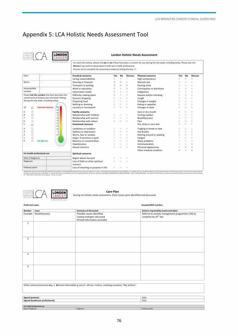
LCA BRAIN/CNS CANCER CLINICAL GUIDELINES
76
Appendix 5: LCA Holistic Needs Assessment Tool
Care Plan During my holistic needs assessment, these issues were identified and discussed:
Preferred name: Hospital/NHS number:
Number Issue Summary of discussion Actions required/by (name and date)
Example Breathlessness Possible causes identified Coping strategies discussed Printed information provided
Referral to anxiety management programme; CNS to complete by 24th Dec
1
2
3
4
Other actions/outcomes e.g. ad ditional information gi ven, he alth pr o motion, smoking cessation, ‘My actions’:
Signed (patient): Date:
Signed (healthcare professional): Date:
For health professional use
Date of diagnosis: Diagnosis: Pathway point:
APPENDIX 6: CANCER SURVIVORSHIP GUIDELINES
77
Appendix 6: Cancer Survivorship Guidelines
As cancer treatments become more effective, more people are living with and beyond cancer with specific
needs as a direct result of the cancer and its treatment. The consequences of cancer treatment are
dependent on multiple factors and affect each person differently. Consequences may be physical
(e.g. cardiovascular conditions, impact on fertility, bone health and gastro-intestinal); emotional and
psychological (e.g. anxiety, self-confidence and depression); social; spiritual; or cognitive. They can have an
impact on every aspect of a person and on their family’s lives, from the ability to work, through to the
ability to have a family or to participate in social activities. It is widely acknowledged that cancer survivors
have a multitude of unmet needs following treatment, with a majority still having some needs 6 months
later. Good survivorship care enables the person to live as full and active a life as possible.
Survivorship can be defined as:
“cover[ing] the physical, psychological and economic issues of cancer, from diagnosis
until end of life. It focuses on the health and life of a person with cancer beyond the
diagnosis and treatment phases. Survivorship includes issues related to the ability to get
healthcare and follow-up treatment, late effects of treatment, second cancer and quality
of life. Family members, friends and caregivers are also part of the survivorship
experiences.”
National Cancer Institute, Dictionary of Cancer Terms, definition of ‘survivorship’ (www.cancer.gov/dictionary?CdrID=445089)
The National Cancer Survivorship Initiative (NCSI) vision document (DH 2010) mandated five shifts in care
for individuals completing cancer treatment. NCSI advocates cancer being treated as a chronic illness, with
patients empowered and supported to take an active role in their care. Improving Outcomes: a Strategy for
Cancer (DH 2011) states that people living with and beyond a cancer diagnosis should have their full needs
addressed to prevent long-term disability, enabling them to live a full, active, good quality life for as long as
possible. Work within the NCSI has to date focused on survivorship from the end of treatment, but its
report, Living With and Beyond Cancer: Taking Action to Improve Outcomes (DH 2013), acknowledges that
survivorship care from the point of diagnosis is also vital. It challenges services to develop further and
focuses on five new areas:
information and support from diagnosis
promoting recovery
sustaining recovery
managing consequences
supporting people with active and advanced disease.
The importance of good survivorship care is well known: those who have unmet needs are 20% more likely
to visit their GP and twice as likely to attend A&E than their healthy counterparts. They are more likely to
be unemployed and many report economic hardship. Much has been achieved both nationally and locally
to address this agenda. It is essential that in the LCA our patients have access to high-quality, equitable
survivorship services on a par with the best in the country. We will continue to build on the successes
to date.
LCA BRAIN/CNS CANCER CLINICAL GUIDELINES
78
The Consequences of Cancer and its Treatment (CCaT) collaborative group (a Macmillan Community of
Interest) produced a ’10 Top Tips’ guidance document for patients. These cover the key components of
good survivorship care, and the LCA expects services to address these areas. The following nine points for
professionals are based on the CCaT’s work.
1 Discuss a person’s needs.
The holistic needs assessment (HNA) has been shown to be effective in identifying a person’s areas of
concern. It can take many forms and the LCA has developed its own tool, based on the concerns checklist
and distress thermometer. The tool allows patients to specify what is of most concern to them, and so
directs subsequent discussion and intervention to addressing these needs. It has scope to cover physical,
emotional, spiritual, finance and welfare, and practical concerns. It is anticipated that as the HNA becomes
embedded within the pathway, patients will start to ask for an HNA and professionals need to be able to
respond to this.
Recommendation: Every patient should be offered an HNA at key pathway points, including at diagnosis
and end of treatment, and whenever a person requests one.
2 Provide a treatment summary and care plan
A treatment summary provides a summary of the cancer treatments received by the end of first treatment,
planned follow-ups (including mechanisms for these) and signs and symptoms of which to be aware. Their
aim is to provide information not only to the patient, but also to the GP about possible consequences of
cancer and its treatment, signs of recurrence and other important information.
A care plan is generated as a result of an HNA and is the agreed plan between the patient and healthcare
professional about how the identified areas of concern will be addressed. This may cover provision of
information (e.g. through an information prescription), onward referral for specialist assessment and
intervention (e.g. breathlessness management), or things which the patient themselves can do (e.g. contact
their HR department about graduated return to work options).
Recommendation: An end of treatment consultation should be offered to every patient. This should
include an end of treatment HNA and associated written care plan, and should also include the discussion
and provision of a comprehensive treatment summary.
3 Provide a main contact
Several pieces of UK-wide work have shown the necessity of a key contact, or key worker, not least the
national Cancer Patient Experience Survey. It is now agreed that both patients and GPs (and other
healthcare professionals) benefit from having a named person to contact if they need help or advice about
issues related to the consequences of cancer and its treatment.
Recommendation: The treatment summary should include the details of a key worker in addition to details
of who to contact out of hours. This should be sent to the GP, the patient and any others whom the patient
identifies as necessary.
APPENDIX 6: CANCER SURVIVORSHIP GUIDELINES
79
4 Identify post-treatment symptoms
As discussed above, cancer and its treatments can have far-reaching consequences and people with
associated unmet needs are more likely to access healthcare services than their healthy counterparts.
Providing information on likely post-treatment symptoms (e.g. neuropathic pain following thoracic surgery
or peripheral neuropathy following chemotherapy), and how these can be managed or avoided, allows
people to seek the right help from the right people at the right time.
Recommendation: Information on anticipated or possible consequences of cancer treatment and what to
do if they occur should be routinely provided to all patients. This should be done from the time of
discussion of treatment onwards, with the information clearly reiterated during the end of treatment
consultation.
5 Provide support about day-to-day concerns
Life changes following a cancer diagnosis. It is recognised that people need help and support to find a ‘new
normal’. This may cover any one of a multitude of aspects, from work and education, through to financial
worries and needing help with caring responsibilities. Help should be offered at all key points in the
pathway, but may be of particular relevance at the end of treatment and may well be highlighted in the
HNA. There are various options for written information provision (e.g. Macmillan Cancer Support
information leaflets and information prescriptions) as well as some specialist services (e.g. Citizens Advice).
Reports published by the NCSI, available on the NCSI website, may be of use to professionals.
Recommendation: Patients should be routinely asked about whether they need support with day-to-day
issues and referrals made to specialist services when necessary.
6 Talk about how you feel
Having a cancer diagnosis has an emotional impact, and at the end of treatment people experience a wide
range of emotions. Sometimes, these can be dealt with by the person alone or with support from the key
worker and others, but some people will need referral to psychological support services. This may be true
not only for patients but their family and carers too.
Recommendation: Use an HNA to identify emotional concerns. Further screening tools (e.g. the Hospital
Anxiety and Depression Scale) should be considered, with subsequent referrals made as necessary.
7 Healthy lifestyle
There is a growing body of evidence which supports the adoption of a healthy lifestyle for those who have
had a cancer diagnosis.
Recommendation: Patients are provided with dietary advice, based on the WCRF recommendations, at the
end of treatment with referral to specialist dietitians as needed.
Physical activity
There has been a dramatic rise in the amount of high-quality published research on the role of exercise in
cancer in recent years. Physical activity results in improvement in quality of life, fitness and function and
LCA BRAIN/CNS CANCER CLINICAL GUIDELINES
80
symptoms related to cancer and its treatments. It reduces cancer recurrence, incidence of second cancers
and reduces both all-cause and cancer-specific mortality.
There is wide consensus that cancer survivors should exercise to the same level as the general population
for health benefits. Research suggests that a combination of cardiovascular and muscular strength training
has additional benefits over and above undertaking only one type of exercise.
Recommendations: Patients should be encouraged to maintain or increase their level of physical activity
both during and after treatment in line with national guidance. They should be referred for specialist
assessment by a physiotherapist as necessary
Patients should also be offered access to a health promotion event, such as a health and well-being clinic,
at the end of treatment.
8 Self-managed follow-up
There is a move towards increased self-management and follow-up closer to home. This has clear benefits
to patients, including reduced anxiety in the lead-up to routine appointments and less interference in their
day-to-day life caused by travelling to hospitals. In addition, research has shown that recurrence is more
likely to be detected by the patient themselves between appointments, rather than at the outpatient
appointment. By reducing unnecessary appointments, Trusts are able to see new patients more quickly and
spend more time with more complex patients.
For self-management to be effective, patients need to be given the right information about the signs and
symptoms of recurrence and clear pathways to follow if they have concerns. They should also be
guaranteed a fast, explicit route to re-access services if necessary. A telephone helpline is suggested, which
should be staffed by senior, experienced staff.
Recommendation: In addition to the use of treatment summaries (as described above), services should
investigate the feasibility of rolling out self-managed/patient-led follow-up.
9 Encourage survivors to share their experience
Sharing the experience of living with and beyond cancer can be beneficial to the patients themselves, their
carers and others who have a cancer experience. Providing feedback on their experience, and volunteering
and participation in research can all have a positive impact on the patient.
Recommendation: Patients should be offered information about local support groups and where they can
access further information on sharing their experiences.
To summarise, these guidelines set out how to best address survivorship care, based on best available
evidence, current national policy and guidance and in response to work such as the national Cancer Patient
Experience Survey.
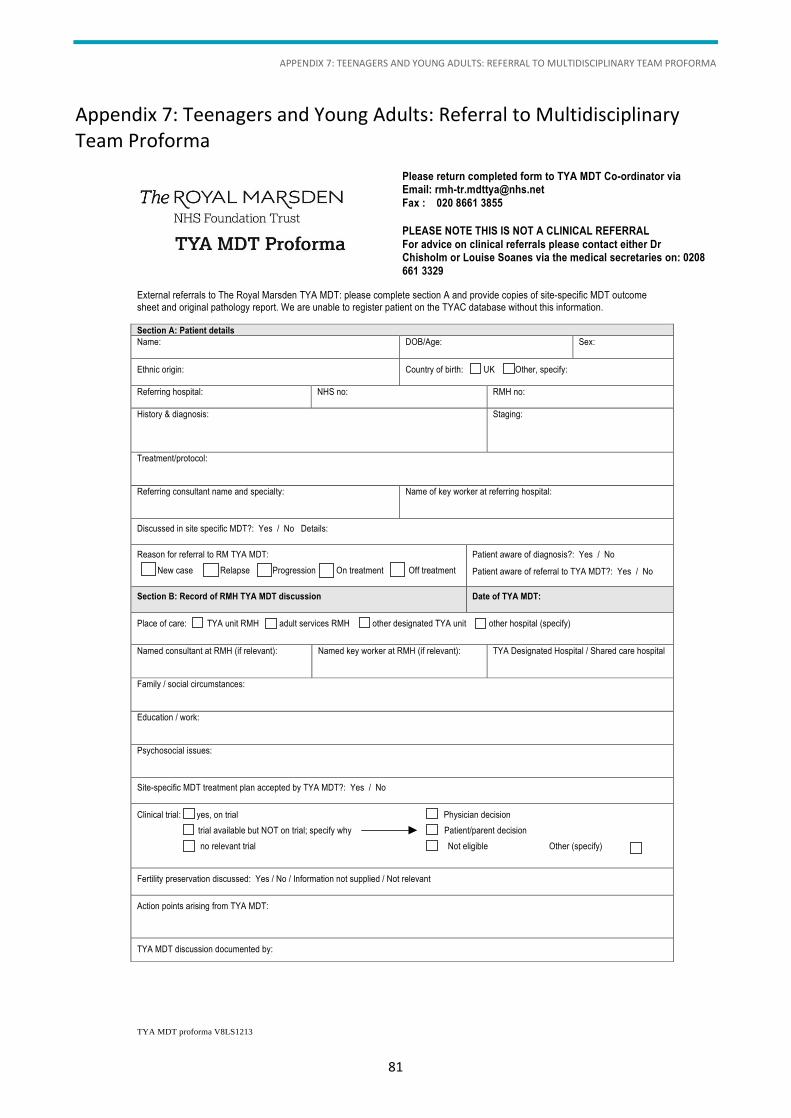
APPENDIX 7: TEENAGERS AND YOUNG ADULTS: REFERRAL TO MULTIDISCIPLINARY TEAM PROFORMA
81
Appendix 7: Teenagers and Young Adults: Referral to Multidisciplinary Team Proforma
TYA MDT proforma V8LS1213
External referrals to The Royal Marsden TYA MDT: please complete section A and provide copies of site-specific MDT outcome
sheet and original pathology report. We are unable to register patient on the TYAC database without this information.
Section A: Patient details
Name: DOB/Age: Sex:
Ethnic origin:
Country of birth: UK Other, specify:
Referring hospital:
NHS no: RMH no:
History & diagnosis:
Staging:
Treatment/protocol:
Referring consultant name and specialty: Name of key worker at referring hospital:
Discussed in site specific MDT?: Yes / No Details:
Reason for referral to RM TYA MDT:
New case Relapse Progression On treatment Off treatment
Patient aware of diagnosis?: Yes / No
Patient aware of referral to TYA MDT?: Yes / No
Section B: Record of RMH TYA MDT discussion
Date of TYA MDT:
Place of care: TYA unit RMH adult services RMH other designated TYA unit other hospital (specify)
Named consultant at RMH (if relevant):
Named key worker at RMH (if relevant):
TYA Designated Hospital / Shared care hospital
Family / social circumstances:
Education / work:
Psychosocial issues:
Site-specific MDT treatment plan accepted by TYA MDT?: Yes / No
Clinical trial: yes, on trial Physician decision
trial available but NOT on trial; specify why Patient/parent decision
no relevant trial Not eligible Other (specify)
Fertility preservation discussed: Yes / No / Information not supplied / Not relevant
Action points arising from TYA MDT:
TYA MDT discussion documented by:
Please return completed form to TYA MDT Co-ordinator via Email: [email protected]
Fax : 020 8661 3855
PLEASE NOTE THIS IS NOT A CLINICAL REFERRAL
For advice on clinical referrals please contact either Dr Chisholm or Louise Soanes via the medical secretaries on: 0208
661 3329
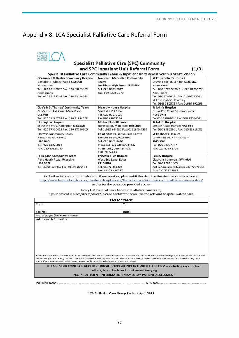
LCA BRAIN/CNS CANCER CLINICAL GUIDELINES
82
Appendix 8: LCA Specialist Palliative Care Referral Form
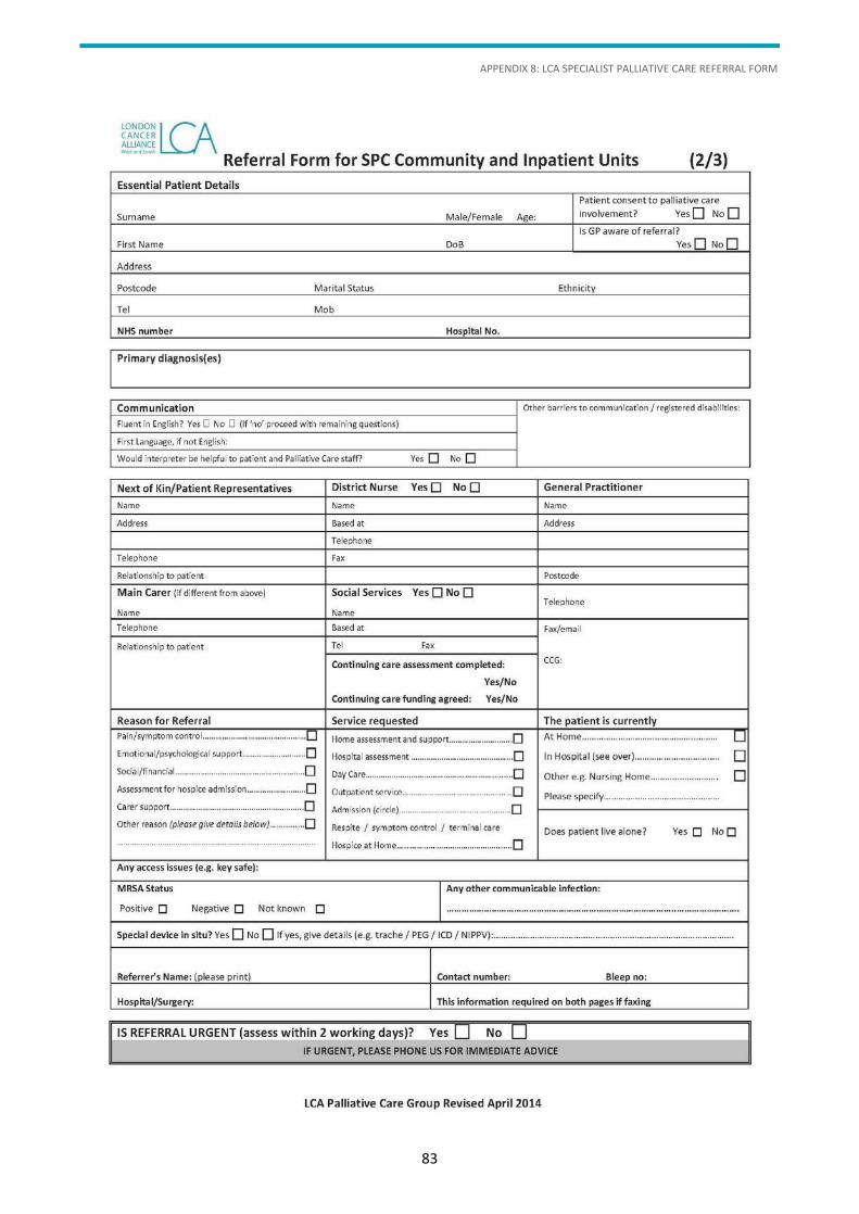
APPENDIX 8: LCA SPECIALIST PALLIATIVE CARE REFERRAL FORM
83
LCA BRAIN/CNS CANCER CLINICAL GUIDELINES
84

© London Cancer Alliance 2014
Published by London Cancer Alliance
London Cancer Alliance
5th Floor Alliance House
12 Caxton Street
London SW1H 0QS
www.londoncanceralliance.nhs.uk