Embed Size (px)
Citation preview
1
20th Advance Course
Àl
“Multimodality imaging of brain tumours: High Grade Brain Tumours”
Unitat de Ressonància MagnèticaServei de Radiologia
Hospital Vall d’Hebron. Barcelona.
alex.rovira@idi‐cat.org
Àlex Rovira
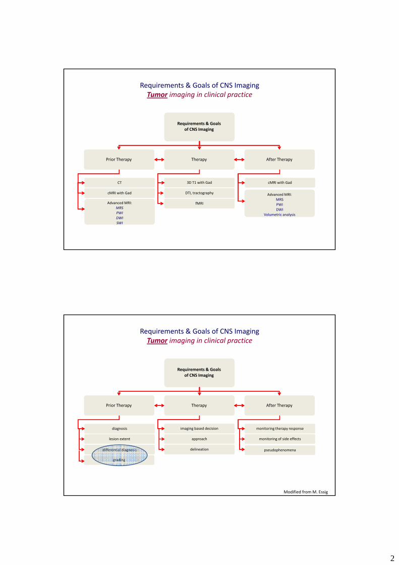
Requirements & Goals of CNS Imaging
Requirements & Goals of CNS ImagingTumor imaging in clinical practice
of CNS Imaging
Therapy After TherapyPrior Therapy
diagnosis imaging based decision monitoring therapy response
lesion extent
differential diagnosis
grading
approach
delineation
monitoring of side effects
Modified fromM. Essig
pseudophenomena
2
Requirements & Goals of CNS ImagingTumor imaging in clinical practice
Requirements & Goals of CNS Imagingof CNS Imaging
Therapy After TherapyPrior Therapy
CT 3D T1 with Gad cMRI with Gad
cMRI with Gad
Advanced MRI:MRSPWIDWISWI
DTI, tractography
fMRI
Advanced MRI:MRSPWIDWI
Volumetric analysis
Requirements & Goals of CNS Imaging
Requirements & Goals of CNS ImagingTumor imaging in clinical practice
of CNS Imaging
Therapy After TherapyPrior Therapy
diagnosis imaging based decision monitoring therapy response
lesion extent
differential diagnosis
grading
approach
delineation
monitoring of side effects
Modified fromM. Essig
pseudophenomena
3
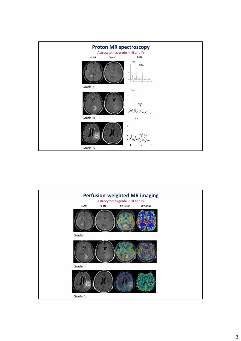
T1 gad MRS
Proton MR spectroscopyAstrocytomas grade II, III and IV
ChoNAA
FLAIR
Grade IICho
NAA
Grade III Cho
NAA Lac
Grade IV
T1 gad CBF (ASL) CBV (DSC)FLAIR
Perfusion‐weighted MR imagingAstrocytomas grade II, III and IV
Grade II
Grade III
Grade IV
4
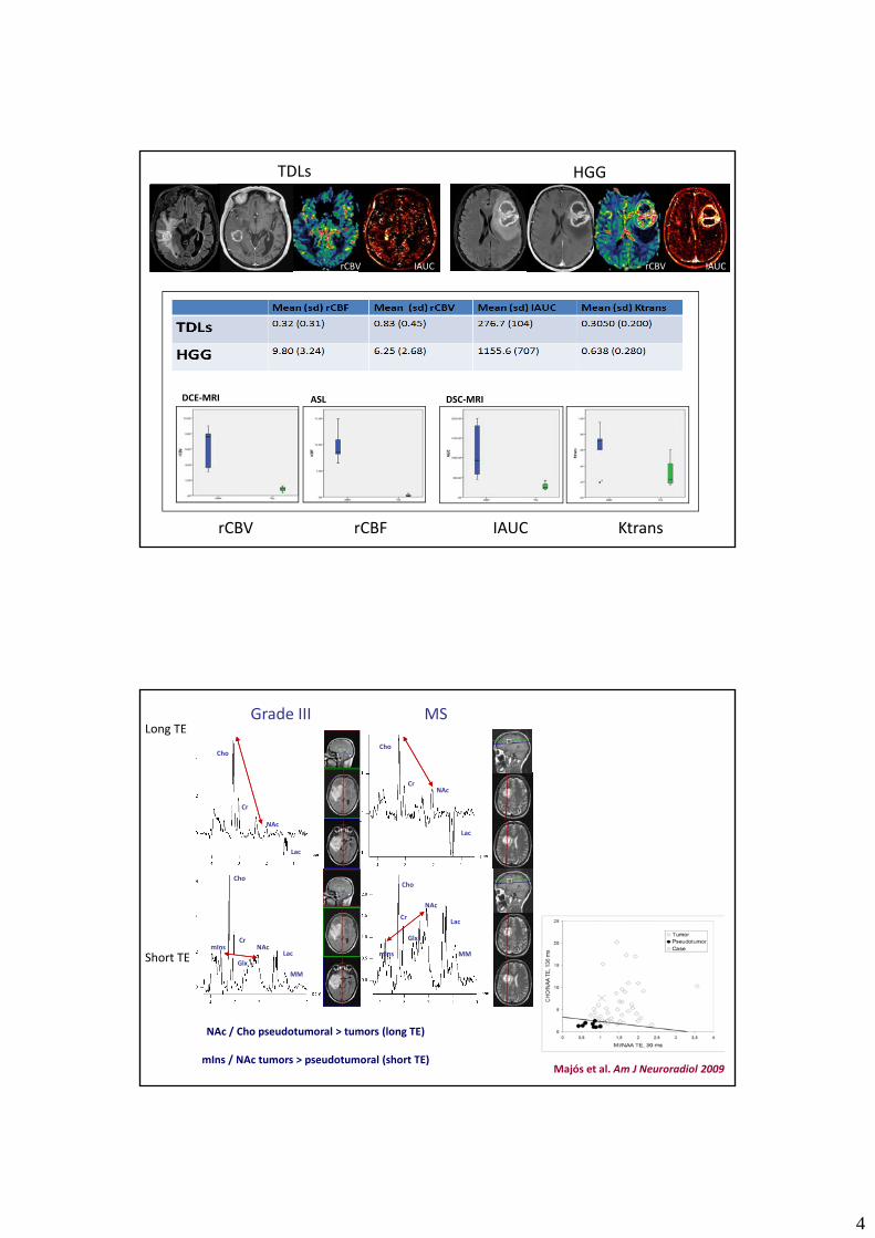
TDLs HGG
rCBV IAUC rCBV IAUC
DSC‐MRIDCE‐MRI ASL
rCBV IAUC KtransrCBF
Cr
Cho
NAcCr
Cho
Grade III MSLong TE
Lac
NAc
LacNAc
Cr
Cho
mIns
Lac
Lac
NAc
Cr
Cho
Glx
MMmInsSh t TE LacGlx
MM
MMmIns
NAc / Cho pseudotumoral > tumors (long TE)
mIns / NAc tumors > pseudotumoral (short TE)Majós et al. Am J Neuroradiol 2009
Short TE
5
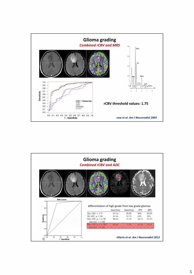
Glioma gradingCombined rCBV and MRS
Law et al. Am J Neuroradiol 2003
rCBV threshold values: 1.75
Glioma gradingCombined rCBV and ADC
differentiation of high‐grade from low grade gliomas
Hilario et al. Am J Neuroradiol 2012
6
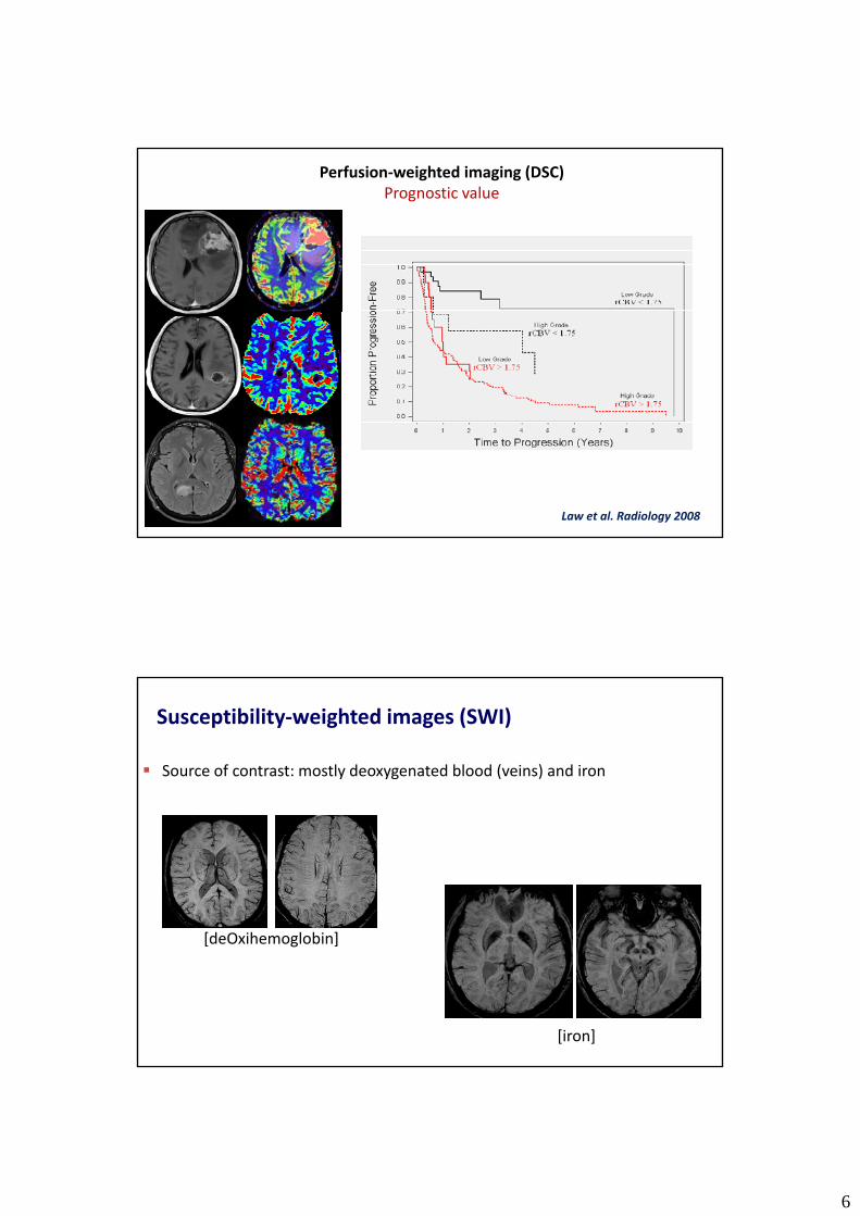
Perfusion‐weighted imaging (DSC)Prognostic value
Law et al. Radiology 2008
Susceptibility‐weighted images (SWI)
Source of contrast: mostly deoxygenated blood (veins) and iron
[deOxihemoglobin][deOxihemoglobin]
[iron]
7
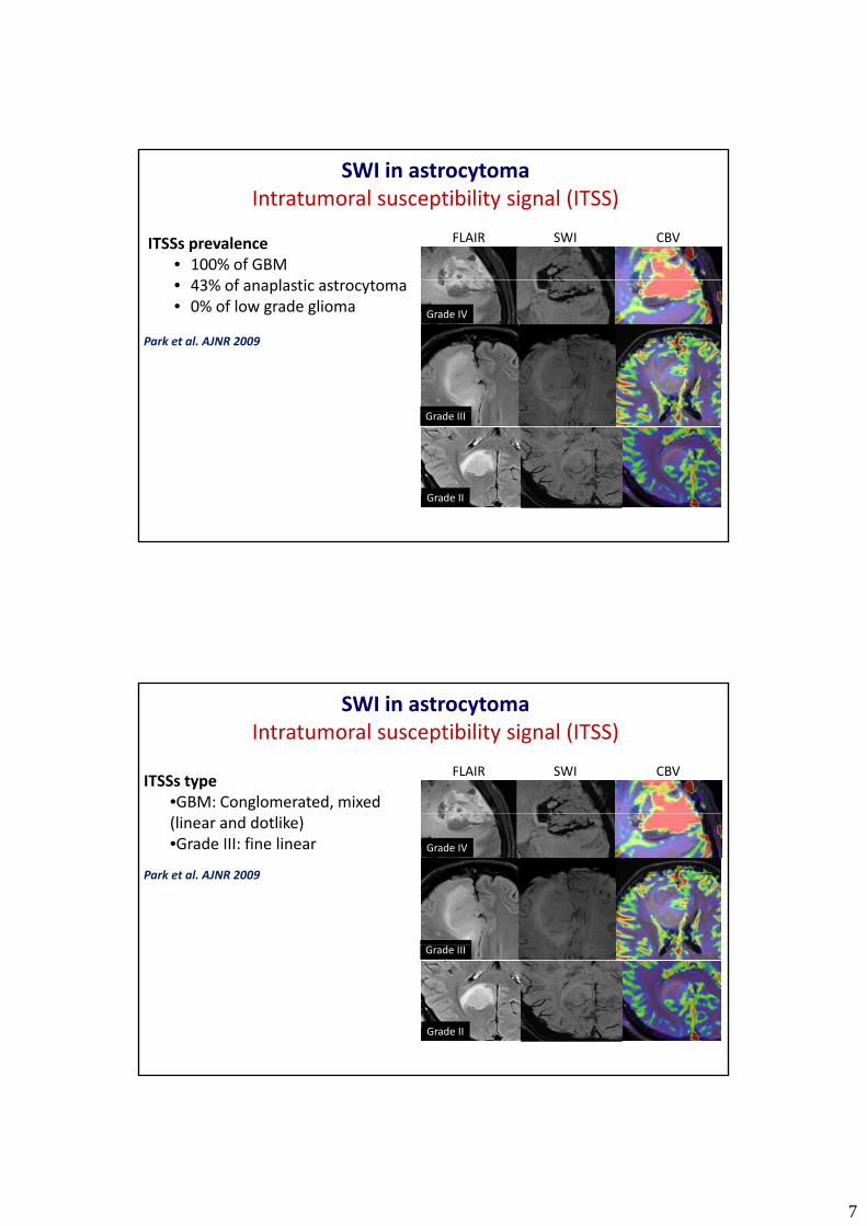
SWI in astrocytomaIntratumoral susceptibility signal (ITSS)
ITSSs prevalence• 100% of GBM43% f l i
SWI CBVFLAIR
Park et al. AJNR 2009
• 43% of anaplastic astrocytoma• 0% of low grade glioma Grade IV
Grade III
Grade II
ITSSs type•GBM: Conglomerated, mixed
SWI in astrocytomaIntratumoral susceptibility signal (ITSS)
SWI CBVFLAIR
Park et al. AJNR 2009
Grade IV
(linear and dotlike)•Grade III: fine linear
Grade III
Grade II

8
Pattern of ITSS in GBMs (inner portion of CE rim)
SWI in astrocytomaIntratumoral susceptibility signal (ITSS)
•Fine, linearRelated to↑CBVTumor vascularity (angiogenesis)
•Densely packedNot related to↑CBV
SWI CBVT1‐Gd
Not related to↑CBVMacro‐micronecrosis
SWI in solitary enhancing lesionsIntratumoral susceptibility signal (ITSS)
TDLGrade I(no ITSS)
GBM
Grade II(1‐10 dotlike, fine linear)
GBMGrade III(>10 dotlike, packed)
Kim et al. AJNR 2009
9
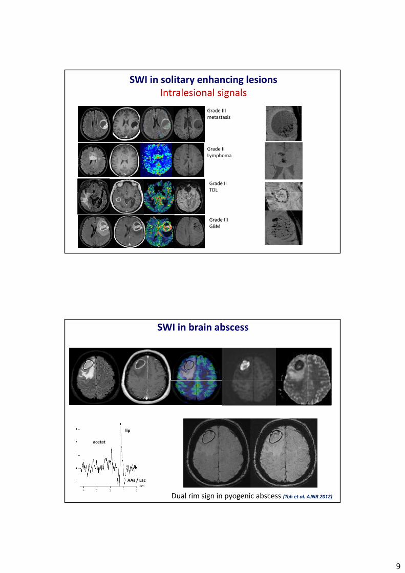
SWI in solitary enhancing lesionsIntralesional signals
Grade IIImetastasis
Grade IILymphoma
Grade IITDL
Grade IIIGBM
SWI in brain abscess
lip
acetatacetat
AAs / Lac
Dual rim sign in pyogenic abscess (Toh et al. AJNR 2012)
10
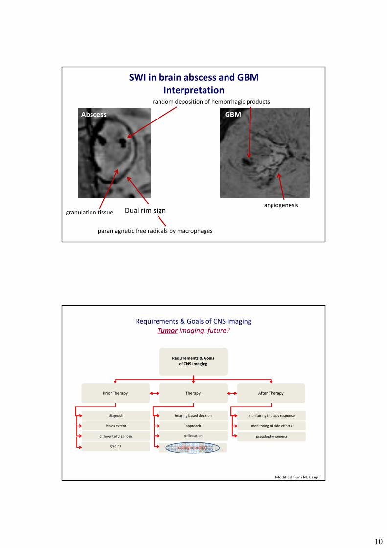
Abscess GBM
random deposition of hemorrhagic products
SWI in brain abscess and GBMInterpretation
paramagnetic free radicals by macrophages
granulation tissueangiogenesis
Dual rim sign
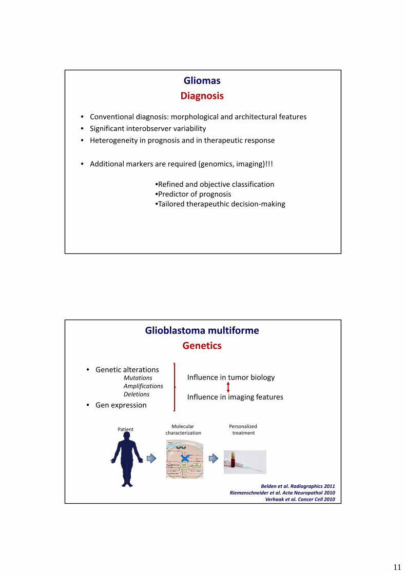
Requirements & Goals of CNS Imaging
Requirements & Goals of CNS ImagingTumor imaging: future?
of CNS Imaging
Therapy After TherapyPrior Therapy
diagnosis imaging based decision monitoring therapy response
lesion extent
differential diagnosis
grading
approach
delineation
monitoring of side effects
Modified fromM. Essig
radiogenomics?
pseudophenomena
11
Gliomas Diagnosis
• Conventional diagnosis: morphological and architectural features
• Significant interobserver variabilityS g ca t te obse e a ab ty
• Heterogeneity in prognosis and in therapeutic response
• Additional markers are required (genomics, imaging)!!!
•Refined and objective classificationP di t f i•Predictor of prognosis
•Tailored therapeuthic decision‐making
Glioblastoma multiformeGenetics
• Genetic alterationsMutations Influence in tumor biology
• Gen expression
Patient Molecular characterization
Personalized treatment
AmplificationsDeletions
gy
Influence in imaging features
Belden et al. Radiographics 2011Riemenschneider et al. Acta Neuropathol 2010
Verhaak et al. Cancer Cell 2010
12
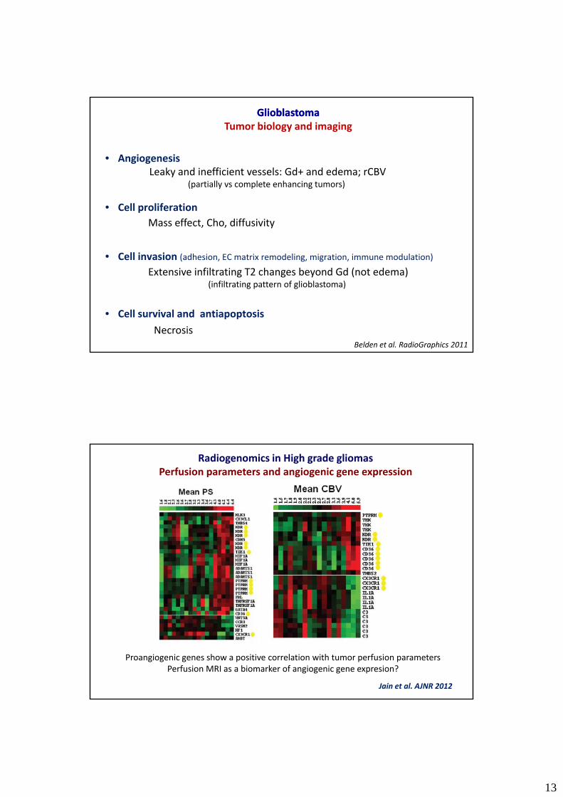
Classification based on gene expression profileClassification based on gene expression profilesignaling pathways: RTK/P13K/PTEN; P53; RB1
GlioblastomasGlioblastomas
GBM
Verhaak et al. Cancer Cell 2010
PurposePurpose
Associations Between MR Imaging and Genomic Features Associations Between MR Imaging and Genomic Features of Glioblastomasof Glioblastomas
• To advance the understanding of the molecular basis of cancerthrough the application of genome analysis technologies,histopathology, and imaging.
• To identify imaging features of primary glioblastomas that may predictgenomic features, including mutation status and gene expression.genomic features, including mutation status and gene expression.
•Predic prognosis and treatment response•Personalized treatment
13
• Angiogenesis
GlioblastomaGlioblastomaTumor biology and imaging
Leaky and inefficient vessels: Gd+ and edema; rCBV( ti ll l t h i t )
• Cell proliferation
• Cell invasion (adhesion, EC matrix remodeling, migration, immune modulation)
(partially vs complete enhancing tumors)
Mass effect, Cho, diffusivity
• Cell survival and antiapoptosis
Extensive infiltrating T2 changes beyond Gd (not edema)(infiltrating pattern of glioblastoma)
NecrosisBelden et al. RadioGraphics 2011
Radiogenomics in High grade gliomasPerfusion parameters and angiogenic gene expression
Jain et al. AJNR 2012
Proangiogenic genes show a positive correlation with tumor perfusion parametersPerfusion MRI as a biomarker of angiogenic gene expresion?
14
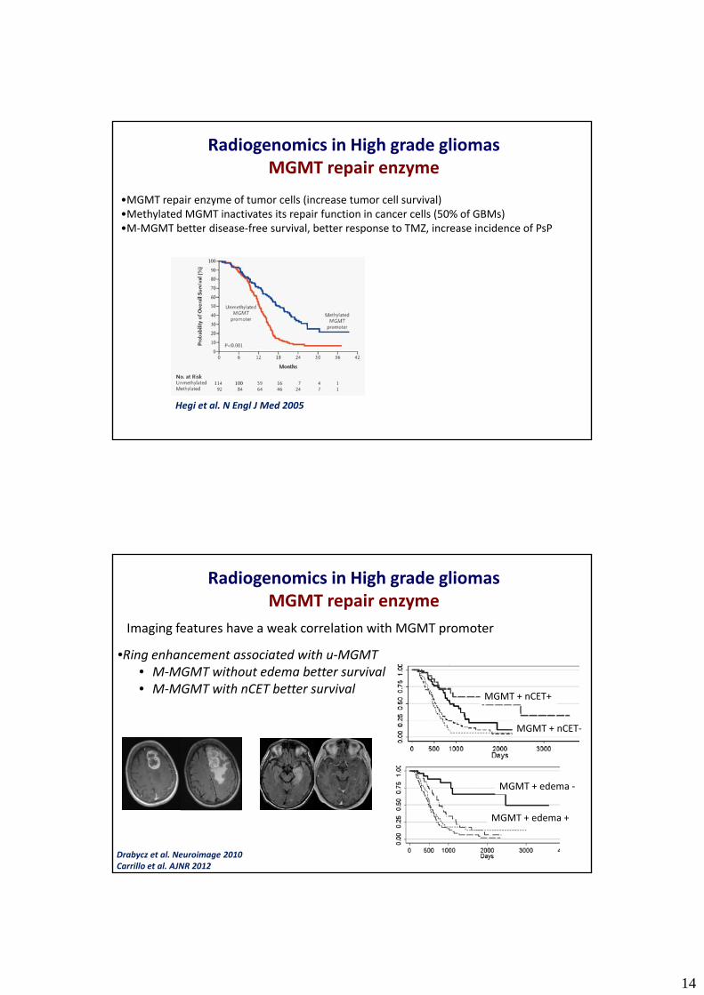
Radiogenomics in High grade gliomasMGMT repair enzyme
•MGMT repair enzyme of tumor cells (increase tumor cell survival)•Methylated MGMT inactivates its repair function in cancer cells (50% of GBMs)•M MGMT better disease free survival better response to TMZ increase incidence of PsP•M‐MGMT better disease‐free survival, better response to TMZ, increase incidence of PsP
Hegi et al. N Engl J Med 2005
Radiogenomics in High grade gliomasMGMT repair enzyme
Imaging features have a weak correlation with MGMT promoter
•Ring enhancement associated with u‐MGMTg• M‐MGMT without edema better survival• M‐MGMT with nCET better survival
MGMT + nCET+
MGMT + nCET‐
MGMT + edema ‐
MGMT + edema +
Drabycz et al. Neuroimage 2010Carrillo et al. AJNR 2012
15
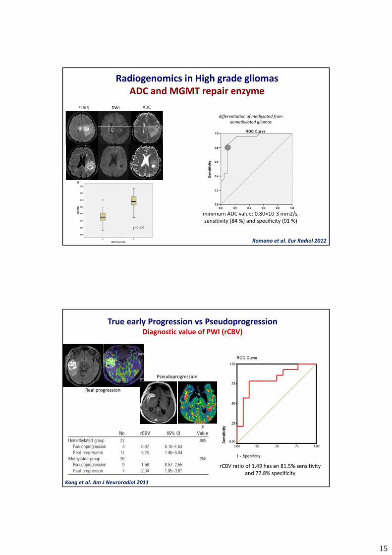
Radiogenomics in High grade gliomasADC and MGMT repair enzyme
differentiation of methylated fromunmethylated gliomas
ADCDWIFLAIR
Romano et al. Eur Radiol 2012
minimum ADC value: 0.80×10‐3 mm2/s, sensitivity (84 %) and specificity (91 %)
True early Progression vs PseudoprogressionDiagnostic value of PWI (rCBV)
Real progression
Pseudoprogression
rCBV ratio of 1.49 has an 81.5% sensitivity and 77.8% specificity
Kong et al. Am J Neuroradiol 2011
16
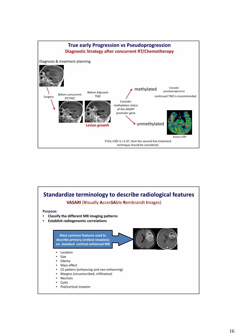
True early Progression vs PseudoprogressionDiagnostic Strategy after concurrent RT/Chemotherapy
Diagnosis & treatment planning
Before concurrent RT/TMZ
Before Adjuvant TMZ
Consider pseudoprogression
Consider methylation status
of the MGMT t
methylated
Surgery continued TMZ is recommended
Lesion growth
promoter gene
unmethylated
Assess rCBV
If the rCBV is >1.47, then the second‐line treatment technique should be considered.
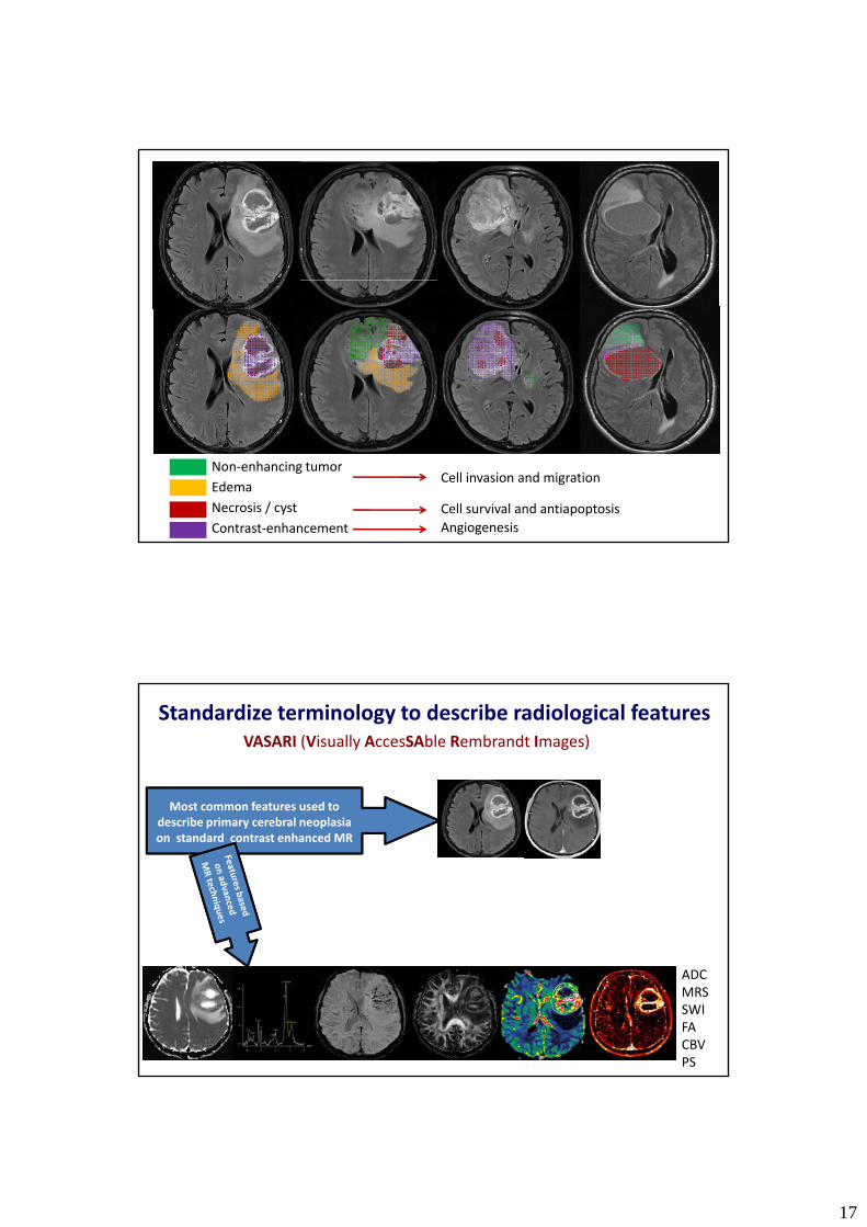
VASARI (Visually AccesSAble Rembrandt Images)
Standardize terminology to describe radiological features
Purpose:• Classify the different MR imaging patterns• Establish radiogenomic correlations
Most common features used to describe primary cerebral neoplasiaon standard contrast enhanced MR
• Location • Size
g
• Edema• Mass effect• CE pattern (enhancing and non‐enhancing)• Margins (circumscribed, infiltrative)• Necrosis• Cysts• Pial/cortical invasion
17
Non‐enhancing tumorEdemaNecrosis / cystContrast‐enhancement
Cell invasion and migration
Cell survival and antiapoptosisAngiogenesis
VASARI (Visually AccesSAble Rembrandt Images)
Most common features used to
Standardize terminology to describe radiological features
describe primary cerebral neoplasia on standard contrast enhanced MR
ADCMRSSWIFACBVPS
18
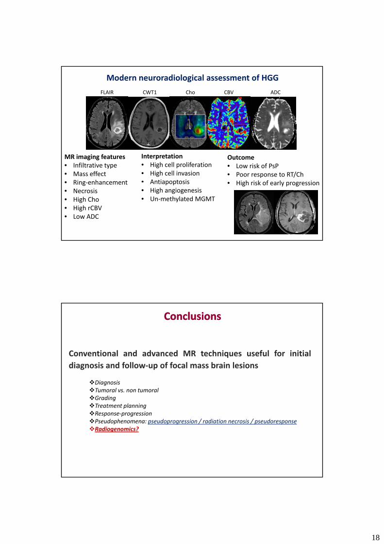
Cho CBVFLAIR CWT1 ADC
Modern neuroradiological assessment of HGG
MR imaging features• Infiltrative type• Mass effect• Ring enhancement
Interpretation• High cell proliferation• High cell invasion• Antiapoptosis
Outcome• Low risk of PsP• Poor response to RT/Ch• High risk of early progression• Ring‐enhancement
• Necrosis• High Cho• High rCBV• Low ADC
Antiapoptosis• High angiogenesis• Un‐methylated MGMT
• High risk of early progression
ConclusionsConclusions
Conventional and advanced MR techniques useful for initialdiagnosis and follow‐up of focal mass brain lesions
DiagnosisTumoral vs. non tumoralGradingTreatment planningResponse‐progressionP d h d i / di i i / dPseudophenomena: pseudoprogression / radiation necrosis / pseudoresponseRadiogenomics?