Embed Size (px)
Citation preview

Rheum Dis Clin N Am
32 (2006) 509–522
Kidney Disease in AntiphospholipidSyndrome
Mary-Carmen Amigo, MD, FACPUniversidad Nacional Autonoma de Mexico, Department of Rheumatology,
Instituto Nacional de Cardiologıa Ignacio Chavez, Juan Badiano #1,
Distrito Federal, Tlalpan, Mexico 14080, Mexico
The kidney is a major target organ in patients with the antiphospholipidsyndrome (APS). However, it is only in the past few years that knowledgeabout renal vascular involvement in APS has acquired a critical mass.This can be explained because APS was first described in patients with sys-temic lupus erythematosus (SLE) and the studies were focused on the im-mune complex-mediated glomerulonephritis rather than the renal vascularlesions. In addition, the risks of a renal biopsy in APS patients because ofthe frequent occurrence of thrombocytopenia and systemic hypertension,discouraged the procedure and impeded the pathologic demonstration ofintrarenal thrombotic lesions.
The renal manifestations may result from thrombosis occurring at anylocation within the renal vasculature, including large vessels, both arterialand venous, the intraparenchymatous arteries and arterioles, as well as theglomerular capillaries. However, other types of renal involvement includingmembranous glomerulonephritis, pauci-immune crescentic glomerulone-phritis, IgA nephropathy, focal segmental glomerulosclerosis, glomerulone-phritis with isolated C3 mesangial deposits, and vasculitis associated withaPL have all been described. It is unclear whether, in addition to thrombosis,other mechanisms could also contribute to the pathogenesis of the nephrop-athy of APS (NAPS). The consequences of the renal compromise include re-novascular hypertension, renal infarcts, cortical atrophy, acute renal failure,proteinuria, and end stage renal disease (Table 1).
There is a prevailing opinion that given its importance and unique fea-tures, the NAPS, should be included in the APS classification criteria. Inthis review, I will discuss systemic hypertension, renal artery lesions, the
E-mail address: [email protected] (M-C. Amigo).
0889-857X/06/$ - see front matter � 2006 Elsevier Inc. All rights reserved.
doi:10.1016/j.rdc.2006.05.004 rheumatic.theclinics.com

510 AMIGO
NAPS, renal vein thrombosis, the significance of antiphospholipid anti-bodies (aPL) in patients with SLE, and the relevance of aPL in end stage re-nal failure and renal transplantation.
Systemic hypertension
Hypertension (HT) is a common feature of the APS both in the primaryand the secondary forms [1–4]. It appears to be a marker of nephropathy asit has been consistently present in the majority of patients with APS and re-nal involvement. It is important to keep in mind that both microvasculop-athy or occlusion of the renal artery can contribute to severe HT in APS[1,5]. The prevalence of HT in 600 patients with primary APS (n ¼ 68), sec-ondary APS (n ¼ 522), or positive for aPL (n ¼ 10) at St Thomas’ LupusClinic was 29%, and it was associated with livedo reticularis in 86% ofthe cases [6]. In primary APS, in our series of five patients with thromboticmicroangiopathy [1], severe hypertension was present in all five patients andin the series by Nochy and colleagues [4], HT was present in 93% of their 16patients with intrarenal vascular lesions. Malignant hypertension in APSwithout overt lupus nephritis or stenosis/thrombosis of the main renal ar-teries was documented by Cacoub and colleagues in five patients. Interest-ingly, high-dose steroids and anticoagulation led to rapid resolution of thehypertensive crisis in three of their patients [2].
The pathophysiology of HT in the context of primary APS includesthrombosis/occlusion of the trunk of a renal artery leading to renovascularhypertension as well as stimulation of the renin–angiotensin–aldosterone
Table 1
Kidney vascular involvement in APS
Vascular lesion Clinical consequences
Renal artery lesions Renovascular hypertension (severe)
Thrombosis/occlusion/stenosis? Renal infarcts (silent, painful, hematuria)
Glomerular capillary thrombosis Increased likelihood of renal insufficiency
leading to glomerular sclerosis
(studied mainly in SLE)
Renal thrombotic microangiopathy Systemic hypertension (usually severe)
-Glomerular capillaries Renal failure
-Arteriolar fibrous occlusions Proteinuria (mild to nephrotic range)
-Fibrous intima hyperplasia of interlobular
arteries and their branches with/without
focal or difusse cortical necrosis
Hematuria
-Cortical atrophy
Renal vein thrombosis Risk of renal failure (if bilateral)
Abbreviations: APS, antiphospholipid syndrome; SLE, systemic lupus erythemaosus.
Adapted from Amigo MC and Garcıa-Torres R. Kidney disease in antiphospholipid syn-
drome. In: Khamashta MA, Editor. Hughes Syndrome Antiphospholipid Syndrome.
Springer-Verlag, London; 2000. p. 70–81.
511KIDNEY DISEASE IN ANTIPHOSPHOLIPID SYNDROME
system secondary to intrarenal vascular lesions. Once HT is established, itmay worsen vascular lesions [4]. Clinicians should thus investigate HT inall APS patients and treat them accordingly.
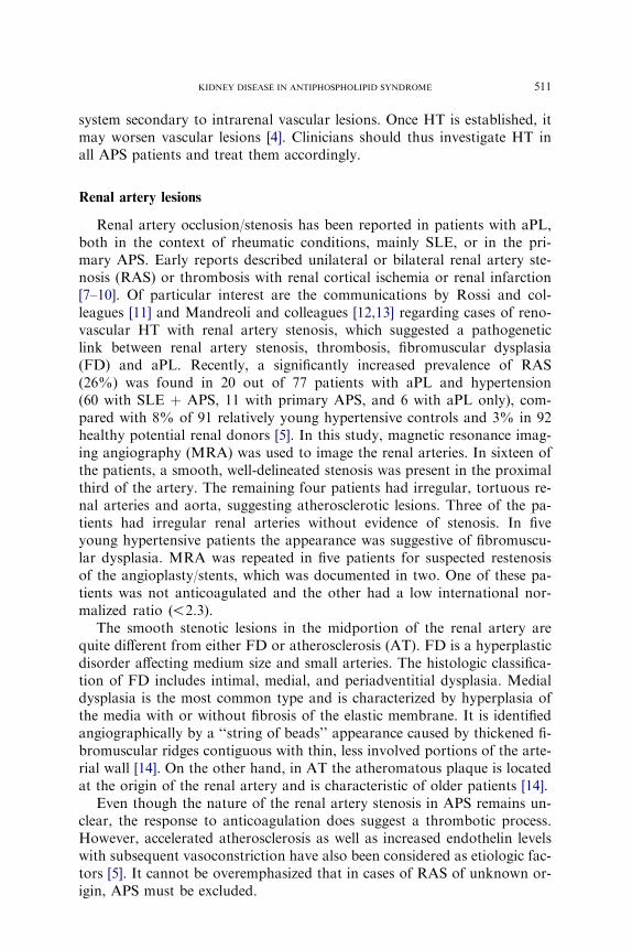
Renal artery lesions
Renal artery occlusion/stenosis has been reported in patients with aPL,both in the context of rheumatic conditions, mainly SLE, or in the pri-mary APS. Early reports described unilateral or bilateral renal artery ste-nosis (RAS) or thrombosis with renal cortical ischemia or renal infarction[7–10]. Of particular interest are the communications by Rossi and col-leagues [11] and Mandreoli and colleagues [12,13] regarding cases of reno-vascular HT with renal artery stenosis, which suggested a pathogeneticlink between renal artery stenosis, thrombosis, fibromuscular dysplasia(FD) and aPL. Recently, a significantly increased prevalence of RAS(26%) was found in 20 out of 77 patients with aPL and hypertension(60 with SLE þ APS, 11 with primary APS, and 6 with aPL only), com-pared with 8% of 91 relatively young hypertensive controls and 3% in 92healthy potential renal donors [5]. In this study, magnetic resonance imag-ing angiography (MRA) was used to image the renal arteries. In sixteen ofthe patients, a smooth, well-delineated stenosis was present in the proximalthird of the artery. The remaining four patients had irregular, tortuous re-nal arteries and aorta, suggesting atherosclerotic lesions. Three of the pa-tients had irregular renal arteries without evidence of stenosis. In fiveyoung hypertensive patients the appearance was suggestive of fibromuscu-lar dysplasia. MRA was repeated in five patients for suspected restenosisof the angioplasty/stents, which was documented in two. One of these pa-tients was not anticoagulated and the other had a low international nor-malized ratio (!2.3).
The smooth stenotic lesions in the midportion of the renal artery arequite different from either FD or atherosclerosis (AT). FD is a hyperplasticdisorder affecting medium size and small arteries. The histologic classifica-tion of FD includes intimal, medial, and periadventitial dysplasia. Medialdysplasia is the most common type and is characterized by hyperplasia ofthe media with or without fibrosis of the elastic membrane. It is identifiedangiographically by a ‘‘string of beads’’ appearance caused by thickened fi-bromuscular ridges contiguous with thin, less involved portions of the arte-rial wall [14]. On the other hand, in AT the atheromatous plaque is locatedat the origin of the renal artery and is characteristic of older patients [14].
Even though the nature of the renal artery stenosis in APS remains un-clear, the response to anticoagulation does suggest a thrombotic process.However, accelerated atherosclerosis as well as increased endothelin levelswith subsequent vasoconstriction have also been considered as etiologic fac-tors [5]. It cannot be overemphasized that in cases of RAS of unknown or-igin, APS must be excluded.

512 AMIGO
Successful treatment with antihypertensive drugs [11,15], aspirin [16], an-ticoagulant therapy [10,12], and transluminal angioplasty with or withoutstenting has been reported [9,17,18]. Surgery, however, is restricted to thosepatients in whom angioplasty and stenting are not feasible. It is crucial toremember that the sooner an arterial lesion is relieved, the likelier a success-ful outcome.
Intrarenal vascular lesionsdThe nephropathy of antiphospholipid syndrome
The most commonly reported intrarenal vascular lesion in patients withaPL is thrombotic microangiopathy (TMA) (Figs. 1–3). This condition wasinitially described in patients with SLE [19–21] and in patients with lupusanticoagulant (LA) and pregnancy-related renal failure [22]. Subsequently,isolated cases of TMA in patients with primary APS were reported[23,24]. In our original series, published in 1992, we described five patientswho had renal disease and HT among 20 consecutive patients with primaryAPS [1]. Mild renal failure was present in three patients while two had end-stage renal disease requiring hemodyalisis. In addition, proteinuria frommild to nephrotic was also present. The study of the kidney biopsies allowedus to observe the lesions due to APS in its pure form. Biopsy findings inall five patients were consistent with TMA. Microangiopathy involvedboth the vascular tree and the glomerular tufts with acute as well as oldand recanalizing thrombi. Subsequently, these findings have consistentlybeen found in the literature. Nochy and colleagues [4] in a retrospectivestudy of 16 patients with primary APS confirmed our initial observationson glomerular and interlobular arteriolar lesions (Figs. 4 and 5). In addition,
Fig. 1. Renal biopsy from an APS patient showing one glomerulus with retraction of the glo-
merular tuft and wrinkling of the glomerular basement membranes of the capillary wall. The
presence of thrombi is also evident (PAS 400�). (Courtesy of M.C. Avila, MD, Mexico City,
Mexico.)

513KIDNEY DISEASE IN ANTIPHOSPHOLIPID SYNDROME
they emphasized the presence of focal cortical atrophy (FCA) involving thesuperficial cortex under the renal capsule, as foci or triangles leading to tis-sue retraction. These authors considered the NAPS as an entity in its ownright being characterized by TMA, fibrous intimal hyperplasia (FIH) ofthe arteries and arterioles, and FCA. Moreover, some glomerular ultraes-tructural changes have recently been proposed as pathognomonic ofNAPS [25], namely glomerular basement membrane wrinkling and redupli-cation. On electron microscopy, redundant wrinkled segments of basementmembrane with straighter thin basement membrane adjacent to the endo-thelium are shown. However, these findings must await confirmation inlarger studies.
Fig. 2. Renal section showing one glomerulus increased in size that presents micro thrombi.
The early presence of double contours and cellular interposition is appreciated by the Jones’s
stain (Jones stain 400�). (Courtesy of M.C. Avila, MD, Mexico City, Mexico.)
Fig. 3. Glomerular thrombotic microangiopathy. Double contours and cellular interposition
are seen (Jones stain 400�). (Courtesy of M.C. Avila, MD, Mexico City, Mexico.)

514 AMIGO
It is worth mentioning that TMA is not exclusive for APS, but occurs inmany other entities caused by coagulation disturbances or endothelial cellinjury, which result in the formation of thrombi within the renal vascula-ture. These conditions include thrombotic thrombocytopenic purpura(TTP), hemolytic uraemic syndrome (HUS), scleroderma renal crisis, malig-nant hypertension, pre-eclampsia, and postpartum renal failure. Contracep-tives, cyclosporine toxicity, chemotherapy, and renal transplant rejection areadditional etiologic factors [26].
It can be very difficult to differentiate between APS, particularly the cat-astrophic APS (CAPS), SLE, and the TTP/HUS coagulopathies. They notonly share TMA, but central nervous system symptoms, thrombocytopenia,and hemolysis. Moreover, they can occur simultaneously. Not surprisingly,APS and the HUS/TTP share endothelial injury and thrombus formation as
Fig. 4. Cross-section of a small artery occluded by fibrin-like material. Hematoxylin and eosin,
�400. (Courtesy of Beatriz De Leon, MD.)
Fig. 5. Small interlobular artery with considerable intimal thickening. Hematoxylin and
eosin, �400. (Courtesy of Beatriz De Leon, MD.)

515KIDNEY DISEASE IN ANTIPHOSPHOLIPID SYNDROME
pathogenic mechanisms. In the CAPS, severe multiorgan dysfunction sec-ondary to small vessel thrombosis and ischaemia dominates the clinical pic-ture. Severe hypertension due to renal artery/vein thrombosis or thromboticmicroangiopathy is common in CAPS patients [27].
Recently, there have been reports of histologic documentation of othertypes of renal involvement in patients with primary APS. These have in-cluded membranous glomerulonephritis, IgA nephropathy, pauci-immunecrescentic glomerulonephritis, glomerulonephritis with isolated C3 mesan-gial deposits, vasculitis, and focal segmental glomerulosclerosis (FSG)[28–30]. In a retrospective study, 5 out of 270 consecutive renal biopsieswere identified as biopsies performed in patients with primary APS [31]. His-tologic examination of these biopsies showed tubulointerstitial lesions in all,vascular lesions and FSG in four. It is not surprising, as FSG may be a se-quela of TMA. It is unclear, however, whether in addition to thrombosis,other mechanisms could also contribute to the pathogenesis of NAPS. Thereis evidence that anticardiolipin antibodies (aCL) recognize b-2 glycoproteinI (b2GP1) on endothelial cells leading to increased expression of adhesionmolecules, increased adhesion of monocytes to endothelial cells, and com-plement activation [32]. These events could explain the inflammation foundin the glomeruli in these uncommon cases. These findings have more than anacademic interest. The distinction between renal inflammation whethercaused by immune complex deposition or other causes, and microvascularthrombosis could give clues about the pathogenesis of APS and couldhelp determine an appropriate treatment for these patients. Renal inflamma-tion may require steroids and cytotoxic therapy, whereas APS vasculopathybenfits from anticoagulation.
The management of renal involvement in primary APS has been empiri-cal. Hamidou and colleagues [33] reported disappearance of proteinuriaand normalization of renal function with the use of aspirin and captoprilin one patient with TMA and primary APS. Olguın and colleagues [34]documented renal function improvement in all five patients with NAPSwith the combination of oral anticoagulants, aspirin, and nifedipine. Kork-maz and colleagues [35] found a beneficial effect of immunosuppressivetherapy (azathioprine or cyclophosphamide) along with warfarin and anangiotensin-converting enzyme inhibitor in four cases with NAPS. Recently,Bhowmik and colleagues [36] reported a patient with primary APS, steroid-responsive FSG, and a successful pregnancy. However, further studies areneeded to unravel the basic mechanisms of renal involvement in APS sothat a targeted, more appropriate treatment be designed.
Renal vein thrombosis
Thrombosis of one or both main renal veins occurs in a variety of settingsincluding trauma, extrinsic compression, renal cell carcinoma, nephroticsyndrome, pregnancy, and oral contraceptives. Renal vein thrombosis

516 AMIGO
(RVT) has been described in aPL positive patients with SLE [37], in a fatalcase of renal transplant [38], and in patients with primary APS including a pa-tient with bilateral RVT in the postpartum period [39,40]. Asherson [41] wasthe first to describe the association between aPL and RVT in two patientswith SLE, proliferative nephritis and nephrotic syndrome. The clinical man-ifestations of RVT depend on the extent of the lesion. Acute or subacute de-terioration of renal function or exacerbation of proteinuria and hematuriashould alert the clinician. Even though the definitive diagnosis can onlybe established through selective renal venography, Doppler ultrasound,contrast-enhanced computed tomography, and magnetic resonance im-aging often provides the diagnosis. Management consists of anticoagulation.
The significance of antiphospholipid antibodies in patients
with systemic lupus erythematosus nephropathy
The prevalence and significance of glomerular capillary thrombosis in pa-tients with SLE have been studied by Kant and colleagues [42] and Glueckand colleagues [37]. These authors found capillary thrombosis in near 50%of the cases with proliferative glomerulonephritis including 78% in patientswith LA and 38% in those without. Notably, the presence of glomerularthrombi in the initial biopsy was a strong predictor of glomerular sclerosis.Other studies have not shown an association between aPL and prognosis inlupus nephritis. However, Moroni and colleagues [43], recently documentedthe impact of aPL in 111 patients with lupus nephritis followed for a meanof 173 � 100 months. Interestingly, a strong association between aPL andthe development of chronic renal failure in the long term was found. Inthe mutlivariate analysis, aPL positivity, high plasma creatinine level atpresentation, and chronicity index, were independent predictors of chronicrenal function deterioration. Miranda and colleagues [44] found no associ-ation between the presence of glomerular thrombi and severity of lupus ne-phritis in their analysis of 108 cases. Curiously, only nine of their patientswere positive for aPL. Daugas and colleagues [3] addressed the presenceand clinical relevance of NAPS in patients with SLE. This study, whichwas retrospective, included 114 patients with SLE nephropathy. The studyshowed that NAPS was present in 32% of the patients, in addition to,and independently from, lupus nephritis. NAPS was statistically associatedwith the presence of LA but not with anticardiolipin antibodies, and wasassociated with extrarenal APS, mainly arterial thrombosis and fetal loss.Finally, these authors found that NAPS is an independent risk factor con-tributing to hypertension, elevated serum creatinine and interstitial fibrosis.Recently, Tektonidou and colleagues [45] examined the prevalence and long-term outcome of NAPS in 151 SLE patients with or without aPL as well asthe histologic evolution of NAPS lesions on serial kidney biopsies. NAPSwas documented independently of lupus nephritis in two thirds of SLE pa-tients with secondary APS, one third of SLE patients with aPL, and in only

517KIDNEY DISEASE IN ANTIPHOSPHOLIPID SYNDROME
3 out of 70 SLE patients without aPL. Progression of the acute thromboticlesions to chronic proliferative, obstructive, and fibrotic forms was observed.TMA in the first biopsy was usually followed by chronic lesions. In addition,a strong association between NAPS, arterial thrombosis, and livedo reticula-ris was found in this series. All of these findings strongly suggest that patientswith lupus nephritis who develop an additional NAPS should be consideredfor long-term anticoagulation in addition to immunosupression. Long-termprospective studies to accurately define the impact of NAPS and the role ofanticoagulant or vasoprotective treatments are urgently needed.
End-stage renal disease
Patients with end-stage renal disease (ESRD) have a higher frequency ofaPL positivity compared with the general population [46–49]. The preva-lence of aPL is higher in hemodialysis patients compared with those on con-tinous ambulatory peritoneal dialysis [48,49]. Quereda and colleagues [46]were the first to report the presence of LA in hemodialysis patients. Thesame group, in a prospective study of 138 patients with nephropathy, foundthat aPL was present 34% of patients with SLE, 9% in patients with chronicprimary glomerulonephritis, 2.6%in patients with nonimmunologicallymediated renal diseases (2.6%) [50]. Why aPL appear in hemodialysis pa-tients is unclear. Several mechanisms have been considered including auto-immunity from an altered immune function due to uremia [51], dialysismembranes incompatibility [49], trauma to blood passing through the he-modialysis circuit [52], as well as induction by microbial agents [53]. TheaPL produced in ESRD patients appear to be b2GP1-independent [54].However, their significance remains to be clarified because, although somestudies suggest that they are not pathogenic [55–58], others indicate thataPL are associated with hemodialysis vascular access thrombosis [1,59–62]. Recently, a case of CAPS following initiation of hemodialysis after bi-lateral nephrectomy due to renal cancer was documented. Endothelial celldamage due to arteriovenous fistula creation/cannulation, in addition to in-fection, and warfarin withdrawal were considered the trigger factors for thedevelopment of CAPS in this patient [63].
Renal transplantation
Reports of aPL-related morbidity among SLE kidney transplant patientsare limited by the relative small number of subjects available for study atany given center. In the University of California, San Francisco study[64], a total of 97 SLE patients who underwent renal transplantation werecompared with matched controls. Patients with SLE had poorer transplan-tation outcomes, with more than twice the risk of allograft loss. In this study,15.4% of allograft failures were attributed to aPL-associated events. In a ret-rospective study of 13 SLE patients who received renal transplant,

518 AMIGO
Radakrishna and colleagues [65], compared eight patients with aCL to fivepatients without, transplanted during the same period. Thrombotic episodesoccurred in three patients in the aCL-positive group but none in the aCL-negative group. There were no differences in the number of rejection episodes,rate of graft loss, or renal function at follow-up. These authors concluded thatpatientswithSLEandaCLcanbe successfully transplanted.However, in a ret-rospective study of 96 consecutive patients with SLE who underwent renaltransplantation, Stone and colleagues [66] assesses the impact of aPL on renaltransplantation. Twenty five patients had at least one aPL. Among these 25patients, 15 (60%) had clinical events associated with APS including 10 casesof posttransplantation morbidity or mortality attributable to the APS (threedeaths, five graft losses, and two TMA).
In 1994, we reported two primary APS patients with ESRD due to TMAwho underwent renal transplantation [67]. Despite intensive anticoagulanttherapy, massive thrombosis of the graft in one case and TMA in the other,suggested recurrence of disease. Furthermore, our two patients had vascularaccess thrombosis and one had previous peritoneal dyalisis catheter mal-function due to fibrin-related obstruction. As far as we know, this was thefirst report of kidney transplantation in patients with primary APS.
Subsequently, there has been increasing evidence that aPL positive pa-tients undergoing renal transplantation are at a significantly increased riskof renal vascular thrombosis, graft loss, and systemic thrombosis.
It has been demonstrated that patients with APS are at a greater risk forthe development of renal allograft thrombosis within 1 week after transplan-tation. In a striking study, within a group of 78 patients who received renaltransplant, 6 had APS. Each of these six patients thrombosed their renal al-lograft within a week of the transplant. In contrast, the remaining 72 patientswere all doing well 1 year after transplantation [68]. In a multicenter study of502 ESRD patients awaiting renal transplantation the potential risks associ-ated with APS were assessed and strategies for therapeutic intervention werereviewed [69]. Twenty-three patients were diagnosed with APS. Of these, 11received a kidney transplant either with (four patients) or without (sevenpatients) concomitant anticoagulation. All seven patients without anticoagu-lation lost their allograft within 1 week as a result of renal thrombosis. Incontrast, three of the four patients who were anticoagulated maintained theirallografts for over 2 years; the fourth patient lost his allograft due to throm-bosis. Of the remaining 70 patients with aCL without thrombosis, 37 weresuccessfully transplanted. Recently, the same group of investigators reportedtheir experience with nine APS renal transplant patients [70]. Seven weretreated with coumadin whereas two received heparin. Of the two patientstreated with heparin, one had an early allograft loss and the other is doingwell at 5 years posttransplant. Of the seven patients treated with coumadin,two are doing well, two had early allograft loss, and the remaining three re-turned to dialysis after they were taken off coumadin because of bleedingcomplications.

519KIDNEY DISEASE IN ANTIPHOSPHOLIPID SYNDROME
In another setting, it has been suggested that aPL may be implicated inthe pathogenesis of TMA/HUS in a subset of hepatitis C-positive (HCV) re-nal allograft recipients [71]. Five HCV-positive renal transplant recipientsdeveloped biopsy-proven de novo TMA shortly after renal transplantation.Anticardiolipin antibodies were detected in pretransplant sera of these fivepatients and in only 1 of 13 HCV-positive recipients without TMA. Inaddition to TMA, three patients also presented other thrombotic com-plications and four of the five patients died within 5 years after transplanta-tion. It is interesting to note that HCV infection has been associated with thedevelopment of several autoantibodies including aPL.
It has been suggested that patients with ESRD of any cause should bescreened for aPL, and if detected, prevention with perioperative heparinand maintenance warfarin should be considered despite the high rate ofbleeding complications, in view of the serious risk of thrombosis of the graft.Finally, in patients with APS, one should question which patients should betransplanted and which therapeutic interventions should be used becausethere is a high risk of posttransplant renal thrombosis even with anticoagu-lation. A prospective randomized multicenter study addressing these issue iswarranted.
Several questions should be answered in the near future regarding the ne-phropathy of APS:
1. When a renal biopsy should be performed in patients with APS?2. Are APS patients at an increased risk of bleeding during kidney biopsy?3. What is the physiopathology of renovascular hypertension in APS?4. What is the optimal treatment for the renal manifestations of APS?5. What is the optimal management for APS patients undergoing kidney
transplantation?
Summary
Renal involvement is a frequent finding in patients with APS. All vascu-lar structures of the kidney may be affected, leading to diverse clinical con-sequences including severe hypertension, proteinuria, hematuria, nephroticsyndrome, and renal failure. In some instances ESRD may occur. Unfor-tunately, APS patients are at high risk of posttransplant renal thrombosis.The nephropathy of APS is characterized by TMA, FIH, and FCA. Thenephropathy of APS should be included in the APS classification criteria.Prospective studies to evaluate management of the diverse renal compromisein APS patients are urgently needed.
References
[1] Amigo MC, Garcıa-Torres R, Robles M, et al. Renal involvement in primary antiphospho-
lipid syndrome. J Rheumatol 1992;19:1181–5.
[2] Cacoub P, Wechler B, Piette JC, et al. Malignant hypertension in antiphospholipid syn-
drome without overt lupus nephritis. Clin Exp Rheumatol 1993;11:479–85.

520 AMIGO
[3] Daugas E, Nochy D, Huong du LT, et al. Antiphospholipid syndrome nephropathy in sys-
temic lupus erythematosus. J Am Soc Nephrol 2002;13:42–52.
[4] Nochy D, Daugas E, Droz D, et al. The intrarenal vascular lesions associated with primary
antiphospholipid syndrome. J Am Soc Nephrol 1999;10:507–18.
[5] Sangle SR, D’Cruz DP, JanW, et al. Renal artery stenosis in the antiphospholipid (Hughes)
syndrome and hypertension. Ann Rheum Dis 2003;62:999–1002.
[6] Sangle S, D’Cruz D, Khamashta M, et al. Prevalence of hypertension in 600 patients with
antiphospholipid syndrome. Rheumatology (Oxford) 2004;43(suppl 2):105.
[7] Ostuni PA, Lazzarin P, PengoV, et al. Renal artey thrombosis and hypertension in a 13 year-
old girl with antiphospholipid syndrome. Ann Rheum Dis 1990;49:184–7.
[8] Hernandez D, DomınguezML, Dıaz F, et al. Renal infarction in a severely hypertensive pa-
tient with lupus erythematosus and antiphospholipid antibodies. Nephron 1996;72:298–301.
[9] Asherson RA, Noble GE, Hughes GRV. Hypertension, renal artery stenosis and ‘‘primary’’
antiphospholipid syndrome. J Rheumatol 1991;18:1413–5.
[10] Ames PRJ, Cianciaruso B, Vellizzi V, et al. Bilateral renal artery occlusion in a patient with
primary antiphospholipid antibody syndrome: thrombosis, vasculitis or both? J Rheumatol
1992;19:1802–6.
[11] Rossi E, Sani C, Zini M, et al. Anticardiolipin antibodies and renovascular hypertension.
Ann Rheum Dis 1992;51:1180–1.
[12] Mandreoli M, Zuccala A, Zucchelli P. Fibromuscular dysplasia of the renal arteries associ-
ated with antiphospholipid auotantibodies: two case reports. Am J Kidney Dis 1992;20:
500–3.
[13] Mandreoli M, Zucchelli P. Renal vascular disease in patients with primary antiphospholipid
antibodies. Nephrol Dial Transplant 1993;8:1277–80.
[14] Safian RD, Textor SC. Renal-artery stenosis. N Engl J Med 2001;344:431–42.
[15] Sonpal GM, Sharma A, Miller A. Primary antiphospholipid antibody syndrome, renal in-
farction and hypertension. J Rheumatol 1993;20:1221–3.
[16] Peribasekar S, ChawlaK, Rosner F, et al. Complete recovery from renal infarcts in a patient
with mixed connective tissue disease. Am J Kidney Dis 1995;26:649–53.
[17] Godfrey T, Khamashta MA, Hughes GRV. Antiphospholipid syndrome and renal artery
stenosis. Q J Med 2000;93:127–9.
[18] Aizawa K, Nakamura T, Sumino H, et al. Renovascular hypertension observed in a patient
with antiphospholipid-antibodymsyndrome. Jpn Circ J 2000;64:541–3.
[19] Bhathena DB, Sobel BJ, Migdal SD. Non-inflammatory renal microangiopathy of systemic
lupus erythematosus. (‘‘lupus vaculitis’’). Am J Nephrol 1981;1:144–59.
[20] Baldwin DS, Gluck MC, Lowenstein J, et al. Lupus nephritis: clinical course as related to
morphological forms and their transitions. Am J Med 1977;62:12–30.
[21] KleinknechtD, BobrieG,MeyerO, et al. Recurrent thrombosis and renal vascular disease in
patients with a lupus anticoagulant. Nephrol Dial Transplant 1989;4:854–8.
[22] Kincaid-Smith P, Fairley KF, Kloss M. Lupus anticoagulant associated with renal throm-
botic microangiopathy and pregnancy-related renal failure. Q J Med 1988;69:795–815.
[23] Becquemont L, Thervet E, Rondeau E, et al. Systemic and renal fibrinolytic activity in a pa-
tient with anticardiolipin syndrome and renal thrombotic microangiopathy. Am J Nephrol
1990;10:254–8.
[24] D’Agati V, Kunis C, Williams G, et al. Anti-cardiolipin antibody and renal disease: a report
of three cases. J Am Soc Nephrol 1990;1:777–84.
[25] Griffiths MH, Papadaki L, Neild GH. The renal pathology of primary antiphospholipid
syndrome: a distinctive form of endothelial injury. Q J Med 2000;93:457–67.
[26] Ruggenenti P, Noria M, Remuzzi G. Thrombotic microangiopathy, haemolytic uremic
syndrome, and thrombotic thrombocytopenic purpura. Kidney Int 2001;60:831–46.
[27] ErkanD, Cervera R, Asherson RA. Catastrophic antiphospholipid syndrome. Where do we
stand? Arthritis Rheum 2003;48:3320–7.

521KIDNEY DISEASE IN ANTIPHOSPHOLIPID SYNDROME
[28] Wilkowski M, Arroyo R, McCabe K. Glomerulonephritis in a patient with anticardiolipin
antibody. Am J Kidney Dis 1990;15:184–6.
[29] Almeshari K, Alfurayh O, Akhtar M. Primary antiphospholipid syndrome and self-limited
renal vasculitis during pregnancy: case report and review of the literature. Am J Kidney Dis
1994;24:505–8.
[30] Fakhouri F, Noel LH, Zuber J, et al. The expanding spectrum of renal diseases associated
with antiphospholipid syndrome. Am J Kidney Dis 2003;41:1205–11.
[31] Saracino A, Ramunni A, Pannarale G, et al. Kidney disease associated with primary anti-
phospholipid syndrome: clinical signs and histopathological features in an experience of
five cases. Clin Nephrol 2005;63:471–6.
[32] Branch DW, Rodgers GM. Induction of endothelial cell tissue factor activity by sera from
patients with antiphospholipid syndrome: a possible mechanism for thrombosis. Am J Ob-
stet Gynecol 1993;168:206–10.
[33] Hamidou MA, Moreau A, Jego P, et al. Captopril and aspirin in the treatment of renal mi-
croangiopathy in primary antiphospholipid syndrome. Am J Kidney Dis 1995;25:486–8.
[34] Olguın L, Calleja C, Hernandez C, et al. Primary antiphospholipid sındrome nephropathy
despite anticoagulant therapy. Artritis Rheum 2003;48(Suppl):S359.
[35] Korkmaz C, Kabukcuoglu S, Isiksoy S, et al. Renal involvement in primary antiphospholi-
pid syndrome and its response to immunosuppressive therapy. Lupus 2003;12:760–5.
[36] Bhowmik D, Dadhwal V, Dinda AK, et al. Steroid-responsive focal segmental glomerulo-
sclerosis in primary antiphospholipid syndrome with successful pregnancy outcome. Neph-
rol Dial Transplant 2005;20:1726–8.
[37] Glueck HI, Kant KS, Weiss MA, et al. Thrombosis in systemic lupus erythematosus. Rela-
tion to the presence of circulating anticoagulants. Arch Intern Med 1985;145:1389–95.
[38] Liano F, Mampaso F, Garcıa Martın F. Allograft membranous glomerulonephritis and re-
nal vein thrombosis in a patient with a lupus anticoagulant factor. Nephrol Dial Transplant
1988;3:684–9.
[39] Morgan RJ, Feneley CL. Renal vein thrombosis caused by primary antiphospholipid syn-
drome. Br J Urol 1994;74:807–8.
[40] Asherson RA, Buchanan N, Baguley E, et al. Postpartum bilateral renal vein thrombosis in
the primary antiphospholipid syndrome. J Rheumatol 1993;20:874–6.
[41] Asherson RA, Lanham JG, Hull RG, et al. Renal vein thrombosis in systemic lupus associ-
ated with the lupus anticoagulant. Clin Exp Rheumatol 1984;2:47–51.
[42] Kant KS, PollaK VE, Weiss MA, et al. Glomerular thrombosis in systemic lupus erythema-
tosus: Prevalence and significance. Medicine 1981;60:71–86.
[43] Moroni G, Ventura D, Riva P, et al. Antiphospholipid antibodies are associated with an
increased risk for chronic renal insufficiency in patients with lupus nephritis. Am J Kidney
Dis 2004;43:28–36.
[44] Miranda JM,Garcıa-Torres R, Jara LJ, et al. Renal biopsy in systemic lupus erythematosus:
significance of glomerular trombosis. Analysis of 108 cases. Lupus 1994;3:25–9.
[45] TektonidouMG, Sotsiou F, Nakopoulou L, et al. Antiphospholipid syndrome nephropathy
in patients with systemic lupus erythematosus and antiphospholipid antibodies. Prevalence,
clinical associations, and long-term outcome. Arthritis Rheum 2004;50:2569–79.
[46] Quereda C, Pardo A, Lamas S, et al. Lupus-like in vitro anticoagulant activity in end-stage
renal disease. Nephron 1988;49:39–44.
[47] Gronhagen-Riska C, Teppo AM, Helantera A, et al. Raised concentrations of antibodies to
cardiolipin in patients receiving dialysis. BMJ 1990;300:1696–7.
[48] Sitter T, Spannal M, Schiffl H. Anticardiolipin antibodies and lupus anticoagulant in pa-
tients treated with different methods of renal replacement therapy compared to patients
with systemic lupus erythematosus. An epiphenomenon? Nephron 1993;64:655–6.
[49] Garcıa Martin F, De Arriba G, Carrascosa T, et al. Anticardiolipin antibodies and lupus
anticoagulant in end-stage renal disease. Nephrol Dial Transpl 1991;6:543–7.

522 AMIGO
[50] Quereda C, Otero GG, Pordo A, et al. Prevalence of antiphospholipid antibodies in nephro-
paties not due to systemic lupus arythematosus. Am J Kidney Dis 1994;23:555–61.
[51] Brunet P, Aillaud M, San Marco M, et al. Antiphospholipids in hemodialysis patients. Re-
lationship between lupus anticoagulant and thrombosis. Kid Int 1995;48:794–800.
[52] Fastenau DR, Wagenknecht DR, McIntyre JA. Increased incidence of antiphospholipid
antibodies in left ventricular assist system recipients. Ann Thorac Surg 1999;68:137–42.
[53] Gharavi AE, Pierangeli S. Origin of antiphospholipid antibodies: induction by viral pep-
tides. Lupus 1998;7(Suppl 2):S52–4.
[54] Matsuda J, Saitoh N, Gohchi K, et al. Beta 2-Glycoprotein-1-dependent and independent
anticardiolipinantibody inpatientswithend-stage renal disease.ThrombRes1993;72:109–17.
[55] Sitter T, Spannagl M, Schiffl H. Anticardiolipin antibodies and lupus anticoagulant in pa-
tients treated with different methods of renal replacement therapy in comparison to patients
with systemic lupus erythematosus. Ann Hematol 1992;65:79–82.
[56] Phillips AO, JonesHW,HambleyH, et al. Prevalence of lupus anticoagulant and anticardio-
lipin antibodies in dialysis patients. Nephron 1993;65:350–3.
[57] Chew SL, Lins RL, Daelemans R, et al. Are antiphospholipid antibodies clinically relevant
in dialysis patients? Nephrol Dial Transp 1992;14:1194–8.
[58] Fabrizi F, Sangiorgio R, Pontoriero G, et al. Antiphospholipid (APL) antibodies in end-
stage renal disease. J Nephrol 1999;12:89–94.
[59] Prieto LN, Suki WN. Frequent hemodialysis graft thrombosis: association with antiphos-
pholipid antibodies. Am J Kidney Dis 1994;23:587–90.
[60] Prakash R, Miller CC, Suki W. Anticardiolipin antibody in patients on maintenance hemo-
dialysis and its association with recurrent arteriovenous graft thrombosis. Am J Kidney Dis
1995;26:347–52.
[61] Haviv YS. Association of anticardiolipin antibodies with vascular access occlusion in hemo-
dialysis patients: cause or effect? Nephron 2000;86:447–54.
[62] Lesar CJ, Merrick HW, Smith MR. Thrombotic complications resulting from hypercoagu-
lable states in chronic hemodialysis vascular access. J Am Coll Sur 1999;189:73–9.
[63] Yotsueda H, Tsuruya K, Tokumoto M, et al. Catastrophic antiphospholipid antibody syn-
drome following initiation of hemodialysis. Clin Exp Nephrol 2005;9:335–9.
[64] Stone JH, Amend WJC, Criswell LA. Outcome of renal transplantation in ninety-seven
cyclosporine-era patients with systemic lupus erythematosus andmatched controls. Arthritis
Rheum 1998;41:1438–45.
[65] Radhakrishnan J, Williams GS, Appel GB, et al. Renal transplantation in anticardiolipin
antibody-positive lupus erythematosus patients. Am J Kidney Dis 1994;23:286–9.
[66] Stone JH, Amend WJC, Criswell LA. Antiphospholipid antibody syndrome in renal trans-
plantation: occurrence of clinical events in 96 consecutive patients with systemic lupus eryth-
ematosus. Am J Kidney Dis 1999;34:1040–7.
[67] Mondragon-Ramırez G, Bochicchio T, Garcıa-Torres R, et al. Recurrent renal thrombotic
angiopathy after kidney transplantation in two patients with primary antiphospholipid syn-
drome (PAPS). Clin Transplant 1994;8:93–6.
[68] Vaidya S, Wang CC, Gugliuzza C, et al. Relative risk of post-transplant renal thrombosis
in patients with antiphospholipid antibodies. Clin Transplant 1998;12:439–44.
[69] Vaidya S, Sellers R, Kimball P. Frequency, potential risk and therapeutic intervention in
end-stage renal disease patients with antiphospholipid antibody syndrome: a multicenter
study. Transplantation 2000;69:1348–52.
[70] Vaidya S, Gugliuzza K, Daller J. Efficacy of anticoagulation therapy in end-stage renal dis-
ease patients with antiphospholipid antibody syndrome. Transplantation 2004;77:1046–9.
[71] Baid Seema, Pascual M, Williams WW Jr, et al. Renal thrombotic micorangiopathy associ-
ated with anticardiolipin antibodies in hepatitis C-positive renal allograft recipients. J Am
Soc Nephrol 1999;10:146–53.