Embed Size (px)
Citation preview


Hematol Oncol Clin N Am 22 (2008) xi
HEMATOLOGY/ONCOLOGY CLINICSOF NORTH AMERICA
Dedication
Dr. Rodger Bick and Dr. William Baker dedicate this issue of Hematology/Oncology Clinics of North America to their wives: Marilyn Bick and Sharon Baker.
0889-8588/08/$ – see front matter ª 2008 Elsevier Inc. All rights reserved.doi:10.1016/j.hoc.2007.11.002 hemonc.theclinics.com
Hematol Oncol Clin N Am 22 (2008) xiii–xiv
HEMATOLOGY/ONCOLOGY CLINICSOF NORTH AMERICA
Preface
Rodger L. Bick, MD, PhD, FACP
William F. Baker, Jr, MD, FACPGuest EditorsThis issue of Hematology Oncology Clinics of North America is dedicated to thetopic of antiphospholipid syndromes. The antiphospholipid syndromesare the most common of the acquired thrombophilias but remain unclear
and confusing to most clinicians and physicians in general. These disorderscommonly lead to arterial and venous thrombosis and other serious clinicalsequelae, but many physicians forget to consider testing for antiphospholipidsyndrome when faced with these events. Indeed, many do not recognize thedifferent antiphospholipid antibodies that may be associated with these events.These antibodies include lupus anticoagulants, anticardiolipin antibodies,beta-2-glycoprotein-1, and antiphospholipid antibody subgroups including anti-phosphatidylserine, antiphosphatidylinositol, antiphosphatidylcholine, anti-phosphatidylglycerol, antiphosphatidylethanolamine, antiphosphatidic acid,antiannexin-V, and hexagonal phospholipids. These antiphospholipid anti-bodies lead to thrombotic, thromboembolic, and other catastrophic events ina wide variety of medical and surgical settings. Thus this issue is written byexperts in the various specialties in which antiphospholipid syndromes aremost commonly seen. In addition sections are dedicated to laboratory diagnos-tic features and available tests, pathophysiology, and controversies.
0889-8588/08/$ – see front matter ª 2008 Elsevier Inc. All rights reserved.doi:10.1016/j.hoc.2007.11.003 hemonc.theclinics.com

xiv PREFACE
We hope this issue will increase awareness and appreciation for these serioussyndromes and thus result in rapid diagnosis and treatment and better patientcare.
Rodger L. Bick, MD, PhD, FACP10455 North Central Expressway, Suite 109-320
Dallas, TX 75231, USA
E-mail address: [email protected]
William F. Baker, Jr, MD, FACPDavid Geffen School of Medicine
Center for Health SciencesUniversity of California, Los Angeles
Los Angeles, CA, USA
Thrombosis, Hemostasis and Special Hematology ClinicKern Medical Center
Bakersfield, CA 93311, USA
California Clinical Thrombosis Center9330 Stockdale Highway, Suite 300
Bakersfield, CA 93311, USA
E-mail address: [email protected]
Hematol Oncol Clin N Am 22 (2008) 1–18
HEMATOLOGY/ONCOLOGY CLINICSOF NORTH AMERICA
The Relationship Between theAntiphospholipid Syndrome andHeparin-Induced Thrombocytopenia
Debra A. Hoppensteadt, PhD, SH, MT(ASCP)a,Jeanine M. Walenga, PhDb,*aDepartment of Pathology, Stritch School of Medicine, Loyola University Chicago,2160 S. First Avenue, Maywood, IL 60153, USAbStritch School of Medicine, Cardiovascular Institute, Building 110, Room 5226,Loyola University Chicago, 2160 S. First Avenue, Maywood, IL 60153, USA
Patients recognized to be at increased risk for thrombosis have been re-ferred to as having a hypercoagulable state or thrombophilia. Thereare two disorders that are characterized by unexplained thrombosis in
the presence of specific antibodies: the antiphospholipid syndrome (APS),and heparin-induced thrombocytopenia (HIT). Both disorders are defined ashaving a hypercoagulable state associated with antibody-mediated thrombosis.
Although caused by two distinct antibodies, it has been suggested that HITand APS are similar in terms of an autoimmune pathophysiologic mechanism[1,2]. Both HIT and APS are caused by antibodies, targeted to a protein-antigen complex, which bind to FccIIa receptors. The antibodies stimulate aninflammatory/hypercoagulable state with platelet activation and remodelingof the vascular endothelium. The resulting clinical presentation is thrombocyto-penia and a high risk of thrombosis in any venous or arterial site. This articlesummarizes the current knowledge in this area.
ANTIPHOSPHOLIPID SYNDROMEAmong the identified acquired blood protein defects that are known to lead tothrombosis, APS is among the most common [3–8]. APS can be seen in asso-ciation with systemic lupus erythematosus (SLE), connective tissue and autoim-mune disorders, malignancy, HIV infection, and drug reactions. A commonmisconception is that patients who have drug-induced APS, often immunoglob-ulin (Ig) M, do not suffer thrombosis, but in fact they have an increased risk ofthrombosis. Thrombotic events are reported in approximately 30% of patientswho have APS, with an overall incidence of 2.5% patients/year (higher if therehas been a past thrombotic episode).
*Corresponding author. E-mail address: [email protected] (J.M. Walenga).
0889-8588/08/$ – see front matter ª 2008 Elsevier Inc. All rights reserved.doi:10.1016/j.hoc.2007.11.001 hemonc.theclinics.com
2 HOPPENSTEADT & WALENGA
The great majority of individuals developing APS are otherwise healthy andharbor no other underlying medical conditions. The antibodies may be tran-sient and asymptomatic. These otherwise healthy individuals are classified ashaving primary rather than secondary APS.
The family of antiphospholipid immunoglobulins is heterogeneous and tar-gets a variety of potential antigenic targets. APS can be caused by the lupus an-ticoagulant (LA), anticardiolipin antibodies (ACA), or other antiphospholipidantibodies. ACA-associated thrombosis is more common than the LA-associ-ated thrombosis, with a ratio of 5:1 [5]. Phospholipids are involved in manyimportant processes throughout the hemostatic system. APS antibodies areassociated with fetal wastage, arterial or venous thrombosis, and thrombocyto-penia. There are distinct clinical, laboratory, and biochemical differencesbetween the disorders mediated by the different antibodies.
Antiphospholipid Syndrome Antibodies
The antibodies that manifest as the APS can target phospholipids directly [5].These anti-phospholipid antibodies (APAs) target cardiolipin, phosphatidylser-ine, phosphatidylinositol, phosphatidylethanolamine, phosphatidylglycerol,and phosphatidylcholine. APAs can be IgG, IgA, and IgM idiotypes. APAsare subgrouped based on type of antibody. The presence of APAs may be as-sociated with either venous or arterial thrombosis [3–8].APS can also be caused by antibodies that target protein antigens which bindto anionic phospholipids, forming a protein-phospholipid complex. The pre-dominant antibodies in APS are those that target the proteins beta-2-glycopro-tein I (b2-GPI) and prothrombin [9,10], although other antigenic targets havebeen identified in APS patients [11–15]. For example, antibodies against annex-in V and protein C have been shown to be associated with APS and SLE.
The term LA is based on a laboratory artifact. Because this antibody inter-feres with the action of phospholipid cofactors in the coagulation cascade inlaboratory assays, a prolongation of the time to clot is produced, mimickingan apparent anticoagulant response. This is a misnomer, because the presenceof an LA is associated with clinical thrombosis and not bleeding [16,17].Specifically, the LA inhibits the formation of the prothrombinase complexwithin the coagulation cascade. It blocks the binding of prothrombin and factorXa to phospholipids, which inhibits the conversion of prothrombin to throm-bin and clot formation [18]. The LA can be an IgG, IgA, or IgM. The LA isfound in at least 10% of patients who have SLE and in many patients whohave autoimmune disorders. LA is commonly associated with venous throm-bosis and only occasionally with arterial disease.
Mechanism of Thrombosis in Antiphospholipid Syndrome
The precise mechanism whereby hemostasis is altered to induce a hypercoagu-lable state in APS remains unclear. Because the antibodies in APS are hetero-geneous and more than one type is probably present in any given patient[5], several mechanisms may be responsible for the clinical manifestations inpatients who have APS.
3APS AND HIT
Because phospholipids are an integral part of platelet and endothelial cellsurface membranes, it is expected that anti-phospholipid antibodies wouldhave a significant effect on platelet and vascular endothelial mechanisms.The currently proposed mechanisms of action involve platelet activation, bloodcoagulation alterations, fibrinolytic deficit, endothelial cell remodeling, orcombined effects. These mechanisms are described below.
Platelet activation
Antibodies to beta-2-glycoprotein I. b2-GPI is an inhibitor of contact activation of thecoagulation system, the factor Xa-generating activity of platelets, and adenosinediphosphate (ADP)-induced platelet aggregation [19,20]. Following plateletactivation, phospholipid binding proteins such as b2-GPI interact with thenegatively charged phospholipids that are exposed on the surface of the platelet.The protein bound to phospholipid exposes normally cryptic epitopes on thephospholipid. These neoepitopes can induce antibody formation [9,10]. Ifantibodies to b2-GPI are formed, the natural anticoagulant properties ofb2-GPI are blocked. Complexes of b2-GPI-phospholipid antibody activate plate-lets via binding to the FccIIa platelet receptor, causing platelet activation and ag-gregation. Platelet activation leads to up-regulation of surface phospholipids andreceptors advancing, further b2-GPI-phospholipid complex formation, antibodygeneration, and additional platelet and cellular activation, establishing a positivefeedback cycle and the eventual formation of platelet-rich thrombi.Interaction of platelets with endothelial cells. Platelets that have bound APAs alterplatelet-endothelial cell interactions [21–27]. A recent study indicates thatalthough endothelial activation occurs, it is the platelet activation that is signifi-cantly more enhanced in APS patients who have thrombosis compared withthose who do not have thrombosis [26].
Another study demonstrated that both antigen and activity levels of vonWillebrand factor (vWF; released from endothelial cells) are positively associ-ated with thrombosis in APS patients [27]. The study authors suggest that vWFmediates increased platelet adhesion, and that this would complement theknown ability of APS antibodies to enhance platelet response to agonists inconventional aggregometry.
Interaction of platelets with leukocytes. Activated platelets interact with leukocytes. In thepresence of APAs, increased tissue factor (TF) expression from monocytes [28–30].
Anti-annexin V antibodies. Annexin V is a calcium-dependent vascular anticoagu-lant protein that binds to phospholipids, preferably phosphatidylserine (PS), onplatelet membrane surfaces. Exposure of PS on the cell surfaces produces proin-flammatory and procoagulant activities. Annexin V binds to PS, thus inhibitingthese responses. Anti-annexin V antibodies interfere with the annexin V-inducedinhibition of the procoagulant and proinflammatory activities of apoptotic cells[12]. High levels of anti-annexin V have been detected in patients who haveSLE and APS, and have been associated with an increased risk of thrombosisthat leads to recurrent abortions, pre-eclampsia, and fetal death [31–33].
4 HOPPENSTEADT & WALENGA
Down-regulation of prostacyclin. Prostacyclin is an important inhibitor of plateletactivation formed from arachidonic acid that is released from platelet andendothelial cell membrane phospholipids. There is evidence that APAs impairprostacyclin synthesis in endothelial cells and up-regulate the generation ofthromboxane, leading to vasoconstriction and platelet aggregation [34–36].
Blood coagulation alterations
Reduced activity of protein C and protein S. Both protein C and protein S are impor-tant inhibitors of the coagulation system. Protein C and its cofactor proteinS are bound to the thrombin-thrombomodulin complex that is bound to a neg-atively charged phospholipid surface. Because APAs interfere with the bindingof the proteins to thrombomodulin, activation of protein C could be inhibited,or the activity of protein C could be inhibited [37]. Vascular damage may leadto the release of thrombomodulin, thus interfering in the regulatory functionsmediated by the thrombin-thrombomodulin complex.Interference with antithrombin III activity. Heparan sulfate on the surface of endo-thelial cells acts as a natural anticoagulant through binding with antithrombinIII (AT). IgG isolated from patients who have APS reacts with a specific disac-charide sequence found in the critical AT binding region of heparin, heparansulfate, and other glycosaminoglycans (GAGs) [38]. This may reduce theendogenous anticoagulant activity of AT, and it may also inhibit the releaseof tissue factor pathway inhibitor (TFPI) from endothelial cells. TFPI is a nat-ural inhibitor of TF. The inhibition of TFPI release may be related to a confor-mational change of endothelial cells caused by an altered binding of GAGs.
Up-regulation of tissue factor expression. IgGs from patients who have LA induceTF activation in endothelial cell culture [39–41]. The expression of TF onendothelial cells induced by isolated IgG from LA patients has been shownto correlate with clinical thrombosis.
Fibrinolytic deficit. Binding of APAs to endothelial cells down-regulates the ex-pression of tissue plasminogen activator (tPA), leading to a decrease in fibrino-lytic activity [23,25,42]. An up-regulation of the inhibitor to tPA, plasminogenactivator inhibitor (PAI-1), has also been shown in patients who have APA.Other fibrinolytic inhibitors such as thrombin activatable fibrinolytic inhibitor(TAFI) may be involved but have not yet been investigated.
Interference with endothelial cell phospholipids
Endothelial cells are involved in many of the hemostatic mechanisms, either di-rectly or in combination with activated platelets, proteins, receptors, enzymes,and so forth. Potential mechanisms of hemostatic abnormalities associatedwith endothelial cells in APS (as described above) may be caused by prothrom-botic endothelial cells. If b2-GPI binds to phospholipid on the vascularendothelial surface, circulating APAs will recognize and bind to the b2-GPI-phospholipid complex and cause endothelial cell damage. This can result inthe exposure of endothelial substances of procoagulant activity such as TF.
5APS AND HIT
Platelet-endothelial cell interactions and platelet-leukocyte interactions can beenhanced, augmenting the procoagulant state and establishing a site for throm-bus formation. The natural anticoagulant substances expressed on the surfaceof normal endothelial cells are reduced because of the cellular injury, leading toblood coagulation activation with loss of fibrinolytic activity. Cytokines fromthe injured endothelium and activated leukocytes produce an inflammatoryresponse that further damages the endothelium and promotes the procoagulantstate.
Up-regulation of inflammation
Inflammation plays a major role in the pathogenesis of autoimmune diseases.Anti-annexin V antibodies are associated with inflammation and a procoagulantstate [43]. Inflammation is intimately associated with the activation of the hemo-static system through endothelial cell interactions with cytokines that increaseC-reactive protein (CRP), nitric oxide (NO), and other substances that subse-quently up- or down-regulate the hemostatic factors. For example, tumor ne-crosis factor a (TNFa) induces TF expression from endothelial cells [40]. Ithas been shown that endothelial cells are activated by APAs, as demonstratedby an up-regulation of the adhesion molecules vascular cell adhesion molecule(VCAM) and E-selectin [22].HEPARIN-INDUCED THROMBOCYTOPENIAHIT is one of the most important life and limb-threatening adverse drug events[44,45]. It is described as an immune disorder associated with exposure toheparin. It occurs in approximately 2% of all patients exposed to heparin(frequency differs by patient population), of which approximately 35% developthrombosis.
The frequency of developing HIT antibodies can be very high (eg, up to40% in post-cardiac surgery patients [46–49]); however, the presence of HITantibody alone does not necessarily correlate with clinical symptoms. Anti-bodies that are not associated with clinical symptoms have been termed‘‘non-functional’’; however, it may be that these are functional antibodies,but that all conditions have not been met to develop clinical symptoms. HITantibodies typically remain in circulation for 90 days.
In the classical sense, HIT presents clinically with a marked thrombocytope-nia, but can be mild and less apparent. Thrombocytopenia is temporally relatedto HIT antibody titer increase [50]. Platelet aggregation occurs when largenumbers of platelets are activated. This results in a decreased number of freecirculating platelets in the blood and increases the likelihood of platelet-richclot formation. HIT may develop into arterial or venous thrombosis if heparinis not discontinued and the patient treated with alternate anticoagulation.Amputation may be necessary, and in severe cases death may ensue.
Heparin-Induced Thrombocytopenia Antibodies
The mechanism of HIT is based on antibody formation with the antigenictarget of a complex of heparin bound to a specific protein, not to heparin alone.
6 HOPPENSTEADT & WALENGA
Most often the protein in the complex is platelet factor 4 (PF4), a substancestored in platelet alpha granules [51–53]. Interleukin-8 (IL-8) and neutrophil-activating peptide-2 (NAP-2) have also been identified as protein antigens[54]. The PF4-heparin complex presents a neo-epitope on PF4, to whichantibodies are generated. These can be IgG, IgM, or IgA antibodies.
The importance of PF4 as the immunogen for inducing HIT is becomingmore clear with recent findings [55,56]. This is of interest when evaluating clin-ical outcomes. For example, so-called ‘‘delayed’’ HIT, or thrombocytopeniathat occurs 10 or more days after heparin exposure (when the patient is athome as opposed to in the acute hospital setting), may be induced when thePF4 concentration increases because of pathologic activity related to cardiovas-cular, inflammatory, or another activation process. This also may explain whydifferent patient populations are more prone to HIT and thrombosis thanothers.
The IgG antibodies determined by diagnostic laboratory assays such as theserotonin release assay (SRA) have been correlated with acute clinical symp-toms, which is not unexpected if one considers the mechanism of the SRA[45,57,58]. The authors believe, however, that IgM and IgA antibodies arealso important in HIT. IgM may be a marker of the initial phase of HIT.The development of IgG and thrombocytopenia are events that occur in a laterphase of HIT, which, depending on the patient’s overall status, may be too lateto successfully manage. Thus once a patient converts to IgG, the clinical symp-toms become much more evident, and therefore more serious. Moreover, it hasbeen demonstrated that certain patients who have HIT clinical symptomshave only IgM or IgA antibodies without IgG antibodies (to the PF4-heparincomplex) [53].
Mechanism of Thrombosis in Heparin-Induced Thrombocytopenia
HIT antibodies bind to PF4-heparin complexes and to FccIIa receptors onplatelets and endothelium. This causes platelet activation, platelet aggregation,and the formation of procoagulant platelet microparticles [59,60]. Plateletactivation results in the release of more PF4 from platelet granules and a contin-uation of the platelet activation cycle. It can also be considered that glycosami-noglycans (GAGs; heparin, heparan sulfate) and PF4 are bound to the surfaceof platelets, and that these are up-regulated with platelet activation. This wouldenhance the platelet activation response.HIT antibodies are involved in other hemostatic activation processes. Plate-lets activated by HIT antibodies induce an inflammatory state in which macro-phages, monocytes, and neutrophils are activated [61]. Activated leukocytesbind to and interact with platelets via up-regulated platelet P-selectin[59,62,63]. TF and cytokines are released from activated monocytes and endo-thelial cells, and neutrophils show an increase in their metabolic activity andalterations in their cell surface adhesive receptors.
Microvascular and macrovascular endothelium have natural surface GAGs(mostly heparan sulfate). PF4 can bind to these GAGs, to which the HIT
7APS AND HIT
antibodies bind, causing local damage to the endothelium. Microvascular andmacrovascular endothelial cell remodeling occurs in the presence of HIT anti-bodies and activated platelets [61]. Antibody and leukocyte binding to activatedendothelial cells causes release of tissue factor, PAI-1, cytokines, and adhesionmolecules from the endothelial cells [64–67]. This promotes localized plateletand monocyte binding to the vascular endothelium. Heparan sulfate on theendothelial cell surface can bind PF4 forming a complex that is recognizedby HIT antibodies [68,69].
Vascular endothelial cells are adversely affected by HIT antibodies. Macro-vascular endothelial cells (human umbilical vein endothelial cells [HUVEC])-incubated with heparin and serum from HIT patients results in a plateletdependent and time-dependent increase in the expression of PAI-1 and TF[64]. On the other hand, microvascular endothelial cells could be activatedby isolated HIT immunoglobulin with no need for platelets or other stimuli[67]. These findings illustrate a differential effect in the ability of HIT anti-bodies to bind macrovascular and microvascular endothelial cells.
This combined cellular activation in HIT leads to a burst of thrombingeneration with induction of a strong hypercoagulable state and inflammation[70]. The inter-relationships of platelets, leukocytes, the endothelium, and theinflammatory state determine the clinical expression of HIT.
COMMONALITIES BETWEEN ANTIPHOSPHOLIPID SYNDROMEAND HEPARIN-INDUCED THROMBOCYTOPENIAClinical PresentationsIn both APS and HIT, the clinical presentation is similar. Many patients canharbor antibody without having clinical signs or symptoms of the disorder.When a reaction does occur, the clinical presentation of APS and HIT is similarin terms of thrombocytopenia and high risk of thrombosis. In both cases cata-strophic clinical outcomes have been commonly reported, particularly if thedisorder was not treated early [44,71]. APS often presents as a chronic andmild condition; HIT can have a mild presentation too, although it is more oftenan acute reaction.
In APS and HIT the threshold for thrombogenesis is lowered. Patients whohave APS are reported to have premature coronary occlusion, cerebrovasculardisease, hepatic vein, portal vein, mesenteric artery, renal vein, retinal vein,and upper limb thrombosis, stroke, myocardial infarction, aorto-coronary graftclosure, and reocclusion of post-angioplasty vessels [5–7,17,28,29,31–33,72,73].Renal artery, mesenteric arteries, upper limb, cardiac, and cerebral sinusthrombosis have been identified in HIT patients [44]. Deep venous thrombosisand pulmonary embolism are the most frequent clinical syndromes identifiedin patients who have either APS or HIT [3,5–7,44]. In both disorders vessels inany organ and any size vessel can be affected.
The one exception is that APS patients often experience recurrent fetal lossthat is not observed in HIT patients.
8 HOPPENSTEADT & WALENGA
In both APS and HIT, IgG is most often associated with thrombosis; how-ever, there are cases of patients who have only IgM antibodies who experiencethrombosis. In APS the IgM-mediated thrombosis is most often related to drugingestion.
The site of thrombosis seems to depend on patient-related factors in bothAPS and HIT. In addition, in both disorders, when antibodies are presentthe association with other risk factors may cause a thrombotic event. BothAPS and HIT can be associated with the ‘‘double hit’’ theory applied to throm-bosis risk. Both disorders often occur in the presence of an underlying diseasesuch as malignancy, or with an infection, inflammation, or trauma.
To avoid serious complications, it is advised to commence anticoagulationtreatment of patients who have APS or HIT upon initial clinical suspicion ofthe disorder. Treatment is obviously different for the two disorders becauseheparin and low molecular weight heparin (LMWH) cannot be given topatients who have HIT, and alternative anticoagulation with a direct thrombininhibitor is the current treatment of choice [45]. On the other hand, LMWH isthe current treatment of choice for APS [5].
Pathological Mechanism
As the mechanism of action of APS and HIT becomes better understood, itappears that both disorders share a commonality in their pathogenic mecha-nism. This has been suggested in previous publications [1,2,65]. In both HITand APS antibodies target vascular sites and platelets. These antibodies induceinflammation, a hypercoagulable state, thrombocytopenia, and thrombosis(Box 1). In both disorders there remains debate whether the target of cellularinjury is predominately platelets or endothelial cells.Immune-mediated response
Both the APS and HIT syndromes are autoimmune disorders. The causativeantibodies of APS and HIT are distinct; however, in both disorders the antibodytargets a bound protein. Normally cryptic epitopes on the protein (b2-GPI forAPS and PF4 for HIT) are exposed after it binds to a natural substrate (phospho-lipid or heparin/heparan, respectively). These neoepitopes induce antibody for-mation; the antigen-antibody complex binds to and activates FccIIa receptors,resulting in platelet activation and perturbations of vascular endothelial cells.These patients are in an exaggerated state of immune response. It is hypoth-esized that patients who have APS can simultaneously develop HIT and thatHIT patients can simultaneously develop APS. In fact, it has been shownthat APAs from patients who have APS bind to heparin [14], and autoanti-bodies to heparan sulfate have been identified at the site of vascular injury inpatients who have SLE and renal disease [38].
Platelet activation occurs in patients who have APS, and with it PF4 isreleased. This PF4 can bind to heparan sulfate on endothelial cells, and thiscomplex could trigger the generation of HIT antibodies (Fig. 1). This processcould be augmented if the APS patient is treated with heparin (unfractionatedheparin would be a stronger stimulant than LMWH). Conversely, it is
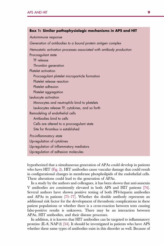
Box 1: Similar pathophysiologic mechanisms in APS and HIT
Autoimmune response
Generation of antibodies to a bound protein antigen complex
Hemostatic activation processes associated with antibody production
Procoagulant state
TF release
Thrombin generation
Platelet activation
Procoagulant platelet microparticle formation
Platelet release reaction
Platelet adhesion
Platelet aggregation
Leukocyte activation
Monocytes and neutrophils bind to platelets
Leukocytes release TF, cytokines, and so forth
Remodeling of endothelial cells
Antibodies bind to cells
Cells are altered to a procoagulant state
Site for thrombus is established
Pro-inflammatory state
Up-regulation of cytokines
Up-regulation of inflammatory mediators
Up-regulation of adhesion molecules
9APS AND HIT
hypothesized that a simultaneous generation of APAs could develop in patientswho have HIT (Fig. 2). HIT antibodies cause vascular damage that could resultin configurational changes in membrane phospholipids of the endothelial cells.These alterations could lead to the generation of APAs.
In a study by the authors and colleagues, it has been shown that anti-annexinV antibodies are consistently elevated in both APS and HIT patients [74].Several authors have shown positive testing of both PF4-heparin antibodiesand APAs in patients [75–77]. Whether the double antibody represents anadditional risk factor for the development of thrombotic complications in thesepatient populations or whether there is a cross-reaction between tests causingfalse-positive results is unknown. There may be an interaction betweenAPAs, HIT antibodies, and their disease processes.
In addition, it is known that HIT antibodies can be targeted to inflammatoryproteins (IL-8, NAP-2) [54]. It should be investigated in patients who have APSwhether these same types of antibodies exist in this disorder as well. Because of
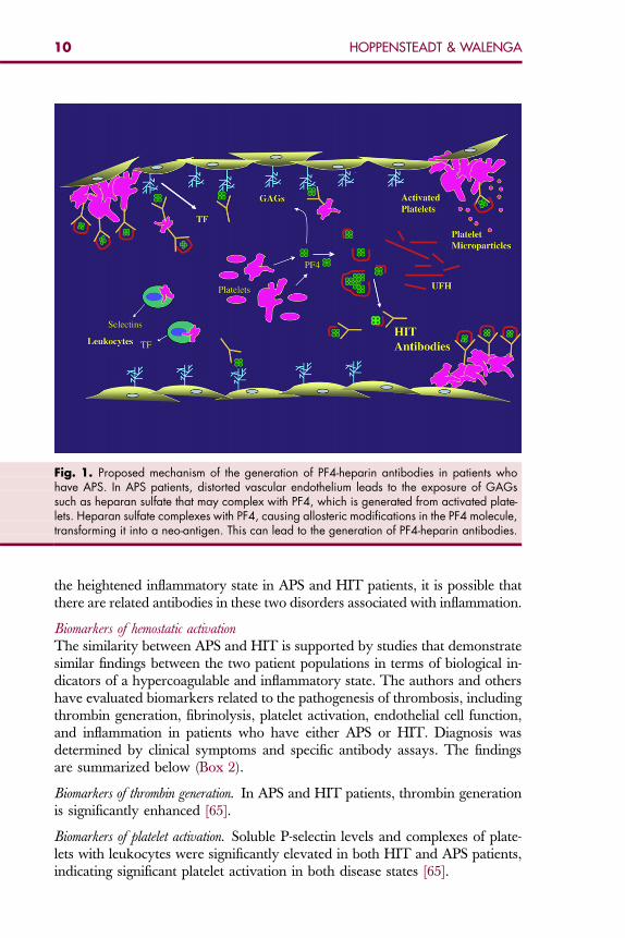
Fig. 1. Proposed mechanism of the generation of PF4-heparin antibodies in patients whohave APS. In APS patients, distorted vascular endothelium leads to the exposure of GAGssuch as heparan sulfate that may complex with PF4, which is generated from activated plate-lets. Heparan sulfate complexes with PF4, causing allosteric modifications in the PF4 molecule,transforming it into a neo-antigen. This can lead to the generation of PF4-heparin antibodies.
10 HOPPENSTEADT & WALENGA
the heightened inflammatory state in APS and HIT patients, it is possible thatthere are related antibodies in these two disorders associated with inflammation.
Biomarkers of hemostatic activation
The similarity between APS and HIT is supported by studies that demonstratesimilar findings between the two patient populations in terms of biological in-dicators of a hypercoagulable and inflammatory state. The authors and othershave evaluated biomarkers related to the pathogenesis of thrombosis, includingthrombin generation, fibrinolysis, platelet activation, endothelial cell function,and inflammation in patients who have either APS or HIT. Diagnosis wasdetermined by clinical symptoms and specific antibody assays. The findingsare summarized below (Box 2).Biomarkers of thrombin generation. In APS and HIT patients, thrombin generationis significantly enhanced [65].
Biomarkers of platelet activation. Soluble P-selectin levels and complexes of plate-lets with leukocytes were significantly elevated in both HIT and APS patients,indicating significant platelet activation in both disease states [65].
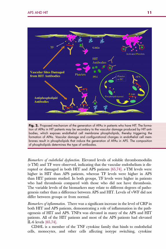
Fig. 2. Proposed mechanism of the generation of APAs in patients who have HIT. The forma-tion of APAs in HIT patients may be secondary to the vascular damage produced by HIT anti-bodies, which exposes endothelial cell membrane phospholipids, thereby triggering theformation of APAs. Vascular damage and configurational changes in endothelial cell mem-branes result in phospholipids that induce the generation of APAs in APS. The compositionof phospholipids determines the type of antibodies.
11APS AND HIT
Biomarkers of endothelial dysfunction. Elevated levels of soluble thrombomodulin(s-TM) and TF were observed, indicating that the vascular endothelium is dis-rupted or damaged in both HIT and APS patients [65,74]. s-TM levels werehigher in HIT than APS patients, whereas TF levels were higher in APSthan HIT patients studied. In both groups, TF levels were higher in patientswho had thrombosis compared with those who did not have thrombosis.The variable levels of the biomarkers may relate to different degrees of patho-genesis rather than a difference between APS and HIT. Levels of vWF did notdiffer between groups or from normal.
Biomarkers of inflammation. There was a significant increase in the level of CRP inboth HIT and APS patients, demonstrating a role of inflammation in the path-ogenesis of HIT and APS. TNFa was elevated in many of the APS and HITpatients. All of the HIT patients and most of the APS patients had elevatedIL-6 levels [65,74].
CD40L is a member of the TNF cytokine family that binds to endothelialcells, monocytes, and other cells affecting isotype switching, cytokine
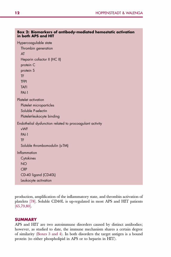
Box 2: Biomarkers of antibody-mediated hemostatic activationin both APS and HIT
Hypercoagulable state
Thrombin generation
AT
Heparin cofactor II (HC II)
protein C
protein S
TF
TFPI
TAFI
PAI-1
Platelet activation
Platelet microparticles
Soluble P-selectin
Platelet-leukocyte binding
Endothelial dysfunction related to procoagulant activity
vWF
PAI-1
TF
Soluble thrombomodulin (s-TM)
Inflammation
Cytokines
NO
CRP
CD-40 ligand (CD40L)
Leukocyte activation
12 HOPPENSTEADT & WALENGA
production, amplification of the inflammatory state, and thrombin activation ofplatelets [78]. Soluble CD40L is up-regulated in most APS and HIT patients[65,79,80].
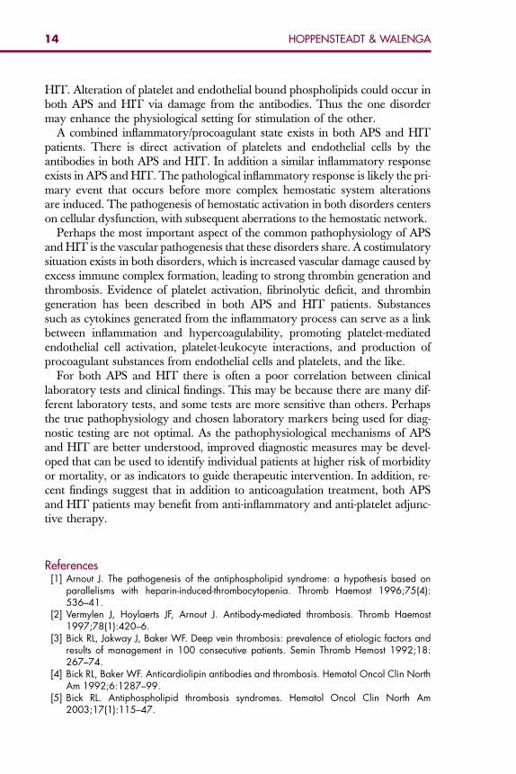
SUMMARYAPS and HIT are two autoimmune disorders caused by distinct antibodies;however, as studied to date, the immune mechanism shares a certain degreeof similarity (Boxes 3 and 4). In both disorders the target antigen is a boundprotein (to either phospholipid in APS or to heparin in HIT).
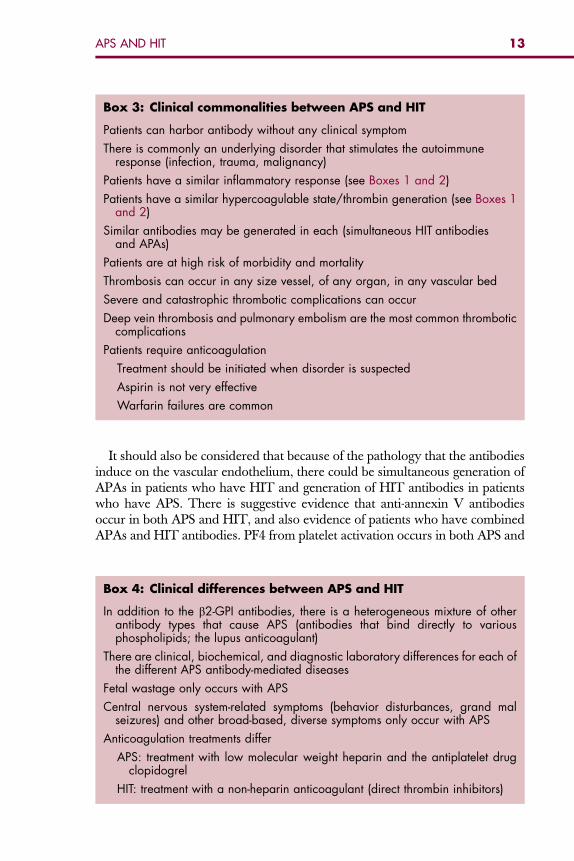
Box 3: Clinical commonalities between APS and HIT
Patients can harbor antibody without any clinical symptom
There is commonly an underlying disorder that stimulates the autoimmuneresponse (infection, trauma, malignancy)
Patients have a similar inflammatory response (see Boxes 1 and 2)
Patients have a similar hypercoagulable state/thrombin generation (see Boxes 1and 2)
Similar antibodies may be generated in each (simultaneous HIT antibodiesand APAs)
Patients are at high risk of morbidity and mortality
Thrombosis can occur in any size vessel, of any organ, in any vascular bed
Severe and catastrophic thrombotic complications can occur
Deep vein thrombosis and pulmonary embolism are the most common thromboticcomplications
Patients require anticoagulation
Treatment should be initiated when disorder is suspected
Aspirin is not very effective
Warfarin failures are common
13APS AND HIT
It should also be considered that because of the pathology that the antibodiesinduce on the vascular endothelium, there could be simultaneous generation ofAPAs in patients who have HIT and generation of HIT antibodies in patientswho have APS. There is suggestive evidence that anti-annexin V antibodiesoccur in both APS and HIT, and also evidence of patients who have combinedAPAs and HIT antibodies. PF4 from platelet activation occurs in both APS and
Box 4: Clinical differences between APS and HIT
In addition to the b2-GPI antibodies, there is a heterogeneous mixture of otherantibody types that cause APS (antibodies that bind directly to variousphospholipids; the lupus anticoagulant)
There are clinical, biochemical, and diagnostic laboratory differences for each ofthe different APS antibody-mediated diseases
Fetal wastage only occurs with APS
Central nervous system-related symptoms (behavior disturbances, grand malseizures) and other broad-based, diverse symptoms only occur with APS
Anticoagulation treatments differ
APS: treatment with low molecular weight heparin and the antiplatelet drugclopidogrel
HIT: treatment with a non-heparin anticoagulant (direct thrombin inhibitors)
14 HOPPENSTEADT & WALENGA
HIT. Alteration of platelet and endothelial bound phospholipids could occur inboth APS and HIT via damage from the antibodies. Thus the one disordermay enhance the physiological setting for stimulation of the other.
A combined inflammatory/procoagulant state exists in both APS and HITpatients. There is direct activation of platelets and endothelial cells by theantibodies in both APS and HIT. In addition a similar inflammatory responseexists in APS and HIT. The pathological inflammatory response is likely the pri-mary event that occurs before more complex hemostatic system alterationsare induced. The pathogenesis of hemostatic activation in both disorders centerson cellular dysfunction, with subsequent aberrations to the hemostatic network.
Perhaps the most important aspect of the common pathophysiology of APSand HIT is the vascular pathogenesis that these disorders share. A costimulatorysituation exists in both disorders, which is increased vascular damage caused byexcess immune complex formation, leading to strong thrombin generation andthrombosis. Evidence of platelet activation, fibrinolytic deficit, and thrombingeneration has been described in both APS and HIT patients. Substancessuch as cytokines generated from the inflammatory process can serve as a linkbetween inflammation and hypercoagulability, promoting platelet-mediatedendothelial cell activation, platelet-leukocyte interactions, and production ofprocoagulant substances from endothelial cells and platelets, and the like.
For both APS and HIT there is often a poor correlation between clinicallaboratory tests and clinical findings. This may be because there are many dif-ferent laboratory tests, and some tests are more sensitive than others. Perhapsthe true pathophysiology and chosen laboratory markers being used for diag-nostic testing are not optimal. As the pathophysiological mechanisms of APSand HIT are better understood, improved diagnostic measures may be devel-oped that can be used to identify individual patients at higher risk of morbidityor mortality, or as indicators to guide therapeutic intervention. In addition, re-cent findings suggest that in addition to anticoagulation treatment, both APSand HIT patients may benefit from anti-inflammatory and anti-platelet adjunc-tive therapy.
References
[1] Arnout J. The pathogenesis of the antiphospholipid syndrome: a hypothesis based onparallelisms with heparin-induced-thrombocytopenia. Thromb Haemost 1996;75(4):536–41.
[2] Vermylen J, Hoylaerts JF, Arnout J. Antibody-mediated thrombosis. Thromb Haemost1997;78(1):420–6.
[3] Bick RL, Jakway J, Baker WF. Deep vein thrombosis: prevalence of etiologic factors andresults of management in 100 consecutive patients. Semin Thromb Hemost 1992;18:267–74.
[4] Bick RL, Baker WF. Anticardiolipin antibodies and thrombosis. Hematol Oncol Clin NorthAm 1992;6:1287–99.
[5] Bick RL. Antiphospholipid thrombosis syndromes. Hematol Oncol Clin North Am2003;17(1):115–47.
15APS AND HIT
[6] Kunkel LA. Acquired circulating anticoagulants. Hematol Oncol Clin North Am 1992;6:1341–57.
[7] Alving BM. Antiphospholipid syndrome, lupus anticoagulants, and anticardiolipinantibodies. In: Loscalzo J, Schafer AI, editors. Thrombosis and hemorrhage. 2nd edition.Baltimore: Williams & Wilkins; 1998. p. 817–33.
[8] Triplett DA. Antiphospholipid antibodies and thrombosis. A consequence, coincidence, orcause? Arch Pathol Lab Med 1993;117:78–88.
[9] Bevers EM, Galli M, Barbui T, et al. Lupus anticoagulant IgG’s are not directed to phospho-lipids only, but to a complex of lipid-bound human prothrombin. Thromb Haemost 1991;66:629–32.
[10] McNeil HP, Simpson RJ, Chesterman CN. Anti-phospholipid antibodies are directed againsta complex antigen that includes a lipid-binding inhibitor of coagulation: b2-glycoproteinI (apolipoprotein H). Proc Natl Acad Sci U S A 1990;87:4120–4.
[11] Matsuura E, Igarashi Y, Fujimoto M, et al. Anticardiolipin cofactor(s) and differentialdiagnosis of autoimmune disease. Lancet 1990;336:177–8.
[12] Nakamura N, Kuragaki C, Shidara Y, et al. Antibody to annexin V has anti-phospholipidand lupus anticoagulant properties. Am J Hematol 1995;49:347–8.
[13] Oosting JD, Derksen RHWM, Bobbick IWG, et al. Antiphospholipid antibodies directedagainst a combination of phospholipids with prothrombin, protein C or protein S. An expla-nation for their pathogenic mechanism. Blood 1993;81:2618–25.
[14] Shibata S, Harpel PC, Gharavi A, et al. Autoantibodies to heparin from patients with anti-phospholipid antibody syndrome inhibits formation of antithrombin III-thrombin complexes.Blood 1994;83:2532–40.
[15] Galli M. Non beta 2-glycoprotein I cofactors for antiphospholipid antibodies. Lupus1996;5:388–92.
[16] Bowie EJW, Thompson JH, Pascuzzi CA, et al. Thrombosis in systemic lupus erythematosusdespite circulating anticoagulants. J Lab Clin Med 1963;62:416–30.
[17] Alarcon-Segovia D. Clinical manifestations of the antiphospholipid syndrome. J Rheumatol1992;18:1916–8.
[18] Triplett DA. Protean clinical presentation of antiphospholipid-protein antibodies (APA).Thromb Haemost 1995;74(1):329.
[19] She W, Chong BH, Hogg PJ, et al. Anticardiolipin antibodies block the inhibition of 2 GPI ofthe factor Xa generating activity of platelets. Thromb Haemost 1993;70:342–5.
[20] Nimpf J, Wurm H, Kostner GM. Beta 2 glycoprotein I (apo-H) inhibits the release reaction ofhuman platelets during ADP-induced aggregation. Atherosclerosis 1987;63:109–14.
[21] Maruyama I. Biology of endothelium. Lupus 1998;7:S41–3.[22] Pierangeli SS, Coleen-Stanfield M, Liu X, et al. Antiphospholipid antibodies from antiphos-
pholipid syndrome patients’ active endothelial cells in vitro and in vivo. Circulation1999;99:1997–2002.
[23] LeRoux G, Wautier MP, Guillevin L, et al. IgG binding to endothelial cells in systemic lupuserythematosus. Thromb Haemost 1986;56:144–6.
[24] Hasselaar P, Derksen RHWM, Blokzijl L, et al. Cross reactivity of antibodies directed againstcardiolipin,DNA,endothelial cells andbloodplatelets. ThrombHaemost1990;63:169–73.
[25] Angles-Cano E, Sultan Y, Clauvel JP. Predisposing factors to thrombosis in systemiclupus erythematosus: possible relation to endothelial damage. J Lab Clin Med 1979;94:317–23.
[26] Bidot CJ, JY W, Horstman LL, et al. Antiphospholipid antibodies and platelet activation asrisk factors for thrombosis in thrombocythaemia. Hematology 2005;10(6):451–6.
[27] Levy Y, Shenkman B, Tamarin I, et al. Increased platelet deposition on extracellular matrixunder flow conditions in patients with antiphospholipid syndrome who experience throm-botic events. Arthritis Rheum 2005;52(12):4011–7.
[28] Gavaghan TP, Krilis SA, Daggard GE. Anticardiolipin antibodies and occlusion of coronaryartery bypass grafts. Lancet 1987;2(8565):977–8.
16 HOPPENSTEADT & WALENGA
[29] Baker WF, Bick RL. Antiphospholipid antibodies in coronary artery disease: a review. SeminThromb Hemost 1994;20(1):27–45.
[30] Reverter JC, Tassies D, Font J, et al. Hypercoagulable state in patients with antiphospholipidsyndrome is related to high induced tissue factor expression on monocytes and to low freeprotein S. Arterioscler Thromb Vasc Biol 1996;16:1319–26.
[31] Kaburaki J, Kuwana M, Yamamoto M, et al. Clinical significance of anti-annexin V anti-bodies in patients with systemic lupus erythematosus. Am J Hematol 1997;54:209–13.
[32] Nakamura N, Shidara Y, Kawaguchi N, et al. Lupus anticoagulant autoantibody inducesapoptosis in umbilical vein endothelial cells: involvement of annexin V. Biochem BiophysRes Commun 1994;205:1488–93.
[33] Matsuda J, Gotoh M, Saith M, et al. Anti-annexin antibody in the sera of patients withhabitual fetal loss or preeclampsia. Thromb Res 1994;75:105–6.
[34] Carreras LO, Defreyn G, Machin SJ, et al. Arterial thrombosis, intrauterine death and‘‘lupus’’ anticoagulant: detection of immunoglobulin interfering with prostacyclin formation.Lancet 1981;I(8214):244–6.
[35] Martinuzzo ME, Maclouf J, Carreras LO, et al. Antiphospholipid antibodies enhance throm-bin induced platelet activation and thromboxane formation. Thromb Haemost 1993;70:667–71.
[36] Rustin MHA, Bull HA, Machin SJ, et al. Effects of lupus anticoagulant in patients withsystemic lupus erythematosus on endothelial cells prostacyclin release and procoagulantactivity. J Invest Dermatol 1988;90:744–8.
[37] Smirov MD, Triplett DA, Comp PC, et al. On the role of phosphatidylethanolamine in theinhibition of activated protein C activity by antiphospholipid antibodies. J Clin Invest1995;95:309–16.
[38] Pengo V, Biasiolo A, Grazia-Fior M. Binding of autoimmune cardiolipin-reactive antibodiesto heparin: a mechanism of thrombosis? Thromb Res 1995;78(5):371–8.
[39] Tannenbaum SH, Finko R, Cines DB. Antibody and immune complexes induce tissue factorproduction by human endothelial cells. J Immunol 1986;137:1532–7.
[40] Ryan J, Brett J, Tijburg P, et al. Tumor necrosis factor induced endothelial tissue factor isassociated with subendothelial matrix vesicles but is not expressed on the apical surface.Blood 1992;80:966–79.
[41] Brandt JT. The effects of lupus anticoagulant on the expression of tissue factor activity bycultured endothelial cells. Thromb Haemost 1991;65:673.
[42] Leurs J, Wissing B, Nerme V, et al. Different mechanisms contribute to the biphasic pattern ofcarboxypeptidase U (TAFIa) generation during in vitro clot lysis in human plasma. ThrombHaemost 2003;89:264–71.
[43] Reutelingsperger CP, van Heerde WL. Annexin V, the regulator of phosphatidylserine-catalyzed inflammation and coagulation during apoptosis. Cell Mol Life Sci 1997;53:527–32.
[44] Warkentin TE, Greinacher A. Heparin-induced thrombocytopenia: recognition, treatment,and prevention. Chest 2004;126:311S–37S.
[45] Walenga JM. Heparin-induced thrombocytopenia and treatment with thrombin inhibitors.Japanese Journal of Thrombosis and Hemostasis 2005;16(6):623–40.
[46] Pouplard C, May MA, Iochmann S, et al. Antibodies to platelet factor 4 heparin after car-diopulmonary bypass in patients anticoagulated with unfractionated heparin or a low mo-lecular weight heparin: clinical implications for HIT. Circulation 1999;99:2530–6.
[47] Lindhoff-Last E, Eichler P, Stein M, et al. A prospective study on the incidence and clinicalrelevance of heparin-induced antibodies in patients after vascular surgery. Thromb Res2000;97:387–93.
[48] Bauer TL, Arepally G, Konkle BA, et al. Prevalence of heparin-associated antibodies withoutthrombosis in patients undergoing cardiopulmonary bypass surgery. Circulation 1997;95:1242–6.
17APS AND HIT
[49] Gluckman TJ, Segal JB, Fredde NL, et al. Incidence of anti-platelet factor 4/heparinantibody induction in patients undergoing percutaneous coronary revascularization. AmJ Cardiol 2005;95:744–7.
[50] Kelton JG. The pathophysiology of heparin-induced thrombocytopenia. Chest 2005;127(2):9S–20S.
[51] Amiral J, Bridey F, Dreyfus M, et al. PF4 complexed to heparin is the target for antibodiesgenerated in heparin-induced thrombocytopenia. Thromb Haemost 1992;68:95–6.
[52] Greinacher A, Potzsch B, Amiral J, et al. Heparin-associated thrombocytopenia: isolation ofthe antibody and characterization of a multimolecular PF4-heparin complex as the majorantigen. Thromb Haemost 1994;71:247–51.
[53] Amiral J, Wolf M, Fischer A-M, et al. Pathogenicity of IgA and/or IgM antibodies to heparin-PF4 complexes in patients with heparin-associated thrombocytopenia. Br J Haematol1996;92:954.
[54] Amiral J, Marfaing-Koka A, Wolf M, et al. Presence of auto-antibodies to interleukin-8 orneutrophil activating peptide-2 in patients with heparin-associated thrombocytopenia.Blood 1996;88:410–6.
[55] Prechel MM, McDonald MK, Jeske WP, et al. Activation of platelets by heparin-inducedthrombocytopenia antibodies in the serotonin release assay is not dependent on thepresence of heparin. J Thromb Haemost 2005;3(10):2168–75.
[56] Prechel MM, Jeske WP, Walenga JM. Platelet-bound PF4 is targeted by HITantibodies in theabsence of heparin. Thromb Haemost 2007;5(Suppl 2):P-W-336.
[57] Ahmad S, Haas S, Hoppensteadt DA, et al. Differential effects of clivarin and heparin inpatients undergoing hip and knee surgery for the generation of anti-heparin-platelet factor4 antibodies. Thromb Res 2003;108:49–55.
[58] Prechel M, Jeske WP, Walenga JM. Laboratory methods for heparin-induced thrombocyto-penia. In: Mousa SA, editor. Anticoagulants, antiplatelets, and thrombolytics: methods inmolecular medicine. Totowa (NJ): Humana Press; 2004. p. 83–93.
[59] Jeske WP, Walenga JM, Szatkowski E, et al. Effect of glycoprotein IIb/IIIa antagonists on theHIT serum induced activation of platelets. Thromb Res 1997;88:271–81.
[60] Warkentin TE, Hayward CPM, Boshkov LK, et al. Sera from patients with heparin-inducedthrombocytopenia generate platelet-derived microparticles with procoagulant activity: anexplanation for the thrombotic complications of heparin-induced thrombocytopenia. Blood1994;84:3691–9.
[61] Walenga JM, Jeske WP, Prechel MM, et al. Newer insights on the mechanism of heparin-induced thrombocytopenia. Semin Thromb Hemost 2004;30(Suppl 1):57–67.
[62] Jeske WP, Vasaiwala S, Schlenker R, et al. Leukocyte activation in heparin-induced throm-bocytopenia. Blood 2000;96(11):29b.
[63] Pouplard C, Lochmann S, Renard B, et al. Induction of monocyte tissue factor expressionby antibodies to heparin-platelet factor 4 complexes developed in heparin-induced throm-bocytopenia. Blood 2001;97(10):3300–2.
[64] Herbert JM, Savi P, Jeske WP, et al. Effect of SR121566A, a potent GPIIb/IIIa antagonist, onthe HIT serum/heparin-induced platelet mediated activation of human endothelial cells.Thromb Haemost 1998;80:326–31.
[65] Walenga JM, Michal K, Hoppensteadt D, et al. Vascular damage correlates betweenheparin-induced thrombocytopenia and the antiphospholipid syndrome. Clin Appl ThrombHemost 1999;5(Suppl 1):S76–84.
[66] Fareed J, Walenga JM, Hoppensteadt DA, et al. Selectins in the HIT syndrome: patho-physiologic role and therapeutic modulation. Semin Thromb Hemost 1999;25(Suppl 1):37–42.
[67] Blank M, Shoenfeld Y, Tavor S, et al. Anti-platelet factor 4/heparin antibodies from patientswith heparin-induced thrombocytopenia provoke direct activation of microvascular endo-thelial cells. Int Immunol 2002;14(2):121–9.
18 HOPPENSTEADT & WALENGA
[68] Visentin GP, Ford SE, Scott PJ, et al. Antibodies from patients with heparin-induced thrombo-cytopenia/thrombosis are specific for platelet factor 4 complexed with heparin or bound toendothelial cells. J Clin Invest 1994;93:81–8.
[69] Suh JS, Aster RH, Visentin GP. Antibodies from patients with heparin-induced thrombocyto-penia recognize different epitopes on heparin:platelet factor 4. Blood 1998;91:916–22.
[70] Walenga JM, Jeske WP, Messmore HL. Mechanisms of venous and arterial thrombosis inheparin-induced thrombocytopenia. J Thromb Thrombolysis 2000;10:S13–20.
[71] Shames DS, Broderick PA. Catastrophic antiphospholipid antibody syndrome. Conn Med2001;65(1):3–5.
[72] Hess D. Stroke associated with antiphospholipid antibodies. Stroke 1992;23(Suppl 1):I23–8.
[73] Toschi V, Motta A, Castelli C, et al. High prevalence of antiphospholipid antibodies in youngpatients with cerebral ischemia of undetermined cause. Stroke 1998;29(9):1759–64.
[74] Baugh MJ, Prechel M, Hoppensteadt D, et al. The role of CD40 ligand and anti-annexinV antibodies in the pathophysiology of APS and HIT. Blood 2000;96(11):272a.
[75] Lasne D, Saffroy R, Bachelot C, et al. Tests for heparin-induced thrombocytopenia in primaryantiphospholipid syndrome [abstract]. Br J Haematol 1997;97:939.
[76] de Larranaga G, Martinuzzo M, Bocassi A, et al. Heparin-platelet factor 4 induced anti-bodies in patients with either autoimmune or alloimmune antiphospholipid antibodies.Thromb Haemost 2002;88:371–3.
[77] Martin-Toutain I, Piette JC, Diemert MC. High prevalence of antibodies to platelet factor4 heparin in patients with antiphospholipid antibodies in absence of heparin-induced throm-bocytopenia. Lupus 2007;16:79–83.
[78] Van Kooten C, Banchereau J. CD40-CD40 ligand. J Leukoc Biol 2000;67:2–17.[79] Desai-Mehta A, Lu L, Ramsey-Goldman R, et al. Hyperexpression of CD40 ligand by B and T
cells in human lupus and its role in pathogenic autoantibody production. J Clin Invest1996;97:2063–73.
[80] Koshy M, Berger D, Crow MK. Increased expression of CD40 Ligand on systemic lupuserythematosus lymphocytes. J Clin Invest 1996;98:826–37.
Hematol Oncol Clin N Am 22 (2008) 19–32
HEMATOLOGY/ONCOLOGY CLINICSOF NORTH AMERICA
Laboratory Evaluationof the Antiphospholipid Syndrome
Debra A. Hoppensteadt, PhD, SH, MT(ASCP)a,*,Nancy Fabbrini, MT(ASCP)b, Rodger L. Bick, MD, PhDc,Harry L. Messmore, MDd, Cafar Adiguzel, MDd,Jawed Fareed, PhD, FAHAe,f
aDepartment of Pathology, Stritch School of Medicine, Loyola University Chicago,2160 S. First Avenue, Maywood, IL 60153, USAbDepartment of Pathology, Edward Hines VA Hospital, Hines, IL, USAc10455 North Central Expressway, Suite 109-320, Dallas, TX 75231, USAdDepartment of Pathology, Loyola University Chicago, 2160 S. First Avenue,Maywood, IL 60153, USAeDepartment of Pathology and Pharmacology, Loyola University Chicago, 2160 S. First Avenue,Maywood, IL 60153, USAfDepartment of Cardiovascular Surgery, Loyola University Chicago, 2160 S. First Avenue,Maywood, IL 60153, USA
Patients recognized as being at increased risk for thrombosis have been re-ferred to as having a ‘‘hypercoagulable state’’ or ‘‘thrombophilia.’’ Box 1lists the acquired blood protein defects that have been identified as lead-
ing to thrombosis [1]. Antiphospholipid syndrome (APLS) is among the mostcommon acquired blood protein defects associated with thrombosis. APLSincludes the lupus anticoagulant (LAC) and anticardiolipin antibodies (ACAs)and the recently recognized subgroups compromised of antibodies to b2-glyco-protein I (b2-GpI), phosphatidylserine, phosphatidylethanolamine, phosphati-dylglycerol, phosphatidylinositol, phosphatidylcholine, and anti-annexin-V[1,2]. The presence of these antibodies, which may be associated with venousthrombosis, arterial thrombosis, or both, is more predictable than that observedin patients who have LAC [2].
Although ACAs and LAC have similarities, there are distinct clinical, labora-tory, and biochemical differences, especially regarding prevalence, origin, possi-ble mechanisms, clinical presentation, laboratory diagnosis, and management.The ACA syndrome is much more common than the LAC syndrome, the ratiobeing 5 or 6 to 1 [2]. Both syndromes may be associated with fetal wastage andthrombocytopenia. In addition, 5% to 15% of patients who have recurrentarterial and venous thrombosis have APLS [3]. In 50% of patients who have
*Corresponding author. E-mail address: [email protected] (D.A. Hoppensteadt).
0889-8588/08/$ – see front matter ª 2008 Elsevier Inc. All rights reserved.doi:10.1016/j.hoc.2007.10.009 hemonc.theclinics.com
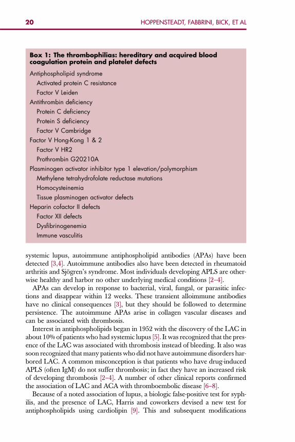
Box 1: The thrombophilias: hereditary and acquired bloodcoagulation protein and platelet defects
Antiphospholipid syndrome
Activated protein C resistance
Factor V Leiden
Antithrombin deficiency
Protein C deficiency
Protein S deficiency
Factor V Cambridge
Factor V Hong-Kong 1 & 2
Factor V HR2
Prothrombin G20210A
Plasminogen activator inhibitor type 1 elevation/polymorphism
Methylene tetrahydrofolate reductase mutations
Homocysteinemia
Tissue plasminogen activator defects
Heparin cofactor II defects
Factor XII defects
Dysfibrinogenemia
Immune vasculitis
20 HOPPENSTEADT, FABBRINI, BICK, ET AL
systemic lupus, autoimmune antiphospholipid antibodies (APAs) have beendetected [3,4]. Autoimmune antibodies also have been detected in rheumatoidarthritis and Sjogren’s syndrome. Most individuals developing APLS are other-wise healthy and harbor no other underlying medical conditions [2–4].
APAs can develop in response to bacterial, viral, fungal, or parasitic infec-tions and disappear within 12 weeks. These transient alloimmune antibodieshave no clinical consequences [3], but they should be followed to determinepersistence. The autoimmune APAs arise in collagen vascular diseases andcan be associated with thrombosis.
Interest in antiphospholipids began in 1952 with the discovery of the LAC inabout 10% of patients who had systemic lupus [5]. It was recognized that the pres-ence of the LAC was associated with thrombosis instead of bleeding. It also wassoon recognized that many patients who did not have autoimmune disorders har-bored LAC. A common misconception is that patients who have drug-inducedAPLS (often IgM) do not suffer thrombosis; in fact they have an increased riskof developing thrombosis [2–4]. A number of other clinical reports confirmedthe association of LAC and ACA with thromboembolic disease [6–8].
Because of a noted association of lupus, a biologic false-positive test for syph-ilis, and the presence of LAC, Harris and coworkers devised a new test forantiphospholipids using cardiolipin [9]. This and subsequent modifications
21LABORATORY EVALUATION OF THE ANTIPHOSPHOLIPID SYNDROME
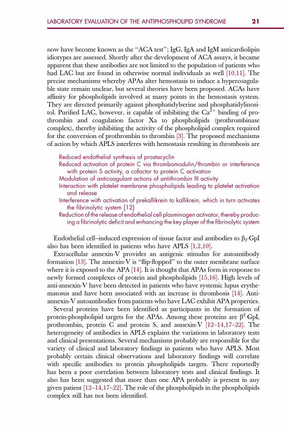
now have become known as the ‘‘ACA test’’; IgG, IgA and IgM anticardiolipinidiotypes are assessed. Shortly after the development of ACA assays, it becameapparent that these antibodies are not limited to the population of patients whohad LAC but are found in otherwise normal individuals as well [10,11]. Theprecise mechanisms whereby APAs alter hemostasis to induce a hypercoagula-ble state remain unclear, but several theories have been proposed. ACAs haveaffinity for phospholipids involved at many points in the hemostasis system.They are directed primarily against phosphatidylserine and phosphatidylinosi-tol. Purified LAC, however, is capable of inhibiting the Ca2þ binding of pro-thrombin and coagulation factor Xa to phospholipids (prothrombinasecomplex), thereby inhibiting the activity of the phospholipid complex requiredfor the conversion of prothrombin to thrombin [3]. The proposed mechanismsof action by which APLS interferes with hemostasis resulting in thrombosis are
Reduced endothelial synthesis of prostacyclinReduced activation of protein C via thrombomodulin/thrombin or interference
with protein S activity, a cofactor to protein C activationModulation of anticoagulant actions of antithrombin III activityInteraction with platelet membrane phospholipids leading to platelet activation
and releaseInterference with activation of prekallikrein to kallikrein, which in turn activates
the fibrinolytic system [12]Reductionof the releaseof endothelial cell plasminogen activator, thereby produc-
ing a fibrinolytic deficit and enhancing the key player of the fibrinolytic system
Endothelial cell–induced expression of tissue factor and antibodies to b2-GpIalso has been identified in patients who have APLS [1,2,10].
Extracellular annexin-V provides an antigenic stimulus for autoantibodyformation [13]. The annexin-V is ‘‘flip-flopped’’ to the outer membrane surfacewhere it is exposed to the APA [14]. It is thought that APAs form in response tonewly formed complexes of protein and phospholipids [15,16]. High levels ofanti-annexin-V have been detected in patients who have systemic lupus erythe-matosus and have been associated with an increase in thrombosis [14]. Anti-annexin-V autoantibodies from patients who have LAC exhibit APA properties.
Several proteins have been identified as participants in the formation ofprotein-phospholipid targets for the APAs. Among these proteins are b2-GpI,prothrombin, protein C and protein S, and annexin-V [12–14,17–22]. Theheterogeneity of antibodies in APLS explains the variations in laboratory testsand clinical presentations. Several mechanisms probably are responsible for thevariety of clinical and laboratory findings in patients who have APLS. Mostprobably certain clinical observations and laboratory findings will correlatewith specific antibodies to protein phospholipids targets. There reportedlyhas been a poor correlation between laboratory tests and clinical findings. Italso has been suggested that more than one APA probably is present in anygiven patient [12–14,17–22]. The role of the phospholipids in the phospholipidscomplex still has not been identified.
22 HOPPENSTEADT, FABBRINI, BICK, ET AL
LABORATORY DIAGNOSIS OF ANTIPHOSPHOLIPIDSYNDROMESAPLS is tested in the laboratory by using both liquid-phase (LAC tests) and thesolid-phase ELISA assays (ACAs and other antibody assays). The testing of an-tibodies to the possible targets of APAs such as b2-GpI and phosphatidylserineis currently under debate. Many consider the current standard tests for ACAsufficiently sensitive and specific for the diagnosis of APA.
Detection of lupus Anticoagulants
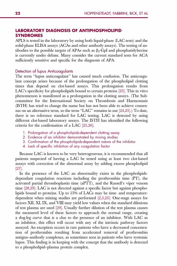
The term ‘‘lupus anticoagulant’’ has caused much confusion. The anticoagu-lant concept arises because of the prolongation of the phospholipid clottingtimes that depend on clot-based assays. This prolongation results fromLAC’s specificity for phospholipids bound to certain proteins [23]. This in vitrophenomenon is manifested as a prolongation in the clotting assays. (The Sub-committee for the International Society on Thrombosis and Haemostasis(ISTH) has tried to change the name but has not been able to achieve consen-sus on an alternative term, so the term ‘‘LAC’’ remains in use [24,25].) To date,there is no reference standard for LAC testing. LAC is detected by usingdifferent clot-based laboratory assays. The ISTH has identified the followingcriteria for the confirmation of a LAC [25,26].1. Prolongation of a phospholipids-dependent clotting assay2. Evidence of an inhibitor demonstrated by mixing studies3. Confirmation of the phospholipids-dependent nature of the inhibitor4. Lack of specific inhibition of any coagulation factor
Because LAC is known to be very heterogeneous, it is recommended that allpatients suspected of having a LAC be tested using at least two clot-basedassays with correction of the abnormal assay by adding excess phospholipid[27].
In the presence of the LAC an abnormality exists in the phospholipids-dependant coagulation reactions including the prothrombin time (PT), theactivated partial thromboplastin time (aPTT), and the Russell’s viper venomtime [28,29]. LAC is not directed against a specific factor but against phospho-lipids bound to proteins. Up to 15% of LACs may be time- and temperature-dependent when mixing studies are performed [2,3,23]. One-stage assays forfactors XII, XI, IX, and VIII may yield low values when the standard dilutionsof test plasma are used [29]. Usually further dilution of the test plasma causesthe measured level of these factors to approach the normal range, creatinga dog-leg curve that is a clue to the presence of an inhibitor. With LAC asan inhibitor, this effect will occur with any of the intrinsic pathway factorsassayed. An exception occurs in rare patients who have a decreased concentra-tion of prothrombin resulting from accelerated removal of prothrombinantigen–antibody complexes, as sometimes seen in patients who have systemiclupus. This finding is in keeping with the concept that the antibody is detectedto a phospholipid–plasma protein complex.
23LABORATORY EVALUATION OF THE ANTIPHOSPHOLIPID SYNDROME
Multiple LAC assays are currently in use. Before these assays as performed, itis essential that blood specimens be collected in 3.2% sodium citrate tubes andcentrifuged to obtain platelet-poor plasma with in 4 hours of blood collection.The platelet count in the platelet-poor plasma should be less than 10,000/lL[3]. If the platelet count is higher, the plasma should be recentrifuged beforetesting or freezing. Outside laboratories should be advised to double-spin theblood before freezing and shipping to ensure a low platelet count. Residual plate-lets will provide phospholipids that can neutralize LAC and result in false-negative results.
The available assays are based on the use of reagents that contain low con-centrations of phospholipids and therefore are sensitive to the LAC in platelet-poor plasma. Initially, a PT was performed with dilute tissue thromboplastinand a reduced number of platelets in the mixture; however, this method missedIgM inhibitors [30]. A new dilute PT assay has become available that uses a re-lipidated recombinant human tissue factor reagent. Similar to the diluteRussell’s viper venom time (dRVVT), this assay incorporates a lower concen-tration of phospholipids in the screening test and a higher concentration in theconfirmatory test. The ratio of these two concentrations is determined, anda cut off is set for the presence of LAC [24,30].
In many patients, an abnormal aPTT is the reason for suspecting a LAC.Such patients probably do not have thrombosis. Therefore it may be reason-able first to do a mixing study to see whether an inhibitor accounts for thisabnormal aPTT. If this test shows a factor deficiency, there is little reason tosuspect a LAC. On the other hand, if an inhibitor is demonstrated, it ismandatory to determine the nature of the inhibitor or inhibitors because factorVIII inhibitors and LACs occasionally have been reported in the same patient[3]. It is reasonable first to exclude the LAC, because it is found much morecommonly. The next step would be to use a platelet-neutralization procedurewith the aPTT test and to perform a dRVV test. If the results of these testsare both positive for LAC, it is, as a rule, not necessary to search for an inhib-itor of factor VIII or factor IX, but it is mandatory to have a PT test as well asa thrombin time to exclude factor deficiencies and heparin, warfarin, or throm-bin inhibitors such as hirudin and argatroban [31].
Zhang and colleagues [32] have proposed that a Staclot LA (DiagnosticaStago, Porsipanny, New Jersey) test be used initially when the LAC is sus-pected . This is a hexagonal phospholipid-based test that is compared witha non–hexagonal-based aPTT using a commercial reagent from a specificsource. This method may be practical when the patient does not have a pro-longed aPTT and a hypercoagulable state is suspected, because that studyshowed the Staclot LA test system to be more sensitive to the than two com-mercially available aPTT reagents that are known to be LAC sensitive. Onedisadvantage of this proposed algorithm is reagent cost, which in this case isclearly an important laboratory budget consideration.
It is reasonable to use an algorithm beginning with a dRVVT or a diluteprothrombin time (dPT) as the initial screening, especially when the LAC
24 HOPPENSTEADT, FABBRINI, BICK, ET AL
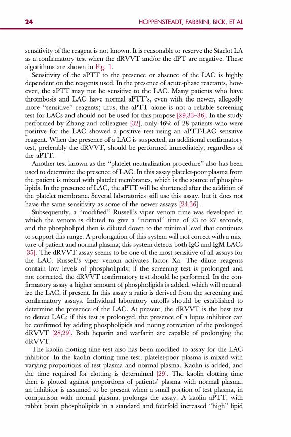
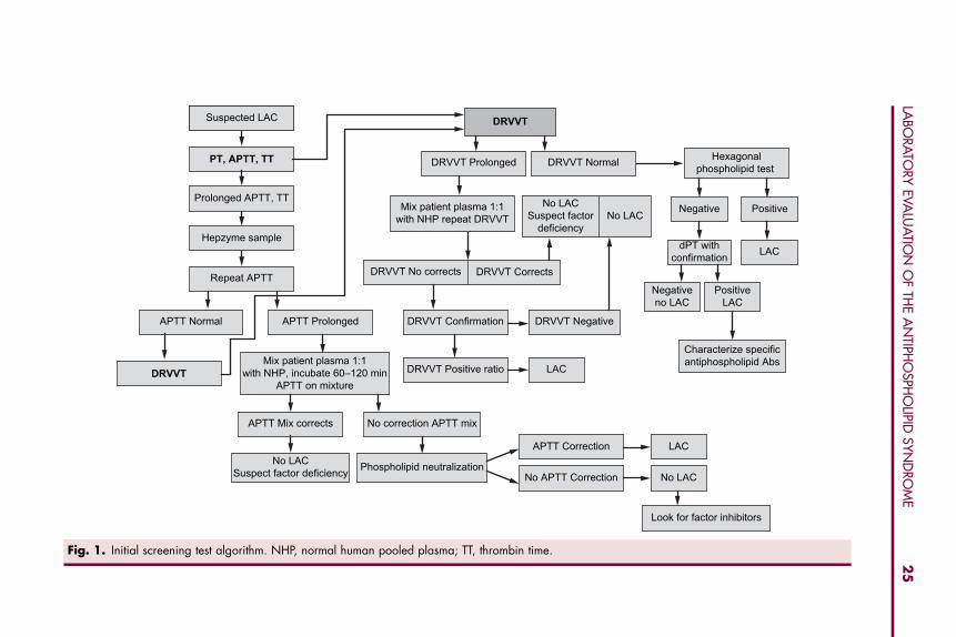
sensitivity of the reagent is not known. It is reasonable to reserve the Staclot LAas a confirmatory test when the dRVVT and/or the dPT are negative. Thesealgorithms are shown in Fig. 1.
Sensitivity of the aPTT to the presence or absence of the LAC is highlydependent on the reagents used. In the presence of acute-phase reactants, how-ever, the aPTT may not be sensitive to the LAC. Many patients who havethrombosis and LAC have normal aPTT’s, even with the newer, allegedlymore ‘‘sensitive’’ reagents; thus, the aPTT alone is not a reliable screeningtest for LACs and should not be used for this purpose [29,33–36]. In the studyperformed by Zhang and colleagues [32], only 46% of 28 patients who werepositive for the LAC showed a positive test using an aPTT-LAC sensitivereagent. When the presence of a LAC is suspected, an additional confirmatorytest, preferably the dRVVT, should be performed immediately, regardless ofthe aPTT.
Another test known as the ‘‘platelet neutralization procedure’’ also has beenused to determine the presence of LAC. In this assay platelet-poor plasma fromthe patient is mixed with platelet membranes, which is the source of phospho-lipids. In the presence of LAC, the aPTT will be shortened after the addition ofthe platelet membrane. Several laboratories still use this assay, but it does nothave the same sensitivity as some of the newer assays [24,36].
Subsequently, a ‘‘modified’’ Russell’s viper venom time was developed inwhich the venom is diluted to give a ‘‘normal’’ time of 23 to 27 seconds,and the phospholipid then is diluted down to the minimal level that continuesto support this range. A prolongation of this system will not correct with a mix-ture of patient and normal plasma; this system detects both IgG and IgM LACs[35]. The dRVVT assay seems to be one of the most sensitive of all assays forthe LAC. Russell’s viper venom activates factor Xa. The dilute reagentscontain low levels of phospholipids; if the screening test is prolonged andnot corrected, the dRVVT confirmatory test should be performed. In the con-firmatory assay a higher amount of phospholipids is added, which will neutral-ize the LAC, if present. In this assay a ratio is derived from the screening andconfirmatory assays. Individual laboratory cutoffs should be established todetermine the presence of the LAC. At present, the dRVVT is the best testto detect LAC; if this test is prolonged, the presence of a lupus inhibitor canbe confirmed by adding phospholipids and noting correction of the prolongeddRVVT [28,29]. Both heparin and warfarin are capable of prolonging thedRVVT.
The kaolin clotting time test also has been modified to assay for the LACinhibitor. In the kaolin clotting time test, platelet-poor plasma is mixed withvarying proportions of test plasma and normal plasma. Kaolin is added, andthe time required for clotting is determined [29]. The kaolin clotting timethen is plotted against proportions of patients’ plasma with normal plasma;an inhibitor is assumed to be present when a small portion of test plasma, incomparison with normal plasma, prolongs the assay. A kaolin aPTT, withrabbit brain phospholipids in a standard and fourfold increased ‘‘high’’ lipid
Suspected LAC
PT, APTT, TT
No LAC
LAC
No LACSuspect factor
deficiency
LAC
No LAC
LAC
Positive
PositiveLAC
Negative
Negativeno LAC
dPT withconfirmation
Characterize specificantiphospholipid Abs
DRVVT Negative
DRVVT Normal
DRVVT Corrects
Mix patient plasma 1:1with NHP repeat DRVVT
Mix patient plasma 1:1with NHP, incubate 60–120 min
APTT on mixture
DRVVT No corrects
DRVVT Prolonged
DRVVT Confirmation
DRVVT Positive ratio
DRVVT
Hexagonalphospholipid test
Prolonged APTT, TT
Hepzyme sample
Repeat APTT
DRVVT
APTT Normal
APTT Correction
No APTT Correction
APTT Mix corrects
APTT Prolonged
No correction APTT mix
Phospholipid neutralizationNo LACSuspect factor deficiency
Look for factor inhibitors
Fig. 1. Initial screening test algorithm. NHP, normal human pooled plasma; TT, thrombin time.
25
LABO
RATO
RYEVA
LUA
TION
OF
THE
AN
TIPHO
SPHO
LIPIDSY
ND
ROM
E
26 HOPPENSTEADT, FABBRINI, BICK, ET AL
concentration to normalize or ‘‘out-inhibit’’ the abnormal ‘‘standard’’ aPTT,also has been used in diagnosis of the lupus inhibitor [29]. This method isknown as the ‘‘rabbit brain neutralization procedure’’; although it is specific(because of rabbit brain neutralization), it is less sensitive than the dRVVT.
An assay known as the ‘‘hexagonal-phase phospholipid neutralization test’’also has been used to detect the presence of the LAC. In this assay, the LAC,if present, is neutralized by the hexagonal-phase phosphatidylethanolamine[24]. This test combines all of the ISTH criteria into one assay. Because this assayis based on the aPTT, elevated factor VIII levels may interfere in this assay [24].
As a practical matter, most clinicians and laboratories are asked to evaluatea patient for LAC after the patient has started anticoagulant therapy. Bothheparin and warfarin prolong most of the tests mentioned previously, includingthe most sensitive test, the dRVVT. If the patient is taking warfarin, and thedRVVT is prolonged and then neutralized by appropriate phospholipids,a LAC is confirmed [28,29]. If, however, the patient is taking heparin andthe dRVVT is prolonged, the neutralization by platelet-derived phospholipidsis not confirmatory, because large amounts of platelet-derived platelet factor IVmay inhibit the heparin effect to correct the test. For example, a commerciallyavailable platelet extract for the platelet neutralization procedure was found tocontain about 100 IU/mL of platelet factor IV, and normal male freeze-thawplatelet extract, commonly prepared for ‘‘platelet or phospholipids neutraliza-tion procedures’’ in the clinical laboratory, contains about 95 IU/mL of plateletfactor IV. These quantities are sufficient to neutralize heparin and shortena prolonged clotting test, thereby rendering a false-positive result for LAC inthe dRVVT or platelet neutralization procedure [28,29]. As a practical matter,therefore, use of the dRVVT offers the most sensitive assay for detection ofa LAC, and neutralization of this test by a non–platelet-derived phospholipids,in particular cephalin, which contains no platelet factor IV, makes this test themost specific as well. In addition, many of the commercial dRVVT reagentshave some sort of heparin neutralizer that is effective up to 1.0 U/mL ofheparin [36].
More recently, with the introduction of newer drugs such as the thrombininhibitors, some publications have warned that these drugs might affect theinterpretation of testing for LAC. Because these agents are capable of inhibitingthrombin and thrombin generation, they interfere in the formation of fibrino-gen to fibrin [32]. In addition, with the development of the new anti-Xa agents,similar caution must be used in interpreting these results [37]. Therefore it isimportant to screen these patients by using the global PT, aPTT, and thrombintime assays.
Because of the marked heterogeneity of APAs, especially in the secondaryAPLS, there is a correlation between elevated ACAs and the LAC in secondaryantiphospholipid-thrombosis syndromes. LAC and ACAs are two separate en-tities, however, and usually one occurs without the other being present, espe-cially in the primary antiphospholipid thrombosis syndromes [38]. LAC hasa stronger association with binding phospholipids of a hexagonal composition
27LABORATORY EVALUATION OF THE ANTIPHOSPHOLIPID SYNDROME
such as phosphatidylcholine, with membrane damage by infection, interleukin-1, or with other mechanisms leading to change from the lamellar to hexagonalform, whereas anticardiolipin antibodies have an affinity to lamellar phospho-lipids in a bilayer (lamellar) composition [39].
Detection of Anticardiolipin Antibodies
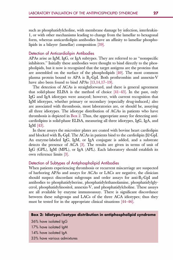
APAs arise as IgM, IgG, or IgA subtypes. They are referred to as ‘‘nonspecificinhibitors.’’ Initially these antibodies were thought to bind directly to the phos-pholipids, but it now is recognized that the target antigens are the proteins thatare assembled on the surface of the phospholipids [40]. The most commonplasma protein bound to APA is B2-GpI. Both prothrombin and annexin-Vhave also been found to bind APAs [13,14,17–19].The detection of ACAs is straightforward, and there is general agreementthat solid-phase ELISA is the method of choice [41–43]. In the past, onlyIgG and IgA idiotypes were assayed; however, with current recognition thatIgM idiotypes, whether primary or secondary (especially drug-induced,) alsoare associated with thrombosis, most laboratories are, or should be, assayingall three idiotypes. The idiotype distribution of ACAs in patients who havethrombosis is depicted in Box 2. Thus, the appropriate assay for detecting anti-cardiolipins is solid-phase ELISA, measuring all three idiotypes, IgG, IgA, andIgM [43].
In these assays the microtiter plates are coated with bovine heart cardiolipinand blocked with B2-GpI. The ACAs in patients bind to the cardiolipin b2-GpI.An enzyme-labeled IgG, IgM, or IgA conjugate is added, and a substratedetects the presence of ACA [3]. The results are given in terms of unit ofIgG (GPL), IgM (MPL), or IgA (APL). Each laboratory should establish itsown reference limits [3].
Detection of Subtypes of Antiphospholipid Antibodies
When patients experiencing thrombosis or recurrent miscarriage are suspectedof harboring APAs and assays for ACAs or LACs are negative, the clinicianshould suspect discordant subgroups and order assays for anti-B2-GpI andantibodies to phosphatidylserine, phosphatidylethanolamine, phosphatidylgly-cerol, phosphatidylinositol, annexin-V, and phosphatidylcholine. These assaysare all available by enzyme immunoassay. There is significant discordancebetween these subgroups and LACs of the three ACA idiotypes; thus theymust be tested for in the appropriate clinical situations [44–46].Box 2: Idiotype/isotype distribution in antiphospholipid syndrome
36% have isolated IgG
17% have isolated IgM
14% have isolated IgA
33% have various admixtures
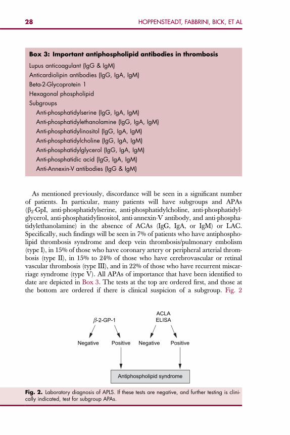
Box 3: Important antiphospholipid antibodies in thrombosis
Lupus anticoagulant (IgG & IgM)
Anticardiolipin antibodies (IgG, IgA, IgM)
Beta-2-Glycoprotein 1
Hexagonal phospholipid
Subgroups
Anti-phosphatidylserine (IgG, IgA, IgM)
Anti-phosphatidylethanolamine (IgG, IgA, IgM)
Anti-phosphatidylinositol (IgG, IgA, IgM)
Anti-phosphatidylcholine (IgG, IgA, IgM)
Anti-phosphatidylglycerol (IgG, IgA, IgM)
Anti-phosphatidic acid (IgG, IgA, IgM)
Anti-Annexin-V antibodies (IgG & IgM)
28 HOPPENSTEADT, FABBRINI, BICK, ET AL
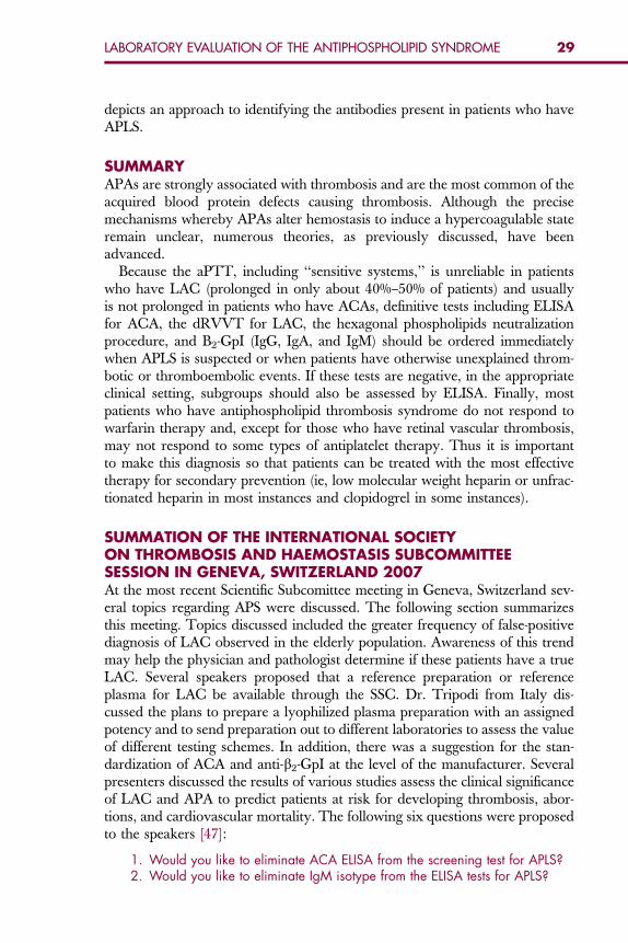
As mentioned previously, discordance will be seen in a significant numberof patients. In particular, many patients will have subgroups and APAs(b2-GpI, anti-phosphatidylserine, anti-phosphatidylcholine, anti-phosphatidyl-glycerol, anti-phosphatidylinositol, anti-annexin-V antibody, and anti-phospha-tidylethanolamine) in the absence of ACAs (IgG, IgA, or IgM) or LAC.Specifically, such findings will be seen in 7% of patients who have antiphospho-lipid thrombosis syndrome and deep vein thrombosis/pulmonary embolism(type I), in 15% of those who have coronary artery or peripheral arterial throm-bosis (type II), in 15% to 24% of those who have cerebrovascular or retinalvascular thrombosis (type III), and in 22% of those who have recurrent miscar-riage syndrome (type V). All APAs of importance that have been identified todate are depicted in Box 3. The tests at the top are ordered first, and those atthe bottom are ordered if there is clinical suspicion of a subgroup. Fig. 2
Negative Positive Negative Positive
-2-GP-1ACLAELISA
Antiphospholipid syndrome
Fig. 2. Laboratory diagnosis of APLS. If these tests are negative, and further testing is clini-cally indicated, test for subgroup APAs.
29LABORATORY EVALUATION OF THE ANTIPHOSPHOLIPID SYNDROME
depicts an approach to identifying the antibodies present in patients who haveAPLS.
SUMMARYAPAs are strongly associated with thrombosis and are the most common of theacquired blood protein defects causing thrombosis. Although the precisemechanisms whereby APAs alter hemostasis to induce a hypercoagulable stateremain unclear, numerous theories, as previously discussed, have beenadvanced.
Because the aPTT, including ‘‘sensitive systems,’’ is unreliable in patientswho have LAC (prolonged in only about 40%–50% of patients) and usuallyis not prolonged in patients who have ACAs, definitive tests including ELISAfor ACA, the dRVVT for LAC, the hexagonal phospholipids neutralizationprocedure, and B2-GpI (IgG, IgA, and IgM) should be ordered immediatelywhen APLS is suspected or when patients have otherwise unexplained throm-botic or thromboembolic events. If these tests are negative, in the appropriateclinical setting, subgroups should also be assessed by ELISA. Finally, mostpatients who have antiphospholipid thrombosis syndrome do not respond towarfarin therapy and, except for those who have retinal vascular thrombosis,may not respond to some types of antiplatelet therapy. Thus it is importantto make this diagnosis so that patients can be treated with the most effectivetherapy for secondary prevention (ie, low molecular weight heparin or unfrac-tionated heparin in most instances and clopidogrel in some instances).
SUMMATION OF THE INTERNATIONAL SOCIETYON THROMBOSIS AND HAEMOSTASIS SUBCOMMITTEESESSION IN GENEVA, SWITZERLAND 2007At the most recent Scientific Subcomittee meeting in Geneva, Switzerland sev-eral topics regarding APS were discussed. The following section summarizesthis meeting. Topics discussed included the greater frequency of false-positivediagnosis of LAC observed in the elderly population. Awareness of this trendmay help the physician and pathologist determine if these patients have a trueLAC. Several speakers proposed that a reference preparation or referenceplasma for LAC be available through the SSC. Dr. Tripodi from Italy dis-cussed the plans to prepare a lyophilized plasma preparation with an assignedpotency and to send preparation out to different laboratories to assess the valueof different testing schemes. In addition, there was a suggestion for the stan-dardization of ACA and anti-b2-GpI at the level of the manufacturer. Severalpresenters discussed the results of various studies assess the clinical significanceof LAC and APA to predict patients at risk for developing thrombosis, abor-tions, and cardiovascular mortality. The following six questions were proposedto the speakers [47]:
1. Would you like to eliminate ACA ELISA from the screening test for APLS?2. Would you like to eliminate IgM isotype from the ELISA tests for APLS?
30 HOPPENSTEADT, FABBRINI, BICK, ET AL
3. Do you agree with the Sydney consensus recommendations that a diagnosisof APLS can be made on the basis of testing for ACA alone?
4. Do you agree with the Sydney consensus recommendations that a diagnosisof APLS can be made on the basis of testing for anti-b2-GpI alone?
5. Should the guidelines for the diagnosis of LAC be updated?6. Would you like to change the laboratory classifications for the diagnosis of
APLS?
References
[1] Bick RL, Jakway J, Baker WF. Deep vein thrombosis: prevalence of etiologic factors andresults of management in 100 consecutive patients. Semin Thromb Hemost 1992;18:267–74.
[2] Bick RL, Baker WF. Anticardiolipin antibodies and thrombosis. Hematol Oncol Clin NorthAm 1992;6:1287–99.
[3] Fritsma GA, Marques MB. Thrombosis risk testing. In: Rodah BF, Fritsma GA, Doig K, editors.Hematology: clinical principles and applications. St. Louis (MO): Saunders Elsevier; 2007.p. 605–28.
[4] Triplett DA. Laboratory diadnosis of lupus anticoagulant. Semin Thromb Hemost 1990;16:182–92.
[5] Conle CL, Hartmann RC. A hemorrhagic disorder caused by circulating anticoagulants inpatients with disseminated lupus erythematosus. J Clin Invest 1952;31:621–2.
[6] Bowie EJW, Thompson JH, Pascuzzi CA, et al. Thrombosis in systemic lupus erythematosusdespite circulating anticoagulants. J Lab Clin Med 1963;62:416–30.
[7] Alarcon-Segovia D. Clinical manifestations of the antiphospholipid syndrome. J Rheumatol1992;18:1916–8.
[8] Triplett DA. Antiphospholipid antibodies and thrombosis. A consequence, coincidence,or cause? Arch Pathol Lab Med 1993;117:78–88.
[9] Harris EN, Gharavi AE, Boey ML. Anticardiolipin antibodies: detection by radioimmunoas-says and association with thrombosis in systemic lupus erythematosus. Lancet 1983;2:1211–4.
[10] Bick RL, Ucar K. Hypercoagulability and thrombosis. Hematol Oncol Clin North Am1992;6:1421–31.
[11] Triplett DA. Antiphospholipid antibodies. In: Hathway WE, Goodnight SH, editors. Disor-ders of hemostasis and thrombosis. 2nd edition. New York: McGraw-Hill; 2001.p. 405–19.
[12] Angles-Cano E, Sultan Y, Clauvel JP. Predisposing factors to thrombosis in systemic lupuserythematosus: possible relation to endothelial damage. J Lab Clin Med 1979;94:317–23.
[13] Nakamura N, Kawasaki C, Shidara Y, et al. Antibody to annexin V has anti-phospholipidand lupus anticoagulant properties. Am J Hematol 1995;49:347–8.
[14] Kaburaki J, Kuwan M, Yamamoto M, et al. Clinical significance of anti-annexin Vantibodiesin patients with systemic lupus erythematosus. Am J Hematol 1997;54:209–13.
[15] Lie JT. Vasculopathy of the antiphospholipid syndromes revisited: thrombosis is the culpritand vasculitis the consort. Lupus 1966;5:368–71.
[16] Lopez-Pedrera C, Buendia P, Barbarroja N, et al. Antiphospholipid mediated thrombosis:interplay between anticardiolipin antibodies and vascular cells. Clin Appl Thromb Hemost2006;12:41–5.
[17] McNeil HP, Simpson RJ, Chesterman CN, et al. Antiphospholipid antibodies are directedagainst a complex antigen that includes a lipid binding inhibitor of coagulation: 2 glycopro-tein I (apolipoprotein H). Proc Natl Acad Sci U S A 1990;87:4120–4.
31LABORATORY EVALUATION OF THE ANTIPHOSPHOLIPID SYNDROME
[18] Miesbach W, Matthias T, Scharrer I. Identification of thrombin antibodies in patients withantiphospholipid syndrome. Ann N Y Acad Sci 2005;1050:250–6.
[19] Bertolaccini ML, Hughes GR. Antiphospholipid antibody testing: which are most useful fordiagnosis? Rheum Dis Clin North Am 2006;32:455–63.
[20] Roubey AS. Autoantibodies to phospholipid-binding plasma proteins: a new view of lupusanticoagulants and other ‘‘antiphospholipid’’ autoantibodies. Blood 1994;84:2854–67.
[21] Oosting JD, Derksen RHWM, Bobbick IWG, et al. Antiphospholipid antibodies directedagainst a combination of phospholipids with prothrombin, protein C or protein S. An expla-nation for their pathogenic mechanism. Blood 1993;81:2618–25.
[22] Matsuda J, Gotoh M, Saith M, et al. Anti-annexin antibody in the sera of patients withhabitual fetal loss or pre-eclampsia. Thromb Res 1994;75:105–6.
[23] Teruya J, West AG, Suell MN. Lupus anticoagulant assays: questions answered and to beanswered. Arch Pathol Lab Med 2007;131(6):885–9.
[24] Exner T, Triplett DA, Taberner D, et al. Guidelines for testing and revised criteria for lupusanticoagulants. Thromb Haemost 1991;65:320–2.
[25] Brandt JT, Triplett DA, Alving B, et al. Criteria for the diagnosis of lupus anticoagulants, anupdate on behalf of the subcommittee on lupus anticoagulants/antiphospholipid antibodiesof the ISTH. Thromb Haemost 1995;74:1597–603.
[26] Greaves M, Cohen H, Machin SJ, et al. Guidelines on the investigation and management ofthe antiphospholipid syndrome. Br J Haematol 2000;109:704–15.
[27] Acosta M, Edwards R, Jaffe EI, et al. Practical approach to pediatric patients referred with anabnormal coagulation profile. Arch Pathol Lab Med 2005;129:1011–6.
[28] Bick RL, Baker WF. Antiphospholipid syndrome and thrombosis. Semin Thromb Hemost1999;25:333–9.
[29] Kunkel L. Acquired circulating anticoagulants. Hematol Oncol Clin North Am 1992;6:1341–58.
[30] Mackie IJ, Lawie AS, Greenfield RS, et al. A new lupus anticoagulant test based on diluteprothrombin time. Thromb Res 2004;114:673–4.
[31] Genzen JR, Miller JL. Presence of direct thrombin inhibitors can affect the results interpreta-tion of lupus anticoagulant testing. Am J Clin Pathol 2005;124(4):586–93.
[32] Zhang L, Whitis JG, Embry MB, et al. A simplified algorithm for the laboratory detection oflupus anticoagulants. Utilization of two automated integrated tests. Am J Clin Pathol2005;124:894–901.
[33] Triplett DA, et al. Laboratory evaluation of circulating anticoagulants. In: Bick RL,Bennett RM, Byrnes RK, editors. Hematology: clinical and laboratory practice. Saint Louis(MO): C.V. Mosby Publisher; 1993. p. 1539–48.
[34] Mannucci PM, Canciani MT, Mari D, et al. The varied sensitivity of partial thromboplastinand prothrombin time reagents in the demonstration of the lupus-like inhibitor. Scand J Hae-matol 1979;22:423–32.
[35] Thiagarajan P, Pengo V, Shapiro SS. The use of the diluted Russel viper venom time for thediagnosis of lupus anticoagulants. Blood 1986;68:869–74.
[36] Teruya J, West AG, Suell MN. Lupus anticoagulant assays. Arch Pathol Lab Med 2007;131:885–9.
[37] Laux V, Hinder M, et al. Rationale for direct factor Xa inhibitors in acute coronarysyndromes. In: Kipshidze NN, Fareed J, Mosses JW, editors. Textbook of internationalcardiovascular pharmacology. London: Informa Healthcare; 2007. p. 119–26.
[38] Bick RL. The antiphospholipid thrombosis syndromes: a common multidisciplinary medicalproblem. Clin Appl Thromb Hemost 1997;3:270–83.
[39] Rauch J, Tannenbaum M, Janoff AS. Distinguishing plasma lupus anticoagulants from anti-factor antibodies using hexagonal (II) phase phospholipids. Thromb Haemost 1989;62:892–6.
[40] Kandiah DA, Sheng YH, Krilis SA. Beta-2 glycoprotein I: target antigen for autoantibodies inthe:antiphospholipid symdrome. Lupus 1996;5:381–6.
32 HOPPENSTEADT, FABBRINI, BICK, ET AL
[41] Falcon CR, Hoffer AM, Forastiero RR, et al. Clinical significance of various ELISA assays fordetecting antiphospholipid antibodies. Thromb Haemost 1990;64:21–5.
[42] Rudge AC, Reynolds R, Bolye CC, et al. Measurement of anti-cardiolipin antibodies by anenzyme-linked immunosorbent assay (ELISA): standardization and quantization of results.Clin Exp Immunol 1985;62:738–45.
[43] Reyes H, Dearing L, Shoenfeld Y. Antiphospholipid antibodies: a critique of their heteroge-neity and hegemony. Semin Thromb Hemost 1994;20:89–100.
[44] Cabral AR, Amigo MC, Cabiedes J, et al. The antiphospholipid/cofactor syndromes: a pri-mary variant with antibodies to beta-2-glycoprotein-I but no antibodies detectable in stan-dard antiphospholipid assays. Am J Med 1996;101:472–81.
[45] Falcon CR, Hoffer AM, Carreras LO. Antiphosphatidylinositol antibodies as markers of theantiphospholipid syndrome. Thromb Res 1990;63:321–2.
[46] Falcon CR, Hoffer AM, Carreras LO. Evaluation of the clinical and laboratory association ofantiphosphatodylethanolamine antibodies. Thromb Res 1990;59:383–8.
[47] Lupus anticoagulant/phospholipid-dependent antibodies 2007 SSC Subcommitteeminutes. Avaialable at: http://www.med.unc.edu/isth/SSC/07sscminutes/07luous.htm.Accessed September 3, 2007.
Hematol Oncol Clin N Am 22 (2008) 33–52
HEMATOLOGY/ONCOLOGY CLINICSOF NORTH AMERICA
The Clinical Spectrumof Antiphospholipid Syndrome
William F. Baker, Jr, MD, FACPa,b,c,*,Rodger L. Bick, MD, PhD, FACPd
aDavid Geffen School of Medicine, Center for Health Sciences, University of California,Los Angeles, Los Angeles, CA, USAbCalifornia Clinical Thrombosis Center, 9330 Stockdale Highway, Suite 300, Bakersfield,CA 93311, USAcThrombosis, Hemostasis and Special Hamatology Clinic, Kern Medical Center, Bakersfield,CA 93305, USAd10455 North Central Expressway, Suite 109-320, Dallas, TX 75231, USA
The antiphospholipid syndrome (APS), first described in 1986 by Hughes,Harris, and Gharavi [1], is an acquired thrombophilic disorder in whichautoantibodies are produced to a variety of phospholipids and phospho-
lipid-binding proteins [2,3]. Clinical manifestations range from no symptomsto imminently life-threatening, catastrophic APS (CAPS) (Fig. 1). By definition,from the 1999 Sapporo International Consensus Statement on Preliminary Cri-teria for the Classification of the Antiphospholipid Syndrome, and from the2006 International Consensus Statement on an Update of the ClassificationCriteria for Definite Antiphospholipid Syndrome, a patient with ‘‘definite’’APS must have persistent high-titer antiphospholipid antibodies (aPL) associ-ated with a history of arterial or venous thrombosis (or both), or recurrentpregnancy morbidity [4,5]. Virtually any organ or organ system may beaffected by the consequences of large-vessel or microvessel thrombosis(Box 1). More venous thrombotic events than arterial thrombotic events areobserved [2,6]. A radiographic series identified 59% of thrombi as venous,28% as arterial, and 13% as both arterial and venous [6]. It has generally beenreported that the most common presentation of APS is deep vein thrombosis(DVT), occurring in from 29% to 55% of patients on follow-up of 6 years.Of these patients, at least 50% are demonstrated to have concomitant pulmo-nary embolism (PE) [6–8].
Primary APS has generally been defined as the presence of aPL in patientswith idiopathic thrombosis but no evidence of autoimmune disease or otherinciting factor, such as infection, malignancy, hemodialysis or drug-inducedaPL [9]. The term secondary APS has been used when patients with a wide
*Corresponding author. E-mail address: [email protected] (W.F. Baker, Jr).
0889-8588/08/$ – see front matter ª 2008 Elsevier Inc. All rights reserved.doi:10.1016/j.hoc.2007.10.007 hemonc.theclinics.com
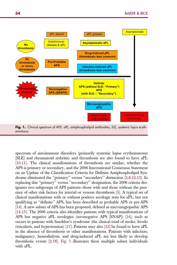
Thrombosis
present
No
thrombosis
Asymptomatic
Life-threateningThrombosis
No
thrombosis
or micro
thrombosis
aPL presentaPL absent
Seronegative
APS (SNAPS)
Definite
APS (without SLE- “Primary”)
APS
(with SLE – “Secondary”)
Microangiopathic
APS
Catastrophic
APS (CAPS)
Autoimmunedisease & aPL
Pre-Probable
APS
Asymptomatic aPL
Drug-induced aPL
(thrombosis less common)
Infection-induced aPL
(thrombosis less common)
Fig. 1. Clinical spectrum of APS. aPL, antiphospholipid antibodies; SLE, systemic lupus eryth-ematosus.
34 BAKER & BICK
spectrum of autoimmune disorders (primarily systemic lupus erythematosus[SLE] and rheumatoid arthritis) and thrombosis are also found to have aPL[10,11]. The clinical manifestations of thrombosis are similar, whether theAPS is primary or secondary, and the 2006 International Consensus Statementon an Update of the Classification Criteria for Definite Antiphospholipid Syn-drome eliminated the ‘‘primary’’ versus ‘‘secondary’’ distinction [2,8,12,13]. Inreplacing this ‘‘primary’’ versus ‘‘secondary’’ designation, the 2006 criteria des-ignates two subgroups of APS patients—those with and those without the pres-ence of other risk factors for arterial or venous thrombosis [5]. A typical set ofclinical manifestations with or without positive serologic tests for aPL, but notqualifying as ‘‘definite’’ APS, has been described as probable APS or pre-APS[14]. A new subset of APS has been proposed, defined as microangiopathic APS[14,15]. The 2006 criteria also identifies patients with typical manifestations ofAPS but negative aPL serologies (seronegative APS [SNAP]) [16], such asoccurs in patients with Sneddon’s syndrome (the clinical triad of stroke, livedoreticularis, and hypertension) [17]. Patients may also [12] be found to have aPLin the absence of thrombosis or other manifestations. Patients with infection,malignancy, hemodialysis, and drug-induced aPL are less likely to developthrombotic events [2,18]. Fig. 1 illustrates these multiple subset individualswith aPL.
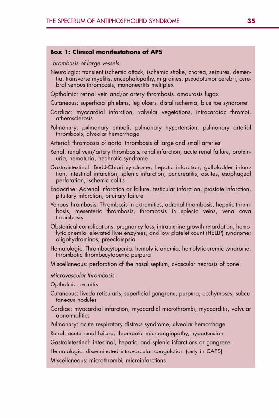
Box 1: Clinical manifestations of APS
Thrombosis of large vessels
Neurologic: transient ischemic attack, ischemic stroke, chorea, seizures, demen-tia, transverse myelitis, encephalopathy, migraines, pseudotumor cerebri, cere-bral venous thrombosis, mononeuritis multiplex
Opthalmic: retinal vein and/or artery thrombosis, amaurosis fugax
Cutaneous: superficial phlebitis, leg ulcers, distal ischemia, blue toe syndrome
Cardiac: myocardial infarction, valvular vegetations, intracardiac thrombi,atherosclerosis
Pulmonary: pulmonary emboli, pulmonary hypertension, pulmonary arterialthrombosis, alveolar hemorrhage
Arterial: thrombosis of aorta, thrombosis of large and small arteries
Renal: renal vein/artery thrombosis, renal infarction, acute renal failure, protein-uria, hematuria, nephrotic syndrome
Gastrointestinal: Budd-Chiari syndrome, hepatic infarction, gallbladder infarc-tion, intestinal infarction, splenic infarction, pancreatitis, ascites, esophagealperforation, ischemic colitis
Endocrine: Adrenal infarction or failure, testicular infarction, prostate infarction,pituitary infarction, pituitary failure
Venous thrombosis: Thrombosis in extremities, adrenal thrombosis, hepatic throm-bosis, mesenteric thrombosis, thrombosis in splenic veins, vena cavathrombosis
Obstetrical complications: pregnancy loss; intrauterine growth retardation; hemo-lytic anemia, elevated liver enzymes, and low platelet count (HELLP) syndrome;oligohydraminos; preeclampsia
Hematologic: Thrombocytopenia, hemolytic anemia, hemolytic-uremic syndrome,thrombotic thrombocytopenic purpura
Miscellaneous: perforation of the nasal septum, avascular necrosis of bone
Microvascular thrombosis
Opthalmic: retinitis
Cutaneous: livedo reticularis, superficial gangrene, purpura, ecchymoses, subcu-taneous nodules
Cardiac: myocardial infarction, myocardial microthrombi, myocarditis, valvularabnormalities
Pulmonary: acute respiratory distress syndrome, alveolar hemorrhage
Renal: acute renal failure, thrombotic microangiopathy, hypertension
Gastrointestinal: intestinal, hepatic, and splenic infarctions or gangrene
Hematologic: disseminated intravascular coagulation (only in CAPS)
Miscellaneous: microthrombi, microinfarctions
35THE SPECTRUM OF ANTIPHOSPHOLIPID SYNDROME
36 BAKER & BICK
PREVALENCEIn young, apparently healthy control subjects, the prevalence for both lupusanticoagulant and anticardiolipin antibodies (aCL) is about 1% to 5% [19].The prevalence increases with age, especially in elderly individuals withchronic disease [19]. Although thrombosis may occur in 50% to 70% of patientswith aPL and SLE over a 20-year follow-up period, as many as 30% do not de-velop APS [19]. The Montpellier Antiphospholipid study, which involved 1014patients admitted to the general medical floor with a variety of diagnoses,found 7.1% with aCL but only 28% of these had clinical features of APS[20]. The risk of thrombosis in patients with APS is estimated to range from0.5% to 30% [18]. According to analysis of 1000 patients reported by the mul-ticenter Euro-Phospholipid Project, APS syndrome is more common in womenthan men in about a 5:1 ratio [12]. In patients with SLE, the male/female ratio iseven higher (7:1). Female patients also appear to more frequently demonstratethe clinical features of arthritis, livedo reticularis, and migraine, whereas malesmore often develop myocardial infarction, epilepsy, and lower extremity arte-rial thrombosis [12]. Although the most common age of onset of the clinicalmanifestations of APS has a mean of 31 years [12], the disorder may be seenin children and older patients as well. While 85% of patients in the Euro-Phos-pholipid Project presented between the ages of 15 and 85, onset is uncommonafter age 60 [21]. Among patients presenting over the age of 50, males domi-nated with more strokes and angina pectoris but less livedo reticularis [12].
ASYMPTOMATIC ANTIPHOSPHOLIPID ANTIBODIESAs the precise genesis of aPL is poorly understood. The reason some individ-uals with no underlying medical disorder develop aPL is also not clear. Whysome individuals develop thrombosis and some do not also remains poorlyunderstood. While aPL may be present as a predisposing risk factor, the addi-tion of a triggering risk factor, or ‘‘double hit,’’ may be required for the devel-opment of thrombosis [2]. Identifiable risk factors for transition fromasymptomatic aPL to APS (aPL with thrombosis) include a prior history ofthrombosis [22,23], the presence of lupus anticoagulant [24], and an elevatedlevel of aCL IgG [10,25,26]. Various reports suggest that each of these riskfactors increase the risk of thrombosis about fivefold [27]. Recent analysis ofthe Warfarin Anti-Phospholipid Syndrome study by Galli contradicts olderstudies and confirms that the association between lupus anticoagulant, beta-2-glycoprotein I antibodies (B-2-GP I), antiprothrombin antibodies, and anti–annexin V antibodies with thrombosis is stronger than the association betweenother aPL and thrombosis [28]. Persistence of aPL over time also progressivelyincreases thrombosis risk [10]. There is no evidence that the presence of riskfactors for thrombosis, with the exception of a history of a prior episode ofthrombosis, warrants therapeutic anticoagulation in an asymptomatic individ-ual with aPL [2].
As the line between patients with asymptomatic aPL and those with APS isdefined by the development of large- or small-vessel thrombosis, or pregnancy
37THE SPECTRUM OF ANTIPHOSPHOLIPID SYNDROME
loss, it is important to consider the individuals who are asymptomatic as also atrisk and deserving of careful clinical surveillance for thrombosis. They are athigh risk for the development of venous thrombosis and require meticulousrisk assessment and DVT prophylaxis in high-risk clinical circumstances. Sim-ilarly, many patients with one thrombophilic disorder are found to have an-other and the cumulative risk increases with each additional risk factor. Asarterial thrombosis is a significant risk for patients with aPL, it is also importantto identify patients with other risk factors for arterial thrombosis and to inter-vene to reduce those risk factors as well. With each additional risk factor forvenous or arterial thrombosis, the likelihood increases that an individualwith otherwise asymptomatic aPL will transition to primary APS as the resultof developing a thrombotic event [2].
PROBABLE ANTIPHOSPHOLIPID SYNDROMEMany patients positive for aPL exhibit clinical features suggesting APS but lackthe clinical criteria of vascular thrombosis or pregnancy loss necessary to sub-stantiate a diagnosis of ‘‘definite’’ APS. Such patients have been classified asprobable APS or pre-APS [14]. Clinical manifestations include livedo reticula-ris, chorea, thrombocytopenia, fetal loss, and cardiac valvular lesions [14,29].The most frequent dermatologic manifestation of APS is livedo reticularis.Some studies have indicated that cutaneous manifestations are the first manifes-tation of APS in up to 41% of patients with APS [29]. Patients with Sneddon’ssyndrome who present with livedo reticularis and stroke may have aPL [17]but may also present with the clinical features in the absence of aPL [30]. Live-do reticularis alone may predate APS and the complication of stroke and othertypes of thrombosis [31]. These patients may also have hypertension, cardiacvalvular disorders, epilepsy, and renal artery pathology [14]. Further study isnecessary to determine whether patients in the pre-APS category benefitfrom anticoagulation designed to prevent the later development of vascularthrombosis [14].
SERONEGATIVE ANTIPHOSPHOLIPID SYNDROMEA subset of patients has been identified who exhibit clinical manifestations ofAPS, without identifiable aPL, lupus anticoagulant, B-2-GP I, antiphospholipidsubtype antibodies, or any other recognized aPL on laboratory testing. Theseindividuals are said to have SNAP syndrome [16,30]. Such patients developidiopathic arterial or venous thrombosis and initial testing for aPL is negative.Repeat testing months later may return positive [30].
DEFINITE ANTIPHOSPHOLIPID SYNDROMEThe 1999 Sapporo International Consensus Statement on Preliminary Criteriafor the Classification of the Antiphospholipid Syndrome defined patients withprimary APS as those with both clinical and laboratory criteria for the diagno-sis in the absence of defined connective tissue disease, and patients with second-ary APS as those with APS and the presence of connective tissue disease
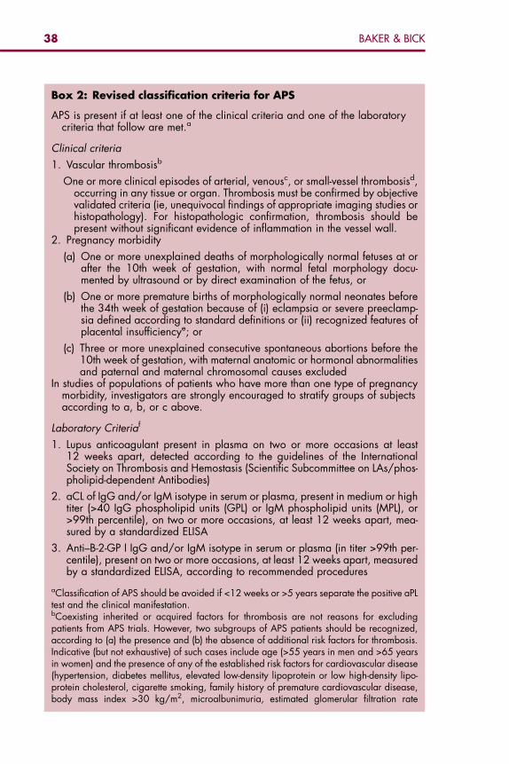
Box 2: Revised classification criteria for APS
APS is present if at least one of the clinical criteria and one of the laboratorycriteria that follow are met.a
Clinical criteria
1. Vascular thrombosisb
One or more clinical episodes of arterial, venousc, or small-vessel thrombosisd,occurring in any tissue or organ. Thrombosis must be confirmed by objectivevalidated criteria (ie, unequivocal findings of appropriate imaging studies orhistopathology). For histopathologic confirmation, thrombosis should bepresent without significant evidence of inflammation in the vessel wall.
2. Pregnancy morbidity
(a) One or more unexplained deaths of morphologically normal fetuses at orafter the 10th week of gestation, with normal fetal morphology docu-mented by ultrasound or by direct examination of the fetus, or
(b) One or more premature births of morphologically normal neonates beforethe 34th week of gestation because of (i) eclampsia or severe preeclamp-sia defined according to standard definitions or (ii) recognized features ofplacental insufficiencye; or
(c) Three or more unexplained consecutive spontaneous abortions before the10th week of gestation, with maternal anatomic or hormonal abnormalitiesand paternal and maternal chromosomal causes excluded
In studies of populations of patients who have more than one type of pregnancymorbidity, investigators are strongly encouraged to stratify groups of subjectsaccording to a, b, or c above.
Laboratory Criteriaf
1. Lupus anticoagulant present in plasma on two or more occasions at least12 weeks apart, detected according to the guidelines of the InternationalSociety on Thrombosis and Hemostasis (Scientific Subcommittee on LAs/phos-pholipid-dependent Antibodies)
2. aCL of IgG and/or IgM isotype in serum or plasma, present in medium or hightiter (>40 IgG phospholipid units (GPL) or IgM phospholipid units (MPL), or>99th percentile), on two or more occasions, at least 12 weeks apart, mea-sured by a standardized ELISA
3. Anti–B-2-GP I IgG and/or IgM isotype in serum or plasma (in titer >99th per-centile), present on two or more occasions, at least 12 weeks apart, measuredby a standardized ELISA, according to recommended procedures
aClassification of APS should be avoided if <12 weeks or >5 years separate the positive aPLtest and the clinical manifestation.bCoexisting inherited or acquired factors for thrombosis are not reasons for excludingpatients from APS trials. However, two subgroups of APS patients should be recognized,according to (a) the presence and (b) the absence of additional risk factors for thrombosis.Indicative (but not exhaustive) of such cases include age (>55 years in men and >65 yearsin women) and the presence of any of the established risk factors for cardiovascular disease(hypertension, diabetes mellitus, elevated low-density lipoprotein or low high-density lipo-protein cholesterol, cigarette smoking, family history of premature cardiovascular disease,body mass index >30 kg/m2, microalbunimuria, estimated glomerular filtration rate
38 BAKER & BICK
<60 mL/min1), inherited thrombophilias, oral contraceptives, nephrotic syndrome, malig-nancy, immobilization, and surgery. Thus, patients who fulfill criteria should be stratified ac-cording to contributing causes of thrombosis.cA thrombotic episode in the past could be considered as a clinical criterion, provided thatthrombosis is proved by appropriate diagnostic means and that no alternative diagnosis orcause of thrombosis is found.dSuperficial venous thrombosis is not included in the clinical criteria.eGenerally accepted features of placental insufficiency include (i) abnormal or non-reassur-ing fetal surveillance test(s) (eg, a non-reactive nonstress test suggestive of fetal hypoxemia),(ii) abnormal Doppler flow velocimetry waveform analysis suggestive of fetal hypoxemia(eg, absent end-diastolic flow in the umbilical artery), (iii) oligohydraminos (eg, an amnioticfluid index of 5 cm or less), or (iv) a postnatal birth weight less than the 10th percentile forthe gestational age.fInvestigators are strongly advised to classify APS patients in studies into one of the followingcategories: I, more than one laboratory criteria present (any combination); IIa, lupus antico-agulant present alone; IIb, aCL present alone; IIc, anti–B-2-GP I present alone.
39THE SPECTRUM OF ANTIPHOSPHOLIPID SYNDROME
(Box 2). The 2006 International Consensus Statement on an Update of theClassification Criteria for Definite Antiphospholipid Syndrome eliminatesthis distinction. Criteria used for diagnosis in this article are those of the2006 Consensus Statement. For a definite diagnosis, patients must have oneor more clinical episodes of arterial, venous, or small-vessel thrombosis occur-ring within any tissue or organ. Thrombosis may include the placenta and re-sult in fetal death or premature birth. Many clinical disorders are associatedwith the presence of aPL, but not rising to the level of evidence necessary tobe included in the 2006 Consensus Statement [5].
Laboratory criteria are well defined and require aCL IgG or IgM or lupusanticoagulant in high titers (>40 IgG phospholipid units [GPL] or IgM phos-pholipid units [MPL] or >99th percentile), confirmed on repeat testing 12 weekslater [4,5,32]. Not included in the 1999 International Criteria, but included inthe 2006 International Criteria are IgG and IgM antibodies to B-2-GP I, whichare also highly predictive of risk for thrombosis. Patients may be found to havenot only aCL or lupus anticoagulant but also other aPL or combinations [5,12].Not included in either criteria are antibodies to the antiphospholipid subtypes,anti-prothrombin or anti–annexin V, which have also been linked to risk forthrombotic events [28,33–39]. The report of 1000 patients by Cervera and col-leagues [12] concluded that over half of the population with APS has primaryAPS (53.1%). Patients designated in the pre-2006 literature with primary APShave no clinical or serologic evidence of SLE, rheumatoid arthritis, or anyother autoimmune disorder, no history of recent infection, the use of drugsknown to trigger aPL, malignancy, or hemodialysis.
The occurrence of clinical manifestations in patients previously designatedwith primary APS is essentially the same as in patients with the so-called ‘‘sec-ondary APS.’’ In the report by Cervera and colleagues [12], the most commonwere DVT (31.7%), thrombocytopenia (21.9%), stroke (13.1%), superficial
40 BAKER & BICK
thrombophlebitis (9.1%), PE (9.0%), fetal loss (8.3%), transient ischemic attack(7.0%), and hemolytic anemia (6.6%). Typically, the presentation is that of anotherwise healthy and asymptomatic individual presenting with DVT, unex-plained thrombocytopenia (platelet count <100,000) or arterial thrombosis(most commonly cerebrovascular). The frequency of idiopathic venous and ar-terial thrombosis in patients with APS is about equal [8,13]. Both large vesselsand small vessels may be involved.
The connective tissue diseases most commonly associated with APS are SLE[40] and rheumatoid arthritis [11]. Other autoimmune disorders reported to beassociated with the development of aPL include polymyalgia rheumatica, Beh-cet’s syndrome, scleroderma, Sjogren’s syndrome, polyarteritis nodosa, relaps-ing polychondritis, giant cell arteritis, Takayasu’s arteritis, autoimmunehemolytic anemia, Evan’s syndrome, and immune thrombocytopenic purpura.The association with SLE and rheumatoid arthritis is well established, while theassociation with other disorders has relied primarily on case reports [2,41].
As many as 12% to 34% of patients with SLE are identified with aPL [40]. From12% to 30% have aCL [11] and the prevalence of lupus anticoagulant ranges hasbeen identified in 15% to 34% of SLE patients [40,42]. Of the SLE patients withaPL, 50% to 70% develop APS over the course of a 20-year follow-up. However,as many as 30% of aPL patients fail to demonstrate clinical evidence of throm-botic complications. Cumulative survival of SLE patients with APS (65%) is sig-nificantly lower at 15 years than for SLE patients without APS (90%) [43].
Patients with APS and SLE more frequently have arthritis, livedo reticularis,thrombocytopenia, leukopenia, or hemolytic anemia [12,44]. Vascular occlu-sion of vessels of any size in patients with SLE and APS is due to thrombosis[2]. The histopathologic identification of vascular wall inflammation found inpatients with SLE vasculitis is not due to APS.
Cardiac thromboemboli may occur in patients with SLE due to Libman-Sacks verrucous vegetations, which may be responsible for valvular stenosis,insufficiency, and cardiac decompensation. Mitral and aortic valvular lesionsare associated with aCL and other manifestations of APS and also correlatewith SLE disease duration and disease activity [45]. Patients with aPL mayalso have a variety of cardiac disorders, independent of the presence of SLE [5].
MICROANGIOPATHIC ANTIPHOSPHOLIPID SYNDROMEPatients with APS may present with a variety of disorders characteristic ofmicrovascular occlusive disease. Such disorders include thrombotic thrombo-cytopenic purpura (TTP); hemolysis, elevated liver enzymes, and low platelets(HELLP) syndrome; thrombotic microangiopathic hemolytic anemia; andCAPS (Asherson’s syndrome). Syndromes may also exhibit overlapping fea-tures characteristic of more than one of these conditions. All share the patho-logic features of microangiopathy [14].
TTP, characterized by thrombocytopenia, microangiopathic hemolytic ane-mia, and cerebrovascular ischemia. demonstrates the pathology of microvascu-lar thrombosis typically associated with the presence of large von Willebrand
41THE SPECTRUM OF ANTIPHOSPHOLIPID SYNDROME
multimers resulting from a deficiency of von Willebrand factor cleaving prote-ase (a disintegrin and metalloproteinase with thrombospondin type 1 motif,13 [ADAMTS13] deficiency) [46]. Patients are also identified with the concom-itant presence of TTP and aPL, as well as TTP, aPL, and SLE [47]. Both pri-mary deficiency of ADAMTS13 [48,49] and ADAMTS13-inhibiting antibodies[50] have been detected in patients with TTP and aPL.
A similar association has been identified in patients with the adult, obstetricform of hemolytic uremic syndrome [51]. Both adults and children with hemo-lytic uremic syndrome have been identified with renal vascular thrombosis andaPL. A similar pathologic appearance is shared among these syndromes,including the presence of capillary congestion and intracapillary fibrin thrombi.Chronic changes are also observed, with characteristic evidence of chronichypoperfusion resulting in atrophy, fibrosis, and scarring [51,52].
Pregnant patients with HELLP syndrome and aPL have also been identified[14]. A review of 16 cases of HELLP and aPL suggests that the development ofHELLP was promoted by the underlying presence of aPL. Review of the pub-lished cases seems to indicate that patients with APS are more likely to havesevere HELLP and this is more likely to be associated with preeclampsia oreclampsia [53].
There also are reported cases of severe HELLP who develop clinical andpathologic features of CAPS. Such patients may develop refractory HELLPand systemic complications of microvascular thrombosis [54]. Just as in otherpatients with CAPS, aggressive therapy involves anticoagulation, intravenousimmune globulin, and plasma exchange [14].
DRUG-INDUCED ANTIPHOSPHOLIPID SYNDROMEA variety of medications have been documented to induce the formation ofaPL. These have included chlorpromazine, phenytoin, hydralazine, procaina-mide, fansidar, quinidine, interferon, and cocaine. Most aPL are of the IgMtype, present at low levels, and not associated with an increased incidence ofthrombosis [55].
INFECTION-ASSOCIATED ANTIPHOSPHOLIPID SYNDROMEA variety of infectious agents trigger the production of aPL [56]. Antibodies in-duced by infection recognize anionic phospholipid epitopes directly rather thanvia cofactors such as B-2-GP I. Autoantibodies are more often IgM than IgG.The clinical features typical of APS are less commonly observed with aPL as-sociated with infections [56]. Some infections, however, have been well docu-mented to be associated with the development of aPL and B-2-GP I and arethus more likely to be associated with the subsequent development of throm-bosis (leprosy, parvovirus B19, HIV, hepatitus C virus, cytomegalovirus)[56]. Infection may be the triggering factor in as many as 40% of cases ofCAPS [57].
42 BAKER & BICK
MALIGNANCY-ASSOCIATED ANTIPHOSPHOLIPIDSYNDROMEIt is well known that malignancy is a high-risk condition for venous thrombo-sis. A variety of solid and hematologic malignancies have been reported to beassociated with the presence of aPL. The relationship between malignancy, thedevelopment of aPL, and thrombosis is poorly understood [58].
CATASTROPHIC ANTIPHOSPHOLIPID SYNDROMECAPS, a syndrome of multisystem involvement as a manifestation of APS, wasfirst described by Asherson [59]. The syndrome was coined Asherson’s syn-drome in 2003 [60]. Occurring in fewer than 1% of APS patients, the syndromeis characterized by multiple small-vessel occlusions leading to multiple organfailure and substantial morbidity and mortality [61]. The syndrome is generallyof acute onset and defined by the involvement of at least three different organsystems over a period of days or weeks. Histopathologically, there is evidenceof small- and large-vessel occlusions. The striking feature of the syndrome isthe presence of an acute microangiopathy, rather than the large-vessel occlu-sions more typically observed in patients with both primary and secondaryAPS [57]. Clinical features are the manifestations of organ and tissue ischemiaand include renal failure due to renal thrombotic microangiopathy, acute respi-ratory failure due to adult respiratory distress syndrome, cerebral injury due tomicrothrombi and microinfarctions, and myocardial failure due to microthrom-bi [61–63]. The 1998 review by Asherson and colleagues [57,61] of 50 patients(5 from their clinic and 45 by review of the literature), identified as many as28% of patients with typical features of disseminated intravascular coagulation.Mortality from the largest two reported series, with a total of 130 patients, wasas high as 48%. Renal involvement was present in 78%, while 66% had lunginvolvement, 56% had central nervous system involvement, 50% had cardiacinvolvement, and 50% had skin involvement [61]. CAPS was more commonin females (66%) compared with males (34%). Twenty-eight of the patients(56%) had primary APS, 15 patients (30%) had defined SLE, 6 patients(12%) had ‘‘lupuslike’’ syndrome, and 1 patient (2%) had rheumatoid arthritis.In the review by Asherson and colleagues [61], thrombocytopenia was reportedin 34 (68%) patients, and hemolytic anemia in 13 (26%). The detection of au-toantibodies was a common finding with lupus anticoagulant (94%), aCL(94%), anti–double-stranded DNA (87% of patients with SLE), antinuclear an-tibodies (58%), anti-Ro/SS-A (8%), anti–ribonucleic protein (8%), and anti-La/SS-B (2%).
Precipitating factors contributed to the development of CAPS in 11 (22%)patients, with infections in 3, oral contraceptives in 3, surgical procedures in4 (three minor and one major), withdrawal in anticoagulation in 2, and hyster-ectomy in 1. A review by Asherson and Cervena in 2003 [56] noted that infec-tion triggered CAPS in as many as 40% of patients with CAPS. An initialthrombosis, felt to be a typical acute thrombosis in a patient with APS, mayrapidly evolve into the systemic microangiopathy, described by Kirchens as
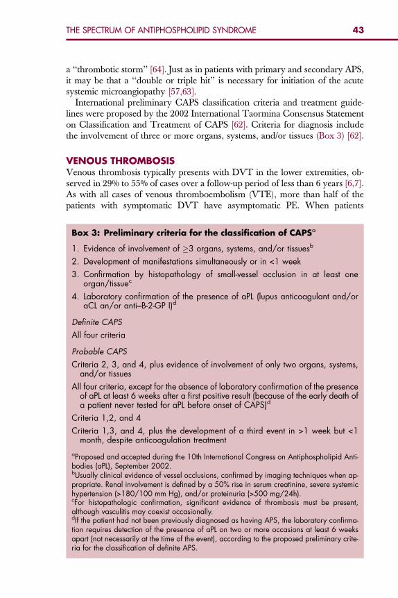
43THE SPECTRUM OF ANTIPHOSPHOLIPID SYNDROME
a ‘‘thrombotic storm’’ [64]. Just as in patients with primary and secondary APS,it may be that a ‘‘double or triple hit’’ is necessary for initiation of the acutesystemic microangiopathy [57,63].
International preliminary CAPS classification criteria and treatment guide-lines were proposed by the 2002 International Taormina Consensus Statementon Classification and Treatment of CAPS [62]. Criteria for diagnosis includethe involvement of three or more organs, systems, and/or tissues (Box 3) [62].
VENOUS THROMBOSISVenous thrombosis typically presents with DVT in the lower extremities, ob-served in 29% to 55% of cases over a follow-up period of less than 6 years [6,7].As with all cases of venous thromboembolism (VTE), more than half of thepatients with symptomatic DVT have asymptomatic PE. When patients
Box 3: Preliminary criteria for the classification of CAPSa
1. Evidence of involvement of �3 organs, systems, and/or tissuesb
2. Development of manifestations simultaneously or in <1 week
3. Confirmation by histopathology of small-vessel occlusion in at least oneorgan/tissuec
4. Laboratory confirmation of the presence of aPL (lupus anticoagulant and/oraCL an/or anti–B-2-GP I)d
Definite CAPS
All four criteria
Probable CAPS
Criteria 2, 3, and 4, plus evidence of involvement of only two organs, systems,and/or tissues
All four criteria, except for the absence of laboratory confirmation of the presenceof aPL at least 6 weeks after a first positive result (because of the early death ofa patient never tested for aPL before onset of CAPS)d
Criteria 1,2, and 4
Criteria 1,3, and 4, plus the development of a third event in >1 week but <1month, despite anticoagulation treatment
aProposed and accepted during the 10th International Congress on Antiphospholipid Anti-bodies (aPL), September 2002.bUsually clinical evidence of vessel occlusions, confirmed by imaging techniques when ap-propriate. Renal involvement is defined by a 50% rise in serum creatinine, severe systemichypertension (>180/100 mm Hg), and/or proteinuria (>500 mg/24h).cFor histopathologic confirmation, significant evidence of thrombosis must be present,although vasculitis may coexist occasionally.dIf the patient had not been previously diagnosed as having APS, the laboratory confirma-tion requires detection of the presence of aPL on two or more occasions at least 6 weeksapart (not necessarily at the time of the event), according to the proposed preliminary crite-ria for the classification of definite APS.

44 BAKER & BICK
present with symptoms typical of PE, at least half will not exhibit clinical man-ifestations of DVT. No particular clinical pattern of venous thrombosis sug-gests that the patient may have underlying aPL. Unusual sites of venousthrombosis have included the upper extremities, intracranial veins, inferiorand superior vena cava, hepatic veins (Budd-Chiari syndrome), portal vein, re-nal vein, and retinal vein [55,65,66]. Thrombosis of the cerebral veins maypresent with acute cerebral infarction [67]. The superficial and deep cerebralvenous system may be extensively involved. Onset of cerebral venous throm-bosis caused by aPL typically presents at a younger age and in a more exten-sive pattern than onset of cerebral venous thrombosis because of other reasons.Thrombosis of the superior saggital sinus has also been reported [68]. In oneseries of 40 patients with cerebral venous thrombosis, 3 had aPL and 2 of these3 also had factor V Leiden mutation [69].
In the setting of idiopathic VTE, laboratory assessment is recommended tosearch for the presence of underlying thrombophilia. Supporting this is theseries of 100 consecutive patients with idiopathic DVT or PE in which 24%were found to have aPL [70]. Testing for aPL is strongly recommended be-cause of the high frequency with which APS is diagnosed [6]. Even in patientswith a family history of venous thrombosis, which suggests an underlying in-herited thrombophilic disorder, testing for aPL is indicated because many pa-tients with one thrombophilic disorder also have another [71,72]. Theclinical manifestations of VTE are the same, whether aPL are present or not.Subsequent to a first episode of VTE, the risk for future venous thrombosis in-creases significantly. In patients with APS, the risk doubles to at least a 30% riskof recurrence without antithrombotic therapy [23].
ARTERIAL THROMBOSISArterial thromboses are less common than venous thromboses and occur ina variety of settings in patients with primary APS [7,8,73]. Of the 1000 patientsin the Euro-Phospholipid Trial, 13.1% presented with stroke, 7% with transientischemic attack, and 2.8% with acute myocardial infarction [12]. Patients witharterial thrombosis most commonly present with transient ischemic attack orstroke (50%) or myocardial infarction (23%) [7,12,65]. These relatively com-mon arterial occlusive events most suggest APS when they occur in individualswithout readily identified risk factors for atherosclerosis. This primarily in-cludes individuals under age 60, without classical risk factors for atherosclerosis(family history, cigarette smoking, hyperlipidemia, hypertension, diabetes mel-litus). The presence of aCL is considered to be a risk factor for first stroke [74].Arterial thrombosis in patients with APS may also involve other large andsmall vessels, which is somewhat unusual for other thrombophilic disordersor atherosclerotic occlusive disease. These potential arterial thromboses includethromboses of brachial and subclavian arteries, axillary artery (aortic arch syn-drome), aorta, iliac, femoral, renal, mesenteric, retinal, and other peripheral ar-teries [2,7,18]. Clinical manifestations, of course, depend on the caliber andlocation of the affected artery.
45THE SPECTRUM OF ANTIPHOSPHOLIPID SYNDROME
CARDIAC DISORDERSArterial occlusion may be either thrombotic or embolic. Premature atheroscle-rosis appears to be accelerated by the presence of aPL and may predispose tocoronary occlusion [75]. Coronary artery thrombosis and embolic occlusion as-sociated with APS results in acute myocardial infarction. Coronary artery graftocclusion [76] and accelerated restenosis of coronary arteries after percutane-ous transluminal coronary angioplasty [77] are also more likely in patientswith APS. Emboli usually arise from mitral or aortic valve vegetations com-posed of platelets and fibrin, and may result in cerebrovascular ischemic events[18]. Echocardiography demonstrates at least one valvular abnormality in asmany as 63% of APS patients. Many of these are asymptomatic and of unclearclinical significance. However, vegetations of the mitral or aortic valve are ob-served in 4% of patients [8]. Valvular thickening, vegetations, regurgitation,premature coronary disease, myocardial infarction, dilated diffuse cardiomyop-athy, congestive heart failure, pericardial effusion, and pulmonary hyperten-sion have all been observed [78].
NEUROLOGIC DISORDERSPrimary thrombosis and embolic occlusion of cerebral arteries result in cerebralinfarction, with clinical manifestations dependent upon the location and caliberof the occluded artery. Most arterial occlusions are of small arteries. Recurrentsmall strokes may contribute to a picture of multiple-infarct dementia [18]. Typ-ical APS patients with stroke are relatively young and lack other classical riskfactors of stroke [79].
Chorea is another clinical disorder that has been strongly linked to the pres-ence of aPL. About 4% of patients with SLE exhibit chorea, which mimics thatseen with rheumatic fever (Sydenhan’s) or Huntington’s chorea. Chorea maypredate the development of aPL and may be followed by acute cerebral ischemia.Other central nervous system manifestations associated with aPL include mi-graine headache, Sneddon’s syndrome, seizures, transverse myelitis, Guillain-Barre syndrome, idiopathic intracranial hypertension, cognitive dysfunction,psychosis, and optic neuritis [80].
OBSTETRICAL DISORDERSAs defined in both the 1999 and 2006 International Consensus Statements, fetaldemise may be attributed to aPL as unexplained fetal death at or after the 10thweek of gestation [4,5]. Premature birth before the 34th week of gestation be-cause of eclampsia or placental insufficiency is also included. Pre-embryonic(<6 weeks’ gestation) and embryonic (6 to 9 weeks’ gestation) losses areincreasingly recognized as an additional complication of aPL. Three or moreunexplained consecutive spontaneous abortions before the 10th week of gesta-tion are also among the recognized diagnostic criteria [4,5]. Other complica-tions of pregnancy may also be observed, including eclampsia, intrauterinegrowth retardation, oligohydraminos, HELLP syndrome, and premature birth.Such patients also demonstrate a high rate of subsequent venous or arterial
46 BAKER & BICK
thrombosis [81]. Pregnancy complications may occur in patients who are onlylater found to develop aPL [14,81].
DERMATOLOGIC DISORDERSDermatologic manifestations may be the first indication of APS. Histopatholog-ically, the most common feature is noninflammatory vascular thrombosis. Clin-ically, patients present with livedo reticularis, necrotizing vasculitis, livedoidvasculitis, cutaneous ulcerations and necrosis, erythematous macules, purpura,ecchymoses, painful skin nodules, and subungual splinter hemorrhages. Aneto-derma, discoid lupus erythematosus, cutaneous T-cell lymphoma, and disor-ders similar to Degos and Sneddon’s syndrome are also rarely observed[41,82]. Patients with livedo reticularis and APS frequently also have cardiacand cerebral thrombotic events, epilepsy, and migraine headaches [83].
PULMONARY MANIFESTATIONSThe most common pulmonary disorder associated with APS is that of PE. Inaddition to thromboembolism, in situ thrombosis has been reported. aPL areassociated with pulmonary hypertension and, in one study of 38 patientswith precapillary pulmonary hypertension, 30% were found to have aPL[84]. Refractory noninflammatory pulmonary vasculopathy characterized bymicrovascular thrombosis may present primarily or in association withCAPS [41].
GASTROINTESTINAL DISORDERSThrombosis of the hepatic veins as a manifestation of APS results in Budd-Chiari syndrome. Mesenteric and portal venous thrombosis in APS are welldescribed. Other manifestations of large- and small-vessel thrombosis includehepatic infarction, pancreatitis, esophageal necrosis, intestinal ischemia and in-farction, colonic ulceration, acalculous cholecystitis with gallbladder necrosis,and giant gastric ulceration [18,41].
RENAL MANIFESTATIONSThe primary renal manifestations of APS are associated with thrombotic micro-angiopathy, designated as aPL-associated nephropathy [5,85]. Other complica-tions may include renal vein thrombosis, renal infarction, renal artery stenosis,and allograft vascular thrombosis [86].
RETINAL DISORDERSVenous and arterial thrombosis of the retinal vasculature is a well-recognizedmanifestation of APS. Presentation strongly suggestive of the presence ofaPL includes the diffuse occlusion of retinal arteries, veins, or both, and neo-vascularization at the time of presentation. Other opthalmic manifestationsincluded optic neuropathy and cilioretinal artery occlusion [87].
47THE SPECTRUM OF ANTIPHOSPHOLIPID SYNDROME
MISCELLANEOUS MANIFESTATIONSA wide variety of clinical disorders have been reported in association with APS.Less common but important are adrenal hemorrhage, bone marrow necrosis(especially in CAPS), and sudden acute sensorineural hearing loss [18].
HEMATOLOGIC DISORDERSThrombocytopenia (platelet count <100,000) [12] is present in from 20% to 40%of patients with APS and is usually mild [88,89]. Severe thrombocytopenia ismost often seen in patients with CAPS and those with concomitant disseminatedintravascular anticoagulation or TTP [14]. The association of aPL with autoim-mune thrombocytopenic purpura has been recognized since the 1985 report byHarris and colleagues [90] and has more recently been reported to be presentin as many as 38% of patients [91]. Follow-up has documented thrombosis in60% of the patients with aPL and only 2.3% of those without aPL [91,92]. To clar-ify the significance of thrombocytopenia in patients with aPL, patients are desig-nated as having aPL-associated thrombocytopenia if there is coexistence of aPLlaboratory criteria with thrombocytopenia (platelet count <100,000) confirmedat least twice, 12 weeks apart and exclusion of patients with TTP, disseminatedintravascular coagulation, pseudothrombocytopenia, or heparin-induced throm-bocytopenia [5].
BLEEDINGPatients with APS rarely develop severe bleeding. Hemorrhage may resultfrom a concurrent coagulopathy or disseminated intravascular coagulation,as may be observed in patients with CAPS. Most cases of severe bleeding solelyattributable to APS are the result of severe, acquired hypo-prothrombinemia[93]. Prolongation of the prothrombin time and activated partial thromboplas-tin time may be attributed to the presence of lupus anticoagulant and the sever-ity of prothrombin deficiency not appreciated. A specific assay for prothrombinmust be obtained for diagnosis. Other disorders that are linked to aPL and thatpredispose to bleeding rather than thrombosis, include acquired thrombocyto-pathies, thrombocytopenia, and acquired inhibitors to coagulation factors [41].
CHILDREN WITH ANTIPHOSPHOLIPID SYNDROMEChildren (2.8% in the Euro-Phospholipid cohort) have more commonly pre-sented with chorea and internal jugular thrombosis than is the case in adults[12]. A variety of presentations have been reported in children, includingDVT, PE, cerebral stroke, myocardial infarction, and peripheral arterial occlu-sion [94,95]. An increased incidence of APS has been observed in children withstroke and migraine headaches [96]. Children with lupus anticoagulant morecommonly present with venous thrombosis and arterial thrombosis in thosewith aCL [97].
48 BAKER & BICK
SUMMARYAPS is a disorder characterized by a wide variety of clinical manifestations.Virtually any organ system or tissue may be affected by the consequences oflarge- or small-vessel thrombosis. There is a broad spectrum of disease amongindividuals with aPL, from asymptomatic to imminently life-threatening CAPS.Patients may exhibit clinical features suggesting APS but not fulfill the Interna-tional Criteria for a ‘‘definite’’ diagnosis. SNAPS patients demonstrate typicalidiopathic thromboses but aPL are not initially detected. Patients definedwith definite APS demonstrate nearly identical sites of venous and arterialthrombosis, regardless of the presence or absence of SLE. MicroangiopathicAPS may present with isolated tissue and organ injury or as the overwhelming‘‘thrombotic storm’’ observed in CAPS.
References
[1] HughesG,Harris EN, GharaviA. Theanticardiolipin syndrome. J Rheumatol 1986;13:486–9.[2] Levine JS, Branch DW, Rauch J. The antiphospholipid syndrome. N Engl J Med2002;346(10):752–63.[3] Roubey RAS. Immunology of the antiphospholipid antibody syndrome. Arthritis Rheum
1996;39:1444–54.[4] Wilson W, Gharavi A, Koike T, et al. International consensus statement on preliminary clas-
sification criteria for definite antiphospholipid syndrome: report of an international work-shop. Arthritis Rheum 1999;42:1309–11.
[5] Miyakis S. International consensus statement on an update of the classification criteria fordefinite antiphospholipid syndrome (APS). J Thromb Haemost 2006;4:295–306.
[6] Provenzale JM, Ortel T, Allen N. Systemic thrombosis in patients with antiphospholipidsyndrome: lesion distribution and imaging findings. Am J Roentgenol 1998;170:285–90.
[7] Asherson R, Khamashta M, Ordi-Rios J, et al. The ‘‘primary’’ antiphospholipid syndrome:major clinical and serological features. Medicine (Baltimore) 1989;68:366–74.
[8] Vianna JL, Khamashta M, Ordi J, et al. Comparison of the primary and secondary antiphos-pholipid syndrome: a European multicenter study of 114 patients. Am J Med 1994;96:3–9.
[9] Piette JC, Wechsler B, Frances C, et al. Exclusion criteria for primary antiphospholipid syn-drome. J Rheumatol 1993;20:1802–4.
[10] Alarcon-Segovia D, Deleze M, O CV, et al. Antiphospholipid antibodies and the antiphos-pholipid syndrome in systemic lupus erythematosus: a prospective analysis of 500 consec-utive patients. Medicine (Baltimore) 1989;68:353–65.
[11] Merkel PA, Chang Y, Pierangeli SS, et al. The prevalence and clinical associations of anti-cardiolipin antibodies in a large inception cohort of patients with connective tissue diseases.Am J Med 1996;101(6):576–83.
[12] Cervera R, Piette JC, Font J, et al. Antiphospholipid syndrome: clinical and immunologicmanifestations and patterns of disease expression in a cohort of 1,000 patients. ArthritisRheum 2002;46:1019–27.
[13] Krnic B, O’Connor C, Looney S, et al. A retrospective review of 61 patients with antiphos-pholipid syndrome. Analysis of factors influencing recurrent thrombosis. Arch Intern Med1997;157:2101–8.
[14] Asherson RA. New subsets of the antiphospholipid syndrome in 2006: ‘‘PRE-APS’’ (proba-ble APS) and microangiopathic antiphospholipid syndromes (‘‘MAPS’’). Autoimmun Rev2006;6(2):76–80.
[15] Schofield Y. Systemic antiphospholipid syndrome. Lupus 2003;12:497–8.[16] Hughes GR, Khamashta MA. Seronegative antiphospholipid syndrome. Ann Rheum Dis
2003;62(12):1127.
49THE SPECTRUM OF ANTIPHOSPHOLIPID SYNDROME
[17] Lotz BP, Schutte CM, Colin PF, et al. Sneddon’s syndrome with anticardiolipin antibodies—-complications and treatment. S Afr Med J 1993;83(9):663–4.
[18] Gezer S. Antiphospholipid syndrome. Dis Mon 2003;49(12):696–741.[19] Petri M. Epidemiology of the antiphospholipid antibody syndrome. J Autoimmun
2000;15(2):145–51.[20] Schved J, Dupuy-Fons C, Biron C, et al. A prospective epidemiological study on the occur-
rence of antiphospholipid antibody: the Montpellier Antiphospholipid (MAP) study. Hemo-stasis 1994;24:175–82.
[21] Piette JC, Cacoub P. Antiphospholipid syndrome in the elderly: caution [editorial]. Circula-tion 1998;97:2195–6.
[22] Finazzi G, Brancaccio V, Moia M, et al. Natural history and risk factors for thrombosis in360 patients with antiphospholipid antibodies: a four-year prospective study from the ItalianRegistry. Am J Med 1996;100:530–6.
[23] Schulman S, Svenungsson E, Granqvist S. Group DoAS. Anticardiolipin antibodies predictearly recurrence of thromboembolism and death among patients with venous thromboembo-lism following anticoagulant therapy. Am J Med 1998;104:332–8.
[24] Wahl D, Guillemin F, de Maistre E, et al. Risk for venous thrombosis related to antiphospho-lipid antibodies in systemic lupus erythematosus—a meta-analysis. Lupus 1997;6:467–73.
[25] Varrala O, Manttari M, Manninen V, et al. Anti-cardiolipin antibodies and risk of myo-cardial infarction in a prospective cohort of middle-aged men. Circulation 1995;91:23–7.
[26] Levine SR, Salowich-Palm L, Sawaya KL, et al. IgG anticardiolipin antibody titer >40 GPLand the risk of subsequent thrombo-occlusive events and death. A prospective cohort study.Stroke 1997;28(9):1660–5.
[27] Khamashta M, Cuadrado M, Mujic F, et al. The management of thrombosis in the antiphos-pholipid-antibody syndrome. N Engl J Med 1995;332:993–7.
[28] Galli M, Borrelli G, Jacobsen EM, et al. Clinical significance of different antiphospholipidantibodies in the WAPS (Warfarin in the Anti-Phospholipid Syndrome) study. Blood2007;110(4):1178–83.
[29] Asherson R, Mayou S, Merry P, et al. The spectrum of livedo reticularis and anticardiolipinantibodies. J Dermatol 1989;120:215–21.
[30] Miret C, Cervera R, Reverter JC, et al. Antiphospholipid syndrome without antiphospholipidantibodies at the time of the thrombotic event: transient ‘‘seronegative’’ antiphospholipidsyndrome? Clin Exp Rheumatol 1997;15:541–4.
[31] Tourbach A, Piette JC, Iba-zizzen M, et al. The natural course of cerebral lesions in Sned-don’s syndrome. Arch Neurol 1997;54:54–60.
[32] Brandt J, Triplett DA, Alving B, et al. Criteria for the diagnosis of lupus anticoagulants: anupdate. Thromb Haemost 1995;74:1185–90.
[33] Pengo V, Biasiolo A, Brocco T, et al. Autoantibodies to phospholipid-binding plasma pro-teins in patients with thrombosis and phospholipid-reactive antibodies. Thromb Haemost1996;75:721–4.
[34] Roubey R, Harper M, Eisenberg R, et al. ‘‘Anticardiolipin’’ autoantibodies recognize beta2-glycoprotein I in the absence of phospholipid. Importance of Ag density and bivalentbinding. J Immunol 1995;154:954–60.
[35] Galli M, Comfurius P, Barbui T, et al. Anticoagulant activity of B2-glycoprotein-I is potenti-ated by a distinct subgroup of anticardiolipin antibodies. Thromb Haemost 1992;68:297–300.
[36] Viard J-P, Amoura Z, Bach J-F. Association of anti-B2 glycoprotein-I antibodies with lupus-type circulating anticoagulant and thrombosis in systemic lupus erythematosus. Am J Med1992;93:181–6.
[37] Meroni PL, Ronda N, De Angelis V, et al. Role of anti-beta2 glycoprotein I antibodies in anti-phospholipid syndrome: In vitro and in vivo studies. Clin Rev Allergy Immunol 2007;32(1):67–74.
50 BAKER & BICK
[38] Parkpian V, Verasertniyom O, Vanichapuntu M, et al. Specificity and sensitivity of anti-beta(2)-glycoprotein I as compared with anticardiolipin antibody and lupus anticoagulantin Thai systemic lupus erythematosus patients with clinical features of antiphospholipid syn-drome. Clin Rheumatol Mar 2, 2007.
[39] Bas de Laat H, Derksen RH, de Groot PG. beta2-glycoprotein I, the playmaker of the anti-phospholipid syndrome. Clin Immunol 2004;112(2):161–8.
[40] Cervera R, Khamashta MA, Font J, et al. Systemic lupus erythematosus: clinical and immu-nologic patterns of disease expression in a cohort of 1,000 patients. The European WorkingParty on Systemic Lupus Erythematosus. Medicine (Baltimore) 1993;72(2):113–24.
[41] Rand JH. The antiphospholipid syndrome. In: Beutler E, editor. Williams hematology. 6thedition. New York: McGraw-Hill; 2001. p. 1715–33.
[42] Love P, Santoro S. Antiphospholipid antibodies: anticardiolipin and the lupus anticoagulantin systemic lupus erythematosus (SLE) and in non-SLE disorders: prevalence and clinical sig-nificance. Ann Intern Med 1990;112:682–98.
[43] Ruiz-Irastorza G, Egurbide MV, Ugalde J, et al. High impact of antiphospholipid syndromeon irreversible organ damage and survival of patients with systemic lupus erythematosus.Arch Intern Med 2004;164(1):77–82.
[44] Arnout J, Jankowski M. Antiphospholipid syndrome. Hematol J 2004;(5 Suppl 3):S1–5.[45] Moyssakis I, Tektonidou M, Vasilliou V, et al. Libman-Sacks endocarditis in systemic
lupus erythematosus: prevalence, associations and evolution. Am J Med 2007;120:636–42.
[46] Lammle B, Kremer Hovinga JA, Alberio L. Thrombotic thrombocytopenic purpura. J ThrombHaemost 2005;3(8):1663–75.
[47] Musa M, Nounou R, Sahovic E, et al. Fulminant thrombotic thrombocytopenic purpura intwo patients with systemic lupus erythematosus and phospholipid autoantibodies. Eur J Hae-matol 2000;64:433–5.
[48] Matsuda J, Sanaka T, Gonichi K, et al. Occurrence of thrombotic thrombocytopenic purpurain a systemic lupus patient with antiphospholipid antibodies in association with a decreasedactivity of von Willebrand-factor cleaving protease. Lupus 2002;11:463–4.
[49] Amoura Z, Costedoat-Chalimeau N, Veyradier A, et al. Thrombotic thrombocytopenic pur-pura with ADAMTS-13 deficiency in two patients with primary antiphospholipid syndrome.Arthritis Rheum 2004;50:3260–4.
[50] Rieger M, Mannucci PM, Kremer Hovinga JA, et al. ADAMTS-13 autoantibodies in patientswith thrombotic microangiopathies and other diseases. Blood 2005;106:1262–7.
[51] Kniaz D, Eisenberg R, Elrad H, et al. Post-partum hemolytic-uremic syndrome associatedwith antiphospholipid antibodies. Am J Nephrol 1992;12:126–33.
[52] Ardiles L, Olavarria F, Elgueta M, et al. Anticardiolipin antibodies in classic pediatric hemo-lytic-uremic syndrome: possible pathogenetic role. Nephron 1998;78:278–83.
[53] Le Thi Thuong D, Tieulie N, Costedoat-Chalimeau N, et al. The HELLP syndrome in the anti-phospholipid syndrome: retrospective study of 16 cases in 15 women. Ann Rheum Dis2005;64:273–8.
[54] Koenig M, Roy M, Baccot S, et al. Thrombotic microangiopathy with liver, gut and bone in-farction (catastrophic antiphospholipid syndrome) associated with HELLP syndrome. ClinRheumatol 2005;2:166–8.
[55] Bick R, Baker W. Anticoardiolipin antibodies and thrombosis. Hematol Oncol Clin NorthAm 1992;6:1287–99.
[56] Asherson R, Cervera R. Antiphospholipid antibodies and infections. Ann Rheum Dis2003;62:388–93.
[57] Asherson R, Cervera R, Piette JC. Catastrophic antiphospholipid syndrome: clues to thepathogenesis from a series of 80 patients. Medicine (Baltimore) 2001;80:355–77.
[58] Zuckerman E, Toubi E, Golan T, et al. Increased thromboembolic incidence in anti-cardioli-pin-positive patients with malignancy. Br J Cancer 1995;72:447–51.
51THE SPECTRUM OF ANTIPHOSPHOLIPID SYNDROME
[59] Asherson R. The catastrophic antiphospholipid syndrome [editorial]. J Rheumatol 1992;19:508–12.
[60] Piette JC, Cervera R, Levy RA, et al. The catastrophic antiphospholipid syndrome-Asherson’ssyndrome. Ann Med Interne (Paris) 2003;154:195–6.
[61] Asherson RA, Cervera R, Piette JC, et al. Catastrophic antiphospholipid syndrome. Clinicaland laboratory features of 50 patients. Medicine (Baltimore) 1998;77(3):195–207.
[62] Asherson RA. Catastrophic antiphospholipid syndrome: international consensus statementon classification criteria and treatment guidelines. Lupus 2003;12:530–4.
[63] Erkan D, Cervera R, Asherson RA. Catastrophic antiphospholipid syndrome: Where do westand? Arthritis Rheum 2003;48(12):3320–7.
[64] Kirchens C. Thrombotic storm: when thrombosis begets thrombosis. Am J Med 1998;104:381–5.
[65] Bick R, Baker W. Antiphospholipid syndrome and thrombosis. Semin Thromb Hemost1999;25:333–50.
[66] Provenzale JM. Anatomic distribution of venous thrombosis in patients with antiphopholipidantibody: image findings. AJR Am J Roentgenol 1995;165:365–8.
[67] Carhuapoma J, Mitsias P, Levine SR. Cerebral venous thrombosis and anticardiolipin anti-bodies. Stroke 1997;28:2363–9.
[68] Nagai S, Horie Y, Akai T, et al. Superior saggital sinus thrombosis associated with primaryantiphospholipid syndrome—case report. Neurol Med Chir (Tokyo) 1998;38:34–9.
[69] Deschiens M, Conrad J, Horellou M, et al. Coagulation studies, factor V Leiden, and anticar-diolipin antibodies in 40 cases of cerebral venous thrombosis. Stroke 1996;27:1724–9.
[70] Bick R, Jakway J, Baker W. Deep vein thrombosis: prevalence of etiologic factors and resultsof management in 100 consecutive patients. Semin Thromb Hemost 1992;18:267–74.
[71] Montaruli B, Borchiellini A, Tamponi G, et al. Factor VArg-Gln mutation in patients with anti-phospholipid antibodies. Lupus 1996;5:303–6.
[72] Schutt M, Kluter H, Hagedorn G, et al. Familial coexistence of primary antiphospholipid syn-drome and factor V Leiden. Lupus 1998;7:176–82.
[73] Alarcon-Segovia D, Perez-Vasquez M, Villa A, et al. Preliminary classification criteria for theantiphospholipid syndrome with systemic lupus erythematosus. Semin Arthritis Rheum1992;21:275–86.
[74] Group APASS. Anticardiolipin antibodies and the risk of recurrent thrombo-occlusive eventsand death. Neurology 1997;48:91–4.
[75] Ames PR, Margarita A, Sokoll KB, et al. Premature atherosclerosis in primary antiphospho-lipid syndrome: preliminary data. Ann Rheum Dis 2005;64(2):315–7.
[76] Morton K, Gavaghan S, Kirilis S, et al. Coronary artery bypass graft failure: an autoimmunephenomenon? Lancet 1986;2:1353–7.
[77] Ludia C, Domenico P, Monia C, et al. Antiphospholipid antibodies: a new risk factor forrestenosis after percutaneous transluminal coronary angioplasty? Autoimmunity1998;27:141–8.
[78] Kaplan S, Chartash E, Pizzarello R, et al. Cardiac manifestations of the antiphospholipidsyndrome. Am Heart J 1992;124:1331–8.
[79] Brey RL. Management of the neurological manifestations of APS—What do the trials tell us?Thromb Res 2004;114(5–6):489–99.
[80] Sanna G, Bertolaccini ML, Hughes GR. Hughes syndrome, the antiphospholipid syndrome:a new chapter in neurology. Ann N Y Acad Sci 2005;1051:465–86.
[81] Carp HJA. Antiphospholipid syndrome in pregnancy. Curr Opin Obstet Gynecol 2004;16:129–35.
[82] Gibson G, Su W, Pittelkow M. Antiphospholipid syndrome and the skin. J Am Acad Derma-tol 1997;36:970–82.
[83] Toubi E, Krause I, Fraser A, et al. Livedo reticularis as a marker for predicting multisystemthrombosis in antiphospholipid syndrome. Clin Exp Rheumatol 2005;23:499–504.
52 BAKER & BICK
[84] Karmochkine, Cacoub P, Dorent R, et al. High prevalence of antiphospholipid antibodies inprecapillary pulmonary hypertension. J Rheumatol 1996;23:286–90.
[85] Nochy D, Daugas E, Droz D, et al. The intrarenal vascular lesions associated with primaryantiphospholipid syndrome. J Am Soc Nephrol 1999;10(3):507–18.
[86] Nzerue C, Hewan-Lowe K, Pierangeli S, et al. ‘‘Black swan in the kidney:’’ renal involve-ment in the antiphospholipid antibody syndrome. Kidney Int 2002;62:733–44.
[87] Dunn J, Noorily S, Petri M, et al. Antiphospholipid antibodies and retinal vascular disease.Lupus 1996;5:313–22.
[88] Galli M, Finazzi G, Barbui T. Thrombocytopenia in the antiphospholipid syndrome: patho-physiology, clinical relevance and treatment. Ann Med Interne (Paris) 1996;147(Suppl 1):24–7.
[89] Cuadrado M, Mujic F, Munoz E, et al. Thrombocytopenia in the antiphospholipid syndrome.Ann Rheum Dis 1997;56:194–6.
[90] Harris EN, Gharavi A, U H, et al. Anticardiolipin antibodies in autoimmune thrombocytope-nic purpura. Br J Haematol 1985;59:231–4.
[91] Diz-Kucukkaya R, Hachihanafioglu A, Yenerei E, et al. Antiphospholipid antibodies andantiphospholipid syndrome in patients presenting with immune thrombocytopenic purpura:a prospective cohort study. Blood 2001;98:1760–4.
[92] Atsumi T, Furukawa S, Amengual O, et al. Antiphospholipid antibody associated thrombo-cytopenia. Lupus 2005;14:499–504.
[93] Vivaldi P, Rossetti G, Galli M, et al. Severe bleeding due to acquired hypoprothrombinemia-lupus anticoagulant syndrome. Haemotologica 1997;82:345–7.
[94] Campos LMA. Antiphospholipid antibodies and antiphospholipid syndrome in 57 childrenand adolescents with systemic lupus erythematosus. Lupus 2003;12:820–6.
[95] Gattorno M, Falcini F, Ravelli A, et al. Outcome of primary antiphospholipid syndrome inchildhood. Lupus 2003;12(6):449–53.
[96] Pilarska E, Lemka M, Bakowska A. Prothrombotic risk factors in ischemic stroke andmigraine in children. Acta Neurol Scand 2006;114(1):13–6.
[97] Barlas S, Tansel T. Antiphospholipid syndrome in a child: an insight into the pathology, iden-tification, and means of cure. J Pediatr Surg 2004;39(8):1280–2.
Hematol Oncol Clin N Am 22 (2008) 53–65
HEMATOLOGY/ONCOLOGY CLINICSOF NORTH AMERICA
Antiphospholipid Antibody Syndromeand Autoimmune Diseases
Rochella A. Ostrowski, MDa,*, John A. Robinson, MDb
aDivision of Rheumatology, Department of Medicine, Loyola University Medical Center,2160 South First Avenue, Building 54, Room 119, Maywood, IL 60153, USAbDivision of Rheumatology, Department of Medicine, Loyola University Medical Center,2160 South First Avenue, Building 54, Room 113, Maywood, IL 60153, USA
For several decades, clinical distinctions have been drawn between patientswho have antiphospholipid antibody syndrome (APS) and no other defin-ing disease and patients who have an autoimmune disease and APS. The
lack of insight into the mechanisms of APS has prompted the identification ofthree clinical phenotypes: primary APS (PAPS), secondary APS, and cata-strophic APS (CAPS).
Two other groups of diverse patients, those with acute and chronic infectiousdiseases [1] and the elderly [2], can also have phospholipid autoantibody pro-files that might suggest the diagnosis of APS to the unwary clinician.
As the understanding of the immunoregulation of autoantibody productionincreases, appreciation of the extreme polymorphism of immune responses thatdictate a wide spectrum of clinical disease expression grows, and the criticalrole of b2 glycoprotein I (b2GPI) antibodies is confirmed in the clinicalexpression of the hypercoagulable state, the value of segregating patients intoclinical subgroups [3] has become questionable.
APS associated with autoimmune disease is often called secondary APS. Theformer descriptor is better than the latter because the term ‘‘secondary’’ impliesthat the primary autoimmune disease is causally linked to the presence of thesecondary APS, which may be misleading because the fundamental thromboticmechanism or mechanisms of primary APS and secondary APS are likely thesame.
A large body of literature confirms that distinctions between PAPS and so-called ‘‘secondary APS’’ can be difficult to make, especially in the case of sys-temic lupus erythematosus (SLE) but also in rheumatoid arthritis (RA) andother rheumatologic diseases.
Classifying APS based on clinical expression may not be worth the time andeffort and may increase the risk for unnecessary treatment of an erroneousdiagnosis based on the epiphenomenon of reactive or immunosenescent
*Corresponding author. E-mail address: [email protected] (R.A. Ostrowski).
0889-8588/08/$ – see front matter ª 2008 Elsevier Inc. All rights reserved.doi:10.1016/j.hoc.2007.10.003 hemonc.theclinics.com
54 OSTROWSKI & ROBINSON
autoantibody formation. The absence of a clear understanding of the specificfactors that collaborate to cause thrombosis and fetal loss in APS confronts theclinician with the difficult task of separating the serious from the benign. Thisdilemma is likely to be resolved in the future but, until that time, a discussionof the prevalence and implications of APS in autoimmune diseases can pro-vide a perspective of the magnitude of the problem. For ease of discussionin the remainder of this article, antiphospholipid antibodies (aPL) refer toany and all antibodies that have been implicated in APS, including anticardio-lipin antibodies (aCL), lupus anticoagulant (LAC), and anti-b2GPI antibodies.Specific types of aPL are cited when deemed relevant to the discussion athand.
ANTIPHOSPHOLIPID ANTIBODY SYNDROMEAND AUTOIMMUNE DISEASE: SYSTEMICLUPUS ERYTHEMATOSUSSLE exemplifies the difficulty of clinically distinguishing between PAPS and anautoimmune disease associated with aPL. Comparing the diagnostic criteria forAPS and the American College of Rheumatology revised criteria for SLEreveal many overlapping features between PAPS and lupus. These includeproteinuria, pleuritis, thrombocytopenia, hemolytic anemia, and seizures [4].The American College of Rheumatology criteria even include the presenceof an aPL, usually with anticardiolipin specificity, as a valid criterion for thediagnosis of SLE. Conversely, the high incidence of APS in SLE has led tomany APS patients being erroneously diagnosed with SLE on a guilt-by-association basis. Although it is true that antinuclear antibodies detected byroutine screening methods are common in PAPS, SLE-specific antibodies likeanti-Smith or anti–double-stranded DNA are almost never present [5,6]. Thedifferentiation between PAPS and SLE is not just an academic diagnostic exer-cise because their treatment and prognosis are significantly different.
Arbitrary inclusion criteria to distinguish between PAPS and SLE co-occur-ring with APS (SLE/APS) have been proposed but have not been widelytested for their power to differentiate [7]. The definite presence of APS is de-termined by two or more clinical manifestations such as recurrent fetal loss,venous thrombosis, arterial occlusion, leg ulcer, livedo reticularis, hemolyticanemia, or thrombocytopenia coupled with a high level of IgG or IgM aPLdefined as more than five standard deviations from the normal mean of theassay being used. Probable presence of APS in SLE requires one of the clin-ical manifestations and a high-titer aPL or two or more clinical manifestationsand an IgG or IgM aPL between two and five standard deviations from thenormal mean of the assay being used. Exclusion criteria with similar limita-tions have also been proposed [8]. Experienced rheumatologists and hematol-ogists have little difficulty deciding whether a patient has PAPS or APS andSLE. Moreover, the clinical usefulness of differentiating between PAPS andSLE/APS may be moot because the treatment of the APS component issimilar.
55ANTIPHOSPHOLIPID ANTIBODY SYNDROME
Another clinical question that often arises in regard to PAPS is whether it canprogress over time to SLE/APS. Literature reviews conclude that if SLE doeseventually occur in patients who have PAPS [9], it is a rare event, but theduration of follow-up of patients who had PAPS was relatively short and thetrue incidence may be underestimated [10].
SLE is a commonly reported autoimmune disease associated with APS[11,12]. A wide range of aPL, usually aCL, has been found in SLE. Frequenciesthat range from 16% to 55% [13–16] have been reported but their significancecannot be assessed because different and outdated criteria for the diagnosis ofSLE, and different aPL assays in time, method, and interpretation have beenused. The lack of inclusion of b2GPI antibodies in many studies rendersaPL frequencies of little value in understanding APS in the context of SLE[9]. A closer look at the frequency of aCL and b2GPI antibody isotypes,epitope specificity, and LAC in SLE/APS reveals that the anti-b2GPI antibodywas common, much more frequent in Caucasians, and rarely found in patientswho do not have aCL or LAC. More significantly, anti-b2GPI antibodies thatremained positive for at least 3 months were associated with arterial andvenous thrombosis but not pregnancy loss [16]. The latter study also reinforcedthe clinical notion that the significance of the IgA isotype of anti-b2GPI remainsan enigma. IgA antibodies of any aPL specificity have not been convincinglyshown to have any relevance in either PAPS or SLE/APS.
Some clinicians advocate routine screening of SLE patients who have APSantibody panels [17]. This practice has merit but, if conducted without perspec-tive, may be misleading because no evidence-based combination of antibodyindicators consistently predicts future thrombotic events in SLE. Conventionaltherapeutic options are clear when there is a definitive history of a thromboticevent or typical pregnancy losses coupled with serologic evidence for APS. Themore likely scenario encountered during routine screening in SLE is that one ormore aCL, LAC, and anti-b2GPI antibodies will be detected. In the absence ofconvincing past history or clinical findings, this can create a therapeuticdilemma. The increasing evidence that the presence and persistence of anti-b2GPI is linked to future thrombosis [16,18,19] may provide the rationalefor identifying patients at risk and pre-empting thrombosis (with the added ben-efit of slowing the emergence of premature atherosclerosis perhaps) with theuse of low-dose aspirin and statin therapy.
The effect of APS on the clinical expression of SLE is now being realized[20,21]. Superimposed thrombotic complications like renal infarction and renalartery or vein thrombosis of APS can exacerbate the glomerulonephritis ofSLE. Many of the earlier described central nervous system manifestations ofSLE, especially seizures, strokes, and transverse myelitis, were most likelycaused by endothelial perturbation and vaso-occlusive disease of APS andnot SLE [22]. Pulmonary hypertension in patients who have SLE is alsomore likely secondary to APS [23].
A review in 2005 of 250 patients who had CAPS reported that almost 50% ofthem had SLE or lupus-like disease [24]. It is impossible to determine how
56 OSTROWSKI & ROBINSON
many patients who have SLE/APS will develop CAPS because of the unavoid-able case selection bias present in the literature. When CAPS does occur withinthe context of SLE, mortality appears to be higher [21].
The urgent clinical question of whether pre-emptive anticoagulation of SLEpatients who have ominous aPL profiles will prevent APS needs to be resolved,but when definite APS is present in SLE, there is consensus that two different,but potentially complementary, treatment strategies be used. APS needs to bemanaged aggressively with anticoagulation while the immune complex patho-genesis of SLE is blunted with corticosteroid therapy combined with some formof B-cell suppression, usually cyclophosphamide (CTX) (Cytoxan), or mofetilmycophenolate (CellCept). The limitations of contemporary treatment regi-mens of SLE are many, not the least being lack of efficacy in many patientsand toxicity in most. Although the fundamental problem in APS and SLEmay be defective immunoregulation by T regulatory cells, the burgeoningliterature on the efficacy of B-cell depletion with the CD20 monoclonal anti-body rituximab (Rituxan) in SLE is encouraging because this strategy mightsimultaneously suppress the production of APS autoantibodies.
ANTIPHOSPHOLIPID ANTIBODY SYNDROMEAND AUTOIMMUNE DISEASE: SJOGREN’S SYNDROMEPrimary Sjogren’s syndrome (PSS), defined with revised criteria by the Euro-pean Study Group in 2002 [25], is an autoimmune exocrinopathy with proteanextraglandular organ manifestations and antibodies to Ro or La or both, and ischaracterized histologically by focal lymphocytic infiltration of exocrine glands.Many PSS patients have a plethora of autoantibodies, including those charac-teristic of APS. In the largest study to date, 13% of 281 patients who hadPSS had some type of aPL [26]. Most of the aCL-positive patients hada wide range of IgM and IgG aCL and a significant number had LA. Unfortu-nately, anti-b2GPI antibodies were not measured in these patients. Almostthree quarters of the aCL-positive PSS had extraglandular features and 6 of36 progressed to definite or probable APS. Multiple other studies and casereports have tried to assess the significance of aPL in PSS. A unified clinicaland serologic tabulation of these additional 98 PSS patients is nested withinthe report of Ramon-Casals and colleagues [26]. Anti-b2GPI antibody preva-lence is not available in most of these studies. When anti-b2GPI antibodieshave been determined in PSS, only 4% of 74 patients were positive in one study[27] and none of 80 patients were positive in another [28]. Overall, it is difficultto make a meaningful assessment of the true incidence of APS in PSS, but what-ever the true frequency is, it is likely to be low. Longitudinal follow-up of aPL-positive patients who had PSS is insufficient to estimate how many aPL-positiveones evolve to APS. Secondary Sjogren’s syndrome, a symptom complexsimilar to PSS but one occurring in the context of another autoimmune disease,usually SLE or RA, does not have any compelling differences from PSS interms of autoantibody frequency. The presence of aPL more likely mirrors
57ANTIPHOSPHOLIPID ANTIBODY SYNDROME
the frequency of aPL in the disease that the secondary Sjogren’s syndrome islinked to, which appears to be the case in SLE [13].
ANTIPHOSPHOLIPID ANTIBODY SYNDROMEAND AUTOIMMUNE DISEASE: RHEUMATOID ARTHRITISRA is a systemic inflammatory disease with a genetic predisposition and a clin-ical phenotype characterized by symmetric synovitis of large, medium, andsmall joints. Like many other autoimmune diseases, most RA patients havenumerous autoantibodies that are generated by a loss of B-cell homeostasis.It is no surprise that aPL are found in RA [13,15,29–31], but the significanceof aPL in RA remains to be determined. The frequency of aPL of any typeranges from 7% to 37%, and their presence does not correlate with a thromboticstate. When measured, anti-b2GPI antibodies were absent [30]. Two decadesago, a study of 243 patients revealed that 33% of those with RA were aCL pos-itive and aCL-RA patients had a higher incidence of recurrent spontaneousabortions (70%) when compared with non–aCL RA patients (22%). The lackof matched control groups, the use of early-generation aCL testing, and otherclinical variables make these correlations interesting observations only [28].Another study found a strong correlation between the presence of rheumatoidnodules and aCL [31]. One suspects that this may not be a causal link but sim-ply reflects the intense inflammation present in many RA patients before thewidespread use of anti–tumor necrosis factor (TNF) a agents and low-dosecorticosteroids. Neither APS nor CAPS is common in RA [32].
The contemporary use of TNFa antagonists has given rise to the interestingobservation that 3 months or more of treatment with infliximab (Remicade),but not etanercept (Enbrel), is associated with an increased frequency of IgMand IgG aCL in RA [33]. Four patients in this study had thromboembolic events.Although these findings require confirmation, the emergence of aCL duringTNFa antagonist treatment, coupled with the previously reported increase inantinuclear antibodies under similar circumstances [34], should alert the clinicianto be wary of their confounding potential when they are used to treat RA.
For several reasons, interest may be renewed in aPL in RA. Reduced lifeexpectancy and excess cardiovascular disease mortality occur in RA [35].The cause is multifactorial, but the rapidly expanding awareness of interactionsbetween aPL and lipoproteins [36] and aPL and endothelial perturbation [37]that can increase the potential for thrombosis suggest these autoantibodiesmay be more than just epiphenomena in RA. The presence of aPL mayhave importance outside the definitions of the APS syndrome [38]. Theymay be risk factors for premature atherosclerosis and increased cardiovascularmortality in RA and possibly other autoimmune diseases like SLE.
ANTIPHOSPHOLIPID ANTIBODY SYNDROMEAND AUTOIMMUNE DISEASE: SYSTEMIC SCLEROSISSystemic sclerosis (SSc) or scleroderma is an autoimmune disease characterizedclinically by ischemia-driven fibrosis of skin, gut, and lungs. The
58 OSTROWSKI & ROBINSON
pathophysiology of SSc remains elusive, as does even a hint of an immunologicabnormality as its fundamental cause. For these reasons, attempts to identifysubsets of scleroderma for the purposes of classification, prognosis, and therapycontinue to be nettlesome issues [39]. Reports are limited on the occurrence ofaPL in scleroderma. In a large cohort of patients who had connective tissuediseases, scleroderma patients had an aCL incidence of slightly less than 7%,a frequency no different than that of controls [13]. In a smaller study, SScpatients did have slightly elevated aCL levels when compared with controlsbut were markedly less positive than other patients who had SLE or Sjogren’ssyndrome [15]. A more recent study analyzed aCL in 68 patients who had SSc[40]. Almost one half had severe peripheral ischemia and 13 required amputa-tion. Overall, the incidence of aCL was low and aCL frequency was no differ-ent between those who required surgery and those who did not. It wouldappear that neither APS nor CAPS is common in SSc.
The two clinical subsets of SSc are diffuse cutaneous scleroderma and limitedcutaneous scleroderma (sometimes referred to as the CREST syndrome).Whether this clinical division is useful is being debated, but most rheumatolo-gists, although agreeing that the two syndromes have significant overlap,believe that severe pulmonary hypertension is much more common in thelimited form. A recent analysis of aCL and anti-b2GPI antibodies in 108 SScpatients sheds light on a possible etiologic factor that supports the clinicaldistinction between the diffuse and limited forms of scleroderma [23]. Althoughthese results confirm those of older studies cited previously that aCL frequencyis low, they provide new evidence that anti-b2GPI antibody prevalence is alsolow in SSc. The presence of aCL and pulmonary hypertension were sig-nificantly correlated, which supports the notion that aCL, causing chronicendothelial perturbation, can mediate more than just a thrombotic state. Addi-tional circumstantial evidence of a possible link between aCL and endothelialdamage can be found in two studies. In a small group of SLE patients whohad aCL, 10 of 15 had Raynaud’s phenomenon and 5 of 15 had livedo retic-ularis [14]. In another SLE cohort, aCL and nail fold capillary changes werecorrelated, prompting the investigators to suggest that aCL may damage vascu-lar endothelium [15].
Almost 10 years ago, the prevalence of LAC, aCL, and anti-b2GPIantibodies was determined in 54 patients who had primary or secondarypulmonary hypertension, and there was a strong correlation of LAC, aCL,or anti-b2GPI antibody with thromboembolic pulmonary hypertension [41].The link between procoagulant antibodies and in situ thrombosis in the pulmo-nary tree might seem self-evident, but it is now clear that some patients whohave primary pulmonary hypertension have little or no in situ or embolicthrombosis. The possibility that LAC, aCL, and anti-b2GPI antibodies mayincrease local production of endothelin and cause pulmonary hypertension inthe absence of actual thrombosis needs to be addressed. Until recently, theclinical relevance of detecting pulmonary hypertension and, to a lesser extent,Raynaud’s phenomenon in diffuse and limited SSc was an academic exercise of
59ANTIPHOSPHOLIPID ANTIBODY SYNDROME
sorts because limited therapeutic options were available. Now, with the avail-ability of the phosphodiesterase inhibitor sildenafil and the dual endothelinantagonist bosentan, this is no longer true.
Patients who have so-called ‘‘idiopathic Raynaud’s phenomenon’’ do notappear to have an increased frequency of aCL [42,43], and CAPS has nounique association with SSC.
ANTIPHOSPHOLIPID ANTIBODY SYNDROMEAND AUTOIMMUNE DISEASE: SYSTEMIC VASCULITISOTHER THAN SYSTEMIC LUPUS ERYTHEMATOSUSAPS is a vasculopathy, not a vasculitis. Vascular damage in APS is mediated bythrombosis and is not causally related to inflammation of the vascular wall [44].In theory, the pathology of APS should be readily distinguishable from that ofvasculitis but often this is difficult because secondary inflammation generatedby the ischemic pathology of APS can mimic systemic vasculitis. The conten-tion that a systemic vasculitis of any cause could predispose a patient toaCL-promoted thrombosis superimposed on the inflammation of a blood vesselhas been difficult to prove. Inflammatory damage to the endothelium exposesphospholipid epitopes to aCL and it would seem logical that superimposedthrombosis might occur. The ‘‘which came first’’ question may never be an-swered, but the occurrence of thrombosis in a patient who has vasculitisfrom any cause should not be relegated automatically to an inflammatory causebut should prompt the clinician to consider superimposed APS. Case reportsand retrospective group analyses of patients who had systemic vasculitis andAPS are numerous, but all are unavoidably compromised by strong case selec-tion bias. In a study of 80 patients who had giant cell arteritis (GCA) or poly-myalgia rheumatica (PMR), 33% of patients who had GCA only, or GCA andPMR, had LAC, and no patient who had isolated PMR had the antibody [45].No relationship could be found between ischemic symptoms and the presenceof any kind of aPL. APS has been described in three patients who had GCA/PMR. The investigators made the important observation that clinical expres-sion of either APS or GCA/PMR were not linked and treatment of one didnot affect the other, suggesting that APS should be considered an independentdisease [46]. Another study reported that 50% of patients who had acute tem-poral arteritis had IgG aCL. The aCL fell during treatment of the GCA withcorticosteroids, which suggests that aCL emerge as an epiphenomenon or acutephase reactant during the inflammatory phase of GCA [47]. Low frequencies ofaCL have been reported in a wide array of other systemic vasculitis, includinganti neutrophil cytoplasmic antibodies positive renal vasculitis [13] andWegener’s granulomatosas [48]. The latter had no clinical manifestations ofAPS. Although APS has been described in patients who had polyarteritisnodosa or microscopic polyarteritis nodosa, no significant relationship betweenAPS and either disease was found in several larger groups of patients [49–51].The broad and heterogeneous spectrum of diseases associated with aPL but notAPS prompted an editorial that made a very sensible case for regarding aPL as
60 OSTROWSKI & ROBINSON
expected, unimportant, and probably not a group of tests that should beordered routinely in systemic vascular disorders [44].
Tabulations of aCL in several other diseases of suspect autoimmune causeare available. Anecdotal reports describe the development of APS in onepatient who had Behcet’s disease, and CAPS in another [52,53]. Although 10of 25 patients who had Behcet’s disease had IgG aCL, there was no correlationwith any of the numerous thrombotic complications in the patient group asa whole. Patients who have polymyositis have a frequency of aPL no higherthan blood bank controls [13]. In two groups of patients who had either derma-tomyositis or polymyositis, aPL was found to be higher than in controls butless than in SLE [14] in one group, and one of seven patients in the otherhad aPL [29].
In summary, the caveats that apply to the value of aPL detection in systemicvasculitis are also true for Behcet’s disease and inflammatory muscle disease.
TREATMENT OF ANTIPHOSPHOLIPID ANTIBODY SYNDROMEAND AUTOIMMUNE DISEASEBefore the treatment of APS in autoimmune diseases can be discussed, the com-plexities of making the diagnosis have to be addressed. The diagnosis of APS isbased on the often difficult assessment of whether aPL, so frequently present inso many autoimmune diseases, are mediating clinical expression of thromboticor pregnancy-related complications. Not all ischemic complications of append-ages, the gut, and emboli in SLE, or the third trimester, are due to APS, and theclinician must maintain a perspective about the common occurrence of manytypes of aPL as epiphenomena in autoimmune diseases. The diagnosis of ‘‘sec-ondary’’ APS is best made in the same context as the diagnosis of PAPS. Thecontemporary and conventional treatment of APS, whether primary or second-ary, is anticoagulation. The treatment of secondary APS is done in parallelwith, but independent of, treatment of the associated autoimmune disease.New approaches to the treatment of SLE, the disease most commonly associ-ated with concomitant APS, may provide significant insight into better waysto treat APS.
The standard treatment of active SLE with significant renal or central ner-vous system involvement has been the use of CTX and corticosteroids.CTX inhibits proliferation of naıve and activated T cells by the direct induc-tion of apoptosis [54]. The effect of CTX on B cells may involve direct apopto-sis of B cells [55] and indirect loss of T helper cell signals. The pervasiveopinion that self-reactive B-cell clones are depleted by CTX has never beenproved in humans, but clinicians strongly believe that CTX therapy can reduceautoantibody titers to several self-antigens. When a small group of patients whohad optic neuritis and SLE were treated with CTX, aCL in four patientsdecreased with treatment, and two of the four ultimately became aCL negative[56]. Despite all the studies documenting the prevalence of aPL in autoimmunediseases, not one has measured, as a primary outcome, the long-term effects ofCTX on aCL in either PAPS or SLE/APS. Plasmapheresis, especially when
61ANTIPHOSPHOLIPID ANTIBODY SYNDROME
used in conjunction with CTX [57], has been reported to be efficacious inCAPS and APS [58,59], but a systematic analysis has not been done of whateffect CTX and plasmapheresis, when used for SLE, has on aPL or the emer-gence of APS.
The growing awareness that B-cell suppression may be effective in APS maybe indirectly confirmed when the results of clinical trials using rituximab inSLE become available. The compelling logic for the use of the anti-CD20monoclonal antibody in SLE should apply to an antibody-driven disease likeAPS. ‘‘Off-label’’ use of rituximab in SLE is already widespread and early clinicalresults are encouraging [60,61]. Many rheumatologists are devising protocols toexploit the synergistic effects of CTX therapy combined with rituximab. Overtime, it may be shown that APS in CTX-rituximab–treated SLE is encounteredmuch less frequently. Rituximab use in RA is also increasing rapidly and it willbe interesting to see if aPL frequency decreases in these patients also.
Another novel pharmaceutic therapy is now being widely applied in patientswho have SLE and RA that may influence the clinical expression of APS. Theconvincing epidemiologic evidence of premature atherosclerosis in SLE [62,63]and marked increase in cardiovascular mortality in RA make statin therapy animperative in both diseases. Fluvastatin (Lescol) has been shown to inhibit invitro anti-b2GPI antibody–mediated endothelial activation [64] and the expres-sion of adhesion molecules on human monocytes [65], reduce the size ofthrombi and the number of adherent leucocytes, and decrease serum levelsof intercellular adhesion molecule in a murine APS model [66]. Whether the in-creasing use of statins in autoimmune diseases will indirectly reduce secondaryAPS is a tantalizing question that won’t be answered for some time.
Autologous stem cell transplantation (ASCT) has been used in highlyselected patients who have SLE. One patient who had SLE and APS wentinto remission for almost 2 years after ASCT [67]. Forty-six patients whohave SLE have undergone ASCT [68]. Sixty-one percent of these patientsalso had features of APS. A significant portion of the patients who had aCLand LAC antibodies became seronegative after transplant. Most importantly,18 of 22 patients who had been difficult to maintain appropriately anticoagu-lated were able to discontinue anticoagulation therapy within 4 months oftransplantation. No cases of ASCT in patients who have PAPS have beenreported. The encouraging results with novel approaches like rituximab andASCT in refractory autoimmune diseases should encourage their considerationfor use in PAPS and APS associated with autoimmune diseases.
SUMMARYaPL are frequently encountered in SLE and, to a lesser extent, in other autoim-mune diseases. In most instances, aPL represent an epiphenomenon of a dysre-gulated immune system but they can mediate APS, especially in SLE. Evidenceis also mounting that aPL may not only be causally linked to pulmonary hyper-tension in SLE and scleroderma but also act as a cofactor in the acceleratedatherosclerosis of autoimmune diseases.
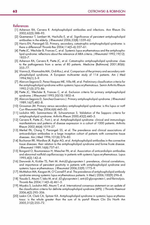
62 OSTROWSKI & ROBINSON
References
[1] Asherson RA, Cervera R. Antiphospholipid antibodies and infections. Ann Rheum Dis2002;62(5):388–93.[2] Quemeneur T, Lambert M, Hachulla E, et al. Significance of persistent antiphospholipid
antibodies in the elderly. J Rheumatol 2006;33(8):1559–62.[3] Harris EN, Pierangeli SS. Primary, secondary, catastrophic antiphospholipid syndrome: is
there a difference? Thromb Res 2004;114(5–6):357–61.[4] Piette JC, Wechsler B, Frances C, et al. Systemic lupus erythematosus and the antiphospho-
lipid syndrome: reflections about the relevance of ARA criteria. J Rheumatol 1992;19(12):1835–7.
[5] Asherson RA, Cervera R, Piette JC, et al. Catastrophic antiphospholipid syndrome: cluesto the pathogenesis from a series of 80 patients. Medicine (Baltimore) 2001;80(6):355–77.
[6] Vianna JL, Khamashta MA, Ordi-Ros J, et al. Comparison of the primary and secondary anti-phospholipid syndrome. A European multicenter study of 114 patients. Am J Med1994;96(1):3–9.
[7] Alarcon-Segovia D, Perez-Vasquez ME, Villa AR, et al. Preliminary classification criteria forthe antiphospholipid syndrome within systemic lupus erythematosus. Semin Arthritis Rheum1992;21(5):275–86.
[8] Piette JC, Wechsler B, Frances C, et al. Exclusion criteria for primary antiphospholipidsyndrome. J Rheumatol 1993;20(10):1802–4.
[9] Alarcon-Segovia D, Sanchez-Guerroro J. Primary antiphospholipid syndrome. J Rheumatol1989;16(7):482–8.
[10] Grossman JM. Primary versus secondary antiphospholipid syndrome: is this lupus or not?Curr Rheumatol Rep 2004;6(6):445–50.
[11] Lockshin MD, Sammaritano LR, Schwartzman S. Validation of the Sapporo criteria forantiphospholipid syndrome. Arthritis Rheum 2000;43(2):440–3.
[12] Cervera R, Piette JC, Font J, et al. Antiphospholipid syndrome: clinical and immunologicmanifestations and patterns of disease expression in a cohort of 1000 patients. ArthritisRheum 2002;46(4):1019–27.
[13] Merkel PA, Chang Y, Pierangeli SS, et al. The prevalence and clinical associations ofanticardiolipin antibodies in a large inception cohort of patients with connective tissuediseases. Am J Med 1996;101(6):576–83.
[14] Buchanan RR, Wardlaw JR, Riglar AG, et al. Antiphospholipid antibodies in the connectivetissue diseases: their relation to the antiphospholipid syndrome and forme fruste disease.J Rheumatol 1989;16(6):757–61.
[15] Bongard O, Boumaneaux H, Miescher PA, et al. Association of anticardiolipin antibodiesand abnormal nailfold capillaroscopy in patients with systemic lupus erythematosus. Lupus1995;4(2):142–4.
[16] Danowski A, Kickler TS, Petri M. Anti-b2-glycoprotein I: prevalence, clinical correlations,and importance of persistent positivity in patients with antiphospholipid syndrome andsystemic lupus erythematosus. J Rheumatol 2006;33(9):1775–9.
[17] McMahon MA, Keogan M, O’Connell P, et al. The prevalence of antiphospholipid antibodysyndrome among systemic lupus erythematosus patients. Ir Med J 2006;100(9):296–8.
[18] Yasuda S, Atsumi T, Iako M, et al. b2-glycoprotein I, anti-b2-glycoprotein I, and fibrinolysis.Thromb Res 2004;114(5–6):461–5.
[19] Miyakis S, Lockshin MD, Atsumi T, et al. International consensus statement on an update ofthe classification criteria for definite antiphospholipid syndrome (APS). J Thromb Haemost2006;4(2):295–306.
[20] Laskin CA, Clark CA, Spitzer KA. Antiphospholipid syndrome in systemic lupus erythema-tosus: is the whole greater than the sum of its parts? Rheum Clin Dis North Am2005;31(2):255–72.
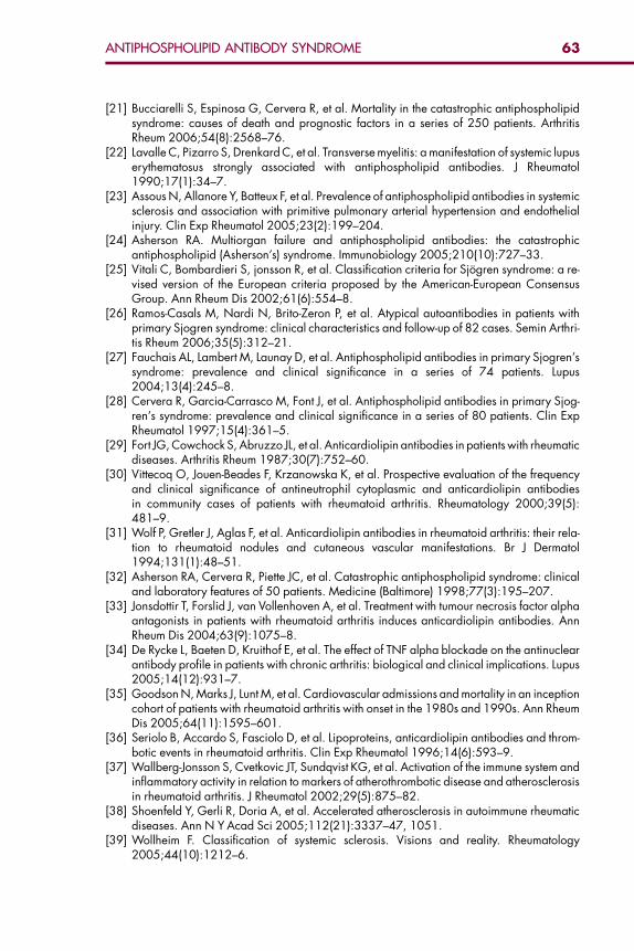
63ANTIPHOSPHOLIPID ANTIBODY SYNDROME
[21] Bucciarelli S, Espinosa G, Cervera R, et al. Mortality in the catastrophic antiphospholipidsyndrome: causes of death and prognostic factors in a series of 250 patients. ArthritisRheum 2006;54(8):2568–76.
[22] Lavalle C, Pizarro S, Drenkard C, et al. Transverse myelitis: a manifestation of systemic lupuserythematosus strongly associated with antiphospholipid antibodies. J Rheumatol1990;17(1):34–7.
[23] Assous N, Allanore Y, Batteux F, et al. Prevalence of antiphospholipid antibodies in systemicsclerosis and association with primitive pulmonary arterial hypertension and endothelialinjury. Clin Exp Rheumatol 2005;23(2):199–204.
[24] Asherson RA. Multiorgan failure and antiphospholipid antibodies: the catastrophicantiphospholipid (Asherson’s) syndrome. Immunobiology 2005;210(10):727–33.
[25] Vitali C, Bombardieri S, jonsson R, et al. Classification criteria for Sjogren syndrome: a re-vised version of the European criteria proposed by the American-European ConsensusGroup. Ann Rheum Dis 2002;61(6):554–8.
[26] Ramos-Casals M, Nardi N, Brito-Zeron P, et al. Atypical autoantibodies in patients withprimary Sjogren syndrome: clinical characteristics and follow-up of 82 cases. Semin Arthri-tis Rheum 2006;35(5):312–21.
[27] Fauchais AL, Lambert M, Launay D, et al. Antiphospholipid antibodies in primary Sjogren’ssyndrome: prevalence and clinical significance in a series of 74 patients. Lupus2004;13(4):245–8.
[28] Cervera R, Garcia-Carrasco M, Font J, et al. Antiphospholipid antibodies in primary Sjog-ren’s syndrome: prevalence and clinical significance in a series of 80 patients. Clin ExpRheumatol 1997;15(4):361–5.
[29] Fort JG, Cowchock S, Abruzzo JL, et al. Anticardiolipin antibodies in patients with rheumaticdiseases. Arthritis Rheum 1987;30(7):752–60.
[30] Vittecoq O, Jouen-Beades F, Krzanowska K, et al. Prospective evaluation of the frequencyand clinical significance of antineutrophil cytoplasmic and anticardiolipin antibodiesin community cases of patients with rheumatoid arthritis. Rheumatology 2000;39(5):481–9.
[31] Wolf P, Gretler J, Aglas F, et al. Anticardiolipin antibodies in rheumatoid arthritis: their rela-tion to rheumatoid nodules and cutaneous vascular manifestations. Br J Dermatol1994;131(1):48–51.
[32] Asherson RA, Cervera R, Piette JC, et al. Catastrophic antiphospholipid syndrome: clinicaland laboratory features of 50 patients. Medicine (Baltimore) 1998;77(3):195–207.
[33] Jonsdottir T, Forslid J, van Vollenhoven A, et al. Treatment with tumour necrosis factor alphaantagonists in patients with rheumatoid arthritis induces anticardiolipin antibodies. AnnRheum Dis 2004;63(9):1075–8.
[34] De Rycke L, Baeten D, Kruithof E, et al. The effect of TNF alpha blockade on the antinuclearantibody profile in patients with chronic arthritis: biological and clinical implications. Lupus2005;14(12):931–7.
[35] Goodson N, Marks J, Lunt M, et al. Cardiovascular admissions and mortality in an inceptioncohort of patients with rheumatoid arthritis with onset in the 1980s and 1990s. Ann RheumDis 2005;64(11):1595–601.
[36] Seriolo B, Accardo S, Fasciolo D, et al. Lipoproteins, anticardiolipin antibodies and throm-botic events in rheumatoid arthritis. Clin Exp Rheumatol 1996;14(6):593–9.
[37] Wallberg-Jonsson S, Cvetkovic JT, Sundqvist KG, et al. Activation of the immune system andinflammatory activity in relation to markers of atherothrombotic disease and atherosclerosisin rheumatoid arthritis. J Rheumatol 2002;29(5):875–82.
[38] Shoenfeld Y, Gerli R, Doria A, et al. Accelerated atherosclerosis in autoimmune rheumaticdiseases. Ann N Y Acad Sci 2005;112(21):3337–47, 1051.
[39] Wollheim F. Classification of systemic sclerosis. Visions and reality. Rheumatology2005;44(10):1212–6.

64 OSTROWSKI & ROBINSON
[40] Herrick AL, Heaney M, Hollis S, et al. Anticardiolipin, anticentromere and anti-Scl-70antibodies in patients with systemic sclerosis and severe digital ischaemia. Ann RheumDis 1995;53(8):540–2.
[41] Martinuzzo ME, Pombo G, Forastiero RR. Lupus anticoagulant, high levels of cardiolipin,and anti-beta2- glycoprotein I antibodies are associated with chronic thromboembolicpulmonary hypertension. J Rheumatol 1998;25(7):1313–9.
[42] Manoussakis MN, Gharavi AE, Drosos AA, et al. Anticardiolipin antibodies in unselectedautoimmune rheumatic disease patients. Clin Immunol Immunopathol 1987;44(3):297–307.
[43] Asherson RA, Mayou SC, Merry P, et al. The spectrum of livedo reticularis and anticardioli-pin antibodies. Br J Dermatol 1989;120(2):215–21.
[44] Cuchacovich R, Espinoza LR. Is antiphospholipid antibody determination clinically relevantto the vasculitides? Semin Arthritis Rheum 2001;31(1):1–3.
[45] Espinosa G, Tassies D, Font J, et al. Antiphospholipid antibodies and thrombophilic factorsin giant cell arteritis. Semin Arthritis Rheum 2001;31(1):12–20.
[46] Ruffatti A, Montecucco C, Volante D, et al. Antiphospholipid antibody syndrome and poly-myalgia rheumatica/giant cell arteritis. Rheumatol 2000;39(5):565–7.
[47] McHugh NJ, James IE, Plant GT. Anticardiolipin and antineutrophil antibodies in giant cellarteritis. J Rheumatol 1990;17(7):916–22.
[48] Meyer MF, Schnabel A, Schatz H, et al. Lack of association between antiphospholipidantibodies and thrombocytopenia in patients with Wegener’s granulomatosis. Semin Arthri-tis Rheum 2001;31(1):4–11.
[49] Handa R, Aggarwal P, Biswas A, et al. Microscopic polyangiitis associated with antiphos-pholipid syndrome. Rheumatology 1999;38(5):478–9.
[50] Norden DK, Ostrov BE, Shafritz AB, et al. Vasculitis associated with antiphospholipidsyndrome. Semin Arthritis Rheum 1995;24(4):273–81.
[51] Dasgupta B, Almond MK, Tanqueray A. Polyarteritis nodosa and the antiphospholipidsyndrome. Br J Rheumatol 1997;36(11):1210–2.
[52] Famularo G, Antonelli S, Barracchini A, et al. Catastrophic antiphospholipid syndrome ina patient with Behcet’s disease. Scand J Rheumatol 2002;31(2):100–2.
[53] Mader R, Ziv M, Adawi M, et al. Thrombophilic factors and their relation to thromboembolicand other clinical manifestations in Behcet’s disease. J Rheumatol 1999;26(11):2404–8.
[54] Strauss G, Osen W, Debatin K-M. Induction of apoptosis and modulation of activation andeffector function in T cells by immunosuppressive drugs. Clin Exp Immunol 2002;128(2):255–66.
[55] Hemendinger RA, Bloom SE. Selective mitomycin C and cyclophosphamide induction ofapoptosis in differentiating B lymphocytes compared to T lymphocytes in vivo. Immunophar-macology 1996;35(1):71–82.
[56] Galindo-Rodriguez G, Avina-Subieta A, Pizarro S, et al. Cyclophosphamide pulse therapyin optic neuritis due to systemic lupus erythematosus: an open trial. Am J Med 1999;106(1):65–9.
[57] Koschmieder S, Miesbach W, Fauth F, et al. Combined plasmapheresis and immunosup-pression as rescue treatment of a patient with catastrophic antiphospholipid syndromeoccurring despite anticoagulation: a case report. Blood Coagul Fibrinolysis 2003;14(4):395–9.
[58] Lacueva J, Enriquez R, Cabezuelo JB, et al. Acute renal failure as first clinical manifestationof the primary antiphospholipid syndrome. Nephron 1993;64(3):479–80.
[59] Yagi K, Kawano M, Haraki T, et al. Long-term efficacy of immunoadsorbent plasmapheresisin a patient with Budd-Chiari syndrome due to antiphospholipid syndrome: case report withnine-year follow up. Lupus 2004;13(2):135–8.
[60] Marks SD, Patey S, Brogan PA, et al. B lymphocyte depletion therapy in children with refrac-tory systemic lupus erythematosus. Arthritis Rheum 2005;52(10):3168–74, B.
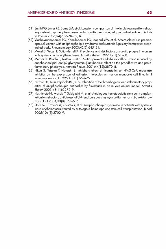
65ANTIPHOSPHOLIPID ANTIBODY SYNDROME
[61] Smith KG, Jones RB, Burns SM, et al. Long-term comparison of rituximab treatment for refrac-tory systemic lupus erythematosus and vasculitis: remission, relapse and retreatment. Arthri-tis Rheum 2006;54(9):2970–82, B.
[62] Vlachoyiannopoulos PG, Kanellopoulos PG, Ioannidis PA, et al. Atherosclerosis in premen-opausal women with antiphospholipid syndrome and systemic lupus erythematosus: a con-trolled study. Rheumatology 2003;42(5):645–51.
[63] Manzi S, Selzer F, Sutton-Tyrrell K. Prevalence and risk factors of carotid plaque in womenwith systemic lupus erythematosus. Arthritis Rheum 1999;42(1):51–60.
[64] Meroni PL, Raschi E, Testoni C, et al. Statins prevent endothelial cell activation induced byantiphospholipid (anti-b2-glycoprotein I) antibodies: effect on the proadhesive and proin-flammatory phenotype. Arthritis Rheum 2001;44(12):2870–8.
[65] Niwa S, Totsuka T, Hayashi S. Inhibitory effect of fluvastatin, an HMG-CoA reductaseinhibitor on the expression of adhesion molecules on human monocyte cell line. Int JImmunopharmacol 1996;18(11):669–75.
[66] Ferrara DE, Liu X, Espinola RG, et al. Inhibition of the thrombogenic and inflammatory prop-erties of antiphospholipid antibodies by fluvastatin in an in vivo animal model. ArthritisRheum 2003;48(11):3272–9.
[67] Hashimoto N, Iwasaki T, Sekiguchi M, et al. Autologous hematopoietic stem cell transplan-tation for refractory antiphospholipid syndrome causing myocardial necrosis. Bone MarrowTransplant 2004;33(8):863–6, B.
[68] Statkute L, Traynor A, Oyama Y, et al. Antiphospholipid syndrome in patients with systemiclupus erythematosus treated by autologous hematopoietic stem cell transplantation. Blood2005;106(8):2700–9.
Hematol Oncol Clin N Am 22 (2008) 67–77
HEMATOLOGY/ONCOLOGY CLINICSOF NORTH AMERICA
Cutaneous Manifestationsof Antiphospholipid AntibodySyndrome
Sari Weinstein, MDa, Warren Piette, MDa,b,*aDivision of Dermatology, John H. Stroger, Jr. Hospital of Cook County,1900 West Polk Street, Room 519, Chicago, IL 60612, USAbDepartment of Dermatology, Rush University Medical Center,Annex Building, Suite 220, 707 S. Wood, Chicago, IL 60612, USA
Although no skin finding is pathognomonic for antiphospholipid anti-body syndrome (APS), many cutaneous findings can be helpful in sug-gesting this diagnostic possibility. The cutaneous syndromes that
should prompt consideration of APS are the focus of this article.
LIVEDO RETICULARIS AND RACEMOSALivedo reticularis is the most frequently associated cutaneous manifestation inpatients who have APS, observed as the presenting sign in up to 40% ofpatients and seen in up to 70% of patients who have systemic lupus erythema-tosus (SLE) and APS [1]. In patients who have SLE, the finding of moderate-to-extensive livedo reticularis probably warrants testing for antiphospholipidantibodies (aPLs). In a recent consensus statement on the updated classificationcriteria for APS, Miyakis and colleagues [2] defined livedo reticularis as thepersistent, not reversible with rewarming, violaceous, red or blue, reticularor mottled pattern of the skin of the trunk, arms, or legs. It may consist of reg-ular, unbroken circles (regular livedo reticularis), or irregular, broken circles(livedo racemosa). The livedo patterns were further classified, depending onthe width of branching pattern (greater or less than 10 mm), into fine and largelivedo reticularis and fine and large livedo racemosa. Although the terms arefrequently used interchangeably, compared with livedo reticularis, livedo race-mosa is a coarser and more irregular pattern, is more often irregularly distrib-uted, and may be more generalized (Fig. 1). Some investigators believe theracemosa variant to be a major clinical feature of APS, strongly associatedwith the arterial subset of APS, which includes cerebrovascular events, arterial
*Corresponding author. Division of Dermatology, John H. Stroger, Jr. Hospital of CookCounty, 1900 West Polk Street, Room 519, Chicago, IL 60612. E-mail address: [email protected] (W. Piette).
0889-8588/08/$ – see front matter ª 2008 Elsevier Inc. All rights reserved.doi:10.1016/j.hoc.2007.10.011 hemonc.theclinics.com

Fig. 1. Livedo. Sneddon’s syndrome with widespread livedo. Although focal areas havea mostly regular reticulate pattern (eg, knee), most areas have a less regular, coarse patternof racemosa.
68 WEINSTEIN & PIETTE
thrombosis, systemic hypertension, heart valve abnormalities, Raynaud’s phe-nomenon, and pregnancy morbidity, and consider it an independent, additive,thrombotic risk factor [1,3]. Livedo reticularis has been observed less fre-quently in those who have only venous thrombosis.
An impairment of blood flow and lowered oxygen tension at the peripheryof skin segments is responsible for the mottling observed clinically. The cuta-neous blood supply is arranged in inverted cones supplied by a central arteriolearising perpendicularly from vessels in the fascia. This arteriole is responsiblefor the blood supply to a 1- to 4-cm–diameter zone on the skin surface. Thenetlike pattern results from cyanotic discoloration at the junctions betweencones, where deoxygenated blood accumulates. The ring size of the reticulatepattern depends on the body site and is usually smallest on the palms and larg-est on the trunk or thighs.
The pathophysiology of livedo is not well characterized, but the relationshipwith arterial thrombosis suggests a possible role for the endothelial cell. Thevasoconstriction of livedo racemosa may be induced by an interaction of anaPL with the endothelial cell or other cellular element of vessels, altering theirfunction [1,4]. Many autoantibodies associated with APS are directed againstphospholipid-protein complexes expressed on, or bound to, the surface ofendothelial cells. When bound, these cells may become prothrombotic, leadingto the production of procoagulant substances such as tissue factor, plasminogenactivator inhibitor 1, and endothelin 1; the increased expression of theseprocoagulants may be responsible, in part, for the hypercoagulability andthromboses observed in APS.
69CUTANEOUS MANIFESTATIONS OF ANTIPHOSPHOLIPID
Unfortunately, livedo reticularis is a nonspecific cutaneous finding. In neo-nates, generalized livedo reticularis is an expected finding, termed cutis mar-morata. Physiologic livedo reticularis in adults is common, triggered by coldand reversed by warming. Associations of nonphysiologic livedo reticulariswith systemic diseases or medications are many; one recent review cited justshort of 100 conditions [5].
IDIOPATHIC LIVEDO RETICULARIS WITH CEREBROVASCULARACCIDENTS (SNEDDON’S SYNDROME)In 1965, Sneddon reported the association of livedo reticularis or racemosawith cerebrovascular accidents [6]. Sneddon’s syndrome is a rare, but poten-tially severe, condition typically affecting young or middle-aged women, withthe first cerebrovascular accident typically occurring before 45 years of age.Livedo of either type, typically on the trunk and buttocks but also on the extrem-ities and face, may precede the onset of stroke by years. Although the relationshipbetween APS and Sneddon’s syndrome is not clear, the prevalence of aPLs inSneddon’s syndrome has ranged from 0% to 85%, and most authorities suggestthat 40% to 50% of patients who have Sneddon’s are antibody positive [1].
Skin biopsies are often nondiagnostic in Sneddon’s syndrome. Multiple deeppunch biopsies from the center and the violaceous areas are required to in-crease sensitivity. According to Zelger and colleagues [4], small-to-medium–sized arteries at the dermal-subcutis boundary are involved in a stage-specificpattern. Initial lesions are characterized by the attachment of lymphohistiocyticcells and the detachment of endothelial cells (endotheliitis). Early-phase lesionsdisplay partial or complete occlusion of vascular lumens by a plug of lympho-histiocytic cells and fibrin. Intermediate-phase lesions show replacement of plugby proliferating subendothelial cells and dilated capillaries in the adventitia ofthe occluded vessel. Late-phase lesions show fibrosis and shrinkage of vessels[1]. True vasculitis is not a feature.
Sneddon’s syndrome may have two distinct causes, one aPL related withpreferential arteriolar involvement, the other a primary non–aPL-related smallartery disease primarily affecting the brain and skin vessels [7].
No treatment has consistently been effective for livedo. In patients who haveSneddon’s or APS, chronic anticoagulation may be warranted, even though thelivedo may worsen on anticoagulant or antiplatelet therapy. In light of thehigher association of arterial occlusion in patients who have livedo racemosa,patients should reduce other risk factors, including smoking and use of estro-gen-containing oral contraceptives. Low-dose aspirin is frequently prescribed,although its efficacy is unproven [8]. Further study is needed on the potentialbenefit of clopidogrel, statins, and angiotensin-converting enzyme inhibitors.
ATROPHIE BLANCHE AND LIVEDOID VASCULOPATHYAtrophie blanche is a term that has been used for several different conditions,leading to some confusion in the literature. Originally described as atrophieblanche en plaque by Milian in 1929, synonyms for this disease include livedo
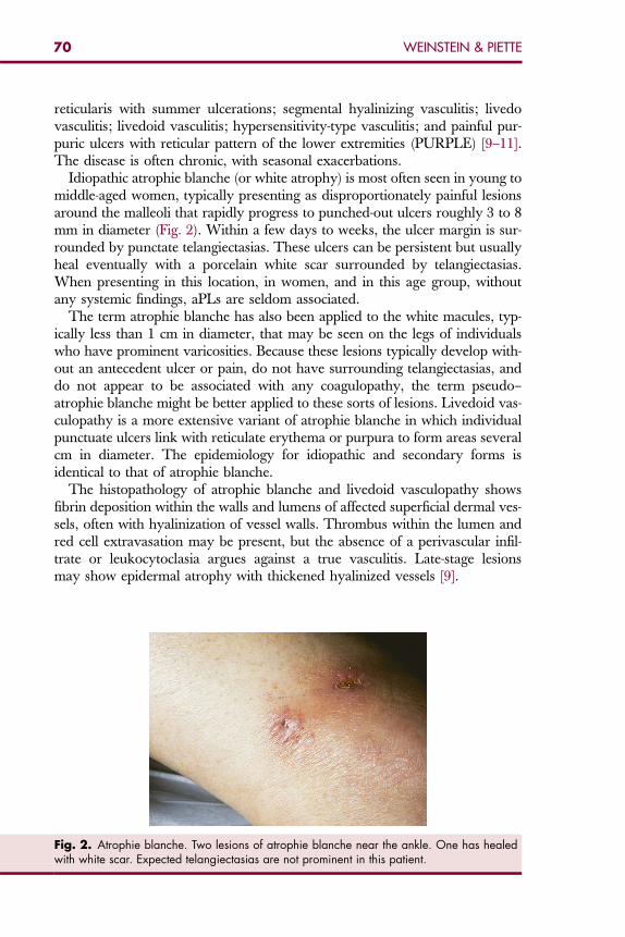
70 WEINSTEIN & PIETTE
reticularis with summer ulcerations; segmental hyalinizing vasculitis; livedovasculitis; livedoid vasculitis; hypersensitivity-type vasculitis; and painful pur-puric ulcers with reticular pattern of the lower extremities (PURPLE) [9–11].The disease is often chronic, with seasonal exacerbations.
Idiopathic atrophie blanche (or white atrophy) is most often seen in young tomiddle-aged women, typically presenting as disproportionately painful lesionsaround the malleoli that rapidly progress to punched-out ulcers roughly 3 to 8mm in diameter (Fig. 2). Within a few days to weeks, the ulcer margin is sur-rounded by punctate telangiectasias. These ulcers can be persistent but usuallyheal eventually with a porcelain white scar surrounded by telangiectasias.When presenting in this location, in women, and in this age group, withoutany systemic findings, aPLs are seldom associated.
The term atrophie blanche has also been applied to the white macules, typ-ically less than 1 cm in diameter, that may be seen on the legs of individualswho have prominent varicosities. Because these lesions typically develop with-out an antecedent ulcer or pain, do not have surrounding telangiectasias, anddo not appear to be associated with any coagulopathy, the term pseudo–atrophie blanche might be better applied to these sorts of lesions. Livedoid vas-culopathy is a more extensive variant of atrophie blanche in which individualpunctuate ulcers link with reticulate erythema or purpura to form areas severalcm in diameter. The epidemiology for idiopathic and secondary forms isidentical to that of atrophie blanche.
The histopathology of atrophie blanche and livedoid vasculopathy showsfibrin deposition within the walls and lumens of affected superficial dermal ves-sels, often with hyalinization of vessel walls. Thrombus within the lumen andred cell extravasation may be present, but the absence of a perivascular infil-trate or leukocytoclasia argues against a true vasculitis. Late-stage lesionsmay show epidermal atrophy with thickened hyalinized vessels [9].
Fig. 2. Atrophie blanche. Two lesions of atrophie blanche near the ankle. One has healedwith white scar. Expected telangiectasias are not prominent in this patient.
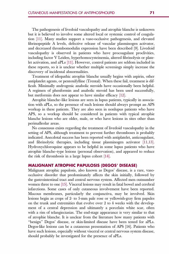
71CUTANEOUS MANIFESTATIONS OF ANTIPHOSPHOLIPID
The pathogenesis of livedoid vasculopathy and atrophie blanche is unknownbut it is believed to involve some altered local or systemic control of coagula-tion [11]. Many studies support a vaso-occlusive pathogenesis, and elevatedfibrinopeptide A levels, defective release of vascular plasminogen activator,and decreased thrombomodulin expression have been described [9]. Livedoidvasculopathy is observed in patients who have procoagulant proclivities,including factor V Leiden, hyperhomocysteinemia, altered fibrinolysis or plate-let activation, and aPLs [11]. However, control patients are seldom included inthese reports, so it is unclear whether multiple screenings simply increase thediscovery of incidental abnormalities.
Treatment of idiopathic atrophie blanche usually begins with aspirin, otherantiplatelet agents, or pentoxifylline (Trental). When these fail, treatment is dif-ficult. Minimally androgenic anabolic steroids have occasionally been helpful.A regimen of phenformin and anabolic steroid has been used successfully,but metformin does not appear to have similar efficacy [12].
Atrophie blanche–like lesions are seen in lupus patients, typically in associa-tion with aPLs, so the presence of such lesions should always prompt an APSworkup in these patients. They are also seen in nonlupus patients who haveAPS, so a workup should be considered in patients with typical atrophieblanche lesions who are older, male, or who have lesions in sites other thanperimalleolar areas.
No consensus exists regarding the treatment of livedoid vasculopathy in thesetting of APS, although treatment to prevent further thromboses is probablyindicated. Anecdotal success has been reported with antiplatelet, anticoagulant,and fibrinolytic therapies, including tissue plasminogen activator [11,13].Hydroxychloroquine appears to be helpful in some lupus patients who haveatrophie blanche–type lesions (personal observation), and appeared to reducethe risk of thrombosis in a large lupus cohort [14].
MALIGNANT ATROPHIC PAPULOSIS (DEGOS’ DISEASE)Malignant atrophic papulosis, also known as Degos’ disease, is a rare, vaso-occlusive disorder that predominately affects the skin initially, followed bythe gastrointestinal tract and central nervous system. Affected men outnumberwomen three to one [15]. Visceral lesions may result in fatal bowel and cerebralinfarctions. Some cases of only cutaneous involvement have been reported.Mucous membranes, particularly the conjunctiva, may be involved. Skinlesions begin as crops of 2- to 5-mm pale rose or yellowish-gray firm papuleson the trunk and extremities that evolve over 2 to 4 weeks with the develop-ment of a central depression and ultimately a porcelain white scar, oftenwith a rim of telangiectasias. The end-stage appearance is very similar to thatof atrophie blanche. It is unclear from the literature how many patients with‘‘benign’’ Degos’ disease, or skin-limited disease have been tested for aPLs.Degos-like lesions can be a cutaneous presentation of APS [16]. Patients whohave such lesions, especially without visceral or central nervous system disease,should probably be investigated for the presence of aPLs.
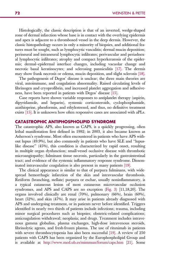
72 WEINSTEIN & PIETTE
Histologically, the classic description is that of an inverted, wedge-shapedzone of dermal infarction whose base is in contact with the overlying epidermisand apex is adjacent to a thrombosed vessel in the deep dermis. However, theclassic histopathology occurs in only a minority of biopsies, and additional fea-tures must be sought, such as lymphocytic vasculitis; dermal mucin deposition;perineural and intraneural lymphocytic infiltrates; perivascular and periadnex-al lymphocytic infiltrates; atrophy and compact hyperkeratosis of the epider-mis; dermal–epidermal interface changes, including vacuolar change andnecrotic basal keratinocytes; and sclerosing panniculitis [17]. The dermismay show frank necrosis or edema, mucin deposition, and slight sclerosis [18].
The pathogenesis of Degos’ disease is unclear; the three main theories areviral, autoimmune, and coagulation abnormality. Raised circulating levels offibrinogen and cryoprofibrin, and increased platelet aggregation and adhesive-ness, have been reported in patients with Degos’ disease [15].
Case reports have shown variable responses to antiplatelet therapy (aspirin,dipyridamole, and heparin), systemic corticosteroids, cyclophosphamide,azathioprine, phenformin, and ethylestrenol, and thus, no definitive treatmentexists [15]. It is unknown how often responsive cases are associated with aPLs.
CATASTROPHIC ANTIPHOSPHOLIPID SYNDROMEThe catastrophic APS, also known as CAPS, is a quickly progressing, oftenlethal manifestation first defined in 1992; in 2003, it also became known asAsherson’s syndrome. Most often encountered in patients who have APS with-out lupus (49.9%), but also commonly in patients who have SLE and ‘‘lupus-like disease’’ (45%), this condition is characterized by rapid onset, resultingin multiple organ dysfunction; small-vessel occlusive disease with thromboticmicroangiopathy; fulminant tissue necrosis, particularly in the gastrointestinaltract; and evidence of the systemic inflammatory response syndrome. Dissem-inated intravascular coagulation is also present in many patients [19].
The clinical appearance is similar to that of purpura fulminans, with wide-spread hemorrhagic infarction of the skin and intravascular thrombosis.Retiform (branching, stellate) purpura or eschar, usually noninflammatory, isa typical cutaneous lesion of most cutaneous microvascular occlusionsyndromes, and APS and CAPS are no exception (Fig. 3) [11,18,20]. Theorgans involved clinically are renal (70%), pulmonary (66%), brain (60%),heart (52%), and skin (47%). It may arise in patients already diagnosed withAPS and undergoing treatment, or in patients never before identified. Triggersidentified in nearly two thirds of patients include infections; trauma, includingminor surgical procedures such as biopsies; obstetric-related complications;anticoagulation withdrawal; neoplasia; and drugs. Treatment includes intrave-nous gamma globulins, plasma exchanges, high-dose intravenous steroids,fibrinolytic agents, and fresh-frozen plasma. The use of rituximab in patientswith severe thrombocytopenia has also been successful [19]. A review of 250patients with CAPS has been organized by the Europhospholipid Group andis available at http://www.med.ub.es/mimmun/forum/caps.htm [21]. Some
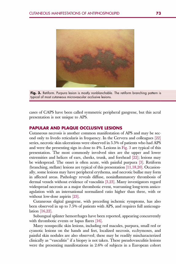
Fig. 3. Retiform. Purpura lesion is mostly nonblanchable. The retiform branching pattern istypical of most cutaneous microvascular occlusive lesions.
73CUTANEOUS MANIFESTATIONS OF ANTIPHOSPHOLIPID
cases of CAPS have been called symmetric peripheral gangrene, but this acralpresentation is not unique to APS.
PAPULAR AND PLAQUE OCCLUSIVE LESIONSCutaneous necrosis is another common manifestation of APS and may be sec-ond only to livedo reticularis in frequency. In the Cervera and colleagues [22]series, necrotic skin ulcerations were observed in 5.5% of patients who had APSand were the presenting sign in close to 4%. Lesions in Fig. 3 are typical of thispresentation. The most commonly involved sites are the upper and lowerextremities and helices of ears, cheeks, trunk, and forehead [22]; lesions maybe widespread. The onset is often acute, with painful purpura [3]. Retiform(branching, stellate) lesions are typical of this presentation [11,18,20]. Occasion-ally, some lesions may have peripheral erythema, and necrotic bullae may formin affected areas. Pathology reveals diffuse, noninflammatory thrombosis ofdermal vessels without evidence of vasculitis [3,23]. Many investigators regardwidespread necrosis as a major thrombotic event, warranting long-term antico-agulation with an international normalized ratio higher than three, with orwithout low-dose aspirin [23].
Cutaneous digital gangrene, with preceding ischemic symptoms, has alsobeen observed in up to 7.5% of patients with APS, and requires full anticoagu-lation [16,22].
Subungual splinter hemorrhages have been reported, appearing concurrentlywith thrombotic events or lupus flares [16].
Many nonspecific skin lesions, including red macules, purpura, small red orcyanotic lesions on the hands and feet, localized necrosis, ecchymoses, andpainful skin nodules are also observed; these may be readily mischaracterizedclinically as ‘‘vasculitis’’ if a biopsy is not taken. These pseudovasculitis lesionswere the presenting manifestations in 2.6% of subjects in a European cohort
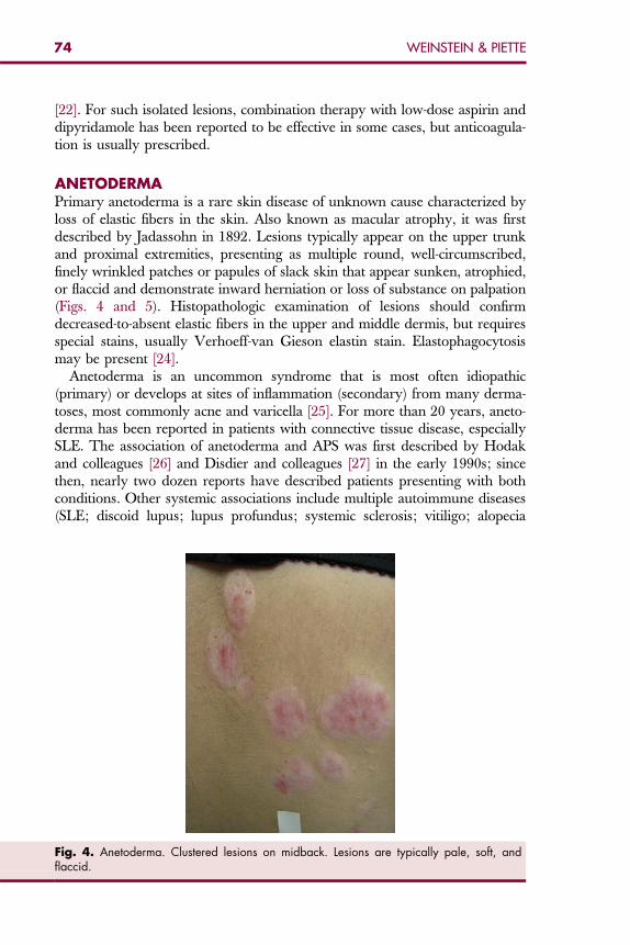
74 WEINSTEIN & PIETTE
[22]. For such isolated lesions, combination therapy with low-dose aspirin anddipyridamole has been reported to be effective in some cases, but anticoagula-tion is usually prescribed.
ANETODERMAPrimary anetoderma is a rare skin disease of unknown cause characterized byloss of elastic fibers in the skin. Also known as macular atrophy, it was firstdescribed by Jadassohn in 1892. Lesions typically appear on the upper trunkand proximal extremities, presenting as multiple round, well-circumscribed,finely wrinkled patches or papules of slack skin that appear sunken, atrophied,or flaccid and demonstrate inward herniation or loss of substance on palpation(Figs. 4 and 5). Histopathologic examination of lesions should confirmdecreased-to-absent elastic fibers in the upper and middle dermis, but requiresspecial stains, usually Verhoeff-van Gieson elastin stain. Elastophagocytosismay be present [24].
Anetoderma is an uncommon syndrome that is most often idiopathic(primary) or develops at sites of inflammation (secondary) from many derma-toses, most commonly acne and varicella [25]. For more than 20 years, aneto-derma has been reported in patients with connective tissue disease, especiallySLE. The association of anetoderma and APS was first described by Hodakand colleagues [26] and Disdier and colleagues [27] in the early 1990s; sincethen, nearly two dozen reports have described patients presenting with bothconditions. Other systemic associations include multiple autoimmune diseases(SLE; discoid lupus; lupus profundus; systemic sclerosis; vitiligo; alopecia
Fig. 4. Anetoderma. Clustered lesions on midback. Lesions are typically pale, soft, andflaccid.
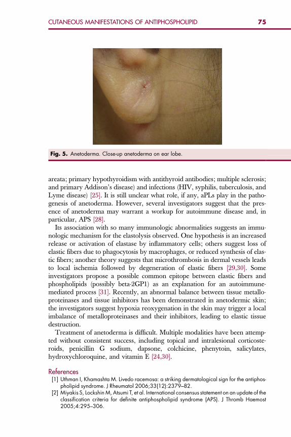
Fig. 5. Anetoderma. Close-up anetoderma on ear lobe.
75CUTANEOUS MANIFESTATIONS OF ANTIPHOSPHOLIPID
areata; primary hypothyroidism with antithyroid antibodies; multiple sclerosis;and primary Addison’s disease) and infections (HIV, syphilis, tuberculosis, andLyme disease) [25]. It is still unclear what role, if any, aPLs play in the patho-genesis of anetoderma. However, several investigators suggest that the pres-ence of anetoderma may warrant a workup for autoimmune disease and, inparticular, APS [28].
Its association with so many immunologic abnormalities suggests an immu-nologic mechanism for the elastolysis observed. One hypothesis is an increasedrelease or activation of elastase by inflammatory cells; others suggest loss ofelastic fibers due to phagocytosis by macrophages, or reduced synthesis of elas-tic fibers; another theory suggests that microthrombosis in dermal vessels leadsto local ischemia followed by degeneration of elastic fibers [29,30]. Someinvestigators propose a possible common epitope between elastic fibers andphospholipids (possibly beta-2GP1) as an explanation for an autoimmune-mediated process [31]. Recently, an abnormal balance between tissue metallo-proteinases and tissue inhibitors has been demonstrated in anetodermic skin;the investigators suggest hypoxia reoxygenation in the skin may trigger a localimbalance of metalloproteinases and their inhibitors, leading to elastic tissuedestruction.
Treatment of anetoderma is difficult. Multiple modalities have been attemp-ted without consistent success, including topical and intralesional corticoste-roids, penicillin G sodium, dapsone, colchicine, phenytoin, salicylates,hydroxychloroquine, and vitamin E [24,30].
References
[1] Uthman I, Khamashta M. Livedo racemosa: a striking dermatological sign for the antiphos-pholipid syndrome. J Rheumatol 2006;33(12):2379–82.[2] Miyakis S, Lockshin M, Atsumi T, et al. International consensus statement on an update of the
classification criteria for definite antiphospholipid syndrome (APS). J Thromb Haemost2005;4:295–306.
76 WEINSTEIN & PIETTE
[3] Frances C, Niang S, Laffitte E, et al. Dermatologic manifestations of the antiphospholipidsyndrome. Arthritis Rheum 2005;52:1785–90.
[4] Zelger B, Sepp N, Stockhammer G, et al. Sneddon’s syndrome: long-term follow-up of 21patients. Arch Dermatol 1993;129:437–47.
[5] Gibbs M, English J, Zirwas M. Livedo reticularis: an update. J Am Acad Dermatol2005;52(6):1009–19.
[6] Sneddon I. Cerebro-vascular lesions and livedo reticularis. Br J Dermatol 1965;77:180–5.[7] Frances C, Piette JC. The mystery of Sneddon syndrome: relationship with antiphospholipid
syndrome and systemic lupus erythematosus. J Autoimmun 2000;15(2):139–43.[8] Erkan D, Harrison MJ, Levy R, et al. Aspirin for primary thrombosis prevention in the anti-
phospholipid syndrome: a randomized, double-blind, placebo-controlled trial in asymptom-atic antiphospholipid antibody-positive individuals. Arthritis Rheum 2007;56(7):2383–91.
[9] Acland KM, Darvay A, Wakelin SH, et al. Livedoid vasculitis: a manifestation of the anti-phospholipid syndrome? Br J Dermatol 1999;140(1):131–5.
[10] Hairston B, Davis M, Pittelkow M, et al. Livedoid vasculopathy: further evidence for procoa-gulant pathogenesis. Arch Dermatol 2006;142:1413–8.
[11] Piette W. Cutaneous manifestations of microvascular occlusion syndromes. In: Bolognia J,Jorrizo J, Rapini R, editors. Dermatology. 1st edition. London: Mosby; 2003. p. 2003.
[12] Shornick J, Nicholes B, Bergstresser P, et al. Idiopathic atrophie blanche. J Am Acad Derma-tol 1983;8(6):792–8.
[13] Klein K, Pittelkow M. Tissue plasminogen activator for treatment of livedoid vasculitis. MayoClin Proc 1992;67:923–33.
[14] Petri M. Hydroxychloroquine use in the Baltimore Lupus Cohort: effects on lipids, glucoseand thrombosis. Lupus 1996;5(Suppl 1):S16–22.
[15] Katz S, Mudd L, Roenigk H. Malignant atrophic papulosis (Degos’ disease) involving threegenerations of a family. J Am Acad Dermatol 1997;37:480–4.
[16] Gibson G, Su W, Pittelkow M. Antiphospholipid syndrome and the skin. J Am Acad Derma-tol 1997;36:970–82.
[17] Harvell J, Williford P, White W. Benign cutaneous Degos’ disease: a case report withemphasis on histopathology as papules chronologically evolve. Am J Dermatopathol2001;23(2):116–23.
[18] Robson K, Piette W. The presentation and differential diagnosis of cutaneous vascular occlu-sion syndromes. Adv Dermatol 1999;15(6):153–82.
[19] Asherson RA, Frances C, Iaccarino L, et al. The antiphospholipid antibody syndrome: diag-nosis, skin manifestations, and current therapy. Clin Exp Rheumatol 2006;24(Suppl 40):S46–51.
[20] Piette W. Purpura and coagulation. In: Bolognia J, Jorrizo J, Rapini R, editors. Dermatology.1st edition. London: Mosby; 2003. p. 355–63.
[21] Asherson R. Multiorgan failure and antiphospholipid antibodies: the catastrophic antiphos-pholipid (Asherson’s) syndrome. Immunobiology 2005;210(10):727–33.
[22] Cervera R, Piette JC, Font J, et al. Antiphospholipid syndrome: clinical and immunologicmanifestations and patterns of disease expression in a cohort of 1,000 patients. ArthritisRheum 2002;46(4):1019–27.
[23] Rossini J, Roverano S, Graf C, et al. Widespread cutaneous necrosis associated with anti-phospholipid antibodies: report of four cases. J Clin Rheumatol 2002;8(6):326–31.
[24] Pascual J, Gimenez E, Sivera F, et al. Atrophic macules and papules in a 24-year-oldwoman. Arch Dermatol 2007;143:109–14.
[25] Sparsa A, Piette JC, Weschsler B, et al. Anetoderma and its prothrombotic abnormalities.J Am Acad Dermatol 2003;49(6):1008–12.
[26] Hodak E, Shamai-Lubovitz O, David M, et al. Immunologic abnormalities associated withprimary anetoderma. Arch Dermatol 1992;128(6):799–803.
[27] Disdier P, Harle J, Andrac L, et al. Primary anetoderma associated with the antiphospholipidsyndrome. J Am Acad Dermatol 1994;30:133–4.

77CUTANEOUS MANIFESTATIONS OF ANTIPHOSPHOLIPID
[28] de Souza E, Christofoletti P, Cintra M. Anetoderma associated with primary antiphospholi-pid syndrome. J Am Acad Dermatol 2007;56(5):881–2.
[29] Venhoff N, Miehle N, Juttner E, et al. Clinical images: anetoderma in systemic lupus eryth-ematosus with antiphospholipid antibodies. Arthritis Rheum 2005;52(7):365–80.
[30] Bilen N, Bayramgurler D, Sikar A, et al. Anetoderma associated with antiphospholipidsyndrome and systemic lupus erythematosus. Lupus 2003;12(9):714–6.
[31] Romani J, Perez F, Llobet M, et al. Anetoderma associated with the antiphospholipidantibodies: case report and review of the literature. J Eur Acad Dermatol Venereol2001;15(2):175–8.
Hematol Oncol Clin N Am 22 (2008) 79–94
HEMATOLOGY/ONCOLOGY CLINICSOF NORTH AMERICA
The Role of Antiphospholipid Syndromein Cardiovascular Disease
Brian R. Long, MDa,*, Ferdinand Leya, MDb
aTennessee Heart & Vascular Institute, P.C., Skyline Medical Center, 3443 Dickerson Pike,Nashville, TN 37207, USAbCardiac Catheterization Laboratory, Loyola University Medical Center,2160 South 1st Avenue, Building 107, Room 1858, Maywood, IL 60153, USA
Antiphospholipid antibodies (aPL) are a family of autoantibodies thatrecognize various phospholipids or phospholipid binding proteins [1].Lupus anticoagulant antibodies, anticardiolipin antibodies (aCL), and
anti-b2-glycoprotein I (b2GPI) antibodies are the most commonly detectedsubgroups of aPL. Patients who have aPL are at risk of developing thrombo-cytopenia, venous and arterial thrombosis, and recurrent fetal loss [2]. Anti-phospholipid syndrome (APS) is defined as the occurrence of these clinicalevents in association with aPL [3]. Although many cases of APS occur inpatients who have systemic lupus erytematosus (SLE), up to 50% of APSpatients have primary APS with no underlying systemic disease [4]. It is recog-nized that aPLs are associated with a spectrum of cardiovascular manifestationsincluding accelerated atherosclerosis, valvular heart disease, intracardiacthrombi, myocardial and pericardial involvement, premature vein graft reste-nosis, and peripheral vascular disease (Box 1) [5–7]. Furthermore, some studieshave demonstrated an association between APS and an increased incidence ofmyocardial infarction (MI), but this remains controversial. The cardiovascularmanifestations of APS will be covered in this article, including recommenda-tions for treatment based on the expert consensus report published in 2003 [8].
CORONARY ARTERY DISEASE IN ANTIPHOSPHOLIPIDSYNDROME
Association of Antiphospholipid Antibodies and Atherosclerosis The prothrombotic state of APS has been implicated in the pathogenesis of pre-mature atherosclerosis in afflicted patients. Although patients who have APSare at higher risk for atherosclerotic cardiovascular events, traditional Framing-ham risk factors have been shown to be similar between APS patients and thegeneral population [9]. This suggests a direct link between APS and*Corresponding author. E-mail address: [email protected] (B.R. Long).
0889-8588/08/$ – see front matter ª 2008 Elsevier Inc. All rights reserved.doi:10.1016/j.hoc.2007.10.002 hemonc.theclinics.com
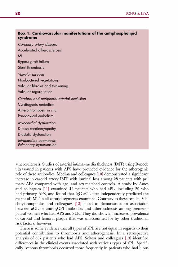
Box 1: Cardiovascular manifestations of the antiphospholipidsyndrome
Coronary artery disease
Accelerated atherosclerosis
MI
Bypass graft failure
Stent thrombosis
Valvular disease
Nonbacterial vegetations
Valvular fibrosis and thickening
Valvular regurgitation
Cerebral and peripheral arterial occlusion
Cardiogenic embolism
Atherothrombosis in situ
Paradoxical embolism
Myocardial dysfunction
Diffuse cardiomyopathy
Diastolic dysfunction
Intracardiac thrombosisPulmonary hypertension
80 LONG & LEYA
atherosclerosis. Studies of arterial intima–media thickness (IMT) using B-modeultrasound in patients with APS have provided evidence for the atherogenicrole of these antibodies. Medina and colleagues [10] demonstrated a significantincrease in carotid artery IMT with luminal loss among 28 patients with pri-mary APS compared with age- and sex-matched controls. A study by Amesand colleagues [11] examined 42 patients who had aPL, including 29 whohad primary APS, and found that IgG aCL titer independently predicted theextent of IMT in all carotid segments examined. Contrary to these results, Vla-choyiannopoulos and colleagues [12] failed to demonstrate an associationbetween aCL or anti-b2GPI antibodies and atherosclerosis among premeno-pausal women who had APS and SLE. They did show an increased prevalenceof carotid and femoral plaque that was unaccounted for by other traditionalrisk factors, however.
There is some evidence that all types of aPL are not equal in regards to theirpotential contribution to thrombosis and atherogenesis. In a retrospectiveanalysis of 637 patients who had APS, Soltesz and colleagues [13] identifieddifferences in the clinical events associated with various types of aPL. Specifi-cally, venous thrombosis occurred more frequently in patients who had lupus
81CARDIOVASCULAR DISEASE
anticoagulant, while coronary, carotid, and peripheral artery thrombosisoccurred more frequently in subjects who had IgG or IgM aCL. These findingssupport the association of aPL, specifically antibodies to cardiolipin and itscofactor, b2-glycoprotein I, with the initiation of atherosclerosis.
Role of Antiphospholipid Antibodies in Atherogenesis
aPLs exert proinflammatory and procoagulant effects directly on endothelialcells, and the inflammatory and immune components of autoantibody-mediated thrombosis may play an indirect role in atherogenesis [9,14]. Anti-bodies against cardiolipin and endothelial cells have been found among patientswith premature atherosclerotic peripheral vascular disease [15]. Vaarala andcolleagues [16] first suggested in 1993 that aPL might be involved in atheroscle-rosis by means of interaction with serum lipoproteins. They found raised con-centrations of IgG antibodies to oxidized low-density lipoprotein (LDL) in 80%of patients with SLE, 46% of whom also had elevated levels of IgG aCLantibodies. Furthermore, they reported a cross-reactivity between aCL and an-tibodies to oxidized LDL, suggesting a potential mechanism for aPL interactionwith serum lipoproteins. The presence of autoantibodies directed against serumlipoproteins also was demonstrated in a study by Alves and colleagues [17].They discovered antibodies directed against high-density lipoprotein (HDL)and Apo A-1 in patients who had SLE and APS, and a high percentage of theseantibodies also cross-reacted with aCL.Antibodies to b2GPI also have been implicated in atherogenesis. OxidizedLDL forms nondissociable complexes with b2GPI, and the cross-reaction ofautoantibodies against these complexes increases LDL uptake by macrophages,leading to foam cell formation [18,19]. In addition, these autoantibodies canbind b2GPI on endothelial cells, inducing adhesion molecule expression andthereby contributing to monocyte adherence to the endothelium [20].
Myocardial Infarction and Antiphospholipid Antibodies
Whether there is an association between APS and an increased incidence of MIremains controversial. Elevated levels of aPL first were reported in patientswith MI in the 1980s, although it was felt that the rise in antibody titers wasa consequence of the acute injury and not indicative of a pathogenic mecha-nism. Hamsten and colleagues [21] measured aCL levels in 62 patients underage 45 who had survived acute MI. Twenty-one percent of patients hadelevated aCL levels, and a higher rate of subsequent cardiovascular eventsoccurred in these patients. A study by Mattila and colleagues [22] showedelevated aCL levels in 15 of 40 acute MI patients compared with one of 41 con-trol patients who had chronic stable coronary artery disease. Zuckerman andcolleagues evaluated 124 survivors of acute MI, and found a high prevalence(14%) of positive aCL titers compared with 3% among controls matched forage and coronary risk factors [23]. During the 3-month follow-up period, therates of thromboembolic events and myocardial reinfarction were significantlyhigher in the aPL-positive group compared with the aPL-negative group. Con-versely, in a cohort of 111 survivors of MI, Raghavan and colleagues [24]
82 LONG & LEYA
found no evidence that patients who had acute or previous MI had higher aPLtiters than those found in patients with chronic coronary disease. They alsofound no correlation of raised aPL titers and post-MI complications.
Bypass Graft Failure and Stent Thrombosis in AntiphospholipidSyndrome
In addition to their association with native coronary artery atherosclerosis andthrombosis, aPLs have been implicated in premature restenosis of vein graftsused in coronary artery bypass surgery (CABG). Morton and colleagues [25]measured aCL titers in 83 patients undergoing CABG, and correlated theresults with the incidence of early (1 to 2 weeks) and late (12 months) graftocclusion as determined by angiography. They found a direct correlationbetween the preoperative antibody level and the incidence of late graft occlu-sion both in terms of the number of patients with an occlusion and the numberof distal anastomotic occlusions. Of the 15 patients who had aCL titers fourstandard deviations (SD) above mean for controls, eight (53.3 percent) hadat least one late graft occlusion. Interestingly, maximum titers that were 2 to4, 0 to 2, or less than 0 SD above the mean led to late occlusion rates of23.1%, 9.1%, and 6.7%, respectively. In another study, Bick and colleagues[26] reviewed the cases of 40 patients who had early failure of CABG (15patients), percutaneous transluminal coronary angioplasty (PTCA) (19 pa-tients), or both (six patients). Of these 40 patients, 12 (30%) were positivefor either lupus anticoagulant or aCL.Stent thrombosis is a rare but serious complication of coronary stenting.When it occurs, cardiologists must evaluate the clinical and technical aspectsof the case carefully to better understand the possible causes of stent thrombo-sis. In some cases, a mechanical problem with stent deployment is at fault,while in others, a previously unrecognized prothrombotic condition is discov-ered in the patient. Two cases of acute stent thrombosis have been reported inpatients with APS. Muir and colleagues [27] described a case of recurrent stentthrombosis that was unexplained by the angiographic appearance of the vessel.The patient later was diagnosed with APS associated with renal cell carcinoma.The second case, reported by Weissman and Coplan [28], was of a 46-year-oldwoman who had subacute stent thrombosis in three coronary arteries two daysafter multivessel stenting despite 100% compliance with antiplatelet medica-tions. After repeat PTCA and stenting, the patient developed recurrentischemia and ultimately required CABG. A thorough evaluation fora hypercoagulable disorder revealed normal levels of proteins C and S, anti-thrombin III, and homocysteine. Tests for factor V Leiden and prothrombingene mutations were negative, as were tests for aspirin resistance and clopidog-rel resistance. The patient was found to have positive lupus anticoagulant titers,and following CABG, she was discharged on therapeutic warfarin. These casessupport the routine screening for aPL in addition to other prothrombotic con-ditions in patients who experience stent thrombosis. Furthermore, CABG andcoronary stenting should be performed with extreme caution in patients who
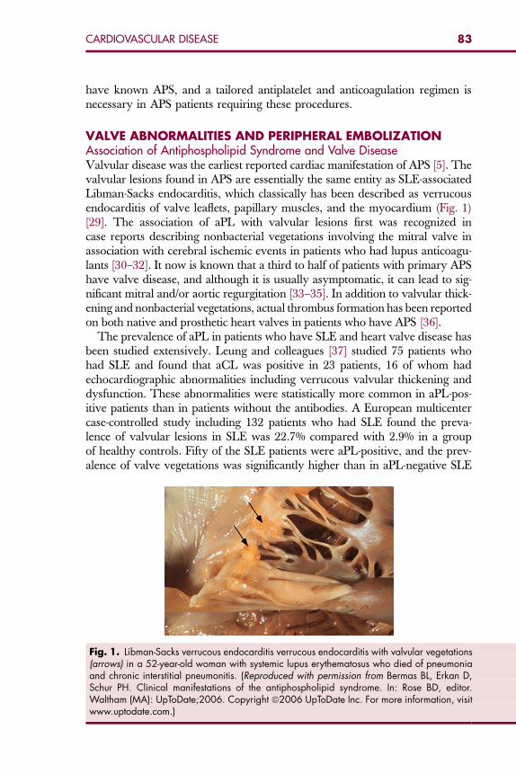
83CARDIOVASCULAR DISEASE
have known APS, and a tailored antiplatelet and anticoagulation regimen isnecessary in APS patients requiring these procedures.
VALVE ABNORMALITIES AND PERIPHERAL EMBOLIZATIONAssociation of Antiphospholipid Syndrome and Valve DiseaseValvular disease was the earliest reported cardiac manifestation of APS [5]. Thevalvular lesions found in APS are essentially the same entity as SLE-associatedLibman-Sacks endocarditis, which classically has been described as verrucousendocarditis of valve leaflets, papillary muscles, and the myocardium (Fig. 1)[29]. The association of aPL with valvular lesions first was recognized incase reports describing nonbacterial vegetations involving the mitral valve inassociation with cerebral ischemic events in patients who had lupus anticoagu-lants [30–32]. It now is known that a third to half of patients with primary APShave valve disease, and although it is usually asymptomatic, it can lead to sig-nificant mitral and/or aortic regurgitation [33–35]. In addition to valvular thick-ening and nonbacterial vegetations, actual thrombus formation has been reportedon both native and prosthetic heart valves in patients who have APS [36].
The prevalence of aPL in patients who have SLE and heart valve disease hasbeen studied extensively. Leung and colleagues [37] studied 75 patients whohad SLE and found that aCL was positive in 23 patients, 16 of whom hadechocardiographic abnormalities including verrucous valvular thickening anddysfunction. These abnormalities were statistically more common in aPL-pos-itive patients than in patients without the antibodies. A European multicentercase-controlled study including 132 patients who had SLE found the preva-lence of valvular lesions in SLE was 22.7% compared with 2.9% in a groupof healthy controls. Fifty of the SLE patients were aPL-positive, and the prev-alence of valve vegetations was significantly higher than in aPL-negative SLE
Fig. 1. Libman-Sacks verrucous endocarditis verrucous endocarditis with valvular vegetations(arrows) in a 52-year-old woman with systemic lupus erythematosus who died of pneumoniaand chronic interstitial pneumonitis. (Reproduced with permission from Bermas BL, Erkan D,Schur PH. Clinical manifestations of the antiphospholipid syndrome. In: Rose BD, editor.Waltham (MA): UpToDate;2006. Copyright ª2006 UpToDate Inc. For more information, visitwww.uptodate.com.)
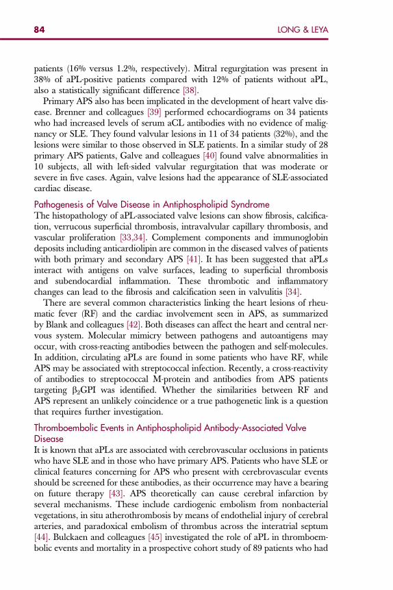
84 LONG & LEYA
patients (16% versus 1.2%, respectively). Mitral regurgitation was present in38% of aPL-positive patients compared with 12% of patients without aPL,also a statistically significant difference [38].
Primary APS also has been implicated in the development of heart valve dis-ease. Brenner and colleagues [39] performed echocardiograms on 34 patientswho had increased levels of serum aCL antibodies with no evidence of malig-nancy or SLE. They found valvular lesions in 11 of 34 patients (32%), and thelesions were similar to those observed in SLE patients. In a similar study of 28primary APS patients, Galve and colleagues [40] found valve abnormalities in10 subjects, all with left-sided valvular regurgitation that was moderate orsevere in five cases. Again, valve lesions had the appearance of SLE-associatedcardiac disease.
Pathogenesis of Valve Disease in Antiphospholipid Syndrome
The histopathology of aPL-associated valve lesions can show fibrosis, calcifica-tion, verrucous superficial thrombosis, intravalvular capillary thrombosis, andvascular proliferation [33,34]. Complement components and immunoglobindeposits including anticardiolipin are common in the diseased valves of patientswith both primary and secondary APS [41]. It has been suggested that aPLsinteract with antigens on valve surfaces, leading to superficial thrombosisand subendocardial inflammation. These thrombotic and inflammatorychanges can lead to the fibrosis and calcification seen in valvulitis [34].There are several common characteristics linking the heart lesions of rheu-matic fever (RF) and the cardiac involvement seen in APS, as summarizedby Blank and colleagues [42]. Both diseases can affect the heart and central ner-vous system. Molecular mimicry between pathogens and autoantigens mayoccur, with cross-reacting antibodies between the pathogen and self-molecules.In addition, circulating aPLs are found in some patients who have RF, whileAPS may be associated with streptococcal infection. Recently, a cross-reactivityof antibodies to streptococcal M-protein and antibodies from APS patientstargeting b2GPI was identified. Whether the similarities between RF andAPS represent an unlikely coincidence or a true pathogenetic link is a questionthat requires further investigation.
Thromboembolic Events in Antiphospholipid Antibody-Associated ValveDisease
It is known that aPLs are associated with cerebrovascular occlusions in patientswho have SLE and in those who have primary APS. Patients who have SLE orclinical features concerning for APS who present with cerebrovascular eventsshould be screened for these antibodies, as their occurrence may have a bearingon future therapy [43]. APS theoretically can cause cerebral infarction byseveral mechanisms. These include cardiogenic embolism from nonbacterialvegetations, in situ atherothrombosis by means of endothelial injury of cerebralarteries, and paradoxical embolism of thrombus across the interatrial septum[44]. Bulckaen and colleagues [45] investigated the role of aPL in thromboem-bolic events and mortality in a prospective cohort study of 89 patients who had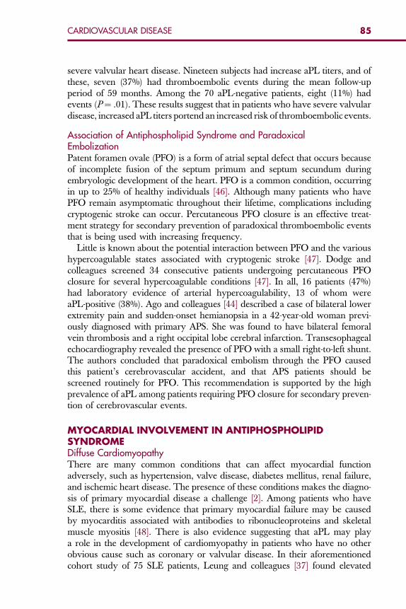
85CARDIOVASCULAR DISEASE
severe valvular heart disease. Nineteen subjects had increase aPL titers, and ofthese, seven (37%) had thromboembolic events during the mean follow-upperiod of 59 months. Among the 70 aPL-negative patients, eight (11%) hadevents (P¼ .01). These results suggest that in patients who have severe valvulardisease, increased aPL titers portend an increased risk of thromboembolic events.
Association of Antiphospholipid Syndrome and ParadoxicalEmbolization
Patent foramen ovale (PFO) is a form of atrial septal defect that occurs becauseof incomplete fusion of the septum primum and septum secundum duringembryologic development of the heart. PFO is a common condition, occurringin up to 25% of healthy individuals [46]. Although many patients who havePFO remain asymptomatic throughout their lifetime, complications includingcryptogenic stroke can occur. Percutaneous PFO closure is an effective treat-ment strategy for secondary prevention of paradoxical thromboembolic eventsthat is being used with increasing frequency.Little is known about the potential interaction between PFO and the varioushypercoagulable states associated with cryptogenic stroke [47]. Dodge andcolleagues screened 34 consecutive patients undergoing percutaneous PFOclosure for several hypercoagulable conditions [47]. In all, 16 patients (47%)had laboratory evidence of arterial hypercoagulability, 13 of whom wereaPL-positive (38%). Ago and colleagues [44] described a case of bilateral lowerextremity pain and sudden-onset hemianopsia in a 42-year-old woman previ-ously diagnosed with primary APS. She was found to have bilateral femoralvein thrombosis and a right occipital lobe cerebral infarction. Transesophagealechocardiography revealed the presence of PFO with a small right-to-left shunt.The authors concluded that paradoxical embolism through the PFO causedthis patient’s cerebrovascular accident, and that APS patients should bescreened routinely for PFO. This recommendation is supported by the highprevalence of aPL among patients requiring PFO closure for secondary preven-tion of cerebrovascular events.
MYOCARDIAL INVOLVEMENT IN ANTIPHOSPHOLIPIDSYNDROMEDiffuse CardiomyopathyThere are many common conditions that can affect myocardial functionadversely, such as hypertension, valve disease, diabetes mellitus, renal failure,and ischemic heart disease. The presence of these conditions makes the diagno-sis of primary myocardial disease a challenge [2]. Among patients who haveSLE, there is some evidence that primary myocardial failure may be causedby myocarditis associated with antibodies to ribonucleoproteins and skeletalmuscle myositis [48]. There is also evidence suggesting that aPL may playa role in the development of cardiomyopathy in patients who have no otherobvious cause such as coronary or valvular disease. In their aforementionedcohort study of 75 SLE patients, Leung and colleagues [37] found elevated
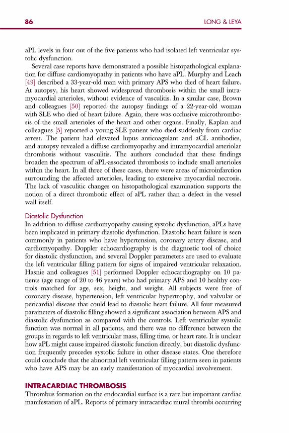
86 LONG & LEYA
aPL levels in four out of the five patients who had isolated left ventricular sys-tolic dysfunction.
Several case reports have demonstrated a possible histopathological explana-tion for diffuse cardiomyopathy in patients who have aPL. Murphy and Leach[49] described a 33-year-old man with primary APS who died of heart failure.At autopsy, his heart showed widespread thrombosis within the small intra-myocardial arterioles, without evidence of vasculitis. In a similar case, Brownand colleagues [50] reported the autopsy findings of a 22-year-old womanwith SLE who died of heart failure. Again, there was occlusive microthrombo-sis of the small arterioles of the heart and other organs. Finally, Kaplan andcolleagues [5] reported a young SLE patient who died suddenly from cardiacarrest. The patient had elevated lupus anticoagulant and aCL antibodies,and autopsy revealed a diffuse cardiomyopathy and intramyocardial arteriolarthrombosis without vasculitis. The authors concluded that these findingsbroaden the spectrum of aPL-associated thrombosis to include small arterioleswithin the heart. In all three of these cases, there were areas of microinfarctionsurrounding the affected arterioles, leading to extensive myocardial necrosis.The lack of vasculitic changes on histopathological examination supports thenotion of a direct thrombotic effect of aPL rather than a defect in the vesselwall itself.
Diastolic Dysfunction
In addition to diffuse cardiomyopathy causing systolic dysfunction, aPLs havebeen implicated in primary diastolic dysfunction. Diastolic heart failure is seencommonly in patients who have hypertension, coronary artery disease, andcardiomyopathy. Doppler echocardiography is the diagnostic tool of choicefor diastolic dysfunction, and several Doppler parameters are used to evaluatethe left ventricular filling pattern for signs of impaired ventricular relaxation.Hasnie and colleagues [51] performed Doppler echocardiography on 10 pa-tients (age range of 20 to 46 years) who had primary APS and 10 healthy con-trols matched for age, sex, height, and weight. All subjects were free ofcoronary disease, hypertension, left ventricular hypertrophy, and valvular orpericardial disease that could lead to diastolic heart failure. All four measuredparameters of diastolic filling showed a significant association between APS anddiastolic dysfunction as compared with the controls. Left ventricular systolicfunction was normal in all patients, and there was no difference between thegroups in regards to left ventricular mass, filling time, or heart rate. It is unclearhow aPL might cause impaired diastolic function directly, but diastolic dysfunc-tion frequently precedes systolic failure in other disease states. One thereforecould conclude that the abnormal left ventricular filling pattern seen in patientswho have APS may be an early manifestation of myocardial involvement.INTRACARDIAC THROMBOSISThrombus formation on the endocardial surface is a rare but important cardiacmanifestation of aPL. Reports of primary intracardiac mural thrombi occurring
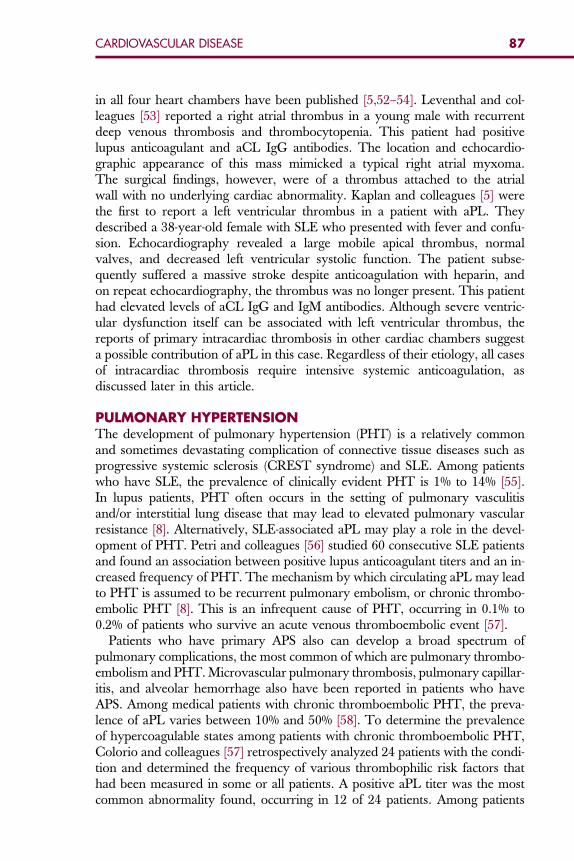
87CARDIOVASCULAR DISEASE
in all four heart chambers have been published [5,52–54]. Leventhal and col-leagues [53] reported a right atrial thrombus in a young male with recurrentdeep venous thrombosis and thrombocytopenia. This patient had positivelupus anticoagulant and aCL IgG antibodies. The location and echocardio-graphic appearance of this mass mimicked a typical right atrial myxoma.The surgical findings, however, were of a thrombus attached to the atrialwall with no underlying cardiac abnormality. Kaplan and colleagues [5] werethe first to report a left ventricular thrombus in a patient with aPL. Theydescribed a 38-year-old female with SLE who presented with fever and confu-sion. Echocardiography revealed a large mobile apical thrombus, normalvalves, and decreased left ventricular systolic function. The patient subse-quently suffered a massive stroke despite anticoagulation with heparin, andon repeat echocardiography, the thrombus was no longer present. This patienthad elevated levels of aCL IgG and IgM antibodies. Although severe ventric-ular dysfunction itself can be associated with left ventricular thrombus, thereports of primary intracardiac thrombosis in other cardiac chambers suggesta possible contribution of aPL in this case. Regardless of their etiology, all casesof intracardiac thrombosis require intensive systemic anticoagulation, asdiscussed later in this article.
PULMONARY HYPERTENSIONThe development of pulmonary hypertension (PHT) is a relatively commonand sometimes devastating complication of connective tissue diseases such asprogressive systemic sclerosis (CREST syndrome) and SLE. Among patientswho have SLE, the prevalence of clinically evident PHT is 1% to 14% [55].In lupus patients, PHT often occurs in the setting of pulmonary vasculitisand/or interstitial lung disease that may lead to elevated pulmonary vascularresistance [8]. Alternatively, SLE-associated aPL may play a role in the devel-opment of PHT. Petri and colleagues [56] studied 60 consecutive SLE patientsand found an association between positive lupus anticoagulant titers and an in-creased frequency of PHT. The mechanism by which circulating aPL may leadto PHT is assumed to be recurrent pulmonary embolism, or chronic thrombo-embolic PHT [8]. This is an infrequent cause of PHT, occurring in 0.1% to0.2% of patients who survive an acute venous thromboembolic event [57].
Patients who have primary APS also can develop a broad spectrum ofpulmonary complications, the most common of which are pulmonary thrombo-embolism and PHT. Microvascular pulmonary thrombosis, pulmonary capillar-itis, and alveolar hemorrhage also have been reported in patients who haveAPS. Among medical patients with chronic thromboembolic PHT, the preva-lence of aPL varies between 10% and 50% [58]. To determine the prevalenceof hypercoagulable states among patients with chronic thromboembolic PHT,Colorio and colleagues [57] retrospectively analyzed 24 patients with the condi-tion and determined the frequency of various thrombophilic risk factors thathad been measured in some or all patients. A positive aPL titer was the mostcommon abnormality found, occurring in 12 of 24 patients. Among patients
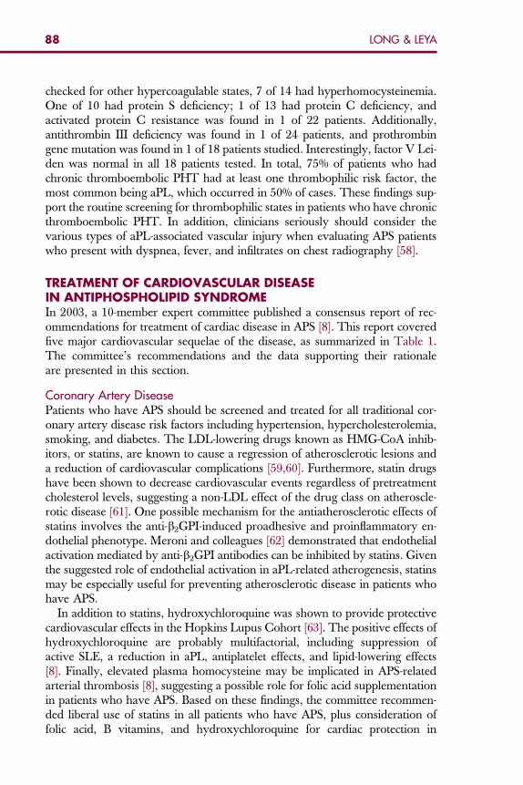
88 LONG & LEYA
checked for other hypercoagulable states, 7 of 14 had hyperhomocysteinemia.One of 10 had protein S deficiency; 1 of 13 had protein C deficiency, andactivated protein C resistance was found in 1 of 22 patients. Additionally,antithrombin III deficiency was found in 1 of 24 patients, and prothrombingene mutation was found in 1 of 18 patients studied. Interestingly, factor V Lei-den was normal in all 18 patients tested. In total, 75% of patients who hadchronic thromboembolic PHT had at least one thrombophilic risk factor, themost common being aPL, which occurred in 50% of cases. These findings sup-port the routine screening for thrombophilic states in patients who have chronicthromboembolic PHT. In addition, clinicians seriously should consider thevarious types of aPL-associated vascular injury when evaluating APS patientswho present with dyspnea, fever, and infiltrates on chest radiography [58].
TREATMENT OF CARDIOVASCULAR DISEASEIN ANTIPHOSPHOLIPID SYNDROMEIn 2003, a 10-member expert committee published a consensus report of rec-ommendations for treatment of cardiac disease in APS [8]. This report coveredfive major cardiovascular sequelae of the disease, as summarized in Table 1.The committee’s recommendations and the data supporting their rationaleare presented in this section.
Coronary Artery Disease
Patients who have APS should be screened and treated for all traditional cor-onary artery disease risk factors including hypertension, hypercholesterolemia,smoking, and diabetes. The LDL-lowering drugs known as HMG-CoA inhib-itors, or statins, are known to cause a regression of atherosclerotic lesions anda reduction of cardiovascular complications [59,60]. Furthermore, statin drugshave been shown to decrease cardiovascular events regardless of pretreatmentcholesterol levels, suggesting a non-LDL effect of the drug class on atheroscle-rotic disease [61]. One possible mechanism for the antiatherosclerotic effects ofstatins involves the anti-b2GPI-induced proadhesive and proinflammatory en-dothelial phenotype. Meroni and colleagues [62] demonstrated that endothelialactivation mediated by anti-b2GPI antibodies can be inhibited by statins. Giventhe suggested role of endothelial activation in aPL-related atherogenesis, statinsmay be especially useful for preventing atherosclerotic disease in patients whohave APS.In addition to statins, hydroxychloroquine was shown to provide protectivecardiovascular effects in the Hopkins Lupus Cohort [63]. The positive effects ofhydroxychloroquine are probably multifactorial, including suppression ofactive SLE, a reduction in aPL, antiplatelet effects, and lipid-lowering effects[8]. Finally, elevated plasma homocysteine may be implicated in APS-relatedarterial thrombosis [8], suggesting a possible role for folic acid supplementationin patients who have APS. Based on these findings, the committee recommen-ded liberal use of statins in all patients who have APS, plus consideration offolic acid, B vitamins, and hydroxychloroquine for cardiac protection in
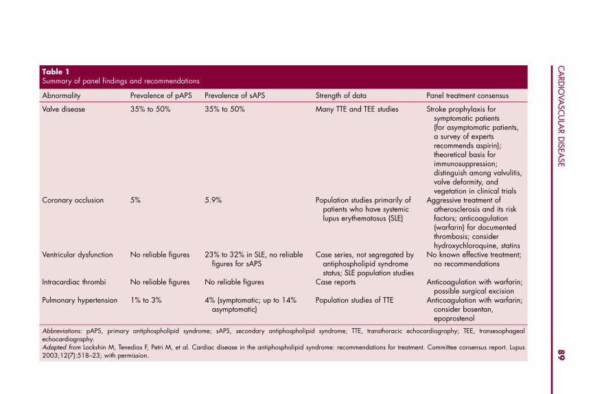
Table 1Summary of panel findings and recommendations
Abnormality Prevalence of pAPS Prevalence of sAPS Strength of data Panel treatment consensus
Valve disease 35% to 50% 35% to 50% Many TTE and TEE studies Stroke prophylaxis forsymptomatic patients(for asymptomatic patients,a survey of expertsrecommends aspirin);theoretical basis forimmunosuppression;distinguish among valvulitis,valve deformity, andvegetation in clinical trials
Coronary occlusion 5% 5.9% Population studies primarily ofpatients who have systemiclupus erythematosus (SLE)
Aggressive treatment ofatherosclerosis and its riskfactors; anticoagulation(warfarin) for documentedthrombosis; considerhydroxychloroquine, statins
Ventricular dysfunction No reliable figures 23% to 32% in SLE, no reliablefigures for sAPS
Case series, not segregated byantiphospholipid syndromestatus; SLE population studies
No known effective treatment;no recommendations
Intracardiac thrombi No reliable figures No reliable figures Case reports Anticoagulation with warfarin;possible surgical excision
Pulmonary hypertension 1% to 3% 4% (symptomatic; up to 14%asymptomatic)
Population studies of TTE Anticoagulation with warfarin;consider bosentan,epoprostenol
Abbreviations: pAPS, primary antiphospholipid syndrome; sAPS, secondary antiphospholipid syndrome; TTE, transthoracic echocardiography; TEE, transesophagealechocardiography.Adapted from Lockshin M, Tenedios F, Petri M, et al. Cardiac disease in the antiphospholipid syndrome: recommendations for treatment. Committee consensus report. Lupus2003;12(7):518–23; with permission.
89
CA
RDIO
VASC
ULA
RD
ISEASE
90 LONG & LEYA
patients at elevated cardiovascular risk. For patients who develop arterialthrombosis in the absence of atherosclerotic disease, prolonged anticoagulationwith warfarin is recommended [64].
Treatment of Valve Abnormalities in Antiphospholipid Syndrome
As described in the section on valvular disease, there are several pathologicalchanges that can occur in patients who have APS, including valvulitis withthickening, valve deformity and failure, and vegetation formation. Unfortu-nately, many of the publications about the prevalence and pathogenesis ofvalve disease in APS do not clearly delineate between these different entities,making recommendations for treatment somewhat challenging. A study byEspnola-Zavaleta and colleagues [65] provided some insight into the utility ofantiplatelet and anticoagulant therapy in APS patients with valvular disease.They performed transesophageal echocardiograms (TEE) on 13 primaryAPS patients before starting aspirin and/or warfarin therapy, and then repeatedthe test after 12 months of treatment. They found no modification of valvelesions in six cases (46.2%), with the remaining seven patients (53.8%) develop-ing new lesions. The authors concluded that oral antiplatelet or anticoagulationtherapy does not diminish the noninfective valve lesions seen in primary APS.There is also controversy over the use of corticosteroids or other anti-inflam-matory drugs to treat valvular disease in primary or secondary APS. Todate, there are no published systematic studies on anti-inflammatory or immu-nosuppressive treatment for this condition [8].Because of the increased risk of systemic embolization from vegetations andvalve thickening, the committee recommends anticoagulation with warfarin orheparin for APS patients with documented valvulopathy and any evidence ofthromboembolic disease. They also support the prophylactic use of antiplatelettherapy for asymptomatic patients. No agreement, however, was reachedregarding corticosteroid treatment for acute valve inflammation.
Myocardial Involvement in Antiphospholipid Syndrome
Because of the lack of published studies addressing treatment of this manifes-tation of APS, it is unknown whether antiplatelet, anticoagulant, or other treat-ment may be useful in preventing the diffuse cardiomyopathy or diastolicdysfunction seen in patients who have APS [8]. Therefore, the committeemade no recommendations on this condition. Standard medical therapy forsystolic or diastolic heart failure should be initiated and optimized accordingto current treatment guidelines.Intracardiac Thrombosis
In case reports of intracardiac thrombi in patients who have APS, physicianshave described treatment strategies of aggressive anticoagulation and/or surgi-cal incision. There are no published studies comparing medical, surgical, orcombined treatment strategies for this complication of APS. The committee’srecommendations were to screen patients who have APS for intracardiac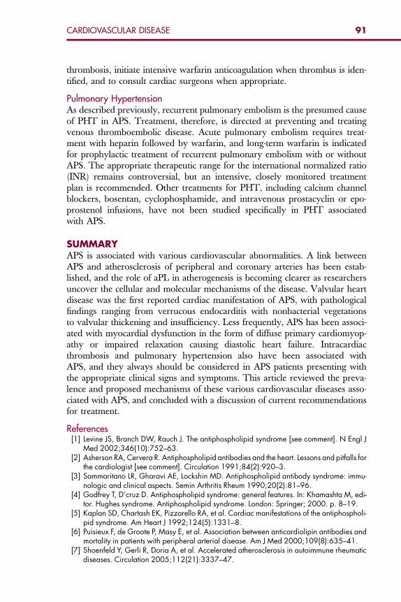
91CARDIOVASCULAR DISEASE
thrombosis, initiate intensive warfarin anticoagulation when thrombus is iden-tified, and to consult cardiac surgeons when appropriate.
Pulmonary Hypertension
As described previously, recurrent pulmonary embolism is the presumed causeof PHT in APS. Treatment, therefore, is directed at preventing and treatingvenous thromboembolic disease. Acute pulmonary embolism requires treat-ment with heparin followed by warfarin, and long-term warfarin is indicatedfor prophylactic treatment of recurrent pulmonary embolism with or withoutAPS. The appropriate therapeutic range for the international normalized ratio(INR) remains controversial, but an intensive, closely monitored treatmentplan is recommended. Other treatments for PHT, including calcium channelblockers, bosentan, cyclophosphamide, and intravenous prostacyclin or epo-prostenol infusions, have not been studied specifically in PHT associatedwith APS.SUMMARYAPS is associated with various cardiovascular abnormalities. A link betweenAPS and atherosclerosis of peripheral and coronary arteries has been estab-lished, and the role of aPL in atherogenesis is becoming clearer as researchersuncover the cellular and molecular mechanisms of the disease. Valvular heartdisease was the first reported cardiac manifestation of APS, with pathologicalfindings ranging from verrucous endocarditis with nonbacterial vegetationsto valvular thickening and insufficiency. Less frequently, APS has been associ-ated with myocardial dysfunction in the form of diffuse primary cardiomyop-athy or impaired relaxation causing diastolic heart failure. Intracardiacthrombosis and pulmonary hypertension also have been associated withAPS, and they always should be considered in APS patients presenting withthe appropriate clinical signs and symptoms. This article reviewed the preva-lence and proposed mechanisms of these various cardiovascular diseases asso-ciated with APS, and concluded with a discussion of current recommendationsfor treatment.
References
[1] Levine JS, Branch DW, Rauch J. The antiphospholipid syndrome [see comment]. N Engl JMed 2002;346(10):752–63.[2] Asherson RA, Cervera R. Antiphospholipid antibodies and the heart. Lessons and pitfalls for
the cardiologist [see comment]. Circulation 1991;84(2):920–3.[3] Sammaritano LR, Gharavi AE, Lockshin MD. Antiphospholipid antibody syndrome: immu-
nologic and clinical aspects. Semin Arthritis Rheum 1990;20(2):81–96.[4] Godfrey T, D’cruz D. Antiphospholipid syndrome: general features. In: Khamashta M, edi-
tor. Hughes syndrome. Antiphospholipid syndrome. London: Springer; 2000. p. 8–19.[5] Kaplan SD, Chartash EK, Pizzarello RA, et al. Cardiac manifestations of the antiphospholi-
pid syndrome. Am Heart J 1992;124(5):1331–8.[6] Puisieux F, de Groote P, Masy E, et al. Association between anticardiolipin antibodies and
mortality in patients with peripheral arterial disease. Am J Med 2000;109(8):635–41.[7] Shoenfeld Y, Gerli R, Doria A, et al. Accelerated atherosclerosis in autoimmune rheumatic
diseases. Circulation 2005;112(21):3337–47.
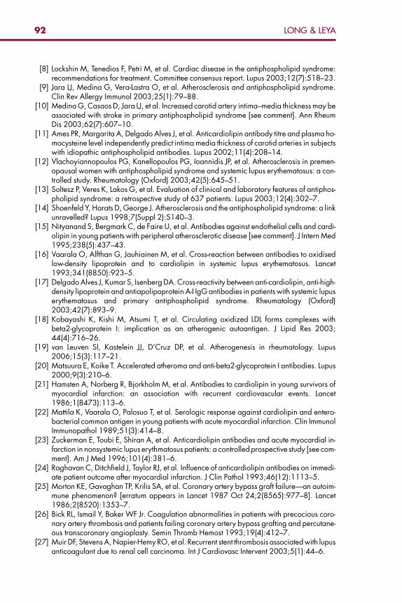
92 LONG & LEYA
[8] Lockshin M, Tenedios F, Petri M, et al. Cardiac disease in the antiphospholipid syndrome:recommendations for treatment. Committee consensus report. Lupus 2003;12(7):518–23.
[9] Jara LJ, Medina G, Vera-Lastra O, et al. Atherosclerosis and antiphospholipid syndrome.Clin Rev Allergy Immunol 2003;25(1):79–88.
[10] Medina G, Casaos D, Jara LJ, et al. Increased carotid artery intima–media thickness may beassociated with stroke in primary antiphospholipid syndrome [see comment]. Ann RheumDis 2003;62(7):607–10.
[11] Ames PR, Margarita A, Delgado Alves J, et al. Anticardiolipin antibody titre and plasma ho-mocysteine level independently predict intima media thickness of carotid arteries in subjectswith idiopathic antiphospholipid antibodies. Lupus 2002;11(4):208–14.
[12] Vlachoyiannopoulos PG, Kanellopoulos PG, Ioannidis JP, et al. Atherosclerosis in premen-opausal women with antiphospholipid syndrome and systemic lupus erythematosus: a con-trolled study. Rheumatology (Oxford) 2003;42(5):645–51.
[13] Soltesz P, Veres K, Lakos G, et al. Evaluation of clinical and laboratory features of antiphos-pholipid syndrome: a retrospective study of 637 patients. Lupus 2003;12(4):302–7.
[14] Shoenfeld Y, Harats D, George J. Atherosclerosis and the antiphospholipid syndrome: a linkunravelled? Lupus 1998;7(Suppl 2):S140–3.
[15] Nityanand S, Bergmark C, de Faire U, et al. Antibodies against endothelial cells and cardi-olipin in young patients with peripheral atherosclerotic disease [see comment]. J Intern Med1995;238(5):437–43.
[16] Vaarala O, Alfthan G, Jauhiainen M, et al. Cross-reaction between antibodies to oxidisedlow-density lipoprotein and to cardiolipin in systemic lupus erythematosus. Lancet1993;341(8850):923–5.
[17] Delgado Alves J, Kumar S, Isenberg DA. Cross-reactivity between anti-cardiolipin, anti-high-density lipoprotein and antiapolipoprotein A-I IgG antibodies in patients with systemic lupuserythematosus and primary antiphospholipid syndrome. Rheumatology (Oxford)2003;42(7):893–9.
[18] Kobayashi K, Kishi M, Atsumi T, et al. Circulating oxidized LDL forms complexes withbeta2-glycoprotein I: implication as an atherogenic autoantigen. J Lipid Res 2003;44(4):716–26.
[19] van Leuven SI, Kastelein JJ, D’Cruz DP, et al. Atherogenesis in rheumatology. Lupus2006;15(3):117–21.
[20] Matsuura E, Koike T. Accelerated atheroma and anti-beta2-glycoprotein I antibodies. Lupus2000;9(3):210–6.
[21] Hamsten A, Norberg R, Bjorkholm M, et al. Antibodies to cardiolipin in young survivors ofmyocardial infarction: an association with recurrent cardiovascular events. Lancet1986;1(8473):113–6.
[22] Mattila K, Vaarala O, Palosuo T, et al. Serologic response against cardiolipin and entero-bacterial common antigen in young patients with acute myocardial infarction. Clin ImmunolImmunopathol 1989;51(3):414–8.
[23] Zuckerman E, Toubi E, Shiran A, et al. Anticardiolipin antibodies and acute myocardial in-farction in nonsystemic lupus erythmatosus patients: a controlled prospective study [see com-ment]. Am J Med 1996;101(4):381–6.
[24] Raghavan C, Ditchfield J, Taylor RJ, et al. Influence of anticardiolipin antibodies on immedi-ate patient outcome after myocardial infarction. J Clin Pathol 1993;46(12):1113–5.
[25] Morton KE, Gavaghan TP, Krilis SA, et al. Coronary artery bypass graft failure—an autoim-mune phenomenon? [erratum appears in Lancet 1987 Oct 24;2(8565):977–8]. Lancet1986;2(8520):1353–7.
[26] Bick RL, Ismail Y, Baker WF Jr. Coagulation abnormalities in patients with precocious coro-nary artery thrombosis and patients failing coronary artery bypass grafting and percutane-ous transcoronary angioplasty. Semin Thromb Hemost 1993;19(4):412–7.
[27] Muir DF, Stevens A, Napier-Hemy RO, et al. Recurrent stent thrombosis associated with lupusanticoagulant due to renal cell carcinoma. Int J Cardiovasc Intervent 2003;5(1):44–6.
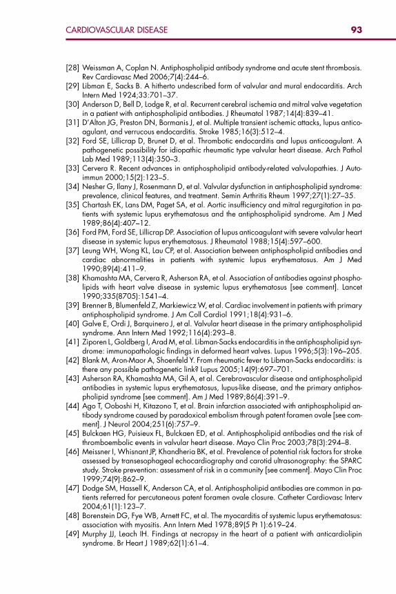
93CARDIOVASCULAR DISEASE
[28] Weissman A, Coplan N. Antiphospholipid antibody syndrome and acute stent thrombosis.Rev Cardiovasc Med 2006;7(4):244–6.
[29] Libman E, Sacks B. A hitherto undescribed form of valvular and mural endocarditis. ArchIntern Med 1924;33:701–37.
[30] Anderson D, Bell D, Lodge R, et al. Recurrent cerebral ischemia and mitral valve vegetationin a patient with antiphospholipid antibodies. J Rheumatol 1987;14(4):839–41.
[31] D’Alton JG, Preston DN, Bormanis J, et al. Multiple transient ischemic attacks, lupus antico-agulant, and verrucous endocarditis. Stroke 1985;16(3):512–4.
[32] Ford SE, Lillicrap D, Brunet D, et al. Thrombotic endocarditis and lupus anticoagulant. Apathogenetic possibility for idiopathic rheumatic type valvular heart disease. Arch PatholLab Med 1989;113(4):350–3.
[33] Cervera R. Recent advances in antiphospholipid antibody-related valvulopathies. J Auto-immun 2000;15(2):123–5.
[34] Nesher G, Ilany J, Rosenmann D, et al. Valvular dysfunction in antiphospholipid syndrome:prevalence, clinical features, and treatment. Semin Arthritis Rheum 1997;27(1):27–35.
[35] Chartash EK, Lans DM, Paget SA, et al. Aortic insufficiency and mitral regurgitation in pa-tients with systemic lupus erythematosus and the antiphospholipid syndrome. Am J Med1989;86(4):407–12.
[36] Ford PM, Ford SE, Lillicrap DP. Association of lupus anticoagulant with severe valvular heartdisease in systemic lupus erythematosus. J Rheumatol 1988;15(4):597–600.
[37] Leung WH, Wong KL, Lau CP, et al. Association between antiphospholipid antibodies andcardiac abnormalities in patients with systemic lupus erythematosus. Am J Med1990;89(4):411–9.
[38] Khamashta MA, Cervera R, Asherson RA, et al. Association of antibodies against phospho-lipids with heart valve disease in systemic lupus erythematosus [see comment]. Lancet1990;335(8705):1541–4.
[39] Brenner B, Blumenfeld Z, Markiewicz W, et al. Cardiac involvement in patients with primaryantiphospholipid syndrome. J Am Coll Cardiol 1991;18(4):931–6.
[40] Galve E, Ordi J, Barquinero J, et al. Valvular heart disease in the primary antiphospholipidsyndrome. Ann Intern Med 1992;116(4):293–8.
[41] Ziporen L, Goldberg I, Arad M, et al. Libman-Sacks endocarditis in the antiphospholipid syn-drome: immunopathologic findings in deformed heart valves. Lupus 1996;5(3):196–205.
[42] Blank M, Aron-Maor A, Shoenfeld Y. From rheumatic fever to Libman-Sacks endocarditis: isthere any possible pathogenetic link? Lupus 2005;14(9):697–701.
[43] Asherson RA, Khamashta MA, Gil A, et al. Cerebrovascular disease and antiphospholipidantibodies in systemic lupus erythematosus, lupus-like disease, and the primary antiphos-pholipid syndrome [see comment]. Am J Med 1989;86(4):391–9.
[44] Ago T, Ooboshi H, Kitazono T, et al. Brain infarction associated with antiphospholipid an-tibody syndrome caused by paradoxical embolism through patent foramen ovale [see com-ment]. J Neurol 2004;251(6):757–9.
[45] Bulckaen HG, Puisieux FL, Bulckaen ED, et al. Antiphospholipid antibodies and the risk ofthromboembolic events in valvular heart disease. Mayo Clin Proc 2003;78(3):294–8.
[46] Meissner I, Whisnant JP, Khandheria BK, et al. Prevalence of potential risk factors for strokeassessed by transesophageal echocardiography and carotid ultrasonography: the SPARCstudy. Stroke prevention: assessment of risk in a community [see comment]. Mayo Clin Proc1999;74(9):862–9.
[47] Dodge SM, Hassell K, Anderson CA, et al. Antiphospholipid antibodies are common in pa-tients referred for percutaneous patent foramen ovale closure. Catheter Cardiovasc Interv2004;61(1):123–7.
[48] Borenstein DG, Fye WB, Arnett FC, et al. The myocarditis of systemic lupus erythematosus:association with myositis. Ann Intern Med 1978;89(5 Pt 1):619–24.
[49] Murphy JJ, Leach IH. Findings at necropsy in the heart of a patient with anticardiolipinsyndrome. Br Heart J 1989;62(1):61–4.
94 LONG & LEYA
[50] Brown JH, Doherty CC, Allen DC, et al. Fatal cardiac failure due to myocardial microthrombiin systemic lupus erythematosus. Br Med J (Clin Res Ed) 1988;296(6635):1505.
[51] Hasnie AM, Stoddard MF, Gleason CB, et al. Diastolic dysfunction is a feature of the anti-phospholipid syndrome. Am Heart J 1995;129(5):1009–13.
[52] Coppock MA, Safford RE, Danielson GK. Intracardiac thrombosis, phospholipid anti-bodies, and two-chambered right ventricle. Br Heart J 1988;60(5):455–8.
[53] Leventhal LJ, Borofsky MA, Bergey PD, et al. Antiphospholipid antibody syndrome with rightatrial thrombosis mimicking an atrial myxoma. Am J Med 1989;87(1):111–3.
[54] Lubbe WF, Asherson RA. Intracardiac thrombus in systemic lupus erythematosus associatedwith lupus anticoagulant. Arthritis Rheum 1988;31(11):1453–4.
[55] Petri M. Systemic lupus erythematosis and the cardiovascular system: the heart. In: Lahita R,editor. Systemic lupus erythematosis. 3rd edition. San Diego: Academic Press; 1999.p. 687–706.
[56] Petri M, Rheinschmidt M, Whiting-O’Keefe Q, et al. The frequency of lupus anticoagulant insystemic lupus erythematosus. A study of sixty consecutive patients by activated partialthromboplastin time, Russell viper venom time, and anticardiolipin antibody level. AnnIntern Med 1987;106(4):524–31.
[57] Colorio CC, Martinuzzo ME, Forastiero RR, et al. Thrombophilic factors in chronic thrombo-embolic pulmonary hypertension. Blood Coagul Fibrinolysis 2001;12(6):427–32.
[58] Espinosa G, Cervera R, Font J, et al. The lung in the antiphospholipid syndrome. Ann RheumDis 2002;61(3):195–8.
[59] Cannon CP, Braunwald E, McCabe CH, et al. Intensive versus moderate lipid lowering withstatins after acute coronary syndromes [see comment] [erratum appears in N Engl J Med2006 Feb 16;354(7):778]. N Engl J Med 2004;350(15):1495–504.
[60] Nissen SE, Tuzcu EM, Schoenhagen P, et al. Effect of intensive compared with moderatelipid-lowering therapy on progression of coronary atherosclerosis: a randomized controlledtrial [see comment]. JAMA 2004;291(9):1071–80.
[61] Collins R, Armitage J, Parish S, et al. MRC/BHF Heart protection study of cholesterol lower-ing with simvastatin in 20,536 high-risk individuals: a randomised placebo-controlled trial.Lancet 2002;360(9326):7–22.
[62] Meroni PL, Raschi E, Testoni C, et al. Statins prevent endothelial cell activation induced byantiphospholipid (anti-beta2-glycoprotein I) antibodies: effect on the proadhesive andproinflammatory phenotype. Arthritis Rheum 2001;44(12):2870–8.
[63] Petri M. Detection of coronary artery disease and the role of traditional risk factors in theHopkins Lupus Cohort. Lupus 2000;9(3):170–5.
[64] Ruiz-Irastorza G, Khamashta MA, Hunt BJ, et al. Bleeding and recurrent thrombosis in def-inite antiphospholipid syndrome: analysis of a series of 66 patients treated with oral antico-agulation to a target international normalized ratio of 3.5. Arch Intern Med 2002;162(10):1164–9.
[65] Espinola-Zavaleta N, Vargas-Barron J, Colmenares-Galvis T, et al. Echocardiographic eval-uation of patients with primary antiphospholipid syndrome. Am Heart J 1999;137(5):973–8.
Hematol Oncol Clin N Am 22 (2008) 95–105
HEMATOLOGY/ONCOLOGY CLINICSOF NORTH AMERICA
Antiphospholipid Syndrome: Roleof Antiphospholipid Antibodiesin Neurology
Rima M. Dafer, MD, MPH, FAHA*,Jose Biller, MD, FACP, FAAN, FAHADepartment of Neurology, Loyola University Chicago,Stritch School of Medicine, 2160 South First Avenue, 2700 McGuire,Maywood, IL 60153, USA
Antiphospholipid syndrome (APS), or Hughes syndrome, is an acquiredprothrombotic syndrome first described in 1983, characterized byrecurrent arterial or venous thromboembolism, unexplained fetal loss
usually within the first 10 weeks of gestation, and thrombocytopenia, withthe presence of circulating antiphospholipid antibodies.
Primary APS is an immune-mediated coagulopathy associated with cerebralischemia in young adults, the etiology of which remains unknown. SecondaryAPS can occur within the context of several diseases, mainly autoimmune orrheumatologic disorders, infections, malignancy, and drugs.
Although only ischemic stroke is reasonably well established and acceptedneurological diagnostic criterion for the syndrome, other neurological condi-tions have been associated with aPLs, most commonly multiple sclerosis,migraine, psychiatric diseases, and various movement disorders.
ANTIPHOSPHOLIPID ANTIBODIESThe antiphospholipid antibodies (aPLs) are antibodies directed against proteinantigens that bind to membrane anionic phospholipids, anticardiolipins, ortheir associated plasma proteins [1]. Several aPLs have been described; themost thoroughly studied subsets include lupus anticoagulant (LA) and anticar-diolipin (aCL) IgG, IgA, and IgM isotypes; antiphospahtidylethanolamine(aPE); antiphospatidylserine (aPS); and antiphospathylcholine (aPC). Bindingof aCL to cell membrane requires the presence of beta-2 glycoprotein 1 anti-body (b2GP1) [2] and prothrombin [3]. LA often is found in patients whohave systemic lupus erythematosus (SLE) and in sera from patients whohave syphilis binding to cardiolipin extracts of beef hearts. LA is identifiedby prolonged clotting time on coagulation assays. In contrast, aCLs and
*Corresponding author. E-mail address: [email protected] (R.M. Dafer).
0889-8588/08/$ – see front matter ª 2008 Elsevier Inc. All rights reserved.doi:10.1016/j.hoc.2007.10.005 hemonc.theclinics.com
96 DAFER & BILLER
b2GP1 are identified by solid-phase ELISAs that measure immunoreactivity toa phospholipid or phospholipid-binding protein.
EPIDEMIOLOGYThe aPL titers tend to be nonspecific; they are present in 1% to 5% of healthyindividuals, and 12% to 50% of elderly patients and patients who have SLE[4–6]. In the Framingham original cohort and offspring combined, the preva-lence of aCL positivity was 19.7% in men and 17.6% in women, and washigher in subjects of both genders with first ischemic stroke or transient ische-mic attack (TIA) [7]. Familial incidence has been reported in 33% of subjects.
The primary APS affects predominantly younger adults of both genders,with female predominance, although it has been reported in extremes of age.The syndrome is common among all ethnicities, with higher prevalence inAfrican Americans and the Hispanic population. LA appears to be the strongerpredictor of thrombotic risk for both venous and arterial thrombosis with anodds ratio of thrombosis five to 16 times higher than control [8,9], followedby anti- b2GP1 antibodies, which correlate mainly with arterial thrombosis.The aCLs are the weakest predictors of thrombosis, correlating mainly withvenous rather than arterial thrombosis. The presence of multiple aPLs isthought to carry a higher risk of thrombosis.
PATHOPHYSIOLOGYThe IgG isotypes of aCL and LA are the mostly associated aPLs with risk forfirst ischemic stroke. The real underlying mechanism of thrombosis remainsunclear. Several mechanisms have been postulated, including multifactorialinteractions with phospholipids in endothelial cells and platelet membranes,multifactorial interactions with coagulation factors, direct abrogation of endo-thelial cell prostacyclin production, and down-regulation of interleukins in vas-cular beds.
CLASSIFICATION CRITERIA FOR ANTIPHOSPHOLIPIDSYNDROMEThe preliminary Sapporo classification divided the APS criteria into clinicaland laboratory, with a definite diagnosis considered when at least one of theclinical and one of the laboratory criteria are present [10]. The criteria wererevised at the 11th International Congress on Antiphospholipid Antibodiesin Sydney, Australia, in 2004. According to the updated classification, definitediagnosis of APS requires the combination of at least one clinical and one lab-oratory criterion; the autoantibodies must be detected on at least 2 occasions,12 weeks apart, to distinguish persistent autoimmune antibody responses fromtransient responses caused by other conditions such as infection or drug expo-sures. The diagnosis of APS should be avoided if less than 12 weeks or morethan 5 years separate the positive aPL test and the clinical manifestation [11].
The following revised criteria were suggested for the classification of the anti-phospholipid syndrome (Box 1).
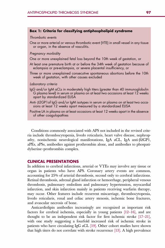
Box 1: Criteria for classifying antiphospholipid syndrome
Thrombotic events
One or more arterial or venous thrombotic event (VTE) in small vessel in any tissueor organ, in the absence of vasculitis.
Pregnancy morbidity
One or more unexplained fetal loss beyond the 10th week of gestation, or
At least one premature birth at or before the 34th week of gestation because ofeclampsia or pre-eclampsia, or severe placental insufficiency, or
Three or more unexplained consecutive spontaneous abortions before the 10thweek of gestation, with other causes excluded
Laboratory criteria
IgG and/or IgM aCLs in moderately high titers (greater than 40 immunoglobulinG plasma level) in serum or plasma on at least two occasions at least 12 weeksapart by standardized ELISA
Anti- b2GP1of IgG and/or IgM isotypes in serum or plasma on at least two occa-sions at least 12 weeks apart measured by a standardized ELISA
Positive LA in plasma on at least occasions at least 12 weeks apart in the absenceof other coagulopathies
97ANTIPHOSPHOLIPID THROMBOSIS SYNDROME
Conditions commonly associated with APS not included in the revised crite-ria include thrombocytopenia, livedo reticularis, heart valve disease, nephrop-athy, nonischemic neurological manifestations, IgA aCL, IgA anti-b2GP1,aPEs, aPSs, antibodies against prothrombin alone, and antibodies to phospati-dylserine–prothrombin complex.
CLINICAL PRESENTATIONSIn addition to cerebral infarctions, arterial or VTEs may involve any tissue ororgan in patients who have APS. Coronary artery events are common,accounting for 25% of arterial thrombosis, second only to cerebral infarctions.Retinal thrombosis, adrenal gland infarction or hemorrhage, peripheral venousthrombosis, pulmonary embolism and pulmonary hypertension, myocardialinfarction, and skin infarction mainly in patients receiving warfarin therapy,may occur. Other features include recurrent miscarriage, thrombocytopenia,livedo reticularis, renal and celiac artery stenosis, ischemic bone fractures,and avascular necrosis of bone.
Anticardiolipin antibodies increasingly are recognized as important riskfactors for cerebral ischemia, especially in young patients [12–16], and arethought to be an independent risk factor for first ischemic stroke [17–21],with one study suggesting a fourfold increased risk of ischemic stroke inpatients who have circulating IgG aCL [19]. Other cohort studies have shownthat high titers do not correlate with stroke recurrence [13]. A high prevalence
98 DAFER & BILLER
of aPLs of 44% was demonstrated in young adult patients under the age of 51with cerebral ischemia of undetermined causes [22,23]. The prevalence of thedisorder, however, is variable because of heterogeneity of aPL, variability andlack of specificity of the assays, and the change in the status of aPL from pos-itive to negative with time. Whether the presence of aPL plays a direct role instroke or whether it is an epiphenomenon remains unclear, but it is postulatedthat the presence of circulating aPLs may cause a possible tendency towardaccelerated atherosclerosis. Prospective studies have found an associationbetween aPLs and stroke [24], in particular in young adult women [25]. Inthe antiphospholipid antibodies and stroke study (APASS), the risk of cerebralinfarction was reported to be 2.31 times higher in patients who have positivecirculating antibodies than in those negative for antibodies [17], but the pres-ence of aPLs (either LA or aCL) among patients who had ischemic strokedid not predict either increased risk of subsequent vascular occlusive eventsover 2 years or a differential response to antiaggregants [26]. In the Framing-ham study, elevated serum concentrations of aCLs, independently of other car-diovascular risk factors, significantly predicted the risk of future ischemicstroke and TIA in women but not in men [7]. In the Honolulu study, therisk of stroke and myocardial infarction was 1.5 times higher in men whohad positive b2GP1-dependent aCL IgG [24].
The most common cerebral ischemia associated with aPLs is arterial infarc-tion, with small vessels and the middle cerebral arteries being the most in-volved in the arterial circulation. Multiple recurrent cerebral infarctions mayoccur [27]. Cerebral vein or dural sinus thrombosis are not uncommon.
Although the neurological association of aPLs and APS is diverse, only ische-mic stroke is established reasonably well and accepted as a diagnostic criterion forthe syndrome. A wide spectrum of neurological disorders, including seizures[28], psychiatric diseases [29], dementia [30], transverse myelitis [31,32], opticneuropathy and multiple sclerosis-like disorders [33–35], migraine [36,37], Guil-lain-Barre syndrome [38], sensory-neural hearing loss [39–42], and atypicalmovement disorders, most commonly chorea, hemidystonia, parkinsonism,and hemiballismus, also has been associated with positive aPLs [43] or APS [42].
Dermatological manifestations are encountered in 50% of patients and maybe the presenting features in 30.5% of patients. The most frequent dermatolog-ical manifestation is livedo reticularis (livedo racemosa), observed in 25.5% ofpatients. Painful purpura and leg ulcers also may occur. The presence of livedoreticularis is significantly associated with cerebral or ocular ischemic arterialevents and Sneddon’s syndrome [44].
Cardiac involvement is common in APS, and is related significantly to highIgG aCL titers [45]. Coronary artery disease, mitral valve thickening and othercardiac valvulopathies, and embolic sources frequently are observed. Splinterhemorrhages and cardiac murmur rarely may be seen in patients who haveLibman Sacks endocarditis. The use of platelet antiaggregants did not pre-vented valvular lesion regression in patients who had APS [45]. In patientswho have pulmonary embolism, tachypnea and chest pain are common.
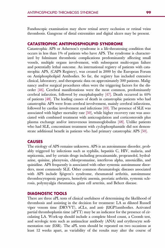
99ANTIPHOSPHOLIPID THROMBOSIS SYNDROME
Funduscopic examination may show retinal artery occlusion or retinal veinsthrombosis. Gangrene of distal extremities and digital ulcers may be present.
CATASTROPHIC ANTIPHOSPHOLIPID SYNDROMECatastrophic APS or Asherson’s syndrome is a life-threatening condition thatoccurs in less than 1% of patients who have APS. The syndrome is character-ized by fulminant thrombotic complications predominantly affecting smallvessels, multiple organs involvement, with subsequent multi-organ failureand potentially lethal outcome. An international registry of patients with cata-strophic APS, (CAPS Registry), was created in 2000 by the European Forumon Antiphospholipid Antibodies. So far, the registry has included extensiveclinical, laboratory, and therapeutic data on approximately 300 patients. Malig-nancy and/or surgical procedures often were the triggering factors for the dis-order [46]. Cerebral manifestations were the most common, predominantlycerebral infarction, followed by encephalopathy [47]. Death occurred in 44%of patients [48]. The leading causes of death in catastrophic patients who hadcatastrophic APS were from cerebral involvement, mainly cerebral infarctions,followed by cardiac involvement and infections [49]. The presence of SLE wasassociated with higher mortality rate [50], while higher recovery rate was asso-ciated with combined treatment with anticoagulation and corticosteroids plusplasma exchange and/or intravenous immunoglobulins [48]. Unlike patientswho had SLE, concomitant treatment with cyclophosphamide did not demon-strate additional benefit in patients who had primary catastrophic APS [50].
CAUSESThe etiology of APS remains unknown. APS is an autoimmune disorder, prob-ably triggered by infections such as syphilis, hepatitis C, HIV, malaria, andsepticemia, and by certain drugs including procainamide, propranolol, hydral-azine, quinine, phenytoin, chlorpromazine, interferon alpha, amoxicillin, andquinidine. APS frequently is associated with other systemic autoimmune disor-ders, most commonly SLE. Other common rheumatologic diseases associatedwith APS include Sjogren’s syndrome, rheumatoid arthritis, autoimmunethrombocytopenic purpura, hemolytic anemia, psoriatic arthritis, systemic scle-rosis, polymyalgia rheumatica, giant cell arteritis, and Behcet disease.
DIAGNOSTIC TOOLSThere are three aPL tests of clinical usefulness of determining the likelihood ofthrombosis and assisting in the decision for treatment: LA as diluted Russellviper venom time (DRVVT), aCLs, and anti- b2GP1antibodies. Activatedpartial thromboplastin time (aPTT) may be an indicator for the presence of cir-culating LA. Work-up should include a complete blood count, a Coomb test,and serologic tests such as antinuclear antibody (ANA) and erythrocyte sedi-mentation rate (ESR). The aPL tests should be repeated on two occasions atleast 12 weeks apart, as variability of the results may alter the course of
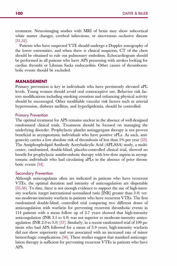
100 DAFER & BILLER
treatment. Neuroimaging studies with MRI of brain may show subcorticalwhite matter changes, cerebral infarctions, or sinovenous occlusive disease[51,52].
Patients who have suspected VTE should undergo a Doppler sonography ofthe lower extremities, and when there is clinical suspicion, CT of the chestshould be obtained to rule out pulmonary embolism. Echocardiogram shouldbe performed in all patients who have APS presenting with strokes looking forcardiac thrombi or Libman Sacks endocarditis. Other causes of thromboem-bolic events should be excluded.
MANAGEMENTPrimary prevention is key in individuals who have persistently elevated aPLlevels. Young women should avoid oral contraceptive use. Behavior risk fac-tors modifications including smoking cessation and enhancing physical activityshould be encouraged. Other modifiable vascular risk factors such as arterialhypertension, diabetes mellitus, and hyperlipidemia, should be controlled.
Primary Prevention
The optimal treatment for APS remains unclear in the absence of well-designedrandomized clinical trials. Treatment should be focused on managing theunderlying disorder. Prophylactic platelet antiaggregant therapy is not provenbeneficial in asymptomatic individuals who have positive aPLs. As such, anti-genecity carries a low absolute risk of thrombosis of less than 1% per year [53].The Antiphospholipid Antibody Acetylsalicylic Acid (APLASA) study, a multi-center, randomized, double-blind, placebo-controlled clinical trial, showed nobenefit for prophylactic antithrombotic therapy with low-dose aspirin in asymp-tomatic individuals who had circulating aPLs in the absence of prior throm-botic events [54].Secondary Prevention
Although anticoagulants often are indicated in patients who have recurrentVTEs, the optimal duration and intensity of anticoagulation are disputable[55,56]. To date, there is not enough evidence to support the use of high-inten-sity warfarin (target international normalized ratio [INR] greater than 3.0) ver-sus moderate-intensity warfarin in patients who have recurrent VTEs. The firstrandomized double-blind, controlled trial comparing two different doses ofanticoagulation with warfarin for preventing recurrent thrombotic events in114 patients with a mean follow up of 2.7 years showed that high-intensityanticoagulation (INR 3.1 to 4.0) was not superior to moderate-intensity antico-agulation (INR 2.0 to 3.0) [57]. Similarly, in a recent randomized trial of 109 pa-tients who had APS followed for a mean of 3.9 years, high-intensity warfarindid not show superiority and was associated with an increased rate of minorhemorrhagic complications [56]. These studies suggest that standard anticoagu-lation therapy is sufficient for preventing recurrent VTEs in patients who haveAPS.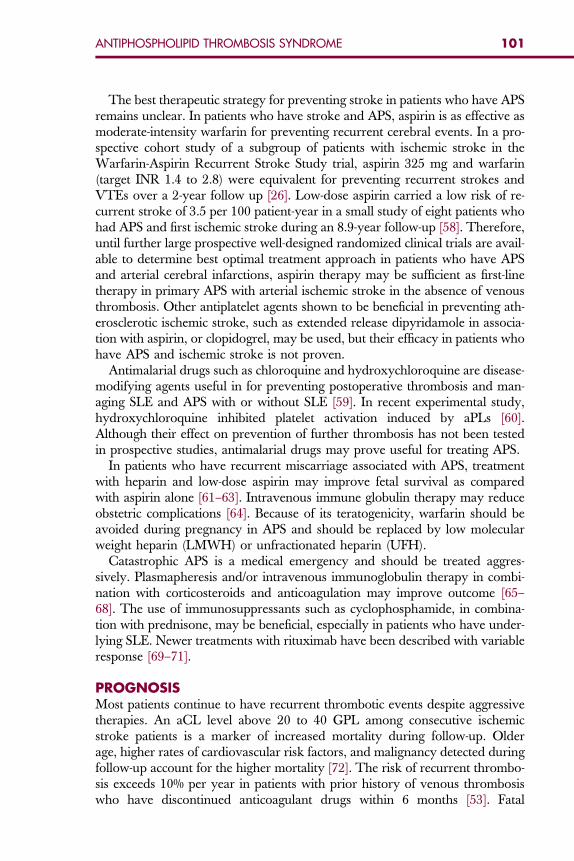
101ANTIPHOSPHOLIPID THROMBOSIS SYNDROME
The best therapeutic strategy for preventing stroke in patients who have APSremains unclear. In patients who have stroke and APS, aspirin is as effective asmoderate-intensity warfarin for preventing recurrent cerebral events. In a pro-spective cohort study of a subgroup of patients with ischemic stroke in theWarfarin-Aspirin Recurrent Stroke Study trial, aspirin 325 mg and warfarin(target INR 1.4 to 2.8) were equivalent for preventing recurrent strokes andVTEs over a 2-year follow up [26]. Low-dose aspirin carried a low risk of re-current stroke of 3.5 per 100 patient-year in a small study of eight patients whohad APS and first ischemic stroke during an 8.9-year follow-up [58]. Therefore,until further large prospective well-designed randomized clinical trials are avail-able to determine best optimal treatment approach in patients who have APSand arterial cerebral infarctions, aspirin therapy may be sufficient as first-linetherapy in primary APS with arterial ischemic stroke in the absence of venousthrombosis. Other antiplatelet agents shown to be beneficial in preventing ath-erosclerotic ischemic stroke, such as extended release dipyridamole in associa-tion with aspirin, or clopidogrel, may be used, but their efficacy in patients whohave APS and ischemic stroke is not proven.
Antimalarial drugs such as chloroquine and hydroxychloroquine are disease-modifying agents useful in for preventing postoperative thrombosis and man-aging SLE and APS with or without SLE [59]. In recent experimental study,hydroxychloroquine inhibited platelet activation induced by aPLs [60].Although their effect on prevention of further thrombosis has not been testedin prospective studies, antimalarial drugs may prove useful for treating APS.
In patients who have recurrent miscarriage associated with APS, treatmentwith heparin and low-dose aspirin may improve fetal survival as comparedwith aspirin alone [61–63]. Intravenous immune globulin therapy may reduceobstetric complications [64]. Because of its teratogenicity, warfarin should beavoided during pregnancy in APS and should be replaced by low molecularweight heparin (LMWH) or unfractionated heparin (UFH).
Catastrophic APS is a medical emergency and should be treated aggres-sively. Plasmapheresis and/or intravenous immunoglobulin therapy in combi-nation with corticosteroids and anticoagulation may improve outcome [65–68]. The use of immunosuppressants such as cyclophosphamide, in combina-tion with prednisone, may be beneficial, especially in patients who have under-lying SLE. Newer treatments with rituximab have been described with variableresponse [69–71].
PROGNOSISMost patients continue to have recurrent thrombotic events despite aggressivetherapies. An aCL level above 20 to 40 GPL among consecutive ischemicstroke patients is a marker of increased mortality during follow-up. Olderage, higher rates of cardiovascular risk factors, and malignancy detected duringfollow-up account for the higher mortality [72]. The risk of recurrent thrombo-sis exceeds 10% per year in patients with prior history of venous thrombosiswho have discontinued anticoagulant drugs within 6 months [53]. Fatal
102 DAFER & BILLER
outcome may occur in patients who have recurrent myocardial infarctions, pul-monary embolism, and in more than half the patients who have catastrophicAPS.
SUMMARYThe aPLs, specifically LA and anti-b2GP1 antibodies, increasingly are recog-nized as risk factors for stroke, especially in young adults. Positivity of aPLsin healthy individuals may not confer increased vascular thrombo–occlusiverisk; therefore prophylactic use of antiaggregants in those individuals maynot be necessary. Patients who have definite APS are at higher risk for recur-rent VTEs, especially in the presence of SLE. The optimal treatment approachremains controversial. Long-term warfarin anticoagulation (INR 2.0 to 3.0)may be optimal in patients who have recurrent VTEs. Aspirin is acceptablefor patients who have APS and first ischemic stroke in the absence of otherunderlying etiology for the ischemic event. Specific therapy and duration oftreatment must be determined on an individual basis, taking into account theage, gender, severity of the event, recurrence, bleeding complications, andother risk factors for arterial of venous thromboembolism.
References
[1] Gharavi AE, Harris EN, Asherson RA, et al. Anticardiolipin antibodies: isotype distributionand phospholipid specificity. Ann Rheum Dis 1987;46(1):1–6.[2] McNeil HP, Chesterman CN, Krilis SA. Anticardiolipin antibodies and lupus anticoagulants
comprise separate antibody subgroups with different phospholipid binding characteristics.Br J Haematol 1989;73(4):506–13.
[3] Bevers EM, Galli M, Barbui T, et al. Lupus anticoagulant IgGs (LA) are not directed to phos-pholipids only, but to a complex of lipid-bound human prothrombin. Thromb Haemost1991;66(6):629–32.
[4] Petri M. Epidemiology of the antiphospholipid antibody syndrome. J Autoimmun2000;15(2):145–51.
[5] Love PE, Santoro SA. Antiphospholipid antibodies: anticardiolipin and the lupus anticoag-ulant in systemic lupus erythematosus (SLE) and in non-SLE disorders. Prevalence and clinicalsignificance. Ann Intern Med 1990;112(9):682–98.
[6] Merkel PA, Chang Y, Pierangeli SS, et al. The prevalence and clinical associations of anti-cardiolipin antibodies in a large inception cohort of patients with connective tissue diseases.Am J Med 1996;101(6):576–83.
[7] Janardhan V, Wolf PA, Kase CS, et al. Anticardiolipin antibodies and risk of ischemic strokeand transient ischemic attack: the Framingham cohort and offspring study. Stroke2004;35(3):736–41.
[8] Carreras LO, Forastiero RR, Martinuzzo ME. Which are the best biological markers of theantiphospholipid syndrome? J Autoimmun 2000;15(2):163–72.
[9] Galli M, Luciani D, Bertolini G, et al. Lupus anticoagulants are stronger risk factors for throm-bosis than anticardiolipin antibodies in the antiphospholipid syndrome: a systematic reviewof the literature. Blood 2003;101(5):1827–32.
[10] Wilson WA, Gharavi AE, Koike T, et al. International consensus statement on preliminaryclassification criteria for definite antiphospholipid syndrome: report of an internationalworkshop. Arthritis Rheum 1999;42(7):1309–11.
[11] Miyakis S, Lockshin MD, Atsumi T, et al. International consensus statement on an update ofthe classification criteria for definite antiphospholipid syndrome (APS). J Thromb Haemost2006;4(2):295–306.
103ANTIPHOSPHOLIPID THROMBOSIS SYNDROME
[12] Brey RL, Hart RG, Sherman DG, et al. Antiphospholipid antibodies and cerebral ischemia inyoung people. Neurology 1990;40(8):1190–6.
[13] Blohorn A, Guegan-Massardier E, Triquenot A, et al. Antiphospholipid antibodies in theacute phase of cerebral ischaemia in young adults: a descriptive study of 139 patients.Cerebrovasc Dis 2002;13(3):156–62.
[14] Singh K, Gaiha M, Shome DK, et al. The association of antiphospholipid antibodies withischaemic stroke and myocardial infarction in young and their correlation: a preliminarystudy. J Assoc Physicians India 2001;49:527–9.
[15] Nagaraja D, Christopher R, Manjari T. Anticardiolipin antibodies in ischemic stroke in theyoung: Indian experience. J Neurol Sci 1997;150(2):137–42.
[16] Kushner MJ. Prospective study of anticardiolipin antibodies in stroke. Stroke 1990;21(2):295–8.
[17] The Antiphospholipid Antibodies in Stroke Study (APASS) Group. Anticardiolipin anti-bodies are an independent risk factor for first ischemic stroke. Neurology 1993;43(10):2069–73.
[18] Devilat M, Toso M, Morales M. Childhood stroke associated with protein C or S deficiencyand primary antiphospholipid syndrome. Pediatr Neurol 1993;9(1):67–70.
[19] Tuhrim S, Rand JH, Wu XX, et al. Elevated anticardiolipin antibody titer is a stroke risk factorin a multiethnic population independent of isotype or degree of positivity. Stroke1999;30(8):1561–5.
[20] Tuhrim S, Rand JH, Wu X, et al. Antiphosphatidyl serine antibodies are independently asso-ciated with ischemic stroke. Neurology 1999;53(7):1523–7.
[21] Zielinska J, Ryglewicz D, Wierzchowska E, et al. Anticardiolipin antibodies are an indepen-dent risk factor for ischemic stroke. Neurol Res 1999;21(7):653–7.
[22] Toschi V, Motta A, Castelli C, et al. High prevalence of antiphosphatidylinositol antibodiesin young patients with cerebral ischemia of undetermined cause. Stroke 1998;29(9):1759–64.
[23] Levine SR, Brey RL, Joseph CL, et al. Risk of recurrent thromboembolic events in patients withfocal cerebral ischemia and antiphospholipid antibodies. The Antiphospholipid Antibodiesin Stroke Study Group. Stroke 1992;23(2 Suppl):I29–32.
[24] Brey RL, Abbott RD, Curb JD, et al. beta(2)-Glycoprotein 1-dependent anticardiolipin anti-bodies and risk of ischemic stroke and myocardial infarction: the Honolulu heart program.Stroke 2001;32(8):1701–6.
[25] Brey RL, Stallworth CL, McGlasson DL, et al. Antiphospholipid antibodies and stroke inyoung women. Stroke 2002;33(10):2396–400.
[26] Levine SR, Brey RL, Tilley BC, et al. Antiphospholipid antibodies and subsequent thrombo-occlusive events in patients with ischemic stroke. JAMA 2004;291(5):576–84.
[27] Fustinoni O, Biller J. Recurrent cerebral infarcts in antiphospholipid syndrome. In:Rabinstein AA, Wijdicks EFM, editors. Tough calls in acute neurology. Philadelphia: Elsev-ier; 2004. p. 199–215.
[28] Milstone A, Fan A, Fuchs H. Antiphospholipid syndrome associated with seizures. SouthMed J 1996;89(7):738–40.
[29] Manna R, Ricci V, Curigliano V, et al. Psychiatric manifestations as a primary symptom inantiphospholipid syndrome. Int J Immunopathol Pharmacol 2006;19(4):915–7.
[30] Gomez-Puerta JA, Cervera R, Calvo LM, et al. Dementia associated with the antiphospholi-pid syndrome: clinical and radiological characteristics of 30 patients. Rheumatology(Oxford) 2005;44(1):95–9.
[31] Rafai MA, Fadel H, Gam I, et al. [Recurrent stroke revealing catastrophic antiphospholipidsyndrome with hepatitis C viral infection]. Rev Neurol (Paris) 2006;162(11):1131–4 [inFrench].
[32] Carter D, Olchovsky D, Yonath H, et al. Simultaneous deep vein thrombosis and transversemyelitis with negative serology as a first sign of antiphospholipid syndrome: a case reportand review of the literature. Clin Rheumatol 2006;25(5):756–8.
104 DAFER & BILLER
[33] Besbas N, Anlar B, Apak A, et al. Optic neuropathy in primary antiphospholipid syndromein childhood. J Child Neurol 2001;16(9):690–3.
[34] IJdo JW, Conti-Kelly AM, Greco P, et al. Antiphospholipid antibodies in patients with multi-ple sclerosis and MS-like illnesses: MS or APS? Lupus 1999;8(2):109–15.
[35] Garg N, Zivadinov R, Ramanathan M, et al. Clinical and MRI correlates of autoreactiveantibodies in multiple sclerosis patients. J Neuroimmunol 2007;187:159–65.
[36] Avcin T, MarkeljG, Niksic V, et al. Estimationof antiphospholipid antibodies in a prospectivelongitudinal study of children with migraine. Cephalalgia 2004;24(10):831–7.
[37] Tietjen GE. Migraine and antiphospholipid antibodies. Cephalalgia 1992;12(2):69–74.[38] Nakos G, Tziakou E, Maneta-Peyret L, et al. Antiphospholipid antibodies in serum from
patients with Guillain-Barre syndrome. Intensive Care Med 2005;31(10):1401–8.[39] Cavallasca JA, Gutierrez DM, Bercellini EA, et al. Sudden sensorineural hearing loss as
a manifestation of primary antiphospholipid syndrome. Joint Bone Spine 2007;74:403–4.[40] Kang KT, Young YH. Sudden sensorineural hearing loss in a patient with primary antiphos-
pholipid syndrome. J Laryngol Otol 2007;1–3.[41] Wiles NM, Hunt BJ, Callanan V, et al. Sudden sensorineural hearing loss and antiphospho-
lipid syndrome. Haematologica 2006;91:e124–7.[42] Chapman J, Rand JH, Brey RL, et al. Nonstroke neurological syndromes associated with anti-
phospholipid antibodies: evaluation of clinical and experimental studies. Lupus2003;12(7):514–7.
[43] Martino D, Chew NK, Mir P, et al. Atypical movement disorders in antiphospholipidsyndrome. Mov Disord 2006;21(7):944–9.
[44] Frances C, Niang S, Laffitte E, et al. Dermatologic manifestations of the antiphospholipidsyndrome: two hundred consecutive cases. Arthritis Rheum 2005;52(6):1785–93.
[45] Turiel M, Sarzi-Puttini P, Peretti R, et al. Five-year follow-up by transesophageal echocardio-graphic studies in primary antiphospholipid syndrome. Am J Cardiol 2005;96(4):574–9.
[46] Asherson RA. Multiorgan failure and antiphospholipid antibodies: the catastrophic anti-phospholipid (Asherson’s) syndrome. Immunobiology 2005;210(10):727–33.
[47] Miesbach W, Asherson RA, Cervera R, et al. The catastrophic antiphospholipid (Asherson’s)syndrome and malignancies. Autoimmun Rev 2006;6(2):94–7.
[48] Bucciarelli S, Espinosa G, Cervera R, et al. Mortality in the catastrophic antiphospholipidsyndrome: causes of death and prognostic factors in a series of 250 patients. ArthritisRheum 2006;54(8):2568–76.
[49] Bucciarelli S, Cervera R, Espinosa G, et al. Mortality in the catastrophic antiphospholipidsyndrome: causes of death and prognostic factors. Autoimmun Rev 2006;6(2):72–5.
[50] Bayraktar UD, Erkan D, Bucciarelli S, et al. The clinical spectrum of catastrophic antiphos-pholipid syndrome in the absence and presence of lupus. J Rheumatol 2007;34(2):346–52.
[51] Csepany T, Bereczki D, Kollar J, et al. MRI findings in central nervous system systemic lupuserythematosus are associated with immunoserological parameters and hypertension. J Neu-rol 2003;250(11):1348–54.
[52] Provenzale JM, Heinz ER, Ortel TL, et al. Antiphospholipid antibodies in patients withoutsystemic lupus erythematosus: neuroradiologic findings. Radiology 1994;192(2):531–7.
[53] Lim W, Crowther MA, Eikelboom JW. Management of antiphospholipid antibodysyndrome: a systematic review. JAMA 2006;295(9):1050–7.
[54] Erkan D, Harrison MJ, Levy R, et al. Aspirin for primary thrombosis prevention in the anti-phospholipid syndrome: a randomized, double-blind, placebo-controlled trial in asymptom-atic antiphospholipid antibody-positive individuals. Arthritis Rheum 2007;56(7):2382–91.
[55] Khamashta MA, Cuadrado MJ, Mujic F, et al. The management of thrombosis in the anti-phospholipid–antibody syndrome. N Engl J Med 1995;332(15):993–7.
[56] Finazzi G, Marchioli R, Brancaccio V, et al. A randomized clinical trial of high-intensitywarfarin vs. conventional antithrombotic therapy for the prevention of recurrent thrombosisin patients with the antiphospholipid syndrome (WAPS). J Thromb Haemost 2005;3(5):848–53.
105ANTIPHOSPHOLIPID THROMBOSIS SYNDROME
[57] Crowther MA, Ginsberg JS, Julian J, et al. A comparison of two intensities of warfarin for theprevention of recurrent thrombosis in patients with the antiphospholipid antibody syndrome.N Engl J Med 2003;349(12):1133–8.
[58] Derksen RH, de Groot PG, Kappelle LJ. Low dose aspirin after ischemic stroke associatedwith antiphospholipid syndrome. Neurology 2003;61(1):111–4.
[59] Wallace DJ. Antimalarial agents and lupus. Rheum Dis Clin North Am 1994;20(1):243–63.
[60] Espinola RG, Pierangeli SS, Gharavi AE, et al. Hydroxychloroquine reverses platelet activa-tion induced by human IgG antiphospholipid antibodies. Thromb Haemost 2002;87(3):518–22.
[61] Rai R, Cohen H, Dave M, et al. Randomised controlled trial of aspirin and aspirin plusheparin in pregnant women with recurrent miscarriage associated with phospholipid anti-bodies (or antiphospholipid antibodies). BMJ 1997;314(7076):253–7.
[62] Girardi G, Redecha P, Salmon JE. Heparin prevents antiphospholipid antibody-inducedfetal loss by inhibiting complement activation. Nat Med 2004;10(11):1222–6.
[63] Glasnovic M, Bosnjak I, Vcev A, et al. Antibody profile of pregnant women with antiphos-pholipid syndrome and pregnancy outcome after treatment with low-dose aspirin and low-weight molecular heparin. Coll Antropol 2007;31(1):173–7.
[64] Branch DW, Peaceman AM, Druzin M, et al. A multicenter, placebo-controlled pilot study ofintravenous immune globulin treatment of antiphospholipid syndrome during pregnancy.The Pregnancy Loss Study Group. Am J Obstet Gynecol 2000;182(1 Pt 1):122–7.
[65] Ruffatti A, Marson P, Pengo V, et al. Plasma exchange in the management of high-risk preg-nant patients with primary antiphospholipid syndrome. A report of 9 cases and a review ofthe literature. Autoimmun Rev 2007;6(3):196–202.
[66] Uthman I, Shamseddine A, Taher A. The role of therapeutic plasma exchange in the cata-strophic antiphospholipid syndrome. Transfus Apher Sci 2005;33(1):11–7.
[67] Erkan D. Therapeutic and prognostic considerations in catastrophic antiphospholipidsyndrome. Autoimmun Rev 2006;6(2):98–103.
[68] Cervera R, Asherson RA, Font J. Catastrophic antiphospholipid syndrome. Rheum Dis ClinNorth Am 2006;32(3):575–90.
[69] Rubenstein E, Arkfeld DG, Metyas S, et al. Rituximab treatment for resistant antiphospholi-pid syndrome. J Rheumatol 2006;33(2):355–7.
[70] Trappe R, Loew A, Thuss-Patience P, et al. Successful treatment of thrombocytopenia inprimary antiphospholipid antibody syndrome with the anti-CD20 antibody rituximab—monitoring of antiphospholipid and anti-GP antibodies: a case report. Ann Hematol2006;85(2):134–5.
[71] Merrill JT. Rituximab in antiphospholipid syndrome. Curr Rheumatol Rep 2003;5(5):381–2.[72] Tanne D, D’Olhaberriague L, Trivedi AM, et al. Anticardiolipin antibodies and mortality
in patients with ischemic stroke: a prospective follow-up study. Neuroepidemiology2002;21(2):93–9.
Hematol Oncol Clin N Am 22 (2008) 107–120
HEMATOLOGY/ONCOLOGY CLINICSOF NORTH AMERICA
Antiphospholipid Syndromein Pregnancy
Rodger L. Bick, MD, PhD, FACP10455 North Central Expressway, Suite 109-320, Dallas, TX 75231, USA
Antiphospholipid syndrome (APLS) causes significant difficulties inobstetrics and pregnancy, including maternal thrombosis, fetal growthretardation, infertility, and recurrent miscarriage syndrome (RMS).
The most common of these, by far, is RMS. RMS is a common obstetric prob-lem, affecting more than 500,000 women in the United States each year [1].Infertility, although less well defined in the population, also is a commonclinical problem.
Recurrent miscarriage, based on literature available and the authors’ experi-ence, generally is caused by well-defined defects. About 7% of cases are secondaryto chromosomal abnormalities, about 10% are caused by anatomic abnormalities,about 15% seem to be caused by hormonal abnormalities (progesterone, estro-gens, diabetes, or thyroid disease), about 6% cannot be explained, and the remain-der, about 55% to 62%, are caused by blood coagulation protein/platelet defects[1]. The most common cause of RMS, however, is APLS.
RECURRENT MISCARRIAGE SYNDROMEAND ANTIPHOSPHOLIPID SYNDROMEThe thrombotic defect of APLS associated with fetal wastage is thought to becaused by thrombosis of early placental vessels. Fetal wastage peaks in the firsttrimester, but small peaks also occur in the second and third trimesters [1,2]. Itseems that the earlier the pregnancy, the smaller are the placental and uterinevessels, and therefore the greater is the vessels’ propensity to undergo partial ortotal occlusion by thrombus formation. Thrombotic occlusion of placental ves-sels, both venous and arterial, precludes adequate nutrition and thus affects theviability of the fetus [1,2].
The defects in thrombotic hemostasis associated with RMS include lupusanticoagulants, anticardiolipin antibodies, and subgroup antiphospholipid anti-bodies (all of these comprise APLS associated with fetal wastage syndrome) [3–5].
Of all hereditary and acquired procoagulant defects (thrombophilias), APLSis clearly the most common thrombotic defect leading to fetal wastage
E-mail address: [email protected]
0889-8588/08/$ – see front matter ª 2008 Elsevier Inc. All rights reserved.doi:10.1016/j.hoc.2007.10.008 hemonc.theclinics.com
108 BICK
syndrome/infertility, and a variety of treatment programs have been advo-cated. One difficulty in evaluating these programs has been that some studiesprimarily addressed patients who had secondary APLS and fetal wastage, inparticular patients who had underlying systemic lupus erythematosus or otherautoimmune disorders. Only a few investigators have addressed populations ofpatients who had primary APLS, with no known underlying disease.
APLS has long been recognized as a cause of miscarriage and infertility; italso has long been recognized that treatment is often successful [6]. Many clini-cians consider APLS to be the most common prothrombotic disorder amongboth hereditary and acquired defects and the most common thrombotic disor-der causing recurrent miscarriage [1,2,7–11]. When assessing causes of infertil-ity alone, APLS is thought to account for about 30% of infertility. In one series,however, abnormal CD56þ/CD16 cell ratios were the single most commondefect found in infertility patients (40%) [12]. In another recent series, only21% of patients who had RMS had APLS; however, an historical assessmentof assessing women who had APLS found that 80% had suffered at least onemiscarriage [13]. In a series reported by Granger [14], 384 unselected patientswere assessed for APLS. Of these, 16% harbored APLS, and 56% of those whohad APLS were treated with low-dose aspirin and had a term delivery [14].Borelli [15] found that 60% of patients who had ‘‘habitual’’ unexplained miscar-riage harbored APLS. Although most cases of APLS are clearly acquired [9,10],familial APLS associated with RMS has been reported [16]. Clearly, however,screening for APLS is indicated in patients who have RMS [1–4,7,8,10,17]. Onerecent study has suggested that using prothrombin fragment 1.2 to monitorpatients who have APLS and have experienced miscarriage may predict pre-clinical placental thrombi [18]. If this finding is confirmed, it may precludethe need for frequent sonograms, which is the current mode of careful follow-ing pregnant patients who are receiving therapy for RMS . In addition, becauseAPLS is so common and because many of the hereditary thrombophilias, suchas factor V Leiden, are so prevalent in North America, it is to be expected thatsome women who have RMS harbor APLS in combination with other procoa-gulant defects. Aznar [19] has reported a case of RMS complicated by deep veinthrombosis and thrombotic stroke in a patient who had APLS, factor V Leiden,and congenital protein S deficiency [19]. Many mechanisms whereby APLSinterferes with the hemostasis system and predisposes patients to thrombosishave been proposed [1–4,7–10,17]. Some investigators, however, have pro-posed mechanisms specific for RMS. These suggested mechanisms have in-cluded the proposal the APLS induces acquired activated protein Cresistance [20] and/or interferes with prothrombin (factor II), protein C andprotein S, tissue factor, factor XI [21], and the tissue factor/tissue factor path-way inhibitor system [22]. Another study also found that patients who hadAPLS also harbored antibodies to prothrombin, protein C, and protein S[23]. Others have proposed these patients also may develop antibodies tothromboplastin and thrombin [24]. Another proposal is that antiphospholipidantibodies interfere with annexin-V (also known as ‘‘placental anticoagulant
109ANTIPHOSPHOLIPID SYNDROME IN PREGNANCY
protein’’) [25]. Two studies have shown that immunoglobulin fractions of anti-phospholipid antibody or beta-2-glycoprotein-1 decrease trophoblastic annexin-V[25–27], but several others have shown this anti-annexin-V activity to be limitedto the anti-phosphatidlyserine subgroup antibody idiotype [28,29]. Two carefullydone studies failed to demonstrate abnormalities of annexin-V in patients whohad APLS and experienced miscarriage. These investigators concluded that an-nexin-V plays no role in RMS [30,31]. In an additional study, anti-annexin anti-bodies were detected in only 19% of patients who had lupus; however, thesepatients had not suffered miscarriage [32]. In an additional study in patientswho had lupus but who did not have RMS, only 3.8% harbored anti-annexin-Vantibodies [21]. In yet another study that assessed patients who had thrombosisand APLS, only 8% harbored beta-2-glycoprotein-1 [33]. Testing for an antiphos-pholipid subgroup, in this case anti-phosphatidylserine, was found useful in onestudy in which patients who had RMS were negative for lupus anticoagulantsand anticardiolipin antibodies [34]. In another study, however, the use of APLsubgroup testing was not considered helpful [35]. It should be noted that onrare occasions APLS may be inherited (this author has seen three such families,and other cases have been reported) [16]. This experience suggests that a positivematernal history may warrant evaluation at first pregnancy, as should a historyof familial thrombosis.
Patients harboring other congenital or acquired thrombophilic states are alsoat high risk for placental thrombosis and RMS. In one study assessing a varietyof these defects in 46 selected women who had RMS (anatomic and hormonaldefects were ruled out before hemostasis assessment), 76% had anticardiolipinantibodies (void of lupus anticoagulants), 3% had a lupus anticoagulant (voidof anticardiolipin antibodies), 11% had congenital protein S deficiency (threequantitative and one dysfunctional), 6.5% had sticky platelet syndrome (twohad type I, and one had type II), 3% had dysfibrinogenemia, and 3% hadcongenital tissue plasminogen activator (tPA) deficiency [36].
The antiphospholipid antibodies that are important in pregnancy and obstet-rics are listed in Box 1. The potential and proposed mechanism(s) of antiphospho-lipid antibody – induced thrombosis in pregnancy are summarized in Box 2.
ANTIPHOSPHOLIPID ANTIBODIES VERSUS OTHERTHROMBOPHILIAS LEADING TO RECURRENT MISCARRIAGESYNDROME: THE DALLAS THROMBOSIS HEMOSTASISCLINICAL CENTER EXPERIENCEDuring the past 5 years the author and colleagues at the Dallas ThrombosisHemostasis Clinical Center have carefully assessed 351 women referred forevaluation of thrombosis and hemostasis after they had suffered recurrent mis-carriages. In the Dallas/Fort Worth Metroplex (DFW Metroplex), with a pop-ulation of about 6 million, a flow protocol is followed to maximize success andkeep the costs of evaluation of RMS/infertility at a minimum while providingthe best chances for defining a cause and thus providing optimal therapy forsuccessful term pregnancy outcome [1,2,36]. This protocol is presented in
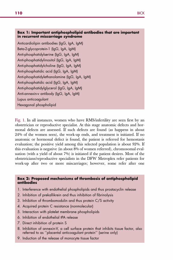
Box 1: Important antiphospholipid antibodies that are importantin recurrent miscarriage syndrome
Anticardiolipin antibodies (IgG, IgA, IgM)
Beta-2-glycoprotein-1 (IgG, IgA, IgM)
Anti-phosphatidylserine (IgG, IgA, IgM)
Anti-phosphatidylinositol (IgG, IgA, IgM)
Anti-phosphatidylcholine (IgG, IgA, IgM)
Anti-phosphatidic acid (IgG, IgA, IgM)
Anti-phosphatidylethanolamine (IgG, IgA, IgM)
Anti-phosphatidic acid (IgG, IgA, IgM)
Anti-phosphatidylglycerol (IgG, IgA, IgM)
Anti-annexin-v antibody (IgG, IgA, IgM)
Lupus anticoagulant
Hexagonal phospholipid
110 BICK
Fig. 1. In all instances, women who have RMS/infertility are seen first by anobstetrician or reproductive specialist. At this stage anatomic defects and hor-monal defects are assessed. If such defects are found (as happens in about24% of the women seen), the work-up ends, and treatment is initiated. If noanatomic or hormonal defect is found, the patient is referred for hemostasisevaluation; the positive yield among this selected population is about 92%. Ifthis evaluation is negative (in about 8% of women referred), chromosomal eval-uation (with a yield of about 7%) is initiated if the patient desires. Most of theobstetricians/reproductive specialists in the DFW Metroplex refer patients forwork-up after two or more miscarriages; however, some refer after one
Box 2: Proposed mechanisms of thrombosis of antiphospholipidantibodies
1. Interference with endothelial phospholipids and thus prostacyclin release
2. Inhibition of prekallikrein and thus inhibition of fibrinolysis
3. Inhibition of thrombomodulin and thus protein C/S activity
4. Acquired protein C resistance (nonmolecular)
5. Interaction with platelet membrane phospholipids
6. Inhibition of endothelial tPA release
7. Direct inhibition of protein S
8. Inhibition of annexin-V, a cell surface protein that inhibits tissue factor, alsoreferred to as ‘‘placental anticoagulant protein’’ (serine only)
9. Induction of the release of monocyte tissue factor
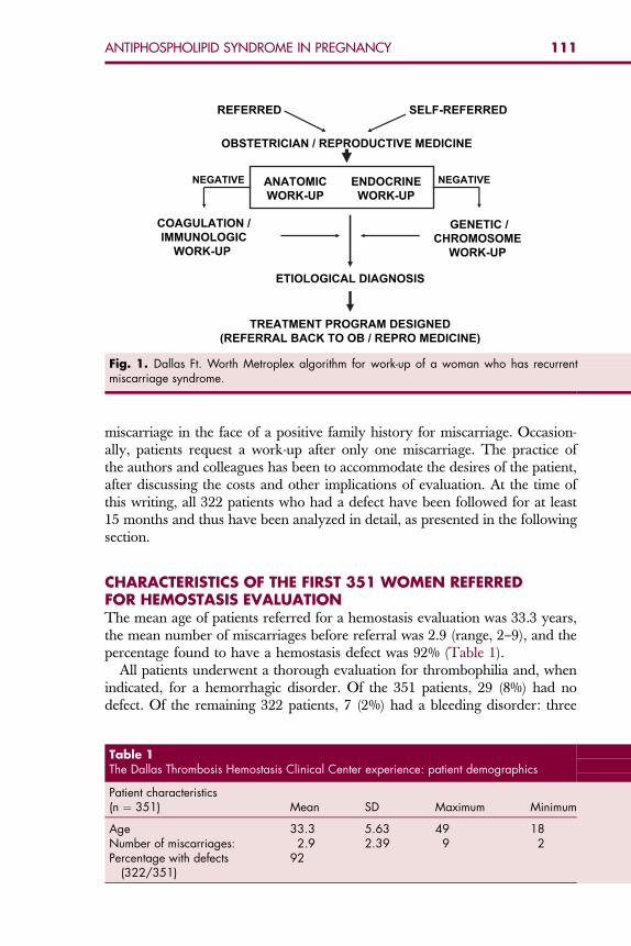
OBSTETRICIAN / REPRODUCTIVE MEDICINE
REFERRED SELF-REFERRED
ANATOMIC
WORK-UP
ENDOCRINE
WORK-UP
COAGULATION /
IMMUNOLOGIC
WORK-UP
GENETIC /
CHROMOSOME
WORK-UP
NEGATIVE NEGATIVE
ETIOLOGICAL DIAGNOSIS
TREATMENT PROGRAM DESIGNED
(REFERRAL BACK TO OB / REPRO MEDICINE)
Fig. 1. Dallas Ft. Worth Metroplex algorithm for work-up of a woman who has recurrentmiscarriage syndrome.
111ANTIPHOSPHOLIPID SYNDROME IN PREGNANCY
miscarriage in the face of a positive family history for miscarriage. Occasion-ally, patients request a work-up after only one miscarriage. The practice ofthe authors and colleagues has been to accommodate the desires of the patient,after discussing the costs and other implications of evaluation. At the time ofthis writing, all 322 patients who had a defect have been followed for at least15 months and thus have been analyzed in detail, as presented in the followingsection.
CHARACTERISTICS OF THE FIRST 351 WOMEN REFERREDFOR HEMOSTASIS EVALUATIONThe mean age of patients referred for a hemostasis evaluation was 33.3 years,the mean number of miscarriages before referral was 2.9 (range, 2–9), and thepercentage found to have a hemostasis defect was 92% (Table 1).
All patients underwent a thorough evaluation for thrombophilia and, whenindicated, for a hemorrhagic disorder. Of the 351 patients, 29 (8%) had nodefect. Of the remaining 322 patients, 7 (2%) had a bleeding disorder: three
Table 1The Dallas Thrombosis Hemostasis Clinical Center experience: patient demographics
Patient characteristics(n ¼ 351) Mean SD Maximum Minimum
Age 33.3 5.63 49 18Number of miscarriages: 2.9 2.39 9 2Percentage with defects
(322/351)92
112 BICK
had platelet dysfunction, one had factor XIII deficiency, three had von Wille-brand’s factor, and three had Osler-Weber-Rendu syndrome. The remainderof the patients had a thrombophilia: the great majority (195, 60%) had anti-phospholipid syndrome; 64 (20%) had sticky platelet syndrome; 38 (12%)had methylene tetrahydrofolate reductase (MTHFR) mutation; 23 (7.1%)had plasminogen activator inhibitor 1 (PAI-1) polymorphism; 12 (3.7%) hadprotein S deficiency; 12 (3.7%) had factor V Leiden, 3 (1%); had antithrombin(AT) deficiency; 3 (1%) had heparin-cofactor II deficiency; 3 (1%) had tPAdeficiency; and 6 (2%) had protein C deficiency. A total of 364 defects werefound in the 312 patients harboring thrombophilia; thus, several patientsharbored two defects, and a few harbored three separate defects.
Similar to reports by most other investigators, as discussed previously, themost common defect found was APLS. Unlike some groups, however, theauthor and colleagues assess for all phospholipid antibody subgroups includinganti-phosphatidylserine, anti-phosphatidylethanolamine, anti-phosphatidylgly-cerol, anti-phosphatidic acid, anti-phosphatidylcholine, anti-phosphatidylinosi-tol, anti-annexin-V antibody, beta-2-glycoprotein-1, hexagonal phospholipid,and lupus anticoagulant (by dilute Russell’s viper venom time) with correctionby non–platelet-derived phospholipid to avoid false-positive results). Theyalways assess all three idiotypes of anticardiolipin antibody (IgG, IgA andIgM); many who evaluate such patients continue to make an incomplete eval-uation, leaving out either IgA or IgM idiotypes. The defects identified by theauthor’s group, in order of frequency, are summarized in Box 3. When all anti-phospholipid subgroups are included, 29% of patients are found to have a sub-group antiphospholipid antibody but no anticardiolipin antibody or lupusanticoagulant; thus 29% of patients would remain undiagnosed if assays forthese subgroups were not performed. This finding is about equivalent to thefinding recently noted in young (< 51 years) patients who had had thromboticstroke [37]. The hemostasis defects found in the author’s population are sum-marized in Box 3. The particulars of the patients who had antiphospholipidantibodies, with demonstration of the idiotypes found, are summarized inTable 2.
All patients who had a thrombophilic defect, including APLS, were treatedwith preconception aspirin at 81 mg/d. At documentation of conception, thefirst 120 patients were treated with the addition of subcutaneous self-injectedunfractionated porcine mucosal heparin at 5000 units every 12 hours. The sub-sequent 192 patients were treated with subcutaneous self-injected low molecu-lar weight (LMW) heparin dalteparin (Fragmin) at 5000 units every 24 hoursby self-injection. Both drugs (aspirin plus unfractionated heparin or LMW hep-arin) are used to term. All patients are instructed in injecting heparin and areinformed extensively of the benefits and risks of heparin/LMW heparin ther-apy, including side effects that, although rare, can include heparin-inducedthrombocytopenia (HIT) with and without paradoxical thrombosis/thrombo-embolism (HITT); osteoporosis; mild to moderate alopecia; skin and allergicreactions, including erythema and itching, at injection sites; eosinophilia (of
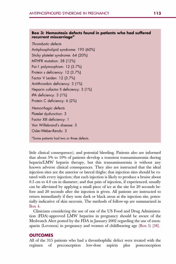
Box 3: Hemostasis defects found in patients who had sufferedrecurrent miscarriagea
Thrombotic defects
Antiphospholipid syndrome: 195 (60%)
Sticky platelet syndrome: 64 (20%)
MTHFR mutation: 38 (12%)
Pai-1 polymorphism: 12 (3.7%)
Protein s deficiency: 12 (3.7%)
Factor V Leiden: 12 (3.7%)
Antithrombin deficiency: 3 (1%)
Heparin cofactor II deficiency: 3 (1%)
tPA deficiency: 3 (1%)
Protein C deficiency: 6 (2%)
Hemorrhagic defects
Platelet dysfunction: 3
Factor XIII deficiency: 1
Von Willebrand’s disease: 3
Osler-Weber-Rendu: 3
aSome patients had two or three defects.
113ANTIPHOSPHOLIPID SYNDROME IN PREGNANCY
little clinical consequence); and potential bleeding. Patients also are informedthat about 5% to 10% of patients develop a transient transaminasemia duringheparin/LMW heparin therapy, but this transaminasemia is without anyknown adverse clinical consequences. They also are instructed that the idealinjection sites are the anterior or lateral thighs; that injection sites should be ro-tated with every injection; that each injection is likely to produce a bruise about0.5 cm to 4.0 cm in diameter; and that pain of injection, if experienced, usuallycan be alleviated by applying a small piece of ice at the site for 20 seconds be-fore and 20 seconds after the injection is given. All patients are instructed toreturn immediately if they note dark or black areas at the injection site, poten-tially indicative of skin necrosis. The methods of follow-up are summarized inBox 4.
Clinicians considering the use of one of the US Food and Drug Administra-tion (FDA)-approved LMW heparins in pregnancy should be aware of theMedwatch Alert posted by the FDA in January 2002 regarding the use of enox-aparin (Lovenox) in pregnancy and women of childbearing age (Box 5) [38].
OUTCOMESAll of the 315 patients who had a thrombophilic defect were treated with theregimen of preconception low-dose aspirin plus postconception
Table 2Antiphospholipids found in patients who have antiphospholipid syndrome (APLS)
Antiphospholipid identifiedPercentage of patientswho have APLS
ACLA-IgG only 32.6ACLA-IgM only 23.4ACLA-IgA only 7ACLA-IgG þ IgM 3ACLA-IgG þ IgA 1ACLA IgA þ IgM 0Lupus anticoagulant only 2ACLA þ lupus anticoagulant 2Subgroup onlya
Anti-phosphatidylserine 4Anti-phosphatidylinositol 2Anti-phosphatidylethanolamine 5Anti-phosphatidic acid 5Anti-phosphatidylcholine 7Anti-phosphatidylglycerol 1Anti-annexin-V 5Beta-2-glycoprotein 1 0Hexagonal phospholipid 0Patients who had APLS and only a subgroup
antibody (n ¼ 9):29
Abbreviation: ACLA, anticardiolipin antibody.aNo ACLA or LA present.
114 BICK
thromboprophylactic (low-dose) subcutaneous porcine heparin or thrombopro-phylactic doses of dalteparin. There were four losses (2.6%), only two of whichwere treatment failures. A loss during the second trimester that accompanieda cholecystectomy and a loss during the first trimester of a fetus that had a chro-mosomal defect in a woman who had APLS were not considered treatment (as-pirin plus heparin/LMW heparin) failures. In two first-trimester losses inpatients receiving aspirin plus heparin/LMW heparin, however, placentalthrombi and infarcts were present; thus these two losses clearly representedtreatment failure. The program’s overall success in treating patients who hadRMS with thrombophilic defects therefore was 99% (312/315) with respect tonormal-term delivery. All patients were followed for a minimum of 3 monthsafter delivery. No patient sustained a thrombotic episode during pregnancy,delivery, or postpartum, except the two treatment failures, both of whomhad placental vascular thrombi. In addition, no patient developed HIT/HITT, and none had a clinically significant hemorrhage. Almost all patientsdeveloped small ecchymoses at injection sites, but these were considered insig-nificant by both patient and physician. Ten percent of patients developed eosin-ophilia that abated by 3 months postpartum, and 7% developed mild tomoderate elevations of hepatic transaminases; these levels also returned to nor-mal by 3 months postpartum. Reports from obstetricians, reproductive
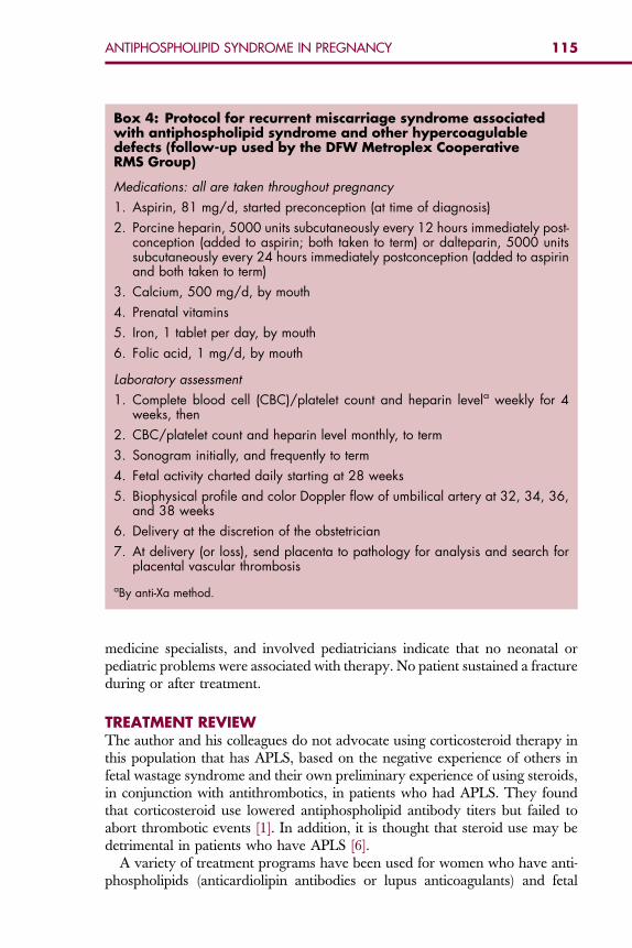
Box 4: Protocol for recurrent miscarriage syndrome associatedwith antiphospholipid syndrome and other hypercoagulabledefects (follow-up used by the DFW Metroplex CooperativeRMS Group)
Medications: all are taken throughout pregnancy
1. Aspirin, 81 mg/d, started preconception (at time of diagnosis)
2. Porcine heparin, 5000 units subcutaneously every 12 hours immediately post-conception (added to aspirin; both taken to term) or dalteparin, 5000 unitssubcutaneously every 24 hours immediately postconception (added to aspirinand both taken to term)
3. Calcium, 500 mg/d, by mouth
4. Prenatal vitamins
5. Iron, 1 tablet per day, by mouth
6. Folic acid, 1 mg/d, by mouth
Laboratory assessment
1. Complete blood cell (CBC)/platelet count and heparin levela weekly for 4weeks, then
2. CBC/platelet count and heparin level monthly, to term
3. Sonogram initially, and frequently to term
4. Fetal activity charted daily starting at 28 weeks
5. Biophysical profile and color Doppler flow of umbilical artery at 32, 34, 36,and 38 weeks
6. Delivery at the discretion of the obstetrician
7. At delivery (or loss), send placenta to pathology for analysis and search forplacental vascular thrombosis
aBy anti-Xa method.
115ANTIPHOSPHOLIPID SYNDROME IN PREGNANCY
medicine specialists, and involved pediatricians indicate that no neonatal orpediatric problems were associated with therapy. No patient sustained a fractureduring or after treatment.
TREATMENT REVIEWThe author and his colleagues do not advocate using corticosteroid therapy inthis population that has APLS, based on the negative experience of others infetal wastage syndrome and their own preliminary experience of using steroids,in conjunction with antithrombotics, in patients who had APLS. They foundthat corticosteroid use lowered antiphospholipid antibody titers but failed toabort thrombotic events [1]. In addition, it is thought that steroid use may bedetrimental in patients who have APLS [6].
A variety of treatment programs have been used for women who have anti-phospholipids (anticardiolipin antibodies or lupus anticoagulants) and fetal
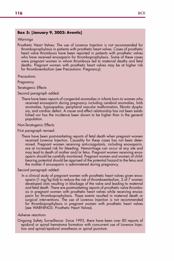
Box 5: [January 9, 2002: Aventis]
Warnings
Prosthetic Heart Valves: The use of Lovenox Injection is not recommended forthromboprophylaxis in patients with prosthetic heart valves. Cases of prostheticheart valve thrombosis have been reported in patients with prosthetic valveswho have received enoxaparin for thromboprophylaxis. Some of these caseswere pregnant women in whom thrombosis led to maternal deaths and fetaldeaths. Pregnant women with prosthetic heart valves may be at higher riskfor thromboembolism (see Precautions: Pregnancy).
Precautions
Pregnancy
Teratogenic Effects
Second paragraph added:
There have been reports of congenital anomalies in infants born to women whoreceived enoxaparin during pregnancy including cerebral anomalies, limbanomalies, hypospadias, peripheral vascular malformation, fibrotic dyspla-sia, and cardiac defect. A cause and effect relationship has not been estab-lished nor has the incidence been shown to be higher than in the generalpopulation.
Non-Teratogenic Effects
First paragraph revised:
There have been post-marketing reports of fetal death when pregnant womenreceived Lovenox Injection. Causality for these cases has not been deter-mined. Pregnant women receiving anti-coagulants, including enoxaparin,are at increased risk for bleeding. Hemorrhage can occur at any site andmay lead to death of mother and/or fetus. Pregnant women receiving enox-aparin should be carefully monitored. Pregnant women and women of child-bearing potential should be apprised of the potential hazard to the fetus andthe mother if enoxaparin is administered during pregnancy.
Second paragraph added:
In a clinical study of pregnant women with prosthetic heart valves given enox-aparin (1 mg/kg bid) to reduce the risk of thromboembolism, 2 of 7 womendeveloped clots resulting in blockage of the valve and leading to maternaland fetal death. There are postmarketing reports of prosthetic valve thrombo-sis in pregnant women with prosthetic heart valves while receiving enoxa-parin for thromboprophylaxis. These events resulted in maternal death orsurgical interventions. The use of Lovenox Injection is not recommendedfor thromboprophylaxis in pregnant women with prosthetic heart valves(see WARNINGS: Prosthetic Heart Valves).
Adverse reactions
Ongoing Safety Surveillance: Since 1993, there have been over 80 reports ofepidural or spinal hematoma formation with concurrent use of Lovenox Injec-tion and spinal/epidural anesthesia or spinal puncture.
116 BICK
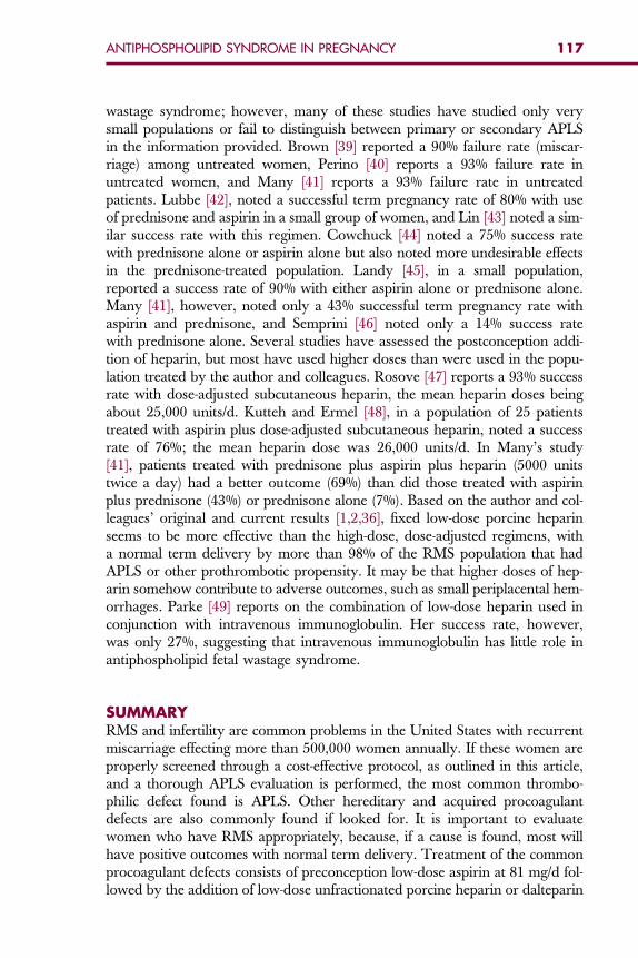
117ANTIPHOSPHOLIPID SYNDROME IN PREGNANCY
wastage syndrome; however, many of these studies have studied only verysmall populations or fail to distinguish between primary or secondary APLSin the information provided. Brown [39] reported a 90% failure rate (miscar-riage) among untreated women, Perino [40] reports a 93% failure rate inuntreated women, and Many [41] reports a 93% failure rate in untreatedpatients. Lubbe [42], noted a successful term pregnancy rate of 80% with useof prednisone and aspirin in a small group of women, and Lin [43] noted a sim-ilar success rate with this regimen. Cowchuck [44] noted a 75% success ratewith prednisone alone or aspirin alone but also noted more undesirable effectsin the prednisone-treated population. Landy [45], in a small population,reported a success rate of 90% with either aspirin alone or prednisone alone.Many [41], however, noted only a 43% successful term pregnancy rate withaspirin and prednisone, and Semprini [46] noted only a 14% success ratewith prednisone alone. Several studies have assessed the postconception addi-tion of heparin, but most have used higher doses than were used in the popu-lation treated by the author and colleagues. Rosove [47] reports a 93% successrate with dose-adjusted subcutaneous heparin, the mean heparin doses beingabout 25,000 units/d. Kutteh and Ermel [48], in a population of 25 patientstreated with aspirin plus dose-adjusted subcutaneous heparin, noted a successrate of 76%; the mean heparin dose was 26,000 units/d. In Many’s study[41], patients treated with prednisone plus aspirin plus heparin (5000 unitstwice a day) had a better outcome (69%) than did those treated with aspirinplus prednisone (43%) or prednisone alone (7%). Based on the author and col-leagues’ original and current results [1,2,36], fixed low-dose porcine heparinseems to be more effective than the high-dose, dose-adjusted regimens, witha normal term delivery by more than 98% of the RMS population that hadAPLS or other prothrombotic propensity. It may be that higher doses of hep-arin somehow contribute to adverse outcomes, such as small periplacental hem-orrhages. Parke [49] reports on the combination of low-dose heparin used inconjunction with intravenous immunoglobulin. Her success rate, however,was only 27%, suggesting that intravenous immunoglobulin has little role inantiphospholipid fetal wastage syndrome.
SUMMARYRMS and infertility are common problems in the United States with recurrentmiscarriage effecting more than 500,000 women annually. If these women areproperly screened through a cost-effective protocol, as outlined in this article,and a thorough APLS evaluation is performed, the most common thrombo-philic defect found is APLS. Other hereditary and acquired procoagulantdefects are also commonly found if looked for. It is important to evaluatewomen who have RMS appropriately, because, if a cause is found, most willhave positive outcomes with normal term delivery. Treatment of the commonprocoagulant defects consists of preconception low-dose aspirin at 81 mg/d fol-lowed by the addition of low-dose unfractionated porcine heparin or dalteparin
118 BICK
immediately after conception. Based on the author and colleagues’ experience,LMH heparin may be a suitable alternative.
References
[1] Bick RL. Recurrent miscarriage syndrome and infertility caused by blood coagulation/plate-let defects. In: Bick RL, Frenkel EP, Baker WF, et al, editors. Hematological complications ofobstetrics, pregnancy and gynecology. Cambridge (UK): Cambridge University Press;2006. p. 55–74.
[2] Bick RL. Recurrent miscarriage syndrome and infertility caused by blood coagulation proteinor platelet defects. Hematol Oncol Clin North Am 2000;14:1117–32.
[3] Bick RL, Baker WF. Antiphospholipid syndrome and thrombosis. Semin Thromb Hemost1999;25:333–50.
[4] Bick RL. The antiphospholipid thrombosis syndromes: a common multidisciplinary medicalproblem. Clin Appl Thromb Hemost 1997;3:270–83.
[5] Scott JR, Rote NS, Branch DW. Immunological aspects of recurrent abortion and fetal death.Obstet Gynecol 1987;70:645–56.
[6] Khamashta MA. Management of thrombosis and pregnancy loss in the antiphospholipidsyndrome. Lupus 1998;2(Suppl 7):S162–5.
[7] Amengual O, Atsumi T, Khamashta MA, et al. Advances in antiphospholipid (Hughes’) syn-drome. Ann Acad Med Singap 1998;27:61–6.
[8] Bick RL. Antiphospholipid thrombosis syndromes: etiology, pathophysiology, diagnosis andmanagement. Int J Hematol 1997;65:193–8.
[9] Bick RL, Baker WF. The antiphospholipid and thrombosis syndromes. Med Clin North Am1994;78:667–84.
[10] Bick RL, Arun B, Frenkel EP. Antiphospholipid thrombosis syndromes. Haemostasis1999;29:111–34.
[11] Festin MR, Limson GM, Maruo T. Autoimmune causes of recurrent pregnancy loss. Kobe JMed Sci 1997;43:143–57.
[12] Roussev RG, Kaider BD, Price DE, et al. Laboratory evaluation of women experiencingreproductive failure. Am J of Reproductive Immunology (Copenhagen) 1996;35:415–20.
[13] Oshiro BT, Silver RM, Scott JR, et al. Antiphospholipid antibodies and fetal death. ObstetGynecol 1996;87:489–93.
[14] Granger KA, Farquharson RG. Obstetric outcome in antiphospholipid syndrome. Lupus1997;6:509–13.
[15] Borrelli AL, Brillante M, Borzacchiello C, et al. Hemocoagulative pathology and immunolog-ical recurrent abortion. Clin Exp Obstet Gynecol 1997;24:39–40.
[16] Hellan M, Kuhnel E, Speiser W, et al. Familial lupus anticoagulant: a case report and reviewof the literature. Blood Coagul Fibrinolysis 1998;9:195–200.
[17] Ogasawara M, Aoki K, Matsuura E, et al. Anti beta-2-glycoprotein I antibodies and lupusanticoagulant in patients with recurrent pregnancy loss: prevalence and clinical signifi-cance. Lupus 1996;5:587–92.
[18] Zangari M, Lockwood CJ, Scher J, et al. Prothrombin activation fragment (F1.2) is increasedin pregnant patients with antiphospholipid antibodies. Thromb Res 1997;85:177–83.
[19] Aznar J, Factor V. Leiden and antibodies against phospholipids and protein S in a youngwoman with recurrent thromboses and abortion. Haematologica 1999;84:80–4.
[20] Aznar J, Villa P, Espana F, et al. Activated protein C resistance phenotype in patients withantiphospholipid antibodies. J Lab Clin Med 1997;130:202–8.
[21] Schultz DR. Antiphospholipid antibodies: basic immunology and assays. Semin ArthritisRheum 1997;26:724–39.
[22] Amengual O, Atsumi T, Khamashta MA, et al. The role of the tissue factor pathway in thehypercoagulable state in patients with the antiphospholipid syndrome. Thromb Haemost1998;79:276–81.
119ANTIPHOSPHOLIPID SYNDROME IN PREGNANCY
[23] Martini A, Ravelli A. The clinical significance of antiphospholipid antibodies. Ann Med1997;29:159–63.
[24] Bussen SS, Steck T. Thyroid antibodies and their relation to antithrombin antibodies, anticar-diolipin antibodies and lupus anticoagulant in women with recurrent spontaneous abortions(antithyroid, anticardiolipin and antithrombin autoantibodies and lupus anticoagulant in ha-bitual aborters). Eur J Obstet Gynecol Reprod Biol 1997;74:139–43.
[25] Rand JH, Wu XX. Antibody-mediated disruption of the annexin-V antithrombotic shield:a new mechanism for thrombosis in the antiphospholipid syndrome. Thromb Haemost1999;82:649–54.
[26] Rand JH, Wu XX, Andree HA, et al. Antiphospholipid antibodies accelerate plasma coag-ulation by inhibiting annexin-V binding to phospholipids: a ‘‘lupus procoagulant’’ phenom-enon. Blood 1998;92:1652–60.
[27] Rauch J. Lupus anticoagulant antibodies: recognition of phospholipid-binding protein com-plexes. Lupus 1998;7(Suppl 2):S29–33.
[28] Rote NS, Vogt E, DeVere G, et al. The role of placental trophoblast in the pathophysiology ofthe antiphospholipid antibody syndrome. Am J of Reproductive Immunology (Copenhagen)1998;39:125–36.
[29] Vogt E, Ng AK, Rote NS. Antiphosphatidylserine antibody removes annexin-V and facili-tates the binding of prothrombin at the surface of a choriocarcinoma model of trophoblastdifferentiation. Am J Obstet Gynecol 1997;177:964–72.
[30] Lakasing L, Campa JS, Poston R, et al. Normal expression of tissue factor, thrombomodulin,and annexin V in placentas from women with antiphospholipid syndrome. Am J ObstetGynecol 1999;181:180–9.
[31] Siaka C, Lambert M, Caron C, et al. Low prevalence of anti-annexin V antibodies in anti-phospholipid syndrome with fetal loss. Revue de Medecine Interne 1999;20:762–5.
[32] Kaburaki J, Kuwana M, Yamamoto M, et al. Clinical significance of anti-annexin V anti-bodies in patients with systemic lupus erythematosus. Am J Hematol 1997;54:209–13.
[33] Eschwege V, Peynaud-Debayle E, Wolf M, et al. Prevalence of antiphospholipid-related an-tibodies in unselected patients with history of venous thrombosis. Blood Coagul Fibrinolysis1998;9:429–34.
[34] Silver RM, Pierangeli SS, Edwin SS, et al. Pathogenic antibodies in women with obstetricfeatures of antiphospholipid syndrome who have negative test results for lupus anticoagu-lant and anticardiolipin antibodies. Am J Obstet Gynecol 1997;176:628–33.
[35] Branch DW, Silver R, Pierangeli S, et al. Antiphospholipid antibodies other than lupus anti-coagulant and anticardiolipin antibodies in women with recurrent pregnancy loss, fertilecontrols, and antiphospholipid syndrome. Obstet Gynecol 1997;89:549–55.
[36] Bick RL, Hoppensteadt D. Recurrent miscarriage syndrome and infertility due to bloodcoagulation/platelet defects: a review and update. Clin Appl Thromb Hemost 2005;11:1–13.
[37] Toschi V, Motta A, Costelli C, et al. High prevalence of antiphosphatidylinositol antibodiesin young patients with cerebral ischemia of undetermined cause. Stroke 1998;29:1759–64.
[38] FDA MedWatch. Lovenox (enoxaparin sodium) injection [January 9, 2002: Aventis]: warn-ings FDA MedWatch 1/9/2002. Available at: http://www.fda.gov/medwatch/SAFETY/2002/jan02.htm#lovenox. Accessed January 2, 2002.
[39] Brown HL. Antiphospholipid antibodies and recurrent pregnancy loss. Clin Obstet Gynecol1991;34:17–26.
[40] Perino A, Barba G, Cimino C, et al. Immunological problems in the recurrent abortion syn-drome. Acta Eur Fertil 1989;20:199–202.
[41] Many A, Pauzner R, Carp H, et al. Treatment of patients with antiphospholipid antibodiesduring pregnancy. Am J Reprod Immunol 1992;28:216–8.
[42] Lubbe WF, Liggins GC. Role of lupus anticoagulant and autoimmunity in recurrent fetal loss.Semin Reprod Endocrinol 1988;6:181–90.
120 BICK
[43] Lin Q. Investigation of the association between autoantibodies and recurrent abortions. Chi-nese J Obstet Gynecol 1993;28:674–9.
[44] Cowchock FS, Reece EA, Balaban D, et al. Repeated fetal losses associated with antiphos-pholipid antibodies: a collaborative randomized trial comparing prednisone with low-doseheparin treatment. Am J Obstet Gynecol 1992;166:1318–23.
[45] Landy HJ, Kessler C, Kelly WK, et al. Obstetric performance in patients with the lupus anti-coagulant and/or anticardiolipin antibodies. Am J Perinatol 1992;9:146–51.
[46] Semprini AE, Vucetich A, Garbo S, et al. Effect of prednisone and heparin treatment in 14patients with poor reproductive efficiency related to lupus anticoagulant. Fetal Ther1989;4(Suppl 1):73–6.
[47] Rosove MH, Tabsh K, Wasserstrum N, et al. Heparin therapy for pregnant women with lupusanticoagulant or anticardiolipin antibodies. Obstet Gynecol 1990;75:630–4.
[48] Kutteh WH, Ermel LD. A clinical trial for the treatment of the antiphospholipid antibodyassociated recurrent pregnancy loss with lower dose heparin and aspirin. Am J ReprodImmunol 1996;35:402–7.
[49] Parke A. The role of IVIG in the management of patients with antiphospholipid antibodiesand recurrent pregnancy losses. In: Ballow M, editor. IVIG Therapy Today. Totowa (NJ):Humana Press, Inc.; 1992. p. 105–18.
Hematol Oncol Clin N Am 22 (2008) 121–130
HEMATOLOGY/ONCOLOGY CLINICSOF NORTH AMERICA
Antiphospholipid Antibodiesand Malignancy
Chi Pham, MD, Yu-Min Shen, MD*The University of Texas Southwestern Medical Center at Dallas, 5323 Harry Hines Boulevard,Dallas, TX 75390-8852, USA
Antiphospholipid antibody syndrome is characterized clinically by ve-nous or arterial thrombosis, recurrent fetal loss, or placental insuffi-ciency in women. The diagnosis requires the persistent laboratory
evidence of antibodies to phospholipids or plasma proteins that have affinityto the negatively charged phospholipids [1]. These include lupus anticoagulantdetected by prolongation of phospholipid-dependent clotting assays, and anti-bodies detected by solid-phase immunoassays (ELISA) such as anticardiolipinantibodies and anti-b2-glycoprotein I antibodies [2]. These first were describedin patients who had systemic lupus erythematosus (SLE) [3], but they now areknown to be associated with other systemic autoimmune diseases, includingrheumatoid arthritis, systemic sclerosis, Sjogren’s syndrome, and systemic vas-culitis [4]. In addition to thrombosis and pregnancy complications, antiphos-pholipid antibodies also are seen in patients who have chronic infections,especially those secondary to hepatitis C virus [5,6] and HIV [7]. Antiphospho-lipid antibodies also can occur because of treatment with certain medicationssuch as interferon [5,8]. Recently, antiphospholipid antibodies have beenreported in conjunction with various solid and hematologic malignancies[9,10].
Numerous healthy individuals also harbor the antiphospholipid antibodieswithout any evidence of an underlying disease process. In a study of 552asymptomatic healthy volunteers, it was noted that up to 9.4% tested positivefor anticardiolipin antibody with 1.4% showing persistence of these antibodieson repeat testing [11].
CANCER AND HYPERCOAGULABILITYSince the first description by Armand Trousseau of the association betweenthrombophlebitis and malignancy, it has been accepted that patients whohave cancer are at a much higher risk of developing thromboembolic diseasethan the normal population [12]. In a large population-based, case-controlled
*Corresponding author. E-mail address: [email protected] (Y-M Shen).
0889-8588/08/$ – see front matter ª 2008 Elsevier Inc. All rights reserved.doi:10.1016/j.hoc.2007.10.004 hemonc.theclinics.com
122 PHAM & SHEN
study, the Dutch demonstrated that the risk of venous thrombosis was in-creased sevenfold in patients who had malignancy compared with normal con-trols. The risk of thrombosis was highest in patients who had hematologicalmalignancies, followed by lung cancer and gastrointestinal cancer [13]. A Medi-care claim-based study of over 8 million patients also showed that the percent-age of patients who had deep venous thrombosis and pulmonary embolism wasslightly higher for patients who had malignancy compared with those withoutmalignancy [14]. Although thromboembolic events often are seen at the time ofcancer diagnosis or shortly thereafter, they also can precede the cancer diagno-sis by months to years [15,16]. A Swedish study of 3795 patients suspected tohave venous thrombosis showed that among 1383 patients with venography-confirmed thrombosis, 66 developed cancer in the first 6 months of follow-upcompared with 37 patients without venographic evidence of thrombosis [17].
The mechanism underlying the prothrombotic state of malignancy is a com-plex interplay between the tumor cells and host cells, such that the delicatebalance between coagulation and fibrinolysis is disrupted [18]. Recent ad-vances have established that the essential factors of hemostasis participate di-rectly in cancer growth and progression, in addition to their known hemostaticproperties. These include tissue factor, cancer procoagulant, thrombin, tissuetype, and urokinase plasminogen activators, plasminogen activator inhibitor 1and 2. The effects of these coagulation factors contribute to the tumor cells’ abilityto proliferate, migrate, and induce proteolysis and permeability [19]. Observa-tions that therapy with unfractionated heparin and low molecular weightheparins in cancer patients may offer survival benefit as anticancer drugs havegenerated renewed interest in this field and lend support to the laboratorydata [20–22].
Recently, a series of case reports have suggested that antiphospholipid anti-bodies also may play a role in the thrombotic predisposition of malignancy.There are several possible theories as to the generation of antiphospholipidantibodies in the setting of malignancy [23]:
Production of autoantibodies by immune system in response to tumor antigensProduction of monoclonal antibodies with lupus anticoagulant and anticardioli-
pin activitiesSecretion of anticardiolipin antibody from tumor cells
The exact mechanism(s) by which antiphospholipid antibody leads to throm-bosis remain an active area of investigation. Because many steps of the coagu-lation cascade take place on negatively charged phospholipid surfaces,antiphospholipid antibodies potentially could act at any level to disrupt normalhemostasis. One may speculate that antibodies to prothrombin may have ananticoagulant effect, whereas antibodies to protein C and annexin V may im-pair normal control of coagulation [24,25]. Recent advances in the understand-ing of the pathophysiology of antiphospholipid syndrome have centered on thedimerization of b2-glycoprotein I on the phospholipid membrane as a result ofantiphospholipid antibody binding, which then leads to receptor-mediated
123ANTIPHOSPHOLIPID ANTIBODIES AND MALIGNANCY
platelet activation and up-regulation of procoagulant properties on endothelialcells [26].
PREVALENCEAlthough antiphospholipid antibodies usually are seen in association withthrombosis, they also can be detected in asymptomatic healthy volunteers asdiscussed previously [11]. The incidence of antiphospholipid antibodies isnoted to be in the range of 4.5% to 9.4% in the healthy young population basedon various studies [11,27]. The incidence, however, increases dramatically withage. A study of 64 healthy elderly patients with mean age of 81 years showedthat 51.6% tested positive for anticardiolipin antibodies of the IgG and IgM iso-topes [28]. The limitation of most of these studies is that the volunteers weretested only once. The definition of antiphospholipid syndrome requires the per-sistence of the antibodies measured at least 12 weeks apart [1]. It is not knownwhat the true incidence of such antibodies is when restricted to only subjectswith their persistence on repeat testing.
The incidence of antiphospholipid antibodies and the diagnosis of antiphos-pholipid syndrome may be much higher in patients who have cancer. To eval-uate this, a large French study tested the sera of 1014 consecutive patientsadmitted to the department of internal medicine for the presence of antiphos-pholipid antibodies. Seventy-two patients (7.1%) were noted to be positivefor the antibodies at least once. Twenty-two patients (2.8%) fulfilled the criteriafor antiphospholipid syndrome. Fourteen (63.6%) of these 22 patients were di-agnosed with a malignancy [29]. Similarly, several case series of patients withknown cancer also demonstrated the high incidence of antiphospholipid anti-bodies. A case–control study of 145 patients who had cancer showed that higherlevels of IgG anti-cardiolipin antibody were found in patients with malignancywhen compared with the controlled population (P <.02). Twenty patients withanticardiolipin antibodies had evidence of thromboembolic disease; most ofthem had solid malignancy, including lung, breast, prostate, stomach, colon,gynecological, bladder, and others [30]. Higher incidence of antiphospholipidantibodies also has been described in patients who have hematological malig-nancies [31]. An Italian study of 100 consecutive patients who had lymphomaindicated that 27 patients tested positive for either anticardiolipin antibody orlupus anticoagulant [32]. This was much higher than the 8% prevalence seenin their age- and sex-matched control group. If restricted to patients whohave malignancy and thrombosis, antiphospholipid antibodies may be seenin up to 60% of patients according to one study of Asian cancer patients[33]. Given the association between malignancy and antiphospholipid anti-bodies, antiphospholipid antibodies have been described in all types of neo-plasm. Table 1 is compilation of the tumor types seen in two large series ofpatients who have antiphospholipid antibodies, and Table 2 shows the fre-quency of antiphospholipid antibodies in four series of cancer patients. Despitethe high incidence of antiphospholipid antibodies in patients who have malig-nancies, it must be kept in mind that the average age of these patients is around
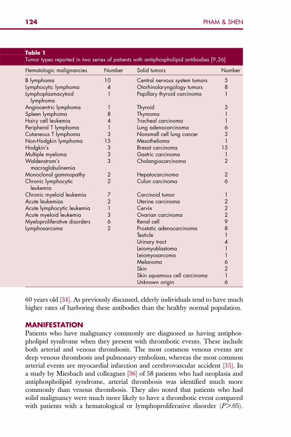
Table 1Tumor types reported in two series of patients with antiphospholipid antibodies [9,36]
Hematologic malignancies Number Solid tumors Number
B lymphoma 10 Central nervous system tumors 5Lymphocytic lymphoma 4 Otorhinolaryngology tumors 8Lymphoplasmacytoid
lymphoma1 Papillary thyroid carcinoma 1
Angiocentric lymphoma 1 Thyroid 3Spleen lymphoma 8 Thymoma 1Hairy cell leukemia 4 Tracheal carcinoma 1Peripheral T lymphoma 1 Lung adenocarcinoma 6Cutaneous T lymphoma 3 Nonsmall cell lung cancer 3Non-Hodgkin lymphoma 15 Mesothelioma 1Hodgkin’s 3 Breast carcinoma 15Multiple myeloma 3 Gastric carcinoma 1Waldenstrom’s
macroglobulinemia3 Cholangiocarcinoma 2
Monoclonal gammopathy 2 Hepatocarcinoma 2Chronic lymphocytic
leukemia2 Colon carcinoma 6
Chronic myeloid leukemia 7 Carcinoid tumor 1Acute leukemias 2 Uterine carcinoma 2Acute lymphocytic leukemia 1 Cervix 2Acute myeloid leukemia 3 Ovarian carcinoma 2Myeloproliferative disorders 6 Renal cell 9Lymphosarcoma 2 Prostatic adenocarcinoma 8
Testicle 1Urinary tract 4Leiomyoblastoma 1Leiomyosarcoma 1Melanoma 6Skin 2Skin squamous cell carcinoma 1Unknown origin 6
124 PHAM & SHEN
60 years old [34]. As previously discussed, elderly individuals tend to have muchhigher rates of harboring these antibodies than the healthy normal population.
MANIFESTATIONPatients who have malignancy commonly are diagnosed as having antiphos-pholipid syndrome when they present with thrombotic events. These includeboth arterial and venous thrombosis. The most common venous events aredeep venous thrombosis and pulmonary embolism, whereas the most commonarterial events are myocardial infarction and cerebrovascular accident [35]. Ina study by Miesbach and colleagues [36] of 58 patients who had neoplasia andantiphospholipid syndrome, arterial thrombosis was identified much morecommonly than venous thrombosis. They also noted that patients who hadsolid malignancy were much more likely to have a thrombotic event comparedwith patients with a hematological or lymphoproliferative disorder (P >.05).
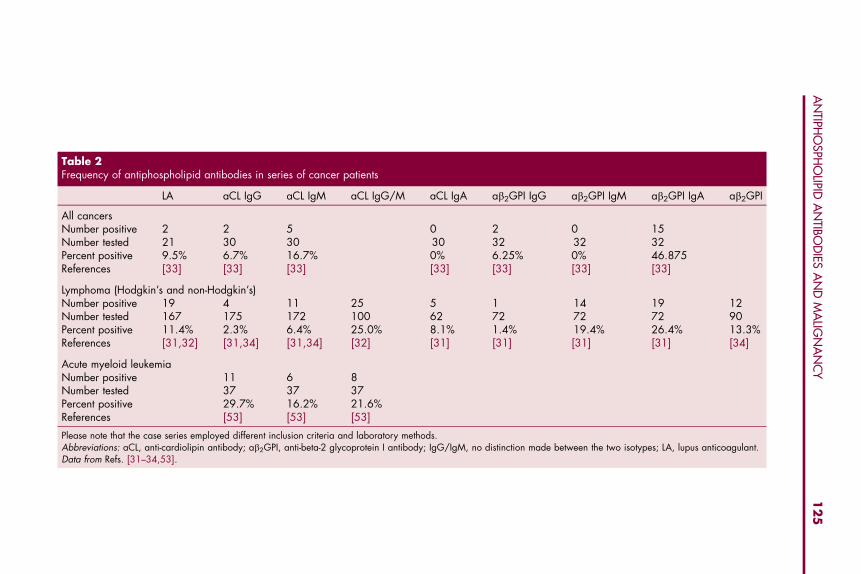
Table 2Frequency of antiphospholipid antibodies in series of cancer patients
LA aCL IgG aCL IgM aCL IgG/M aCL IgA ab2GPI IgG ab2GPI IgM ab2GPI IgA ab2GPI
All cancersNumber positive 2 2 5 0 2 0 15Number tested 21 30 30 30 32 32 32Percent positive 9.5% 6.7% 16.7% 0% 6.25% 0% 46.875References [33] [33] [33] [33] [33] [33] [33]
Lymphoma (Hodgkin’s and non-Hodgkin’s)Number positive 19 4 11 25 5 1 14 19 12Number tested 167 175 172 100 62 72 72 72 90Percent positive 11.4% 2.3% 6.4% 25.0% 8.1% 1.4% 19.4% 26.4% 13.3%References [31,32] [31,34] [31,34] [32] [31] [31] [31] [31] [34]
Acute myeloid leukemiaNumber positive 11 6 8Number tested 37 37 37Percent positive 29.7% 16.2% 21.6%References [53] [53] [53]
Please note that the case series employed different inclusion criteria and laboratory methods.Abbreviations: aCL, anti-cardiolipin antibody; ab2GPI, anti-beta-2 glycoprotein I antibody; IgG/IgM, no distinction made between the two isotypes; LA, lupus anticoagulant.Data from Refs. [31–34,53].
12
5A
NTIPH
OSPH
OLIPID
AN
TIBOD
IESA
ND
MA
LIGN
AN
CY
126 PHAM & SHEN
Others, however, have reported similar thrombotic complication rates in pa-tients who have hematological malignancies. Pusterla and colleagues [32] dem-onstrated in a case-controlled study that in patients who had lymphoma, therate of thrombosis was significantly higher in patients who had antiphospholi-pid antibodies than those without the antibodies (5.1% per year versus 0.75%per year, respectively).
There are case reports of other rare thrombotic manifestations in patientswho have cancer and antiphospholipid antibodies. These include necrotizingleg ulcers [37], recurrent stent thrombosis [38], transverse myelitis [39], andnonbacterial thrombotic endocarditis [40]. The most feared complication isthe catastrophic antiphospholipid antibody syndrome, or Asherson’s syn-drome. This syndrome is characterized by multiple sites of thromboses withmultiorgan failure that is often fatal [41]. Several case reports of malignancyhave been linked to catastrophic antiphospholipid syndrome [42,43], althoughit remains an extremely rare occurrence. According to one study, which con-sisted of 1000 patients with antiphospholipid syndrome, only 0.8% of patientshad criteria for Asherson’s syndrome [44].
It remains to be determined whether having antiphospholipid antibodies inthe setting of malignancy indeed leads to a higher risk of thrombosis. In con-trast to the previous studies, a study by Genvresse and colleagues [34] showedthat 26.6% of patients with non-Hodgkin’s lymphoma had evidence of elevatedantiphospholipid antibodies, but none developed any thromboembolic eventduring 14 months of follow-up. Similarly, a study of four patients who hadelevated IgG and IgM anticardiolipin antibody and lupus anticoagulant didnot demonstrate any thromboembolic manifestation during the entire follow-up period [45]. At this point, the role antiphospholipid antibodies play inpatients with malignancy is still controversial. It is unclear if these antibodiesrepresent an epiphenomenon or contribute to the pathogenesis of thrombosisin patients who have malignancy.
ANTIPHOSPHOLIPID ANTIBODY AND PROGNOSISThe Italian Registry of Antiphospholipid Antibodies followed 360 patients pro-spectively over 5 years to assess the natural history of antiphospholipid syn-drome. With a median follow up of 3.9 years, the Italians noted four newcases of non-Hodgkin’s lymphoma and one new case of breast cancer. Fornon-Hodgkin’s lymphoma, the estimated event rate was 0.28% patient-year,which is much higher than the expected incidence of non-Hodgkin’s lymphomaof 5 to 15 cases per 100,000 population per year. The study investigators con-cluded that antiphospholipid antibodies should be considered a risk factor fornon-Hodgkin’s lymphoma [46].
Treatment of the underlying malignancy often leads to the disappearance ofantiphospholipid antibody [9,29]. In a report by Sciarra and colleagues [47], pa-tients with acute myelogenous leukemia or non-Hodgkin’s lymphoma who hadelevated titers of antiphospholipid antibodies at diagnosis were shown to haveundetectable antibody levels after treatment of their leukemia or lymphoma if
127ANTIPHOSPHOLIPID ANTIBODIES AND MALIGNANCY
they responded to the prescribed therapy. In contrast, the antibodies were per-sistent in those patients who did not respond. Furthermore, patients who re-sponded and had disappearance of their antiphospholipid antibodies laterwere noted to have recurrence of the antibodies when their disease relapsed[47]. Some patients who tested negative for antiphospholipid antibodies werenoted to be positive when their cancer progressed or became refractory tothe treatment [48]. Thus, the presence of antiphospholipid antibodies mayserve as a surrogate marker for disease status and response to therapy forhematologic malignancies.
Similar findings also were seen in patients who had solid tumors. In a reportby Miesbach and colleagues [36], two patients who had colon carcinoma andantiphospholipid antibody were noted to have disappearance of the antiphos-pholipid antibody after effective treatment of their colon cancer. As long asthe patients were in remission, the antiphospholipid testing remained negative.Despite the fact that they presented with clots initially, once the antiphospho-lipid antibody became undetectable, they did not have recurrence of the throm-botic event.
Even though the presence of antiphospholipid antibody is indicative of thepersistence of the underlying malignancy, antiphospholipid antibody has notbeen correlated with tumor stage, age, performance status, or InternationalPrognostic Index (IPI) score in the case of non-Hodgkin’s lymphoma [31,34].Antiphospholipid antibody, however, may serve as an independent prognosticvariable in aggressive lymphoma. A study of patients who had non-Hodgkin’slymphoma showed that the 2-year survival was 90% plus or minus 5% for pa-tients without antiphospholipid antibody at diagnosis compared with 63% plusor minus 11% for patients with an elevated antibody level (P ¼ .0025) [31].This observation requires validation with further studies.
SUMMARYPresence of antiphospholipid antibodies is a significant thrombophilic risk fac-tor with a high prevalence in patients who have malignancies [32]. Patients whoharbor these antibodies may be at increased risk for thromboembolic complica-tions [49]. The mechanism by which antiphospholipid antibody develops in thesetting of malignancy is poorly understood. It is postulated that the antibodiesmay be a direct consequence of the tumor itself [50], a normal immune reactionto the tumor, or it also could be caused by specific cancer therapies [51]. Thepresence of antiphospholipid antibody does not predict the likelihood of re-sponse to treatment necessarily, nor does it affect prognosis, with the possibleexception of high grade lymphomas. It may help in monitoring disease activity,because cancers in remission often are accompanied by disappearance of anti-phospholipid antibody [47]. Manipulating the antibody levels with immuno-suppressive therapy has not been shown to affect the thrombosis risk or itsrecurrence [52]. Patients should be treated only if they develop a thromboem-bolic event. Prospective studies should be conducted to elucidate these issues.
128 PHAM & SHEN
References
[1] Miyakis S, Lockshin MD, Atsumi T, et al. International consensus statement on an update ofthe classification criteria for definite antiphospholipid syndrome (APS). J Thromb Haemost2006;4(2):295–306.
[2] Passam F, Krilis S. Laboratory tests for the antiphospholipid syndrome: current concepts.Pathology 2004;36:129–38.
[3] Conley CL. A hemorrhagic disorder caused by circulating anticoagulant in patients with dis-seminated lupus erythematosus. J Clin Invest 1952;31:621–2.
[4] Cervera R, Asherson RA. Clinical and epidemiological aspects in the antiphospholipid syn-drome. Immunobiology 2003;207(1):5–11.
[5] Matsuda J, Saitoh N, Gotoh M, et al. High prevalence of antiphospholipid antibodies andantithyroglobulin antibody in patients with hepatitis C virus infection treated with interferon-alpha. Am J Gastroenterol 1995;90(7):1138–41.
[6] Prieto J, Yuste JR, Beloqui O, et al. Anticardiolipin antibodies in chronic hepatitis C: implica-tion of hepatitis C virus as the cause of the antiphospholipid syndrome. Hepatology1996;23(2):199–204.
[7] Uthman IW, Gharavi AE. Viral infections and antiphospholipid antibodies. Semin ArthritisRheum 2002;31(4):256–63.
[8] Naldi L, Locati F, Finazzi G, et al. Antiphospholipid syndrome associated with immunother-apy for patients with melanoma. Cancer 1995;75(11):2784–5.
[9] Gomez-Puerta JA, Cervera R, Espinosa G, et al. Antiphospholipid antibodies associatedwith malignancies: clinical and pathological characteristics of 120 patients. Semin ArthritisRheum 2006;35(5):322–32.
[10] Pugliese LBI, Pacifico E, Viola-Magni M, et al. Antiphospholipid antibodies in patients withcancer. Int J Immunopathol Pharmacol 2006;19(4):879–88.
[11] Vila P, Hernandez MC, Lopez-Fernandez MF, et al. Prevalence, follow-up, and clinical sig-nificance of the anticardiolipin antibodies in normal subjects. Thromb Haemost1994;72(2):209–13.
[12] Sack GH Jr, Levin J, Bell WR. Trousseau’s syndrome and other manifestations of chronic dis-seminated coagulopathy in patients with neoplasms: clinical, pathophysiologic, and thera-peutic features. Medicine (Baltimore) 1977;56(1):1–37.
[13] Blom JW, Doggen CJ, Osanto S, et al. Malignancies, prothrombotic mutations, and the riskof venous thrombosis. JAMA 2005;293(6):715–22.
[14] Levitan N, Dowlati A, Remick SC, et al. Rates of initial and recurrent thromboembolic dis-ease among patients with malignancy versus those without malignancy. Risk analysis usingMedicare claims data. Medicine (Baltimore) 1999;78(5):285–91.
[15] White RH, Chew HK, Zhou H, et al. Incidence of venous thromboembolism in the year be-fore the diagnosis of cancer in 528,693 adults. Arch Intern Med 2005;165(15):1782–7.
[16] Prandoni P, Lensing AW, Buller HR, et al. Deep vein thrombosis and the incidence of subse-quent symptomatic cancer. N Engl J Med 1992;327(16):1128–33.
[17] Nordstrom M, Lindblad B, Anderson H, et al. Deep venous thrombosis and occult malig-nancy: an epidemiological study. BMJ 1994;308(6933):891–4.
[18] Gouin-Thibault I, Achkar A, Samama MM. The thrombophilic state in cancer patients. ActaHaematol 2001;106(1–2):33–42.
[19] Buller HR, van Doormaal FF, van Sluis GL, et al. Cancer and thrombosis: from molecularmechanisms to clinical presentations. J Thromb Haemost 2007;5(Suppl 1):246–54.
[20] Kakkar AK, Levine MN, Kadziola Z, et al. Low molecular weight heparin, therapy with dal-teparin, and survival in advanced cancer: the fragmin advanced malignancy outcome study(FAMOUS). J Clin Oncol 2004;22(10):1944–8.
[21] Klerk CP, Smorenburg SM, Otten HM, et al. The effect of low molecular weight heparin onsurvival in patients with advanced malignancy. J Clin Oncol 2005;23(10):2130–5.
129ANTIPHOSPHOLIPID ANTIBODIES AND MALIGNANCY
[22] Lee AY, Rickles FR, Julian JA, et al. Randomized comparison of low molecular weight heparinand coumarin derivatives on the survival of patients with cancer and venous thromboembo-lism. J Clin Oncol 2005;23(10):2123–9.
[23] Zuckerman E, Toubi E, Golan TD, et al. Increased thromboembolic incidence in anticardio-lipin-positive patients with malignancy. Br J Cancer 1995;72(2):447–51.
[24] Hanly JG,Smith SA,Anderson D. Inhibitionof annexinV binding to cardiolipinand thrombingeneration in an unselected population with venous thrombosis. J Rheumatol 2003;30(9):1990–3.
[25] Rand JH, Wu XX, Quinn AS, et al. Human monoclonal antiphospholipid antibodies disruptthe annexin A5 anticoagulant crystal shield on phospholipid bilayers: evidence from atomicforce microscopy and functional assay. Am J Pathol 2003;163(3):1193–200.
[26] Giannakopoulos B, Passam F, Rahgozar S, et al. Current concepts on the pathogenesis ofthe antiphospholipid syndrome. Blood 2007;109(2):422–30.
[27] Shi W, Krilis SA, Chong BH, et al. Prevalence of lupus anticoagulant and anticardiolipinantibodies in a healthy population. Aust N Z J Med 1990;20(3):231–6.
[28] Manoussakis MN, Tzioufas AG, Silis MP, et al. High prevalence of anticardiolipin and otherautoantibodies in a healthy elderly population. Clin Exp Immunol 1987;69(3):557–65.
[29] Schved JF, Dupuy-Fons C, Biron C, et al. A prospective epidemiological study on the occur-rence of antiphospholipid antibody: the Montpellier Antiphospholipid (MAP) Study. Haemo-stasis 1994;24(3):175–82.
[30] Armas JB, Dantas J, Mendonca D, et al. Anticardiolipin and antinuclear antibodies in cancerpatients—a case control study. Clin Exp Rheumatol 2000;18(2):227–32.
[31] Bairey O, Blickstein D, Monselise Y, et al. Antiphospholipid antibodies may be a new prog-nostic parameter in aggressive non-Hodgkin’s lymphoma. Eur J Haematol 2006;76(5):384–91.
[32] Pusterla S, Previtali S, Marziali S, et al. Antiphospholipid antibodies in lymphoma: preva-lence and clinical significance. Hematol J 2004;5(4):341–6.
[33] Yoon KH, Wong A, Shakespeare T, et al. High prevalence of antiphospholipid antibodies inAsian cancer patients with thrombosis. Lupus 2003;12(2):112–6.
[34] Genvresse I, Luftner D, Spath-Schwalbe E, et al. Prevalence and clinical significance of anti-cardiolipin and anti-beta2-glycoprotein-I antibodies in patients with non-Hodgkin’s lym-phoma. Eur J Haematol 2002;68(2):84–90.
[35] Shortell CK, Ouriel K, Green RM, et al. Vascular disease in the antiphospholipid syndrome:a comparison with the patient population with atherosclerosis. J Vasc Surg 1992;15(1):158–65 [discussion: 165–6].
[36] Miesbach W, Scharrer I, Asherson R. Thrombotic manifestations of the antiphospholipid syn-drome in patients with malignancies. Clin Rheumatol 2006;25(6):840–4.
[37] Andrejevic S, Bonaci-Nikolic B, Bukilica M, et al. Purpura and leg ulcers in a patient withcryoglobulinaemia, non-Hodgkin’s lymphoma, and antiphospholipid syndrome. Clin ExpDermatol 2003;28(2):151–3.
[38] Muir DF, Stevens A, Napier-Hemy RO, et al. Recurrent stent thrombosis associated with lupusanticoagulant due to renal cell carcinoma. Int J Cardiovasc Intervent 2003;5(1):44–6.
[39] Carter D, Olchovsky D, Yonath H, et al. Simultaneous deep vein thrombosis and transversemyelitis with negative serology as a first sign of antiphospholipid syndrome: a case reportand review of the literature. Clin Rheumatol 2006;25(5):756–8.
[40] Gonzalez Quintela A, Candela MJ, Vidal C, et al. Nonbacterial thrombotic endocarditis incancer patients. Acta Cardiol 1991;46(1):1–9.
[41] Asherson RA, Cervera R, Piette JC, et al. Catastrophic antiphospholipid syndrome: clues tothe pathogenesis from a series of 80 patients. Medicine (Baltimore) 2001;80(6):355–77.
[42] Soltesz P, Szekanecz Z, Vegh J, et al. Catastrophic antiphospholipid syndrome in cancer.Haematologia (Budap) 2000;30(4):303–11.
130 PHAM & SHEN
[43] Yamamoto T, Ito M, Nagata S, et al. Catastrophic exacerbation of antiphospholipid syn-drome after lung adenocarcinoma biopsy. J Rheumatol 2000;27(8):2035–7.
[44] Cervera R, Piette JC, Font J, et al. Antiphospholipid syndrome: clinical and immunologicmanifestations and patterns of disease expression in a cohort of 1000 patients. ArthritisRheum 2002;46(4):1019–27.
[45] Miesbach W, Scharrer I, Asherson RA. High titres of IgM-antiphospholipid antibodies areunrelated to pathogenicity in patients with non-Hodgkin’s lymphoma. Clin Rheumatol2007;26(1):95–7.
[46] Finazzi G. The Italian registry of antiphospholipid antibodies. Haematologica 1997;82(1):101–5.
[47] Sciarra A, Stasi R, Stipa E, et al. [Antiphospholipid antibodies: their prevalence, clinical sig-nificance and correlation with cytokine levels in acute myeloid leukemia and non-Hodgkin’slymphoma]. Recenti Prog Med 1995;86(2):57–62.
[48] Stasi R, Stipa E, Masi M, et al. Antiphospholipid [in Italian] antibodies: prevalence, clinicalsignificance, and correlation to cytokine levels in acute myeloid leukemia and non-Hodg-kin’s lymphoma. Thromb Haemost 1993;70(4):568–72.
[49] Palosuo T, Virtamo J, Haukka J, et al. High antibody levels to prothrombin imply a risk ofdeep venous thrombosis and pulmonary embolism in middle-aged men—a nested case–control study. Thromb Haemost 1997;78(4):1178–82.
[50] Sawamura M, Yamaguchi S, Murakami H, et al. Multiple autoantibody production in a pa-tient with splenic lymphoma. Ann Hematol 1994;68(5):251–4.
[51] Becker JC, Winkler B, Klingert S, et al. Antiphospholipid syndrome associated with immu-notherapy for patients with melanoma. Cancer 1994;73(6):1621–4.
[52] Joseph RE, Radhakrishnan J, Appel GB. Antiphospholipid antibody syndrome and renaldisease. Curr Opin Nephrol Hypertens 2001;10(2):175–81.
[53] Lossos IS, Bogomolski-Yahalom V, Matzner Y. Anticardiolipin antibodies in acute myeloidleukemia: prevalence and clinical significance. Am J Hematol 1998;57(2):139–43.
Hematol Oncol Clin N Am 22 (2008) 131–143
HEMATOLOGY/ONCOLOGY CLINICSOF NORTH AMERICA
Antiphospholipid Syndromesin Infectious Diseases
Navin M. Amin, MD, DTM&Ha,b,c,d,*aDavid Geffen School of Medicine, University of California, Los Angeles,10833 Le Conte Avenue, Los Angeles, CA 90095, USAbUniversity of California Irvine Medical Center, Bld. 200, Suite 512, Route 81, Orange,CA 92868-3298, USAcStanford University, 300 Pasteur Drive, Stanford, CA 94305, USAdDepartment of Family Medicine, Kern Medical Center, Bakersfield, CA, USA
Antiphospholipid syndrome (APS), or the classic ‘‘Hughes syndrome,’’first described between 1983 and 1996, is a systemic disorder, autoim-mune in nature, characterized by various vaso-occlusive and hemo-
cytopenic manifestations, predominately with multiple arterial or venousthrombosis, recurrent spontaneous abortions and fetal wastage, mild to moder-ate thrombocytopenia, hemolytic anemia, positive Coombs test, pulmonaryhypertension, arthritis, central nervous system manifestations (stroke, trans-verse myelitis, Guillain-Barre syndrome), and detection of elevated levels ofantiphospholipid antibodies (aPL), namely anticardiolipin antibodies (aCL)or lupus anticoagulant (LA) [1,2]. It can be a primary clinical disorder, com-monly associated with connective tissue disease like systemic lupus erythema-tosus (41.2%) [3] and other autoimmune diseases like rheumatoid arthritis andSjogren’s syndrome.
Many other conditions can be associated with aPL but are not necessarilyassociated with thrombotic manifestations. aPL may occur in various infec-tions, in certain malignancies like hairy cell leukemia, and with exposure tovarious drugs like chlorpromazine, quinine, quinidine, hydralazine, procaina-mide, phenytoin, and interferon-alpha. aPL in these circumstances are notnecessarily pathogenic but should be considered in differential diagnosis ofAPS [4].
HISTORICAL BACKGROUNDaPL have been strongly related to various infections. These antibodies weredetected for the first time in sera from a patient who had syphilis. In 1906,Wassermann used saline extract of the liver and spleen of a fetus with congenital
*1111 Columbus Street, Suite 1200, Bakersfield, CA 93305. E-mail address: [email protected]
0889-8588/08/$ – see front matter ª 2008 Elsevier Inc. All rights reserved.doi:10.1016/j.hoc.2007.10.001 hemonc.theclinics.com
132 AMIN
syphilis as an antigen in a complement fixation test and demonstrated a positivereaction with syphilitic sera. The antibody was called Wassermann reagin andwas introduced as a serologic test for syphilis [4–6]. The antigenic componentof this test was isolated and identified as a phospholipids-cardiolipin, markingthe beginning of detection of aPL in various infections.
PATHOGENIC HYPOTHESISaPL have been detected and strongly associated with numerous microbial andviral infections, although a pathogenic role for these antibodies has not usuallybeen obvious, except in a few cases.
aPL will bind to phospholipid by a cofactor known as B2 glycoprotein I (B2
GPI), a glycoprotein with anticoagulant properties predominantly seen in auto-immune diseases, resulting in thrombotic manifestations of APS [6]. In othersituations, like varied infection or drug-induced aPL production, a cofactorwas not needed to enhance binding and therefore did not cause typical throm-botic symptoms [7,8].
The two types of aPL were referred to as ‘‘autoimmune’’ and ‘‘infectious.’’This distinction, however, has subsequently been found to be absolute. Recentpostulation is that aPL associated with infection do not possess anti–B2 GPIactivity and are not associated with thrombotic complications. However, in lep-rosy patients, investigators have found increased levels of anti–B2 GPI antibodiesthat were associated with thrombosis [9]. Similarly, aCL binding characteristicswere seen in human parvovirus B19 infection, which led to the hypothesis thatinfections may be a trigger for the induction of pathogenic aPL in certain predis-posed subjects. The B2 GPI induced by infections may bind to ‘‘self’’ aPL, thusforming an immunogenic complex against the aPL that are then produced. Whatconstitutes the predisposition is not known, but clearly, genetic factors play a ma-jor role. The antibodies produced by infectious ‘‘triggers’’ are therefore heteroge-neous in their dependency on B2 GPI and few may resemble the autoimmunetype [10]. Microbial agents or viruses can induce autoimmune diseases by vari-ous mechanisms [11]. For example, proteins of certain infectious agents can actas polyclonal activators on unique lymphocyte subsets. Viruses can preferen-tially infect and destroy a particular T-cell subset, leading to an imbalance inthe immune response. In other instances, infectious agents can up-regulateTh1 cytokines, thereby increasing the selected expression of molecules such asMHC glycoproteins. Microbes can also direct the release of cytokines and che-mokines, which can act as growth, differentiation, or chemotactic factors fordifferent Th populations and regulate MHC class I and class II molecules [12].The healthy immune system is tolerant of the molecules of which the body iscomposed. However, among major antigens recognized during various bacterial,viral, and parasitic diseases, many belong to conserved protein families sharingextensive sequence identity or conformation fits with the host’s molecules,namely molecular mimicry [13]. Antigenic similarity between antigens of infec-tious agents and host tissues might trigger an immune response against theshared determinants. As a result, the tolerance to autoantigens breaks down
133ANTIPHOSPHOLIPID SYNDROMES IN INFECTIOUS DISEASES
and the pathogen-specific immune response that is generated cross-reacts withhost structures to cause tissue damage and disease. Molecular mimicrybetween common pathogens and B2 GPI may be one of the main causes of theinduction of APS [13,14].
INFECTIONS AND ANTIPHOSPHOLIPID ANTIBODIESaPL may be demonstrated during the course of many infections, in addition tooccurring in conditions such as systemic lupus erythematosus, primary APS,and various other rheumatic diseases. Among the predominant aPL in patientswho have various infections is a propensity for IgM isotype aCL. An increasein IgG isotype may also be detected. LA, either alone or with aCL, is encoun-tered in few infections. Cryoglobulins, rheumatoid factor, and antinuclearantibodies are detected at some time during the course of acute illness [3,6,15].
Many infections may be associated with an increase in aPL (Box 1); how-ever, in some, these increases may be accompanied by clinical manifestationsof APS [6,16,17]. Skin infection (18%), HIV infection (17%), pneumonia(14%), hepatitis C virus (HCV) (13%), and urinary tract (10%) have consti-tuted the most common infections found as triggering factors. In a few cases,more than one agent or organ was identified as the source.
ANTIPHOSPHOLIPID ANTIBODIES IN VARIOUS INFECTIONSAntiphospholipid Antibodies in Hepatitis C InfectionHepatitis C is a hepatotropic and lymphotropic virus that is associated withhepatic and extrahepatic manifestations [15,18]. HCV tends to induce nonspe-cific autoimmune reactions, as demonstrated by a high prevalence of variousnon–organ-specific autoantibodies usually in low titers, including antinuclear,smooth muscle, antineutrophil, and liver–kidney microsomal antibodies [18].
aCL have been widely reported [19–21] in HCV infection. The prevalenceof these antibodies ranges from 3.3% to 46%, as reported in 15 studies with1475 patients [15]. The high variation in aCL prevalence can be attributedto the laboratory methods used and to the design of the studies. For instance,it is important to know whether a commercial enzyme-linked immunosorbentassay (ELISA) kit or a homemade, standardized, internationally acceptedELISA was used to determine aCL.
In addition, no relationship between HCV and APS was established fromthese studies [15] and no clinical manifestation of APS was reported in aCL-and HCV-positive patients before or during follow-up visits [22–24]. aCLdetected in the patients who had HCV infections were of the nonthrombogenictype, which means that they were cofactor independent [18,19,25]. This finding,along with the absence of LA activity, justifies the lack of clinical manifestationsof APS in HCV-infected patients. Prieto and colleagues [20] found 22% of sub-jects with cirrhosis-related portal hypertension, thrombocytopenia, and previousthrombotic events to have higher titers of aCL [19,20]. Cirrhosis, whatever itscause, is frequently associated with the presence of nonpathogenic aCL [19,26].
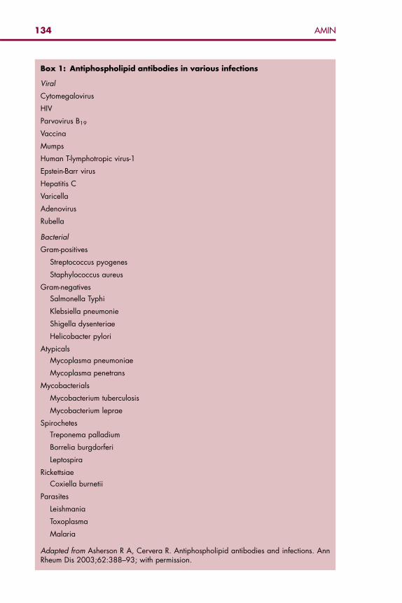
Box 1: Antiphospholipid antibodies in various infections
Viral
Cytomegalovirus
HIV
Parvovirus B19
Vaccina
Mumps
Human T-lymphotropic virus-1
Epstein-Barr virus
Hepatitis C
Varicella
Adenovirus
Rubella
Bacterial
Gram-positives
Streptococcus pyogenes
Staphylococcus aureus
Gram-negatives
Salmonella Typhi
Klebsiella pneumonie
Shigella dysenteriae
Helicobacter pylori
Atypicals
Mycoplasma pneumoniae
Mycoplasma penetrans
Mycobacterials
Mycobacterium tuberculosis
Mycobacterium leprae
Spirochetes
Treponema palladium
Borrelia burgdorferi
Leptospira
Rickettsiae
Coxiella burnetii
Parasites
Leishmania
Toxoplasma
Malaria
Adapted from Asherson R A, Cervera R. Antiphospholipid antibodies and infections. AnnRheum Dis 2003;62:388–93; with permission.
134 AMIN
135ANTIPHOSPHOLIPID SYNDROMES IN INFECTIOUS DISEASES
However, several cases of patients who had HCV and thrombosis have beenreported. One patient who had thalassemia and HCV and who developedthrombosis [27] had aCL and LA present. Baid and colleagues [28], studyingaCL and renal allograft thrombosis in 18 patients who had HCV positivitybefore transplant, found that renal microangiopathy with aCL positivity haddeveloped in 5 patients, compared with only 1 of 13 without angiopathy. Mal-wick and colleagues [29] also reported a case of a 54-year-old man who hada chronic HCV infection and a high level of aCL, who developed a lacunarbrain infarction [29,30].
In summary, the prevalence of aCL in patients who have HCV infection isfound at a higher proportion than in controlled groups. In most cases, the titersof these antibodies are low, cofactor independent, and unrelated to APS man-ifestations. A positive correlation between HCV and APS seems unlikely;therefore, it is not recommended to test for HCV in patients who have APS.In a few patients, thrombotic complications may occur.
Antiphospholipid Antibodies in HIV Infection
HIV, a retrovirus, causes abnormalities of T- and B-cell function that result indevelopment of various autoimmune phenomena of unknown but pathogenicsignificance. In 1986, Bloom and colleagues [31] first documented the presenceof LA in 40% of AIDS patients and in 43% of asymptomatic HIV-positiveindividuals (in which they may be transient) [17,31]. In 1992, Argov and col-leagues [32] reported the association of aCL with HIV infection in male homo-sexuals [16,32]. Since then, many studies have alluded to a high prevalence ofaCL in patients who have HIV (range 12%–67%) [33–36]. In contrast, anti-B2GPI antibodies are rarely detected in HIV patients, which indicates that aCL insuch individuals have characteristics of the natural type, not the pathogenic [37]variety. aCL in HIV patients have been associated with the presence of cerebralperfusion defects and transient neurologic deficit, but association with throm-bosis is uncommon, even with high aCL titers [38].
Antibodies against phospholipids other than cardiolipin, such as phosphati-dylserine, phosphatidylinositol, and phosphatidylcholine, have also beendetected in HIV-infected patients [36]. According to Guerin and colleagues[39], who found LA positivity in 72% and aCL positivity of 67% in theirHIV patients, the detection of antibodies to prothrombin and B2 GPI is signif-icantly less when compared with patients who have definite APS. Thus, in HIVinfection, both types of aCL-nonpathogenic (non–B2 GPI dependent) and path-ogenic (B2 GPI dependent) antibodies may be detected [6]. Petrovas andcolleagues [35] demonstrated that aPL reactivity does not correlate with the dis-ease duration or stage of HIV infection or with history of pneumocystis pneu-monia or coinfection with HCV. aCL did not appear to be a prognostic markerin HIV-infected subjects [40]. A significant decrease of aCL binding after treat-ment with urea and sodium chloride was observed in the sera of HIV-infectedpatients when compared with APS patients, indicating that aCL from HIV pa-tients have a low resistance to dissociating agents [41].
136 AMIN
In patients who have HIV or AIDS, thrombosis occurs frequently, includingperipheral vein, pulmonary embolism [42], retinal vein [43], cerebral vein [44],portal vein [45], mesenteric [46] occlusions, and testicular vessel [47]. Bossonand colleagues [48] reported a patient who had both arterial and venous throm-boembolism. In 1994, Thirumalai and colleagues [49] reported the presence ofaCL and stroke in an HIV-positive patient. In 1993, Cappell and colleagues[50] reported a case of 33-year-old woman with AIDS who had a cerebrovascu-lar accident and developed splenic infarction. In 1993, Belmonte and colleagues[51] documented three cases of avascular necrosis of bone associated withelevated aPL. Opportunistic infections, particularly with coexisting cytomega-lovirus (CMV), or, on occasion, pneumocystis, have been associated withthrombosis [52].
In summary, therefore, it seems that the pathogenesis of thrombotic compli-cations in HIV-infected patients and patients who have AIDS is multifactorial,with aPL playing a role in selected patients only. Although thrombotic compli-cations encountered with aPL positivity have been reported from variouscenters, their frequency remains low at present. Lipid disturbances associatedwith antiretroviral therapy may be an additional cause of these complications.
Antiphospholipid Antibodies in Cytomegalovirus Infection
IgG and IgM isotypes of aCL have been detected in CMV infections. Labarcaand colleagues [53] described a healthy young man who developed mesentericand femoropopliteal thrombosis during the course of CMV infection. Heresponded well to treatment with anticoagulants; 6 months after the onset ofAPS, IgM and IgG aCL titers declined. A similar case with transitory manifes-tation of APS was reported by Uthman and colleagues [54] in 1999. Dual viralinfection with CMV and HIV have frequently resulted in thrombotic manifes-tations of APS [44], including digital infarcts [55].Within the last year, a high prevalence (56%) of aPL was reported in CMVinfection among patients who received unrelated bone marrow and cord bloodallogenic stem cell transplantation for hematologic malignancies [56]. No asso-ciation was noted between these antibodies and the clinical expression ofautoimmunity.
Antiphospholipid Antibodies in Parvovirus B19 Infection
Infection with parvovirus B19 was associated with predominantly IgG antibodiesagainst the negatively charged phospholipids, cardiolipin and phosphatidylser-ine, which were B2 GPI dependent, as seen in patients who have systemic lupuserythematosus [10]. Unlike antibodies from patients who have other viral infec-tions (like HCV and HIV) or from those with syphilis, parvovirus infection wasassociated with aCL and increased their binding to antigen in the presence of B2GPI as a binding cofactor, resulting in thrombotic manifestations.
Antiphospholipid Antibodies in Varicella Virus Infection
Transient elevation of IgM and IgG aCL and LA is detected in patients whohave acute varicella infection. In a few cases, raised anti–B2 GPI antibodies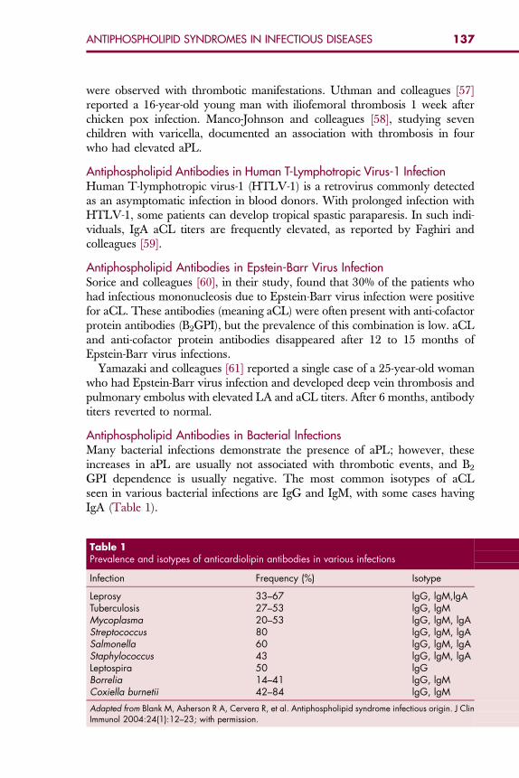
137ANTIPHOSPHOLIPID SYNDROMES IN INFECTIOUS DISEASES
were observed with thrombotic manifestations. Uthman and colleagues [57]reported a 16-year-old young man with iliofemoral thrombosis 1 week afterchicken pox infection. Manco-Johnson and colleagues [58], studying sevenchildren with varicella, documented an association with thrombosis in fourwho had elevated aPL.
Antiphospholipid Antibodies in Human T-Lymphotropic Virus-1 Infection
Human T-lymphotropic virus-1 (HTLV-1) is a retrovirus commonly detectedas an asymptomatic infection in blood donors. With prolonged infection withHTLV-1, some patients can develop tropical spastic paraparesis. In such indi-viduals, IgA aCL titers are frequently elevated, as reported by Faghiri andcolleagues [59].Antiphospholipid Antibodies in Epstein-Barr Virus Infection
Sorice and colleagues [60], in their study, found that 30% of the patients whohad infectious mononucleosis due to Epstein-Barr virus infection were positivefor aCL. These antibodies (meaning aCL) were often present with anti-cofactorprotein antibodies (B2GPI), but the prevalence of this combination is low. aCLand anti-cofactor protein antibodies disappeared after 12 to 15 months ofEpstein-Barr virus infections.Yamazaki and colleagues [61] reported a single case of a 25-year-old womanwho had Epstein-Barr virus infection and developed deep vein thrombosis andpulmonary embolus with elevated LA and aCL titers. After 6 months, antibodytiters reverted to normal.
Antiphospholipid Antibodies in Bacterial Infections
Many bacterial infections demonstrate the presence of aPL; however, theseincreases in aPL are usually not associated with thrombotic events, and B2GPI dependence is usually negative. The most common isotypes of aCLseen in various bacterial infections are IgG and IgM, with some cases havingIgA (Table 1).
Table 1Prevalence and isotypes of anticardiolipin antibodies in various infections
Infection Frequency (%) Isotype
Leprosy 33–67 lgG, lgM,lgATuberculosis 27–53 lgG, lgMMycoplasma 20–53 lgG, lgM, lgAStreptococcus 80 lgG, lgM, lgASalmonella 60 lgG, lgM, lgAStaphylococcus 43 lgG, lgM, lgALeptospira 50 lgGBorrelia 14–41 lgG, lgMCoxiella burnetii 42–84 lgG, lgM
Adapted from Blank M, Asherson R A, Cervera R, et al. Antiphospholipid syndrome infectious origin. J ClinImmunol 2004:24(1):12–23; with permission.
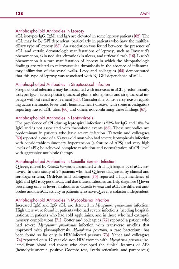
138 AMIN
Antiphospholipid Antibodies in Leprosy
aCL isotypes IgG, IgM, and IgA are elevated in some leprosy patients [62]. TheaCL may be B2 GPI dependent, particularly in patients who have the multiba-cillary type of leprosy [63]. An association was found between the presence ofaCL and certain dermatologic manifestations of leprosy, such as Raynaud’sphenomenon, skin nodules, chronic skin ulcers, and urticarial rash [16]. Lucio’sphenomenon is a rare manifestation of leprosy in which the histopathologicfindings are related to microvascular thrombosis in the absence of inflamma-tory infiltration of the vessel walls. Levy and colleagues [64] demonstratedthat this type of leprosy was associated with B2 GPI dependence of aCL.Antiphospholipid Antibodies in Streptococcal Infection
Streptococcal infections may be associated with increases in aCL, predominantlyisotypes IgG in acute poststreptococcal glomerulonephritis and streptococcal im-petigo without renal involvement [65]. Considerable controversy exists regard-ing acute rheumatic fever and rheumatic heart disease, with some investigatorsreporting raised aCL titers [66] and others not confirming these findings [67].Antiphospholipid Antibodies in Leptospirosis
The prevalence of aPL during leptospiral infection is 23% for IgG and 10% forIgM and is not associated with thrombotic events [68]. These antibodies arepredominant in patients who have severe infection. Tattevin and colleagues[69] reported a case of a 63-year-old man who had severe leptospirosis infectionwith considerable pulmonary hypertension (a feature of APS) and very highlevels of aPL; he achieved complete resolution and normalization of aPL levelwith aggressive antibiotic therapy.Antiphospholipid Antibodies in Coxiella Burnetii Infection
Q fever, caused by Coxiella burnetii, is associated with a high frequency of aCL pos-itivity. In their study of 26 patients who had Q fever diagnosed by clinical andserologic criteria, Ordi-Ros and colleagues [70] reported a high incidence ofIgM and IgG isotypes of aCL and that these antibodies can help diagnose Q feverpresenting only as fever; antibodies to Coxiella burnetii and aCL are different anti-bodies and the aCL activity in patients who have Q fever is cofactor independent.Antiphospholipid Antibodies in Mycoplasma Infection
Increased IgM and IgG aCL are detected in Mycoplasma pneumoniae infection.High titers were found in patients who had severe infections (needing hospital-ization), in patients who had cold agglutinins, and in those who had extrapul-monary complications [71]. Cotter and colleagues [72] reported a patient whohad severe Mycoplasma pneumoniae infection with transverse myelitis thatimproved with plasmapheresis. Mycoplasma penetrans, a rare bacterium, hasbeen found so far only in HIV-infected persons [73]. Yanez and colleagues[74] reported on a 17-year-old non-HIV woman with Mycoplasma penetrans iso-lated from blood and throat who developed the clinical features of APS(hemolytic anemia, positive Coombs test, livedo reticularis, and paraparesis)
139ANTIPHOSPHOLIPID SYNDROMES IN INFECTIOUS DISEASES
and elevated aPL and LA titers. The patient clinically improved after intensiveantibiotic therapy.
CATASTROPHIC ANTIPHOSPHOLIPID SYNDROMEAND INFECTIONSCatastrophic APS (CAPS), an unusual and potentially fatal subset of APS, wasfirst described in 1992 [75]. As of 2004, 220 patients with this syndrome hadbeen described and it comprises 1% of cases of APS [76]. It is characterizedby evidence of the involvement of three or more organs, systems, or tissues,and the development of manifestations simultaneously or in less thana week, with confirmation by histopathology of small vessel occlusion in atleast one organ or tissue and laboratory confirmation of the presence of aPL(LA or aCL) [77]. The disease is predominantly seen in women (70% of cases).Presentation of CAPS is often rapid and complex with involvement of renal(70%), pulmonary (65%), cerebral (55%), cardiac (50%), and gastrointestinal(44%) vessels. LA is detected in 79% of cases and aCL in high titers is seenin 86% of patients. Mortality is 47% and occurs from cardiac or respiratory fail-ure or gastrointestinal involvement [78].
Several triggering factors can precipitate CAPS, including trauma, with-drawal of anticoagulation, carcinoma, and infections. Rojas-Rodriguez [79]reported infections to precede the appearance of CAPS; 35% to 40% of casesdevelop CAPS after infectious episodes [80], including respiratory (15%); cuta-neous, including infected leg ulcers (8%); urinary tract (6%); gastrointestinal(1%); general sepsis (1%); and others (9%) [6]. More viral, as opposed to bac-terial, infections act as trigger factors for CAPS [6]. It is suggested that antigensof Salmonella Typhi, such as lipopolysaccharides, may have immunologic andprothrombotic effects [81].
Most of the infectious agents may act as superantigens and, by way of a mo-lecular mimicry phenomenon, lead to a disproportionate immune response byinducing nonpathogenic aPL and pathogenic anti–B2 GPI antibodies. There-fore, it may be possible to prevent this devastating evolution if the infectiousprocess is recognized promptly and treated aggressively.
SUMMARYaPL with their protein cofactors are essential for the diagnosis of APS. aPL ofdifferent kinds are detected in many infections, although in low titers and with-out protein cofactors in most cases.
aPL found in APS can be differentiated from those found in infections by de-termining LA activity and anti–B2 GPI antibodies. LA activity and anti–B2 GPIare present in APS resulting in thrombotic manifestations but not in most casesof infection (nonthrombogenic aPL). However, in several infections (leprosy,parvovirus B19, HIV, HCV, CMV), the aCL may be B2 GPI dependent, resem-bling those found in autoimmune diseases; are clearly heterogenous and patho-genic; and may be accompanied by thrombotic manifestation.
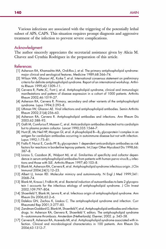
140 AMIN
Various infections are associated with the triggering of the potentially lethalsubset of APS, CAPS. This situation requires prompt diagnosis and aggressivetreatment of the infection to prevent severe complications.
Acknowledgment
The author sincerely appreciates the secretarial assistance given by Alicia M.Chavez and Cynthia Rodriguez in the preparation of this article.References
[1] Asherson RA, Khamashta MA, Ordi-Ros J, et al. The primary antiphospholipid syndrome:major clinical and serological features. Medicine 1989;68:366–74.[2] Wilson WA, Gharavi AE, Koike T, et al. International consensus statement on preliminary
criteria for definite antiphospholipid syndrome. Report of an international workshop. Arthri-tis Rheum 1999;42:1309–11.
[3] Cervera R, Piette JC, Font J, et al. Antiphospholipid syndrome, clinical and immunologicmanifestations and pattern of disease expression in a cohort of 1000 patients. ArthritisRheum 2002;46:1019–27.
[4] Asherson RA, Cervera R. Primary, secondary and other varients of the antiphospholipidsyndrome. Lupus 1994;3:293–8.
[5] Uthman IW, Gharavi AE. Viral infections and antiphospholipid antibodies. Semin ArthritisRheum 2002;31(4):256–63.
[6] Asherson RA, Cervera R. Antiphospholipid antibodies and infections. Ann Rheum Dis2003;62:388–93.
[7] Galli M, Comfurius P, Massen C, et al. Anticardiolipin antibodies directed not to cardiolipinbut to plasma protein cofactor. Lancet 1990;335:1544–7.
[8] Hunt JE, Mc Neil HP, Morgan GI, et al. A phospholipid B—B2 glycoprotein I complex in anantigen for cardiolipin antibodies occurring in autoimmune disease but not with infection.Lupus 1992;1:75–81.
[9] Fiallo P, Nunzi E, Cardo PP. B2 glycoprotein 1 dependent anticardiolipin antibodies as riskfactors for reactions in borderline leprosy patients. Int J Lepr Other Mycobact Dis 1998;66:387–8.
[10] Loizou S, Cazabon JK, Walport MJ, et al. Similarities of specificity and cofactor depen-dence in serum antiphospholipid antibodies from patients with human parvo virus B19 infec-tions and those with SLE. Arthritis Rheum 1997;40:103–8.
[11] Blank M, Asherson RA, Cervera R, et al. Antiphospholipid syndrome infectious origin. J ClinImmunol 2004;24(1):12–23.
[12] Albert LJ, Inman RD. Molecular mimicry and autoimmunity. N Engl J Med 1999;341:2068–74.
[13] Blank M, Krauss I, Fridkin M, et al. Bacterial induction of autoantibodies to beta 2 glycopro-tein I accounts for the infectious etiology of antiphospholipid syndrome. J Clin Invest2002;109:797–804.
[14] Shoenfeld Y, Blank M, Lervra R, et al. Infectious origin of antiphospholipid syndrome. AnnRheum Dis 2006;65:2–6.
[15] Dalekos GN, Zachou K, Liaskos C. The antiphospholipid syndrome and infection. CurrRheumatol Rep 2001;3:277–85.
[16] Zandman-GoddardG,BlankM,ShoenfeldY, et al. Antiphospholipidantibodies and infection-drugs. In: Asherson RA, Gervera R, Shoenfeld Y, editors. The antiphospholipid syndromeII—autoimmune thrombosis. Amsterdam (Netherlands): Elsevier; 2002. p. 343–58.
[17] Cervera R, Asherson RA, Acevedo MI, et al. Antiphospholipid syndrome associated with in-fection. Clinical and microbiological characteristics in 100 patients. Ann Rheum Dis2004;63:1312–7.
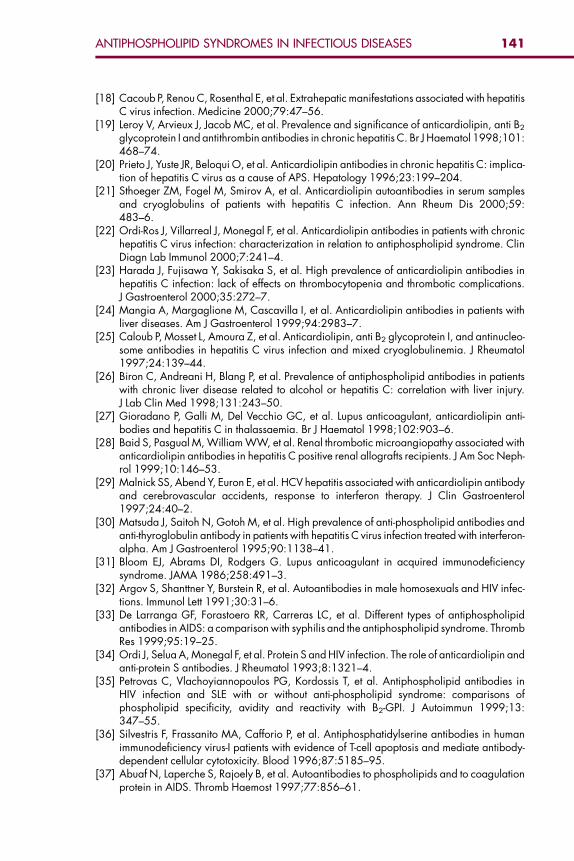
141ANTIPHOSPHOLIPID SYNDROMES IN INFECTIOUS DISEASES
[18] Cacoub P, Renou C, Rosenthal E, et al. Extrahepatic manifestations associated with hepatitisC virus infection. Medicine 2000;79:47–56.
[19] Leroy V, Arvieux J, Jacob MC, et al. Prevalence and significance of anticardiolipin, anti B2
glycoprotein I and antithrombin antibodies in chronic hepatitis C. Br J Haematol 1998;101:468–74.
[20] Prieto J, Yuste JR, Beloqui O, et al. Anticardiolipin antibodies in chronic hepatitis C: implica-tion of hepatitis C virus as a cause of APS. Hepatology 1996;23:199–204.
[21] Sthoeger ZM, Fogel M, Smirov A, et al. Anticardiolipin autoantibodies in serum samplesand cryoglobulins of patients with hepatitis C infection. Ann Rheum Dis 2000;59:483–6.
[22] Ordi-Ros J, Villarreal J, Monegal F, et al. Anticardiolipin antibodies in patients with chronichepatitis C virus infection: characterization in relation to antiphospholipid syndrome. ClinDiagn Lab Immunol 2000;7:241–4.
[23] Harada J, Fujisawa Y, Sakisaka S, et al. High prevalence of anticardiolipin antibodies inhepatitis C infection: lack of effects on thrombocytopenia and thrombotic complications.J Gastroenterol 2000;35:272–7.
[24] Mangia A, Margaglione M, Cascavilla I, et al. Anticardiolipin antibodies in patients withliver diseases. Am J Gastroenterol 1999;94:2983–7.
[25] Caloub P, Mosset L, Amoura Z, et al. Anticardiolipin, anti B2 glycoprotein I, and antinucleo-some antibodies in hepatitis C virus infection and mixed cryoglobulinemia. J Rheumatol1997;24:139–44.
[26] Biron C, Andreani H, Blang P, et al. Prevalence of antiphospholipid antibodies in patientswith chronic liver disease related to alcohol or hepatitis C: correlation with liver injury.J Lab Clin Med 1998;131:243–50.
[27] Gioradano P, Galli M, Del Vecchio GC, et al. Lupus anticoagulant, anticardiolipin anti-bodies and hepatitis C in thalassaemia. Br J Haematol 1998;102:903–6.
[28] Baid S, Pasgual M, William WW, et al. Renal thrombotic microangiopathy associated withanticardiolipin antibodies in hepatitis C positive renal allografts recipients. J Am Soc Neph-rol 1999;10:146–53.
[29] Malnick SS, Abend Y, Euron E, et al. HCV hepatitis associated with anticardiolipin antibodyand cerebrovascular accidents, response to interferon therapy. J Clin Gastroenterol1997;24:40–2.
[30] Matsuda J, Saitoh N, Gotoh M, et al. High prevalence of anti-phospholipid antibodies andanti-thyroglobulin antibody in patients with hepatitis C virus infection treated with interferon-alpha. Am J Gastroenterol 1995;90:1138–41.
[31] Bloom EJ, Abrams DI, Rodgers G. Lupus anticoagulant in acquired immunodeficiencysyndrome. JAMA 1986;258:491–3.
[32] Argov S, Shanttner Y, Burstein R, et al. Autoantibodies in male homosexuals and HIV infec-tions. Immunol Lett 1991;30:31–6.
[33] De Larranga GF, Forastoero RR, Carreras LC, et al. Different types of antiphospholipidantibodies in AIDS: a comparison with syphilis and the antiphospholipid syndrome. ThrombRes 1999;95:19–25.
[34] Ordi J, Selua A, Monegal F, et al. Protein S and HIV infection. The role of anticardiolipin andanti-protein S antibodies. J Rheumatol 1993;8:1321–4.
[35] Petrovas C, Vlachoyiannopoulos PG, Kordossis T, et al. Antiphospholipid antibodies inHIV infection and SLE with or without anti-phospholipid syndrome: comparisons ofphospholipid specificity, avidity and reactivity with B2-GPI. J Autoimmun 1999;13:347–55.
[36] Silvestris F, Frassanito MA, Cafforio P, et al. Antiphosphatidylserine antibodies in humanimmunodeficiency virus-I patients with evidence of T-cell apoptosis and mediate antibody-dependent cellular cytotoxicity. Blood 1996;87:5185–95.
[37] Abuaf N, Laperche S, Rajoely B, et al. Autoantibodies to phospholipids and to coagulationprotein in AIDS. Thromb Haemost 1997;77:856–61.
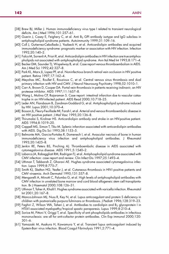
142 AMIN
[38] Brew BJ, Miller J. Human immunodeficiency virus type I related to transient neurologicaldeficits. Am J Med 1996;101:257–61.
[39] Guerin J, Casey E, Feighery C, et al. Anti B2 GPI antibody isotype and IgG subclass inantiphospholipid syndrome patients. Autoimmunity 1999;31:109–16.
[40] Coll J, Gutierrez-Cebollada J, Yazbeck H, et al. Anticardiolipin antibodies and acquiredimmunodeficiency syndrome: prognostic marker or association with HIV infection. Infection1992;20:140–2.
[41] FalcoM,SorrenliA, Priori R, et al.Anticardiolipinantibodies inHIV infectionare trueantiphos-pholipids not associated with antiphospholipid syndrome. Ann Ital Med Int 1993;8:171–4.
[42] Becker DM, Saunder TJ, Wispelway B, et al. Case report venous thromboembolism in AIDS.Am J Med Sci 1992;42:327–8.
[43] Park KL, Marx JL, Lopez PF, et al. Noninfectious branch retinal vein occlusion in HIV positivepatient. Retina 1997;17:162–4.
[44] Meyohas MC, Roullet E, Rouzioux C, et al. Central venous sinus thrombosis and dualprimary infection with HIV and CMV. J Neurol Neurosurg Psychiatry 1998;52:1010–1.
[45] Carr A, Brown D, Cooper DA. Portal vein thrombosis in patients receiving indinavir, an HIVprotease inhibitor. AIDS 1997;11:1657–8.
[46] Wang L, Molina CP, Rajaraman S. Case report: intestinal infarction due to vascular catas-trophe in an HIV-infected patient. AIDS Read 2000;10:718–25.
[47] Leder AN, Flansbaum B, Zandman-Goddard G, et al. Antiphospholipid syndrome inducedby HIV. Lupus 2001;10:370–4.
[48] Bosson JL, Fleury-Feuillade MI, Farah I, et al. Arterial and venous thromboembolic disease inan HIV positive patient. J Mal Vasc 1995;20:136–8.
[49] Thirumalai S, Kirshner HS. Anticardiolipin antibody and stroke in an HIV-positive patient.AIDS 1994;8:1019–20.
[50] Cappell MS, Simon T, Tiku M. Splenic infarction associated with anticardiolipin antibodieswith AIDS. Dig Dis Sci 1993;38:1153–5.
[51] Belmonte MA, Garcia-Portales R, Domenech I, et al. Avascular necrosis of bone in humanimmunodeficiency virus infection and antiphospholipid antibodies. J Rheumatol1993;20:1425–8.
[52] Jenkin RE, Peters BS, Pinching AJ. Thromboembolic disease in AIDS associated withcytomegalovirus disease. AIDS 1991;5:1540–2.
[53] Labarca JA, Rabaggliati RM, Radrigan FJ, et al. Antiphospholipid syndrome associated withCMV infection: case report and review. Clin Infect Dis 1997;25:1493–4.
[54] Uthman T, Tabbarah Z, Gharavi AE. Hughes syndrome associated cytomegalovirus infec-tion. Lupus 1999;8:775–7.
[55] Smith KJ, Skelton HG, Yeafer J, et al. Cutaneous thrombosis in HIV-I positive patients andCMV viraemia. Arch Dermatol 1995;131:357–8.
[56] Mengarelli A, Minotti C, Palumbo G, et al. High levels of antiphospholipid antibodies withCMV infection in unrelated bone marrow and cord blood allogeneic stem cell transplanta-tion. Br J Haematol 2000;108:126–31.
[57] Uthman T, Taher A, Khalil I. Hughes syndrome associated with varicella infection. RheumatolInt 2001;20:167–8.
[58] Manco-Johnson MJ, Nuss R, Key N, et al. Lupus anticoagulant and protein S deficiency inchildren with postvaricella purpura fulminans or thrombosis. J Pediatr 1996;128:319–23.
[59] Faghiri Z, Wilson WA, Taheri J, et al. Antibodies to cardiolipin and B2 glycoprotein I inHTLV-I associated myelopathy/tropical spastic paraparesis. Lupus 1999;8:210–4.
[60] Sorice M, Pittoni V, Griggi T, et al. Specificity of anti phospholipids antibodies in infectiousmononucleosis: are all for anti-cofactor protein antibodies. Clin Exp immunol 2000;120:301–6.
[61] Yamazaki M, Asakura H, Kawamura Y, et al. Transient lupus anticoagulant induced byEpstein-Barr virus infection. Blood Coagul Fibrinolysis 1991;2:771–4.
143ANTIPHOSPHOLIPID SYNDROMES IN INFECTIOUS DISEASES
[62] de Larranga GF, Forasttero RR, Martinuzzo ME, et al. High prevalence of antiphospholipidantibodies in leprosy-evaluation of antigen reactivity. Lupus 2000;9:594–600.
[63] Fiallo P, Travaglino C, Nunzi E, et al. B2 glycoprotein dependence of anticardiolipinantibodies in multibacillary leprosy patients. Lepr Rev 1998;69:376–81.
[64] Levy RA, Pierangeli SA, Espinola RG, et al. Antiphospholipid beta-2 glycoprotein I depen-dency assay to determine antibody pathogenicity. Arthritis Rheum 2000;43(Suppl):1476.
[65] Ardiles L, Ramirez P, Moya P, et al. Anticardiolipin antibodies in acute poststreptococcal glo-merulonephritis and streptococcal impetigo. Nephron 1999;83:47–52.
[66] Figueroa F, Berrios X, Gutierrez M, et al. Anticardiolipin antibodies in acute rheumatic fever.J Rheumatol 1992;19:1175–80.
[67] Ilarraza H, Marquez MF, Alcocer A, et al. Anticardiolipin antibodies are not associated withrheumatic heart disease. Lupus 2001;10:873–5.
[68] Rugman FP, Pinn G, Palmer G, et al. Anticardiolipin antibodies in leptospirosis. J Clin Pathol1991;44:517–9.
[69] Tattevin P, Dupeux S, Hoff J, et al. Leptospirosis and the antiphospholipid syndrome. AmJ Med 2003;114:164.
[70] Ordi-Ros J, Selva-O’Callaghan A, Monegal-Ferran F, et al. Prevalence, significance andspecificity of antibodies to phospholipids in Q fever. Clin Infect Dis 1994;18:213–8.
[71] Snowden N, Wilson PB, Longson M, et al. Antiphospholipid antibodies and Mycoplasmapneumoniae infection. Postgrad Med J 1990;66:356–62.
[72] Cotter FE, Bainbridge D, Newland AC, et al. Neurological deficit associated with Myco-plasma pneumoniae reversed by plasma exchange. Br Med J 1983;286:22.
[73] Grau O, Slizewicz B, Tuppin P, et al. Association of Mycoplasma penetrans with immuno-deficiency virus infection. J Infect Dis 1995;172:672–81.
[74] Yanez A, Codillo L, Neyrolles O, et al. Mycoplasma penetrans bacteremia and primaryantiphospholipid syndrome. Emerg Infect Dis 1999;5(1):1–6.
[75] Asherson RA. The catastrophic antiphospholipid syndrome. J Rheumatol 1992;19:508–12.[76] Asherson RA. The catastrophic antiphospholipid syndrome registry project group. Lupus
2003;12:530–6.[77] Cervera R, Gomez-Puerta JA, Cucho M, et al. Catastrophic antiphospholipid syndrome:
analysis of international consensus statement on preliminary classification criteria forCAPS using CAPS registry. Ann Rheum Dis 2003;62(Suppl):84.
[78] Michael-Belmont H. Catastrophic antiphospholipid syndrome. In: Asherson RA, Cervera R,Shoenfield Y, editors. The antiphospholipid syndrome II-autoimmune thrombosis. Amster-dam (Netherlands): Elsevier; 2002. p. 171–80.
[79] Rojas-Rodriguez J, Garcia-Carrasco M, Ramos-Casal M, et al. Catastrophic antiphospholi-pid syndrome: clinical description and triggering factors in 8 patients. J Rheumatol1999;26:238–40.
[80] Asherson RA, Cervera R, Piette JC, et al. Catastrophic antiphospholipid syndrome: clues tothe pathogenesis from a series of 80 patients. Medicine 2001;80:355–77.
[81] Hayem G, Kassis N, Nicaise P, et al. Systemic lupus erythematosus-associated catastrophicantiphospholipid syndrome occurring after typhoid fever: a possible role of salmonella lipo-polysaccharide in the occurrence of diffuse vasculopathy-coagulopathy. Arthritis Rheum1999;42:1056–65.
Hematol Oncol Clin N Am 22 (2008) 145–153
HEMATOLOGY/ONCOLOGY CLINICSOF NORTH AMERICA
Treatment Options for PatientsWho Have AntiphospholipidSyndromes
Rodger L. Bick, MD, PhD, FACPa,*,William F. Baker, MD, FACPb,c,d
a10455 North Central Expressway, Suite 109-320, Dallas, TX 75231, USAbDavid Geffen School of Medicine Center for Health Sciences University of California,Los Angeles, Los Angeles, CA, USAcThrombosis, Hemostasis and Special Hematology Clinic, Kern Medical Center, Bakersfield,CA, USAdCalifornia Clinical Thrombosis Center, 9330 Stockdale Highway, Suite 300, Bakersfield,CA 93311, USA
The antiphospholipid thrombosis syndrome, associated with anticardiolipin(aCL) or subgroup antibodies (Box 1), can be divided into one of sixsubgroups:
� Type I syndrome comprises patients with deep venous thrombosis (DVT) andpulmonary embolism (PE).
� Type II syndrome comprises patients with coronary artery or peripheralarterial (including aorta and carotid artery) thrombosis.
� Type III syndrome comprises patients with retinal or cerebrovascular (intracra-nial) thrombosis.
� Type IV patients are those with admixtures of the first three types. Type IVpatients are uncommon, with most patients fitting into one of the first three types.
� Type V patients are those with recurrent miscarriage syndrome.� Type VI patients are those harboring antiphospholipid syndromes (APSs)
without any (as yet) clinical expression, including thrombosis.
There is little overlap (about 10% or less) between these subtypes, andpatients usually conveniently fit into only one of these clinical types. The typesof antiphospholipid and thrombosis syndromes associated with aCL antibodiesare summarized in Box 2 [1–3]. Although there appears to be no correlationwith the type, or titer, of aCL antibody and type of syndrome (I–VI), thesubclassification of thrombosis and aCL antibody patients into these groupsis important from the therapy standpoint [1–3]. Type I patients are best man-aged by use of long-term fixed-dose low molecular weight heparin (LMWH) or
*Corresponding author. E-mail address: [email protected] (R.L. Bick).
0889-8588/08/$ – see front matter ª 2008 Elsevier Inc. All rights reserved.doi:10.1016/j.hoc.2007.10.010 hemonc.theclinics.com
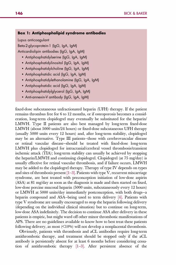
Box 1: Antiphospholipid syndrome antibodies
Lupus anticoagulant
Beta-2-glycoprotein-1 (IgG, IgA, IgM)
Anticardiolipin antibodies (IgG, IgA, IgM)� Antiphosphatidylserine (IgG, IgA, IgM)� Antiphosphatidylinositol (IgG, IgA, IgM)� Antiphosphatidylcholine (IgG, IgA, IgM)� Antiphosphatidic acid (IgG, IgA, IgM)� Antiphosphatidylethanolamine (IgG, IgA, IgM)� Antiphosphatidic acid (IgG, IgA, IgM)� Antiphosphatidylglycerol (IgG, IgA, IgM)� Anti-annexin-V antibody (IgG, IgA, IgM)
146 BICK & BAKER
fixed-dose subcutaneous unfractionated heparin (UFH) therapy. If the patientremains thrombus free for 6 to 12 months, or if osteoporosis becomes a consid-eration, long-term clopidogrel may eventually be substituted for the heparin/LMWH. Type II patients are also best managed by long-term fixed-doseLMWH (about 5000 units/24 hours) or fixed-dose subcutaneous UFH therapy(usually 5000 units every 12 hours) and, after long-term stability, clopidogrelmay be an alternative. Type III patients—those with cerebrovascular diseaseor retinal vascular disease—should be treated with fixed-dose long-termLMWH plus clopidogrel for intracranial/cerebral vessel thrombosis/transientischemic attack (TIA); long-term stability can usually be achieved by stoppingthe heparin/LMWH and continuing clopidogrel. Clopidogrel (at 75 mg/day) isusually effective for retinal vascular thrombosis, and if failure occurs, LMWHmay be added to the clopidogrel therapy. Therapy of type IV depends on typesand sites of thrombosis present [1–3]. Patients with type V, recurrent miscarriagesyndrome, are best treated with preconception initiation of low-dose aspirin(ASA) at 81 mg/day as soon as the diagnosis is made and then started on fixed,low-dose porcine mucosal heparin (5000 units, subcutaneously every 12 hours)or LMWH at 5000 units/day immediately postconception, with both drugs—aheparin compound and ASA—being used to term delivery [4]. Patients withtype V syndrome are usually encouraged to stop the heparin following delivery(depending on the individual clinical situation) but to continue on long-term,low-dose ASA indefinitely. The decision to continue ASA after delivery in thesepatients is empiric, but might ward off other minor thrombotic manifestations ofAPS. There are no guidelines available to know how to best treat these patientsfollowing delivery, as most (<10%) will not develop a nonplacental thrombosis.
Obviously, patients with thrombosis and aCL antibodies require long-termantithrombotic therapy, and treatment should be stopped only if the aCLantibody is persistently absent for at least 6 months before considering cessa-tion of antithrombotic therapy [1–3]. After persistent absence of the
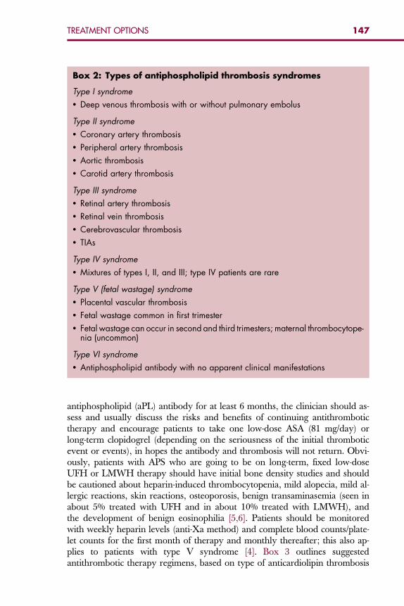
Box 2: Types of antiphospholipid thrombosis syndromes
Type I syndrome� Deep venous thrombosis with or without pulmonary embolus
Type II syndrome� Coronary artery thrombosis� Peripheral artery thrombosis� Aortic thrombosis� Carotid artery thrombosis
Type III syndrome� Retinal artery thrombosis� Retinal vein thrombosis� Cerebrovascular thrombosis� TIAs
Type IV syndrome� Mixtures of types I, II, and III; type IV patients are rare
Type V (fetal wastage) syndrome� Placental vascular thrombosis� Fetal wastage common in first trimester� Fetal wastage can occur in second and third trimesters; maternal thrombocytope-
nia (uncommon)
Type VI syndrome� Antiphospholipid antibody with no apparent clinical manifestations
147TREATMENT OPTIONS
antiphospholipid (aPL) antibody for at least 6 months, the clinician should as-sess and usually discuss the risks and benefits of continuing antithrombotictherapy and encourage patients to take one low-dose ASA (81 mg/day) orlong-term clopidogrel (depending on the seriousness of the initial thromboticevent or events), in hopes the antibody and thrombosis will not return. Obvi-ously, patients with APS who are going to be on long-term, fixed low-doseUFH or LMWH therapy should have initial bone density studies and shouldbe cautioned about heparin-induced thrombocytopenia, mild alopecia, mild al-lergic reactions, skin reactions, osteoporosis, benign transaminasemia (seen inabout 5% treated with UFH and in about 10% treated with LMWH), andthe development of benign eosinophilia [5,6]. Patients should be monitoredwith weekly heparin levels (anti-Xa method) and complete blood counts/plate-let counts for the first month of therapy and monthly thereafter; this also ap-plies to patients with type V syndrome [4]. Box 3 outlines suggestedantithrombotic therapy regimens, based on type of anticardiolipin thrombosis
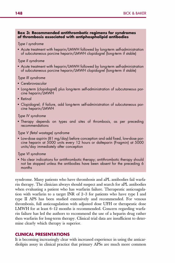
Box 3: Recommended antithrombotic regimens for syndromesof thrombosis associated with antiphospholipid antibodies
Type I syndrome� Acute treatment with heparin/LMWH followed by long-term self-administration
of subcutaneous porcine heparin/LMWH clopidogrel (long-term if stable)
Type II syndrome� Acute treatment with heparin/LMWH followed by long-term self-administration
of subcutaneous porcine heparin/LMWH clopidogrel (long-term if stable)
Type III syndrome� Cerebrovascular� Long-term (clopidogrel) plus long-term self-administration of subcutaneous por-
cine heparin/LMWH� Retinal� Clopidogrel; if failure, add long-term self-administration of subcutaneous por-
cine heparin/LMWH
Type IV syndrome� Therapy depends on types and sites of thrombosis, as per preceding
recommendations
Type V (fetal wastage) syndrome� Low-dose aspirin (81 mg/day) before conception and add fixed, low-dose por-
cine heparin at 5000 units every 12 hours or dalteparin (Fragmin) at 5000units/day immediately after conception
Type VI syndrome� No clear indications for antithrombotic therapy; antithrombotic therapy should
not be stopped unless the antibodies have been absent for the preceding 6months
148 BICK & BAKER
syndrome. Many patients who have thrombosis and aPL antibodies fail warfa-rin therapy. The clinician always should suspect and search for aPL antibodieswhen evaluating a patient who has warfarin failure. Therapeutic anticoagula-tion with warfarin to a target INR of 2–3 for patients who have type I andtype II APS has been studied extensively and recommended. For venousthrombosis, full anticoagulation with adjusted dose UFH or therapeutic doseLMWH for at least 6–12 months is recommended. Concern regarding warfa-rin failure has led the authors to recommend the use of a heparin drug ratherthen warfarin for long-term therapy. Clinical trial data are insufficient to deter-mine clearly which therapy is superior.
CLINICAL PRESENTATIONSIt is becoming increasingly clear with increased experience in using the anticar-diolipin assay in clinical practice that primary APSs are much more common
149TREATMENT OPTIONS
than suspected. Diagnostic evaluation of the patient to determine the etiologyof a wide variety of thrombotic problems must now include assays for aCL an-tibodies, lupus anticoagulants, and, when indicated, subgroups. Although it isappropriate to suspect aPL antibodies in virtually any clinical problem compli-cated by thrombosis, certain presentations are stronger indicators than others.
In patients with type I disease, a strong index of suspicion is appropriate, par-ticularly in individuals with DVT unaccompanied by another potential risk fac-tor (eg, exogenous estrogen administration, surgery, prolonged immobility,malignancy, or another hypercoagulable state). Likewise, patients may presentwith recurrent DVT with or without a significant clinical risk factor. As is fre-quently observed in clinical practice, patients may be referred for evaluationonly after a second episode of thrombosis. The initial thrombotic event mayhave appeared to result from a recognizable predisposing problem, only laterproven to be present concomitantly with aCL antibodies. Although the severityor location (iliofemoral, popliteal calf vein, or other sites) of thrombosis or thepresence of pulmonary embolization does not correlate with the presence ofaCL antibodies, recurrent thromboembolic events or multiple sites of thrombo-sis should strongly suggest an aPL antibody. Another common presentation isa patient referred because of failure (rethrombosis) while on warfarin therapy.Failure to apparently adequate doses of warfarin should immediately alert thephysician to strongly consider APS.
Patients with type II disease frequently present with catastrophic illness. Ahistory of myocardial infarction at a young age, recurrent myocardial infarc-tion, early graft occlusion following coronary artery bypass graft surgery,and early re-occlusion following post-transluminal angioplasty is typical [7].Aorta, subclavian, mesenteric, femoral, or other large-vessel thrombosis maypresent with complete occlusion and acute symptoms of ischemia and threat-ened limb loss. Emergent diagnosis and appropriate therapy may decrease un-necessary morbidity and be life-saving.
Type III patients may be referred for a variety of problems. Acute loss or dis-tortion of vision may lead to ophthalmologic confirmation of retinal arterial orvenous thrombosis. Focal neurologic symptoms may suggest the presence of ce-rebrovascular thrombosis resulting in symptoms of stroke or TIA. Alternatively,multiple infarct dementia may present more gradually, without clearly definedacute ischemic events. Early diagnosis is critical in type III patients, as failure totreat may result in irreversible central nervous system or retinal injury.
Type IV patients, having a mixture of the aforementioned types, are extremelyrare and comprise only about 1% of patients with anticardiolipin thrombosis syn-drome. A strong index of suspicion is required for the diagnosis, and therapymust be individualized depending on the particular combination of thromboses.
Type V patients are usually those with one or more spontaneous miscar-riages and are most often referred by the obstetrician or high-risk reproductiveexperts. Most women relate a history of spontaneous miscarriage in the firsttrimester (most commonly the 6th–12th week), but some also spontaneouslymiscarry in the second and third trimesters.
150 BICK & BAKER
PREVALENCE OF THE ANTIPHOSPHOLIPID THROMBOSISSYNDROMEUnfortunately, little information is available on prevalence of aPL antibodies,especially in asymptomatic individuals. Additionally, nothing is known aboutthe potential propensity to develop thrombosis or other clinical manifestationswhen seemingly healthy individuals are found to harbor these antibodies. Tworecent studies have addressed this issue. The first such study was the Montpel-lier Antiphospholipid study [8], wherein 1014 patients (488 males, 526 females)admitted to a general internal medicine department for a variety of reasonswere assessed for IgG, IgA, and IgM aCL antibodies. Lupus anticoagulant as-says were not performed. Of the patients tested, 72 (7.1%) were positive for atleast one idiotype. When assessing these 72 patients, 20 (28%) were determinedto have clinical manifestations of APS. Fifty-two patients, when questioned, hadnot yet demonstrated any manifestations of APL-T syndrome, suggestinga false-positive incidence of 5.1%. However, long-term follow-up of the thusfar asymptomatic patients has not occurred, and a follow-up report of theMontpellier Antiphospholipid study will be awaited with interest. In anotherrecent study [9], 552 healthy blood donors were screened for study; IgG andIgM idiotypes and lupus anticoagulant were assessed. It was found that 6.5%(28 donors) of the population harbored IgG, 9.4% (38 donors) of the popula-tion harbored IgM aCL antibodies, and 5 donors had both idiotypes. No donorwas positive for lupus anticoagulant. The donors were followed for 12 months;during the follow-up time, no aCL-positive patient developed a thromboticevent. However, 9 aCL-positive donors had a positive family history forthrombosis and 3 of the aCL-positive donors had a history of unexplainedmiscarriage.
In a patient harboring aPL antibodies and no symptoms, there is no clearevidence that any antithrombotic treatment is needed.
In a survey of 100 consecutive patients presenting with DVT or PE, 24% ofpatients were found to have aPL antibodies [10]. It is suggested that aPL anti-bodies are common in patients presenting with unexplained DVT or PE, andcertainly any patient presenting with unexplained DVT or PE should be eval-uated for presence of aPL antibodies.
General treatment programs have been outlined in this section. These areprimarily developed from the present authors’ clinical experience. For addi-tional or alternate treatment strategies and recommendations, the reader isencouraged to see other articles in this issue.
International preliminary catastrophic antiphospholipid syndrome (CAPS)classification criteria and treatment guidelines were proposed by the 2002 Inter-national Taormina Consensus Statement on Classification and Treatment ofCAPS [11]. Criteria for diagnosis include the involvement of three or more or-gans, systems, and/or tissues [11].
The approach to treatment has included a variety of modalities, including an-ticoagulation, glutocorticoids, plasmapheresis, cyclophosphamide, intravenousgammaglobulins, and splenectomy. Most patients, however, received
151TREATMENT OPTIONS
a combination of nonsurgical therapies. In spite of combination therapy (mostnonsurgical), death still occurred in 25 of the 50 (50%) patients reported byAsherson and colleagues [12]. The current rationale for the recommended ther-apy of combined anticoagulation, steroids, and plasmapheresis or intravenousgammaglobulins is derived from the reported survival rate of 70% in patientsso treated [11]. The most successful approach is similar to that used in treatmentof patients with thrombotic thrombocytopenic purpura and hemolytic uremicsyndrome. The fibrinolytic agents streptokinase and urokinase have also beenused [13,14]. There is, however, no large series comparing various therapiesand combinations that can be relied on to provide firm recommendations [2].
Venous Thrombosis
Venous thrombosis typically presents with DVT in the lower extremities, ob-served in 29% to 55% of cases over a follow-up period of less than 6 years [7,8],[10]. As with all cases of venous thromboembolism (VTE), more than half ofthe patients with symptomatic DVT have asymptomatic PE [15]. When pa-tients present with symptoms typical of PE, at least half will not exhibit clinicalmanifestations of DVT [16,17]. There is not a particular clinical pattern ofvenous thrombosis that suggests that the patient may have underlying aPLantibodies. Unusual sites of thrombosis have included venous thrombosis ofthe upper extremities, intracranial veins, inferior and superior vena cava, he-patic veins (Budd Chiari syndrome), portal vein, renal vein, and retinal vein[18–20]. Thrombosis of the cerebral veins may present with acute cerebral in-farction [21]. The superficial and deep cerebral venous system may be exten-sively involved. Onset of cerebral venous thrombosis due to aPL antibodiestypically presents at a younger age and in a more extensive pattern than inother patients. Thrombosis of the superior saggital sinus has also been re-ported. In one series of 40 patients with cerebral venous thrombosis, 3 hadaPL antibodies and 2 of these 3 also had Factor V Leiden mutation [22].In the setting of idiopathic VTE, laboratory assessment is recommended tosearch for the presence of underlying thrombophilia [15]. Supporting this is theseries of 100 consecutive patients with idiopathic DVT or PE in which 24%were found to have aPL antibodies [23]. Testing for aPL antibodies is stronglyrecommend because of the high frequency with which APS is diagnosed [6].Even in patients with a family history of venous thrombosis, which suggestsan underlying inherited thrombophilic disorder, testing for aPL antibodies isindicated, as many patients with one thrombophilic disorder also have another.The clinical manifestations of VTE are the same, whether aPL antibodies arepresent or not. Subsequent to a first episode of VTE, the risk for future venousthrombosis increases significantly. In patients with APS, the risk doubles to atleast a 30% risk of recurrence, without antithrombotic therapy.
Arterial Thrombosis
Arterial thromboses are less common than venous thrombosis [7,8], and occurin a variety of settings in patient with primary APS. Of the 1000 patients in theEuro-Phospholipid Trial, 13.1% presented with stroke, 7% with TIA, and 2.8%
152 BICK & BAKER
with acute myocardial infarction [24]. Patients with arterial thrombosis mostcommonly present with TIA or stroke (50%) or myocardial infarction (23%)[7,24,25]. These relatively common arterial occlusive events most suggestAPS when they occur in individuals without readily identified risk factors foratherosclerosis. This population primarily includes individuals under age 60,without classic risk factors for atherosclerosis (family history, cigarette smok-ing, hyperlipidemia, hypertension, diabetes mellitus) [19,26]. The presence ofaCL antibody is considered to be a risk factor for first stroke [23]. Arterialthrombosis in patients with APS may also involve other large and small vessels,somewhat unusual for other thrombophilic disorders or atherosclerotic occlu-sive disease. These include thrombosis of brachial and subclavian arteries, ax-illary artery (aortic arch syndrome), aorta, iliac, femoral, renal, mesenteric,retinal, and other peripheral arteries [2,7]. Clinical manifestations, of course,depend on the caliber and location of the affected artery.
References
[1] Bick RL. Antiphospholipid thrombosis syndromes: etiology, pathophysiology, diagnosis andmanagement. Int J Hematol 1997;65:193–213.[2] Bick RL. The antiphospholipid thrombosis syndromes: a common multidisciplinary medical
problem. J Clin Appl Thromb Hemost 1997;3:270–83.[3] Bick RL. Hereditary and acquired thrombophilia. Clin Appl Thromb Hemost 2006;12:
125–35.[4] Bick RL. Recurrent miscarriage syndrome and Infertility caused by blood coagulation
protein / platelet defects. Chapter 2. In: Bick RL, Frenkel E, Baker W, editors. Hematologicalcomplications in obstetrics, pregnancy and gynecology. Cambridge (UK): CambridgeUniversity Press; 2006. p. 55–74.
[5] Walenga JM, Bick RL. Heparin-induced thrombocytopenia, paradoxical thromboembolismand other side effects of heparin therapy. Med Clin North Am 1998;82(3):635–58.
[6] Girolami B, Prandoni P, Rossi L, et al. Transaminase elevation in patients treated with unfrac-tionated heparin or low molecular weight heparin for venous thromboembolism. Clin ApplThromb Hemost 1998;4:126–8.
[7] Bick RL. Antiphospholopid thrombosis syndromes. Hematol Oncol Clin North Am 2003;17:115–47.
[8] Schved JF, Dupuy-Fons C, Biron C. A prespective epidemiological study on the occurrence ofantiphospholipid antibody: the Montpellier Antiphospholipiod (MAP) Study. Haemostasis1994;24:175–82.
[9] Vila P, Hernandez MC, Lopez-Fernandez MF. Prevalence, follow-up and clinical signifi-cance of the anticardiolipin antibodies in normal subjects. Thromb Haemost 1994;72:209–13.
[10] Bick RL, Baker WF. Deep vein thrombosis: prevalence of etiologic factors and results of man-agement in 100 consecutive patients. Semin Thromb Hemost 1992;18:267–74.
[11] Asherson RA, Cervera R, de Groot PG, et al. Catastrophic and antiphospholipid syndrome:international consensus statement on classification criteria and treatment guidelines. Lupus2003;12(7):530–4.
[12] Asherson RA, Cervera R, Piette JC, et al. Catastrophic antiphospholipid syndrome. Clinicaland laboratory features of 50 patients. Medicine (Baltimore) 1998;77(3):195–207.
[13] Harris EN, Gharavi A, Hegde U, et al. Anticardiolipin antibodies in autoimmune thrombo-cytopenic purpura. Br J Haematol 1985;59(2):231–4.
[14] Matsuda J, Sanaka T, Gonichi K, et al. Occurence of thrombotic thrombocytopenic purpurain a systemic lupus patient with antiphospholipid antibodies in association with a decreasedactivity of von Willebrand-factor cleaving protease. Lupus 2002;11:463–4.
153TREATMENT OPTIONS
[15] Schulman S, Svenungsson E, Granqvist S, et al. Anticardolipin antibodies predict early re-currence of thromboembolism and death among patients with venous thromboembolism fol-lowing anticoagulant therapy. Am J Med 1998;104:332–8.
[16] Khamashta M, Cuadrado M, Mujic F, et al. The management of thrombosis in the antiphos-pholipid-antibody syndrome. N Engl J Med 1995;332:993–7.
[17] Nzerue C, Hewan-Lowe K, Pierangeli S, et al. ‘‘Black swan in the kidney:’’ renal involve-ment in the antiphospholipid antibody syndrome. Kidney Int 2002;62:733–44.
[18] Dunn J, Noorily S, Petri M, et al. Antiphospholipid antibodies and retinal vascular disease.Lupus 1996;5:313–22.
[19] Nochy D, Daugas E, Droz D, et al. The intrarenal vascular lesions associated with primaryantiphospholipid syndrome. J Am Soc Nephrol 1999;10(3):507–18.
[20] Carhuapoma J, Mitsias P, Levine SR. Cerebral venous thrombosis and anticardiolipin anti-bodies. Stroke 1997;28:2363–9.
[21] Deschiens M, Conrad J, Horellou M, et al. Coagulation studies, factor V Leiden, and anticar-diolipin antibodies in 40 cases of cerebral venous thrombosis. Stroke 1996;27:1724–9.
[22] Provenzale JM. Anatomic distribution of venous thrombosis in patients with antiphospholi-pid antibody: image findings. AJR 1995;165:365–8.
[23] Bick R, baker W. Antiphospholipid syndrome and thrombosis. Semin Thromb Hemost1999;25:333.
[24] Group TAAiSSA. Anticardiolipin antibodies are an independent risk factor for ischemicstroke. Neurology 1993;43:2069–73.
[25] Group TAAiSSA. Anticardiolipin antibodies and the risk of recurrent thrombo-occlusiveevents and death. Neurology 1997;48:91.
[26] Cervera R, Piette JC, Font J, et al. Antiphospholipid syndrome: clinical and immunologicmanifestations and patterns of disease expression in a cohort of 1,000 patients. ArthritisRheum 2002;46:1019–27.
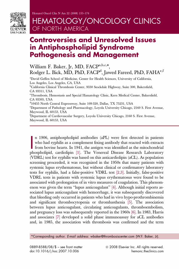
Hematol Oncol Clin N Am 22 (2008) 155–174
HEMATOLOGY/ONCOLOGY CLINICSOF NORTH AMERICA
Controversies and Unresolved Issuesin Antiphospholipid SyndromePathogenesis and Management
William F. Baker, Jr, MD, FACPa,b,c,*,Rodger L. Bick, MD, PhD, FACPd, Jawed Fareed, PhD, FAHAe,f
aDavid Geffen School of Medicine, Center for Health Sciences, University of California,Los Angeles, Los Angeles, CA, USAbCalifornia Clinical Thrombosis Center, 9330 Stockdale Highway, Suite 300, Bakersfield,CA 93311, USAcThrombosis, Hemostasis and Special Hamatology Clinic, Kern Medical Center, Bakersfield,CA 93305, USAd10455 North Central Expressway, Suite 109-320, Dallas, TX 75231, USAeDepartment of Pathology and Pharmacology, Loyola University Chicago, 2160 S. First Avenue,Maywood, IL 60153, USAfDepartment of Cardiovascular Surgery, Loyola University Chicago, 2160 S. First Avenue,Maywood, IL 60153, USA
In 1906, antiphospholipid antibodies (aPL) were first detected in patientswho had syphilis as a complement fixing antibody that reacted with extractsfrom bovine hearts. In 1941, the antigen was identified as the mitochondrial
phospholipid, cardiolipin [1]. The Venereal Disease Research Laboratory(VDRL) test for syphilis was based on this anticardiolipin (aCL). As populationscreening proceeded, it was recognized in the 1950s that many patients withsystemic lupus erythematosus, but without clinical or confirmatory laboratorytests for syphilis, had a false-positive VDRL test [2,3]. Initially, false-positiveVDRL tests in patients with systemic lupus erythematosus were found to beassociated with prolongation of in vitro measures of coagulation. This phenom-enon was given the term ‘‘lupus anticoagulant’’ [4]. Although initial reports as-sociated lupus anticoagulant with hemorrhage, it was subsequently discoveredthat bleeding only occurred in patients who had in vivo hypo-prothrombinemiaand significant thrombocytopenia or thrombasthenia [5]. The associationbetween lupus anticoagulant, circulating anticoagulants, thromboembolism,and pregnancy loss was subsequently reported in the 1960s [6]. In 1983, Harrisand associates [7] developed a solid phase immunoassay for aCL antibodiesand, in 1985, the association with thrombosis was confirmed and the term
*Corresponding author. E-mail address: [email protected] (W.F. Baker, Jr).
0889-8588/08/$ – see front matter ª 2008 Elsevier Inc. All rights reserved.doi:10.1016/j.hoc.2007.10.006 hemonc.theclinics.com
156 BAKER, BICK, & FAREED
antiphospholipid syndrome (APS) was coined [8]. Continuing investigationin the 1990s demonstrated that the plasma phospholipid-binding proteinbeta-2-glycoprotein (B-2-GP I) was necessary to bind to cardiolipin [9,10]. Thisrequirement was not found to be present in syphilis or in patients with infections[11]. It was shown that antibodies are formed not only to cardiolipin bound toB-2-GP I complexed with phospholipid but also to B-2-GP I directly [9,12].Investigation into the pathobiology of APS has also led to the identificationanti–annexin V antibodies [13], anti-prothrombin antibodies, and autoanti-bodies to the antiphospholipid subtypes (phosphatidylserine, phosphatidyletha-nolamine, phosphatidylglycerol, phosphatidylinositol, phosphatidylcholine, andphosphatidic acid) [4,14].
The presence of autoantibodies to various phospholipids and phospholipid-binding proteins has been identified in patients with large-vessel arterial andvenous thrombosis, microvascular thrombosis, placental thrombosis, anda variety of other manifestations. The consequences of these vascular occlu-sions are found to affect virtually every organ and tissue.
While much is understood concerning the clinical features of patients withAPS, many questions and controversial issues remain. There are many unan-swered questions regarding pathogenesis. The proper designation of patientswith ‘‘definite’’ APS and the correct categorization of patients by both labora-tory and clinical features are matters of ongoing debate. Recent proposals haveidentified new subsets of patients who have many typical features of APS buteither do not fit the criteria for a ‘‘definite’’ diagnosis [15] or who have initiallynegative laboratory tests for aPL [16]. The approach to diagnosis is also inquestion. Decisions about which laboratory test to order on which patientand which laboratory test to order when are based on expert opinion, ratherthan controlled trials. While many publications deal with treatment of APS,many issues remain unresolved. While many guidelines are offered, few arebacked by data from strong clinical trials. This article summarizes the clinicalquestions remaining to be answered (Box 1) and debates concerning pathogen-esis (Box 2), diagnosis (Box 3), and management (Box 4).
PATHOGENESISThe early laboratory investigations of the serum of patients with syphilis trig-gered a sequence of discoveries leading to much of the current understandingof APS. Case reports and a small series of patients with biologically false-positivetests for syphilis and later lupus anticoagulant and aCL antibodies were simplyreports of clinical associations with laboratory results. Intensive investigation hassubsequently attempted to precisely determine the molecular biology involvedwith both the laboratory phenomena and the clinical manifestations (see Box 2).
Debate has centered on whether aPL directly cause thrombosis, are a conse-quence of thrombosis, or are simply an epiphenomenon. aPL may also functionsimply as a ‘‘second hit,’’ both causing thrombosis and representing the resultof antibody-mediated cellular injury [28]. Exposed anionic phospholipids maylead to immunological recognition and the generation of additional antibodies
Box 1: APS: questions to be answered
Is the presence of aPL a primary risk factor for thrombosis?
Is the presence of aPL a primary risk factor for atherosclerosis?
Is the presence of aPL a primary risk factor for pregnancy loss?
Does the presence of asymptomatic aPL require measures to prevent a first throm-botic event? Does the benefit of such therapy outweigh the potential risks?
Is aPL associated with recurrent arterial or venous thrombosis or recurrent preg-nancy loss?
Is acute management for thrombosis in the presence of aPL different from suchmanagement in the absence of aPL?
Do aPL play a role in the development of cardioembolic strokes?
What is optimal management to prevent recurrent arterial and venous thrombosisin patients with APS?
If aPL become negative in a patient with a prior thrombotic event, should therapybe altered?
What is optimal type, duration, and intensity of antithrombotic therapy?
What are the best tests to order when APS is suspected?
How should patients with clinical manifestations of APS but negative laboratorytests for aPL be managed?
How should patients be managed who have clinical features of APS (‘‘pre-APS’’and microangiopathic phospholipid syndromes) and who test positive for aPL,but who do not fit the 2006 International Consensus criteria for APS?
157CONTROVERSIES IN ANTIPHOSPHOLIPID SYNDROME
[38]. Evidence has increasingly accumulated that the aPL are, in fact, directlyresponsible for vascular thrombosis [38].
A variety of experimental findings point to possible triggering events for theformation of autoantibodies to phospholipid and B-2-GP I [18,39]. These mech-anisms have included:
Overproduction of natural antibodies [17]Molecular mimicry resulting from infection [18]Exposure of phospholipids during platelet activation and cellular apoptosis (nor-
mally intracellular anionic phospholipids redistributed to the extracellularcompartment) [19]
Cardiolipin peroxidation [20]Platelet activation with or without antiplatelet antibodies [21]Genetic predisposition [22]
It is likely that a combination of pathologic interactions is necessary for thedevelopment of aPL. Once aPL are present, a complex cycle may develop suchthat apoptosis may elicit the formation of aPL but also then induce additionalapoptosis. Apoptosis may then cause oxidative cellular injury, which alsoinduces apoptosis. Pathologic activation of platelets and injury to endothelial
Box 2: Unresolved issues in pathogenesis
Triggers to the development of aPL
Overproduction of natural antibodies [17]
Molecular mimicry resulting from infection [18]
Exposure of phospholipids during platelet activation and cellular apoptosis (nor-mally intracellular anionic phospholipids redistributed to the extracellular com-partment) [19]
Cardiolipin peroxidation [20]
Platelet activation with or without antiplatelet antibodies [21]
Genetic predisposition [22]
Molecular mechanisms leading to thrombosis
Inhibition of endothelial cell prostacyclin production [23]
Procoagulant effect on platelets [21]
Impairment of fibrinolysis [24]
Interference with the thrombomodulin–protein S–protein C pathway [25]
Induction of procoagulant activity on endothelial cells and/or monocytes [26]
Disruption of the annexin V cellular shield [13]
Abnormal cytotrophoblast expression of adhesion molecules in pregnancy [27]
Is a ‘‘second hit’’ necessary for clinical thrombosis? [28]
158 BAKER, BICK, & FAREED
cells by aPL may result in exposure of anionic phospholipids and further stim-ulate the formation of aPL [40].
Proposed mechanisms of aPL-mediated thrombosis have included:
Inhibition of endothelial cell prostacyclin production [23]Procoagulant effect on platelets [21]Impairment of fibrinolysis [24]Interference with the thrombomodulin–protein S–protein C pathway [25]Induction of procoagulant activity on endothelial cells and/or monocytes [26]Disruption of the annexin V cellular shield [13]Abnormal cytotrophoblast expression of adhesion molecules in pregnancy [27]
While it is widely accepted that the presence of aPL is associated with throm-bosis, the importance of all of the cellular consequences and interactions result-ing from aPL is not clearly understood [1,39,41].
The importance of B-2-GP I in binding of aCL as a cofactor is well recog-nized. However, the mechanisms by which anti–B-2-GP I antibodies bindB-2-GP I are unclear [26]. It appears that B-2-GP I functions as a mild naturalinhibitor of contact activation of coagulation [42]. B-2-GP I inhibits plateletfunction [43,44], down-regulates the activation of Hageman factor [45] andthe contact phase of coagulation, prevents the binding of protein S by its
Box 3: Unresolved issues in diagnosis
Established criteria
1999 Sapporo International Consensus Statement on Preliminary Criteria for theClassification of the Antiphospholipid Syndrome [29]
2006 International Consensus Statement on an Update of the ClassificationCriteria for Definite Antiphospholipid Syndrome
Unresolved issues
Addition of a category for pre-antiphospholipid syndrome [15]
Addition of a category for microangiopathic phospholipid syndrome [15]
Criteria for a ‘‘definite’’ diagnosis amended to eliminate aCL and replace thiswith anti–B-2-GP I [30]
Addition of IgA to testing for aPL [31,32]
Addition of testing for anti-prothrombin antibodies [30]
Addition of testing for anti–annexin V antibodies in patients with recurrent preg-nancy loss [30]
Addition of testing for other aPL
Defining the ‘‘best’’ testing strategy to detect lupus anticoagulant
159CONTROVERSIES IN ANTIPHOSPHOLIPID SYNDROME
plasma inhibitor, C4b-binding protein [46], and inhibits tissue factor activity[47]. Interestingly, B-2-GP I deficiency is not associated with predispositionto thrombosis [48]. More recent investigation has implicated other activity ofB-2-GP I, including binding of factor XI and inhibition of the factor XI by fac-tor XIIa [40]. It appears that B-2-GP I may colocalize with factor XI and throm-bin on the platelet surface and regulate thrombin generation. Rahgozar andcolleagues [40] have demonstrated that anti–B-2-GP I antibodies actually poten-tiate the B-2-GP I–mediated inhibition of factor XI activation by thrombin, thusinhibiting coagulation, rather than inducing thrombosis. This finding, onceagain, leaves open the question of the pathogenesis of APS and the role ofB-2-GP I. Also, because a variety of experimental studies have used systemsthat do include the presence of B-2-GP I, it is possible that previous studiesmay have led to erroneous conclusions regarding APS pathogenesis [40].
Just as questions remain regarding the molecular and cellular effects of auto-antibodies to aPL and B-2-GP I, the relationship between the activation ofintrinsic coagulation and clinical thrombosis remains a subject of investigation.It may be that more than one mechanism is involved and that these are differ-ent in patients with different clinical manifestations [39]. Antibodies to a varietyof phospholipids and phospholipid-binding proteins now have been recognized,and each may be associated with unique clinical consequences. Although a hall-mark of thrombophilia is the propensity for both venous and arterial thrombo-sis in the same patient, patients with APS tend to have either recurrent arterialor venous events [49]. Arterial thrombosis is primarily driven by platelet
Box 4: Unresolved issues in management
Type of therapy: antiplatelet versus antithrombin agents [33]
The intensity of therapy: target level of international normalized ratio for warfarintherapy [1]
The most accurate method of confirming treatment efficacy: the ratio of prothrom-bin time (PT) to partial thromboplastin time (PTT) or other laboratory studies [34]
The duration of treatment: no therapy, time-limited therapy versus lifelong therapy[35]
Optimal management during pregnancy and delivery [36]
Optimal management of catastrophic APS [37]
Anticoagulation
Corticosteroid therapy
Plasma exchange with fresh frozen plasma
Intravenous immune globulin
Rituximab
160 BAKER, BICK, & FAREED
aggregation while venous thrombosis is driven by activation of the coagulationcascade. aPL procoagulant effects on platelets and endothelial cells may be themechanisms that trigger arterial events in APS patients. Meanwhile, othermechanisms, such as impairment of fibrinolysis, induction of the expressionof tissue factor, and interference with the thrombomodulin–protein S–proteinC systems, may be the predominant triggers for venous events in APS patients[39,40]. In addition, it remains unclear why some patients with aPL developthrombosis and others do not. Furthermore, the triggers for thrombosis inpatients with previously asymptomatic aPL are poorly understood.
In considering the pathobiology of APS, it is clear that much remains to beunderstood. There is debate concerning the triggering events, the molecularmechanisms, and the critical links between pathologic changes at the cellularlevel and clinical thrombosis. Because these critical questions remain unan-swered, precise diagnosis and therapy remain challenging and prognosticationfor the individual patient continues to be difficult.
DIAGNOSISBefore the 2006 publication of the International Consensus Statement on anUpdate of the Classification Criteria for Definite Antiphospholipid Syndrome[50], the diagnosis of ‘‘definite’’ APS was made by following the 1999 SapporoInternational Consensus Statement on Preliminary Criteria for the Classifica-tion of the Antiphospholipid Syndrome [29]. While the 2006 criteria representan improvement over the 1999 criteria, a variety of patient subsets still do notfit the strict 2006 criteria, as Asherson [15] has pointed out. The ‘‘pre-APS’’
161CONTROVERSIES IN ANTIPHOSPHOLIPID SYNDROME
(probable APS) group includes individuals who have such conditions as livedoreticularis, chorea, thrombocytopenia, fetal loss, and cardiac valve lesions, andwho do not fulfill all of the criteria for diagnosis but at a later time developthrombosis and are then diagnosed with APS. The patients who have microan-giopathic phospholipid syndromes include those with thrombotic microangiop-athy (thrombotic thrombocytopenic purpura, hemolytic uremic syndrome,HELLP [hemolysis, elevated liver enzyme, and low platelets] syndrome, andcatastrophic APS), and patients who have aPL without larger vessel thrombosis[15]. Patients with typical, life-threatening, catastrophic APS may also not fit thestrict 2006 International Consensus criteria.
The 2006 International Consensus Statement on an Update of the Classifi-cation Criteria for Definite Antiphospholipid Syndrome redefined the diagnos-tic criteria in light of recent studies. Outstanding issues addressed by thechanges from the 1999 to the 2006 criteria include the addition of anti–B-2-GP I antibodies to the laboratory criteria and the more precise definitionsregarding the maternal–fetal complications cited among the clinical criteria[50]. Special emphasis is also now given to the other risk factors that mayplay important roles in the propensity for thrombosis, such as atheroscleroticrisk factors and other thrombophilic disorders. Certainly, continuing attentionis required to update and possibly modify the criteria for a ‘‘definite’’ diagnosis.A variety of questions remain with respect to diagnosis of APS (see Box 3).
Current Laboratory Criteria for the diagnosis of APS include high titers ofaCL or B-2-GP I IgG or IgM antibodies or lupus anticoagulant present ontwo or more occasions 12 weeks apart [50]. Problems with the laboratory diag-nosis of APS remain [28]. Solid phase assays are widely available for the detec-tion and quantification of antibodies to aCL, B-2-GP I, prothrombin, annexinV, and phosphatidylserine, phosphatidylethanolamine, phosphatidylcholine,phosphatidylglycerol, phosphatidylinositol, and phosphatidic acid.
Unfortunately, results vary considerably among laboratories. The FirstFrench Anticardiolipin Standardization Workshop found an interlaboratorycoefficient of variation for aPL IgG of greater than 70% and a within-run coef-ficient of variation of 10% [51]. A study by Favoloro and Silvestrini [52] dem-onstrated an interlaboratory coefficient of variation for aCL IgG and IgM ofover 50%. Different laboratories frequently disagreed on whether the aCLwere positive or negative. General laboratory standards require interlaboratoryconsensus of 90% or more in most tests. This is clearly an area of concern inAPS testing and the primary basis for requiring repeat testing and agreementon two or more specimens for a ‘‘definite’’ diagnosis [29,50].
The laboratory diagnosis of lupus anticoagulant has proven challenging. Theprimary coagulation screening study for diagnosis is the activated partialthromboplastin time (aPTT). This phospholipid-based assay becomes pro-longed in the presence of a lupus anticoagulant. A mixing study is performedusing a 1:1 mix with normal plasma followed by prolonged incubation todetermine a correction in aPTT prolongation. In the presence of aPL, theaddition of normal plasma and 2 hours or more incubation time will fail to
162 BAKER, BICK, & FAREED
correct the aPTT prolongation. The addition of phospholipid usually correctsthe aPTT. Detection of ‘‘weak’’ lupus anticoagulant may be difficult withoutthe use of ‘‘platelet poor’’ normal plasma [28]. The dilute Russell viper venomtime (dRVVT) inhibits factor X directly and is not prolonged by the presenceof intrinsic factor deficiencies. Prolongation of the dRVVT is more sensitivethan the prolongation of the aPTT for detection of lupus anticoagulant [53].A variety of test systems have been developed to improve diagnostic accuracy.The highest sensitivity and specificity were found with the ratio of dRVVT todRVVT with high lipid concentration [54]. Because of the many pitfalls indiagnosis, a laboratory decision tree is recommended. Further investigationof a prolonged aPTT is required, with mixing studies, dRVVT, and plateletneutralization procedures. Criteria for the diagnosis of lupus anticoagulantare well established [55] and are included in the 2006 International ConsensusCriteria [29,50]. When the diagnosis remains in question, additional testingmay be useful. One such test is the hexagonal phase phospholipid neutraliza-tion assay [28]. Close correlation between laboratory testing and clinical fea-tures is required for accurate diagnosis [56]. Newer approaches to laboratorydiagnosis continue to evolve [57].
In patients who present with a history of thrombosis or recurrent miscar-riage and who fulfill laboratory criteria for the diagnosis of aPL, there is nocontroversy regarding the diagnosis. Patients who test positive for aPL andwho do not have a history of thrombosis that fits the 2006 International Con-sensus criteria [29,50] present another challenge. These patients may be testedbecause of symptoms consistent with pre-APS or microangiopathic phospho-lipid syndromes, as discussed earlier [15]. Patients may also present with clin-ical features typical of APS but fail to fulfill the laboratory criteria fora diagnosis of definite APS, or may test positive for aPL but lack clinical criteriafor a definite diagnosis. The 2006 criteria addressed many of these patient sub-sets by specifically defining categories for patients with cardiac valvular diseaseand aPL, livedo reticularis with aPL, nephropathy with aPL, and thrombocy-topenia with aPL. Patients in these groups share clinical features and test pos-itive for aPL, but do not fulfill the 2006 criteria for a definite diagnosis of APS[50].
Because laboratory testing provides less than optimal sensitivity and specific-ity and is potentially misleading, testing is only indicated in patients suspectedof having APS on clinical grounds. Repeat testing is required to make a ‘‘defi-nite’’ diagnosis. Thus, any positive test results should be repeated two or moretimes 12 weeks apart [50]. Additional confirmatory studies may be indicated inpatients with equivocal results, especially in attempting to confirm the sus-pected diagnosis of lupus anticoagulant. Just as other indices of autoimmunity,the levels of aPL and lupus anticoagulant may vary. Titers may be initially neg-ative and later positive and low titers may become higher or high titers latermay become lower [16,58]. There remains no consensus on whether laterrepeat testing should be performed and whether the results of such repeat stud-ies should affect therapy.
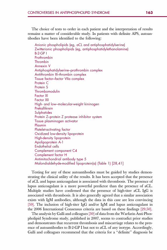
163CONTROVERSIES IN ANTIPHOSPHOLIPID SYNDROME
The choice of tests to order in each patient and the interpretation of resultsremains a matter of considerable study. In patients with definite APS, autoan-tibodies have been identified to the following:
Anionic phospholipids (eg, aCL and antiphosphatidylserine)Zwitterionic phospholipids (eg, antiphosphatidylethanolamine)B-2-GP IProthrombinThrombinAnnexin VAntiphosphatidylserine–prothrombin complexAntithrombin III–thrombin complexTissue factor–factor VIIa complexProtein CProtein SThrombomodulinFactor XIFactor XIIHigh- and low–molecular-weight kininogenPrekallikreinSulphatidesProtein Z–protein Z protease inhibitor systemTissue plasminogen activatorPlasminPlatelet-activating factorOxidized low-density lipoproteinHigh-density lipoproteinApolipoprotein A-1Endothelial cellsComplement component C4Complement factor HAntimitochondrial antibody type 5Malondialdehyde-modified lipoprotein(a) (Table 1) [28,41]
Testing for any of these autoantibodies must be guided by studies demon-strating the clinical utility of the results. It has been accepted that the presenceof aCL and lupus anticoagulant is associated with thrombosis. The presence oflupus anticoagulant is a more powerful predictor than the presence of aCL.Multiple studies have confirmed that the presence of high-titer aCL IgG isassociated with thrombosis. It is also generally agreed that a similar associationexists with IgM antibodies, although the data in this case are less convincing[59]. The inclusion of high-titer IgG and/or IgM and lupus anticoagulant inthe 2006 International Consensus criteria are based on these findings [29,50].
The analysis by Galli and colleagues [30] of data from the Warfarin Anti-Phos-pholipid Syndrome study, published in 2007, seems to contradict prior studiesand demonstrates that recurrent thrombosis and miscarriage relates to the pres-ence of autoantibodies to B-2-GP I but not to aCL of any isotype. Accordingly,Galli and colleagues recommend that the criteria for a ‘‘definite’’ diagnosis be
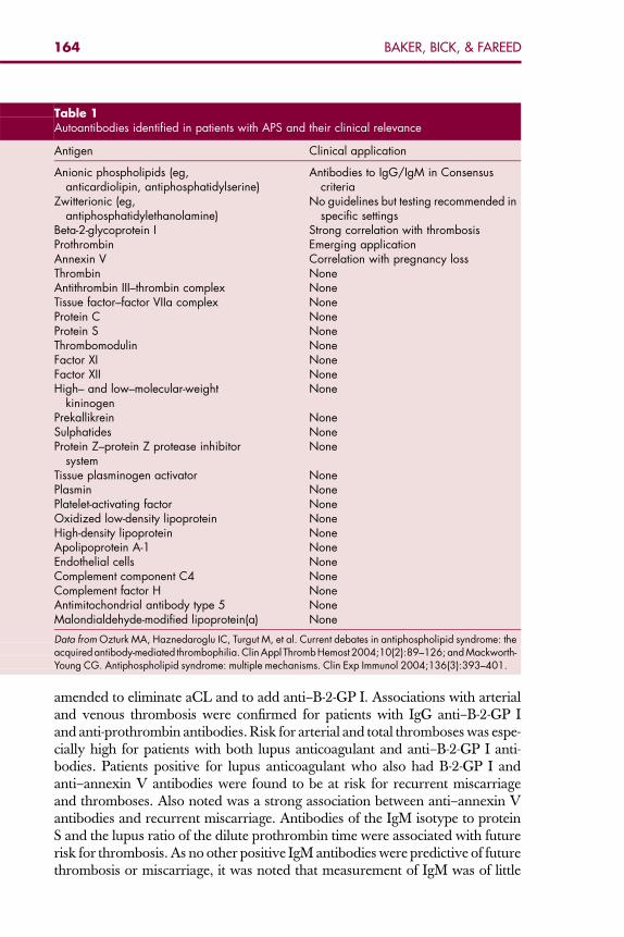
Table 1Autoantibodies identified in patients with APS and their clinical relevance
Antigen Clinical application
Anionic phospholipids (eg,anticardiolipin, antiphosphatidylserine)
Antibodies to IgG/IgM in Consensuscriteria
Zwitterionic (eg,antiphosphatidylethanolamine)
No guidelines but testing recommended inspecific settings
Beta-2-glycoprotein I Strong correlation with thrombosisProthrombin Emerging applicationAnnexin V Correlation with pregnancy lossThrombin NoneAntithrombin III–thrombin complex NoneTissue factor–factor VIIa complex NoneProtein C NoneProtein S NoneThrombomodulin NoneFactor XI NoneFactor XII NoneHigh– and low–molecular-weight
kininogenNone
Prekallikrein NoneSulphatides NoneProtein Z–protein Z protease inhibitor
systemNone
Tissue plasminogen activator NonePlasmin NonePlatelet-activating factor NoneOxidized low-density lipoprotein NoneHigh-density lipoprotein NoneApolipoprotein A-1 NoneEndothelial cells NoneComplement component C4 NoneComplement factor H NoneAntimitochondrial antibody type 5 NoneMalondialdehyde-modified lipoprotein(a) None
Data from Ozturk MA, Haznedaroglu IC, Turgut M, et al. Current debates in antiphospholipid syndrome: theacquiredantibody-mediated thrombophilia.Clin Appl ThrombHemost 2004;10(2):89–126; and Mackworth-Young CG. Antiphospholipid syndrome: multiple mechanisms. Clin Exp Immunol 2004;136(3):393–401.
164 BAKER, BICK, & FAREED
amended to eliminate aCL and to add anti–B-2-GP I. Associations with arterialand venous thrombosis were confirmed for patients with IgG anti–B-2-GP Iand anti-prothrombin antibodies. Risk for arterial and total thromboses was espe-cially high for patients with both lupus anticoagulant and anti–B-2-GP I anti-bodies. Patients positive for lupus anticoagulant who also had B-2-GP I andanti–annexin V antibodies were found to be at risk for recurrent miscarriageand thromboses. Also noted was a strong association between anti–annexin Vantibodies and recurrent miscarriage. Antibodies of the IgM isotype to proteinS and the lupus ratio of the dilute prothrombin time were associated with futurerisk for thrombosis. As no other positive IgM antibodies were predictive of futurethrombosis or miscarriage, it was noted that measurement of IgM was of little
165CONTROVERSIES IN ANTIPHOSPHOLIPID SYNDROME
value. The measurement of anti–annexin V antibodies and anti-prothrombin an-tibodies was recommended in certain situations [30].
Another issue has been whether aCL of the IgA isotype are associated withthrombosis. Some studies have noted an association [60], while others have not[61]. aCL IgA has not been measured routinely and debate continues as to theclinical relevance of an elevated titer [31]. It is not generally recommended thatthe diagnostic criteria be expanded to include the IgA isotype. However, thisremains a topic of debate. Bick [32] have recommended including the measure-ment of IgA in the laboratory assessment for aCL. The study by Selva and col-leagues [62] of 795 patients, however, demonstrated aCL IgA positivity in only2 patients, both also positive for IgG aCL. The analysis contained in the Miya-kis [50] summary of the 2006 criteria notes that testing for IgA is primarily use-ful for identification of APS subgroups, such as African Americans withsystemic lupus erythematosus and patients with connective tissue disease asso-ciated with thrombocytopenia, skin ulcers, and cutaneous vasculitis.
Laboratory testing for anti-prothrombin antibodies has not routinely beenrecommended in the evaluation of patients with suspected thrombophilia.The prevalence of anti-prothrombin antibodies in patients with APS hasbeen reported from 50% to 90% of patients [63]. Some studies have demon-strated an association with thrombosis [64,65] and other studies have not[66]. Analysis of the Warfarin Anti-Phospholipid Syndrome study data sug-gests a potential role for anti-prothrombin testing [30].
Anti–annexin V is a tissue and circulating phospholipid-binding protein withhigh affinity for anionic phospholipids and functions as an anticoagulant bydisplacing coagulation factors from phospholipid surfaces [28]. Autoantibodiesto annexin V are reported in a variety of clinical settings, including those in-volving patients with recurrent thrombosis and miscarriage. In patients withsystemic lupus erythematosus, the finding of anti–annexin V IgG antibodieswas reported to be associated with a high incidence of arterial or venous throm-bosis, prolonged aPTT, and recurrent miscarriage [67]. There has been, how-ever, conflicting data regarding the prognostic value of anti–annexin Vantibodies [68]. The data from the Warfarin Anti-Phospholipid Syndromestudy provide stronger evidence for the use of anti–annexin V antibodies asa predictor of future thrombosis or miscarriage [30].
There remains a lack of consensus regarding the utility of testing for otheranionic and zwitterionic aPL, often referred to as ‘‘subtypes.’’ These includephosphatidylserine, phosphatidylethanolamine, phosphatidylcholine, phospha-tidylglycerol, phosphatidylinositol, and phosphatidic acid. Studies have demon-strated association with thrombosis for antiphosphatidylserine [69] andantiphosphatidylethanolamine [70]. Some investigators have noted that these‘‘subtypes’’ may be present in patients without detectable aCL and have advo-cated testing for them [70,71]. Other investigators have advised that testing forthese noncardiolipin phospholipid antibodies is not indicated [72].
The laboratory measurement of autoantibodies in patients with suspectedAPS remains a subject of research. Review of the literature and clinical practice
166 BAKER, BICK, & FAREED
make it clear that, lacking consensus, a variety of approaches to diagnosis maybe used. Generally, a panel of studies is ordered, including the aPTT with mix-ing study and dRVVT in search of lupus anticoagulant, and IgG, IgM, andpossibly IgA assays for aCL. Assessment for B-2-GP I antibodies should be rou-tinely included in the diagnostic panel. In addition, it appears that to this shouldbe added anti–annexin V antibodies, especially in patients with recurrent mis-carriage. While broad recommendations are not yet available, it appears thatassay for anti-prothrombin antibodies may also soon be included. The inclu-sion of analysis for other aPL and for other antibodies associated with APSremains controversial and the subject for further investigation.
MANAGEMENTThe management of patients with APS has proven challenging and the subjectof numerous studies. Generally, treatment may be divided into the followingseveral categories:
Management of acute large-vessel thrombosisManagement of ‘‘pre-APS’’ (probable APS) and microangiopathic APSManagement of catastrophic APSManagement during pregnancyPrevention of initial and recurrent thromboses
Bick [32] has divided APS into 6 subgroups. Treatment guidelines have beenoffered for each group. Well-controlled trials have not, however, verified theserecommendations. Many clinical trials support guidelines and expert recom-mendations. However, in the area of treatment, few large, controlled trialshave taken place. Debate and controversy surround the questions of thetype of therapy, the intensity of therapy, the most accurate method of confirm-ing treatment efficacy, the duration of treatment, and how best to manage spe-cific clinical circumstances, such as surgery and delivery (see Table 1).
Intervention with anticoagulation is the primary treatment for any patientpresenting with acute large-vessel venous or arterial thrombosis. There is a dif-ference, however, between patients with venous versus those with arterialdisease. As discussed earlier, in patients with arterial thrombosis, platelet aggre-gation plays a central role. If the patient has not previously been diagnosed withAPS, the mainstay of therapy for arterial thrombosis is usually antiplatelet ther-apy. The patient with APS, however, requires the addition of anticoagulationtherapy with unfractionated or low–molecular-weight heparin followed by war-farin anticoagulation [1]. Patients with acute myocardial infarction and APShave been treated successfully with thrombolytic therapy [73] and also intra-coronary stents, followed by anticoagulation [74]. At issue has been how longto treat patients and with what intensity of anticoagulation [75].
Patients with acute stroke, the most common arterial thromboembolic disor-der in patients with APS, are managed initially based on clinical criteria. Itappears that whether or not the diagnosis of APS is known before presentation
167CONTROVERSIES IN ANTIPHOSPHOLIPID SYNDROME
with new neurologic symptoms, therapy should be guided by well-establishedcriteria for stroke management. If therapy can be provided within 3 hours ofonset and all inclusion criteria are met, thrombolytic therapy may be indicated[76]. There are, however, no large trials of thrombolytic therapy in patientswith APS, whether known at the time of the stroke onset or diagnosed at a latertime. Post-stroke therapy also remains a subject of investigation and it is notclear what optimum therapy should be [77]. The data from the Antiphospho-lipid Antibody in Stroke Study Group subset of the Warfarin Versus AspirinRecurrent Stroke Study demonstrate, contrary to other studies, that there isno difference between aspirin and warfarin in prevention of recurrent stroke[78]. Similar conclusions have been reached in other trials [79,80].
The treatment of acute venous thromboembolism follows well-establishedguidelines and is independent of the diagnosis of APS [81]. Anticoagulationwith low–molecular-weight heparin is favored over unfractionated heparinand should be administered in a full anticoagulation dose. Anticoagulationwith warfarin should also be initiated with the dose adjusted to achieve a ther-apeutic international normalized ratio (INR) of at least 2.0 to 2.9.
Prevention of recurrent thrombosis after acute arterial or venous thrombosis isthe subject of considerable investigation and unresolved issues remain. Three ret-rospective studies have demonstrated that the rate of recurrent thrombosis is re-duced by warfarin anticoagulation. In the small series by Derksen and colleagues[82], the recurrence rate at 8 years was 0% for patients anticoagulated and the re-currence rate for patients in whom therapy was stopped was 50% at 2 years and78% at 8 years. Recurrence also correlates with the intensity of anticoagulation. Ithas been well established that patients with an INR of less than 1.9 are vulnerableto recurrent thrombosis [49]. The rate of recurrence in patients without optimalanticoagulation is as high as 70% [49,82]. It remains unclear whether high-inten-sity warfarin therapy is of greater benefit. The report of Crowther and colleagues[83] compared intermediate therapy (2.0 to 3.0) and high-intensity therapy (3.0 to4.0) in a carefully controlled setting. The benefits and bleeding risks for both treat-ment groups were essentially the same. Current guidelines recommend interme-diate levels of anticoagulation and care to avoid subtherapeutic doses [1]. Theappropriate duration of therapy is also unclear. Patients with venous thromboem-bolism and APS who discontinue therapy have a high risk of recurrence. Ther-apy, however, is accompanied by a risk of hemorrhage [35]. Patients withvenous or arterial thrombotic events and aPL may require lifelong therapy. Mon-itoring warfarin presents additional challenges as aPL may interfere with theaPTT and prothrombin time test system phospholipid substrate [34].
Aspirin alone may be inadequate to prevent recurrence [33]. Patients witharterial thrombosis and APS may benefit more than patients with venous throm-bosis from the addition of antiplatelet therapy. Caution is required, however, inview of the increased risk of hemorrhage associated with such dual therapy.Patients who are not candidates for aspirin therapy may benefit from substitu-tion with clopidogrel [32]. In addition, therapy is complicated by the potentialpresence of aspirin and/or clopidrogrel resistance [36,84].
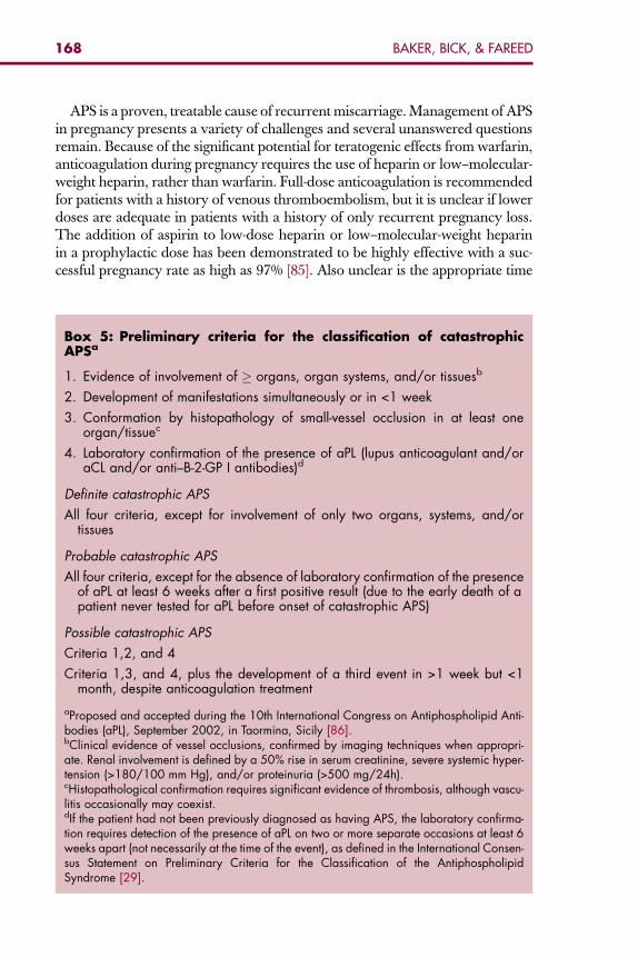
168 BAKER, BICK, & FAREED
APS is a proven, treatable cause of recurrent miscarriage. Management of APSin pregnancy presents a variety of challenges and several unanswered questionsremain. Because of the significant potential for teratogenic effects from warfarin,anticoagulation during pregnancy requires the use of heparin or low–molecular-weight heparin, rather than warfarin. Full-dose anticoagulation is recommendedfor patients with a history of venous thromboembolism, but it is unclear if lowerdoses are adequate in patients with a history of only recurrent pregnancy loss.The addition of aspirin to low-dose heparin or low–molecular-weight heparinin a prophylactic dose has been demonstrated to be highly effective with a suc-cessful pregnancy rate as high as 97% [85]. Also unclear is the appropriate time
Box 5: Preliminary criteria for the classification of catastrophicAPSa
1. Evidence of involvement of � organs, organ systems, and/or tissuesb
2. Development of manifestations simultaneously or in <1 week
3. Conformation by histopathology of small-vessel occlusion in at least oneorgan/tissuec
4. Laboratory confirmation of the presence of aPL (lupus anticoagulant and/oraCL and/or anti–B-2-GP I antibodies)d
Definite catastrophic APS
All four criteria, except for involvement of only two organs, systems, and/ortissues
Probable catastrophic APS
All four criteria, except for the absence of laboratory confirmation of the presenceof aPL at least 6 weeks after a first positive result (due to the early death of apatient never tested for aPL before onset of catastrophic APS)
Possible catastrophic APS
Criteria 1,2, and 4
Criteria 1,3, and 4, plus the development of a third event in >1 week but <1month, despite anticoagulation treatment
aProposed and accepted during the 10th International Congress on Antiphospholipid Anti-bodies (aPL), September 2002, in Taormina, Sicily [86].bClinical evidence of vessel occlusions, confirmed by imaging techniques when appropri-ate. Renal involvement is defined by a 50% rise in serum creatinine, severe systemic hyper-tension (>180/100 mm Hg), and/or proteinuria (>500 mg/24h).cHistopathological confirmation requires significant evidence of thrombosis, although vascu-litis occasionally may coexist.dIf the patient had not been previously diagnosed as having APS, the laboratory confirma-tion requires detection of the presence of aPL on two or more separate occasions at least 6weeks apart (not necessarily at the time of the event), as defined in the International Consen-sus Statement on Preliminary Criteria for the Classification of the AntiphospholipidSyndrome [29].
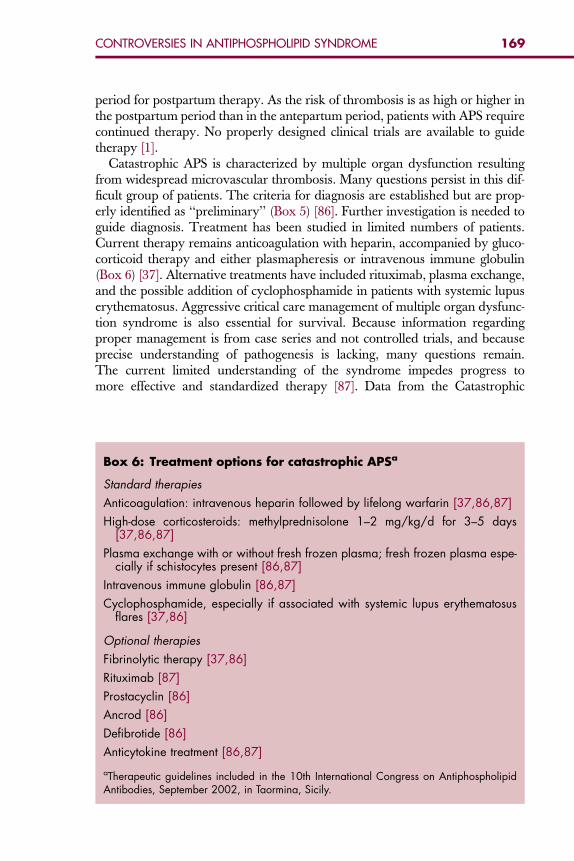
169CONTROVERSIES IN ANTIPHOSPHOLIPID SYNDROME
period for postpartum therapy. As the risk of thrombosis is as high or higher inthe postpartum period than in the antepartum period, patients with APS requirecontinued therapy. No properly designed clinical trials are available to guidetherapy [1].
Catastrophic APS is characterized by multiple organ dysfunction resultingfrom widespread microvascular thrombosis. Many questions persist in this dif-ficult group of patients. The criteria for diagnosis are established but are prop-erly identified as ‘‘preliminary’’ (Box 5) [86]. Further investigation is needed toguide diagnosis. Treatment has been studied in limited numbers of patients.Current therapy remains anticoagulation with heparin, accompanied by gluco-corticoid therapy and either plasmapheresis or intravenous immune globulin(Box 6) [37]. Alternative treatments have included rituximab, plasma exchange,and the possible addition of cyclophosphamide in patients with systemic lupuserythematosus. Aggressive critical care management of multiple organ dysfunc-tion syndrome is also essential for survival. Because information regardingproper management is from case series and not controlled trials, and becauseprecise understanding of pathogenesis is lacking, many questions remain.The current limited understanding of the syndrome impedes progress tomore effective and standardized therapy [87]. Data from the Catastrophic
Box 6: Treatment options for catastrophic APSa
Standard therapies
Anticoagulation: intravenous heparin followed by lifelong warfarin [37,86,87]
High-dose corticosteroids: methylprednisolone 1–2 mg/kg/d for 3–5 days[37,86,87]
Plasma exchange with or without fresh frozen plasma; fresh frozen plasma espe-cially if schistocytes present [86,87]
Intravenous immune globulin [86,87]
Cyclophosphamide, especially if associated with systemic lupus erythematosusflares [37,86]
Optional therapies
Fibrinolytic therapy [37,86]
Rituximab [87]
Prostacyclin [86]
Ancrod [86]
Defibrotide [86]
Anticytokine treatment [86,87]
aTherapeutic guidelines included in the 10th International Congress on AntiphospholipidAntibodies, September 2002, in Taormina, Sicily.
170 BAKER, BICK, & FAREED
Antiphospholipid Syndrome registry may, in time, provide more reliable treat-ment guidelines [87].
No one yet has identified the best therapy for patients with ‘‘pre-APS’’ andmanifestations of microangiopathic APS [15]. Trials have not addressed thequestion of whether some type of prophylactic therapy is indicated beforea thrombotic event occurs and before the patient truly fulfills the criteria fora ‘‘definite’’ diagnosis according to the 2006 International Consensus Statement[29,50]. Considering the risk of a first thrombotic event in patients with theseproblematic subsets, some type of antiplatelet or anticoagulation therapy maybe indicated. This should be the subject of future investigation [15].
SUMMARYAn overview of the subject of APS provides clear evidence that much remainsto be understood. Questions remain regarding the initial triggering events andthe pathogenetic mechanisms leading to thrombosis. Limited understanding ofpathogenesis has also resulted in considerable uncertainty concerning the useand clinical relevance of laboratory tests for aPL and antiphospholipid bindingproteins. The widely accepted 2006 International Consensus Criteria [29] arenow challenged in light of recent analysis of trial data, which contradicts earlierstudies [30]. Treatment guidelines are not well supported by clinical trial data inmany areas of management. Significant debate remains regarding importantissues, such as the type, intensity, and duration of anticoagulation therapy.Finally, management of catastrophic APS is based on case series and muchmore information is needed for the development of strong, evidence-basedrecommendations.
References
[1] Levine JS, Branch DW, Rauch J. The antiphospholipid syndrome. N Engl J Med2002;346(10):752–63.[2] Haserick J, Long R. Systemic lupus erythematosus preceded by false-positive serologic tests
for syphillis: presentation of five cases. Ann Intern Med 1952;37:559–65.[3] Moore J, Mohr C. Biologically false positive tests for syphillis: type, incidence, and cause.
JAMA 1952;150:467.[4] Rand JH. The antiphospholipid syndrome. In: Beutler E, editor. Williams hematology. 6th
edition. New York: McGraw-Hill; 2001. p. 1715–33.[5] Conley C, Hartmann R. A hemorrhagic disorder caused by circulating anticoagulant in
patients with disseminated lupus erythemasosus. J Clin Invest 1952;31:621.[6] Bowie W, Thompson J, Pascuzzi C, et al. Thrombosis in systemic lupus erythematosus
despite circulating anticoagulants. J Clin Invest 1963;62:416.[7] Harris EN, Gharavi A, Boey M, et al. Anticardiolipin antibodies: detection by radioimmu-
noassay and association with thrombosis in systemic lupus erythematosus. Lancet1983;2:1211–4.
[8] Hughes G. The anticardiolipin syndrome. Clin Exp Rheumatol 1985;3:285.[9] Galli M, Comfurius P, Barbui T, et al. Anticoagulant activity of B2-glycoprotein-I is potenti-
ated by a distinct subgroup of anticardiolipin antibodies. Thromb Haemost 1992;68:297–300.
171CONTROVERSIES IN ANTIPHOSPHOLIPID SYNDROME
[10] McNeil H, Simpson R, Chesterman C, et al. Anti-phospholipid antibodies are directedagainst a complex antigen that includes a lipid-binding inhibitor of coagulation: B2-glycoprotein I (apolipoprotein H). Proc Natl Acad Sci USA 1990;87:4120–4.
[11] Hunt J, McNeil H, Morgan G, et al. A phospholipid-beta 2-glycoprotein I complex is anantigen for anticardiolipin antibodies occurring in autoimmune disease but not with infec-tion. Lupus 1992;1:75–81.
[12] Arvieux J, Roussel B, Jacob MC, et al. Measurement of anti-phospholipid antibodies by ELI-SA using beta 2-glycoprotein I as an antigen. J Immunol Methods 1991;143:223–9.
[13] Hanly J, Smith S. Anti-beta2-glycoprotein I (GPI) autoantibodies annexin V binding and theanti-phospholipid syndrome. Clin Exp Immunol 2000;120:537.
[14] Bick R, Baker W. Antiphospholipid syndrome and thrombosis. Semin Thromb Hemost1999;25:333–50.
[15] Asherson RA. New subsets of the antiphospholipid syndrome in 2006: ‘‘PRE-APS’’ (proba-ble APS) and microangiopathic antiphospholipid syndromes (‘‘MAPS’’). Autoimmun Rev2006;6(2):76–80.
[16] Miret C, Cervera R, Reverter JC, et al. Antiphospholipid syndrome without antiphospholipidantibodies at the time of the thrombotic event: transient ‘‘seronegative’’ antiphospholipidsyndrome? Clin Exp Rheumatol 1997;15:541.
[17] Arnout J, Vermylen J. Current status and implications of autoimmune antiphospholipidantibodies in relation to thrombotic disease. J Thromb Haemost 2003;1:931.
[18] Gharavi A, Pierangeli S, Harris EN. Origin of antiphospholipid antibodies. Rheum Dis ClinNorth Am 2001;27:551.
[19] Rauch J, Subang R, D’Agnillo P, et al. Apoptosis and the antiphospholipid syndrome. J Auto-immun 2000;15:231.
[20] Horkko S, Miller E, Dudl E, et al. Antiphospholipid antibodies are directed against epitopesof oxidized phospholipids. Recognition of cardiolipin by monoclonal antibodies to epitopesof oxidized low density lipoprotein. J Clin Invest 1996;98:815.
[21] Walenga J, Michal K, Hoppensteadt D, et al. Vascular damage correlates between heparin-induced thrombocytopenia and the antiphospholipid syndrome. Clin Appl Thromb Hemost1999;5(Suppl 1):S76.
[22] Hellan M, Kuhnel E, Speiser W, et al. Familial lupus anticoagulant: a case report and reviewof the literature. Blood Coagul Fibrinolysis 1998;9:195.
[23] Pierangeli S, Gharavi A, Harris EN. Experimental thrombosis and antiphospholipidantibodies: new insights. J Autoimmun 2000;15:241.
[24] Takeuchi R, Atsumi T, Ieko M, et al. Suppressed intrinsic fibrinolytic activity by monoclonalanti-beta-2 glycoprotein I autoantibodies: possible mechanism for thrombosis in patientswith antiphospholipid syndrome. Br J Haematol 2002;119:781.
[25] Tsakiris DA, Settas L, PM, et al. Lupus anticoagulant-antiphospholipid antibodies andthrombophilia. Relation to protein C-protein S-thrombomodulin. J Rheumatol 1990;17:785.
[26] Koike T, Bohgaki M, Amengual O, et al. Antiphospholipid antibodies: lessons from thebench. J Autoimmun 2007;28(2–3):129–33.
[27] Di Simone N, Castellani R, Caliandro D, et al. Antiphospholipid antibodies regulate theexpression of trophoblast cell adhesion molecules. Fertil Steril 2002;77:805.
[28] Ozturk MA, Haznedaroglu IC, Turgut M, et al. Current debates in antiphospholipidsyndrome: the acquired antibody-mediated thrombophilia. Clin Appl Thromb Hemost2004;10(2):89–126.
[29] Wilson W, Gharavi A, Koike T, et al. International consensus statement on preliminaryclassification criteria for definite antiphospholipid syndrome: report of an internationalworkshop. Arthritis Rheum 1999;42:1309–11.
[30] Galli M, Borrelli G, Jacobsen EM, et al. Clinical significance of different antiphospholipidantibodies in the WAPS (Warfarin in the Anti-Phospholipid Syndrome) study. Blood2007;110(4):1178–85.
172 BAKER, BICK, & FAREED
[31] Carreras LO, Forastiero RR, Martinuzzo M. Which are the best biological markers of theantiphospholipid syndrome? J Autoimmun 2000;15:163.
[32] Bick R. Antiphospholipid thrombosis syndromes. Hematol Oncol Clin North Am 2003;17:115.
[33] Khamashta M, Cuadrado M, Mujic F, et al. The management of thrombosis in the antiphos-pholipid-antibody syndrome. N Engl J Med 1995;332:993–7.
[34] Moll S, Ortel T. Monitoring warfarin therapy in patients with lupus anticoagulants. Ann In-tern Med 1997;127:177–85.
[35] Baker RI. Prevention of recurrent thrombosis in the antiphospholipid antibody syndrome:how long and how high with oral anticoagulant therapy? Med J Aust 2004;180(9):436–7.
[36] Snoep JD, Hovens MM, Eikenboom JC, et al. Association of laboratory-defined aspirin re-sistance with a higher risk of recurrent cardiovascular events: a systemic review andmeta-analysis. Arch Intern Med 2007;167(15):1593–9.
[37] Kitchens C. Thrombotic storm: when thrombosis begets thrombosis. Am J Med 1998;104:381–5.
[38] Rand JH. Molecluar pathogenesis of the antiphospholipid syndrome. Circ Res 2002;90:29.[39] Gharavi AE, Wilson W, Pierangeli S. The molecular basis of antiphospholipid syndrome.
Lupus 2003;12(8):579–83.[40] Rahgozar S, Yang Q, Giannakopoulos B, et al. Beta2-glycoprotein I binds thrombin via
exosite I and exosite II: anti-beta2-glycoprotein I antibodies potentiate the inhibitory effectof beta2-glycoprotein I on thrombin-mediated factor XIa generation. Arthritis Rheum2007;56(2):605–13.
[41] Mackworth-Young CG. Antiphospholipid syndrome: multiple mechanisms. Clin Exp Immu-nol 2004;136(3):393–401.
[42] Schousboe I. B2-Glycoprotein I: a plasma inhibitor of the contact activation of the bloodcoagulation pathway. Blood 1985;66:1086–91.
[43] Nimpf J, Bevers E, Bomans P, et al. Prothrombinase activity of human platelets is inhibited bybeta2-glycoprotein-I. Biochim Biophys Acta 1986;884:142.
[44] Nimpf J, Wurm H, Kostner G. Beta 2-glycoprotein-I (apo-H) inhibits the release reaction ofhuman platelets during ADP-induced aggregation. Atherosclerosis 1987;63:109.
[45] Henry M, Everson B, Ratnoff D. Inhibition of the activation of Hageman factor (factor XII) bybeta2-glycoprotein I. J Lab Clin Med 1988;111:519.
[46] Merrill J, Zhang H, Shen C, et al. Enhancement of protein S anticoagulant function by beta2-glycoprotein I, amajor target antigenof antiphospholipidantibodies: interferes with bindingof protein S to its plasma inhibitor, C4b-binding protein. Thromb Haemost 1999;81:748.
[47] Ieko M, Yasokouchi T, Sawada K, et al. The influence of beta2-glycoprotein I on tissue factoractivity. Semin Thromb Hemost 1998;24:211.
[48] Bancsi L, van der Linden I, Bertina R. Beta 2 glycoprotein I deficiency and the risk of throm-bosis. Thromb Haemost 1992;67:649.
[49] Rosove M, Brewer P. Antiphospholipid thrombosis: clinical course after the first thromboticevent in 70 patients. Ann Intern Med 1992;117:303–8.
[50] Miyakis S. International consensus statement on an update of the classification criteria fordefinite antiphospholipid syndrome (APS). J Thromb Haemost 2006;4:295–306.
[51] Abuaf N, Meyer O, Laperche S, et al. Conclusions of the first French workshop of standard-ization of anticardiolipin antibody determination associated with autoimmune pathology.Ann Biol Clin (Paris) 1994;52:365.
[52] Favaloro E, Silvestrini R. Assessing the usefulness of anticardiolipin antibody assays: a cau-tious approach is suggested by high variation and limited consensus in multilaboratory test-ing. Am J Clin Pathol 2002;118:548.
[53] Gerbutavicus R, Fareed J, Messmore H, et al. Reference ranges of the dilute tissue thrombo-plastin inhibition and dilute Russell’s viper venom titer tests revisited. Clin Appl ThrombHemost 2002;8:51.
173CONTROVERSIES IN ANTIPHOSPHOLIPID SYNDROME
[54] Ruiz-Irastorza G, Egurbide MV, Ugalde J, et al. High impact of antiphospholipid syndromeon irreversible organ damage and survival of patients with systemic lupus erythematosus.Arch Intern Med 2004;164(1):77–82.
[55] Brandt J, Triplett DA, Alving B, et al. Criteria for the diagnosis of lupus anticoagulants: anupdate. Thromb Haemost 1995;74:1185–90.
[56] Wasmuth J, Minarro D, Homrighausen A, et al. Phospholipid autoantibodies and the anti-phospholipid antibody syndrome: diagnostic accuracy of 23 methods studied by variationin ROC curves with number of clinical manifestations. Clin Chem 2002;48:1004.
[57] Passam F, Krilis S. Laboratory tests for the antiphospholipid syndrome: current concepts.Pathology 2004;36(2):129–38.
[58] Silver RM, Pierangeli S, Edwin SS, et al. Pathogenic antibodies in women with obstetricfeatures of antiphospholipid syndrome who have negative test results for lupus anticoagu-lant and anticardiolipin antibodies. Obstet Gynecol 1997;176:628.
[59] Galli M, Luciani D, Bertolini G, et al. Lupus anticoagulants are stronger risk factors for throm-bosis than anticardiolipin antibodies in the antiphospholipid syndrome: a systematic reviewof the literature. Blood 2003;101:1827.
[60] Diri E, Cucurull E, Gharavi A, et al. Antiphospholipid (Hughes’) syndrome in African-Americans: IgA aCL and abeta2 glycoprotein-I is the most frequent isotype. Lupus1999;8:263.
[61] Bertolaccini ML, Atsumi T, Escudero Contreras A, et al. The value of IgA antiphospholipidtesting for diagnosis of the antiphospholipid (Hughes) syndrome in systemic lupus erythema-tosus. J Rheumatol 2001;28:2637.
[62] Selva O, Ordi R, Monegal F, et al. Anticardiolipin antibodies-relation with other antiphos-pholipid antibodies and clinical significance. Thromb Haemost 1998;79:282.
[63] Galli M. Should we include anti-prothrombin antibodies in the screening for the antiphos-pholipid syndrome? J Autoimmun 2000;15:101.
[64] Nojima J, Kuratsune H, Suehisa E, et al. Anti-prothrombin antibodies combined with lupusanticoagulant activity is an essential risk factor for venous thromboembolism in patients withsystemic lupus erythematosus. Br J Haematol 2001;114:647.
[65] Lakos G, Kiss E, Regeczy N, et al. Antiprothrombin and antiannexin Vantibodies imply riskof thrombosis in patients with systemic autoimmune disease. J Rheumatol 2000;27:924.
[66] Forastiero RR, Martinuzzo M, Cerrato G, et al. Relationship of anti beta2-glycoprotein I andanti-prothrombin antibodies to thrombosis and pregnancy loss in patients with antiphospho-lipid antibodies. Thromb Haemost 1997;78:1008.
[67] Kaburaki J, Kuwana M, Yamamato M, et al. Clinical significance of anti-annexin V anti-bodies in patients with systemic lupus erythematosus. Am J Hematol 1997;54:209.
[68] Ogawa H, Zhao D, Dlott J, et al. Elevated anti-annexin Vantibody levels in antiphospholipidsyndrome and their involvement in antiphospholipid antibody specificities. Am J Clin Pathol2000;114:619.
[69] Branch DW, Rote N, Dostal D, et al. Association of lupus anticoagulant with antibodyagainst phosphatidylserine. Clin Immunol Immunopathol 1987;42:63.
[70] Berard M, Chantome R, Marcelli A, et al. Antiphosphatidylethanolamine antibodies as theonly antiphospholipid antibodies. I. Association with thrombosis and vascular cutaneousdiseases [see comments]. J Rheumatol 1996;23:1369.
[71] Laroche P, Berard M, Rouquette A, et al. Advantage of using both anionic and zwitterionicphospholipid antigens for the detection of antiphospholipid antibodies. Am J Clin Pathol1996;106:549.
[72] Branch DW, Silver RM, Pierangeli S, et al. Antiphospholipid antibodies other than lupusanticoagulant and anticardiolipin antibodies in women with recurrent pregnancy loss,fertile controls and antiphospholipid syndrome. Obstet Gynecol 1997;89:549.
[73] HoY,ChenM,WuC,etal. Successful treatment of acutemyocardial infarctionby thrombolytictherapy in a patient with primary antiphospholipid syndrome. Cardiology 1996;87:354.
174 BAKER, BICK, & FAREED
[74] Jankowski M, Dudek D, Dubiel J, et al. Successful coronary stent implantation in a patientwith primary antiphospholipid syndrome. Blood Coagul Fibrinolysis 1998;9:753.
[75] Derksen R, de Groot P. Do we know which patients with the antiphospholipid syndromeshould receive long-term high dose anti-coagulation? J Autoimmun 2000;15:255.
[76] Wardlaw J, del Zoppo G, Yamaguchi T, et al. Thrombolysis for acute ischemic stroke.Cochrane Database Syst Rev 2000(2):CD 000213.
[77] Gatenby PA. Controversies in the antiphospholipid syndrome and stroke. Thromb Res2004;114(5–6):483–8.
[78] Committee TAW. Antiphospholipid antibodies and subsequent thrombo-occlusive events inpatients with ischaemic stroke. JAMA 2004;291:576–84.
[79] Pullicino P, Mohr J, Thompson J, et al, ftW-ARSSG. A comparison of warfarin and aspirin forthe prevention of recurrent ischaemic stroke. N Engl J Med 2001;345:1444–51.
[80] Group TEASPiRITES. Oral anticoagulation in patients after cerebral ischaemia of arterialorigin and risk of intracranial hemorrhage. Stroke 2003;34:45–7.
[81] Haas S. Treatment of deep venous thrombosis and pulmonary embolism: current recommen-dations. Med Clin North Am 1998;82:495–510.
[82] Derksen R, de Groot P, Kater L, et al. Patients with antiphospholipid antibodies and venousthrombosis should receive long term anticoagulant treatment. Ann Rheum Dis 1993;52:689–92.
[83] Crowther MA, Ginsberg JS, Julian J, et al. A comparison of two intensities of warfarin for theprevention of recurrent thrombosis in patients with the antiphospholipid antibody syndrome.N Engl J Med 2003;349(12):1133–8.
[84] Fefer P, Hod H, Matetzky S. Clopidogrel resistance—the cardiologist’s perspective. Platelets2007;18(3):175–81.
[85] Bick R. Recurrent miscarriage syndrome due to blood coagulation protein/platelet defects:prevalence, treatment and outcome results. DRW Metroplex Recurrent MiscarriageSyndrome Cooperative Group. Clin Appl Thromb Hemost 2000;6:115.
[86] Asherson RA. Catastrophic antiphospholipid syndrome: international consensus statementon classification criteria and treatment guidelines. Lupus 2003;12:530–4.
[87] Erkan D. Therapeutic and prognostic considerations in catastrophic antiphospholipidsyndrome. Autoimmun Rev 2006;6:98–103.