Embed Size (px)
Citation preview
Antiphospholipid Syndrome
Sheila Knupp Feitosa de Oliveira
Universidade Federal do Rio de Janeiro
PRES-LA
Outline
1. Classification criteria
2. Registries of Pediatric APS
– Primary and Secondary APS
– Catastrophic APS
– Neonatal APS
3. Treatment
Outline
1. Classification criteria
Case report
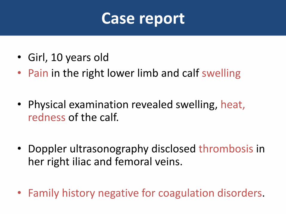
• Girl, 10 years old
• Pain in the right lower limb and calf swelling
• Physical examination revealed swelling, heat, redness of the calf.
• Doppler ultrasonography disclosed thrombosis in her right iliac and femoral veins.
• Family history negative for coagulation disorders.
Case report - Labs
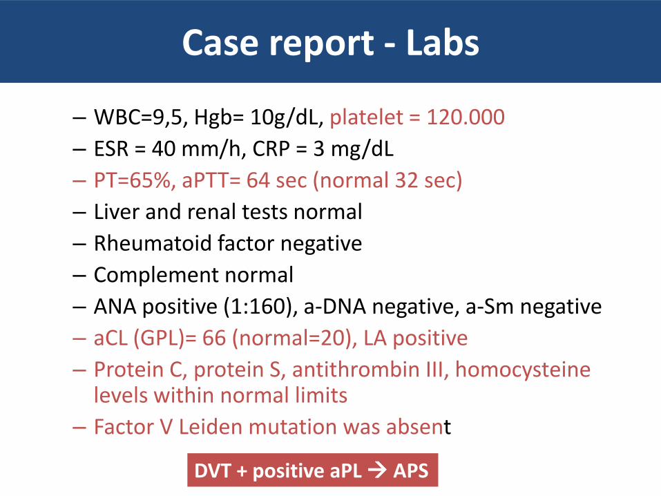
– WBC=9,5, Hgb= 10g/dL, platelet = 120.000
– ESR = 40 mm/h, CRP = 3 mg/dL
– PT=65%, aPTT= 64 sec (normal 32 sec)
– Liver and renal tests normal
– Rheumatoid factor negative
– Complement normal
– ANA positive (1:160), a-DNA negative, a-Sm negative
– aCL (GPL)= 66 (normal=20), LA positive
– Protein C, protein S, antithrombin III, homocysteine levels within normal limits
– Factor V Leiden mutation was absent
DVT + positive aPL APS
Definition
Antiphospholipid syndrome (APS) is a multisystemic autoimmune condition characterized by:
1 - vascular thrombosis and/or pregnancy loss
associated with
2- persistently positive antiphospholipid antibodies (aPL)
clinical + laboratory
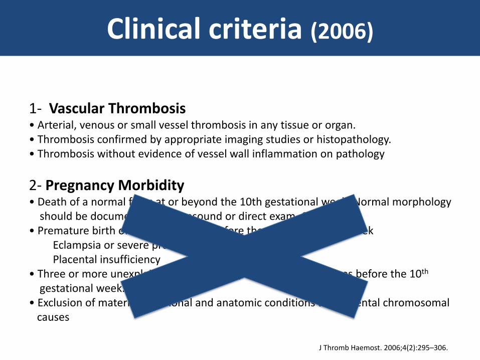
Clinical criteria (2006)
1- Vascular Thrombosis • Arterial, venous or small vessel thrombosis in any tissue or organ. • Thrombosis confirmed by appropriate imaging studies or histopathology. • Thrombosis without evidence of vessel wall inflammation on pathology
2- Pregnancy Morbidity • Death of a normal fetus at or beyond the 10th gestational week. Normal morphology should be documented by ultrasound or direct exam, OR • Premature birth of normal neonate before the 34th gestational week Eclampsia or severe pre-eclampsia Placental insufficiency • Three or more unexplained consecutive spontaneous fetal losses before the 10th gestational week. • Exclusion of maternal hormonal and anatomic conditions and parental chromosomal causes
J Thromb Haemost. 2006;4(2):295–306.
Vascular thrombosis
• One or more clinical episodes of arterial, venous, or small-vessel thrombosis, in any tissue or organ.
• Thrombosis must be confirmed by objective
validated criteria (i.e., unequivocal findings of appropriate imaging studies or histopathology).
• For histopathologic confirmation, thrombosis
should be present without significant evidence of inflammation in the vessel wall.
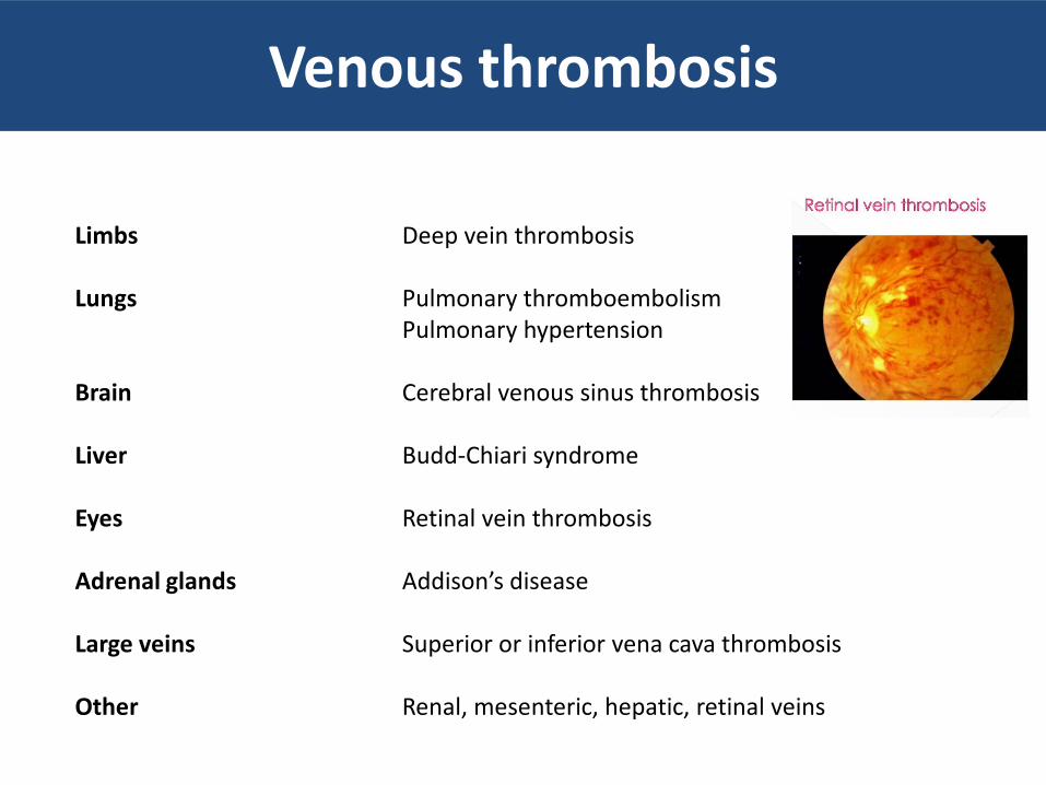
Venous thrombosis
Limbs Deep vein thrombosis Lungs Pulmonary thromboembolism Pulmonary hypertension Brain Cerebral venous sinus thrombosis Liver Budd-Chiari syndrome Eyes Retinal vein thrombosis Adrenal glands Addison’s disease Large veins Superior or inferior vena cava thrombosis Other Renal, mesenteric, hepatic, retinal veins
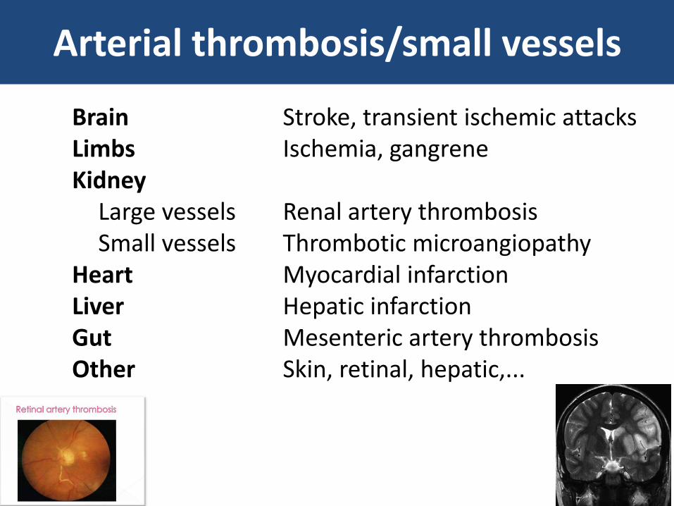
Arterial thrombosis/small vessels
Brain Stroke, transient ischemic attacks Limbs Ischemia, gangrene Kidney Large vessels Renal artery thrombosis Small vessels Thrombotic microangiopathy Heart Myocardial infarction Liver Hepatic infarction Gut Mesenteric artery thrombosis Other Skin, retinal, hepatic,...
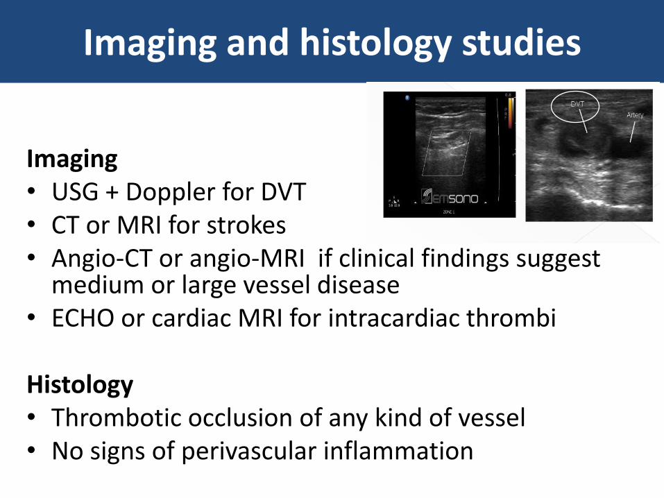
Imaging and histology studies
Imaging • USG + Doppler for DVT • CT or MRI for strokes • Angio-CT or angio-MRI if clinical findings suggest
medium or large vessel disease • ECHO or cardiac MRI for intracardiac thrombi
Histology • Thrombotic occlusion of any kind of vessel • No signs of perivascular inflammation
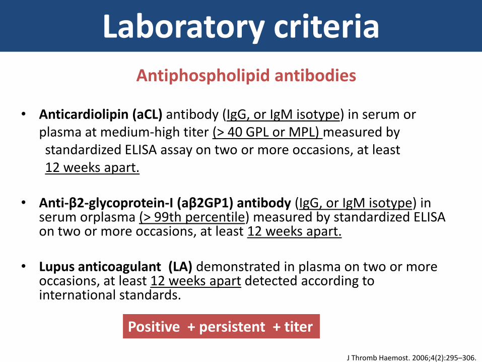
Laboratory criteria
• Anticardiolipin (aCL) antibody (IgG, or IgM isotype) in serum or plasma at medium-high titer (> 40 GPL or MPL) measured by standardized ELISA assay on two or more occasions, at least 12 weeks apart. • Anti-β2-glycoprotein-I (aβ2GP1) antibody (IgG, or IgM isotype) in
serum orplasma (> 99th percentile) measured by standardized ELISA on two or more occasions, at least 12 weeks apart.
• Lupus anticoagulant (LA) demonstrated in plasma on two or more occasions, at least 12 weeks apart detected according to international standards.
J Thromb Haemost. 2006;4(2):295–306.
Antiphospholipid antibodies
Positive + persistent + titer
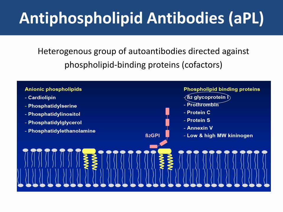
Antiphospholipid Antibodies (aPL)
Heterogenous group of autoantibodies directed against
phospholipid-binding proteins (cofactors)
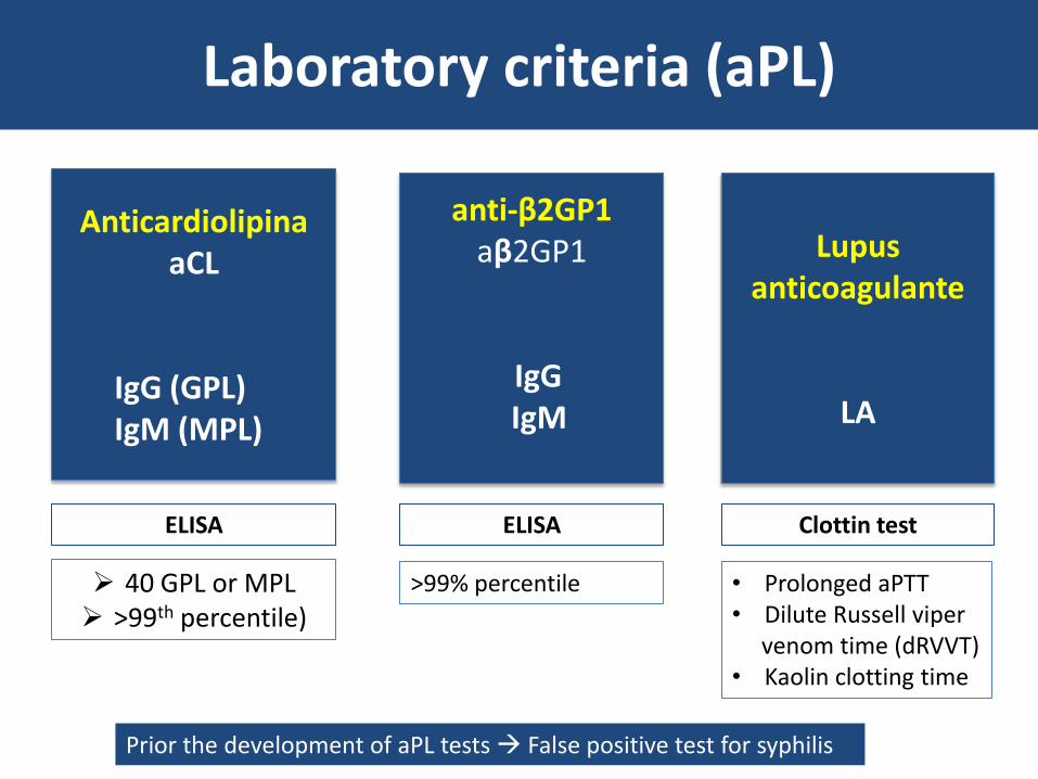
Laboratory criteria (aPL)
Lupus anticoagulante
LA
anti-β2GP1 aβ2GP1
IgG IgM
Anticardiolipina aCL
IgG (GPL) IgM (MPL)
Clottin test ELISA ELISA
40 GPL or MPL >99th percentile)
>99% percentile • Prolonged aPTT • Dilute Russell viper venom time (dRVVT) • Kaolin clotting time
Prior the development of aPL tests False positive test for syphilis
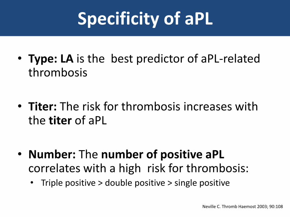
Specificity of aPL
• Type: LA is the best predictor of aPL-related thrombosis
• Titer: The risk for thrombosis increases with the titer of aPL
• Number: The number of positive aPL correlates with a high risk for thrombosis: • Triple positive > double positive > single positive
Neville C. Thromb Haemost 2003; 90:108
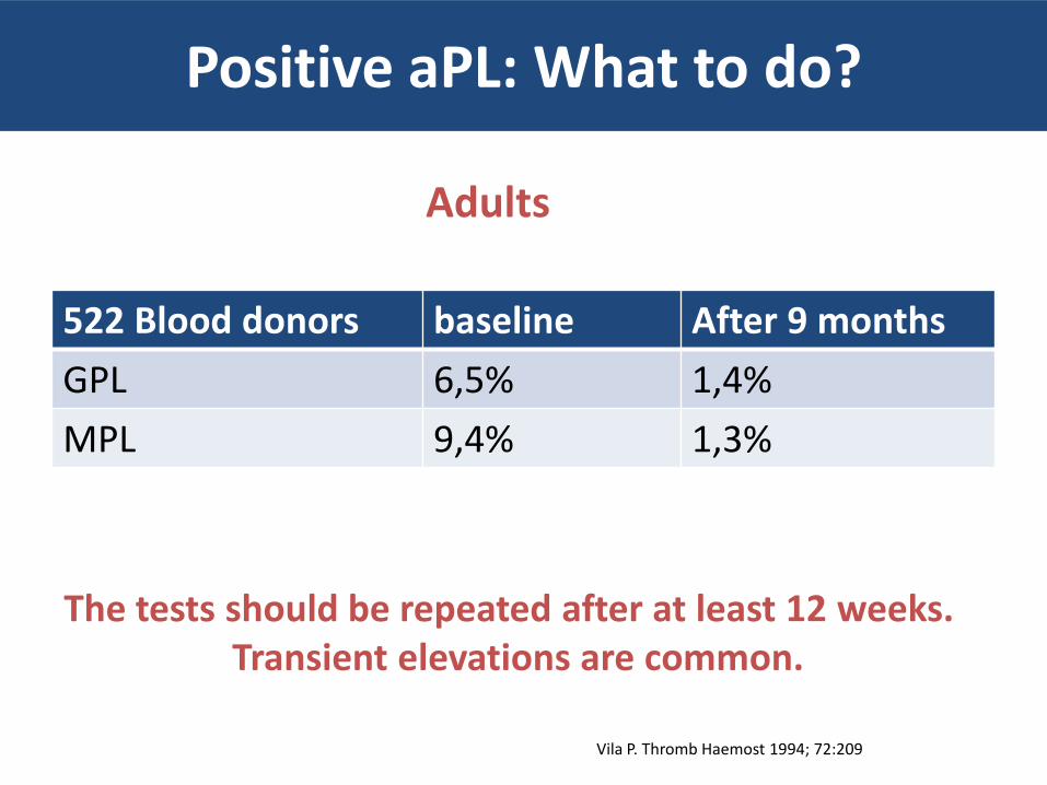
Positive aPL: What to do?
522 Blood donors baseline After 9 months
GPL 6,5% 1,4%
MPL 9,4% 1,3%
The tests should be repeated after at least 12 weeks. Transient elevations are common.
Vila P. Thromb Haemost 1994; 72:209
Adults
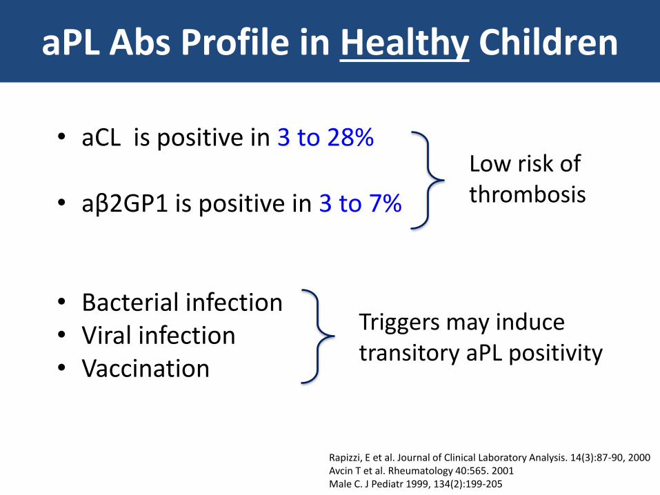
aPL Abs Profile in Healthy Children
• aCL is positive in 3 to 28%
• aβ2GP1 is positive in 3 to 7%
• Bacterial infection • Viral infection • Vaccination
Triggers may induce transitory aPL positivity
Rapizzi, E et al. Journal of Clinical Laboratory Analysis. 14(3):87-90, 2000 Avcin T et al. Rheumatology 40:565. 2001 Male C. J Pediatr 1999, 134(2):199-205
Low risk of thrombosis
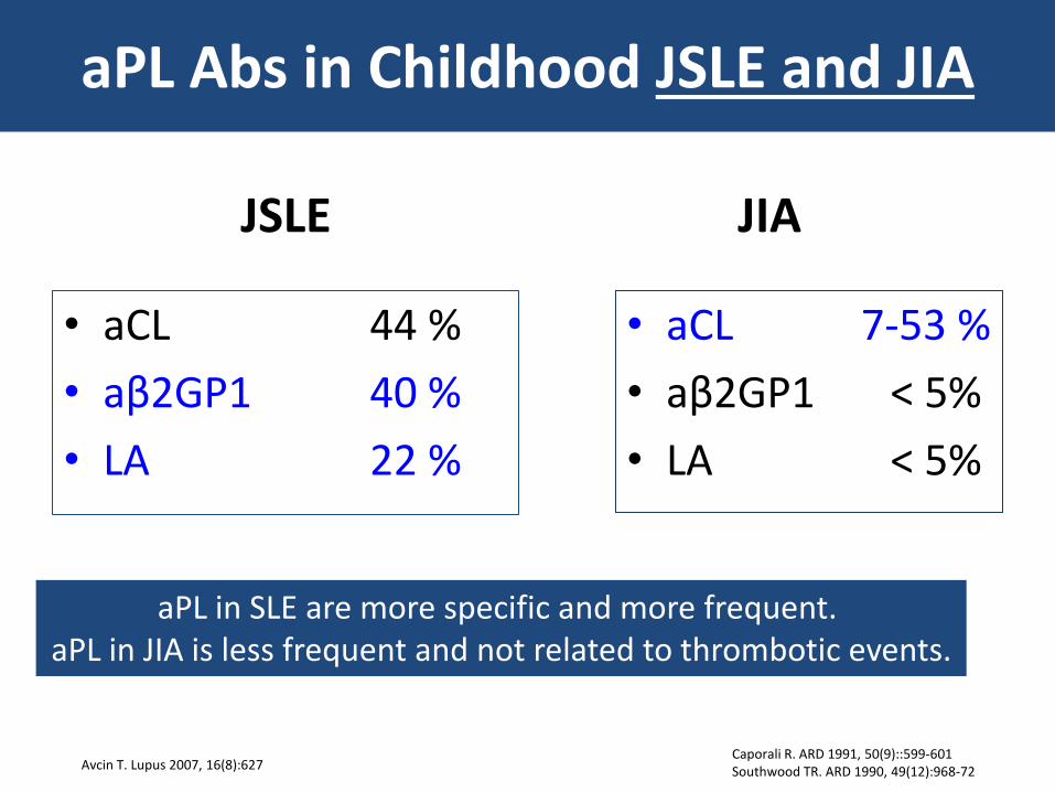
aPL Abs in Childhood JSLE and JIA
JSLE
• aCL 44 %
• aβ2GP1 40 %
• LA 22 %
JIA
• aCL 7-53 %
• aβ2GP1 < 5%
• LA < 5%
Caporali R. ARD 1991, 50(9)::599-601 Southwood TR. ARD 1990, 49(12):968-72
aPL in SLE are more specific and more frequent. aPL in JIA is less frequent and not related to thrombotic events.
Avcin T. Lupus 2007, 16(8):627
“ Criteria and Non-criteria" clinical manifestations
• Hematologic
• Cutaneous
• Pulmonary
• Renal
• Cardiovasvular
• Neurological
Non-criteria clinical manifestations can suggest the diagnosis
and precede later vascular thrombosis.
“ Criteria and Non-criteria" manifestations of APS
– Thrombocytopenia • 50.000 to 140.000
• < 50.00 – bleeding
• Severe: C-APS?
– Microangiopathic hemolytic anemia
– Bleeding episodes due to antibodies to prothrombin
Hematologic manifestations
Thrombocytopenia does not Preclude the occurrence of thrombotic complications of APS
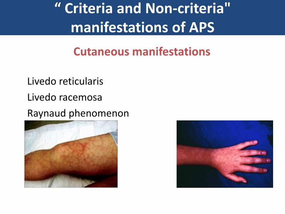
“ Criteria and Non-criteria" manifestations of APS
Livedo reticularis
Livedo racemosa
Raynaud phenomenon
Cutaneous manifestations
Pulmonary manifestations of APS
– Pulmonary thromboembolic disease
– Pulmonary arterial thrombosis
– Pulmonary microthrombosis
– Pulmonary hypertension
– Acute respiratory distress syndrome (ARDS)
– Diffuse alveolar hemorrhage
Renal manifestations of APS
• Thrombosis at any site
• Flank pain, proteinuria – check for aPL
• Urine analysis – proteinuria, hematuria
• Ischemic mesangiolysis, vessel hyperplasia
• Acute renal failure
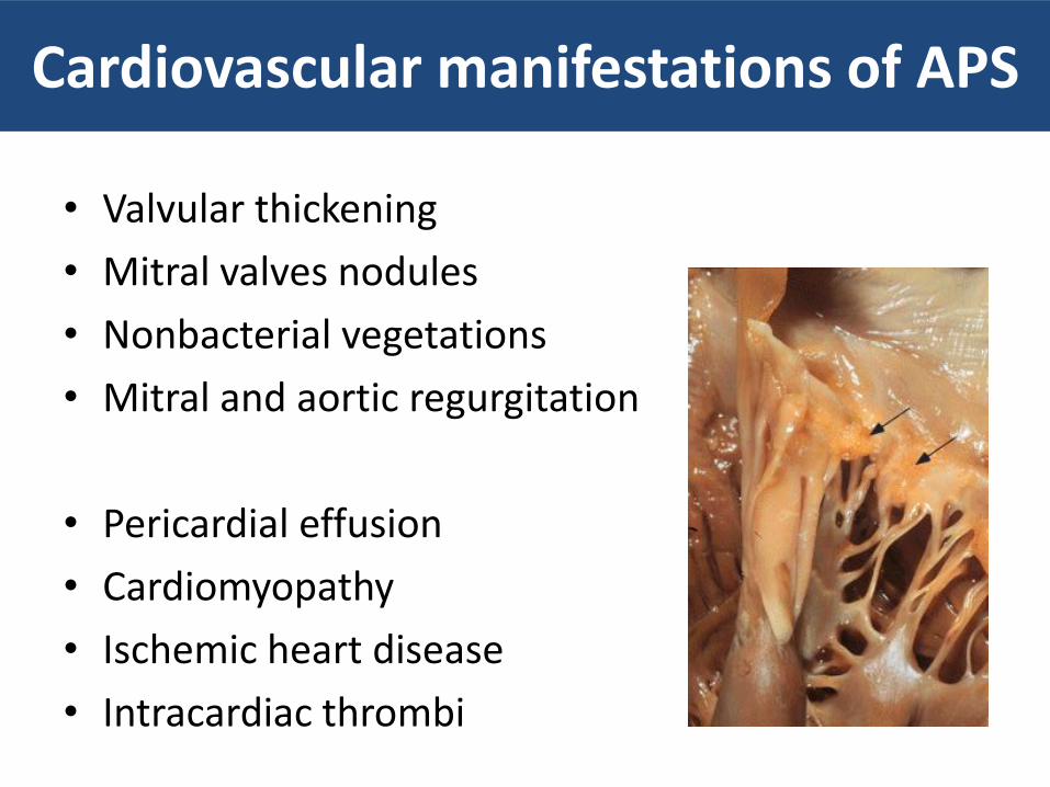
Cardiovascular manifestations of APS
• Valvular thickening
• Mitral valves nodules
• Nonbacterial vegetations
• Mitral and aortic regurgitation
• Pericardial effusion
• Cardiomyopathy
• Ischemic heart disease
• Intracardiac thrombi
“Non-criteria” manifestations of APS
– Cognitive deficit • Subtle findings
• Permanent and profound cognitive functioning
– White matter lesions • MRI suggestive of vasculopathy (high intensity lesions)
– Other manifestations • Epilepsia
• Chorea and hemiballismus
• Transverse myelopathy
• Sensorioneural hearing loss
• Migraine
Neurological non-thrombotic
Avcin T. Pediatrics 2008, 122(5)
Non-criteria lab manifestations of APS
• Low titers of aCL (GPL or MPL): 20 - 39 units
• Test for additional IgA aPL – IgA aCL – IgA anti- β2GP1
• Test for another “non-criteria” aPL (prothrombin, phosphatidylserine,
phosphatidylinositol) – Not well standardized – Sensitivity? – Specificity? – Clinical significance?
• Test for heritable thrombophilias
• Differential diagnosis and additional risk factor in patients with APS
Negative aPL and strong suspicion of aCL
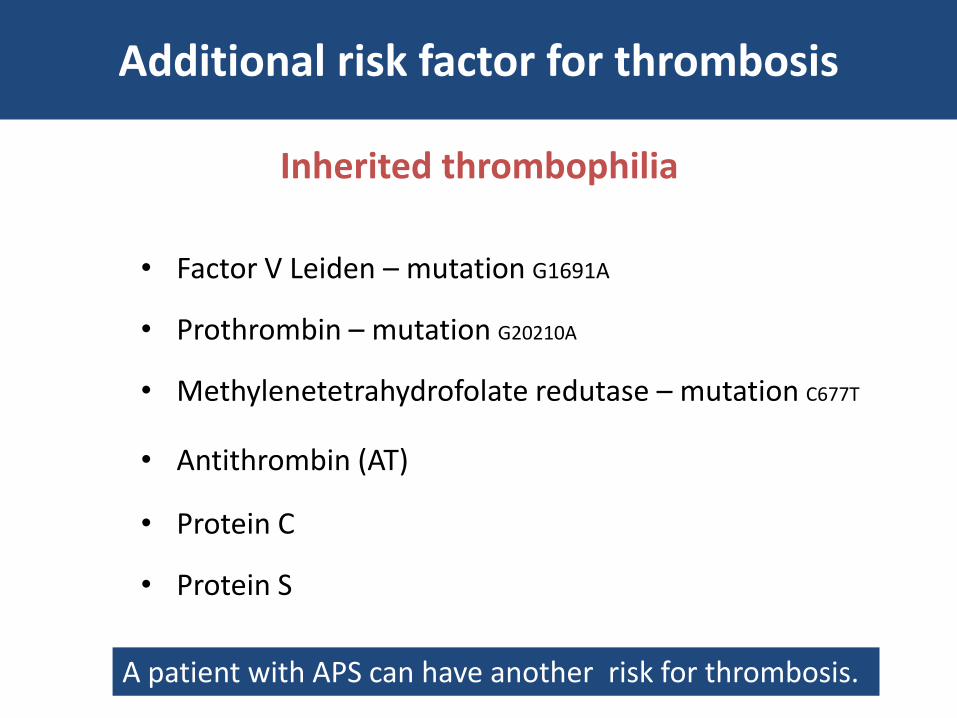
Additional risk factor for thrombosis
• Factor V Leiden – mutation G1691A
• Prothrombin – mutation G20210A
• Methylenetetrahydrofolate redutase – mutation C677T
• Antithrombin (AT)
• Protein C
• Protein S
Inherited thrombophilia
A patient with APS can have another risk for thrombosis.
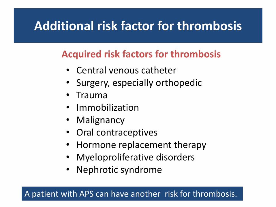
Additional risk factor for thrombosis
• Central venous catheter • Surgery, especially orthopedic • Trauma • Immobilization • Malignancy • Oral contraceptives • Hormone replacement therapy • Myeloproliferative disorders • Nephrotic syndrome
Acquired risk factors for thrombosis
A patient with APS can have another risk for thrombosis.
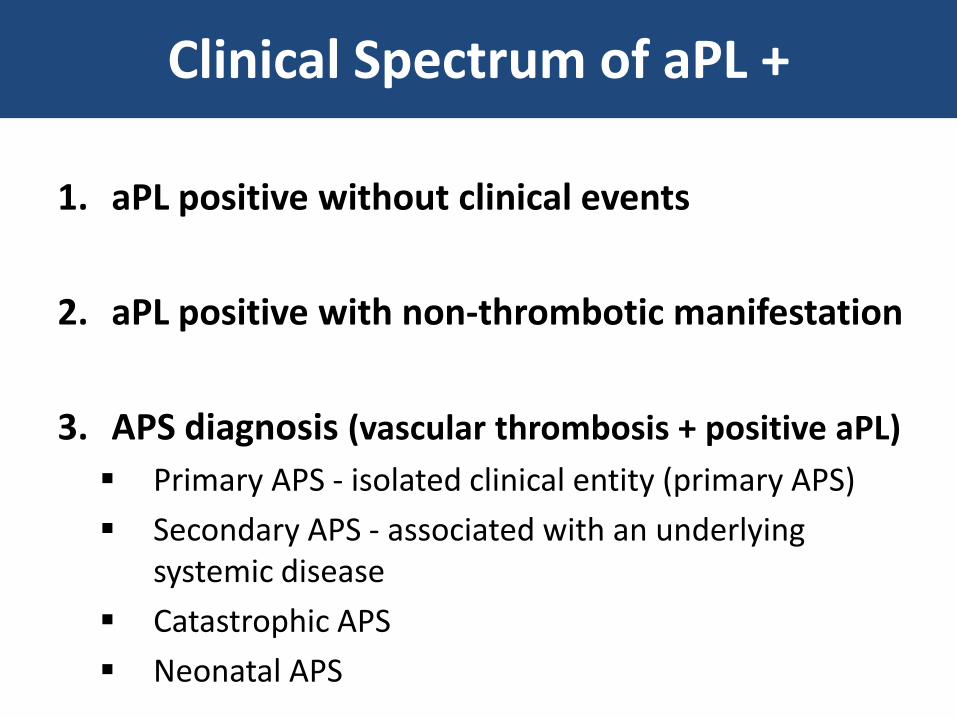
Clinical Spectrum of aPL +
1. aPL positive without clinical events
2. aPL positive with non-thrombotic manifestation
3. APS diagnosis (vascular thrombosis + positive aPL)
Primary APS - isolated clinical entity (primary APS)
Secondary APS - associated with an underlying systemic disease
Catastrophic APS
Neonatal APS
Outline
2. Registries of Pediatric APS
– Primary and Secondary APS
– Catastrophic APS (CAPS)
– Neonatal APS
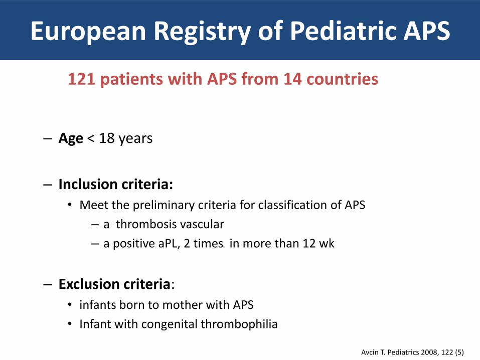
European Registry of Pediatric APS
121 patients with APS from 14 countries
– Age < 18 years
– Inclusion criteria: • Meet the preliminary criteria for classification of APS
– a thrombosis vascular
– a positive aPL, 2 times in more than 12 wk
– Exclusion criteria: • infants born to mother with APS
• Infant with congenital thrombophilia
Avcin T. Pediatrics 2008, 122 (5)
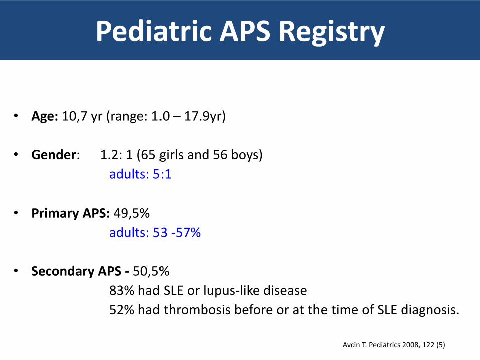
Pediatric APS Registry
• Age: 10,7 yr (range: 1.0 – 17.9yr)
• Gender: 1.2: 1 (65 girls and 56 boys)
adults: 5:1
• Primary APS: 49,5%
adults: 53 -57%
• Secondary APS - 50,5%
83% had SLE or lupus-like disease
52% had thrombosis before or at the time of SLE diagnosis.
Avcin T. Pediatrics 2008, 122 (5)
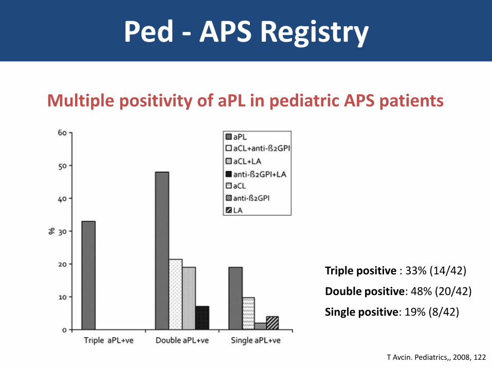
Multiple positivity of aPL in pediatric APS patients
Triple positive : 33% (14/42)
Double positive: 48% (20/42)
Single positive: 19% (8/42)
T Avcin. Pediatrics,, 2008, 122
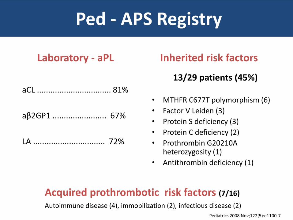
Ped - APS Registry
Ped - APS Registry
Laboratory - aPL
aCL ................................. 81%
aβ2GP1 ........................ 67%
LA ................................ 72%
Inherited risk factors
13/29 patients (45%)
• MTHFR C677T polymorphism (6)
• Factor V Leiden (3)
• Protein S deficiency (3)
• Protein C deficiency (2)
• Prothrombin G20210A heterozygosity (1)
• Antithrombin deficiency (1)
Pediatrics 2008 Nov;122(5):e1100-7
Acquired prothrombotic risk factors (7/16)
Autoimmune disease (4), immobilization (2), infectious disease (2)
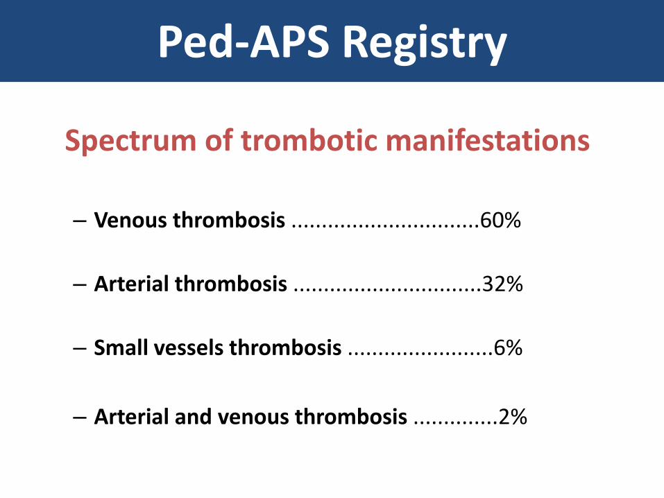
Ped-APS Registry
Spectrum of trombotic manifestations
– Venous thrombosis ...............................60%
– Arterial thrombosis ...............................32%
– Small vessels thrombosis ........................6%
– Arterial and venous thrombosis ..............2%
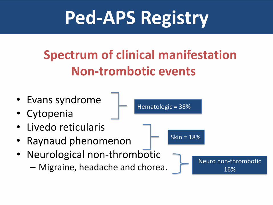
Ped-APS Registry
Spectrum of clinical manifestation Non-trombotic events • Evans syndrome • Cytopenia • Livedo reticularis • Raynaud phenomenon • Neurological non-thrombotic
– Migraine, headache and chorea.
Hematologic = 38%
Skin = 18%
Neuro non-thrombotic 16%
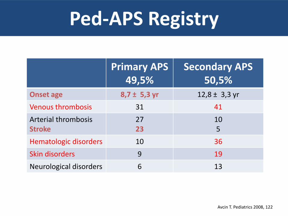
Ped-APS Registry
Primary APS 49,5%
Secondary APS 50,5%
Onset age 8,7 ± 5,3 yr 12,8 ± 3,3 yr
Venous thrombosis 31 41
Arterial thrombosis Stroke
27 23
10 5
Hematologic disorders 10 36
Skin disorders 9 19
Neurological disorders 6 13
Avcin T. Pediatrics 2008, 122
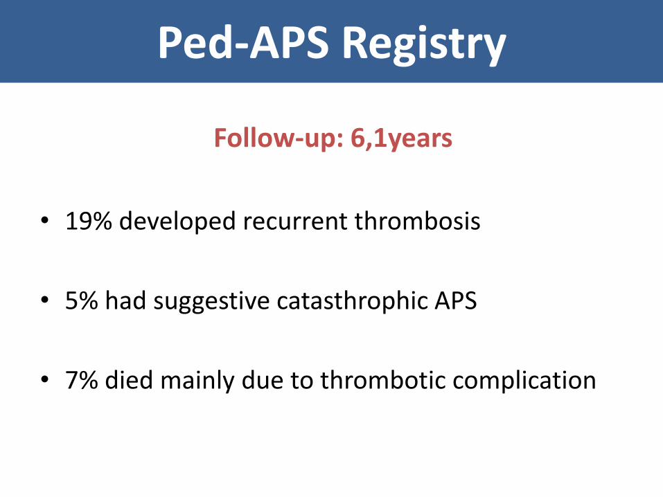
Ped-APS Registry
Follow-up: 6,1years
• 19% developed recurrent thrombosis
• 5% had suggestive catasthrophic APS
• 7% died mainly due to thrombotic complication
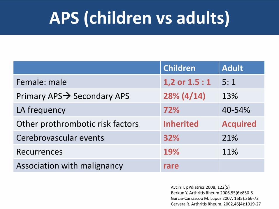
APS (children vs adults)
Children Adult
Female: male 1,2 or 1.5 : 1 5: 1
Primary APS Secondary APS 28% (4/14) 13%
LA frequency 72% 40-54%
Other prothrombotic risk factors Inherited Acquired
Cerebrovascular events 32% 21%
Recurrences 19% 11%
Association with malignancy rare
Avcin T. pPdiatrics 2008, 122(5) Berkun Y. Arthritis Rheum 2006,55(6):850-5 Garcia-Carrascoo M. Lupus 2007, 16(5):366-73 Cervera R. Arthritis Rheum. 2002,46(4):1019-27
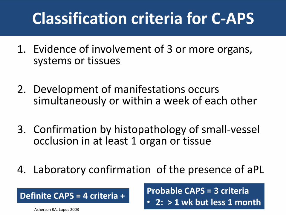
Classification criteria for C-APS
1. Evidence of involvement of 3 or more organs, systems or tissues
2. Development of manifestations occurs simultaneously or within a week of each other
3. Confirmation by histopathology of small-vessel occlusion in at least 1 organ or tissue
4. Laboratory confirmation of the presence of aPL
Asherson RA. Lupus 2003
Definite CAPS = 4 criteria + Probable CAPS = 3 criteria • 2: > 1 wk but less 1 month
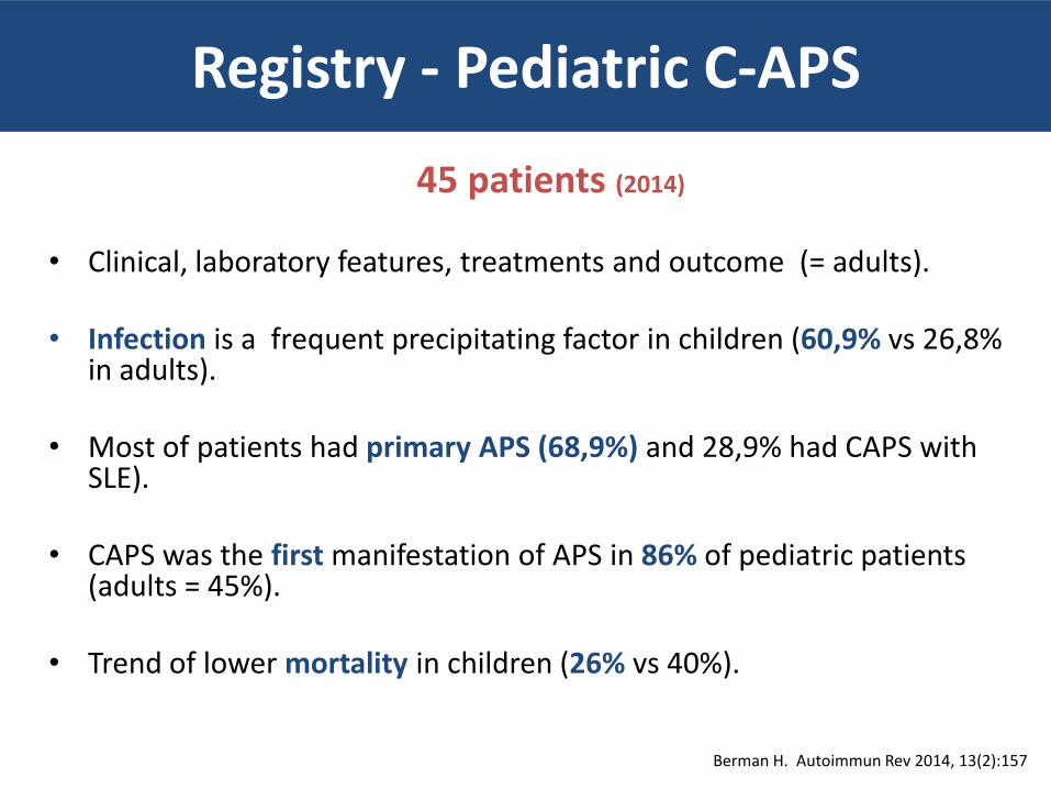
Registry - Pediatric C-APS
45 patients (2014)
• Clinical, laboratory features, treatments and outcome (= adults).
• Infection is a frequent precipitating factor in children (60,9% vs 26,8%
in adults).
• Most of patients had primary APS (68,9%) and 28,9% had CAPS with SLE).
• CAPS was the first manifestation of APS in 86% of pediatric patients (adults = 45%).
• Trend of lower mortality in children (26% vs 40%).
Berman H. Autoimmun Rev 2014, 13(2):157
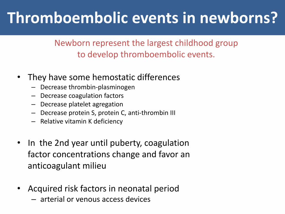
Thromboembolic events in newborns?
Newborn represent the largest childhood group to develop thromboembolic events.
• They have some hemostatic differences
– Decrease thrombin-plasminogen – Decrease coagulation factors – Decrease platelet agregation – Decrease protein S, protein C, anti-thrombin III – Relative vitamin K deficiency
• In the 2nd year until puberty, coagulation factor concentrations change and favor an anticoagulant milieu
• Acquired risk factors in neonatal period
– arterial or venous access devices
Babies born to mothers with APS
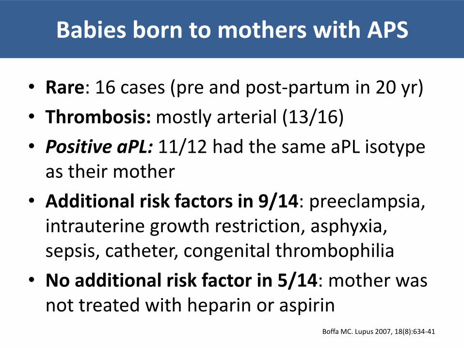
• Rare: 16 cases (pre and post-partum in 20 yr)
• Thrombosis: mostly arterial (13/16)
• Positive aPL: 11/12 had the same aPL isotype as their mother
• Additional risk factors in 9/14: preeclampsia, intrauterine growth restriction, asphyxia, sepsis, catheter, congenital thrombophilia
• No additional risk factor in 5/14: mother was not treated with heparin or aspirin
Boffa MC. Lupus 2007, 18(8):634-41
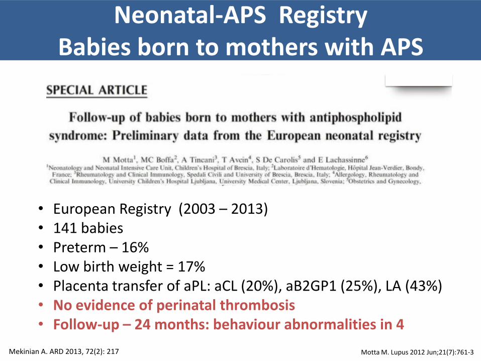
Neonatal-APS Registry Babies born to mothers with APS
• European Registry (2003 – 2013) • 141 babies • Preterm – 16% • Low birth weight = 17% • Placenta transfer of aPL: aCL (20%), aB2GP1 (25%), LA (43%) • No evidence of perinatal thrombosis • Follow-up – 24 months: behaviour abnormalities in 4
Motta M. Lupus 2012 Jun;21(7):761-3 Mekinian A. ARD 2013, 72(2): 217
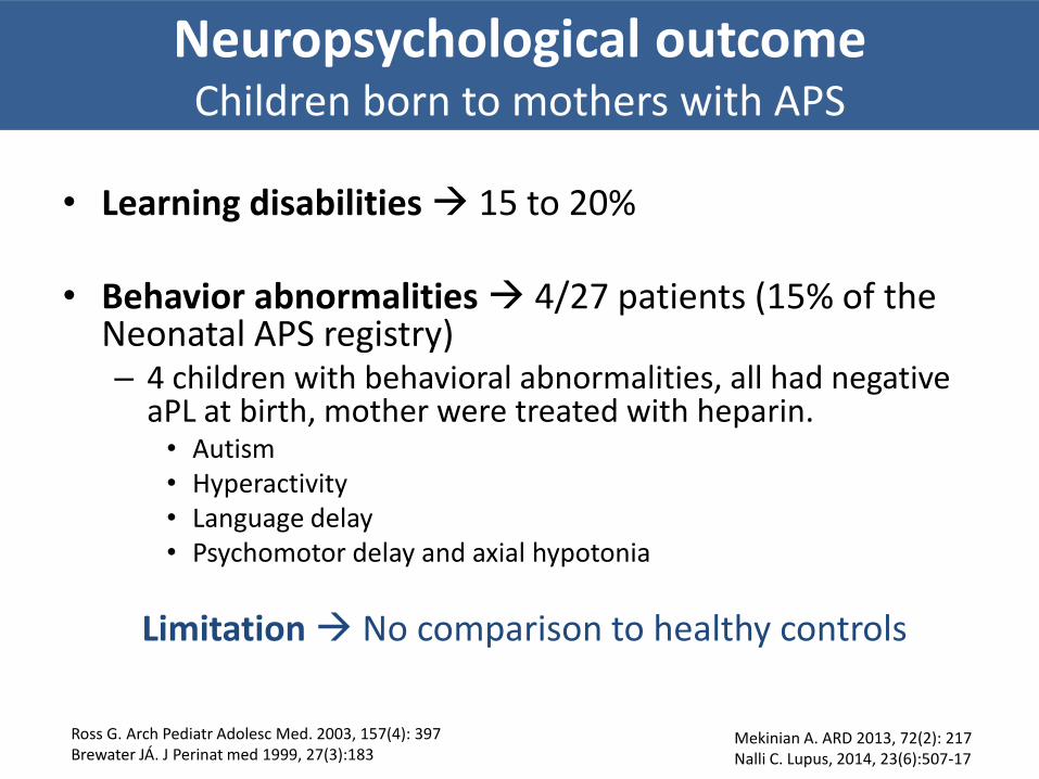
Neuropsychological outcome Children born to mothers with APS
• Learning disabilities 15 to 20%
• Behavior abnormalities 4/27 patients (15% of the Neonatal APS registry) – 4 children with behavioral abnormalities, all had negative
aPL at birth, mother were treated with heparin. • Autism • Hyperactivity • Language delay • Psychomotor delay and axial hypotonia
Limitation No comparison to healthy controls
Mekinian A. ARD 2013, 72(2): 217 Nalli C. Lupus, 2014, 23(6):507-17
Ross G. Arch Pediatr Adolesc Med. 2003, 157(4): 397 Brewater JÁ. J Perinat med 1999, 27(3):183
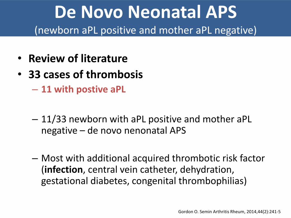
De Novo Neonatal APS (newborn aPL positive and mother aPL negative)
• Review of literature
• 33 cases of thrombosis – 11 with postive aPL
– 11/33 newborn with aPL positive and mother aPL
negative – de novo nenonatal APS
– Most with additional acquired thrombotic risk factor (infection, central vein catheter, dehydration, gestational diabetes, congenital thrombophilias)
Gordon O. Semin Arthritis Rheum, 2014,44(2):241-5
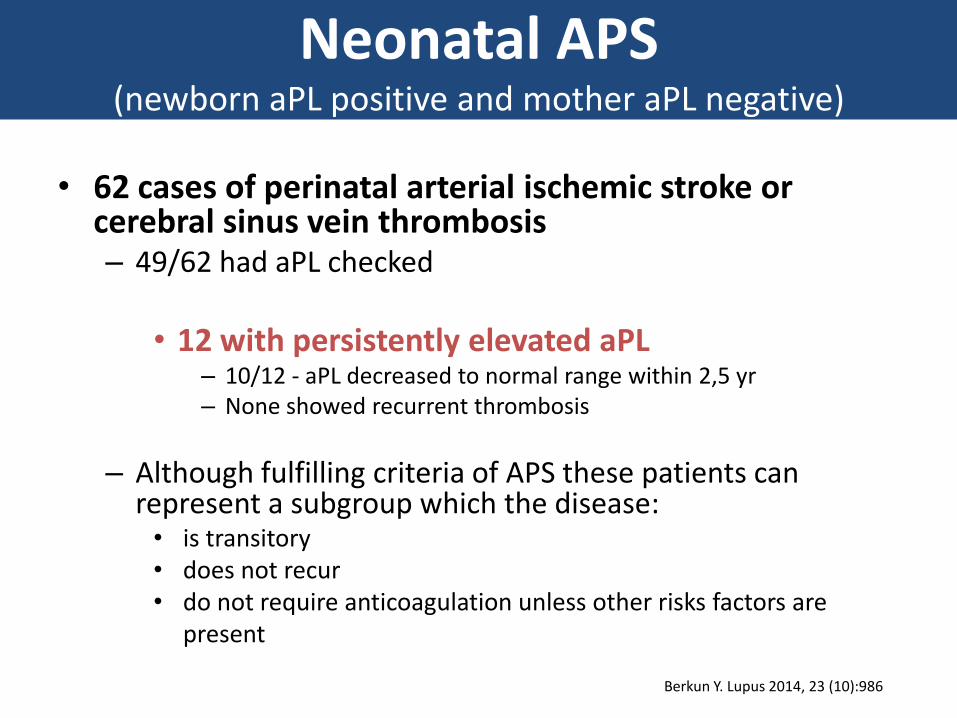
Neonatal APS (newborn aPL positive and mother aPL negative)
• 62 cases of perinatal arterial ischemic stroke or cerebral sinus vein thrombosis – 49/62 had aPL checked
• 12 with persistently elevated aPL – 10/12 - aPL decreased to normal range within 2,5 yr – None showed recurrent thrombosis
– Although fulfilling criteria of APS these patients can represent a subgroup which the disease: • is transitory • does not recur • do not require anticoagulation unless other risks factors are present
Berkun Y. Lupus 2014, 23 (10):986
Outline
3. Treatment
How to treat
• There are no recommendations specific for children.
• Medications – Heparin
– Warfarin
– Aspirin
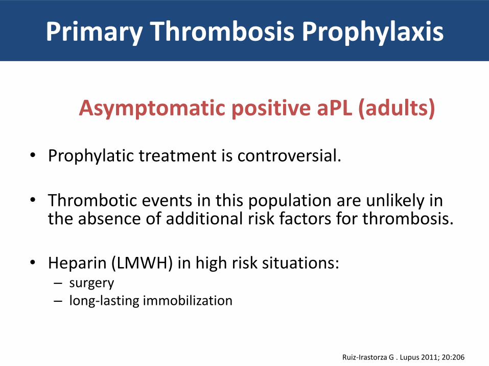
Primary Thrombosis Prophylaxis
Asymptomatic positive aPL (adults)
• Prophylatic treatment is controversial.
• Thrombotic events in this population are unlikely in the absence of additional risk factors for thrombosis.
• Heparin (LMWH) in high risk situations:
– surgery – long-lasting immobilization
Ruiz-Irastorza G . Lupus 2011; 20:206
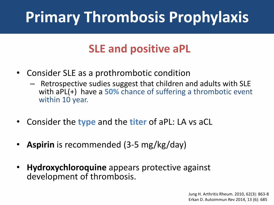
Primary Thrombosis Prophylaxis
SLE and positive aPL
• Consider SLE as a prothrombotic condition – Retrospective sudies suggest that children and adults with SLE
with aPL(+) have a 50% chance of suffering a thrombotic event within 10 year.
• Consider the type and the titer of aPL: LA vs aCL
• Aspirin is recommended (3-5 mg/kg/day)
• Hydroxychloroquine appears protective against
development of thrombosis.
Jung H. Arthritis Rheum. 2010, 62(3): 863-8 Erkan D. Autoimmun Rev 2014, 13 (6): 685
Acute thrombotic event
Heparin
• LMWH – low molecular weight heparin
• Unfractionated heparin
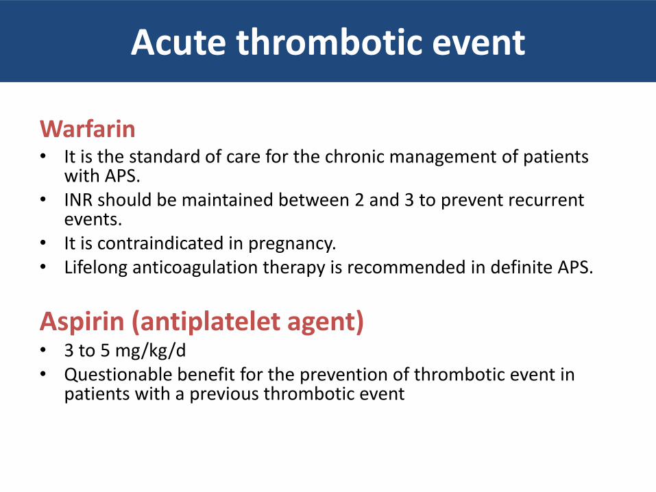
Acute thrombotic event
Warfarin
• It is the standard of care for the chronic management of patients with APS.
• INR should be maintained between 2 and 3 to prevent recurrent events.
• It is contraindicated in pregnancy. • Lifelong anticoagulation therapy is recommended in definite APS.
Aspirin (antiplatelet agent) • 3 to 5 mg/kg/d • Questionable benefit for the prevention of thrombotic event in
patients with a previous thrombotic event
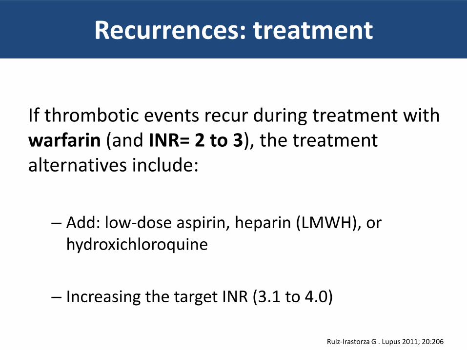
Recurrences: treatment
If thrombotic events recur during treatment with warfarin (and INR= 2 to 3), the treatment alternatives include:
– Add: low-dose aspirin, heparin (LMWH), or hydroxichloroquine
– Increasing the target INR (3.1 to 4.0)
Ruiz-Irastorza G . Lupus 2011; 20:206
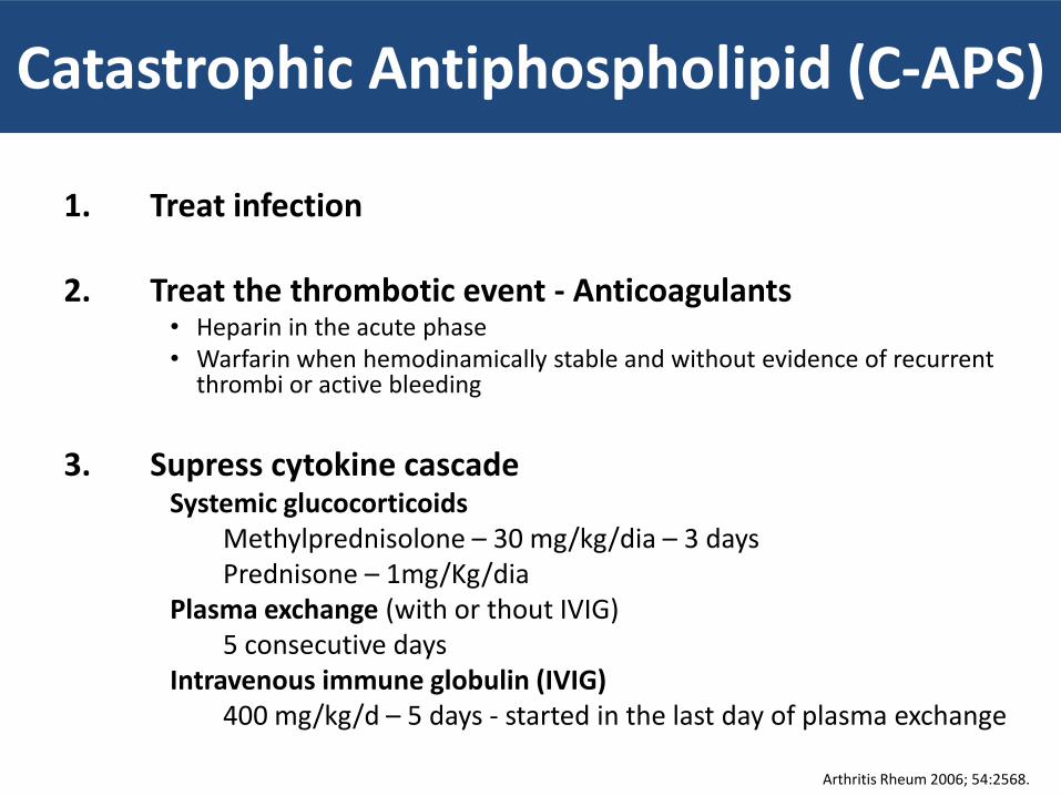
Catastrophic Antiphospholipid (C-APS)
1. Treat infection 2. Treat the thrombotic event - Anticoagulants
• Heparin in the acute phase • Warfarin when hemodinamically stable and without evidence of recurrent
thrombi or active bleeding
3. Supress cytokine cascade
Systemic glucocorticoids Methylprednisolone – 30 mg/kg/dia – 3 days Prednisone – 1mg/Kg/dia
Plasma exchange (with or thout IVIG) 5 consecutive days
Intravenous immune globulin (IVIG) 400 mg/kg/d – 5 days - started in the last day of plasma exchange
Arthritis Rheum 2006; 54:2568.
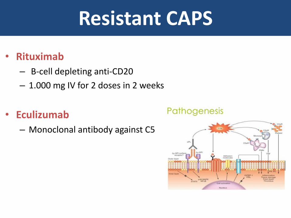
Resistant CAPS
• Rituximab
– B-cell depleting anti-CD20
– 1.000 mg IV for 2 doses in 2 weeks
• Eculizumab
– Monoclonal antibody against C5

Thank you!