Embed Size (px)
Citation preview
J Borrero 2/09 NUR240
Respiratory Stressors I
Pulmonary EmbolismLung Cancer
Thoracic SurgeryChest Tubes
Pleural Effusions
Pulmonary Embolism Pulmonary Embolism -emboli that reach the lungs and
obstruct pulmonary circulation -blood, air, fat, tumor cells, amniotic
fluid, foreign objects -many die within 1 hr of onset of
symptoms or before dx.

Risk Factors for PE
Virchow’s Triad of causes DVT and PE
1. Stasis of blood flow 2. Endothelial injury 3. Hypercoagulability
What else????
Symptoms of a PE Chest pain with respirations S3 or S4 heart sounds EKG-non specific- T or ST abnormalities SOB-crackles, friction rub, breath sounds Dyspnea, hemoptysis, CP in<20% pts. Mild temp with sweating Shock: Tachycardia, hypotension, skin
cold/clammy N & V Feeling of anxiety, impending doom,
restlessness
Assessment Laboratory: Elevated WBCs ABGs-Resp alkalosis
Resp.acidosis. O2 Sats low CXR EKG Ventilation/Perfusion Scan CT Scan or CTA “Gold Standard” Pulmonary angiography- invasive Thoracentesis
Management- Non surgical Nursing Dx: ABG analysis Prevention of DVT, prophylactic use of
heparin Thrombolytic agents for massive clots O2, VS, lung/heart sounds, Mechanical ventilation Assess bleeding risk
Nursing Diagnosis
1.Decreased Cardiac Output R/T … IVF Positive inotropic agents VasodilatorsOutcome:Adequate tissue perfusion in all
major organs Predictors: Adequate circulation Predictors:
Nursing Diagnoses
2. Risk for injury (bleeding) R/T… Maintain H&H WNL Monitoring and pt. teaching
3. Anxiety R/T… Verbalization of fears Teach coping mechanisms
Management
Stable pts- Heparin for 5-10 days, then Coumadin started on the third day (from 3-6 weeks or indefinitely)
Health Teaching
Management- Surgical
Embolectomy-removal of clot
IVC fillter
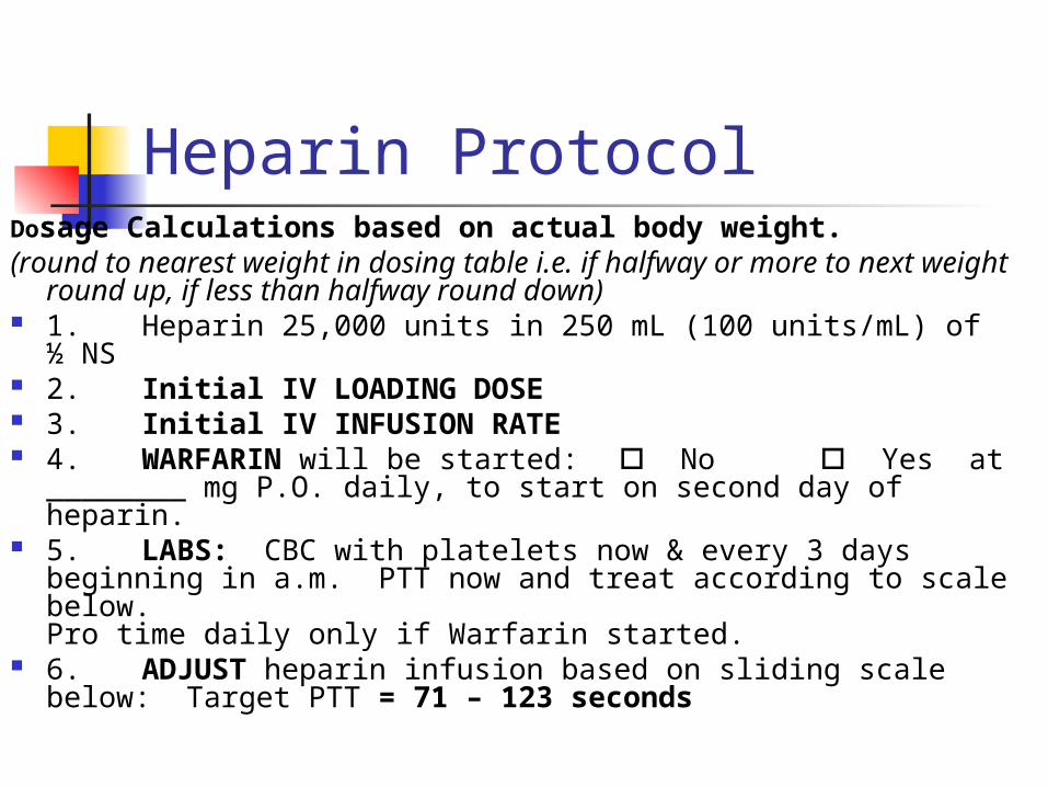
Heparin ProtocolDosage Calculations based on actual body weight.(round to nearest weight in dosing table i.e. if halfway or more
to next weight round up, if less than halfway round down) 1. Heparin 25,000 units in 250 mL (100 units/mL) of ½
NS 2. Initial IV LOADING DOSE 3. Initial IV INFUSION RATE 4. WARFARIN will be started: No Yes at
________ mg P.O. daily, to start on second day of heparin. 5. LABS: CBC with platelets now & every 3 days
beginning in a.m. PTT now and treat according to scale below. Pro time daily only if Warfarin started.
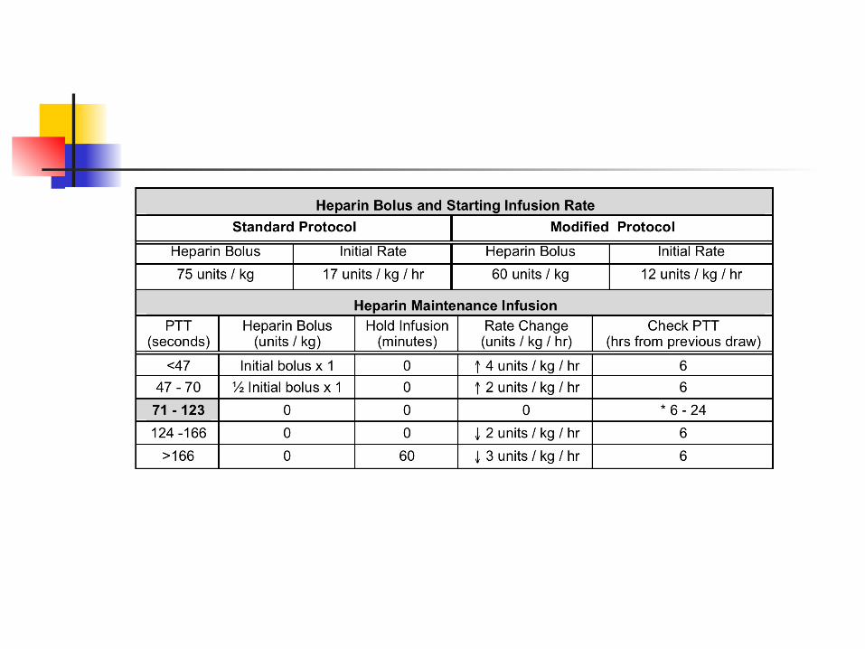
6. ADJUST heparin infusion based on sliding scale below: Target PTT = 71 – 123 seconds
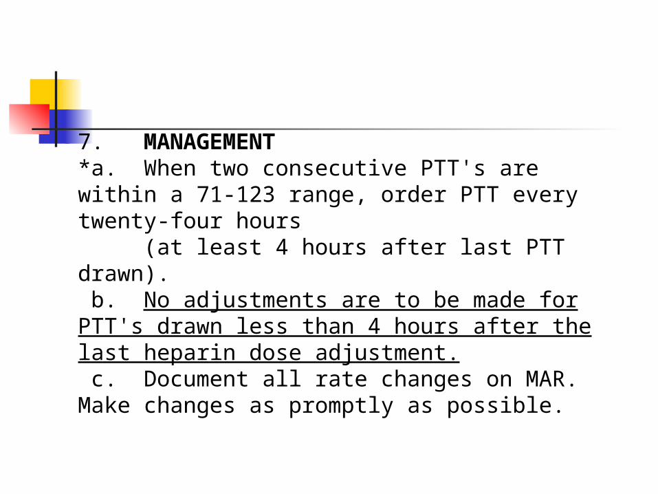
7. MANAGEMENT*a. When two consecutive PTT's are within a 71-123 range, order PTT every twenty-four hours
(at least 4 hours after last PTT drawn). b. No adjustments are to be made for PTT's drawn less than 4 hours after the last heparin dose adjustment. c. Document all rate changes on MAR. Make changes as promptly as possible.
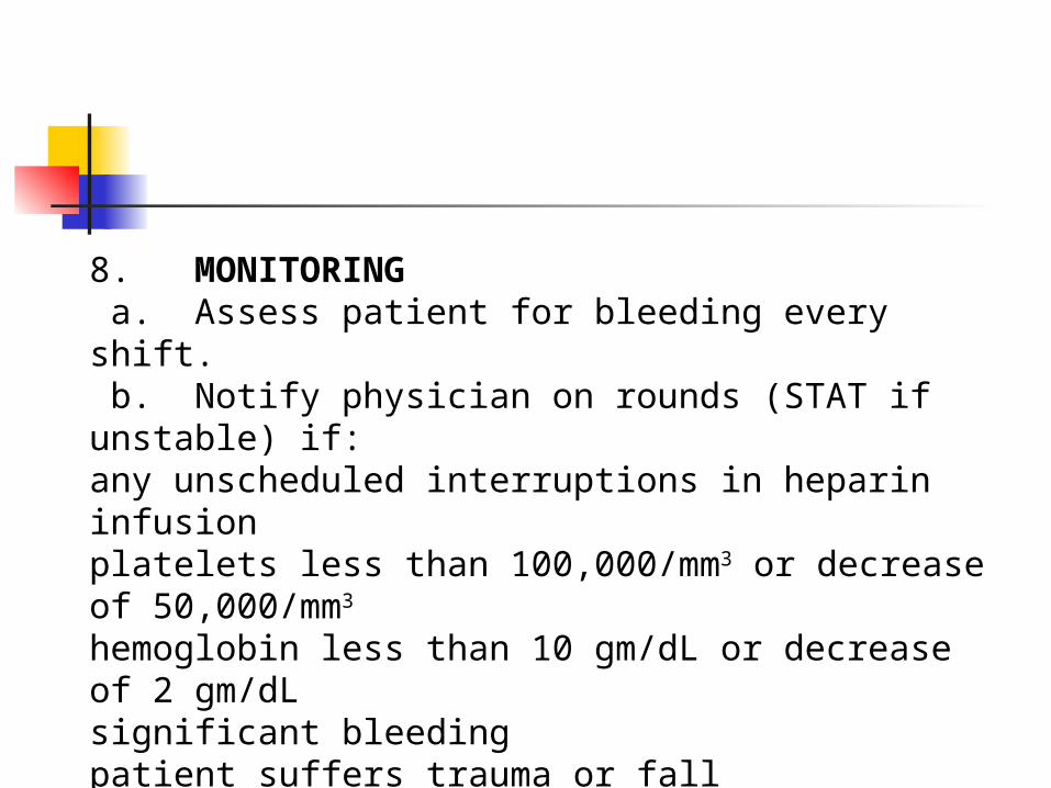
8. MONITORING a. Assess patient for bleeding every shift. b. Notify physician on rounds (STAT if unstable) if:any unscheduled interruptions in heparin infusionplatelets less than 100,000/mm3 or decrease of 50,000/mm3
hemoglobin less than 10 gm/dL or decrease of 2 gm/dLsignificant bleedingpatient suffers trauma or fall
Lung Cancer Leading cancer killer for men and
women Number of men has stayed stable but
number of women continues to rise Lung cancer has surpassed breast
cancer as the major killer of women and remains at the top of the list
70% have mets at time of dx. Long term survival is low. Most die within 1yr of dx
5 year survival rate is <15%
Leading cause of cancer-related deaths worldwide Kills more women than breast, ovarian and
uterine combined Rate of lung Ca among women has not been
declining as in men…but women are more likely to survive the disease
No rationale offered for the difference The rate of lung Ca among non-smokers is
increasing, esp. young women, reason is unclear
New studies have identified some causes of increased incidence
Risk Factors for Lung Cancer 85% are caused by inhalation of carcinogenic
chemicals Cigarette smoke has 43 known chemical
carcinogens Directly related to pack-years Second hand smoke is also a risk factor Exposure to ionizing radiation Air pollution (2-3x risk in urban areas) Chronic exposure to asbestos, coal distillates
and radiation Genetic predisposition Underlying respiratory disease- COPD or TB
Pathophysiology of Lung Ca Epithelial cell is attacked by carcinogen
and binds to the cell’s DNA and damages it The cells mutate, have abnormal cell
growth and develop into malignant cells The cells replicate and continue to change,
causing the pulmonary epithelium to become an invasive carcinoma
Metastasize by direst extension through blood and by invading lymph gland and vessels
Lung Ca Classification
1.Small cell lung cancer (SCLC) or oat cell -2% of all lung Ca -99% associated with cigarette smoking -fast growing2. Non small cell lung cancer (NSCLC) - has the best survival rate if tx early - includes squamous cell,
adenocarcinoma and large cell cancer
Assessment History Risk Factors Respiratory Assessment Presence of Abnormal findings: Inspection Palpation Percussion Auscultation Psychosocial Assessments
Warning Signs
Persistant cough or change in coughChange in resp patternHemoptysisWheezing/dyspneaBlood streaked sputumChest pain- dull or pleuriticHoarseness or dysphagiaRecurrent episodes of PN, Pleural effusionCompression of SVCWeight lossClubbing of the fingers
Clinical Manifestations
Paraneoplastic- additional manifestation caused by hormones secreted by tumor cells
1.Endocrine Hypercalcemia Cushing’s Syndrome SIADH- Syndrome of Inappropriate
Antidiuretic Hormone Ectopic Insulin- Hypoglycemia
Clinical Manifestations
2. Neuromusular Peripheral neuropathy, cerebellur
degeneration, seizures Myasthenia-like muscle weakness3. Cardiovascular Thrombophlebitis Endocarditis Dysrhythmias
Clinical Manifestations
4. Hematologic Anemia DIC5. Musculoskeletal Bone pain from mets and
pathological fractures

Late Manifestations
Fatigue Weight loss Anorexia Dysphagia N&V
When to seek immediate attention:
Superior Vena Cava Syndrome
Spinal Cord Compression
Loss of bladder/bowel tone
Staging & MetastasisStaging- done at time of dx to assess size and
extent of diseaseStaging by tumor size, location, degree of
invasion of primary Tumor, Nodes and Metastasis
From Stage 0 to Stage IV TNM Mets usually to long bones vertebral column liver adrenal glands brain (personality changes, in 50% of cases)
Diagnostic Evaluation CXR Chest CT Scan- fine needle aspiration MRI Bronchoscopy/Thoracoscopy Sputum cytology Thoracentesis- with pleural effusion Percutaneous needle bx, lymph node
bx, and bx of metastatic sites.
Diagnostic Evaluation Mediastinoscopy- under general
anesthesia, a scope is passed through a supra sternal incision along the trachea, visualize the mediastinum and bx lymph nodes or tumor
Video Thoracosopy- endoscopic procedure for bx and to dx masses
PET Scans to detect mets
Management
Depends on the cell type Stage of the disease Physiologic status of patient
Nursing Interventions
Maintain airway Administer O2 as ordered calorie/protein diet Smoking cessation
Chemotherapy
Used to slow tumor growth Treat patients with distant mets or small cell
cancer of the lung Supplement sx or radiation therapy Not a cure and does not prolong life to a
measurable degree Many side effects Choice of drug depends on the growth of the
cell and the specific phase of the cell cycle that the medication affects and overall health of the patient
Drugs are generally used in combination
Chemotherapy Drugs * platinum analogues cisplatin (Platinol-AQ),
carboplatin (Paraplatin) *taxanes- paclitaxel (Taxol), docetaxel
(Taxotere) alkylating agents ifosfamide (Ifex) mitomycin (Mitomycin C) inca akloids- vinblastine sulfate doxorubicin (Adriamycin) vinorelbine (Navelbine) cyclophosphamide (Cytoxan), Methotrexate * generally first line drugs
Chemotherapy Side Effects
Alopecia N&V Mucositis Anemia Immunosuppression Thrombocytopenia
Other Management
Bronchodilators Antibiotics Pain Management Radiation therapy
Radiation Therapy
Curative if only local disease, palliative for mets Can be used in combo with sx and chemo to
improve outcome Shrink tumor size preop Relieve superior vena cava syndrome Pt monitoring and teaching: Maintain dye marks, no lotion, no soap, no sun
exposure Observe for complications- skin irritation,
peeling, fatigue, nausea, taste changes, esophagitis
Maintain adequate fluids
Surgical ManagementDepends on stage of Cancer
Localized (Stage I or II)-NSCLC - lobectomy - wedge resection - segmental resection - pneumonectomy - thoracotomy
PNEUMONECTOMY Entire lung is removed Bronchus is severed and sutured No chest tube, fluid is allowed to
collect Diaphragm is paralyzed in elevated
position to prevent shift Positioning depends on physician Removal of RL is more dangerous
because of larger vascular bed

Surgical Management
Lobectomy
Segmental
Wedge
Thoracic Surgery Management Pre Op Baseline studies Explanation of the surgery/incision/dsg Use of chest tubes ICU/ Ventilator/O2 Teaching re: C&DB, splinting,pursed
lip breathing Pain management-PCA Relieve anxiety
Thoracic Surgery Management Post Op Care
Impaired Gas Exchange R/T… 1. Airway Management Semi-fowler’s Suction prn C&DB Humidified O2 Use of IS Regulate fluid intake 2.Respiratory assessment Mechanical ventilation
Post Op Care
Ineffective Breathing PatternsAssess for respiratory complications Tension Pneumothorax Subq emphysema Pulmonary embolism Pulmonary edemaAssess for CV complications Decreased Cardiac Output Cardiac dysrhythmias Hemorrhage and hemothorax
Post Op CareActivity Intolerance R/T restricted arm and shoulder
movement Monitor for fatigue Monitor nutrition Encourage rest alternating with activity Dangle at bedside Monitor VS
Acute Pain R/T surgical incision, CT Pain management RTC IV preferable, PCA Comfort Measures- dsg, irritants, tubing, positioning
Anticipatory Grieving Refer to ACS for support after dicharge
Chest Drainage
Opening of the chest causes some degree of pneumothorax
Air and fluid that collects prevents lung expansion and gas exchange
Catheters or chest tubes are inserted and attached to drainage systems
Purpose:Reinflate lungs and remove collections of fluid or air from the pleural space due to a pneumothorax, hemothorax or pleural effusion
Chest Drainage
System is usually 3 bottle/chamber system
New systems allow for dry suction (water seal). Preset at -20cm H20
Heimlich valve- is a one way flutter valve made of rubber tubing in a plastic chamber.
Chest Drainage Water in the second chamber acts as a seal
and allows air and fluid to drain from the chest into the first chamber but cannot reenter the chest tube
Think of a cup of water and a straw. If you blow bubbles into a submerged straw, air would bubble out through the water. Now if you wanted to draw back air through the straw, you would only draw water
Drainage accumulates in the first chamber and air exits through the second chamber.
The first chamber remains empty in case of pneumothorax
Chest Drainage The water level fluctuates as the pt
breathes (tidaling) Up on inhalation Down on exhalation Outside suction may be added to promote
drainage of fluid and removal of air Addition of suction creates constant
bubbling in 3rd chamber If bubbling occurs in the absence of
suction there may be a leak in the system
Nursing Care
Assess patency of CT/ Pleurovac Keep 2 padded clamps and bottle
of sterile H2O at bedside Vaseline and sterile gauze Assess amt/type of chest drainage
q1h 1st 24hrs. Notify MD >100/hr Assess respiratory status
Assessment of Water Seal Function
Fluctuation of fluid in water seal compartment during respiration is normal
If tidaling does not occur- observe for bubbling, possible leak
Rapid bubbling in absence of leak-EMERGENCY-notify MD
May have loss of air from incision or tear in pleura
System kept below the insertion site If postitioning pt on affected side, check for kinks
& occluded tubing Tape all connections securely with adhesive tape Coil tube at pts side Monitor tension on tubing when pt sits up or turns
over If unit accidentally tips over, stand it up right
away If drainage has moved from the collection
chamber, replace unit Change dsg prn, monitor insertion site Documentation
Care of the Chest Tube and Drainage System
Duration and Removal of CT
Duration of CT is dependent upon CXR Normal Resp Status Drainage <100ml/24 hrPlace occlusive dsg over insertion site Monitor pt CXR Change dsg prn
Chest Tube Complications
Dislodged Tube from Chest Wall 1.Apply pressure over insertion site2.Notify MD3.Have pt cough forcefully and cover wound
with vaseline gauze and DSD4.Tape on 3 sides only5.Stay with pt and assess for resp distress6.Prepare for CT reinsertion7.If S&S of tension pneumo/mediastinal shift
are present, release dsg to let air escape
Interventions for Emergency Situations
Disconnected Chest Tubes- check agency policy
Clean both ends with alcohol and allow to dry
Reconnect and tape Assess continuously for resp
distress Anticipate a STAT portable CXR
Interventions for Emergency Situations
Tension Pneumothorax Assess for resp distress, tracheal shift,
diminished to absent breath sounds, assymetrical breathing, hypotension, pain
Assure system is patent, not clamped or obstructed
Notify MD STAT and increase O2 Prepare for needle thoracostomy (14G ) Stay with pt and assess continuously Place in hi fowler’s if not contraindicated Prepare for ABG and/or CXR
Interventions for Emergency Situations
Disconnection from drainage system Submerge open end of the chest tube
in sterile water Prepare new equipment and attach, use
adhesive tape Wipe ends with alcohol and allow to dryChest tube becomes obstructed by clot Observe tubing for signs of clot,
decreased flow of fluid through tube Gentle milking of tube, do not strip
PleuritisInflammation of the pleura generally 2nd to viral
respiratory illness, pneumonia or rib injury. Self limiting and short duration
Pain unilateral and localized, sharp or stabbing, may refer to neck or shoulder
Dx: based on presenting symptoms.CXR and EKG to r/o other problemsTx: Analgesics and NSAIDS. Codeine for pain
and to suppress coughReport increased fever, productive cough,
dyspnea or SOB
Pleural Effusions
Excess fluid in pleural space Systemic Causes: CHF, liver or
renal disease, connective tissue disorders RA and SLE
Local Causes: PN, atelectasis, TB, lung CA and trauma
Pleural EffusionsThe accumulated fluid can be transudate or
exudate: Transudate: protein free fluid forced from lung
by increased (overload) pressures in the lung “weeps out”
Heart failure, ascites from liver failure, renal disease, PN
Exudate: contains cells > 3% proteins Inflammation, infection, malignancy in pleural
space, TB, pancreatitis, subphrenic abscess, empyema
Pleural Effusions Symptoms- dyspnea, pleuritic CP Diagnosis- diminished BS, dullness over
effusion CXR/CT/Ultrasound- to differentiate, localize
pleural effusions Thoracentesis- analysis of pleural fluid Fluid removal is limited to 1200-1500cc to
prevent cardiovascular collapse, relieve symptoms
-may be diagnostic, cells are sent for cultures -done under radiology or ultrasound
Pleural Effusions Chemical Pleurodesis- tx to
prevent recurrence of pleural effusions
A sclerosing agent is instilled.Creates an inflammation that causes
adhesions between the pleura layers so no fluid can accumulate there.
Pleural Effusions
Treat the underlying cause- antibiotics, thoracotomy
Pt teaching re the recurrence of symptoms and control of systemic causes
NCLEX TIME While assisting a client in changing
positions, the chest tube is pulled from the client's chest. What should the nurse do first?
A.Check breath sounds. B.Place the end of the chest tube in a
cup of water. C.Place the client in a reverse
Trendelenburg position. D.Cover the opening in the chest with a
dressing.
NCLEX TIME Which of the following findings in
the client after lung reduction surgery would require an immediate intervention?
A.Pain on inspiration B.Decreased cough C.Absence of breath sounds D.Drainage from operative site
NCLEX TIME The nurse teaches the client being
discharged after pneumonectomy to: A.Always sleep with the operative side
down. B.Take temperature daily to monitor for
signs of infection. C.Avoid using arm on affected side. D.Perform deep breathing exercises
with the operative side up
NCLEX TIME
The nurse assesses the client receiving chronic oral steroids for which of the following complications?
A.Weight loss B.Renal calculi C.Hyperglycemia D.Tachycardia
NCLEX TIME
In teaching the client about radiation therapy for lung cancer, the nurse explains that side effects may include:
A.Weight gain B.Dyspnea C.Oral bleeding D.Taste changes
NCLEX TIMEThe registered nurse is caring for a client with lung
cancer who has just been admitted to the ICU after having a pneumonectomy. The client is intubated and being ventilated with a positive pressure ventilator. All of the following orders are received. Which one will the nurse implement first?
A.Morphine sulfate 6 to 10 mg IV for painB.Continuous pulse oximetry to keep O2 saturation
at 92% to 100%C.Ceftriaxone (Rocephin) 500 mg IV every 6 hoursD.Infusion of one unit packed red blood cells over 2
hours
NCLEX TIMEThe RN and nursing assistant are working together to
provide care for a group of clients. Which of these nursing activities could the RN delegate to the nursing assistant?
A.Monitor the effectiveness of oxygen therapy for a client admitted with chronic bronchitis.
B.Reinforce the use of slow expiration through pursed lips to maximize gas exchange for a client with sarcoidosis.
C.Auscultate for improvement in breath sounds in a client who has had a right upper lobectomy.
D.Document discharge instructions for a client being discharged with new medication prescriptions.
NCLEX TIME
The nurse identifies which of the following as risk factors for development of pulmonary emboli? (Choose all that apply.)
A.Delayed wound healingB.ImmobilityC.Renal stonesD.ThrombocytopeniaE.ObesityF.Lung cancer
![Pleural Effusions [Read-Only] · An Update in Evaluation and Management Shruti Patel, MD Pulmonary & Critical Care PLEURAL EFFUSIONS](https://img.pdfslide.us/doc/110x75/5acddd407f8b9ab10a8e239f/pleural-effusions-read-only-update-in-evaluation-and-management-shruti-patel.jpg)