Embed Size (px)
Citation preview

J Borrero 2/08 NUR240
Respiratory Stressors II
Chest TraumaRespiratory FailureARDSVentilators

Chest Trauma
About 25% of all traumatic deaths result from chest injuries
- Pulmonary contusion - Rib fracture - Flail chest - Pneumothorax - Tension Pneumothorax - Hemothorax - Tracheobronchial trauma

Assessment
Assessment of overall condition and type of injury
Car accident-blunt trauma Assess for blood loss Assess for underlying structures Monitor for airway obstruction, tension
pneumothorax,open pneumothorax, flail chest with pulmonary contusion

Emergency Assessment
Maintain ABCs Obtain a quick hx What happened? What was the mechanism of
injury? How long ago did it happen? Where is the pain? What does it feel like? Pain scale? Does it radiate? Is there anything that makes the pain better or
worse? Medical hx?

Emergency 1 Minute Assessment
Shortness of breath and cyanosis VS, Heart sounds, skin color and temp Wound size and location Look and listen for sucking chest sounds Bilateral breath sounds, stridor, paradoxical chest
movement (flail chest), use of accessory muscles Tracheal deviation SQ emphysema Assess for bowel sounds in the chest-ruptured
diaphragm
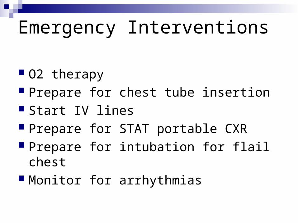
Emergency Interventions
O2 therapy Prepare for chest tube insertion Start IV lines Prepare for STAT portable CXR Prepare for intubation for flail chest Monitor for arrhythmias

Pathophysiology
Hypoxia Hypovolemia Pulmonary ventilation/perfusion mismatch Changes in intrathoracic pressure
relationships Respiratory acidosis, Hypercarbia Metabolic acidosis

Flail Chest
Complication of blunt trauma, 2 or more ribs next to each other are broken in half
Inward movement of thorax during inspiration and outward during expiration
Fractured ribs Fractured sternum-blunt deceleration May occur after CPR

Flail Chest Assessment
Chest wall is unstable and leads to repiratory distress, dyspnea, anxiety
Breath sounds diminished and crackles may be heard
Hypoventilation and hypoxemia Hypotension/ inadequate tissue perfusion and
metabolic acidosis—shock Pain assessment

Management of Flail Chest
Depending on the amount of distressMild-moderate: Humidified O2 Pain management Promotion of lung expansion through DB and positioningSevere: Mechanical ventilation IV hydration Monitor ABGs, pulse ox, pain management Psychosocial support

Pulmonary Contusion
Due to blunt trauma-potentially lethal
Damage leading to lung tissue hemorrhage and local edema
Damage to the lung leads to leakage of serum proteins and plasma
Increased oncotic pressure pulls fluid into lungs…. Results in hypoxemia and CO2 retention
May not be evident for 12-24hrs

S&S of Pulmonary Contusion
MILD Tachypnea Tachycardia Pleuritic chest pain Hypoxemia Blood tinged sputum
SEVERE Tachypnea Tachycardia Severe hypoxemia Crackles Respiratory acidosis Mental changes
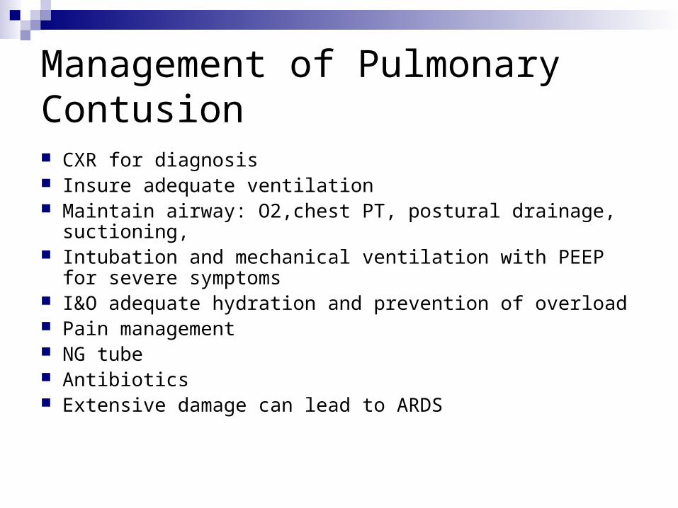
Management of Pulmonary Contusion CXR for diagnosis Insure adequate ventilation Maintain airway: O2,chest PT, postural drainage, suctioning, Intubation and mechanical ventilation with PEEP for severe
symptoms I&O adequate hydration and prevention of overload Pain management NG tube Antibiotics Extensive damage can lead to ARDS

Diaphragmatic Rupture
Herniation of the abdominal viscera into the chest
Most often occurs on left side

S&S Diaphragmatic Rupture
Dyspnea Cyanosis Dysphagia Sharp shoulder pain Bowel sounds in lower to middle chest Decreased breath sounds

Management
Maintain adequate oxygenation with endotracheal tube placement and mechanical ventilation
NGT Immediate surgical repair

Acute Respiratory Failure
Pressure of arterial oxygen < 60 mm Hg Pressure of arterial carbon dioxide > 50 mm
Hg pH < 7.30 O2 sats < 90% Ventilatory failure, oxygenation failure, or a
combination of both ventilatory and oxygenation failure
Mortality rate is 50-60%

Acute Respiratory FailureClassification
1.Ventilatory Failure-perfusion is normal but ventilation is inadequate
Causes: extrapulmonary
intrapulmonary
2.Oxygenation Failure
3. Combined Ventilatory and Oxygenation Failure

Ventilatory Failure Type of mismatch in which perfusion is
normal but ventilation is inadequate Thoracic pressure insufficiently changed
to permit air movement into and out of the lungs
Mechanical abnormality of the lungs or chest wall
Defect in the brain’s respiratory control center
Impaired ventilatory muscle function

Causes of Ventilatory Failure
Decreased respiratory drive
Brain disorders
Dysfunction of the chest wall

Oxygenation Failure
Thoracic pressure changes are normal, and air moves in and out without difficulty, but does not oxygenate the pulmonary blood sufficiently.
Ventilation is normal but lung perfusion is decreased.

Causes of Oxygenation Failure
Dysfunction of the lung parenchyma, conditions of the lung that interfere with ventilation by preventing expansion of the lung
Pain-restricting chest movement Ascites Upper airway obstruction

Combined Ventilatory and Oxygenation Failure Hypoventilation involves poor
respiratory movements. Gas exchange at the alveolar-capillary
membrane is inadequate—too little oxygen reaches the blood and carbon dioxide is retained.

Causes of Ventilation/Oxygenation Failure CAL Cardiac failure- can’t reverse hypoxia by
increasing CO

Dyspnea Encourage deep breathing exercises. Assess for:
Perceived difficulty breathingOrthopnea: client finds it easier to breathe
when in upright positionOxygenPosition of comfortEnergy-conserving measuresPulmonary drugs

Assessment of ARF
HYPOXEMIA Dyspnea Tachypnea Cyanosis Restlessness Apprehension Confusion Impaired judgement Tachycardia Dysrhythmias Hypertension
HYPERCAPNIA Dyspnea Respiratory depression Headache Pailedema Tachycardia Hypertension Drowsiness Coma Heart failure

Management of ARF
GOALS: treat the underlying cause and restore adequate gas exchange
Keep O2 >60% C&DB, respiratory tx Prevent complications of immobility Monitor ABGs and pulse Ox Maintain endotracheal intubation and mechanical
ventilation Relaxation techniques Energy conserving measures

ARDSAcute Respiratory Distress SyndromeOther names-wet lung, shock lungForm of acute respiratory failurePathophysiology is complex and not clearly understoodAcute respiratory failure occurs 1-96hrs after a pulmonary or non
pulmonary eventChemical mediators and endotoxins are released by the body
which cause increased capillary permeability and pansystemic microvascular injury
Alveoli fill with RBCs, neutrophils and protein-rich fluid which impairs perfusion and damages surfactant
Decreased surfactantBlood in capillaries pass damaged alveoli “shunting”Hypoxemia not responsive to O2 tx

Acute Respiratory Distress Syndrome Refractory Hypoxemia that persists even
when oxygen is administered at 100% Severe dyspnea, with air hunger, retractions
and cyanosis. Works at breathing Noncardiac-associated bilateral pulmonary
edema Dense pulmonary infiltrates seen on x-ray Decreased lung compliance (stiff lung)

Causes of Lung Injury in Acute Respiratory Distress Syndrome Systemic inflammatory response is the
common pathway. Intrinsically the alveolar-capillary membrane is
injured from conditions such as sepsis and shock.
Extrinsically the alveolar-capillary membrane is injured from conditions such as aspiration or inhalation injury.
“Leaky capillaries”- increased permeability leads to alveolar flooding and collapse

Common Causes Of ARDSDamage directly or indirectly to the Lung Shock, trauma Cardiopulmonary bypass Serious nerve injury Pancreatitis Fat and amniotic fluid emboli Pulmonary infections Sepsis and multi-system failure (30-40% mortality) Inhalation of toxic gases Pulmonary aspiration Drug ingestion (opioids, heroin, ASA) Hemolytic disorders Multiple transfusions Near drowning
Diagnostic Assessment
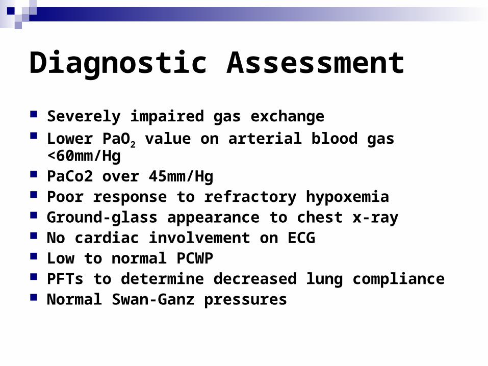
Severely impaired gas exchange Lower PaO2 value on arterial blood gas <60mm/Hg PaCo2 over 45mm/Hg Poor response to refractory hypoxemia Ground-glass appearance to chest x-ray No cardiac involvement on ECG Low to normal PCWP PFTs to determine decreased lung compliance Normal Swan-Ganz pressures
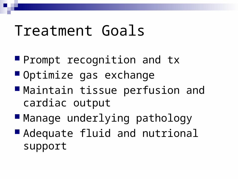
Treatment Goals
Prompt recognition and tx Optimize gas exchange Maintain tissue perfusion and cardiac
output Manage underlying pathology Adequate fluid and nutrional support

Medical Management
Endotracheal intubation and mechanical ventilation (PEEP, CPAP)
Monitor for complications of PEEP Neuromuscular blocking drugs Sedation Corticosteroids Antibiotics Fluid volume Induced diuresis TPN or enteral feedings Prone position prn Surfactant and nitrous oxide NSAIDS

Phase I
Dyspnea and Tachypnea
Tx Support
Provide O2

Phase II Interventions Increasing Pulmonary Edema Endotracheal intubation and
mechanical ventilation with positive end-expiratory pressure or continuous positive airway pressure
Drug therapy Nutrition therapy; fluid therapy

Phase III
Occurs over 2-10 days Progressive hypoxemia not responsive to
high levels O2 Support failing lung until it can heal

Phase IV
Occurs after 10 days Pulmonary fibrosis- irreversible Late or chronic ARDS Goals: To prevent sepsis, PN, MODS May require long term ventilation

Mechanical VentilationIndications Airway protection when the pt loses reflexes To provide positive pressure or high O2
concentration To bypass airway obstruction Facilitating pulmonary hygiene and
suctioning of secretions when the client can’t handle secretions
Mechanical Ventilation Requires Endotracheal Intubation
“Artificial Airway”
Components of the endotracheal tube Preparation for intubation Verifying tube placement Stabilizing the tube Nursing care

Mechanical Ventilation
Types of ventilators:Negative-pressure ventilatorsPositive-pressure ventilators
1.Pressure-cycled ventilators 2.Time-cycled ventilators
3.Microprocessor ventilators
4.Volume-cycled (most common)

Modes of Ventilation
How the machine will ventilate the patient in relation to the pts own repiratory efforts
The ways in which the client receives breath from the ventilator include: Assist-control ventilation (AC) Synchronized intermittent mandatory ventilation
(SIMV) Bi-level positive airway pressure (BiPAP), CPAP
and others

Ventilator Settings
Settings are adjusted towards pt needs and include:
Mode of ventilation Tidal Volume- Normal 7-10ml/kg FiO2- 21%-100% Rate- breaths per minute Sighs- increases air 1.5-2x Specialized delivery modes: CPAP or PEEP PEEP is used if FiO2 is>50%

Nursing Management
First concern is for the client; second for the ventilator.
Monitor and evaluate response to the ventilator.
Manage the ventilator system safely. Prevent complications.

Nursing Management:
Monitor/evaluate response to ventilation
Monitor respiratory patterns and lung sounds Does pt assist/buck vent Assess airway tubes frequently, minimal leak
technique BP and HR CXR, observe for SQ emphysema ABGs/Pulse ox Plan methods for communication Sedation/anti-anxiety meds as needed Observe for ICU psychosis
Nursing Management:
Manage the ventilator system safely
Monitor ventilator settings Suction prn- preoxygenate, when?/ Provide humidification Check alarms-always have alarms
activiated Remove condensation in tubing
Nursing Management:
PreventComplications Complications can include:
PulmonaryCardiacGastrointestinal and nutritional
Infection Muscular complications Ventilator dependence Inadvertant Extubation
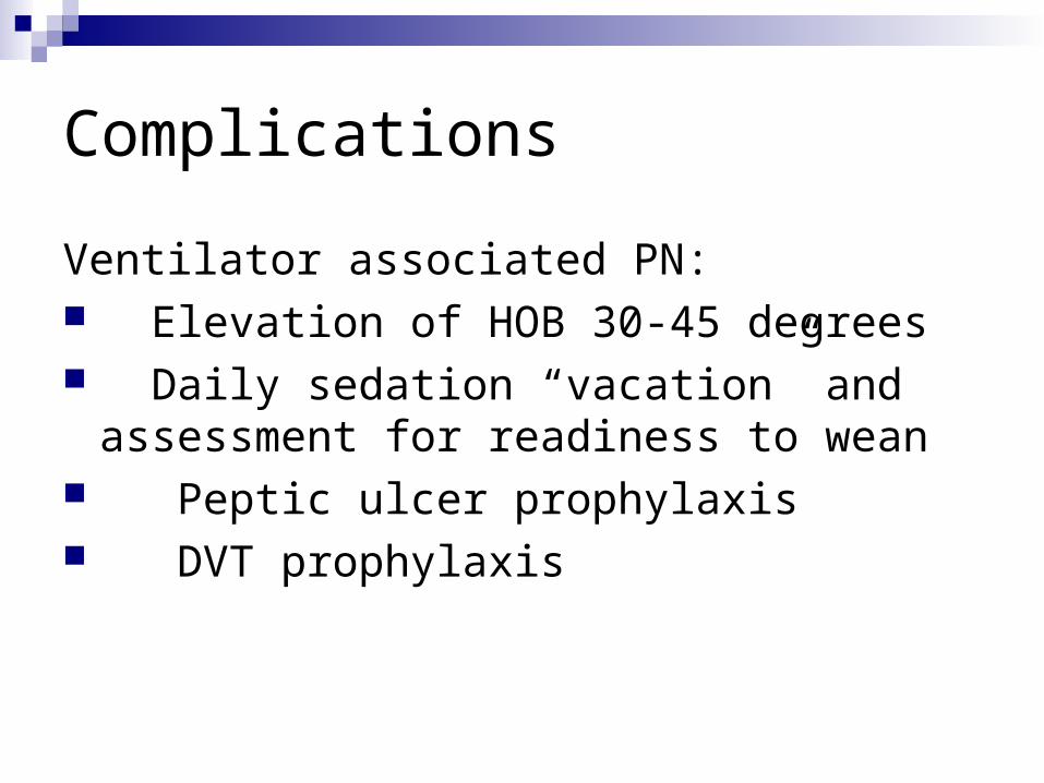
Complications
Ventilator associated PN: Elevation of HOB 30-45 degrees Daily sedation “vacation” and
assessment for readiness to wean Peptic ulcer prophylaxis DVT prophylaxis

ComplicationsMalnutrition is major reason why pts cannot be
weaned Nutrition: daily weights maintain TF or TPN
GI Bleed- stress ulcer preventionNose, lip, trachea problemsDecreased saliva and mouth ulcersBarotrauma-hypoxemia,crepitus,no breath soundsPneumothoraxVentilator dependence

Troubleshooting the VentCHECK PT FIRSTDO NOT IGNORE ALARMS
HIGH PRESSURE ALARM
1.pt needs to be suctioned
2.pt bucking/fighting the vent
3.displacement of ET tube
4.pt coughing when machine gives breath
5.water in the tubing

Troubleshooting
LOW PRESSURE ALARM
1. Leak-in the system
2. Disconnected tubing

Weaning From A VentilatorGOAL: SPONTANEOUS BREATHING
Factors related to weaning: 1. Pre-existing lung condition 2. Duration of mechanical ventilation 3. Pt physical and psychological condition
Short term vs long term

Weaning From A Ventilator
Ability to sustain spontaneous ventilation Monitor for respiratory distress Position to facilitate breathing Energy conservation-assist with care Avoid sedatives and respiratory
depressant meds

The Big Moment Has ArrivedEXTUBATION TIME Explain procedure Have O2 available Suction ET/oral Deflate cuff Have pt cough while tube is pulled Assess for respiratory fatigue and
obstruction Assess voice/sore throat
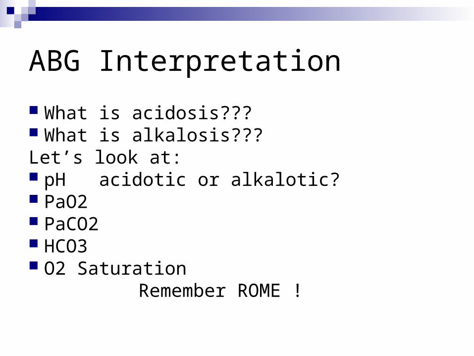
ABG Interpretation
What is acidosis??? What is alkalosis???Let’s look at: pH acidotic or alkalotic? PaO2 PaCO2 HCO3 O2 Saturation Remember ROME !

ABG Normal Ranges
pH 7.35-7.45
PaCO2 35-45 mm Hg
PaO2 80-100 mm Hg
SaO2 95%-100%
HCO3 22-26 mEq/L

Acid/Base MnemonicRemember ROME
R Respiratory O Opposite
pH up PCO2 down = Alkalosis pH down PCO2 up = Acidosis
M Metabolic E Equal
pH up HCO3 up = Alkalosis pH down HCO3 down = Acidosis

NCLEX Time
Of the following clients, which would be appropriate to assign to an LPN?
A.A 20-year-old man on a ventilator with a history of tension pneumothorax and currently awaiting transport to another hospital
B.A 59-year-old postoperative woman with a history of pulmonary embolism who is receiving subcutaneous heparin
C.A 65-year-old woman with acute respiratory distress syndrome who is on a ventilator and has a history of gastrointestinal bleeding
D.An 80-year-old man with a history of cancer of the larynx who is receiving CPAP ventilation through his tracheostomy

NCLEX Time
Of the following orders which would the nurse do first on a client who was intubated 30 minutes ago for acute respiratory distress syndrome?
A.Hang Levaquin 500 mg IV and D5 ½ normal saline.B.Obtain aerobic and anaerobic sputum culture.C.Increase ventilator rate as needed to keep between
16 and 20 breaths/min.D.Obtain arterial blood gases (ABGs) and pulmonary
wedge pressure via the arterial line.

NCLEX Time
Of the following tasks, which is appropriate to delegate to a new graduate nurse working with you?
A.Assessing respiratory system on a ventilated client with a history of barotrauma
B.Telephoning the cardiologist regarding a client you have just assessed who is complaining of shortness of breath and has noted ST depression
C.Administering Plavix to a client with a pulmonary embolism and paraplegia secondary to a spinal cord injury
D.Stripping the chest tube on a client with a left hemothorax from a motor vehicle collision sustained 12 hours earlier
NCLEX Time
Which of the following patients need immediate attention?
A.The 89-year-old male ventilated patient intermittently coughing
B.The 74-year-old female ventilated patient with noted tracheal deviation
C.The 57-year-old male patient recently extubated and complaining of a sore throat
D.The 40-year-old woman on BiPAP for asthma and with increased anxiety
NCLEX Time
Which of the following clients should the medical-surgical nurse consider transferring to the intensive care unit?
A.The 75-year-old client with a diagnosed pulmonary embolism who is receiving heparin and who currently is experiencing hemoptysis
B.The 63-year-old client with deep vein thrombosis receiving low–molecular-weight heparin and who has no calf pain
C.The 59-year-old client with a right pneumothorax currently being treated with a chest tube and oximetry of 96% on room air
D.The 30-year-old client with a history of being intubated 3 days ago and is currently on nasal cannula oxygen with clear lung sounds bilaterally

NCLEX Time
After starting oxygen 40% by face mask to a client with respiratory failure, an arterial blood gas is obtained. Which change would require immediate attention?
A.pH changes from 7.37 to 7.32
B.PaO2 increases from 56 to 60
C.PaCO2 increases from 47 to 55
D.O2 Sat remains at 88%