Embed Size (px)
Citation preview
Eugenia Conti, MD, PhDa,b
Kerstin Pannek, PhDc
Sara Calderoni, MD, PhDb
Anna Gaglianese, PhDb
Simona Fiori, MDa
Paola Brovedani, MScb
Danilo Scelfob
Stephen Rose, PhDc
Michela Tosetti, PhDb
Giovanni Cioni, MDa,b
Andrea Guzzetta, MD, PhDb
a Division of Child Neurology and Psychiatry,
University of Pisa, Italyb SMILE Lab, Department of Developmental
Neuroscience, Stella Maris Scientific Institute, Pisa,
Italyc The Australian e-Health Research Centre, CSIRO,
Brisbane, Australia
Correspondence to: Andrea Guzzetta
E-mail: [email protected]
Summary
In recent years, the use of brain diffusion MRI has led
to the hypothesis that children with autism spectrum
disorder (ASD) show abnormally connected brains.
We used the model of disease-discordant identical
twins to test the hypothesis that higher-order diffu-
sion MRI protocols are able to detect abnormal con-
nectivity in a single subject.
We studied the structural connectivity of the brain of
a child with ASD, and of that of his unaffected identi-
cal twin, using high angular resolution diffusion
imaging (HARDI) probabilistic tractography. Cortical
regions were automatically parcellated from high-res-
olution structural images, and HARDI-based connec-
tion matrices were produced for statistical compari-
son. Differences in diffusion indexes between sub-
jects were tested by Wilcoxon signed rank test. Tracts
were defined as discordant when they showed a
between-subject difference of 10 percent or more.
Around 11 percent of the discordant intra-hemispher-
ic tracts showed lower fractional anisotropy (FA) val-
ues in the ASD twin, while only 1 percent showed
higher values. This difference was significant.
High angular resolution diffusion imaging in a child with autism spectrum disorder and comparison with his unaffected identical twin
Our findings in a disease-discordant identical twin
pair confirm previous literature consistently report-
ing lower FA values in children with ASD.
KEY WORDS: abnormal connectivity, autism, connectome, correla-
tion matrix
Introduction
The autism spectrum disorders (ASDs) are a group of
complex neurodevelopmental disorders characterized
by social and communication impairments and restrict-
ed and repetitive interests (American Psychiatric
Association, 2013). Although the pathogenesis of
autism is still unknown, there is substantial agreement
that it has a multifactorial origin involving interaction
between multiple susceptibility genes, epigenetic
effects and environmental factors (Currenti, 2010).
Recent concurrent evidence suggests that a disruption in
brain connectivity is present in ASD individuals, mainly
consisting of diffuse cortical under-connectivity (Aoki et al.,
2013; Kana et al., 2011; Travers et al., 2012). It is not yet
clear whether this disrupted connectivity represents the
state-independent endophenotype of the disease (i.e. a
genetically determined hallmark of vulnerability) or
whether it is, rather, an actual marker of disease manifes-
tation (i.e. a biological correlate of the first signs and symp-
toms of the disease) (Gottesman and Gould, 2003). Were
this latter notion found to be correct, it may prove very use-
ful in leading to more specific diagnostic procedures and
effective resource allocation for early intervention.
A model that can be proposed to help disentangle the con-
tribution of genetic make-up from the influence of epige-
netic and environmental factors is that of disease-discor-
dant identical twins, i.e. monozygotic (MZ) twin pairs in
which only one individual presents with the disease. To
date, no studies have used this twin model in ASDs to
shed light on the role of disrupted connectivity in the dis-
ease pathogenesis. In the present study we address this
issue by analyzing inter-individual differences in brain
connectivity between a child with ASD and his unaffected
identical twin using a higher-order diffusion MRI approach.
Methods
Two MZ male twins (S1 and S2) aged five years and
two months were referred to our institute in February
Functional Neurology 2015; 30(3): 203-208 203
12_Conti 3b_FN 3 2015 13/10/15 10:27 Pagina 203
© CIC
Edizion
i Inter
nazio
nali
2012 for clinical assessment (Table I). They were born
preterm at 27 weeks’ gestation. At birth, S1 weighed
1125 g; his length was 35.5 cm and his head circum-
ference was 25 cm. S2 weighed 1000 g; he was 35 cm
long and had a head circumference of 25 cm. Brain
ultrasounds showed a transient flare, which normalized
within the 10th day of life, in both subjects. At term-
equivalent age, brain ultrasounds were confirmed as
normal. The twins were raised in the same home and
history taking revealed no notable differences in envi-
ronmental exposure. In both children, growth meas-
ures had always been within the normal range.
Following admission to our Child Neurology Unit both
twins underwent a five-day neurodevelopmental
observation, involving clinical evaluation and adminis-
tration of standardized tests, including the Griffiths
Developmental Scale (Griffiths, 1976), the Wechsler
Pre-school and Primary Scale of Intelligence –
Revised (Wechsler, 1990), the Movement Assessment
Battery for Children (Henderson and Sugden, 1992),
and the Autism Diagnostic Observation Schedule-
Generic (Lord et al., 2000; Tancredi et al., 2005).
Screening for homozygosity was performed by means
of i) genome sequence identity of the following loci:
CSF1PO, D2S1338, D3S1358, D5S818, D7S820,
D81179, D13S317, D16S539, D18S51, D21S11,
D19S433, FGA, THO1, TPOX, VWA, Amg, ii) poly-
merase chain reaction analysis of 15 microsatellites,
and iii) fragment analysis performed using an ABI
PRISM 3100 Genetic Analyzer (Applied Biosystems,
Foster City, California).
Brain MRI was obtained using a 1.5T scanner (General
Electric Medical Systems, Milwaukee, Wisconsin), an
examination that is part of our institute’s diagnostic
protocol for children with neurodevelopmental delay of
unknown origin. Both patients received inhalational
anesthesia with an odorless oxygen and nitrous mix-
ture for induction and sevoflurane for maintenance. No
side effects were reported. A high-resolution T1 struc-
tural image was acquired for each participant together
with a high angular resolution diffusion imaging
(HARDI) sequence (30 directions, b=1000 s/mm2)
(Tournier et al., 2012). An intensity-based normaliza-
tion software suite called Advanced Normalization
Tools (http://stnava.github.io/ANTs) (Avants et al.,
2008; Avants and Gee, 2004) was used to perform
motion correction of diffusion-weighted images. For
this purpose, a cross correlation similarity matrix for
rigid affine transformation was used (Rose et al.,
2012). Cortical parcellation was performed on structur-
al images using the Freesurfer image analysis suite
(http://surfer.nmr.mgh.harvard.edu), which parcellates
the cortex into 33 units per hemisphere on the basis of
gyral and sulcal structure, and can be reliably used in
children as young as four years of age (Ghosh et al.,
2010).
The fiber orientation distribution was estimated using
constrained spherical deconvolution with MRtrix soft-
ware (https://github.com/MRtrix3/mrtrix3/wiki) (Tournier
et al., 2012), which allows resolution of crossing fibers
and hence more accurate tractographic delineation of
white matter pathways (Jones, 2008). Fiber tracking
was also performed with MRtrix. Five million probabilis-
tic streamlines were generated seeding randomly over
the entire brain volume to create a whole-brain trac-
togram. Information from the Freesurfer cortical parcel-
lation was combined with tractography information to
obtain a connection matrix. The terminal end points of
every streamline were hit-tested with every cortical
region. Only connections containing at least 250
streamlines were retained for further analysis, and
weighted mean fractional anisotropy (FA) and mean dif-
fusivity (MD) were calculated within these connections.
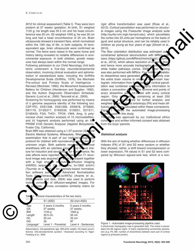
Figure 1 outlines the automated image-processing
pipeline used in this study.
The study was approved by our institutional ethics
committee and written informed consent was obtained
from the boys’ parents.
Statistical analysis
With the aim of testing whether differences in diffusion
indexes (FA) of S1 and S2 were random or whether
they showed, rather, a shift toward overexpression or
lower expression, FA values in S1 and S2 were com-
pared by Wilcoxon signed-rank test, which is a non-
E. Conti et al.
204 Functional Neurology 2015; 30(3): 203-208
Table I - Clinical characteristics of the two twins.
S1 (ASD) S2 (non-ASD)
Age 5 years 2 months 5 years 2 monthsGA 27 weeks 27 weeksBW 1125 g 1000 gLength 35.5 cm 35 cmHC 25 cm 25 cmDQ 35 87Language* Level 1 - Preverbal Level 4 - Sentences
Abbreviations: GA=gestational age, BW=birth weight; HC=head circum-
ference; DQ=developmental quotient. *Assessed according to Tager-
Flusberg et al., 2009
Figure 1 - Automated image-processing pipeline used.A whole-brain tractography map is generated (left). The cortex is parcel-
lated into 66 regions (right). A matrix representing connectivity parame-
ters (e.g. FA, MD, number of streamlines) between each pair of cortical
regions is produced (center).
12_Conti 3b_FN 3 2015 13/10/15 10:27 Pagina 204
© CIC
Edizion
i Inter
nazio
nali
parametric test that compares two dependent obser-
vations and counts the number of negative and posi-
tive differences. The number of discordant tracts,
defined as those showing a between-subject differ-
ence of 10 percent or more, was counted.
Results
The main clinical characteristics of the subjects are
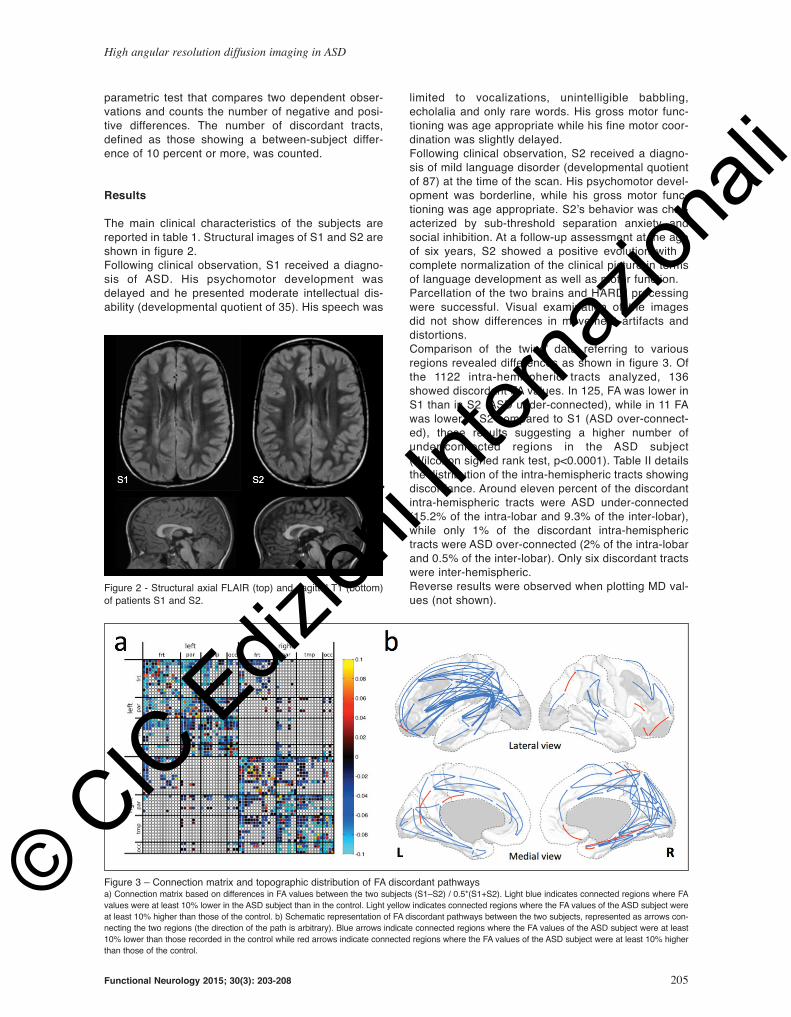
reported in table 1. Structural images of S1 and S2 are
shown in figure 2.
Following clinical observation, S1 received a diagno-
sis of ASD. His psychomotor development was
delayed and he presented moderate intellectual dis-
ability (developmental quotient of 35). His speech was
limited to vocalizations, unintelligible babbling,
echolalia and only rare words. His gross motor func-
tioning was age appropriate while his fine motor coor-
dination was slightly delayed.
Following clinical observation, S2 received a diagno-
sis of mild language disorder (developmental quotient
of 87) at the time of the scan. His psychomotor devel-
opment was borderline, while his gross motor func-
tioning was age appropriate. S2’s behavior was char-
acterized by sub-threshold separation anxiety and
social inhibition. At a follow-up assessment at the age
of six years, S2 showed a positive evolution with a
complete normalization of the clinical picture in terms
of language development as well as motor function.
Parcellation of the two brains and HARDI processing
were successful. Visual examination of the images
did not show differences in movement artifacts and
distortions.
Comparison of the twins’ data referring to various
regions revealed differences as shown in figure 3. Of
the 1122 intra-hemispheric tracts analyzed, 136
showed discordant FA values. In 125, FA was lower in
S1 than in S2 (ASD under-connected), while in 11 FA
was lower in S2 compared to S1 (ASD over-connect-
ed), these results suggesting a higher number of
under-connected regions in the ASD subject
(Wilcoxon signed rank test, p<0.0001). Table II details
the distribution of the intra-hemispheric tracts showing
discordance. Around eleven percent of the discordant
intra-hemispheric tracts were ASD under-connected
(15.2% of the intra-lobar and 9.3% of the inter-lobar),
while only 1% of the discordant intra-hemispheric
tracts were ASD over-connected (2% of the intra-lobar
and 0.5% of the inter-lobar). Only six discordant tracts
were inter-hemispheric.
Reverse results were observed when plotting MD val-
ues (not shown).
High angular resolution diffusion imaging in ASD
Functional Neurology 2015; 30(3): 203-208 205
Figure 2 - Structural axial FLAIR (top) and sagittal T1 (bottom)
of patients S1 and S2.
Figure 3 – Connection matrix and topographic distribution of FA discordant pathwaysa) Connection matrix based on differences in FA values between the two subjects (S1–S2) / 0.5*(S1+S2). Light blue indicates connected regions where FA
values were at least 10% lower in the ASD subject than in the control. Light yellow indicates connected regions where the FA values of the ASD subject were
at least 10% higher than those of the control. b) Schematic representation of FA discordant pathways between the two subjects, represented as arrows con-
necting the two regions (the direction of the path is arbitrary). Blue arrows indicate connected regions where the FA values of the ASD subject were at least
10% lower than those recorded in the control while red arrows indicate connected regions where the FA values of the ASD subject were at least 10% higher
than those of the control.
12_Conti 3b_FN 3 2015 13/10/15 10:27 Pagina 205
© CIC
Edizion
i Inter
nazio
nali

Discussion
The distribution of corticocortical connections showing
different FA values between our twins (discordant
tracts), and specifically our finding of a significant
majority of discordant tracts (94.2%) having lower FA
values in the ASD twin, is broadly consistent with pre-
vious literature reporting lower FA values in children
with ASD. Most studies show that structural connectiv-
ity between brain regions is weaker, in terms of lower
FA values, in individuals with ASD compared to con-
trols. Reduced FA of white matter has been reported
as a global feature of the ASD brain, particularly in the
frontal and temporal cortex, in the main fasciculi con-
necting different lobes, in subcortical regions and in
the corpus callosum [see (Vissers et al., 2012) for a
review]. Our study is mostly consistent with these
reports, in that ASD tracts with lower FA values were
found both within the intra-lobar connections (with
percentages ranging from 10 to 25% in the four lobes)
and within the inter-lobar connections, and in particu-
lar in the parieto-occipital, temporo-occipital and fron-
toparietal tracts.
The fact that the great majority of discordant tracts
observed in our twins were located in the left hemi-
sphere, with discordance involving the frontoparietal
and frontotemporal connections in particular (see Fig.
3b), suggests a possible reduction of FA left lateraliza-
tion in the ASD twin. Indeed, one of the emerging bio-
logical hallmarks of ASD is the loss or inversion of the
typical patterns of brain lateralization, which involve,
in particular, but are not limited to, the language-relat-
ed frontotemporal networks (Toga and Thompson,
2003). Recent diffusion imaging and tractography
studies in adolescents and young adults with high-
functioning ASD have provided reports of loss or
inversion of the left-right asymmetry in the cingulate,
arcuate fasciculus and uncinate fasciculus (Knaus et
al., 2010; Lo et al., 2011; Fletcher et al., 2010), in the
superior temporal gyrus (Lange et al., 2010), and in
the pathways involving the fusiform (Conturo et al.,
2008). Converging evidence has also been provided
by functional MRI studies showing reduced asymme-
tries in brain representation of language-related net-
works (Eyler et al., 2012).
A small number of discordant tracts in our study
showed higher FA values in the ASD twin. Increased
connectivity values have previously been reported in
individuals with ASD, particularly within the frontal lobe,
using functional and structural methods. For example,
increased functional connectivity was found in the fron-
tostriatal circuitry in adolescents and young adults with
ASD (Delmonte et al., 2013), a finding consistent with
earlier studies (Di Martino et al., 2011). Increased con-
nectivity in the frontal regions has been interpreted as a
compensatory mechanism for global under-connectivity
(Kana et al., 2011). Although it needs to be underlined
that our statistical analysis only supports the presence
of a significantly higher number of tracts with lower FA
values in the ASD twin, it is of interest that in our two
patients almost half (46%) of the tracts with higher FA
values were found within the frontal lobe, versus only
15% of the ones showing lower FA values.
Low FA values have been widely interpreted as an
index of weak structural connectivity, thus reflecting
functional evidence of a pattern of under-connectivity in
ASD (Vissers et al., 2012). However, it needs to be
underlined that diffusion data, above all FA data, may
not be interpreted as a direct measure of structural con-
nectivity (Castellanos et al., 2014; Jones et al., 2013).
A potential bias in our analysis is that S2, the non-ASD
twin, did not show a psychomotor profile fully typical for
a healthy subject, as he received a diagnosis of mild
language disorder, and showed some sub-threshold
separation anxiety and social inhibition. As some
reports support partially overlapping neurophysiologi-
cal underpinnings of language impairment and ASD
(Verly et al., 2014), it might be suggested that the dis-
E. Conti et al.
206 Functional Neurology 2015; 30(3): 203-208
Table II - Distribution of discordant tracts.
Total number of ASD under-connected tracts ASD over-connected tractsintra-hemispheric % of the total % of the total tracts (% of the under-connected) (% of the over-connected)
INTRA-LOBARF-F 182 10.4 (15.2) 2.7 (45.5)P-P 56 23.2 (10.4) 1.8 (9.1)T-T 90 17.8 (12.8) 1.1 (9.1)O-O 20 25.0 (4.0) 0
Total intra-lobar 348 15.2 2.0
INTERLOBARF-P 182 12.1 (17.6) 1.1 (18.2)F-T 234 6.0 (11.2) 0F-O 104 2.9 (2.4) 0P-T 126 6.3 (6.4) 0P-O 56 23.2 (10.4) 1.8 (9.1)T-O 72 16.7 (9.6) 1.4 (9.1)
Total inter-lobar 774 9.3 0.5
Total 1122 11.1 1.0
Abbreviations: F=frontal; P=parietal; T=temporal; O=occipital; ASD=autism spectrum disorder
12_Conti 3b_FN 3 2015 13/10/15 10:27 Pagina 206
© CIC
Edizion
i Inter
nazio
nali

cordance in our twins is an underestimation of the real
differences between ASD and typical brains. It should
also be noted that the analysis could be affected by the
presence of white matter abnormal signal intensity in
both subjects, likely related to preterm birth. Indeed,
cerebral white matter abnormalities on structural MRI
have been extensively described in infants born pre-
maturely, in particular punctate high-signal intensities
on T1-weighted images and diffuse high-signal intensi-
ties on T2-weighted images (Rutherford et al., 2010).
A very low birth weight (<1500 g) as well as prematuri-
ty have repeatedly been identified as risk factors for an
ASD (Elgen et al., 2002; Indredavik et al., 2010;
Limperopoulos et al., 2008). In fact, the prevalence of
ASD among children born prematurely is between
3.65% and 8% (Hack et al., 2009; Johnson et al., 2010;
Pinto-Martin et al., 2011), compared to 1% in the gen-
eral population of school-aged children (Baron-Cohen
et al., 2009). Future investigations should attempt to
determine the role of these neonatal and perinatal risk
factors in the development of ASD.
This study is the first attempt to use the model of dis-
cordant twins to explore the role of abnormal connec-
tivity in the pathogenesis of ASD. In a recent longitu-
dinal study on 92 high-risk infant siblings, 28 of which
were eventually diagnosed with ASD, the FA trajecto-
ries, from six to 24 months, for several fiber tracts dif-
fered significantly between the infants who developed
ASD and those who did not, suggesting that aberrant
white matter development may precede the manifes-
tation of autistic symptoms, thus representing a reli-
able biomarker of the disease (Wolff et al., 2012).
Since the affected subjects shared only some of the
genetic heritage of their unaffected siblings, it could
not be determined whether this biomarker is more the
direct result of genetic determination predisposing to
ASD, or rather an early sign of a multifactorial process
(genetic, epigenetic and environmental) leading to dis-
ease expression. Our findings are more in favor of the
latter. Since our twins share the same genetic her-
itage, the differences in brain connectivity, which are
consistent with previous studies, suggest that abnor-
mal connectivity as a biomarker for ASD lies closer to
the final steps in the pathogenetic process than to the
early ones. Unquestionably, our study represents only
a first, preliminary attempt to address these important
issues. The model of discordant MZ twins is promising
and we expect our preliminary data to foster further
research on this topic.
Acknowledgments
We would like to thank Maria Puopolo for the statisti-
cal analysis.
References
American Psychiatric Association (2013). Diagnostic and
Statistical Manual of Mental Disorders (5th ed.). Arlington,
VA, American Psychiatric Publishing.
Aoki Y, Abe O, Nippashi Y, et al (2013). Comparison of white
matter integrity between autism spectrum disorder sub-
jects and typically developing individuals: a meta-analysis
of diffusion tensor imaging tractography studies. Mol
Autism 4:25.
Avants BB, Epstein CL, Grossman M, et al (2008). Symmetric
diffeomorphic image registration with cross-correlation:
evaluating automated labeling of elderly and neurodegen-
erative brain. Med Image Anal 12:26-41.
Avants B, Gee JC (2004). Geodesic estimation for large defor-
mation anatomical shape averaging and interpolation.
Neuroimage 23 Suppl 1:S139-150.
Baron-Cohen S, Scott FJ, Allison C, et al (2009). Prevalence of
autism-spectrum conditions: UK school-based population
study. Br J Psychiatry 194:500-509.
Castellanos FX, Cortese S, Proal E (2014). Connectivity. Curr
Top Behav Neurosci 16:49-77.
Conturo TE, Williams DL, Smith CD, et al (2008). Neuronal fiber
pathway abnormalities in autism: an initial MRI diffusion
tensor tracking study of hippocampo-fusiform and amygda-
lo-fusiform pathways. J Int Neuropsychol Soc 14: 933-946.
Currenti SA (2010). Understanding and determining the etiolo-
gy of autism. Cell Mol Neurobiol 30:161-171.
Delmonte S, Gallagher L, O’Hanlon E, et al (2013). Functional
and structural connectivity of frontostriatal circuitry in
Autism Spectrum Disorder. Front Hum Neurosci 7:430.
Di Martino A, Kelly C, Grzadzinski R, et al (2011). Aberrant stri-
atal functional connectivity in children with autism. Biol
Psychiatry 69:847-856.
Elgen I, Sommerfelt K, Markestad T (2002). Population based,
controlled study of behavioural problems and psychiatric
disorders in low birthweight children at 11 years of age.
Arch Dis Child Fetal Neonatal Ed 87:F128-132.
Eyler LT, Pierce K, Courchesne E (2012). A failure of left tempo-
ral cortex to specialize for language is an early emerging
and fundamental property of autism. Brain 135:949-960.
Fletcher PT, Whitaker RT, Tao R, et al (2010). Microstructural
connectivity of the arcuate fasciculus in adolescents with
high-functioning autism. Neuroimage 51:1117-1125.
Ghosh SS, Kakunoori S, Augustinack J, et al (2010). Evaluating
the validity of volume-based and surface-based brain
image registration for developmental cognitive neuro-
science studies in children 4 to 11 years of age.
Neuroimage 53:85-93.
Gottesman II, Gould TD (2003). The endophenotype concept in
psychiatry: etymology and strategic intentions. Am J
Psychiatry 160:636-645.
Griffiths R (1976). The Abilities of Babies: A Study in Mental
Measurement. London, UK, University of London Press.
Hack M, Taylor HG, Schluchter M, et al (2009). Behavioral out-
comes of extremely low birth weight children at age 8
years. J Dev Behav Pediatr 30:122-130.
Henderson SE, Sugden DA (1992). Movement assessment bat-
tery for children. Sidcup, Kent, The Psychological
Corporation.
Indredavik MS, Vik T, Evensen KA, et al (2010). Perinatal risk
and psychiatric outcome in adolescents born preterm with
very low birth weight or term small for gestational age. J
Dev Behav Pediatr 31:286-294.
Johnson S, Hollis C, Kochhar P, et al (2010). Autism spectrum
disorders in extremely preterm children. J Pediatr 156:
525-531.e2.
Jones DK, Knösche TR, Turner R (2013). White matter integri-
ty, fiber count, and other fallacies: the do’s and don’ts of
diffusion MRI. Neuroimage 73:239-254.
Jones DK (2008). Studying connections in the living human
brain with diffusion MRI. Cortex 44:936-952.
High angular resolution diffusion imaging in ASD
Functional Neurology 2015; 30(3): 203-208 207
12_Conti 3b_FN 3 2015 13/10/15 10:27 Pagina 207
© CIC
Edizion
i Inter
nazio
nali

Kana RK, Libero LE, Moore MS (2011). Disrupted cortical con-
nectivity theory as an explanatory model for autism spec-
trum disorders. Phys Life Rev 8:410-437.
Knaus TA, Silver AM, Kennedy M, et al (2010). Language later-
ality in autism spectrum disorder and typical controls: a
functional, volumetric, and diffusion tensor MRI study.
Brain Lang 112:113-120.
Lange N, Dubray MB, Lee JE, et al (2010). Atypical diffusion
tensor hemispheric asymmetry in autism. Autism Res 3:
350-358.
Limperopoulos C, Bassan H, Sullivan NR, et al (2008). Positive
screening for autism in ex-preterm infants: prevalence and
risk factors. Pediatrics 121:758-765.
Lo YC, Soong WT, Gau SS, et al (2011). The loss of asymme-
try and reduced interhemispheric connectivity in adoles-
cents with autism: a study using diffusion spectrum imag-
ing tractography. Psychiatry Res 192:60-66.
Lord C, Risi S, Lambrecht L, et al (2000). The autism diagnos-
tic observation schedule-generic: a standard measure of
social and communication deficits associated with the
spectrum of autism. J Autism Dev Disord 30:205-223.
Pinto-Martin JA, Levy SE, Feldman JF, et al (2011). Prevalence
of autism spectrum disorder in adolescents born weighing
<2000 grams. Pediatrics 128:883-891.
Rose S, Rowland T, Pannek K, et al (2012). Structural hemi-
spheric asymmetries in the human precentral gyrus hand
representation. Neuroscience 210:211-221.
Rutherford MA, Supramaniam V, Ederies A, et al (2010).
Magnetic resonance imaging of white matter diseases of
prematurity. Neuroradiology 52:505-521.
Tager-Flusberg H, Rogers S, Cooper J, et al (2009). Defining
spoken language benchmarks and selecting measures of
expressive language development for young children with
autism spectrum disorders. J Speech Lang Hear Res
52:643–652.
Tancredi R, Saccani M, Persico AM, et al (2005). Autism diag-
nostic observation schedule-generic. Edizione Italiana.
Florence, Giunti O.S. Organizzazioni Speciali.
Toga AW, Thompson PM (2003). Mapping brain asymmetry. Nat
Rev Neurosci 4:37-48.
Tournier JD, Calamante F, Connelly A (2012). MRtrix: Diffusion
tractography in crossing fiber regions. International
Journal of Imaging Systems and Technology 22:53-66.
Travers BG, Adluru N, Ennis C, et al (2012). Diffusion tensor
imaging in autism spectrum disorder: a review. Autism Res
5:289-313.
Verly M, Verhoeven J, Zink I, et al (2014). Structural and func-
tional underconnectivity as a negative predictor for lan-
guage in autism. Hum Brain Mapp 35:3602-3615.
Vissers ME, Cohen MX, Geurts HM (2012). Brain connectivity
and high functioning autism: a promising path of research
that needs refined models, methodological convergence,
and stronger behavioral links. Neurosci Biobehav Rev 36:
604-625.
Wechsler D (1990). Wechsler Pre-school and Primary Scale of
Intelligence–Revised. Sidcup, Kent, The Psychological
Corporation.
Wolff JJ, Gu H, Gerig G, et al (2012). Differences in white mat-
ter fiber tract development present from 6 to 24 months in
infants with autism. Am J Psychiatry 169:589-600.
E. Conti et al.
208 Functional Neurology 2015; 30(3): 203-208
12_Conti 3b_FN 3 2015 13/10/15 10:27 Pagina 208
© CIC
Edizion
i Inter
nazio
nali