Embed Size (px)
Citation preview
EUKARYOTIC CELL, May 2010, p. 795–805 Vol. 9, No. 51535-9778/10/$12.00 doi:10.1128/EC.00058-10Copyright © 2010, American Society for Microbiology. All Rights Reserved.
Interaction of the Aspergillus nidulans Microtubule-Organizing Center(MTOC) Component ApsB with Gamma-Tubulin and Evidence
for a Role of a Subclass of Peroxisomes in theFormation of Septal MTOCs�
Nadine Zekert,‡ Daniel Veith,†‡ and Reinhard Fischer*Karlsruhe Institute of Technology, Institute for Applied Biosciences, Department of
Microbiology, Hertzstrasse 16, D-76187 Karlsruhe, Germany
Received 6 March 2010/Accepted 19 March 2010
Peroxisomes are a diverse class of organelles involved in different physiological processes in eukaryotic cells.Although proteins imported into peroxisomes carry a peroxisomal targeting sequence at the C terminus (PTS1)or an alternative one close to the N terminus (PTS2), the protein content of peroxisomes varies drastically.Here we suggest a new class of peroxisomes involved in microtubule (MT) formation. Eukaryotic cells assembleMTs from distinct points in the cell. In the fungus Aspergillus nidulans, septum-associated microtubule-organizing centers (sMTOCs) are very active in addition to the spindle pole bodies (SPBs). Previously, weidentified a novel MTOC-associated protein, ApsB (Schizosaccharomyces pombe mto1), whose absence affectedMT formation from sMTOCs more than from SPBs, suggesting that the two protein complexes are organizeddifferently. We show here that sMTOCs share at least two further components, gamma-tubulin and GcpC (S.pombe Alp6) with SPBs and found that ApsB interacts with gamma-tubulin. In addition, we discovered thatApsB interacts with the Woronin body protein HexA and is targeted to a subclass of peroxisomes via a PTS2peroxisomal targeting sequence. The PTS2 motif was necessary for function but could be replaced with a PTS1motif at the C terminus of ApsB. These results suggest a novel function for a subclass of peroxisomes incytoskeletal organization.
Peroxisomes are ubiquitous organelles of eukaryotes whichare surrounded by a single membrane (9, 30). They serve avariety of functions, depending on the species, the cell type,and the environmental or developmental conditions. In mam-mals, peroxisomes are involved in a number of catabolic andanabolic pathways, most importantly, peroxide metabolism,the �-oxidation of long-chain fatty acids, and the biosynthesisof ether phospholipids (17, 37). The vital importance of theorganelle in humans is shown by the existence of a number ofsevere and often lethal disorders that occur when the biogen-esis of the organelle is impaired (36). In plants, peroxisomesare involved in photorespiration and typically contain theglyoxylate cycle, as in protozoa and yeast (8).
Given the complexity of peroxisomal functions, it is obviousthat a large number of proteins need to be targeted to theseorganelles. Peroxisomal membrane and matrix proteins aresynthesized on free ribosomes in the cytosol and are importedposttranslationally into preexisting organelles (9). The appara-tus of protein import is clearly distinct from the import ma-chinery of other organelles because it translocates folded andeven oligomeric proteins and there is evidence that they are
descending from the endoplasmic reticulum (6). A large num-ber of peroxisomal proteins employ a tripeptide sequence atthe C terminus, PTS1 (S/A/C-K/R/H-L/M) (7). A second classof proteins uses a sequence close to the N terminus which isless conserved, consists of R/K-L/I/V-X5-H/Q-L/A, and iscalled PTS2 (33). In both cases, complex protein machineriesare employed and some of the components appear to be usedin PTS1- and PTS2-dependent protein translocation (9).
A very distinct class of peroxisomes is represented by thefungal Woronin body. This structure is named after a Russianmycologist who reported the characteristics of a distinct type oforganelle in the fungus Ascobolus pulcherrimus (1, 39). Woro-nin bodies have been described in more than 50 species ofascomycota and deuteromycota but are missing in single-cellyeasts such as Saccharomyces cerevisiae and Schizosaccharomy-ces pombe. Thus, their function appears to be important for thefilamentous life style. In Neurospora crassa, they appear ashexagonal bodies in the cell and upon cell damage plug theseptal pores after a few seconds (15). This sealing mechanismis very important in syncytial organisms to prevent loss of theentire cytoplasm and hence death of the entire mycelium afterone hypha is damaged (20). Their exact composition, however,remained obscure for many decades until G. Jedd and N.-H.Chua purified the organelle from N. crassa and identified themain constituent as a single protein named Hex1 (15, 42),because it forms hexagonal crystals. The existence of a PTS1peroxisomal targeting sequence at the C terminus of the pro-tein indicated that the Woronin bodies represent specializedperoxisomes. Hex1 displays some sequence similarity to eIF5,and it is thought that Hex1 derived from eIF5 during evolution
* Corresponding author. Mailing address: Karlsruhe Institute ofTechnology, Institute for Applied Biosciences, Department of Micro-biology, Hertzstrasse 16, D-76187 Karlsruhe, Germany. Phone: 49-721-608-4630. Fax: 49-721-608-4509. E-mail: [email protected].
† Present address: Technologie-Lizenz-Buro (TLB) der Baden-Wurttembergischen Hochschulen GmbH, Ettlinger Str. 25, D-76137Karlsruhe, Germany.
‡ Equal contribution.� Published ahead of print on 26 March 2010.
795
at KIT
Library Karlsruhe on M
ay 4, 2010 ec.asm
.orgD
ownloaded from
by gene duplication and subsequent modification of its func-tion (42). Another example of a peroxisome-associated func-tion may be the Pro40 protein in Sordaria macrospora (5). Thisprotein is implicated in the regulation of sexual development.
In addition to the Woronin body close to the septal pore(22), we had evidence in Aspergillus nidulans that microtubule(MT) polymerization is initiated at septa. Using an MT plus-end-associated protein, the kinesin motor KipA (kinesin-7), weshowed that the cytoplasmic area close to septa acts as anactive MT-organizing center (MTOC) (16). Furthermore, weidentified a novel MTOC-associated protein, ApsB, and local-ized it to the spindle pole bodies (SPBs) and to septa (35). Thepresence of septal MTOCs is similar to that in S. pombe, butthere is no evidence for such organelles in the S. cerevisiae-related filamentous fungus Ashbya gossypii (18, 19). Given thatMTOCs are generally composed of a large protein complexwith gamma-tubulin as one characteristic member, we antici-pate that MT polymerization at septa also requires a proteincomplex (40). Here we show for the first time the presence ofgamma-tubulin at septal MTOCs (sMTOCs) and that it phys-ically interacts with ApsB. Surprisingly, ApsB is associated witha subclass of peroxisomes in the cytoplasm, and we proposethat they are involved in septal MTOC formation.
MATERIALS AND METHODS
Strains, plasmids, and culture conditions. The preparation of the supple-mented minimal and complete media used for A. nidulans and the standardstrain construction procedures used are described in reference 11. To isolatetotal DNA and RNA, corresponding strains were grown in liquid culture for 16 h.Mycelium was harvested and immediately processed for total DNA (see below).A list of the A. nidulans strains used in this study is given in Table 1. Standardlaboratory Escherichia coli strains (XL-1 blue and Top10) were used. The plas-mids used are listed in Table 2.
Light and fluorescence microscopy. For live-cell imaging, cells were grown inglass-bottom dishes (FD35-100; World Precision Instruments, Berlin, Germany)in 2 ml of minimal medium (MM) containing either 2% glycerol or 2% glucoseas a carbon source. Medium was supplemented with pyridoxine, p-aminobenzoicacid, biotin, arginine, uracil, or uridine, depending on the auxotrophy of thestrains. Cells were incubated at room temperature for 1 to 2 days, and imageswere captured using an Axiophot microscope (Zeiss, Jena, Germany), a Plan-apochromatic 63� or 100� oil immersion objective lens, and an HBO50 Hglamp. Alternatively, a Zeiss AxioImager Z1 with AxioVision software (V4.5) wasused. Fluorescence was observed using standard Zeiss filter combinations no. 09(fluorescein isothiocyanate, green fluorescent protein [GFP]) and no. 15 (mono-meric red fluorescent protein 1 [mRFP1], DsRed). Laser images were obtainedusing the Zeiss Cell Observer SD, which combines the high-end Cell Observermicroscopy platform and the CSU-X1 spinning-disc technology from Yokogawafor high-speed confocal microscopy. Images were collected and analyzed with aHamamatsu Orca ER II camera system and the Wasabi software (version 1.2) ora Zeiss Axiocam and AxioVision software. Image and video processing was donewith the Wasabi software from Hamamatsu, Adobe Photoshop, ImageJ (NIH,Bethesda, MD), and virtual dub (http://www.virtualdub.org).
Molecular techniques. Standard DNA transformation procedures were usedfor A. nidulans (41) and E. coli (27). For PCR experiments, standard protocolswere applied using a Biometra Personal Cycler (Biometra, Gottingen, Germany)for the reaction cycles. DNA sequencing was done commercially (MWG Biotech,Ebersberg, Germany). Total DNA was extracted from A. nidulans in the follow-ing way. Spores were inoculated into liquid MM plus supplements and grown for12 to 18 h at 30°C without shaking. Hyphal mats were harvested, dried with tissuepaper, and ground in liquid nitrogen. The resulting powder was mixed withextraction buffer (50 mM EDTA, 0.2% sodium dodecyl sulfate [SDS]) andincubated for 30 min to 2 h at 68°C in a water bath. SDS and proteins wereremoved from the suspension by addition of potassium-acetate solution (8 M, pH4.2) and centrifugation. Total DNA was precipitated from the supernatant withisopropanol, and the pellet was washed twice with 70% ethanol, air dried,resuspended in TE buffer with RNase A, and stored at 4°C. Southern hybrid-izations were performed according to the DIG Application Manual for Filter
Hybridization (Roche Applied Science, Technical Resources, Roche DiagnosticsGmbH, Mannheim, Germany).
Bimolecular fluorescence complementation assay (BiFC). The enhanced yel-low fluorescent protein (eYFP)-tagged N-terminal half (YFPN) was ampli-fied using primers 5�-CGGTACCATGGTGAGCAAGGGCGAGGAGCTG-3� (fwd_Kpn_YFP-N) and 5�-CGGCGCGCCCGTGGCGATGGAGCGCATGATATAGACGTTGTGGCTGTTGTAG-3�. For the C-terminally eYFP-tagged (YFPC) half, primers 5�-CGGTACCATGGCCGACAAGCAGAAGAACGGCATCAAGG-3� (fwd_Kpn_YFP-C) and 5�-CGGCGCGCCGTGGTTCATGACCTTCTGTTTCAGGTCGTTCGGGATCTTGCAGGCCGGGCGCTTGTACAGCTCGTCCATGCCGAGAGTGATCCC-3� (rev_YFP-C_Li_Asc) were used.These primers introduced KpnI and AscI restriction sites (in italics) in additionto the protein linker sequences RSIAT (YFPN) and RPACKIPNDLKQKVMNH (YFPC) (underlined). eYFP was split at bp 460 to 462 by using the ATGcodon as the start of the YFPC half. PCR fragments were subcloned intopCR2.1-Topo (Invitrogen, Karlsruhe, Germany), subsequently released withKpnI and AscI, and used to replace GFP2-5 of pMCB17apx-apsB (35), givingpDV7 (YFPN) and pDV8 (YFPC). Full-length apsB3.2 (3.2 kb) was taken frompDV21a and cloned into pDV7, giving pDV22b [alcA(p)::YFPN::apsB3.2], andfull-length �tubulin1.8 was amplified using primers 5�-CGGCGCGCCCGGGATGCCTAGGTATACCCTC-3� (Gamma_tub_Asc_fwd) and 5�-CTTAATTAATTATACTCCAACTTCATCCTTTCC-3� (Gamma_tub_Pac_rev) (the AscI andPacI restriction sites are in italics, and the start and stop codons are underlined)and cloned into pDV8, giving pDV50 [alcA(p)::YFPC::�tubulin1.8]. For BiFCanalysis, pDV22 and pDV50 were combined and transformed into GR5, givingSNZ11. The cloned gene length is indicated as exponents; e.g., the length ofthe cloned gene apsB3.2 is 3.2 kb. Similarly, hexA was amplified using primers5�-CGGCGCGCCCGGGATGGGTTACTACGACGACG-3� (hexA_Asc_fwd)and 5�-CTTAATTAATTATAGACGGGAAGAGTGGATGATC-3� (hexA_Pac_rev1; 680 bp to stop codon) or 5�-GTTAATTAACCTCAATCAAGTGCAAGGTTTCG-3� [hexA_Pac_rev2; 1 kb, including the poly(A) site] and cloned intopDV8, giving pDV17 [alcA(p)::YFPC::hexA680], and into pDV7, giving pDV19a[alcA(p)::YFPN::hexA1.0]. For BiFC analysis, pDV17 and pDV22b were com-bined and transformed into GR5, giving SDV42, or pDV19a and pDV23a werecombined, giving SDV43.
Protein extracts, immunoprecipitation, and Western blotting. To prepareprotein extracts, A. nidulans strains SNZ-SI 42 [alcA(p)::3�HA::apsB3.2],SNZ16 [alcA(p)::GFP::�tubulin1.8], and SNZ37 [alcA(p)::apsB3.2::3�HAalcA(p)::GFP::�tubulin1.8] were incubated in liquid MM for 24 h at 37°C. Themedium was supplemented with 0.2% glucose and 2% threonine to induce thealcA promoter. The mycelium was harvested by filtration through Miracloth(Calbiochem, Heidelberg, Germany), dried between paper towels, and immedi-ately ground in liquid nitrogen. Afterwards, the mycelial powder was resus-pended in protein extraction buffer (20 mM Tris-HCl, pH 8, 150 mM NaCl,0.01% Triton X-100) containing protease inhibitor (2 mM phenylmethylsulfonylfluoride [PMSF]) and vortexed for 5 min at 4°C. Cell debris was pelleted by twocentrifugations (Eppendorf centrifuge 5403; Eppendorf, Hamburg, Germany) at13,000 rpm at 4°C for 10 min. A volume of 1 ml protein extract was adjusted to300 mM NaCl and incubated with monoclonal antibody HA.11 (dilution, 1:200;clone 16B12; Hiss Diagnostics, Freiburg, Germany). After 1 h of incubation at4°C, 50 �l protein-G-agarose (Roche, Mannheim, Germany) was added and themixture was incubated for an additional 3 h. Agarose beads were pelleted bycentrifugation in an Eppendorf centrifuge at 15,000 rpm at 4°C for 30 s andwashed three times with 1 ml extraction buffer containing protease inhibitor (2mM PMSF) with different NaCl molarities (150 mM NaCl, 500 mM NaCl, andno NaCl). After the denaturation of the samples, protein extracts and coimmu-noprecipitated pellets were loaded onto an 8% SDS-polyacrylamide gel. ForWestern blotting, a polyclonal antibody raised against GFP (product G1544;dilution, 1:4,000; Sigma-Aldrich, Munich, Germany) with anti-rabbit IgG perox-idase conjugate secondary antibody (product A0545; dilution, 1:4,000; Sigma-Aldrich, Munich, Germany) in the case of gamma-tubulin and the anti-HAantibody (clone 16B12; dilution, 1:1,000) with anti-mouse IgG peroxidase con-jugate secondary antibody (product A2304; dilution, 1:10,000; Sigma-Aldrich,Munich, Germany) in the case of ApsB were used. Nitrocellulose membranesused for blotting were from Schleicher & Schuell (Dassel, Germany).
Yeast two-hybrid screen. A full-length cDNA fragment of apsB was amplifiedwith primers 5�-GGATCCGAATGACTCTAAAAGAGC-3� and 5�-GTCGACTCAAACTTCGATATCAAC-3� and cloned into the BamHI-SalI restrictionsites of pGBT9 (Clontech), giving pRS89, and into pGAD424 (Clontech), givingpRS88. A cDNA fragment from a cDNA library containing the full-length hexAgene was cloned into of the yeast GAL4-Matchmaker system (Clontech), givingpRS91. Transformation of yeast strains, selection for diploids, a histidine growthassay, and a �-galactosidase (�-Gal) assay were done as described in reference 2.
796 ZEKERT ET AL. EUKARYOT. CELL
at KIT
Library Karlsruhe on M
ay 4, 2010 ec.asm
.orgD
ownloaded from
The yeast strains used for transformation were AH109, Y187, and PJ69-4A(Clontech).
Site-directed mutagenesis. The peroxisomal target sequence of apsB was mu-tated using pDV21a as the template and the QuikChange XL site-directedmutagenesis kit from Stratagene. The last two amino acids were mutated usingprimer 5�-GCGATTTGGAGAAGCTACGTAAGACCAGCAGTCAGATAAGGAG-3� and the corresponding antiparallel primer, giving pDV43. Successfulmutagenesis was confirmed by commercial sequencing (MWG Biotech, Ebers-berg, Germany).
GFP or mRFP1 tagging of proteins. pMCB17apx was used as the basic vectorfor the tagging of apsB or hexA with GFP, and pDM8 was used for tagging withmRFP1 (see reference 35). Full-length hexA was amplified from genomic DNAusing primers 5�-CGGCGCGCCCGGGATGGGTTACTACGACGACG-3� and5�-CTTAATTAATTATAGACGGGAAGAGTGGATGATC-3�.
To obtain in vivo protein expression levels, we expressed the proteins underthe control of the corresponding natural promoters. The apsB promoter (1.33kb) was amplified from genomic DNA using primers 5�-GCCTAGGCAAGCCGCAACTCCC-3� (apsB_nat(p)_AvrII_fwd) and 5�-CGGTACCGGATCTG
TABLE 1. A. nidulans, E. coli, and S. cerevisiae strains used in this study
Strain Genotype Source
AJC1.5 biA1 apsB6 J. Clutterbuck (1969)AJC1.7 biA1 apsB10 J. Clutterbuck (1969)FGSC89 biA1 argB2 FGSCGJA28 biA1 �hexA::argB (FGSC89 transformed with �hexA::argB deletion cassette) G. Jedd, SingaporeGR5 pyrG89 wA3 pyroA4 38MH11269 biA1 niiA4 pyroA4 pexC::bar 12SDV38 alcA(p)::GFP::hexA680 wA3 pyroA4 (GR5 transformed with pDV15) This workSDV42 alcA(p)::YFPN::apsB3.2 alcA(p)::YFPC::hexA680 wA3 pyroA4 (GR5 transformed with pDV17
and pDV22b)This work
SDV43 alcA(p)::YFPN::hexA1.0 alcA(p)::YFPC::apsB3.2 wA3 pyroA4 (GR5 transformed with pDV19aand pDV23a)
This work
SDV49-4 alcA(p)::mRFP1::apsB1.5 alcA(p)::GFP::hexA680 pyroA4 �nkuA::argB (TN02A3 transformedwith pDV15 and pDM8a)
This work
SDV70b yA1 pyroA4 riboB2 areA102 gpd(p)::GFP::acuE alcA(p)::mRFP1::apsB (TALX207-10transformed with pDV42a)
This work
SDV73 alcA(p)::GFP::apsB1.5 alcA(p)::mRFP1::hexA680 pyroA4 �nkuA::argB �TN02A3 transformedwith pDV39 and pMCB17apx(-apsB)�
This work
SDV77 alcA(p)::GFP::apsB-PTS2.1 pyroA4 �nkuA::argB (TN02A3 transformed with pDV43) This workSDV78c alcA(p)::mRFP1::hexA680 gpd(p)::GFP::acuE yA1 riboB2 areA102 (TALX207-10 transformed
with pDV39 and pTN1)This work
SDV79 �hexA alcA(p)::GFP::apsB_PTS2mut (GJA28 crossed with SDV77a) This workSDV80 apsB6 alcA(p)::GFP::apsB_PTS2mut (AJC1.5 crossed with SDV77) This workSDV88 apsB6 alcA(p)::GFP::apsB1.5 (AJC1.5 crossed with SEa3) This workSDV95 �hexA �apsB alcA(p)::GFP::apsB_PTS2mut (SDV82 crossed with SRS25) This workSDV98 apsB10 alcA(p)::GFP::tubA (AJC1.7 crossed with SJW02) This workSDV103 apsB10 alcA(p)::GFP::tubA alcA(p)::GFP::apsB_PTS2mut (SDV98 transformed with pDV43a) This workSEa3 alcA(p)::GFP::apsB wA3 pyroA4 35SJW02 alcA(p)::GFP::tubA �argB::trpC�B wA3 pyroA4 35SNZ11 alcA(p)::YFPN::apsB3.2 alcA(p)::YFPC::�tubulin1.8 wA3 pyroA4 (GR5 transformed with
pDV22 and pDV50)This work
SNZ16 alcA(p)::GFP::�tubulin1.8 pyroA4 (TN02A3 transformed with pNZ17) This workSNZ22 alcA(p)::GFP::�tubulin1.8 gpd(p)::DsRed::stuA(NLS) (SNZ16 transformed with pJH19
and pTN1)This work
SNZ34 apsB10 alcA(p)::GFP::apsB_PTS2mut_SRL (AJC1.7 transformed with pNZ16) This workSNZ37 alcA(p)::GFP::�tubulin apsB::3�HA (SNZ16 transformed with pNZS23 and pTN1) This workSNZ59 apsB(p)::GFP::apsB pyroA4 (TN02A3 transformed with pNZ-SI37) This workSNZ61 �tubulin(p)::GFP::�tubulin pyroA4 (TN02A3 transformed with pNZ-SI36) This workSNZ94 pabaA1 biA1 alcA(p)::GFP::apsB_PTS2mut_SRL gpd(p)::GFP:stuA(NLS) �apsB::argB trpC801
(SRS24 transformed with pNZ16)This work
SNZ-SH80 alpB(p)::alpB::GFP pyroA4 �nkuA::argB (SO451 transformed with alpB::GFP::pyrG::RB-alpBfusion PCR)
This work
SNZ-SI 42 alcA(p)::3�HA::apsB pyroA4 (TN02A3 transformed with pSI-N4) This workSO451 pyrG89 wA3 pyroA4 �nkuA::argB FGSCSRS24 gpd(p)::GFP::stuA(NLS) pabaA1 �apsB::argB trpC801 31TALX207-10 yA1 pyroA4 areA102 transformed with gpd(p)::GFP::acuE and riboB plasmid M. Hynes and A.
Andrianopoulos,Melbourne, Australia
TNO2A3 pyrG89 pyroA4 �nkuA::argB S. OsmaniPJ69-4A MATa trp1-901 leu2-3 ura3-52 his3-200 gal4� gal80� GAL2-ADE-LYS::GAL1-HIS3
met2::GAL7-lacZ2
AH109 MATa trp1-901 leu2-3,112 ura3-52 his3-200 gal4� gal80�LYS2::GAL1UAS-GAL1TATA-HIS3 GAL2UAS-GAL2TATA-ADE2URA3::MEL1UAS-MEL1TATA-lacZ GAL2-ADE-LYS::GAL1-HIS3 met2::GAL7-lacZ
13
Y187 MAT ura3-52 his3-200 ade2-101 trp1-901 leu2-3,112 gal4� met-gal80�URA3::GAL1UAS-GAL1TATA-lacZ
P. Uetz, Karlsruhe,Germany
a FGSC, Fungal Genetics Stock Center.
VOL. 9, 2010 NONNUCLEAR MTOC IN A. NIDULANS 797
at KIT
Library Karlsruhe on M
ay 4, 2010 ec.asm
.orgD
ownloaded from
CCACTGCG-3� (apsB_nat(p)_KpnI_rev) (the AvrII and KpnI restrictionsites are in italics), cloned instead of alcA(p) into pDV21, giving pNZ-SI37[apsB(p)::GFP::apsB], and transformed into TN02A3, giving SNZ59. Thegamma-tubulin promoter (1.16 kb) was amplified from genomic DNA usingprimers 5�-GGAATTCCATACCCAGCATAAATTCGG-3� (Gamma_tub_nat(p)_EcoRI_fwd) and 5�-CCGTACGCTTTCTTGCTTGCCTTAAG-3� (Gamma_tub_nat(p)_BsiwI_rev) (EcoRI and BsiwI restriction sites are in italics), cloned instead ofalcA(p) into pNZ17, giving pNZ-SI36 (�tubulin(p)::GFP::�tubulin1.8), and trans-formed into TN02A3, giving SNZ61. AlpB AN4867 (S. pombe Alp6) was ampli-fied via fusion PCR using primers 5�-GGGAGGACAAATACAAACTCG-3�(Alp6_mitte_fwd) and 5�-ctccagcgcctgcaccagctccTTGCTCAGTCGAATCCTTCTTTTC-3� (Alp6_linker_rev) to amplify the C-terminal fragment of AlpB with-out the stop codon and primers 5�-atcagtgcctcctctcagacagTAGCATACATGCAGTACATTTCTCG-3� (Alp6_RB_link_fwd) (linkers in lower case letters) and5�-ACCGTCATGGCAGAAACGAAG-3� (Alp6_RB_rev) to amplify theright border of AlpB. The two PCR products were fused to a GFP-pyrG PCRcassette (kindly provided by S. Osmani, Ohio State University) to generate a5.5 fusion PCR product using primers 5�-CCAGTCTCGAGACCTCAATTG-3� (Alp6_Nprimer_fwd) and 5�-TTATCACCTGCTGGTTCTGAG-3� (Alp6_Nprimer_rev). The fusion PCR product was transformed into A. nidulans strainSO451, giving SNZ-SH80 [alpB(p)::alpB::GFP].
Generation of the apsB3.2_PTS2mut_SRL (PTS1) construct. A PTS1 targetingsequence (SRL) was added to the C terminus of ApsB by amplifying thefull-length mutated gene apsB3.2_PTS2mut in pDV43 using primers 5�-TTTGGGCGCGCCCGGCATGACTCTAAAAGAGCAAAGTAGTACG-3� (apsB_Asc_fwd) and 5�-CCTTAATTAATCAtagacgggaAACTTCGATATC-3� (SRL_PTS1_PacI_rev) (PTS1 is in lowercase letters). The PCR product was clonedbetween the AscI and PacI restriction sites in the vector pMCB17apx and con-firmed via sequencing, giving plasmid pNZ16, which was transformed intoapsB10 mutant strain AJC1.7, generating strain SNZ34 [apsB10, alcA(p)::GFP::
apsB_PTS2mut_SRL]. Ectopic integration of the construct and the presence ofthe mutated endogenous apsB locus were confirmed by PCR, Southern blotting,and sequencing of the PCR products. Likewise, transformation of the apsBconstruct was done with pNZ16 into apsB deletion strain SRS24, generatingSNZ94 with the same rescue phenotype as in the case of AJC1.7.
Immunostaining. Spores (103/ml) were inoculated with 0.5 ml MM on sterilecoverslips for 12 to 24 h at room temperature (RT). Cells were fixed for 30 minwith formaldehyde and digested for 1 h using digestion solution (Glucanex,�-D-glucanase, lyticase, and Driselase in Na-phosphate buffer with 50% eggwhite), washed with PBS, incubated in �20°C methanol for 10 min, and blockedwith Tris-buffered saline–Tween 20 (TBST) plus 5% skim milk before incubationwith the first monoclonal antibody (anti-gamma-tubulin T6657 at 1:500; Sigma-Aldrich) in TBST overnight at 4°C. Next, cells were washed and incubated withthe Alexa Fluor546-labeled goat anti-mouse secondary antibody (A11003 at 1:200in TBST; Molecular Probes) for 1 h at RT. Cells were washed and mounted onmicroscope slides (with VECTASHIELD mounting medium with DAPI [4�,6-diamidino-2-phenylindole]), sealed with nail polish, and stored at 4°C overnightin the dark before microscopy.
RESULTS
Identification of gamma-tubulin and GcpCAlp6 at septalMTOCs and interaction of ApsB with gamma-tubulin. A. nidu-lans ApsB has been localized at SPBs and at septa, suggestingthe presence of MTOCs at septa (Fig. 1) (35). MTOCs arelarge protein complexes which consist of several proteins, gam-ma-tubulin, and associated gamma-tubulin complex proteins,which are mostly conserved from yeast to humans (26). Un-
TABLE 2. Plasmids used in this study
Plasmid Construction Source
pCR2.1-TOPO Cloning vector InvitrogenpDM8a GFP replaced with mRFP1 in pMCB17apx-apsB 35pDV7 alcA(p)::YFPN::apsB1.5 pyr4 GFP of pMCB17apx-apsB replaced with YFPN This workpDV8 alcA(p)::YFPC::apsB1.5 pyr4 GFP of pMCB17apx-apsB replaced with YFPC This workpDV15 alcA(p)::GFP::hexA680 pyr4 pMCB17apx with full-length hexA This workpDV17 alcA(p)::YFPC::hexA680 pyr4 apsB of pDV8 replaced with hexA680 This workpDV19 alcA(p)::YFPN::hexA1.0 pyr4 apsB of pDV7 replaced with full-length hexA1.0 This workpDV21a pMCB17-apx containing full-length apsB of 3.2 kb between AscI and PacI restriction sites;
alcA(p)::GFP::apsB3.2 pyr4This work; 35
pDV22b alcA(p)::YFPN::apsB3.2 pyr4 apsB of pDV7 replaced with full-length apsB3.2 This workpDV23 alcA(p)::YFPC::apsB3.2 pyr4 apsB of pDV8 replaced with full-length apsB3.2 This workpDV39 alcA(p)::mRFP1::hexA680 pyr4 apsB of pDM8a changed with hexA680 35pDV42a alcA(p)::mRFP::apsB3.2 pyr4 pDM8a with full-length apsB3.2 35pDV43 PTS2 of apsB in pDV21a is mutated alcA(p)::GFP::apsB_PTS2mut pyr4 This workpDV50 alcA(p)::YFPC::�tubulin1.8 pyr4 apsB of pDV8 replaced with full-length �tubulin1.8 This workpENTRMT/D-Topo Cloning vector InvitrogenpJH19 gpd(p)::stuA(NLS)::DsRed argB 34pMCB17apx(-apsB) pMCB17 version for fusion of GFP to N termini of proteins of interest (with 1.5 kb of apsB) 35; V. P. EfimovpMT-3 � HA Gateway destination vector 34pNZ16 PTS1 (SRL) added before stop codon of apsB_PTS2mut in pDV43;
alcA(p)::GFP::apsB_PTS2mut_SRL pyr4This work
pNZ17 pMCB17-apx containing full-length �tubulin of 1.8 kb between AscI and PacI restriction sites;alcA(p)::GFP::�tubulin1.8 pyr4
This work
pNZ21 apsB3.2 without stop codon in pENTRMT/D-TopopNZS23 apsB3.2 from pNZ21 cloned into pMT-3 � HA alcA(p)::apsB3.2::3 � HA argB This workpNZ-SI36 alcA(p) of pNZ17 replaced with 1.16-kb �tubulin(p) EcoRI and BsiwI restriction sites;
�tubulin(p)::GFP::�tubulin1.8 pyr4This work
pNZ-SI37 alcA(p) of pDV21 Replaced with 1.33-kb apsB(p) AvrII and KpnI restriction sites;apsB(p)::GFP::apsB3.2 pyr4
This work
pRS88 apsB in BamHI-SalI sites of pGAD424 This workpRS89 apsB in BamHI-SalI sites of pGBT9 This workpRS91 cDNA clone of hexA in pGAD424 This workpSI-N4 pSM14 containing full-length apsB of 3.2 kb between AscI and PacI restriction sites;
alcA(p)::3�HA::apsB3.2 pyr4This work
pSM14 GFP of pMCB17apx replaced with 3�HA between KpnI and AscI restriction sites 25pTN1 pyroA from A. fumigatus 23
798 ZEKERT ET AL. EUKARYOT. CELL
at KIT
Library Karlsruhe on M
ay 4, 2010 ec.asm
.orgD
ownloaded from
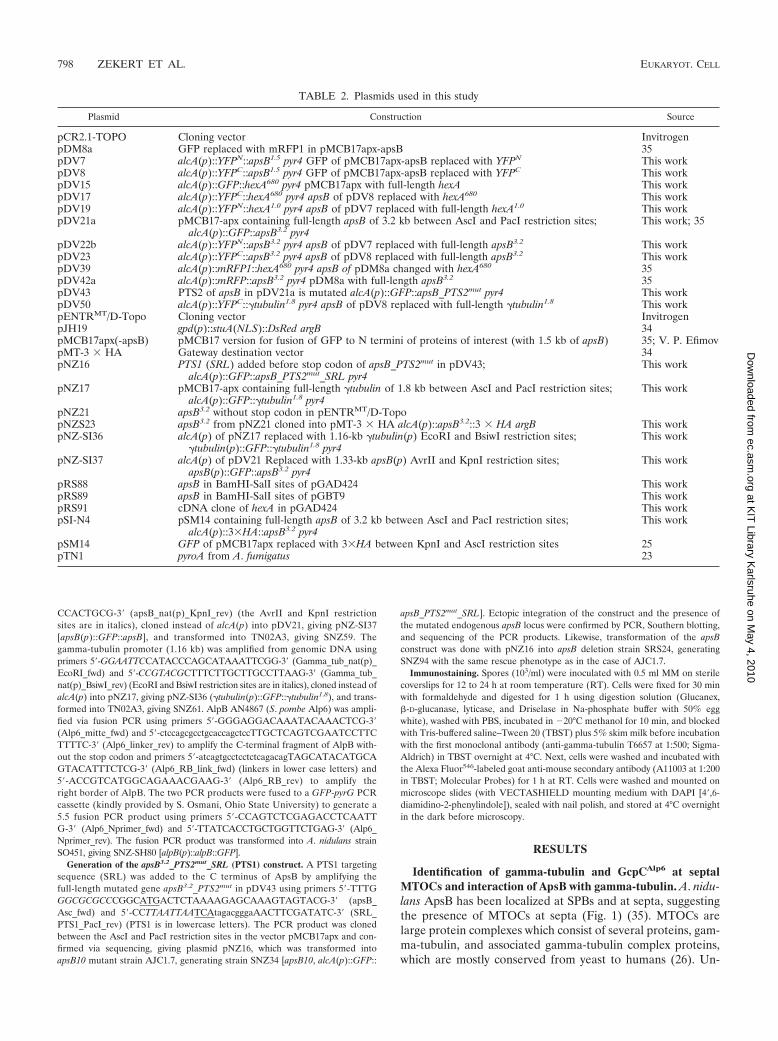
fortunately, the most important protein of MTOCs, gamma-tubulin, has not been identified at septa of A. nidulans before.In our own experiments, we were able to detect a very weaksignal at septa when gamma-tubulin was expressed from itsown promoter and fused to GFP. In S. pombe, it has alsobeen reported that gamma-tubulin was present at nonnuclearMTOCs in very small amounts and thus was also not easy todetect (29). To further elucidate the composition of septalMTOCs, we searched the A. nidulans genome for a homologueof S. pombe Alp6 (S. cerevisiae Spc98, human Gcp3) and iden-tified the open reading frame AN4867 (968 amino acids inlength) with 35% identity to Alp6. In order to localize thecorresponding protein, we constructed a C-terminal GFP fu-sion protein expressed from the native promoter and trans-formed it into A. nidulans (SO451). The protein localized toMTOCs at nuclei and at septa, indicating that the two MTOCs
also share this protein (Fig. 1). During the course of our ex-periments, this gene was analyzed in the laboratory of B. Oak-ley and was named gcpC (40).
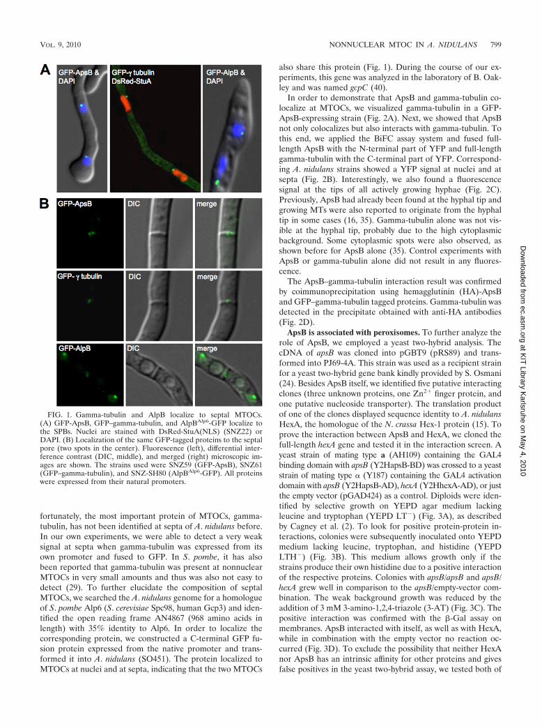
In order to demonstrate that ApsB and gamma-tubulin co-localize at MTOCs, we visualized gamma-tubulin in a GFP-ApsB-expressing strain (Fig. 2A). Next, we showed that ApsBnot only colocalizes but also interacts with gamma-tubulin. Tothis end, we applied the BiFC assay system and fused full-length ApsB with the N-terminal part of YFP and full-lengthgamma-tubulin with the C-terminal part of YFP. Correspond-ing A. nidulans strains showed a YFP signal at nuclei and atsepta (Fig. 2B). Interestingly, we also found a fluorescencesignal at the tips of all actively growing hyphae (Fig. 2C).Previously, ApsB had already been found at the hyphal tip andgrowing MTs were also reported to originate from the hyphaltip in some cases (16, 35). Gamma-tubulin alone was not vis-ible at the hyphal tip, probably due to the high cytoplasmicbackground. Some cytoplasmic spots were also observed, asshown before for ApsB alone (35). Control experiments withApsB or gamma-tubulin alone did not result in any fluores-cence.
The ApsB–gamma-tubulin interaction result was confirmedby coimmunoprecipitation using hemagglutinin (HA)-ApsBand GFP–gamma-tubulin tagged proteins. Gamma-tubulin wasdetected in the precipitate obtained with anti-HA antibodies(Fig. 2D).
ApsB is associated with peroxisomes. To further analyze therole of ApsB, we employed a yeast two-hybrid analysis. ThecDNA of apsB was cloned into pGBT9 (pRS89) and trans-formed into PJ69-4A. This strain was used as a recipient strainfor a yeast two-hybrid gene bank kindly provided by S. Osmani(24). Besides ApsB itself, we identified five putative interactingclones (three unknown proteins, one Zn2 finger protein, andone putative nucleoside transporter). The translation productof one of the clones displayed sequence identity to A. nidulansHexA, the homologue of the N. crassa Hex-1 protein (15). Toprove the interaction between ApsB and HexA, we cloned thefull-length hexA gene and tested it in the interaction screen. Ayeast strain of mating type a (AH109) containing the GAL4binding domain with apsB (Y2HapsB-BD) was crossed to a yeaststrain of mating type (Y187) containing the GAL4 activationdomain with apsB (Y2HapsB-AD), hexA (Y2HhexA-AD), or justthe empty vector (pGAD424) as a control. Diploids were iden-tified by selective growth on YEPD agar medium lackingleucine and tryptophan (YEPD LT�) (Fig. 3A), as describedby Cagney et al. (2). To look for positive protein-protein in-teractions, colonies were subsequently inoculated onto YEPDmedium lacking leucine, tryptophan, and histidine (YEPDLTH�) (Fig. 3B). This medium allows growth only if thestrains produce their own histidine due to a positive interactionof the respective proteins. Colonies with apsB/apsB and apsB/hexA grew well in comparison to the apsB/empty-vector com-bination. The weak background growth was reduced by theaddition of 3 mM 3-amino-1,2,4-triazole (3-AT) (Fig. 3C). Thepositive interaction was confirmed with the �-Gal assay onmembranes. ApsB interacted with itself, as well as with HexA,while in combination with the empty vector no reaction oc-curred (Fig. 3D). To exclude the possibility that neither HexAnor ApsB has an intrinsic affinity for other proteins and givesfalse positives in the yeast two-hybrid assay, we tested both of
FIG. 1. Gamma-tubulin and AlpB localize to septal MTOCs.(A) GFP-ApsB, GFP–gamma-tubulin, and AlpBAlp6-GFP localize tothe SPBs. Nuclei are stained with DsRed-StuA(NLS) (SNZ22) orDAPI. (B) Localization of the same GFP-tagged proteins to the septalpore (two spots in the center). Fluorescence (left), differential inter-ference contrast (DIC, middle), and merged (right) microscopic im-ages are shown. The strains used were SNZ59 (GFP-ApsB), SNZ61(GFP–gamma-tubulin), and SNZ-SH80 (AlpBAlp6-GFP). All proteinswere expressed from their natural promoters.
VOL. 9, 2010 NONNUCLEAR MTOC IN A. NIDULANS 799
at KIT
Library Karlsruhe on M
ay 4, 2010 ec.asm
.orgD
ownloaded from
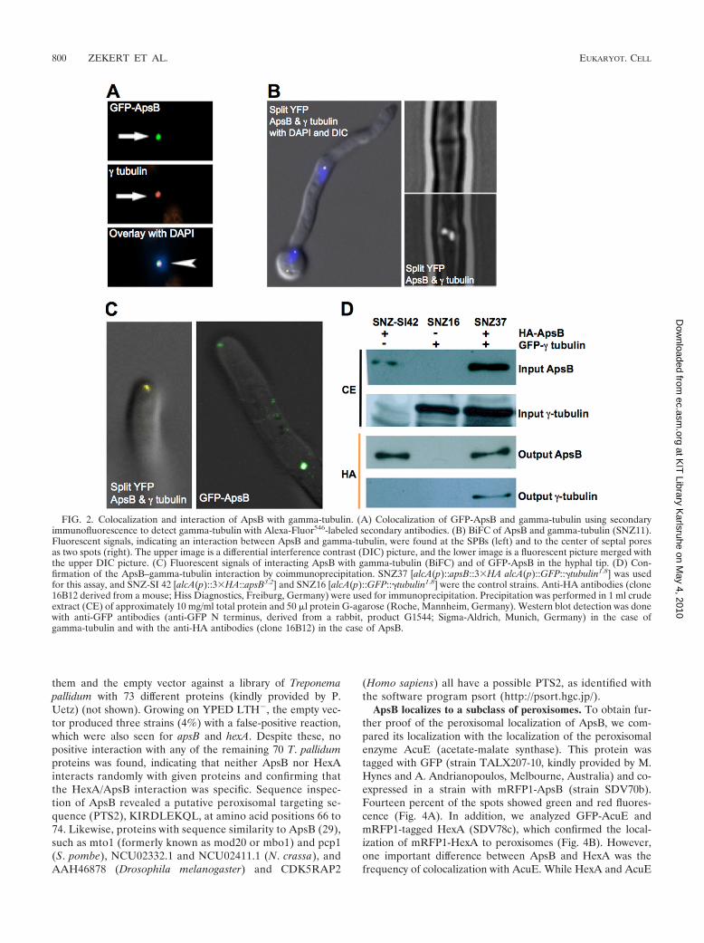
them and the empty vector against a library of Treponemapallidum with 73 different proteins (kindly provided by P.Uetz) (not shown). Growing on YPED LTH�, the empty vec-tor produced three strains (4%) with a false-positive reaction,which were also seen for apsB and hexA. Despite these, nopositive interaction with any of the remaining 70 T. pallidumproteins was found, indicating that neither ApsB nor HexAinteracts randomly with given proteins and confirming thatthe HexA/ApsB interaction was specific. Sequence inspec-tion of ApsB revealed a putative peroxisomal targeting se-quence (PTS2), KIRDLEKQL, at amino acid positions 66 to74. Likewise, proteins with sequence similarity to ApsB (29),such as mto1 (formerly known as mod20 or mbo1) and pcp1(S. pombe), NCU02332.1 and NCU02411.1 (N. crassa), andAAH46878 (Drosophila melanogaster) and CDK5RAP2
(Homo sapiens) all have a possible PTS2, as identified withthe software program psort (http://psort.hgc.jp/).
ApsB localizes to a subclass of peroxisomes. To obtain fur-ther proof of the peroxisomal localization of ApsB, we com-pared its localization with the localization of the peroxisomalenzyme AcuE (acetate-malate synthase). This protein wastagged with GFP (strain TALX207-10, kindly provided by M.Hynes and A. Andrianopoulos, Melbourne, Australia) and co-expressed in a strain with mRFP1-ApsB (strain SDV70b).Fourteen percent of the spots showed green and red fluores-cence (Fig. 4A). In addition, we analyzed GFP-AcuE andmRFP1-tagged HexA (SDV78c), which confirmed the local-ization of mRFP1-HexA to peroxisomes (Fig. 4B). However,one important difference between ApsB and HexA was thefrequency of colocalization with AcuE. While HexA and AcuE
FIG. 2. Colocalization and interaction of ApsB with gamma-tubulin. (A) Colocalization of GFP-ApsB and gamma-tubulin using secondaryimmunofluorescence to detect gamma-tubulin with Alexa-Fluor546-labeled secondary antibodies. (B) BiFC of ApsB and gamma-tubulin (SNZ11).Fluorescent signals, indicating an interaction between ApsB and gamma-tubulin, were found at the SPBs (left) and to the center of septal poresas two spots (right). The upper image is a differential interference contrast (DIC) picture, and the lower image is a fluorescent picture merged withthe upper DIC picture. (C) Fluorescent signals of interacting ApsB with gamma-tubulin (BiFC) and of GFP-ApsB in the hyphal tip. (D) Con-firmation of the ApsB–gamma-tubulin interaction by coimmunoprecipitation. SNZ37 [alcA(p)::apsB::3�HA alcA(p)::GFP::�tubulin1.8] was usedfor this assay, and SNZ-SI 42 [alcA(p)::3�HA::apsB3.2] and SNZ16 [alcA(p)::GFP::�tubulin1.8] were the control strains. Anti-HA antibodies (clone16B12 derived from a mouse; Hiss Diagnostics, Freiburg, Germany) were used for immunoprecipitation. Precipitation was performed in 1 ml crudeextract (CE) of approximately 10 mg/ml total protein and 50 �l protein G-agarose (Roche, Mannheim, Germany). Western blot detection was donewith anti-GFP antibodies (anti-GFP N terminus, derived from a rabbit, product G1544; Sigma-Aldrich, Munich, Germany) in the case ofgamma-tubulin and with the anti-HA antibodies (clone 16B12) in the case of ApsB.
800 ZEKERT ET AL. EUKARYOT. CELL
at KIT
Library Karlsruhe on M
ay 4, 2010 ec.asm
.orgD
ownloaded from
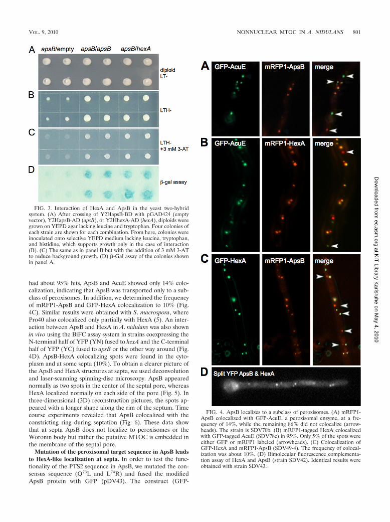
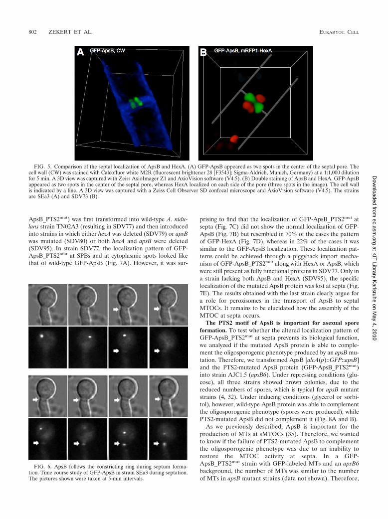
had about 95% hits, ApsB and AcuE showed only 14% colo-calization, indicating that ApsB was transported only to a sub-class of peroxisomes. In addition, we determined the frequencyof mRFP1-ApsB and GFP-HexA colocalization to 10% (Fig.4C). Similar results were obtained with S. macrospora, wherePro40 also colocalized only partially with HexA (5). An inter-action between ApsB and HexA in A. nidulans was also shownin vivo using the BiFC assay system in strains coexpressing theN-terminal half of YFP (YN) fused to hexA and the C-terminalhalf of YFP (YC) fused to apsB or the other way around (Fig.4D). ApsB-HexA colocalizing spots were found in the cyto-plasm and at some septa (10%). To obtain a clearer picture ofthe ApsB and HexA structures at septa, we used deconvolutionand laser-scanning spinning-disc microscopy. ApsB appearednormally as two spots in the center of the septal pore, whereasHexA localized normally on each side of the pore (Fig. 5). Inthree-dimensional (3D) reconstruction pictures, the spots ap-peared with a longer shape along the rim of the septum. Timecourse experiments revealed that ApsB colocalized with theconstricting ring during septation (Fig. 6). These data showthat at septa ApsB does not localize to peroxisomes or theWoronin body but rather the putative MTOC is embedded inthe membrane of the septal pore.
Mutation of the peroxisomal target sequence in ApsB leadsto HexA-like localization at septa. In order to test the func-tionality of the PTS2 sequence in ApsB, we mutated the con-sensus sequence (Q73L and L74R) and fused the modifiedApsB protein with GFP (pDV43). The construct (GFP-
FIG. 4. ApsB localizes to a subclass of peroxisomes. (A) mRFP1-ApsB colocalized with GFP-AcuE, a peroxisomal enzyme, at a fre-quency of 14%, while the remaining 86% did not colocalize (arrow-heads). The strain is SDV70b. (B) mRFP1-tagged HexA colocalizedwith GFP-tagged AcuE (SDV78c) in 95%. Only 5% of the spots wereeither GFP or mRFP1 labeled (arrowheads). (C) Colocalization ofGFP-HexA and mRFP1-ApsB (SDV49-4). The frequency of colocal-ization was about 10%. (D) Bimolecular fluorescence complementa-tion assay of HexA and ApsB (strain SDV42). Identical results wereobtained with strain SDV43.
FIG. 3. Interaction of HexA and ApsB in the yeast two-hybridsystem. (A) After crossing of Y2HapsB-BD with pGAD424 (emptyvector), Y2HapsB-AD (apsB), or Y2HhexA-AD (hexA), diploids weregrown on YEPD agar lacking leucine and tryptophan. Four colonies ofeach strain are shown for each combination. From here, colonies wereinoculated onto selective YEPD medium lacking leucine, tryptophan,and histidine, which supports growth only in the case of interaction(B). (C) The same as in panel B but with the addition of 3 mM 3-ATto reduce background growth. (D) �-Gal assay of the colonies shownin panel A.
VOL. 9, 2010 NONNUCLEAR MTOC IN A. NIDULANS 801
at KIT
Library Karlsruhe on M
ay 4, 2010 ec.asm
.orgD
ownloaded from
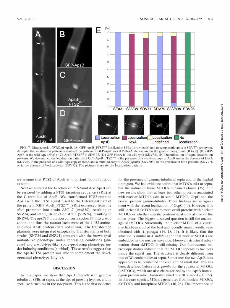
ApsB_PTS2mut) was first transformed into wild-type A. nidu-lans strain TN02A3 (resulting in SDV77) and then introducedinto strains in which either hexA was deleted (SDV79) or apsBwas mutated (SDV80) or both hexA and apsB were deleted(SDV95). In strain SDV77, the localization pattern of GFP-ApsB_PTS2mut at SPBs and at cytoplasmic spots looked likethat of wild-type GFP-ApsB (Fig. 7A). However, it was sur-
prising to find that the localization of GFP-ApsB_PTS2mut atsepta (Fig. 7C) did not show the normal localization of GFP-ApsB (Fig. 7B) but resembled in 70% of the cases the patternof GFP-HexA (Fig. 7D), whereas in 22% of the cases it wassimilar to the GFP-ApsB localization. These localization pat-terns could be achieved through a piggyback import mecha-nism of GFP-ApsB_PTS2mut along with HexA or ApsB, whichwere still present as fully functional proteins in SDV77. Only ina strain lacking both ApsB and HexA (SDV95), the specificlocalization of the mutated ApsB protein was lost at septa (Fig.7E). The results obtained with the last strain clearly argue fora role for peroxisomes in the transport of ApsB to septalMTOCs. It remains to be elucidated how the assembly of theMTOC at septa occurs.
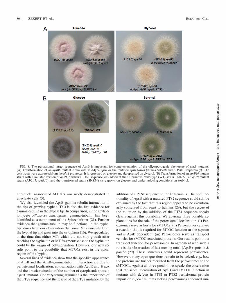
The PTS2 motif of ApsB is important for asexual sporeformation. To test whether the altered localization pattern ofGFP-ApsB_PTS2mut at septa prevents its biological function,we analyzed if the mutated ApsB protein is able to comple-ment the oligosporogenic phenotype produced by an apsB mu-tation. Therefore, we transformed ApsB [alcA(p)::GFP::apsB]and the PTS2-mutated ApsB protein (GFP-ApsB_PTS2mut)into strain AJC1.5 (apsB6). Under repressing conditions (glu-cose), all three strains showed brown colonies, due to thereduced numbers of spores, which is typical for apsB mutantstrains (4, 32). Under inducing conditions (glycerol or sorbi-tol), however, wild-type ApsB protein was able to complementthe oligosporogenic phenotype (spores were produced), whilePTS2-mutated ApsB did not complement it (Fig. 8A and B).
As we previously described, ApsB is important for theproduction of MTs at sMTOCs (35). Therefore, we wantedto know if the failure of PTS2-mutated ApsB to complementthe oligosporogenic phenotype was due to an inability torestore the MTOC activity at septa. In a GFP-ApsB_PTS2mut strain with GFP-labeled MTs and an apsB6background, the number of MTs was similar to the numberof MTs in apsB mutant strains (data not shown). Therefore,
FIG. 5. Comparison of the septal localization of ApsB and HexA. (A) GFP-ApsB appeared as two spots in the center of the septal pore. Thecell wall (CW) was stained with Calcofluor white M2R (fluorescent brightener 28 [F3543]; Sigma-Aldrich, Munich, Germany) at a 1:1,000 dilutionfor 5 min. A 3D view was captured with Zeiss AxioImager Z1 and AxioVision software (V4.5). (B) Double staining of ApsB and HexA. GFP-ApsBappeared as two spots in the center of the septal pore, whereas HexA localized on each side of the pore (three spots in the image). The cell wallis indicated by a line. A 3D view was captured with a Zeiss Cell Observer SD confocal microscope and AxioVision software (V4.5). The strainsare SEa3 (A) and SDV73 (B).
FIG. 6. ApsB follows the constricting ring during septum forma-tion. Time course study of GFP-ApsB in strain SEa3 during septation.The pictures shown were taken at 5-min intervals.
802 ZEKERT ET AL. EUKARYOT. CELL
at KIT
Library Karlsruhe on M
ay 4, 2010 ec.asm
.orgD
ownloaded from
we assume that PTS2 of ApsB is important for its functionat septa.
Next we tested if the function of PTS2-mutated ApsB canbe restored by adding a PTS1 targeting sequence (SRL) atthe C terminus of ApsB. We transformed PTS2-mutatedApsB with the PTS1 signal fused to the C-terminal part ofthe protein (GFP-ApsB_PTS2mut_SRL) expressed from thealcA promoter into strain AJC1.7 (apsB10), resulting inSNZ34, and into apsB deletion strain (SRS24), resulting inSNZ94. The apsB10 mutation converts codon 83 into a stopcodon, and thus the mutant lacks most of the 1,052-amino-acid-long ApsB protein (data not shown). The transformedplasmids were integrated ectopically. Transformants of bothstrains (SNZ34 and SNZ94) appeared with the brown apsBmutant-like phenotype under repressing conditions (glu-cose) and a wild-type-like, spore-producing phenotype un-der inducing conditions (sorbitol). These results suggest thatthe ApsB-PTS1 protein was able to complement the devel-opmental phenotype (Fig. 8).
DISCUSSION
In this paper, we show that ApsB interacts with gamma-tubulin at SPBs, at septa, at the tips of growing hyphae, and inspot-like structures in the cytoplasm. This is the first evidence
for the presence of gamma-tubulin at septa and in the hyphaltip region. We had evidence before that MTOCs exist at septa,but the nature of these MTOCs remained elusive (35). Ournew results show that at least two other proteins associatedwith nuclear MTOCs exist in septal MTOCs, GcpC and thecrucial protein gamma-tubulin. These findings are in agree-ment with the recent localization of GcpC (40). However, it isstill unclear if sMTOCs share more or all proteins with nuclearMTOCs or whether specific proteins exist only at one or theother place. The biggest unsolved question is still the anchor-age of sMTOCs. Structurally, the nuclear MTOC of S. cerevi-siae has been studied the best and recently similar results wereobtained with A. gossypii (14, 18, 19). It is likely that thesituation is similar in A. nidulans and that nuclear MTOCs areembedded in the nuclear envelope. However, structural infor-mation about sMTOCs is still missing. Our fluorescence mi-croscopy studies indicate that the MTOC appears as two dotsinside the septal rim. The structure is clearly different fromthat of Woronin bodies at septa. Sometimes the two ApsB dotsappeared to be connected through a third small dot. This hasbeen described before in S. pombe for the equatorial MTOCs(eMTOCs), which are also characterized by the ApsB-homol-ogous protein mto1 (formerly named mod20 or mbo1) (10, 29).In this yeast species, MTs are generated from nuclear MTOCs,eMTOCs, and interphase MTOCs (10, 28). The importance of
FIG. 7. Mutagenesis of PTS2 of ApsB. (A) GFP-ApsB_PTS2mut localized to SPBs (arrowheads) and to cytoplasmic spots in SDV77 (genotype).At septa, the localization pattern resembled the pattern of GFP-ApsB or GFP-HexA, depending on the genetic background (B to E). (B) GFP-ApsB in the wild type (SEa3). (C) ApsB-PTS2mut in SDV 77. (D) GFP-HexA in the wild type (SDV38). (E) Quantification of septal localizationpatterns. We determined the localization patterns of GFP-ApsB_PTS2mut in the presence of a wild-type copy of ApsB and in the absence of HexA(SDV79), in the presence of a wild-type copy of HexA and a mutated copy of ApsB (apsB6) (SDV80b), in the presence of both proteins (SDV77),or in the absence of both proteins (SDV95). The pictures illustrate the localization patterns.
VOL. 9, 2010 NONNUCLEAR MTOC IN A. NIDULANS 803
at KIT
Library Karlsruhe on M
ay 4, 2010 ec.asm
.orgD
ownloaded from
non-nucleus-associated MTOCs was nicely demonstrated inenucleate cells (3).
We also identified the ApsB–gamma-tubulin interaction inthe tips of growing hyphae. This is also the first evidence forgamma-tubulin in the hyphal tip. In comparison, in the chytrid-iomycete Allomyces macrogynus, gamma-tubulin has beenidentified as a component of the Spitzenkorper (21). Furtherevidence that gamma-tubulin may be functional in the hyphaltip comes from our observation that some MTs emanate fromthe hyphal tip and grow into the cytoplasm (16). We speculatedat the time that either MTs which did not stop growth afterreaching the hyphal tip or MT fragments close to the hyphal tipcould be the origin of polymerization. However, our new re-sults point to the possibility that MTOCs exist in the apicalregion of the hypha.
Several lines of evidence show that the spot-like appearanceof ApsB and the ApsB–gamma-tubulin interaction are due toperoxisomal localization: colocalization with AcuE and HexAand the drastic reduction of the number of cytoplasmic spots ina pexC mutant. One very strong argument is the importance ofthe PTS2 sequence and the rescue of the PTS2 mutation by the
addition of a PTS1 sequence to the C terminus. The nonfunc-tionality of ApsB with a mutated PTS2 sequence could still beexplained by the fact that this region appears to be evolution-arily conserved from yeast to humans (29), but the rescue ofthe mutation by the addition of the PTS1 sequence speaksclearly against this possibility. We envisage three possible ex-planations for the role of the peroxisomal localization. (i) Per-oxisomes serve as hosts for sMTOCs. (ii) Peroxisomes catalyzea reaction that is required for MTOC function at the septumand is ApsB dependent. (iii) Peroxisomes serve as transportvehicles for sMTOC-associated proteins. Our results point to atransport function for peroxisomes. In agreement with such arole is the observation of fast-moving mto1 (ApsB) spots in S.pombe (29). These structures could represent peroxisomes.However, many open questions remain to be solved, e.g., howthe proteins are further recruited from the peroxisomes to thesMTOCs. Against all three possibilities speaks the observationthat the septal localization of ApsB and sMTOC function inmutants with defects in PTS1 or PTS2 peroxisomal proteinimport or in pexC mutants lacking peroxisomes appeared sim-
FIG. 8. The peroxisomal target sequence of ApsB is important for complementation of the oligosporogenic phenotype of apsB mutants.(A) Transformation of an apsB6 mutant strain with wild-type apsB or the mutated apsB forms (strains SDV88 and SDV80, respectively). Theconstructs were expressed from the alcA promoter. It is repressed on glucose and derepressed on glycerol. (B) Transformation of an apsB10 mutantstrain with a mutated version of apsB in which a PTS1 sequence was added at the C terminus. Wild-type (WT) strain TN02A3, an apsB mutantstrain (AJC1.7, apsB10), and the transformed strain (SNZ34) were grown on glucose and under inducing conditions on sorbitol.
804 ZEKERT ET AL. EUKARYOT. CELL
at KIT
Library Karlsruhe on M
ay 4, 2010 ec.asm
.orgD
ownloaded from
ilar to the situation in the wild type (results not shown). How-ever, it has to be considered that the pexC mutant strain dis-plays pleiotropic phenotypes and that the possibility of apiggyback import mechanism might mask the possible effectsof PTS1 or PTS2 defects (12).
From our results we conclude that ApsB defines a new classof peroxisomes that is—besides the Woronin bodies—the sec-ond example of peroxisomes as organelles with a functionbeyond metabolic pathways (30).
ACKNOWLEDGMENTS
We are grateful to M. Hynes (University of Melbourne, Melbourne,Australia) for sending us peroxisomal marker proteins and pex mutantstrains and to G. Jedd for helpful discussions and the hexA deletionstrains. We thank R. Suelmann, Bjorn Titz, and Sabrina Hettinger forinitial help with this project.
The work was funded by the Deutsche Forschungsgemeinschaft, theFonds der Chemischen Industrie, and the special program Lebensmit-tel und Gesundheit of the Landesstiftung Baden Wurttemberg. N.Z.was partly supported by a fellowship from the Syrian ministry.
REFERENCES
1. Buller, A. H. R. 1931. Researches on fungi. Hafner Publishing Co., NewYork, NY.
2. Cagney, B., P. Uetz, and S. Fields. 2000. High-throughput screening forprotein-protein interactions using the two-hybrid assay. Methods Enzymol.328:3–14.
3. Carazo-Salas, R. E., and P. Nurse. 2006. Self-organization of interphasemicrotubule arrays in fission yeast. Nat. Cell Biol. 8:1102–1107.
4. Clutterbuck, A. J. 1994. Mutants of Aspergillus nidulans deficient in nuclearmigration during hyphal growth and conidiation. Microbiology 140:1169–1174.
5. Engh, I., C. Wurtz, K. Witzel-Schlomp, H. Y. Zhang, B. Hoff, M. Nowrou-sian, H. Rottensteiner, and U. Kuck. 2007. The WW domain protein PRO40is required for fungal fertility and associates with Woronin bodies. Eukaryot.Cell 6:831–843.
6. Gabaldon, T., B. Snel, F. van Zimmeren, W. Hemrika, H. Tabak, and M. A.Hyunen. 2006. Origin and evolution of the peroxisomal proteome. Biol.Direct 1:8.
7. Gould, S. J., G. A. Keller, N. Hosken, J. Wilkinson, and S. Subramani. 1989.A conserved tripeptide sorts proteins to peroxisomes. J. Cell Biol. 108:1657–1664.
8. Hayashi, M., and M. Nishimura. 2003. Entering a new era of research onplant peroxisomes. Curr. Opin. Plant Biol. 6:577–582.
9. Heiland, I., and R. Erdmann. 2005. Biogenesis of peroxisomes. FEBS J.272:2362–2372.
10. Heitz, M. J., J. Petersen, S. Valovin, and I. M. Hagan. 2001. MTOC forma-tion during mitotic exit in fission yeast. J. Cell Sci. 114:4521–4532.
11. Hill, T. W., and E. Kafer. 2001. Improved protocols for Aspergillus minimalmedium: trace element and minimal medium salt stock solutions. FungalGenet. Newsl. 48:20–21.
12. Hynes, M. J., S. L. Murray, G. S. Khew, and M. A. Davis. 2008. Geneticanalysis of the role of peroxisomes in the utilization of acetate and fatty acidsin Aspergillus nidulans. Genetics 178:1355–1369.
13. James, P., J. Halladay, and E. A. Craig. 1996. Genomic libraries and a hoststrain designed for highly efficient two-hybrid selection in yeast. Genetics144:1425–1436.
14. Jaspersen, S. L., and M. Winey. 2004. The budding yeast spindle pole body:structure, duplication, and function. Annu. Rev. Cell Dev. Biol. 20:1–28.
15. Jedd, G., and N.-H. Chua. 2000. A new self-assembled peroxisomal vesiclerequired for efficient resealing of the plasma membrane. Nat. Cell Biol.2:226–231.
16. Konzack, S., P. Rischitor, C. Enke, and R. Fischer. 2005. The role of thekinesin motor KipA in microtubule organization and polarized growth ofAspergillus nidulans. Mol. Biol. Cell 16:497–506.
17. Kovacs, W. J., S. Krisans, S. Hogenboom, R. J. Wanders, and H. R. Water-ham. 2003. Cholesterol biosynthesis and regulation: role of peroxisomes.Adv. Exp. Med. Biol. 544:315–327.
18. Lang, C., S. Grava, M. Finlayson, R. Trimble, P. Philippsen, and S. L.
Jaspersen. 2010. Structural mutants of the spindle pole body cause distinctalterations of cytoplasmic microtubules and nuclear dynamics in multinucle-ated hyphae. Mol. Biol. Cell 21:753–766.
19. Lang, C., S. Grava, T. van den Hoorn, R. Trimble, P. Philippsen, and S. L.Jaspersen. 2010. Mobility, microtubule nucleation and structure of microtu-bule-organizing centers in multinucleated hyphae of Ashbya gossypii. Mol.Biol. Cell 21:18–28.
20. Maruyama, J., P. R. Juvvadi, K. Ishi, and K. Kitamoto. 2005. Three-dimen-sional image analysis of plugging at the septal pore by Woroinin body duringhypotonic shock inducing hyphal tip bursting in the filamentous fungusAspergillus oryzae. Biochem. Biophys. Res. Commun. 331:1081–1088.
21. McDaniel, D. P., and R. W. Roberson. 1998. �-Tubulin is a component of theSpitzenkorper and centrosomes in hyphal-tip cells of Allomyces macrogynus.Protoplasma 203:118–123.
22. Momany, M., E. Richardson, C. Van Sickle, and G. Jedd. 2002. MappingWoronin body position in Aspergillus nidulans. Mycologia 94:260–266.
23. Nayak, T., E. Szewczyk, C. E. Oakley, A. Osmani, L. Ukil, S. L. Murray, M. J.Hynes, S. A. Osmani, and B. R. Oakley. 2006. A versatile and efficient genetargeting system for Aspergillus nidulans. Genetics 172:1557–1566.
24. Osmani, A. H., J. Davies, C. E. Oakley, B. R. Oakley, and S. A. Osmani. 2003.TINA interacts with the NIMA kinase in Aspergillus nidulans and negativelyregulates astral microtubules during metaphase arrest. Mol. Biol. Cell 14:3169–3179.
25. Purschwitz, J., S. Muller, and R. Fischer. 2009. Mapping the interaction sitesof Aspergillus nidulans phytochrome FphA with the global regulator VeA andthe white collar protein LreB. Mol. Genet. Genomics 281:35–42.
26. Raynaud-Messina, B., and A. Merdes. 2007. Gamma-tubulin complexes andmicrotubule organization. Curr. Opin. Cell Biol. 19:24–30.
27. Sambrook, J., and D. W. Russell. 2001. Molecular cloning: a laboratory manual,3rd ed. Cold Spring Harbor Laboratory Press, Cold Spring Harbor, NY.
28. Samejima, I., P. C. C. Laurenco, H. A. Snaith, and K. E. Sawin. 2005. Fissionyeast mto2p regulates microtubule nucleation by the centrosomin-relatedprotein mto1p. Mol. Biol. Cell 16:3040–3051.
29. Sawin, K. E., P. C. C. Lourenco, and H. A. Snaith. 2004. Microtubulenucleation at non-spindle pole body microtubule-organizing centers requiresfission yeast centrosomin-related protein mod20p. Curr. Biol. 14:763–775.
30. Schrader, M., and H. D. Fahimi. 2008. The peroxisome: still a mysteriousorganelle. Histochem. Cell Biol. 129:421–440.
31. Suelmann, R., N. Sievers, and R. Fischer. 1997. Nuclear traffic in fungalhyphae: in vivo study of nuclear migration and positioning in Aspergillusnidulans. Mol. Microbiol. 25:757–769.
32. Suelmann, R., N. Sievers, D. Galetzka, L. Robertson, W. E. Timberlake, andR. Fischer. 1998. Increased nuclear traffic chaos in hyphae of apsB mutantsof Aspergillus nidulans: molecular characterization of apsB and in vivo ob-servation of nuclear behaviour. Mol. Microbiol. 30:831–842.
33. Swinkels, B. W., S. J. Gould, A. G. Bodnar, R. A. Rachubinski, and S.Subramani. 1991. A novel, cleavable peroxisomal targeting signal at theamino-terminus of the rat 3-ketoacyl-CoA thiolase. EMBO J. 10:3255–3262.
34. Toews, M. W., J. Warmbold, S. Konzack, P. E. Rischitor, D. Veith, K.Vienken, C. Vinuesa, H. Wei, and R. Fischer. 2004. Establishment of mRFP1as fluorescent marker in Aspergillus nidulans and construction of expressionvectors for high-throughput protein tagging using recombination in Esche-richia coli (GATEWAY). Curr. Genet. 45:383–389.
35. Veith, D., N. Scherr, V. P. Efimov, and R. Fischer. 2005. Role of the spindle-pole body protein ApsB and the cortex protein ApsA in microtubule orga-nization and nuclear migration in Aspergillus nidulans. J. Cell Sci. 118:3705–3716.
36. Wanders, R. J. 2004. Metabolic and molecular basis of peroxisomal disor-ders: a review. Am. J. Med. Genet. 126A:355–375.
37. Wanders, R. J., and H. R. Waterham. 2006. Biochemistry of mammalianperoxisomes revisited. Annu. Rev. Biochem. 75:295–332.
38. Waring, R. B., G. S. May, and N. R. Morris. 1989. Characterization of aninducible expression system in Aspergillus nidulans using alcA and tubulincoding genes. Gene 79:119–130.
39. Woronin, M. 1864. Entwicklungsgeschichte der Ascobolus pucherrimus Cr.und einiger Pezizen. Abh. Senkenb. Naturforsch. 5:333–344.
40. Xiong, Y., and B. R. Oakley. 2009. In vivo analysis of the functions ofgamma-tubulin-complex proteins. J. Cell Sci. 122:4218–4227.
41. Yelton, M. M., J. E. Hamer, and W. E. Timberlake. 1984. Transformation ofAspergillus nidulans by using a trpC plasmid. Proc. Natl. Acad. Sci. U. S. A.81:1470–1474.
42. Yuan, P., G. Jedd, D. Kumaran, S. Swaminathan, H. Shio, D. Hewitt, N.-H.Chua, and K. Swaminathan. 2003. A Hex-1 crystal lattice required forWoronin body function in Neurospora crassa. Nat. Struct. Biol. 10:264–270.
VOL. 9, 2010 NONNUCLEAR MTOC IN A. NIDULANS 805
at KIT
Library Karlsruhe on M
ay 4, 2010 ec.asm
.orgD
ownloaded from
![Index [] · 2011-03-23 · Index 937 Ascochyta rabiei 507 Aspergillus nidulans 755 Aspergillus ochraceus 135, 158 Aspergillus terreus 506, 755 Aspidosperma alkaloids 106 – fragmentation](https://img.pdfslide.us/doc/110x75/5e2b9234e715b60f857d1b3b/index-2011-03-23-index-937-ascochyta-rabiei-507-aspergillus-nidulans-755-aspergillus.jpg)